


Abstract
Autofluorescence induced by 405 nm excitation may provide a non-invasive optical approach for characterizing cultured bacteria. This study evaluated the dynamics of autofluorescence in anaerobic bacterial strains using quantitative hue analysis. Twelve reference strains representing 11 bacterial species were cultured under controlled anaerobic conditions. Eight independent colonies per strain were evaluated repeatedly at 24, 48, 72, 96, and 168 h after inoculation, using 405 nm laser excitation for 45 s at each time point. Autofluorescence color was assessed using ΔH, defined as the mean colony hue minus the agar background hue. The effects of bacterial strain, incubation time, and their interaction were analyzed using ART-ANOVA. Significant effects were observed for both factors and their interaction (p < 0.001). Comparatively higher, less negative ΔH values were observed for A. odontolyticus, B. fragilis, B. breve, C. acnes, and C. sporogenes ATCC 2486, whereas more negative values were observed for P. intermedia, P. micra, and A. israelii. P. intermedia showed the largest visible shift in mean ΔH, particularly between 48 and 72 h. Temporal changes were not consistently monotonic, and several strains showed partial reversals at later time points. These findings indicate strain-dependent temporal changes in colony color relative to the culture medium. Absolute fluorescence intensity was not quantified. Quantitative hue analysis may provide complementary information for characterizing cultured anaerobic bacteria, but its reproducibility and performance for identifying unknown isolates require further validation.
1. Introduction
Anaerobic bacteria are important members of the human microbiota, colonizing the oral cavity, gastrointestinal and genitourinary tracts, and skin [1,2,3,4,5,6]. Although they are part of the normal microbial community, they may contribute to local and systemic infections when microbial balance is disrupted, anatomical barriers are compromised, or host defenses are impaired [3,4,7,8,9,10,11,12,13]. In the oral cavity, anaerobic bacteria are associated with dysbiosis-related diseases, particularly periodontitis and necrotizing periodontal diseases [14,15,16]. They are also frequently involved in endodontic infections, periapical inflammatory lesions, odontogenic abscesses, and deep infections of the head and neck [4,7,17,18,19,20]. Interactions between oral microorganisms, including streptococci and other bacterial groups, further contribute to the complexity of the oral microbiome and its relationship with host health [21,22,23,24,25,26,27,28,29]. Some oral bacteria have also been investigated in relation to carcinogenesis and tumor progression, although these associations vary according to the organism and biological context [30,31,32,33,34,35,36,37].
Laboratory diagnosis of anaerobic infections remains challenging because many species are sensitive to oxygen exposure, clinical specimens require appropriate collection and transport, and some microorganisms grow slowly [3,5,38]. Identification may be further complicated by phenotypic variability, polymicrobial infections, and organisms that are difficult to cultivate under routine laboratory conditions [4,19,38,39,40]. Molecular approaches can complement culture-based methods [41], while optical techniques may provide additional information on microbial phenotypes [42,43,44,45,46]. The selected reference panel also represents diverse biological groups, including organisms involved in oral microbial interactions and species investigated for probiotic effects [35,47,48,49,50,51,52,53]. Studies of paraprobiotic preparations further illustrate the diversity of host–microbe relationships [54]. Thus, the strains examined should not be considered a homogeneous group of oral pathogens.
The clinical importance of microbial characterization is reflected in studies of antimicrobial and antibiofilm strategies for oral microorganisms. These include endodontic materials and irrigants [55,56,57,58,59,60,61,62,63], laser-based and photodynamic approaches [64,65,66,67], and modified biomaterials or natural products [68,69,70,71,72,73,74,75,76,77,78,79,80,81]. Such investigations provide a broader microbiological context, but antimicrobial efficacy and optical identification are distinct outcomes.
Autofluorescence is the emission of light by naturally occurring compounds following excitation at an appropriate wavelength. Previous studies have investigated photoinduced fluorescence in cultured microorganisms [42,43]. Bacterial autofluorescence may arise from several endogenous fluorophores, including porphyrins, flavins, and reduced pyridine nucleotides (NADH and NADPH) [82,83,84,85,86,87]. Excitation at 405 nm can induce emission at longer wavelengths [82,83,84,85], with porphyrins and their derivatives contributing to red and orange-red fluorescence in oral bacteria [82,83,85,88,89]. Flavins may contribute to green emission [84,85,87], whereas NADH and NADPH have excitation maxima near 340 nm [86]. The resulting color reflects the combined contributions of multiple fluorophores and may be influenced by culture medium composition, pH, heme availability, microbial physiology, colony morphology, and imaging conditions [46,82,83,84,85,90,91,92,93,94].
Digital image analysis allows color to be described using hue, saturation, and brightness. One such model is HSV, referred to as HSB in ImageJ: hue describes color, saturation its purity, and value or brightness its light–dark component. Comparing colony hue with that of the adjacent growth-free agar provides a relative measure of color contrast, although it cannot fully separate bacterial fluorescence from contributions of the culture medium. Previous studies have demonstrated that bacterial fluorescence varies with species, strain, and culture conditions [82,83,84,85]. However, temporal changes in the colony-to-background hue difference in anaerobic cultures remain insufficiently characterized, including the magnitude and direction of ΔH changes during incubation.
The aim of the present study was to evaluate the effects of bacterial strain, incubation time, and their interaction on the colony-to-background hue difference (ΔH) following excitation at 405 nm. Eight independent colonies of each reference strain were assessed at five time points after inoculation to characterize temporal color profiles under controlled laboratory conditions. The study provides an initial evaluation of ΔH as a parameter for comparing bacterial colony color over time. Further research will be required to establish its reproducibility, relationship to spectroscopic and biological measurements, and potential diagnostic value using clinical isolates and independent validation [44,45,46].
2. Materials and Methods
2.1. Bacterial Strains
The study included 12 reference strains representing 11 species of anaerobic bacteria from the American Type Culture Collection (ATCC) and the Polish Collection of Microorganisms (PCM). Information on the strains selected for study is presented in Table 1.
Table 1.
Reference anaerobic bacterial strains used in the study.
2.2. Strain Storage and Culture Conditions
The strains selected for the study were stored at −80 °C in Trypticase Soy Broth (TSB) (Biomaxima, Lublin, Poland) with 15% glycerol. Each strain was frozen in multiple replicates. Before each experimental cycle, the strains were cultured on solid Schaedler Agar with Vitamin K1 and 5% Sheep Blood (Becton Dickinson, Heidelberg, Germany) at 37 °C under anaerobic conditions using the GENbag Anaer system (Biomaxima, Lublin, Poland) for 48 h to 5 days.
2.3. Experimental Design and Image Analysis
For each strain, a suspension corresponding to 1.0 McFarland was prepared, and 5 µL aliquots were inoculated onto Brain Heart Infusion (BHI) agar (Biomaxima, Lublin, Poland) to obtain eight independent colonies. The cultures were incubated at 37 °C under anaerobic conditions using the GENbag Anaer system (Biomaxima, Lublin, Poland). The same eight colonies of each strain were evaluated at 24, 48, 72, 96, and 168 h after inoculation. Thus, each colony contributed five repeated observations, with the colony serving as the biological replicate.
Fluorescence was excited using a continuous-wave 405 nm diode laser (SmartM, Lasotronix, Piaseczno, Poland). The laser system was calibrated before the experiments, and the output power was set to 200 mW. At each observation time, the agar plate containing eight colonies of the analysed strain was exposed to laser illumination for 45 s. The laser beam was delivered through a handpiece incorporating a glass optical light guide and positioned approximately perpendicular to the agar surface at a fixed working distance of 11 cm. The illumination spot at the sample surface had an approximate diameter of 8 mm, corresponding to an estimated irradiance of 398 mW/cm2 and a radiant exposure of 17.9 J/cm2 over the 45 s exposure period. Fluorescence images were acquired through a yellow emission filter using an iPhone 14 (Apple, Cupertino, CA, USA). To maintain consistency, image acquisition was performed using the same phone, laser output settings, working distance, and relative positioning of the camera and sample. Photographs were recorded in JPEG format with automatic exposure and white-balance functions enabled, and the flash was not used. No additional colour modifications were performed after image capture. However, the smartphone’s built-in image-processing procedures, such as automatic colour balancing, tone mapping, and computational enhancement, remained partly beyond experimental control. The images were not captured in RAW/DNG format, and no calibrated colour target or fluorescence reference standard was included.
Image analysis was performed using ImageJ software (version 1.54g) and the HSV/HSB color model. At each observation time, each colony was manually outlined to define a region of interest (ROI), which was stored in the ROI Manager. The minimum (Hmin) and mean (Hmean) hue values were recorded for each colony, together with the hue value of the agar background (BC). The colony-to-background hue difference was calculated as ΔH = Hmean − BC and used in all statistical analyses; Hmin was not included in these comparisons. Hue values were expressed on a 0–255 scale, and direct subtraction was used because the observed values did not cross the scale boundary. No saturation- or brightness-based thresholds or additional pixel-exclusion procedures were applied; therefore, pixels with poorly defined color may have influenced the mean hue values. Higher ΔH values indicate a more positive or less negative relative hue difference, not stronger fluorescence. Values close to zero indicate similar colony and background hues, whereas negative values indicate a lower hue value for the colony. Thus, ΔH represents relative color contrast rather than fluorescence intensity. Images were also examined descriptively for visible color, colony morphology, and changes over time. This assessment was qualitative and did not provide an independent measurement of fluorescence intensity. At each observation time, the plates were temporarily removed from the GENbag Anaer system and photographed in a completely darkened microbiology laboratory, with the room lights switched off and the window blinds closed. Immediately after image acquisition, the plates were returned to anaerobic incubation; the time outside the anaerobic system was kept as brief as practicable. (Table S1: Descriptive statistics of colony-to-background hue difference.)
2.4. Statistical Analysis
The data were analyzed using R software (version 4.5.3; R Core Team). Prior to analysis, outliers were assessed based on boxplots using the 1.5 × IQR criterion. Given the small number of colonies per strain (n = 8) and the limited power of normality tests in small samples, a non-parametric analysis of variance was performed using the aligned rank transform ANOVA (ART-ANOVA) method. The analysis was conducted using the ARTool package (functions art() and artlm()), including the following factors: bacterial strain, incubation time, and the strain × incubation time interaction. When significant effects were identified, post hoc analyses were performed using the emmeans package (functions emmeans() and pairs()) and the multcomp package (function cld()), with Holm correction for multiple comparisons. Data visualization was performed using the ggplot2, dplyr, and tidyr packages. Statistical significance was set at p < 0.05. Observations identified by the 1.5 × IQR criterion were not excluded; all 480 measurements were retained in the descriptive summaries, figures, and inferential analyses. The ART-ANOVA model included bacterial strain, incubation time, and their interaction as fixed effects. Colony identity was not included as a repeated-measures or random effect; therefore, the model did not explicitly account for within-colony correlation across time.
3. Results
3.1. Effects of Bacterial Strain, Incubation Time, and Their Interaction on ΔH
The results of the analysis of variance using the aligned rank transform method (ART-ANOVA) showed a significant effect of bacterial strain (F(11, 420) = 222.10; p < 0.001), incubation time (F(4, 420) = 112.68; p < 0.001), and the interaction between bacterial strain and incubation time (F(44, 420) = 29.92; p < 0.001) on the value of the ΔH parameter (Table 2). The obtained results indicate that the colony-to-background hue difference depends on both the tested strain and the incubation time. Descriptive median and interquartile range values for every strain and observation time are presented in Table 3.
Table 2.
Results of the analysis of variance using the aligned rank transform ANOVA (ART-ANOVA) method assessing the effect of bacterial strain, incubation time and their interaction on the value of the ΔH parameter.
Table 3.
Median [interquartile range] values of ΔH for each bacterial strain and incubation time (n = 8 colonies per strain and time point).
3.2. Differences in ΔH Between Bacterial Strains
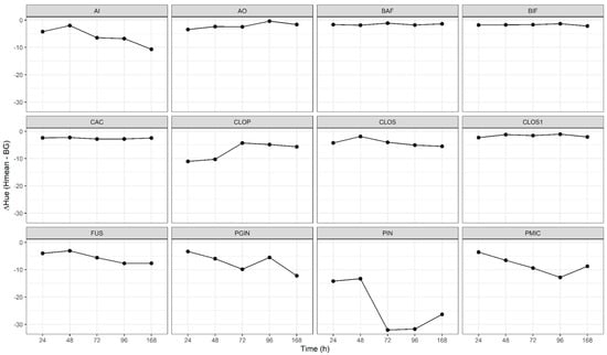
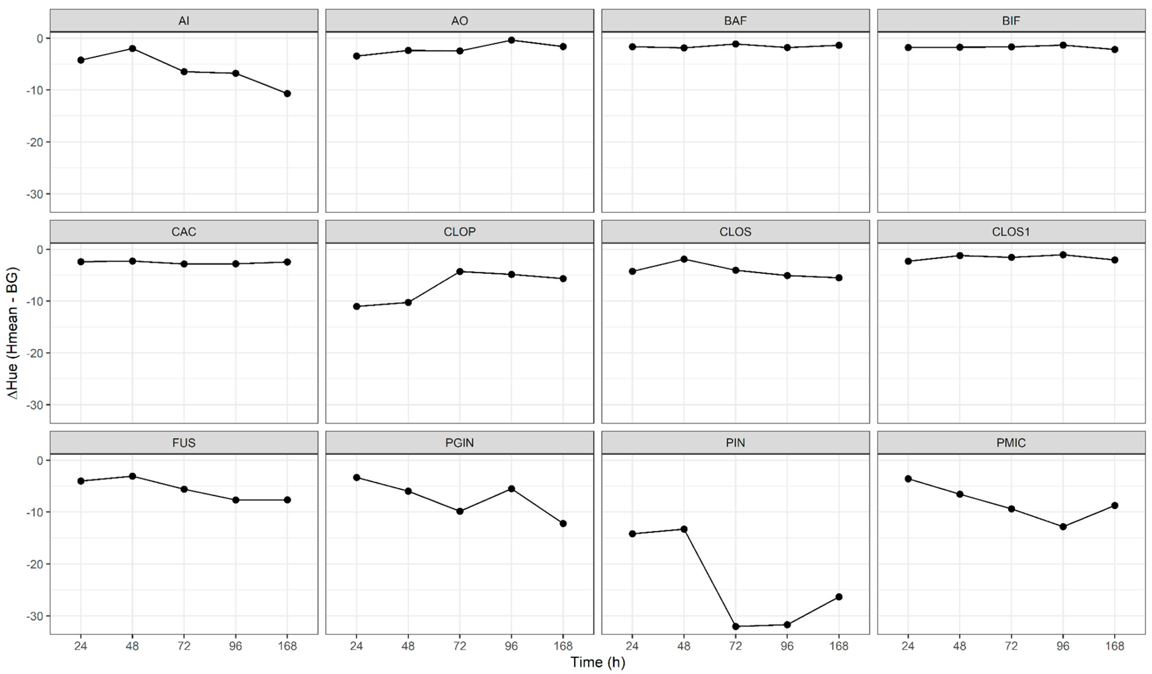
Post hoc analyses revealed numerous significant differences between the examined strains at all analyzed time points. The largest differences were observed between strains with the highest and lowest ΔH values, whereas some strains with similar ΔH values did not differ statistically significantly. The highest, least negative ΔH values were observed for Actinomyces odontolyticus (AO), Bacteroides fragilis (BAF), Bifidobacterium breve (BIF), Cutibacterium acnes (CAC), and Clostridium sporogenes ATCC 2486 (CLOS1). The lowest, most negative ΔH values were recorded for Prevotella intermedia (PIN), Parvimonas micra (PMIC), and Actinomyces israelii (AI) (Figure 1). These differences describe relative colony-to-background hue. Absolute fluorescence intensity was not quantified.
Figure 1.
Mean colony-to-background hue differences (ΔH = Hmean − BC) for each bacterial strain at 24, 48, 72, 96, and 168 h after inoculation (eight colonies per strain). Time points are displayed as equally spaced categories; line segments do not represent the rate of change per hour. Strain abbreviations: AI, A. israelii PCM 2391; AO, A. odontolyticus ATCC 17929/PCM 2399; BAF, B. fragilis ATCC 25285; BIF, B. breve ATCC 15700; CAC, C. acnes PCM 2400; CLOP, C. perfringens ATCC 13124; CLOS, C. sporogenes ATCC 19404; CLOS1, C. sporogenes ATCC 2486; FUS, F. nucleatum ATCC 25586; PGIN, P. gingivalis ATCC 33277; PIN, P. intermedia ATCC 25611; PMIC, P. micra ATCC 33270.
3.3. Changes in ΔH During Incubation
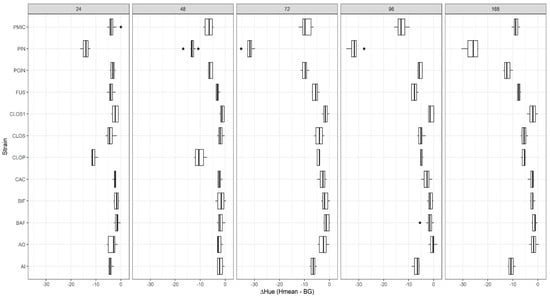
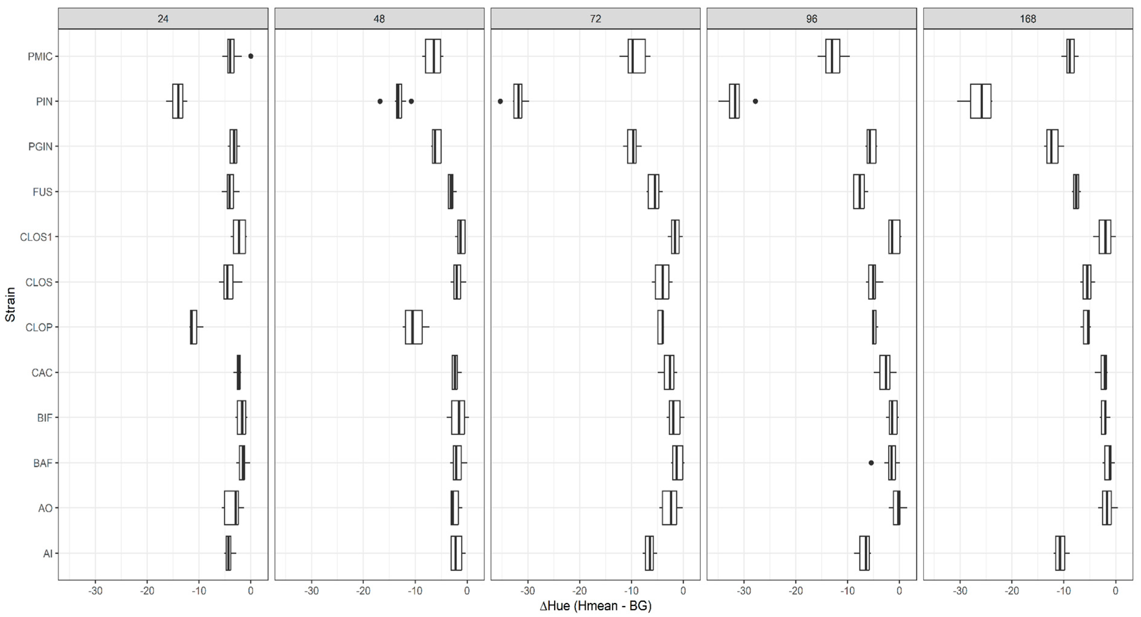
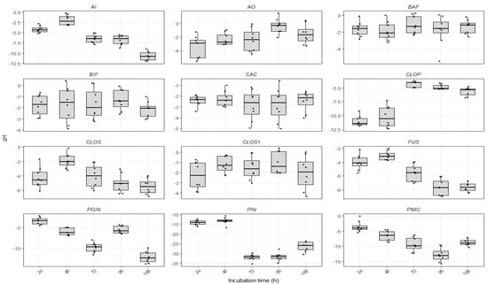
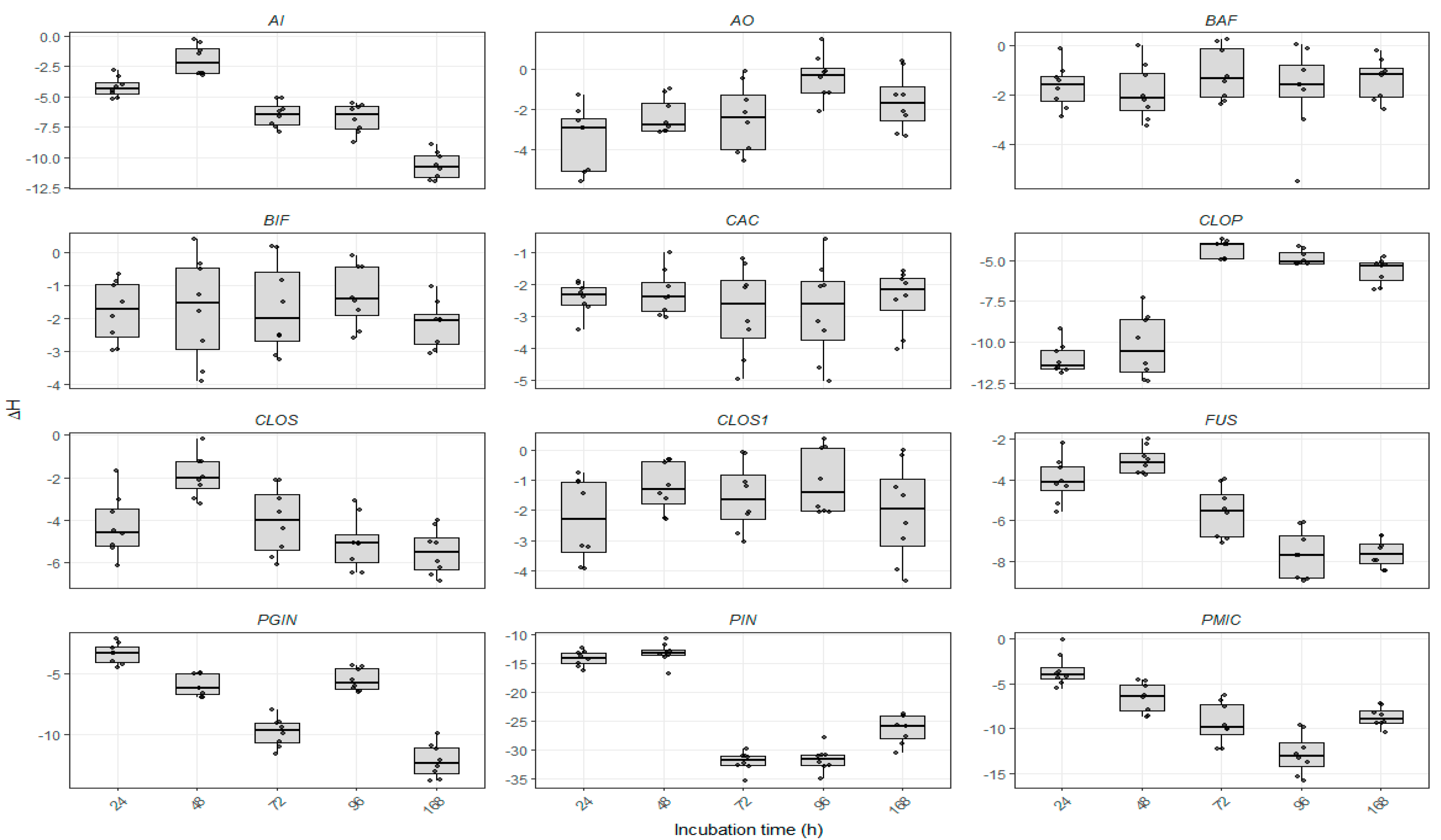
Incubation time had a significant effect on ΔH, with temporal patterns varying among strains. The largest visible shift in mean ΔH was observed for Prevotella intermedia (PIN), particularly between 48 and 72 h. Parvimonas micra (PMIC), Porphyromonas gingivalis (PGIN), and Fusobacterium nucleatum (FUS) generally showed more negative values at later than at initial time points, but these changes were not consistently monotonic. PIN and PMIC showed a partial increase between 96 and 168 h, while PGIN showed an increase between 72 and 96 h before decreasing again. Changes were also evident in Actinomyces israelii (AI) and Clostridium perfringens (CLOP); in CLOP, mean ΔH increased between 48 and 72 h. Bacteroides fragilis (BAF), Bifidobacterium breve (BIF), and Cutibacterium acnes (CAC) showed comparatively small descriptive changes over the observation period. The distributions at each time point are presented in Figure 2. These descriptions concern the observed profiles and do not establish equivalence or absence of temporal change in any strain.
3.4. Interaction Between Bacterial Strain and Incubation Time
The significant bacterial strain × incubation time interaction indicates that the temporal pattern of ΔH differed among the examined strains. Within-strain comparisons identified significant differences between consecutive time points in a subset of strains. P. intermedia showed a marked decrease between 48 and 72 h, followed by similar mean values at 72 and 96 h and a partial increase at 168 h. P. micra and P. gingivalis also exhibited non-monotonic profiles, whereas B. fragilis, B. breve, and C. acnes showed comparatively small shifts. Figure 3 displays within-strain distributions across time. The interaction supports strain-dependent temporal differences in hue contrast under the tested conditions, without demonstrating unique profiles suitable for identifying every strain.
Figure 3.
Within-strain distributions of ΔH at 24, 48, 72, 96, and 168 h after inoculation. Boxplots summarize the median and interquartile range, with whiskers and individual outlying measurements showing variability among the eight colonies. Strain abbreviations are defined in the Figure 1 caption and Table 1.
3.5. Between-Strain Variation and Distribution Overlap
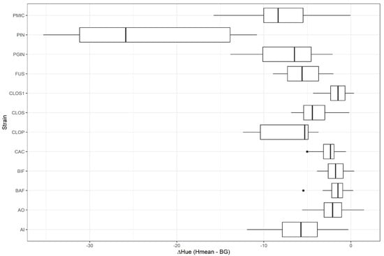
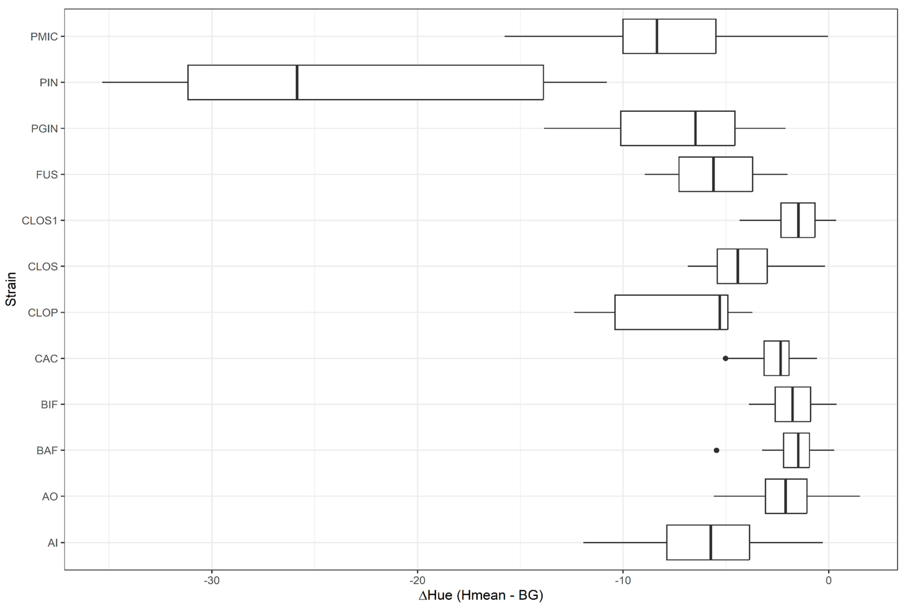
The examined strains differed in ΔH, but their distributions overlapped to varying degrees (Figure 4). Statistically significant between-strain comparisons therefore demonstrate differences in hue contrast at the group level. The strain-dependent temporal patterns may provide additional characterization features, but no classification model or independent validation was performed. The accuracy of ΔH for identifying unknown isolates cannot be determined from these results.
Figure 4.
Distributions of ΔH for the examined bacterial strains pooled across the five incubation time points. Boxplots show the median and interquartile range; the pooled distributions combine temporal variation with variation among colonies and do not represent independent measurements. Strain abbreviations are defined in the Figure 1 caption and Table 1.
3.6. Qualitative Assessment
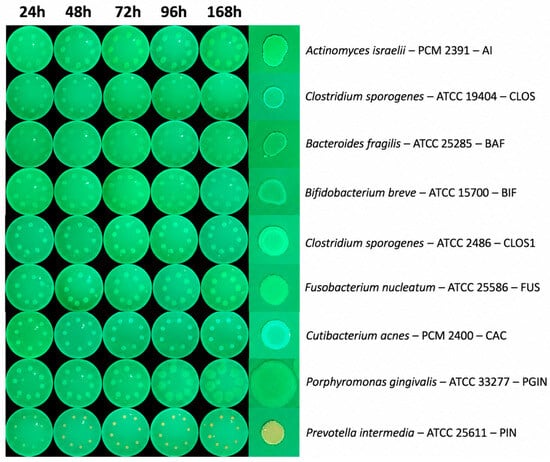
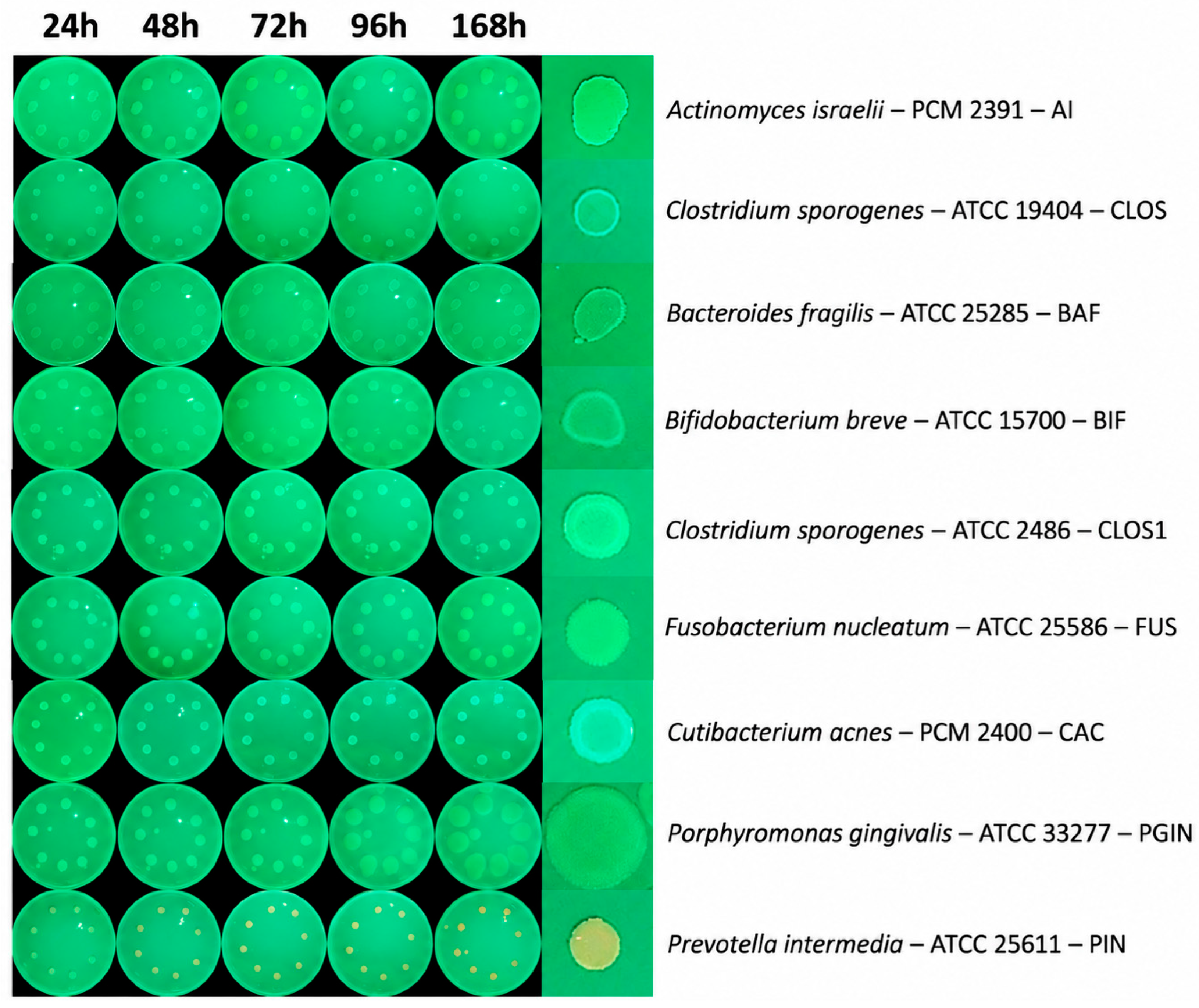
To complement the quantitative ΔH analysis, the images were examined descriptively for color appearance, apparent signal visibility, spatial distribution, and temporal changes. Most examined strains appeared green against the green culture-medium background, with limited visual color contrast. This limited the assessment of fluorescence distribution within individual colonies.
Prevotella intermedia ATCC 25611 showed a distinct yellow-to-orange appearance relative to the green background. The colony color was visually more evident at later observation times, although the images do not establish a quantitative increase in fluorescence intensity. The color appearance was observed across the eight evaluated colonies, but consistency within this experiment does not independently demonstrate inter-experiment reproducibility or exclude systematic imaging effects. In magnified images, the signal appeared broadly distributed over the colony, with some variation between central and peripheral regions.
Among the predominantly green-appearing strains, differences in colony size, morphology, apparent brightness, and central reflections were visible. In some strains, increasing colony size made the signal more conspicuous at later time points. These observations cannot distinguish increased biomass, differences in illumination or reflection, and changes in emission per unit colony area.
The qualitative assessment therefore supports a visible color distinction for P. intermedia within the examined panel. It is descriptive and should be interpreted alongside ΔH as a color feature, without treating perceived brightness or saturation as quantitative fluorescence intensity measurements (Figure 5).
Figure 5.
Representative images of nine selected anaerobic bacterial strains under 405 nm laser excitation at 24, 48, 72, 96, and 168 h after inoculation. The right-hand images show enlarged colony views. Most strains appear green against the medium; P. intermedia exhibits a distinct yellow-to-orange color. The images provide a qualitative illustration of colony appearance; they do not quantify fluorescence intensity.
4. Discussion
The present study aimed to assess differences in ΔH values among selected reference strains of anaerobic bacteria, and to determine whether characteristics of autofluorescence, as assessed by this parameter, change during successive stages of culture growth. The results revealed significant effects of bacterial strain and incubation time on ΔH, as well as a notable interaction between these two factors. The examined strains differed in their overall ΔH values, as well as in the direction and magnitude of changes over time. The values were negative, so higher ΔH denotes a hue difference closer to zero. ΔH does not quantify emission strength. While some strains, including Bacteroides fragilis, Bifidobacterium breve and Cutibacterium acnes, exhibited relatively stable profiles, more pronounced temporal changes were observed for Prevotella intermedia, Parvimonas micra, Porphyromonas gingivalis and Fusobacterium nucleatum. These findings suggest that image- derived colony-to-background hue differences varied according to bacterial strain and incubation time under the experimental conditions used. Although the significant strain × incubation time interaction supports further evaluation of temporal features, whether these changes improve classification performance over a single measurement remains undetermined.
The differences observed in the ΔH values among the examined strains may be related to variations in the composition and relative contribution of endogenous fluorophores. Previous studies have demonstrated substantial variation in the fluorescence profiles of oral bacteria, attributing red and orange-red emission primarily to porphyrins and their derivatives [82,83,84,85,88,89]. These compounds include protoporphyrin IX, coproporphyrins and metalloporphyrins. The production, accumulation and transformation of these compounds depend on microbial characteristics, as well as the availability of heme and other components of the culture medium [82,83]. Therefore, the observed differences should be interpreted as variations in image-derived colony-to-background colour contrast, without attributing them to specific fluorophores or their abundance. Porphyrins may contribute to bacterial fluorescence under 405 nm excitation because of their Soret absorption band, although other fluorophores, including flavins, may also contribute [87]. Previous studies have reported species-dependent differences in porphyrin production and heme utilization, including protoporphyrin IX release by B. fragilis and coproporphyrin III production by C. acnes [95,96]. Red autofluorescence has also been reported in A. odontolyticus and P. intermedia [88]. However, these findings do not establish the origin of the ΔH differences observed here. As fluorophore composition was not directly determined, ΔH should be interpreted as an image-derived colony-to-background hue difference rather than as a measure of specific fluorophores or their abundance. Furthermore, bacterial fluorescence may depend on culture-medium composition [82,83].
The ΔH values also changed over the course of the incubation period. The most pronounced changes were observed for P. intermedia, P. micra, P. gingivalis and F. nucleatum. For these strains, ΔH was generally more negative at later than at initial time points, but the profiles were not consistently monotonic. In particular, PIN and PMIC showed partial increases between 96 and 168 h, and PGIN increased between 72 and 96 h. AI and CLOP also showed appreciable descriptive changes. In contrast, B. fragilis, B. breve and C. acnes exhibited relatively stable profiles. Studies of biofilms partly support the dependence of autofluorescence on the growth stage, as an increase in red fluorescence and changes in microbial composition have been observed with biofilm maturation and increasing thickness [93,94,97]. These studies provide context for growth-dependent structural changes but do not directly explain the observed ΔH profiles. Antimicrobial and antibiofilm studies also emphasize the dependence of measured outcomes on the experimental model, exposure conditions, and biofilm architecture [57,61,62]. Such outcomes should not be inferred from hue alone. Although previous studies have associated microbial optical properties with growth conditions and metabolic state [90,91,92,93], these factors were not measured in the present study and cannot be used to explain the observed ΔH profiles directly. However, a decrease in ΔH does not necessarily indicate reduced metabolic activity or decreased autofluorescence intensity. Similarly, a stable ΔH value reflects a relatively stable hue difference between the colony and the culture medium; fluorophore abundance and metabolic state may still change. Therefore, temporal changes in ΔH primarily describe changes in the color phenotype of the colony during growth. Their relationship to metabolic activity or fluorescence intensity would require independent measurements.
The significant interaction between bacterial strain and incubation time indicates that temporal changes in relative hue difference varied among the examined strains. P. intermedia showed a particularly distinctive profile, with a marked decrease in ΔH between 48 and 72 h and a more evident yellow-to-orange appearance in the images. This observation can be considered alongside previous reports of pronounced red fluorescence in P. intermedia following excitation at 409 nm, with an emission maximum near 636 nm [98], and studies associating its characteristic fluorescence with protoporphyrin IX and other porphyrin derivatives [83,90,98,99]. Lennon et al. also reported fluorescence peaks consistent with protoporphyrin IX and showed that fluorescence depended on the culture medium [83]. However, these findings do not establish the mechanism underlying the ΔH changes observed here. Hue, saturation, and brightness represent distinct image properties [100], and the present study measured relative hue contrast rather than absolute emission intensity. Porphyrin-related pigmentation and microbial interactions in P. gingivalis [91,101], as well as differences in heme-acquisition mechanisms among anaerobic bacteria [95,102], provide broader biological context but were not investigated in this experiment. The temporal profiles may offer descriptive information for distinguishing the examined strains; however, classification performance was not assessed, and the potential advantage of temporal measurements over a single time point remains unknown. Establishing diagnostic value would require a classification model evaluated on an independent sample set, with sensitivity, specificity, and accuracy determined [44,45].
The present study has several limitations that should be considered when interpreting the findings. Firstly, the analysis was conducted under controlled in vitro conditions using a limited number of reference strains, with mostly one strain representing each species. Consequently, the observed ΔH profiles should be interpreted as strain-level observations, obtained under specific culture and imaging conditions used in this study, rather than as representative characteristics of the respective species. The findings also do not establish the clinical diagnostic value of the proposed approach, as its performance was not evaluated using clinical isolates or an independent validation cohort. Another important limitation is the dependence of autofluorescence on culture conditions, including the medium composition and the availability of heme and porphyrin precursors [83,91,93]. Furthermore, ΔH characterizes relative differences in colony and background color but does not provide a direct measure of fluorescence intensity or spectral composition [100]. The reliability of hue-based analysis may be reduced when image regions have weak or poorly defined color, while the circular nature of the hue scale may complicate statistical interpretation. These factors should be considered when comparing measurements across different samples or experimental settings. Future studies should investigate the influence of color saturation, brightness, and data-processing choices on measurement robustness and establish standardized criteria for reliable hue-based comparisons. Colony size, thickness and heterogeneity, as well as the characteristics of the optical system and image-recording device, may also influence the results [46,85]. In particular, the smartphone camera operated with automatic exposure and white-balance settings, and internal image-processing procedures could not be fully controlled. Although the same device and imaging geometry were used throughout the experiment, these automatic adjustments may have introduced variability in the recorded hue values. Therefore, ΔH should be interpreted as a relative image-based parameter obtained under the conditions used in this study, rather than as an absolute colourimetric measurement directly transferable across devices. Future studies should assess measurement reproducibility using fixed camera settings where possible, colour calibration, and validated image-acquisition protocols. Although the approximate laser spot diameter at the sample surface and the corresponding estimated irradiance and radiant exposure were determined, the spatial uniformity of irradiance across the illuminated area was not independently characterized. Consequently, potential variations in local excitation intensity across the sample cannot be excluded and may have influenced the measured autofluorescence signals. In addition, repeated exposure to 405 nm laser radiation may affect subsequent optical measurements through photobleaching or other light-induced photochemical or biological effects [87]. Although the laser output power and exposure duration were standardized across samples, these potential effects were not independently assessed. The possible contribution of laser-induced effects to temporal changes in ΔH therefore remains uncertain. Future studies should characterize the spatial irradiance distribution on bacterial autofluorescence to improve the standardization and reproducibility of the optical measurements. As no direct spectroscopic analysis or measurements of individual fluorophores were performed, the proposed biological mechanisms remain hypotheses that require experimental confirmation. Future studies should include a larger number of strains, including multiple strains of the same species and clinical isolates, and assess inter-batch and inter-laboratory reproducibility while standardizing image acquisition. Extending validation to dental substrates would require accounting for material- and species-dependent microbial colonization [103]. A valuable next step would be to conduct parallel spectral analysis to better characterize the contribution of individual fluorophores to the observed signal. Combining ΔH with other image-derived features and temporal changes across successive time points would also be valuable, followed by the development and validation of classification models. The feasibility of detecting bacterial autofluorescence signals using simple imaging systems with 405 nm excitation has already been demonstrated [46], whereas the application of multidimensional spectroscopic data and machine-learning algorithms may improve discriminatory performance [44,45]. Validating such an approach using an independent clinical sample set would help to determine whether temporal autofluorescence profiles could ultimately support conventional microbiological identification [44,45,46]. Accordingly, the present methodology can be used to quantify and compare relative colony-to-background hue differences under the tested imaging conditions. However, direct assessment of fluorescence intensity, identification or quantification of individual fluorophores, and interpretation of metabolic activity require complementary measurements and further validation. Because the same colonies were assessed at five successive time points, observations obtained from an individual colony were correlated. Colony identity was not included as a repeated-measures or random effect in the ART-ANOVA model. Consequently, within-colony correlation may have been underestimated and the inferential results, particularly the reported p-values, may be anticonservative. The statistical findings should therefore be interpreted as exploratory and require confirmation using models that explicitly account for repeated measurements. In addition, temporary removal of the plates from anaerobic incubation during imaging introduced brief exposure to ambient oxygen. Although imaging was performed promptly and the plates were immediately returned to the GENbag Anaer system, the possible influence of this transient exposure on subsequent measurements was not independently assessed.
5. Conclusions
- Bacterial strain was significantly associated with ΔH, which quantifies the relative hue difference between the colony and the culture medium under the tested culture and imaging conditions.
- ΔH varied with incubation time, and the direction and magnitude of these changes differed among the examined strains.
- The significant strain × incubation time interaction indicates strain-dependent temporal patterns of colony color. However, the potential value of these patterns for bacterial classification has not yet been validated.
- ΔH provides a quantitative measure of relative hue difference but does not directly measure fluorescence intensity, fluorophore abundance, or metabolic activity. Its diagnostic utility and the potential influence of laser-induced effects require further investigation using complementary measurements and independent validation.
Supplementary Materials
The following supporting information can be downloaded at: https://www.mdpi.com/article/10.3390/app162110573/s1, Table S1: Descriptive statistics of colony-to-background hue difference.
Author Contributions
Conceptualization, A.U., J.M. and M.D.; methodology, J.M., M.P. and J.N.; software, A.U.; validation, J.M., M.D., R.W. and D.S.; formal analysis, Ł.J.; investigation, A.U., M.P. and J.N.; resources, M.D., J.N. and M.P.; data curation, Ł.J. and A.U.; writing—original draft preparation, A.U., M.P., J.N., Ł.J., J.K. (Jan Kiryk) and J.K. (Julia Kensy); writing—review and editing, J.M., R.W. and J.K. (Julia Kensy); visualization, J.K. (Jan Kiryk) and J.K. (Julia Kensy); supervision, J.M., R.W. and D.S.; project administration, J.M.; funding acquisition, J.M. and M.D. All authors have read and agreed to the published version of the manuscript.
Funding
This work was financed by a subsidy from Wroclaw Medical University, number SUBD.B180.26.003.
Institutional Review Board Statement
Not applicable.
Informed Consent Statement
Not applicable.
Data Availability Statement
The reported findings are presented in the article. Further inquiries regarding the underlying measurements can be directed to the corresponding authors.
Acknowledgments
This work was carried out as a part of project No. 1/05/2025 entitled “Application of the 405 nm SMARTm laser manufactured by LASOTRONIX for detection of red-orange autofluorescence of pathogenic bacteria”.
Conflicts of Interest
The authors declare no conflicts of interest.
References
- Dewhirst, F.E.; Chen, T.; Izard, J.; Paster, B.J.; Tanner, A.C.R.; Yu, W.-H.; Lakshmanan, A.; Wade, W.G. The Human Oral Microbiome. J. Bacteriol. 2010, 192, 5002–5017. [Google Scholar] [CrossRef] [Scilit] [PubMed]
- Kilian, M.; Chapple, I.L.C.; Hannig, M.; Marsh, P.D.; Meuric, V.; Pedersen, A.M.L.; Tonetti, M.S.; Wade, W.G.; Zaura, E. The Oral Microbiome—An Update for Oral Healthcare Professionals. Br. Dent. J. 2016, 221, 657–666. [Google Scholar] [CrossRef] [Scilit] [PubMed]
- Nagy, E. Anaerobic Infections: Update on Treatment Considerations. Drugs 2010, 70, 841–858. [Google Scholar] [CrossRef] [Scilit] [PubMed]
- Könönen, E.; Wade, W.G. Actinomyces and Related Organisms in Human Infections. Clin. Microbiol. Rev. 2015, 28, 419–442. [Google Scholar] [CrossRef] [Scilit] [PubMed]
- Hall, V. Actinomyces—Gathering Evidence of Human Colonization and Infection. Anaerobe 2008, 14, 1–7. [Google Scholar] [CrossRef] [Scilit] [PubMed]
- Williams, H.C.; Dellavalle, R.P.; Garner, S. Acne Vulgaris. Lancet 2012, 379, 361–372. [Google Scholar] [CrossRef] [Scilit] [PubMed]
- Stefanopoulos, P.K.; Kolokotronis, A.E. The Clinical Significance of Anaerobic Bacteria in Acute Orofacial Odontogenic Infections. Oral Surg. Oral Med. Oral Pathol. Oral Radiol. Endodontology 2004, 98, 398–408. [Google Scholar] [CrossRef] [Scilit] [PubMed]
- Ali, M.; Razok, A.; Sasi, S.; Balushi, S.A.; Ibrahim, E.B.; Al-Maslamani, M.; Hadi, H.A. Significance, Clinical Spectrum, and Microbiological Evaluation of Actinomyces Blood Stream Infections. IJID Reg. 2025, 17, 100769. [Google Scholar] [CrossRef] [Scilit] [PubMed]
- Truong, R.D.; Do, V.A.; Njaravelil, K.A.; Ayesu, K.; Madruga, M.; Carlan, S.J. Ludwig Angina Caused by Clostridium Sporogenes in a 49-Year-Old HIV-Positive Man with Alcoholism and a Tooth Abscess: The First Reported Case. Am. J. Case Rep. 2023, 24, e941731-1. [Google Scholar] [CrossRef] [Scilit] [PubMed]
- Jain, H.; Singh, G.; Eranki, A. Actinomyces odontolyticus Causing Meningitis and Cervical Abscess. Bayl. Univ. Med. Cent. Proc. 2021, 34, 492–493. [Google Scholar] [CrossRef] [Scilit] [PubMed]
- Chen, L.; Zhang, T.; Wang, Y.; Lu, Y.; Chen, X.; Tan, X.; Huang, D.; Li, S. Bacteroides Fragilis Spondylitis after Suspected Oral Ulcer: A Clinical Case and Comprehensive Literature Review. Anaerobe 2024, 88, 102863. [Google Scholar] [CrossRef] [Scilit] [PubMed]
- Paulino, S.; Duran, M.; Allena, N.; Sosa, F.; Singhal, R. Understanding Actinomyces Odontolyticus: A Rare Culprit of Bacteremia. Cureus 2024, 16, e66086. [Google Scholar] [CrossRef] [Scilit] [PubMed]
- Shimizu, K.; Horinishi, Y.; Sano, C.; Ohta, R. Infection Route of Parvimonas Micra: A Case Report and Systematic Review. Healthcare 2022, 10, 1727. [Google Scholar] [CrossRef] [Scilit] [PubMed]
- Sanz, M.; Beighton, D.; Curtis, M.A.; Cury, J.A.; Dige, I.; Dommisch, H.; Ellwood, R.; Giacaman, R.A.; Herrera, D.; Herzberg, M.C.; et al. Role of Microbial Biofilms in the Maintenance of Oral Health and in the Development of Dental Caries and Periodontal Diseases. Consensus Report of Group 1 of the Joint EFP/ORCA Workshop on the Boundaries between Caries and Periodontal Disease. J. Clin. Periodontol. 2017, 44, S5–S11. [Google Scholar] [CrossRef] [Scilit] [PubMed]
- Papapanou, P.N.; Sanz, M.; Buduneli, N.; Dietrich, T.; Feres, M.; Fine, D.H.; Flemmig, T.F.; Garcia, R.; Giannobile, W.V.; Graziani, F.; et al. Periodontitis: Consensus Report of Workgroup 2 of the 2017 World Workshop on the Classification of Periodontal and Peri-Implant Diseases and Conditions. J. Clin. Periodontol. 2018, 45, S162–S170. [Google Scholar] [CrossRef] [Scilit] [PubMed]
- Herrera, D.; Retamal-Valdes, B.; Alonso, B.; Feres, M. Acute Periodontal Lesions (Periodontal Abscesses and Necrotizing Periodontal Diseases) and Endo-Periodontal Lesions. J. Clin. Periodontol. 2018, 45, S78–S94. [Google Scholar] [CrossRef] [Scilit] [PubMed]
- Siqueira, J.F.; Silva, W.O.; Romeiro, K.; Gominho, L.F.; Alves, F.R.F.; Rôças, I.N. Apical Root Canal Microbiome Associated with Primary and Posttreatment Apical Periodontitis: A Systematic Review. Int. Endod. J. 2024, 57, 1043–1058. [Google Scholar] [CrossRef] [Scilit] [PubMed]
- Siqueira, J.F.; Rôças, I.N. Present Status and Future Directions: Microbiology of Endodontic Infections. Int. Endod. J. 2022, 55, 512–530. [Google Scholar] [CrossRef] [Scilit] [PubMed]
- Könönen, E. Polymicrobial Infections with Specific Actinomyces and Related Organisms, Using the Current Taxonomy. J. Oral Microbiol. 2024, 16, 2354148. [Google Scholar] [CrossRef] [Scilit] [PubMed]
- Prada, I.; Mico-Munoz, P.; Giner-Lluesma, T.; Mico-Martinez, P.; Collado-Castellano, N.; Manzano-Saiz, A. Influence of Microbiology on Endodontic Failure. Literature Review. Med. Oral Patol. Oral Cir. Bucal 2019, 24, e364–e372. [Google Scholar] [CrossRef] [Scilit] [PubMed]
- Zygmunt, Ł.; Kiryk, S.; Wesołek, K.; Kiryk, J.; Nawrot-Hadzik, I.; Rybak, Z.; Sztyler, K.; Małyszek, A.; Matys, J.; Dobrzyński, M. The Role of the Oral Microbiome and Dental Caries in Respiratory Health: A Systematic Review. J. Clin. Med. 2025, 14, 7670. [Google Scholar] [CrossRef] [Scilit] [PubMed]
- Khadka, S.; Khan, S.; King, A.; Goldberg, L.R.; Crocombe, L.; Bettiol, S. Poor Oral Hygiene, Oral Microorganisms and Aspiration Pneumonia Risk in Older People in Residential Aged Care: A Systematic Review. Age Ageing 2021, 50, 81–87. [Google Scholar] [CrossRef] [Scilit] [PubMed]
- Canullo, L.; Rossetti, P.; Penarrocha, D. Identification of Enterococcus Faecalis and Pseudomonas Aeruginosa on and in Implants in Individuals with Peri-Implant Disease: A Cross-Sectional Study. Int. J. Oral Maxillofac. Implant. 2015, 30, 583–587. [Google Scholar] [CrossRef] [Scilit] [PubMed]
- Hashizume-Takizawa, T.; Yamaguchi, Y.; Kobayashi, R.; Shinozaki-Kuwahara, N.; Saito, M.; Kurita-Ochiai, T. Oral Challenge with Streptococcus Sanguinis Induces Aortic Inflammation and Accelerates Atherosclerosis in Spontaneously Hyperlipidemic Mice. Biochem. Biophys. Res. Commun. 2019, 520, 507–513. [Google Scholar] [CrossRef] [Scilit] [PubMed]
- Qays, R.A.; Jafar, Z.J. Comparison of the Level of Staphylococcus Aureus and Escherichia Coli in Complete Cleft Infants With Normal Infants. J. Craniofac. Surg. 2023, 34, e268–e271. [Google Scholar] [CrossRef] [Scilit] [PubMed]
- Arboleda, V.; Elsouri, K.N.; Heiser, S.E.; Bernal, I.; Kesselman, M.M.; Demory Beckler, M. Oral Microbiome as a Tool of Systemic Disease on Cleft Patients: A New Landscape. Cureus 2023, 15, e35444. [Google Scholar] [CrossRef] [Scilit] [PubMed]
- Sedgley, C.M.; Samaranayake, L.P. Antimicrobial Susceptibility of Oral Isolates of Enterobacter Cloacae and Klebsiella Pneumoniae from a Southern Chinese Population. Oral Microbiol. Immunol. 1998, 13, 315–321. [Google Scholar] [CrossRef] [Scilit] [PubMed]
- Abranches, J.; Zeng, L.; Kajfasz, J.K.; Palmer, S.R.; Chakraborty, B.; Wen, Z.T.; Richards, V.P.; Brady, L.J.; Lemos, J.A. Biology of Oral Streptococci. Microbiol. Spectr. 2018, 6, GPP3-0042-2018. [Google Scholar] [CrossRef] [Scilit] [PubMed]
- Scoffield, J.A.; Wu, H. Oral Streptococci and Nitrite-Mediated Interference of Pseudomonas Aeruginosa. Infect. Immun. 2015, 83, 101–107. [Google Scholar] [CrossRef] [Scilit] [PubMed]
- Minervini, G.; Shivakumar, S.; Ronsivalle, V.; Franco, R.; Cicciù, M.; Marrapodi, M.M. Microbiological Aspects of Cancer Progression: A Systematic Review Conducted According to the PRISMA 2020 Guidelines and the Cochrane Handbook for Systematic Reviews of Interventions. Dent. Med. Probl. 2024, 61, 739–746. [Google Scholar] [CrossRef] [Scilit] [PubMed]
- Qi, H.; Gao, H.; Li, M.; Sun, T.; Gu, X.; Wei, L.; Zhi, M.; Li, Z.; Fu, D.; Liu, Y.; et al. Parvimonas Micra Promotes Oral Squamous Cell Carcinoma Metastasis through TmpC-CKAP4 Axis. Nat. Commun. 2025, 16, 2305. [Google Scholar] [CrossRef] [Scilit] [PubMed]
- Oral and Intestinal Bacterial Exotoxins: Potential Linked to Carcinogenesis. In Progress in Molecular Biology and Translational Science; Elsevier: Amsterdam, The Netherlands, 2020; Volume 171, pp. 131–193.
- Bergsten, E.; Mestivier, D.; Donnadieu, F.; Pedron, T.; Barau, C.; Meda, L.T.; Mettouchi, A.; Lemichez, E.; Gorgette, O.; Chamaillard, M.; et al. Parvimonas Micra, an Oral Pathobiont Associated with Colorectal Cancer, Epigenetically Reprograms Human Colonocytes. Gut Microbes 2023, 15, 2265138. [Google Scholar] [CrossRef] [Scilit] [PubMed]
- Wang, B.; Deng, J.; Donati, V.; Merali, N.; Frampton, A.E.; Giovannetti, E.; Deng, D. The Roles and Interactions of Porphyromonas Gingivalis and Fusobacterium Nucleatum in Oral and Gastrointestinal Carcinogenesis: A Narrative Review. Pathogens 2024, 13, 93. [Google Scholar] [CrossRef] [Scilit] [PubMed]
- Dessì, A.; Bosco, A.; Pintus, R.; Orrù, G.; Fanos, V. Fusobacterium Nucleatum and Alteration of the Oral Microbiome: From Pregnancy to SARS-COV-2 Infection. Eur. Rev. Med. Pharmacol. Sci. 2021, 25, 4579–4596. [Google Scholar] [CrossRef] [Scilit] [PubMed]
- Nakashima, C.; Yamamoto, K.; Kishi, S.; Sasaki, T.; Ohmori, H.; Fujiwara-Tani, R.; Mori, S.; Kawahara, I.; Nishiguchi, Y.; Mori, T.; et al. Clostridium Perfringens Enterotoxin Induces Claudin-4 to Activate YAP in Oral Squamous Cell Carcinomas. Oncotarget 2020, 11, 309–321. [Google Scholar] [CrossRef] [Scilit] [PubMed]
- Brennan, C.A.; Garrett, W.S. Fusobacterium Nucleatum—Symbiont, Opportunist and Oncobacterium. Nat. Rev. Microbiol. 2019, 17, 156–166. [Google Scholar] [CrossRef] [Scilit] [PubMed]
- Peeters, B.; Magerman, K.; Waumans, L.; Cartuyvels, R. Laboratory Survey and Literature Review of Anaerobic Bacteriology: Foundations of a Clinically Orientated and Evidence-Based Workup for Anaerobic Cultures. Diagn. Microbiol. Infect. Dis. 2016, 86, 15–22. [Google Scholar] [CrossRef] [Scilit] [PubMed]
- Siqueira, J.F.; Rôças, I.N. As-yet-Uncultivated Oral Bacteria: Breadth and Association with Oral and Extra-Oral Diseases. J. Oral Microbiol. 2013, 5, 21077. [Google Scholar] [CrossRef] [Scilit] [PubMed]
- Clarridge, J.E.; Zhang, Q. Genotypic Diversity of Clinical Actinomyces Species: Phenotype, Source, and Disease Correlation among Genospecies. J. Clin. Microbiol. 2002, 40, 3442–3448. [Google Scholar] [CrossRef] [Scilit] [PubMed]
- Matys, J.; Kensy, J.; Gedrange, T.; Zawiślak, I.; Grzech-Leśniak, K.; Dobrzyński, M. A Molecular Approach for Detecting Bacteria and Fungi in Healthcare Environment Aerosols: A Systematic Review. Int. J. Mol. Sci. 2024, 25, 4154. [Google Scholar] [CrossRef] [Scilit] [PubMed]
- Urbańska, A.; Pajączkowska, M.; Nowicka, J.; Kensy, J.; Kulus, M.; Wiench, R.; Skaba, D.; Dobrzyński, M.; Matys, J. In Vitro Study of Autofluorescence Dynamics in Selected Fungal Strains Under 405 Nm Laser Excitation. Appl. Sci. 2026, 16, 5475. [Google Scholar] [CrossRef] [Scilit]
- Urbańska, A.; Pajączkowska, M.; Nowicka, J.; Kensy, J.; Kiryk, J.; Janek, Ł.; Wiench, R.; Skaba, D.; Dobrzyński, M.; Matys, J. Photoinduced Fluorescence Changes in Selected Bacterial Strains Under 405 Nm Laser Excitation: An In Vitro Hue-Based Monitoring Approach. Micromachines 2026, 17, 885. [Google Scholar] [CrossRef] [Scilit] [PubMed]
- Mito, D.; Eda, H.; Okihara, S.; Kurita, M.; Hatayama, N.; Yoshino, Y.; Watanabe, Y.; Ishii, K. Optimizing Excitation Light for Accurate Rapid Bacterial Species Identification with Autofluorescence. J. Fluoresc. 2024, 34, 1737–1745. [Google Scholar] [CrossRef] [Scilit] [PubMed]
- Mito, D.; Okihara, S.; Kurita, M.; Hatayama, N.; Yoshino, Y.; Watanabe, Y.; Ishii, K. Leveraging Broad-Spectrum Fluorescence Data and Machine Learning for High-Accuracy Bacterial Species Identification. J. Biophotonics 2024, 17, e202400300. [Google Scholar] [CrossRef] [Scilit] [PubMed]
- Buchanan, B.C.; Safavinia, B.; Wu, L.; Yoon, J.-Y. Smartphone-Based Autofluorescence Imaging to Detect Bacterial Species on Laboratory Surfaces. Analyst 2022, 147, 2980–2987. [Google Scholar] [CrossRef] [Scilit] [PubMed]
- Guo, Y.; Wei, C.; Liu, C.; Li, D.; Sun, J.; Huang, H.; Zhou, H. Inhibitory Effects of Oral Actinomyces on the Proliferation, Virulence and Biofilm Formation of Candida Albicans. Arch. Oral Biol. 2015, 60, 1368–1374. [Google Scholar] [CrossRef] [Scilit] [PubMed]
- Shen, M.; Yang, Y.; Shen, W.; Cen, L.; McLean, J.S.; Shi, W.; Le, S.; He, X. A Linear Plasmid-Like Prophage of Actinomyces Odontolyticus Promotes Biofilm Assembly. Appl. Environ. Microbiol. 2018, 84, e01263-18. [Google Scholar] [CrossRef] [Scilit] [PubMed]
- Li, Q.; Li, Y.; Qiao, Q.; Zhao, N.; Yang, Y.; Wang, L.; Wang, Y.; Guo, C.; Guo, Y. Oral Administration of Bifidobacterium Breve Improves Anti-Angiogenic Drugs-Derived Oral Mucosal Wound Healing Impairment via Upregulation of Interleukin-10. Int. J. Oral Sci. 2023, 15, 56. [Google Scholar] [CrossRef] [Scilit] [PubMed]
- Nishikawa, Y.; Xu, C.; Yoshimoto, S.; Katsumata, N.; Iwabuchi, N.; Yanagisawa, N.; Koido, S.; Tanaka, M.; Xiao, J.-Z.; Asaoka, D.; et al. Imaging and Microorganism Analyses of the Effects of Oral Bifidobacterium Breve Intake on Facial Skin in Females: A Randomized, Double-Blind, Placebo-Controlled Study. Nutrients 2025, 17, 2976. [Google Scholar] [CrossRef] [Scilit] [PubMed]
- Ananda, N.; Ariawan, D.; Juniantito, V.; Julia, V.; Yunial, A.; Januarti, R.D.; Irfan, I.; Bachtiar, E.W. Effects of the Probiotics on the Proliferation Phase in Oral Wound Healing: In Vivo Study. Dent. Med. Probl. 2025, 62, 681–690. [Google Scholar] [CrossRef] [Scilit] [PubMed]
- Yli-Knuuttila, H.; Snäll, J.; Kari, K.; Meurman, J.H. Colonization of Lactobacillus Rhamnosus GG in the Oral Cavity. Oral Microbiol. Immunol. 2006, 21, 129–131. [Google Scholar] [CrossRef] [Scilit] [PubMed]
- Teanpaisan, R.; Surachat, K.; Wonglapsuwan, M.; Piwat, S.; Pahumunto, N. Short-term Use of Lacticaseibacillus Rhamnosus SD11 and the Oral Microbiome: Low Caries RCT Study. Oral Dis. 2024, 30, 2736–2745. [Google Scholar] [CrossRef] [Scilit] [PubMed]
- Olczak-Kowalczyk, D.; Turska-Szybka, A.; Twetman, S.; Gozdowski, D.; Piekoszewska-Ziętek, P.; Góra, J.; Wróblewska, M. Effect of Tablets Containing a Paraprobiotic Strain and the Cranberry Extract on Caries Incidence in Preschool Children: A Randomized Controlled Trial. Dent. Med. Probl. 2025, 62, 209–215. [Google Scholar] [CrossRef] [Scilit] [PubMed]
- Kowalski, J.; Rygas, J.; Homa, K.; Dobrzyński, W.; Wiglusz, R.J.; Matys, J.; Dobrzyński, M. Antibacterial Activity of Endodontic Gutta-Percha—A Systematic Review. Appl. Sci. 2024, 14, 388. [Google Scholar] [CrossRef] [Scilit]
- Lakshmanan, L.; Gurunathan, D.; Shanmugam, R. Effectiveness of White Tea-Mediated Silver Nanoparticles as an Intracanal Irrigant against Enterococcus Faecalis: An in Vitro Study. Dent. Med. Probl. 2024, 61, 593–598. [Google Scholar] [CrossRef] [Scilit] [PubMed]
- Duggan, J.; Sedgley, C. Biofilm Formation of Oral and Endodontic Enterococcus Faecalis. J. Endod. 2007, 33, 815–818. [Google Scholar] [CrossRef] [Scilit] [PubMed]
- Karlak, V.; Ortarzewska, M.; Jankowski, J.; Nijakowski, K. Propolis Efficiency against Enterococcus faecalis Compared with Other Intra-Canal medicaments: A Systematic Review With meta-Analysis. J. Stomatol. 2024, 77, 136–146. [Google Scholar] [CrossRef] [Scilit]
- Nochahrly, N.; Hachem, C.; Geitani, R.; Chedid, J.-C. Comparison of Antibacterial Activity of Five Root Canal Filling Materials in Primary Dentition: An in Vitro Study. J. Stomatol. 2025, 78, 194–202. [Google Scholar] [CrossRef] [Scilit]
- Mehdipour, A.; Pourreisi, R.; Amini-khoei, H.; Shams, S. Evaluating the Antibacterial and Antibiofilm Activity of Zataria Multiflora in Comparison with Chlorhexidine, Using a Tooth Model: A Preliminary Study. Dent. Med. Probl. 2025, 62, 293–298. [Google Scholar] [CrossRef] [Scilit] [PubMed]
- Awaida, A.; El Hachem, R.; Issa, A.; Kallasy, M.; Zogheib, C.; Hage, W. The Action of Different Irrigant Activation Methods on Engineered Endodontic Biofilm: An in Vitro Study. Biomater. Investig. Dent. 2025, 12, 60–67. [Google Scholar] [CrossRef] [Scilit] [PubMed]
- Liu, H.; Nio, S.; Shen, Y. Sodium Hypochlorite against Enterococcus Faecalis Biofilm in Dentinal Tubules: Effect of Concentration, Temperature, and Exposure Time. Odontology 2024, 112, 390–398. [Google Scholar] [CrossRef] [Scilit] [PubMed]
- Barnard, D.; Davies, J.; Figdor, D. Susceptibility of Actinomyces Israelii to Antibiotics, Sodium Hypochlorite and Calcium Hydroxide. Int. Endod. J. 1996, 29, 320–326. [Google Scholar] [CrossRef] [Scilit] [PubMed]
- Grącka—Mańkowska, J.; Zarzycka, B.; Pawlicka, H. Impact of Diode Laser Irradiation on the Size of Enterococcus Faecalis Population in the Canals of Bovine Teeth—An in Vitro Study. J. Stomatol. Czas. Stomatol. 2015, 68, 48–58. [Google Scholar] [CrossRef] [Scilit]
- Morikawa, M.; Uehara, S.; Yoshida, A.; Sakagami, H.; Masuda, Y. Photodynamic Therapy With Resveratrol and an Nd:YAG Laser for Enterococcus Faecalis Elimination. In Vivo 2024, 38, 559–566. [Google Scholar] [CrossRef] [Scilit] [PubMed]
- Le Goff, A.; Dautel-Morazin, A.; Guigand, M.; Vulcain, J.-M.; Bonnaure-Mallet, M. An Evaluation of the CO2 Laser for Endodontic Disinfection. J. Endod. 1999, 25, 105–108. [Google Scholar] [CrossRef] [Scilit] [PubMed]
- Garcez, A.S.; Ribeiro, M.S.; Tegos, G.P.; Núñez, S.C.; Jorge, A.O.C.; Hamblin, M.R. Antimicrobial Photodynamic Therapy Combined with Conventional Endodontic Treatment to Eliminate Root Canal Biofilm Infection. Lasers Surg. Med. 2007, 39, 59–66. [Google Scholar] [CrossRef] [Scilit] [PubMed]
- Zakrzewski, W.; Dobrzynski, M.; Nowicka, J.; Pajaczkowska, M.; Szymonowicz, M.; Targonska, S.; Sobierajska, P.; Wiglusz, K.; Dobrzynski, W.; Lubojanski, A.; et al. The Influence of Ozonated Olive Oil-Loaded and Copper-Doped Nanohydroxyapatites on Planktonic Forms of Microorganisms. Nanomaterials 2020, 10, 1997. [Google Scholar] [CrossRef] [Scilit] [PubMed]
- Abd Alsatar Shwailiya, S.; Mahdi, A.; Mohammed, Q. The Efficacy of Anti-Microbial Agents against Actinomyces Species Isolated from Patients with Dental Caries. J. Stomatol. 2024, 77, 47–54. [Google Scholar] [CrossRef] [Scilit]
- Carvalho, F.S.; Tarabal, V.S.; Livio, D.F.; Cruz, L.F.; Monteiro, A.P.F.; Parreira, A.G.; Guimarães, P.P.G.; Scheuerman, K.; Chagas, R.C.R.; Da Silva, J.A.; et al. Production and Characterization of the Lipopeptide with Anti-Adhesion for Oral Biofilm on the Surface of Titanium for Dental Implants. Arch. Microbiol. 2024, 206, 354. [Google Scholar] [CrossRef] [Scilit] [PubMed]
- Dimkov, A.; Gjorgievska, E.; Simonoska, J. New Findings about Releasing of Chloride Ions and Quaternary Ammonium Compounds From conventional and Experimental Glass Ionomers. J. Stomatol. 2024, 77, 77–86. [Google Scholar] [CrossRef] [Scilit]
- Aherne, O.; Ortiz, R.; Fazli, M.M.; Davies, J.R. Effects of Stabilized Hypochlorous Acid on Oral Biofilm Bacteria. BMC Oral Health 2022, 22, 415. [Google Scholar] [CrossRef] [Scilit] [PubMed]
- Shalaby, H.A.; Soliman, N.K.; Al–Saudi, K.W. Antibacterial and Preventive Effects of Newly Developed Modified Nano-Chitosan/Glass-Ionomer Restoration on Simulated Initial Enamel Caries Lesions: An in Vitro Study. Dent. Med. Probl. 2024, 61, 353–362. [Google Scholar] [CrossRef] [Scilit] [PubMed]
- Elbahie, D.M.; Badawy, R.E.-S.; Ibrahim, S.A.M.; Hassan, M.; Habib, N.A. Assessment of the Antibacterial Activity of Glass Ionomer Cements Modified by Polyamidoamine and Bioactive Glass: An in Vitro Study. Dent. Med. Probl. 2025, 63, 199–207. [Google Scholar] [CrossRef] [Scilit] [PubMed]
- Aydın, N.; Karaoğlanoğlu, S.; Süloğlu, A.K.; İdil, N.; Demir, M.; Karakaş, İ.H. Investigation of Cytotoxicity and Antibacterial effect of Boron-Containing Nano-Hydroxyapatite. J. Stomatol. 2024, 77, 93–99. [Google Scholar] [CrossRef] [Scilit]
- Poolkerd, P.; Nagaviroj, N.; Srisatjaluk, R.L.; Eiampongpaiboon, T. Anti-Microbial Effect of Piper Betle Crude Extract and Essential Oil Incorporated into Short-Term Soft Lining Material. J. Stomatol. 2023, 76, 226–234. [Google Scholar] [CrossRef] [Scilit]
- Shamma, B.; Arrag, E.; Rajab, A.; Kurdi, S. Anti-Bacterial Activity of Applying Chitosan and Propolis Dressing against Enterococcus Faecalis in Primary Teeth: In Vitro Study. J. Stomatol. 2022, 75, 36–43. [Google Scholar] [CrossRef] [Scilit]
- Rifaat, S.; Abuohashish, H.M.; Alsaleh, R.; Alqahtani, M.; Aloraifi, N.; Aljarbaua, J.; Albeladi, A.; Almajed, Y.; Alharamlah, F.; Aleraky, D. Antimicrobial and Antibiofilm Activities of Neem and Myrrh Extracts against Enterococcus Faecalis in Dentin Specimens. Dent. Med. Probl. 2026. ahead of print. [Google Scholar] [CrossRef] [Scilit] [PubMed]
- Algarni, H.A.; Alonazi, M.A.; Bhavikatti, S.K.; Adil, A.H.; Karobari, M.I. Antimicrobial and Cytotoxic Evaluation of Biosurfactant-Coated Copper Nanoparticles: A Study on Selected Endodontic Pathogens. Dent. Med. Probl. 2026, 63, 407–417. [Google Scholar] [CrossRef] [Scilit] [PubMed]
- Mojgani, N.; Bagheri, M.; Moharrami, M.; Vaseji, N.; Sanjabi, M.-R.; Hosseini, O.; Khoramjouy, M.; Kiani, R.; Ayatollahi, S.A. Antioxidant and Antibacterial Potentials of Four indigenous Propolis Extracts against Oral Tooth decay Pathogens, Streptococcus Mutans and Candida Albicans. J. Stomatol. 2025, 78, 6–14. [Google Scholar] [CrossRef] [Scilit]
- Adiningrat, A.; Maulana, I.; Fadhlurrahman, A.G.; Kurniawan, M.F.; Seno Aji, N.R.A. Evaluation of Bio-Compatibility and Effectiveness of Propolis Tetragonula Sp. as Dental Anti-Microbial Agent. J. Stomatol. 2023, 76, 94–100. [Google Scholar] [CrossRef] [Scilit]
- Volgenant, C.M.C.; van der Veen, M.H.; de Soet, J.J.; ten Cate, J.M. Effect of Metalloporphyrins on Red Autofluorescence from Oral Bacteria. Eur. J. Oral Sci. 2013, 121, 156–161. [Google Scholar] [CrossRef] [Scilit] [PubMed]
- Lennon, Á.M.; Brune, L.; Techert, S.; Buchalla, W. Fluorescence Spectroscopy Shows Porphyrins Produced by Cultured Oral Bacteria Differ Depending on Composition of Growth Media. Caries Res. 2023, 57, 74–86. [Google Scholar] [CrossRef] [Scilit] [PubMed]
- Kang, S.-M.; de Josselin de Jong, E.; Higham, S.M.; Hope, C.K.; Kim, B.-I. Fluorescence Fingerprints of Oral Bacteria. J. Biophotonics 2020, 13, e201900190. [Google Scholar] [CrossRef] [Scilit] [PubMed]
- Yan, Y.-J.; Wang, B.-W.; Yang, C.-M.; Wu, C.-Y.; Ou-Yang, M. Autofluorescence Detection Method for Dental Plaque Bacteria Detection and Classification: Example of Porphyromonas Gingivalis, Aggregatibacter Actinomycetemcomitans, and Streptococcus Mutans. Dent. J. 2021, 9, 74. [Google Scholar] [CrossRef] [Scilit] [PubMed]
- Cannon, T.M.; Lagarto, J.L.; Dyer, B.T.; Garcia, E.; Kelly, D.J.; Peters, N.S.; Lyon, A.R.; French, P.M.W.; Dunsby, C. Characterization of NADH Fluorescence Properties under One-Photon Excitation with Respect to Temperature, pH, and Binding to Lactate Dehydrogenase. OSA Contin. 2021, 4, 1610–1625. [Google Scholar] [CrossRef] [Scilit] [PubMed]
- Plavskii, V.Y.; Mikulich, A.V.; Tretyakova, A.I.; Leusenka, I.A.; Plavskaya, L.G.; Kazyuchits, O.A.; Dobysh, I.I.; Krasnenkova, T.P. Porphyrins and Flavins as Endogenous Acceptors of Optical Radiation of Blue Spectral Region Determining Photoinactivation of Microbial Cells. J. Photochem. Photobiol. B 2018, 183, 172–183. [Google Scholar] [CrossRef] [Scilit] [PubMed]
- Van Der Veen, M.H.; Thomas, R.Z.; Huysmans, M.C.D.N.J.M.; De Soet, J.J. Red Autofluorescence of Dental Plaque Bacteria. Caries Res. 2006, 40, 542–545. [Google Scholar] [CrossRef] [Scilit] [PubMed]
- Sun, J.; Vellappan, S.; Akdemir, J.; Steier, L.; Feinbloom, R.E.; Yadavalli, S.S. Imaging of Porphyrin-Specific Fluorescence in Pathogenic Bacteria in Vitro Using a Wearable, Hands-Free System. J. Microbiol. Methods 2025, 237, 107225. [Google Scholar] [CrossRef] [Scilit] [PubMed]
- Hope, C.K.; Billingsley, K.; De Josselin De Jong, E.; Higham, S.M. A Preliminary Study of the Effects of pH upon Fluorescence in Suspensions of Prevotella Intermedia. PLoS ONE 2016, 11, e0158835. [Google Scholar] [CrossRef] [Scilit] [PubMed]
- Phonok, Y.; Pyne, A.; Liu, S.; Ataide, S.F.; Gu, H.; Zhou, X.; Gao, J.; Kwan, A.H. Colouring Dysbiosis: FetB-Dependent Mn-PPIX Produced by Porphyromonas Gingivalis Shapes the Oral Microbiota. npj Biofilms Microbiomes 2026, 12, 76. [Google Scholar] [CrossRef] [Scilit] [PubMed]
- Bhattacharjee, A.; Datta, R.; Gratton, E.; Hochbaum, A.I. Metabolic Fingerprinting of Bacteria by Fluorescence Lifetime Imaging Microscopy. Sci. Rep. 2017, 7, 3743. [Google Scholar] [CrossRef] [Scilit] [PubMed]
- Volgenant, C.M.C.; Hoogenkamp, M.A.; Buijs, M.J.; Zaura, E.; Ten Cate, J.B.M.; Van Der Veen, M.H. Red Fluorescent Biofilm: The Thick, the Old, and the Cariogenic. J. Oral Microbiol. 2016, 8, 30346. [Google Scholar] [CrossRef] [Scilit] [PubMed]
- Kim, Y.-S.; Lee, E.-S.; Kwon, H.-K.; Kim, B.-I. Monitoring the Maturation Process of a Dental Microcosm Biofilm Using the Quantitative Light-Induced Fluorescence-Digital (QLF-D). J. Dent. 2014, 42, 691–696. [Google Scholar] [CrossRef] [Scilit] [PubMed]
- Rocha, E.R.; Bergonia, H.A.; Gerdes, S.; Jeffrey Smith, C. Bacteroides Fragilis Requires the Ferrous-iron Transporter Feo AB and the CobN-like Proteins BtuS1 and BtuS2 for Assimilation of Iron Released from Heme. MicrobiologyOpen 2019, 8, e00669. [Google Scholar] [CrossRef] [Scilit] [PubMed]
- Lee, W.L.; Shalita, A.R.; Poh-Fitzpatrick, M.B. Comparative Studies of Porphyrin Production in Propionibacterium Acnes and Propionibacterium Granulosum. J. Bacteriol. 1978, 133, 811–815. [Google Scholar] [CrossRef] [Scilit] [PubMed]
- Corsini, P.M.; Wang, S.; Rehman, S.; Fenn, K.; Sagar, A.; Sirovica, S.; Cleaver, L.; Edwards-Gayle, C.J.C.; Mastroianni, G.; Dorgan, B.; et al. Molecular and Cellular Insight into Escherichia Coli SslE and Its Role during Biofilm Maturation. npj Biofilms Microbiomes 2022, 8, 9. [Google Scholar] [CrossRef] [Scilit] [PubMed]
- Nomura, Y.; Takeuchi, H.; Okamoto, M.; Sogabe, K.; Okada, A.; Hanada, N. Chair-Side Detection of Prevotella Intermedia in Mature Dental Plaque by Its Fluorescence. Photodiagnosis Photodyn. Ther. 2017, 18, 335–341. [Google Scholar] [CrossRef] [Scilit] [PubMed]
- Smalley, J.W.; Silver, J.; Birss, A.J.; Withnall, R.; Titler, P.J. The Haem Pigment of the Oral Anaerobes Prevotella Nigrescens and Prevotella Intermedia Is Composed of Iron(III) Protoporphyrin IX in the Monomeric Form. Microbiology 2003, 149, 1711–1718. [Google Scholar] [CrossRef] [Scilit] [PubMed]
- Hakonen, A.; Beves, J.E.; Strömberg, N. Digital Colour Tone for Fluorescence Sensing: A Direct Comparison of Intensity, Ratiometric and Hue Based Quantification. Analyst 2014, 139, 3524–3527. [Google Scholar] [CrossRef] [Scilit] [PubMed]
- Zhu, B.; Anandan, V.; Bao, L.; Xu, P. High-throughput Characterization of the Influence of Streptococcus Sanguinis Genes on the Interaction between Streptococcus Sanguinis and Porphyromonas Gingivalis. Mol. Oral Microbiol. 2024, 39, 461–467. [Google Scholar] [CrossRef] [Scilit] [PubMed]
- Antonyuk, S.V.; Siemińska, K.; Śmiga, M.; Strange, R.W.; Wagner, M.; Barnett, K.J.; Olczak, T. Bacteroides Fragilis Expresses Three Proteins Similar to Porphyromonas Gingivalis HMUY: Hemophore-like Proteins Differentially Evolved to Participate in Heme Acquisition in Oral and Gut Microbiomes. FASEB J. 2023, 37, e22981. [Google Scholar] [CrossRef] [Scilit] [PubMed]
- Dobrzynski, M.; Pajaczkowska, M.; Nowicka, J.; Jaworski, A.; Kosior, P.; Szymonowicz, M.; Kuropka, P.; Rybak, Z.; Bogucki, Z.A.; Filipiak, J.; et al. Study of Surface Structure Changes for Selected Ceramics Used in the CAD/CAM System on the Degree of Microbial Colonization, In Vitro Tests. BioMed Res. Int. 2019, 2019, 9130806. [Google Scholar] [CrossRef] [Scilit] [PubMed]

Disclaimer/Publisher’s Note: The statements, opinions and data contained in all publications are solely those of the individual author(s) and contributor(s) and not of MDPI and/or the editor(s). MDPI and/or the editor(s) disclaim responsibility for any injury to people or property resulting from any ideas, methods, instructions or products referred to in the content. |
© 2026 by the authors. Licensee MDPI, Basel, Switzerland. This article is an open access article distributed under the terms and conditions of the Creative Commons Attribution (CC BY) license.