


Abstract
Ultrasound-guided femoral vascular access (UFVA) is a crucial step in cardiovascular intervention, yet training models remain costly, anatomically limited, or insufficiently durable. This research aimed to develop and evaluate a reusable, self-healing vascularised leg phantom in collaboration with clinicians. This bifurcating vascular model was cast in Plastisol using a customisable silicone mould design. The material exhibited a Shore OO hardness of 18.0 ± 2.2, a speed of sound of 1454 ± 15 m/s, an acoustic attenuation of 1.66 ± 0.02 × 10⁶ kg/m2s, and healed 18G needle lesions within 3 h. Training capabilities were evaluated in a workshop involving 18 medical students: FVA times improved by more than 60% after 5 min of free practice. Qualitative feedback was collected from 31 medical educators via a seven-question Likert survey, with most reporting they would adopt the phantom for teaching. Phantoms cost £7.87 for materials, yet educators valued the models at £100–£500, underscoring its perceived utility. Compared to commercial alternatives, this in-house production approach reduced costs by 10–60 times, achieving comparable durability and anatomical fidelity. This study establishes a scalable, ultra-low-cost method for producing anatomically realistic, self-healing vascular phantoms, validated for effective skill acquisition in both educational and research settings.
1. Introduction
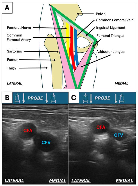
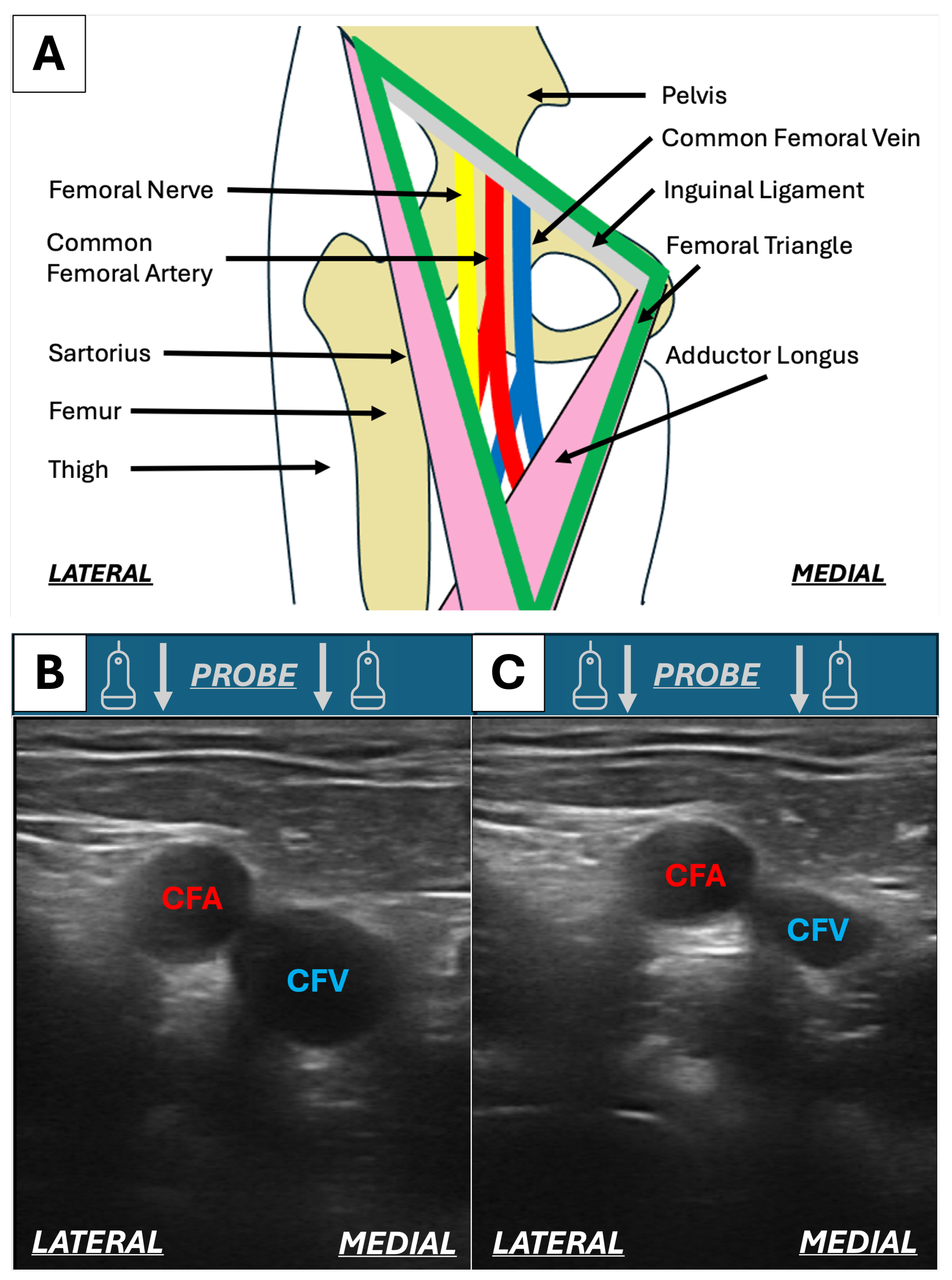
Cardiovascular intervention often begins by introducing tools into the femoral vasculature due to its practical location and versatile anatomy. The Femoral Triangle (shown in Figure 1A) is a region of the thigh marked by the Inguinal Ligament, the Adductor Longus muscle, and the Sartorius muscle [1]. Its floor is defined by the Pectineus and Adductor Longus muscles medially, and the Iliacus and Psoas Major muscles laterally. The roof of the Femoral Triangle is marked by the Fascia Lata. In this area, the Femoral Nerve, the Common Femoral Artery (CFA), and Common Femoral Vein (CFV) (lateral to medial) can be identified. Both vessels bifurcate into the Deep and Superficial Femoral Artery/Vein inferiorly. Normally, the CFA feels more incompressible and pulsatile than the CFV and bifurcates more superiorly [2].
Figure 1.
(A) Anatomy of the Femoral Triangle. (B) Transverse ultrasound image of the uncompressed CFV and CFA of the right thigh. (C) Transverse ultrasound image of the compressed CFV and CFA of the right thigh from the same volunteer.
Vascular access is a technique performed for various catheter-based cardiovascular interventional procedures. The Femoral Triangle is a strategic access site as it allows the operator to insert large interventional tools into the vascular system, maintain stable hand positioning during catheter manipulation, and close the wound post-operatively while ensuring patient safety, as Femoral Triangle-related complications can be managed minimally invasively. Ultrasound-guided femoral vascular access (UFVA) has become a clinical standard for cardiovascular intervention [3] to minimise vascular access site complications. These complications can be severe, requiring vascular surgical repair or stenting, thrombin injection for pseudo-aneurysm, blood transfusion, and more [3,4]. They increase length of hospital stay, delay patient recovery, and increase risk of secondary complications, such as hospital-acquired infections [4], ultimately increasing resource utilisation for the healthcare provider. UFVA begins by scanning the area with a linear, mid-frequency (5–10 MHz) [2,5] ultrasound probe to identify the anatomy of interest (Figure 1B). Using the probe, even pressure is exerted to both arterial and venous vessels. Due to lower haemostatic pressure, the vein will compress, allowing operators to differentiate between the two vessels (Figure 1C). Alternatively, Doppler ultrasound imaging can be implemented to distinguish between vascular structures based on their haemodynamic characteristics. The vessel of interest is centred in the image and punctured at a 30–45° angle using a 6 cm 18–16 G hollow needle, possibly mounted on a 5 mL syringe [5]. The needle is inside the vessel once blood can be withdrawn or exits via the needle. At this point, the syringe is removed, and a guidewire is inserted into the vessel using the needle, to subsequently introduce dilators and sheaths. This is known as the Seldinger technique.
The growing occurrence and complexity of cardiovascular procedures [6,7] increase the demand for more operators and equipment, and thus, more accessible UFVA training and testing solutions. Similarly to other areas of medicine, commercial and experimental ultrasound-guided vascular access phantoms have been proven effective in training, boosting operator confidence with direct positive outcomes on patient care [8,9]. To meet the need for training and testing, commercial ultrasound phantoms are widely available on the market; several can be applied to UFVA.
Multiple companies propose lower-budget vascular phantoms that can be applied to general ultrasound-guided vascular access training [10,11,12]. These systems are commonly shaped as blocks and contain sealed fluid-filled cavities shaped as general, artificially designed vessels. They can resist multiple punctures and can be refilled with the designated fluid priced at approximately 120 £/L [13]. The maximum number of punctures is often unspecified and the price ranges from £500 to £1000 [10,11,12]. The main disadvantages of these models are their dissimilarity to any specific region of the body, and their sealed vessel cavities can trap air bubbles. The Leg Femoral and Saphenous Vein Venous Access Ultrasound Training Model (CAE Healthcare Inc., Saint-Laurent, QC, Canada) is priced at £4000 to £5000 [14]. This model includes a single leg-shaped block containing fluid-filled cavities recreating the Femoral and Saphenous Veins. Similarly to the previously discussed block phantoms, the vessel cavities can be refilled for multiple uses but can trap air. The most complete commercial simulator for UFVA is the Gen II Femoral Vascular Access and Regional Anesthesia Ultrasound Training Model (CAE Healthcare Inc., Saint-Laurent, QC, Canada) [15]. This phantom includes the Femur, the Femoral and Saphenous Veins, the Femoral Artery, and the Femoral Nerve. The vessels can be refilled, and the artery can be pressurised to display the difference in compressibility. Though it proposes many appealing features, this system is priced at approximately £5000 to £6000, and the maximum number of uses is unspecified [15]. Due to the manufacturing techniques adopted, many of these vascular structures are composed of single fluid-filled cylindrical cavities (i.e., non-bifurcating) and bifurcations are simulated by placing single cylinders proximal to each other, meaning the bifurcation is not interconnected. This may not impact visual learning but prevents users from freely navigating interventional tools in the simulated vessels and recreating flow.
Due to the high costs associated with commercial models, several studies have proposed methods to produce low-cost ultrasound-compatible vascular access simulators [16,17,18,19,20,21,22,23,24] using single tubes and balloons as simple, idealised vessels. The most commonly used material is gelatine, a hydrogel with realistic acoustic properties [21]. Despite their ease of use and widespread accessibility, hydrogels, such as gelatine, are extremely fragile, prone to dehydration and microbial degradation. This limits the shelf life of hydrogel phantoms to a single use, and in some cases only a few days if carefully preserved and refrigerated [25], thus restricting their application in geometrically complex models. In light of these factors, D. Pepley et al. [22] propose the use of longer-lasting Polyvinyl Chloride rubber in the form of Plastisol (PVCP), which has a functional lifetime of <50 years when plasticised [26] and over 100 years unplasticised [27]. This material was mixed with mineral oil to produce a self-lubricating, self-healing material with acoustic properties comparable to those of body tissue. By adding chalk powder (CaCO3), the level of scatter in the material can be adjusted to make the material look brighter (more powder) or darker (less powder) in B-mode ultrasound [22].
Though rich in the material science of these tissue-mimicking ultrasound materials, the current literature in this field lacks examples on how to make complex anatomical features (such as continuous bifurcating vascular structures) using common laboratory equipment, greatly limiting users with little manufacturing experience. Between the high costs of commercial phantoms and the fragility of hydrogel phantoms, there is a gap to fill, namely, the combination of complex vascular anatomy, simple ultra-low-cost (<50£) manufacturing, and a self-healing PVCP phantom.
This research’s objective was to propose an easily replicable, reusable, low-cost block phantom for UFVA, containing the Femoral Vein (FV) and Femoral Artery (FA) for industrial and academic uses. The design consisted of an ultrasound-compatible self-healing PVCP block phantom containing vessel-shaped cavities resembling the anatomy of interest for UFVA, maintaining the simplicity of low-cost commercial phantoms whilst improving their vascular anatomy. We then sought to evaluate the relevant material properties, test the phantom’s training capabilities, and estimate a cost per puncture, serving to provide an overall assessment of the phantom’s effectiveness as a tool for UFVA training and testing. Needle insertion represents the most high risk part of UFVA, so this project dedicated a particular focus on this step of the procedure. Following the manufacturing procedures hereby described, training hubs and research centres can create custom ultrasound phantoms with known self-healing abilities, acoustic speed, and hardness, while the evaluation methods can be adopted to quantise the performance of other phantoms and training equipment.
2. Materials and Methods
2.1. Material Selection
When selecting tissue mimicking materials for ultrasound phantom preparation, three rubber elastomers are most commonly considered as follows: PVCP, silicone, and polyurethane [28,29,30]. PVCP exhibits realistic acoustic properties, including acoustic attenuation (0.50–0.063 dB/cm/MHz) and sound speed (1400–1500 m/s) [28,29,30]. When emulsified with oils, PVCP can exhibit self-healing properties; it is commercially accessible and is the cheapest of the three [22,28,29]. Silicone, by contrast, typically demonstrates a lower sound speed (900–1200 m/s) and higher acoustic attenuation (0.5–4.09 dB/cm/MHz), which distort ultrasound images and prevent sound from penetrating deeper into the material [28,29]. Polyurethane provides acoustic characteristics comparable to PVC, but it is the most expensive material of the three, and available in fewer formulations suited for needle puncture [30].
Given this balance of acoustic performance, cost, availability, and modifiability, PVC was selected as the most appropriate base material for the present phantom.
2.2. Design and Manufacture
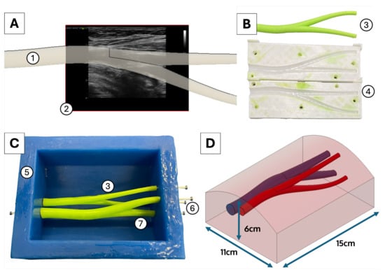
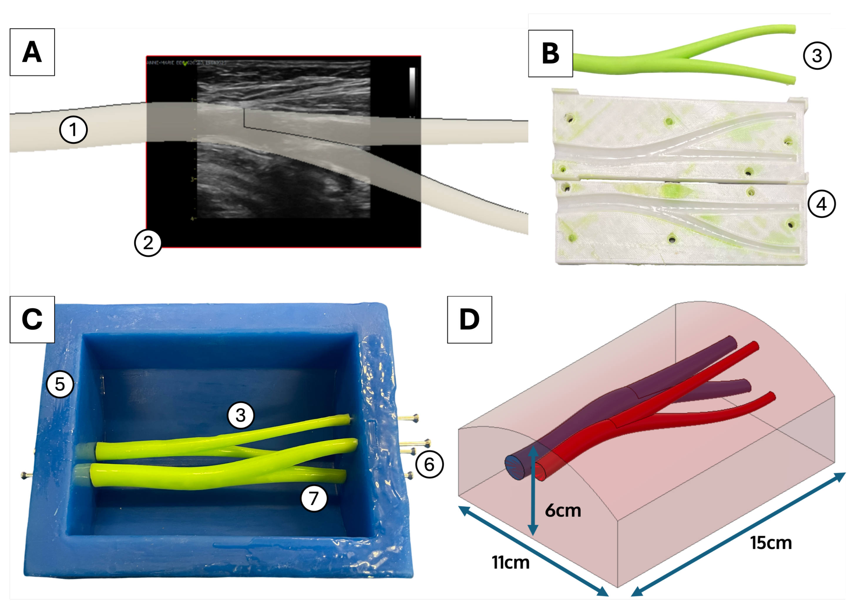
A rounded block sized 11 × 6 × 15 cm was designed on Fusion360 (Autodesk, San Francisco, CA, USA) as the main body of the phantom. To design realistic vessel shapes, an ultrasound image of the femoral vasculature was acquired from anonymised patient data. Figure 2A shows how the canvas tool was used to place the reference ultrasound image on the X-Y plane while the sketch and loft tools were used to design a general bifurcating vessel shape. Heart disease and cardiac intervention are more common among the elderly [31], so the vessel was scaled to create a CFA with a diameter of 10 mm [32] and a vein with a diameter of 13 mm [33] to simulate the general dimensions for patients over 60 years of age. The position and design of the vessels with respect to the block were validated and adjusted with consultation from three senior interventional electrophysiologists and can be observed in Figure 2D. The two vessels were angled at LAO 25° and the CFA was placed 12 mm deep at the centre of the block. The vein was offset from the artery by 6 mm medially to recreate the right thigh. The design was validated by three senior electrophysiologists (consultant or above) before moving on to the manufacturing phase.
Figure 2.
(A) A view of the Fusion 360 CAD process used to design the CFA (10 mm) (1) based on bifurcation shape of anonymised patient data (2). (B) The femoral artery silicone insert (3) is moulded in silicone using a 3D-printed PLA mould (4). (C) This image shows the assembled 30A blue silicone mould (5) with steel pins (6) holding the green silicone vein and artery vessel cavities (3, 7) in position. (D) The final design and dimensions of the UFVA leg phantom.
PVCP is processed at 185 °C [22] and therefore necessitates heat-resistant moulds for casting; conventional 3D-printed thermoplastic moulds are thus not suitable for direct contact with molten PVCP. Silicone rubbers offer both flexibility and thermal resistance within this temperature range. Platinum-cured silicone elastomers are prepared by mixing two components and cure at room temperature, making them well suited for conventional 3D-printed thermoplastic moulds.
To enable PVCP moulding, a reusable heat-resistant platinum-cured silicone rubber mould was designed on Fusion 360 alongside a set of 3D-printable thermoplastic moulds. The plastic moulds were fabricated in white polylactic acid at 0.1 mm layer height on an Ender3Max printer (Creality, Shenzhen, China) using a 0.4 mm nozzle and the concentric ironing function to optimise surface finish. After printing, the plastic moulds were smoothed by sanding to eliminate 3D-printing layer lines and coated in epoxy resin to yield a smooth surface finish. The two-part silicone rubber was then mixed and degassed in a 15 L steel vacuum chamber at negative 1Bar pressure for 30 s to eliminate air bubbles and optimise the surface finish. The silicone was subsequently poured into its smoothened plastic moulds to produce the rubber parts of the final heat-resistant silicone rubber mould, which can be seen in Figure 2C. The silicone leg mould was assembled from three components: the block mould, and two vessel inserts to generate the FA and FV cavities. The vessel inserts were cast in green 35A Shore fast-curing high-viscosity silicone (Reschimica Srl., Sambuca, Italy), which minimised the risk of leaks in the plastic moulds. Figure 2B shows the silicone artery insert and its 3D-printed plastic mould. The block mould was cast using 30A Shore low viscosity blue silicone (BBDINO Direct, Temple City, CA, USA) to trap fewer air bubbles within the rubber and optimise the surface finish of the mould. To ensure structural integrity during casting, the block mould was designed to have a 2 cm wall thickness with pre-cut holes to host support rods for the vessels. The block mould and vessel inserts were connected using 3 mm stainless steel rods as can be seen in Figure 2C.
The PVCP in D. Pepley et al. [22] was mixed in three mixtures; one of the most promising was “mixture 2” containing a 9:2:1 volume ratio of PVCP, softener, and mineral oil. To replicate “mixture 2”, pre-softened PVCP “LureFlex-Soft” by Lure Factors (Doncaster, UK) was mixed in a 11:1 volume ratio with food-grade mineral oil (100% food-grade paraffin oil) (Lubrisolve, Langport, UK), and 1 g of chalk (pure CaCO3) was added for every 150 mL of mixture [22]. In the safe environment of a fume hood, the material was heated to 185 °C using a 950 W microwave oven (monitored every 30 s with a digital immersion thermometer) and placed in a 15 L steel vacuum at negative 1 Bar vacuum pressure for 30 s to remove air bubbles. Any remaining bubbles were skimmed off the surface using a steel spatula. Following the methods described in D. Pepley et al. [22], three phantoms were cast into the assembled silicone mould, pouring slowly without draping to avoid collecting air bubbles. The phantoms were allowed to cool down overnight in a 4 °C fridge and then demoulded. The green vessel inserts (Figure 2C items 3 and 7) were pulled out of the PVCP block to reveal vessel-shaped cavities in the rubber model. The three phantoms were submerged in water to fill the vessel cavities and replicate blood. The models were inspected with B-mode ultrasound imaging at 7.5 MHz using the 4 cm linear probe of the e-POC 3-in-1 system (Ballater Medical, Edinburgh, UK).
2.3. Material Properties
Hardness in the OO scale was measured to quantitatively compare the tactile feel of this phantom to examples present in the literature. Ten cylindrical samples of the phantom PVCP mixture measuring 35 × 20 mm (diameter × height) were cast in silicone moulds as previously described. Hardness was measured on the OO Shore scale using the HT-6510OO durometer (Landtek Instruments Co., Guangzhou, China).
As well as tactile properties, acoustic properties are crucial in creating a realistic and effective ultrasound model. Diagnostic ultrasound systems reconstruct images based on the time waves are reflected back to the probe, using an assumed wave speed of 1540 m/s [34]. The speed of sound through a material sample can be calculated by ratios of assumed speed (1540 m/s) vs. real speed and measured sample depth vs. real sample depth [35,36]. Sound speed through the PVCP rubber was therefore estimated by sampling depth of ten depth-constrained 12 mm-thick rubber samples using the Ballater Medical 4 cm linear probe at 7.5 MHz. Acoustic impedance governs the behaviour of sound reflections within a material and it is defined as the product of mass density and sound speed [37]. Soft tissues have been reported to exhibit an acoustic impedance of 1.40–1.69 [37]. To estimate the acoustic impedance of the PVCP material, the mass density of PVCP was determined by measuring the total mass of the ten 12 mm samples, and dividing by the total volume (ten samples of 12 × 10 × 40 mm).
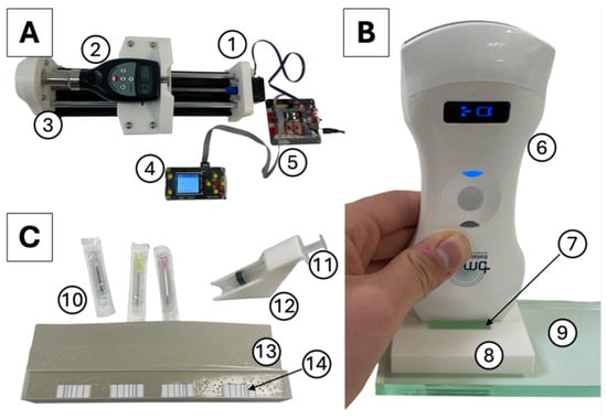
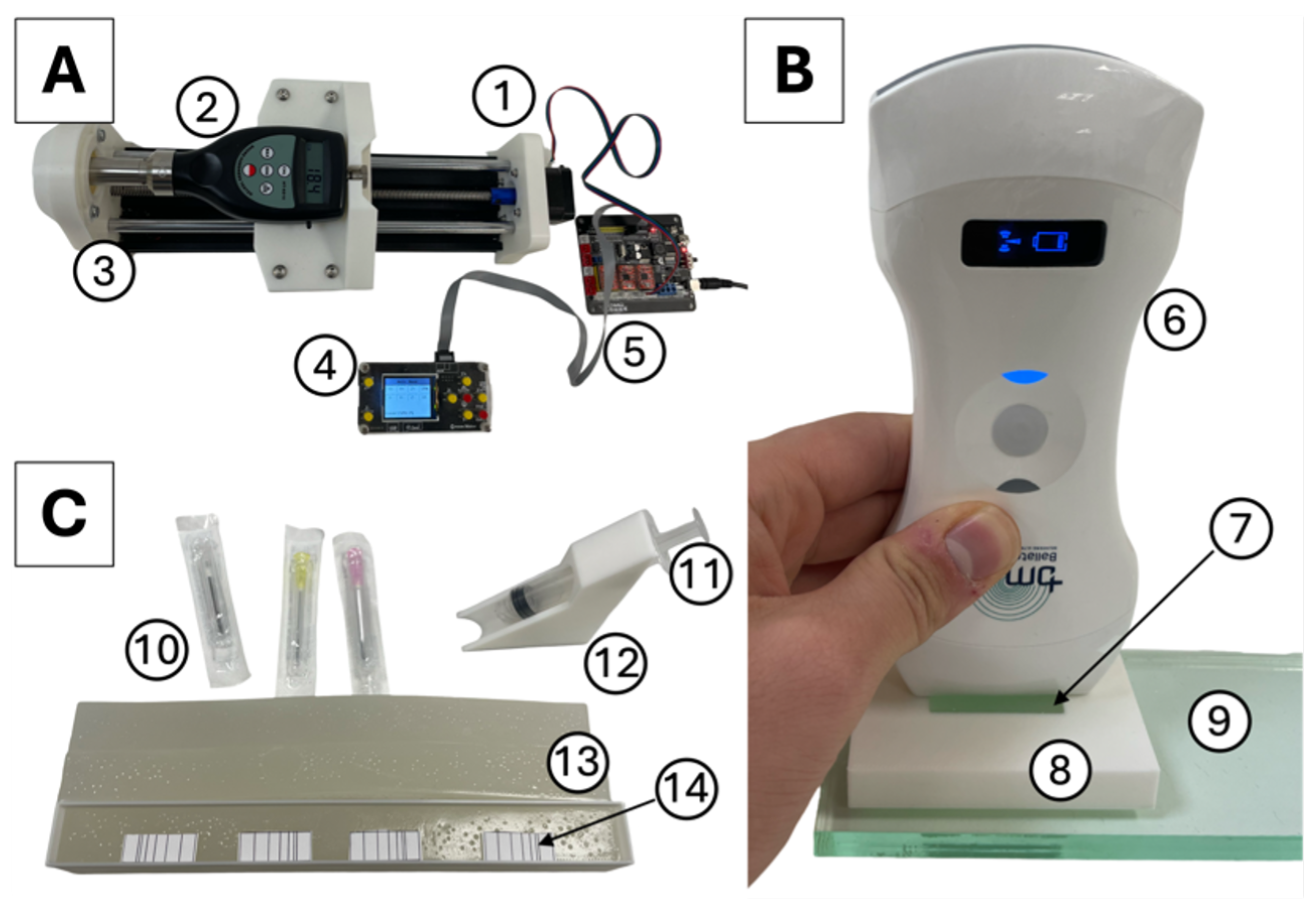
To estimate the maximum number of punctures per unit area that the PVCP material can sustain, the self-healing properties of the phantom were characterised. Three 2 × 2 × 20 cm cuboidal sections of PVCP phantom mixture were cast in silicone moulds following the previously described methods. Three series of six distinct punctures distanced 1, 2, 3, 4, and 5 mm were performed on each PVCP sample using 16 G, 18 G, and 20 G needles. To ensure the test was coherent to clinical practice, the needles were mounted on a 5 mL syringe and inserted using a 45° guide rail [5], ensuring the needle was oriented with the bevel facing up [38]. The needle tracks were inspected with B-mode ultrasound imaging at 7.5 MHz using the Ballater Medical probe at time intervals: t = 0 h (control), t = 1 h, t = 2 h, t = 3 h, and t = 24 h. Although the UFVA task is usually performed in axial view, the needle tracks were inspected longitudinally to better visualise the damage. For the purpose of this simulation study, needle tracks would be considered healed if more than 50% has reintegrated into the background. Since the vessels are visualised in axial view during needle insertion [5] (see Figure 1B,C), needle tracks would appear as dots, so it would be difficult for the user to notice them and follow them among the speckled background in the axial view.
The methods implemented to analyse the material properties of the phantom are depicted in Figure 3.
Figure 3.
(A) The linear actuator (1) moves the OO Shore durometer (2) to the sample placed at the end (3). The system is operated via a controller (4) connected to a GRBL board (5). (B) The Ballater Medical ePoc 3-in-1 probe (6) is used to measure the depth of the 12 mm material sample (7). The depth is controlled by a plastic section (8) and sound waves are reflected off a smooth surface of acrylic (9) (C) Needles (10) are attached to a 5 mL syringe, which is guided into the material sample (11) using the guide rail (12) to control the angle of puncture in the material samples (13). Markings (14) are used to ensure the needle tracks are separated by 1, 2, 3, 4, and 5 mm.
2.4. Timed-Task Performance Evaluation
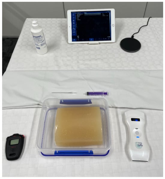
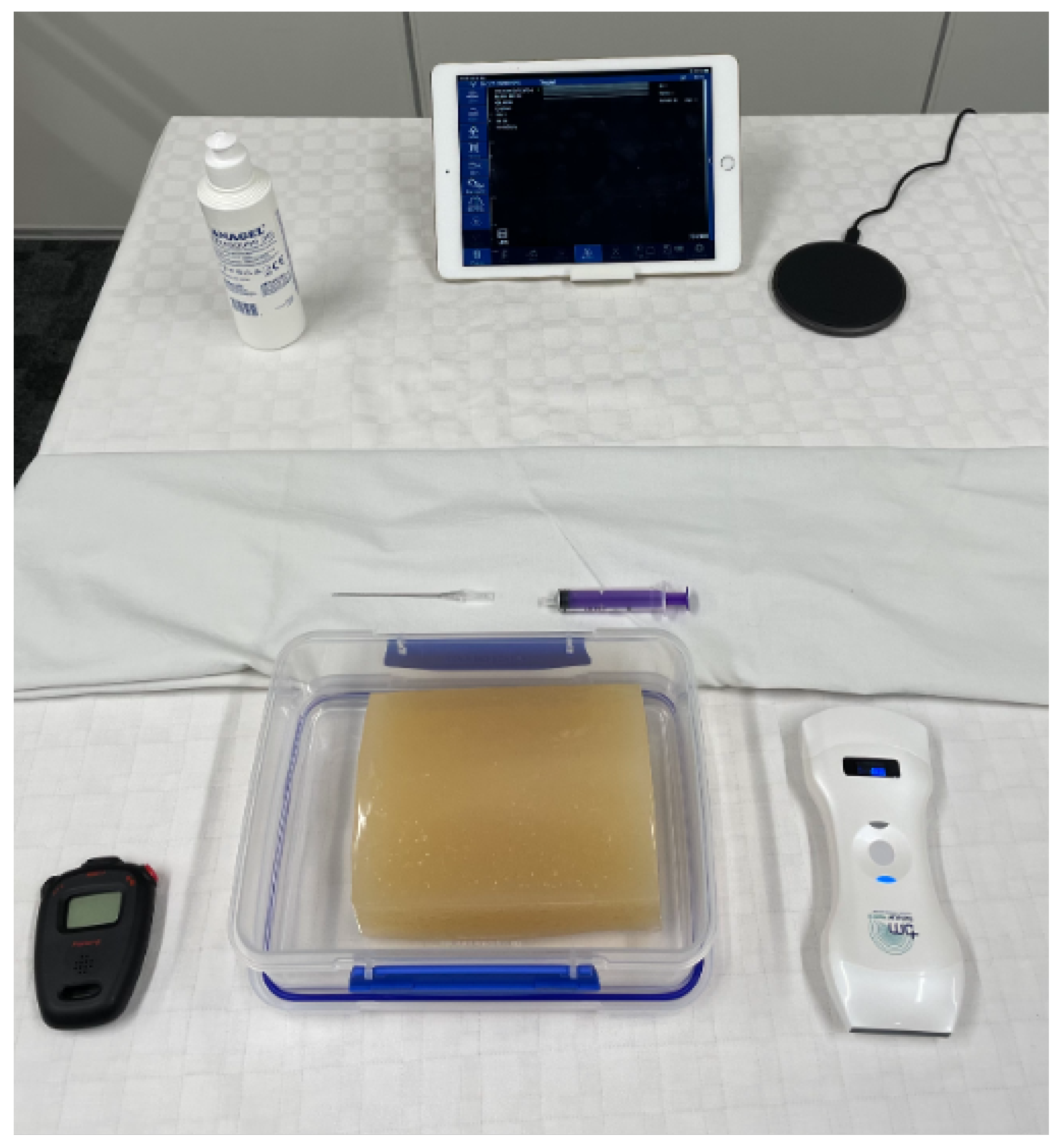
To quantitatively assess the training capabilities of the leg phantom on inexperienced students, a workshop was set up at a local surgical conference at a medical university in England. Three leg phantoms were submerged in water until the vessels were fully covered. Three stations were set up with three 5 mL syringes, three 6.5 cm 19 G needles, and three 4 cm ultrasound probes: one with the L7.5 MHz L12-5L40N-4 ultrasound probe (Telemed, Vilnius, Lithuania), and two with the e-POC 3-in-1 systems (Figure 4). All probes were imaging in B-mode at 7.5 MHz.
Figure 4.
The experimental set up to perform UFVA. Users are provided with a stopwatch to measure their trial times and free practice period, a leg phantom submerged in water, ultrasound gel, and the ultrasound system.
Of all the conference attendees, 18 local undergraduate medical students from years 2–4 with no previous practical experience in ultrasound imaging participated in this study. The students were briefed with a 25 min presentation on ultrasound imaging and UFVA including the instructions video provided by the MSD Professional Manual [5]. A 5 min practical demonstration of needle insertion was then provided (see Video S1, Supplementary Digital Content 1, which includes a 3 min demonstration summary). All students were then allowed to freely familiarise with ultrasound imaging by scanning the phantoms until they felt confident enough to initiate needle insertion on the phantom. The probe was placed down, the needle was mounted on the syringe, and the guard was removed. A timer was initiated, and the students began UFVA. Needle insertion was timed until the student was able to visualise the tip of the needle inside the vessel. Each student was subsequently permitted 5 min of free practice on the phantom. At the end of free practice, a second timed trial was performed. Two metrics were calculated as follows: time difference in seconds, and time difference as a percentage of the first trial.
Subsequently, a t-test was used to statistically assess whether the students experienced a statistically significant (non-zero) improvement between the two trials. To further assess the magnitude of change, the results were also assessed with Cohen’s d value.
In order to provide a control, the same test was performed by three experienced interventional cardiologists (different from the ones previously mentioned) who routinely perform UFVA. The cardiologists were provided with the same presentation, demonstration and opportunity to familiarise themselves with ultrasound imaging. The total procedure times, time differences, and percentage time differences of students and cardiologists were analysed in two-tailed multi-variance unpaired t-tests at 5% significance level.
According to our local ethics rules at King’s College London, this study was deemed minimal risk (study references: MRSP-24/25-47887, MRSP-24/25-48196). A description of the research was provided to all potential participants. Informed consent was waived by the ethics committee for this minimal-risk educational study, with participant information provided and completion of the task/questionnaire taken as implied consent. No identifiable or personal information was collected for any participant to ensure anonymity. At the recruitment stage, it was clarified both verbally and via the study description that the student participants had not personally used an ultrasound imaging system before; this was their first time holding and using an ultrasound probe themselves.
2.5. Survey-Based Evaluation
To qualitatively assess the phantom’s training capabilities from a teacher’s perspective, a stand was set up in a medical education conference in the UK focusing on simulation-based education. The phantoms were presented to 31 UK-based educators in medicine, anatomy, and nursing at varying levels of experience in ultrasound-guided vascular access. Volunteers were free to participate anonymously in this study without collecting any identifiable information. According to our local ethics rules at King’s College London, this study was deemed minimal risk (study references: MRSP-24/25-47887, MRSP-24/25-48196). A description of the research was provided to all potential participants. Informed consent was waived by the ethics committee for this minimal-risk educational study, with participant information provided and completion of the task/questionnaire taken as implied consent. No identifiable or personal information was collected for any participant to ensure anonymity. The phantoms were set up as previously described for the student time trial and a 5 min demonstration in UFVA was provided to all participants. After the demonstration, the educators were free to ask questions and test the phantom. Finally, each participant was asked to answer a seven-part questionnaire using a 5-point Likert scale, multiple-choice answers, and optional comments (see PDF S2, Supplementary Digital Content 2, which includes the full questionnaire) following the structure of C. Saija et al. [39].
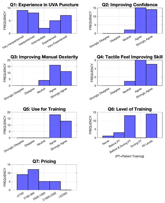
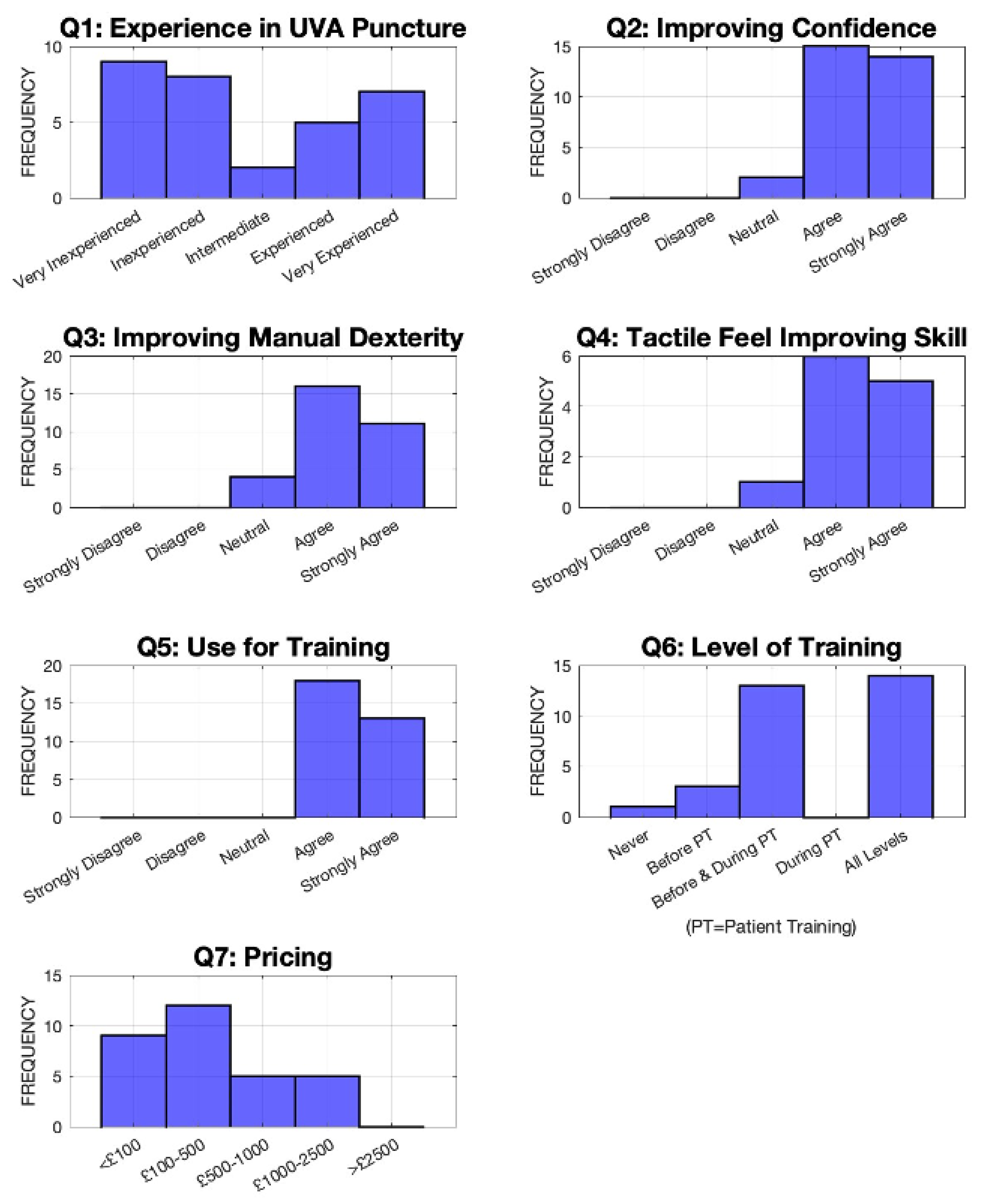
The first question was used to rate the participant’s experience in ultrasound-guided vascular access. The second question assessed if the phantom helps build user confidence in UFVA. The third question evaluated if the model improves manual dexterity in UFVA. The fourth explored if the phantom’s tactile feel helped to hone skills, assessing the importance and realism of the feel. Question 5 asked if they would want to use this model for training. Question 6 inquired the levels of training for which they would use the leg phantom. The final question investigated the price range the educators would be willing to pay for such a phantom.
3. Results
3.1. Manufacturing and Material Properties
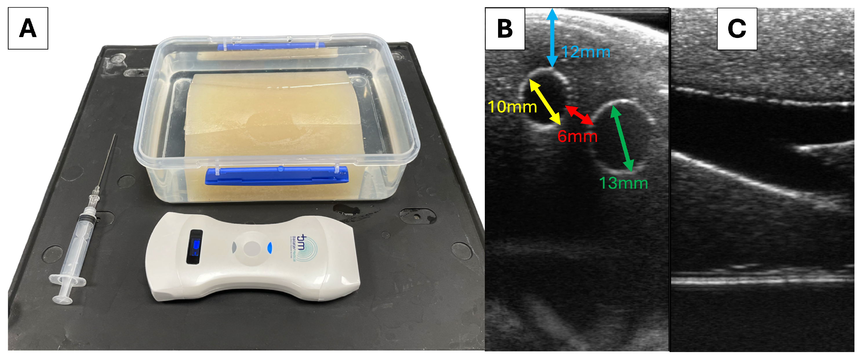
The phantoms were imaged in their entirety using 7.5 MHz ultrasound imaging (Figure 5). The vessel cavities could be visualised in longitudinal and transverse views using B-mode imaging (Figure 5B,C). The 12 mm rubber samples exhibited a sound speed of 1454 ± 15 m/s and an acoustic impedance of 1.66 ± 0.02 ; all the structures in the phantoms matched dimensions in the design (Figure 5B): the CFV had a diameter of 13 mm, the CFA had a diameter of 10 mm and a depth of 12 mm, and the two vessels were 6 mm apart. The PVCP material had an OO Shore hardness of 18.0 ± 2.2, which falls within the range of hardness of soft tissue phantoms for similar applications (Shore hardness OO 10–30) [40,41,42,43]. The surface of the phantom was slightly oily to the touch and was unaffected by exposure to water or ultrasound gel. Some bubbles were formed upon casting the PVCP in the silicone moulds; however, they were easily skimmed off the top of the hot molten rubber using a metal spatula. The final phantoms were free or bubble artefacts. The material cost to make the mould was £36.82, 24 h to 3D-print the PLA mould and 3 h to cast the silicone. This includes £28.27 for the BBDINO silicone, £1.15 for the Reschimica silicone, £4.30 for the PLA, and £2.10 for the metal. One phantom cost £7.87 in materials and the time spent to produce it was 1 h of labour and ∼14 h of cooling. This includes £6.89 for the PVCP, £0.91 for the oil, and £0.06 for the chalk powder.
Figure 5.
(A) The UFVA phantom submerged in water. (B) Axial ultrasound image of the phantom acquired at 7.5 MHz. The distance between vessels (red), depth of the artery (blue), artery diameter (yellow), and vein diameter (green) were measured using the Ballater Medical ultrasound machine. (C) Longitudinal view of the CFV bifurcation in the phantom acquired at 7.5 MHz using the Ballater Medical ultrasound machine.
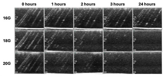
Figure 6 shows that under ultrasound inspection, the needle tracks caused by the 18 G and 20 G needles healed within 3 h, and fully reintegrated with the background within a day, including those spaced 1 mm apart. The colour and texture of the material samples returned to a consistent grey colour. The larger 16 G needle tracks, however, did not heal as defined in the methods, and instead remained noticeable in their entirety even after 24 h (Figure 6). Adjacent needle tracks spaced up to 1mm apart did not interconnect and tear the phantom for any of the tool sizes analysed in this study. The area of interest on the phantom was the surface area directly above the CFV and CFA, which measures approximately 2000 mm2. Therefore, up to 2000 equally spaced parallel punctures could be sustained in the phantom’s region of interest. Based on this, the cost per puncture on this phantom would be £0.0039.
Figure 6.
The appearance of the same sets of needle tracks observed at time intervals 0 h, 1 h, 2 h, 3 h, and 24 h. 16 G needle tracks did not heal in a day. The 18 G and 20 G needle tracks had healed within three hours, and had become completely unapparent within a day. The grey scale on the side of each ultrasound image indicate depth in mm.
3.2. Timed-Task Performance Evaluation
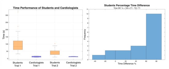
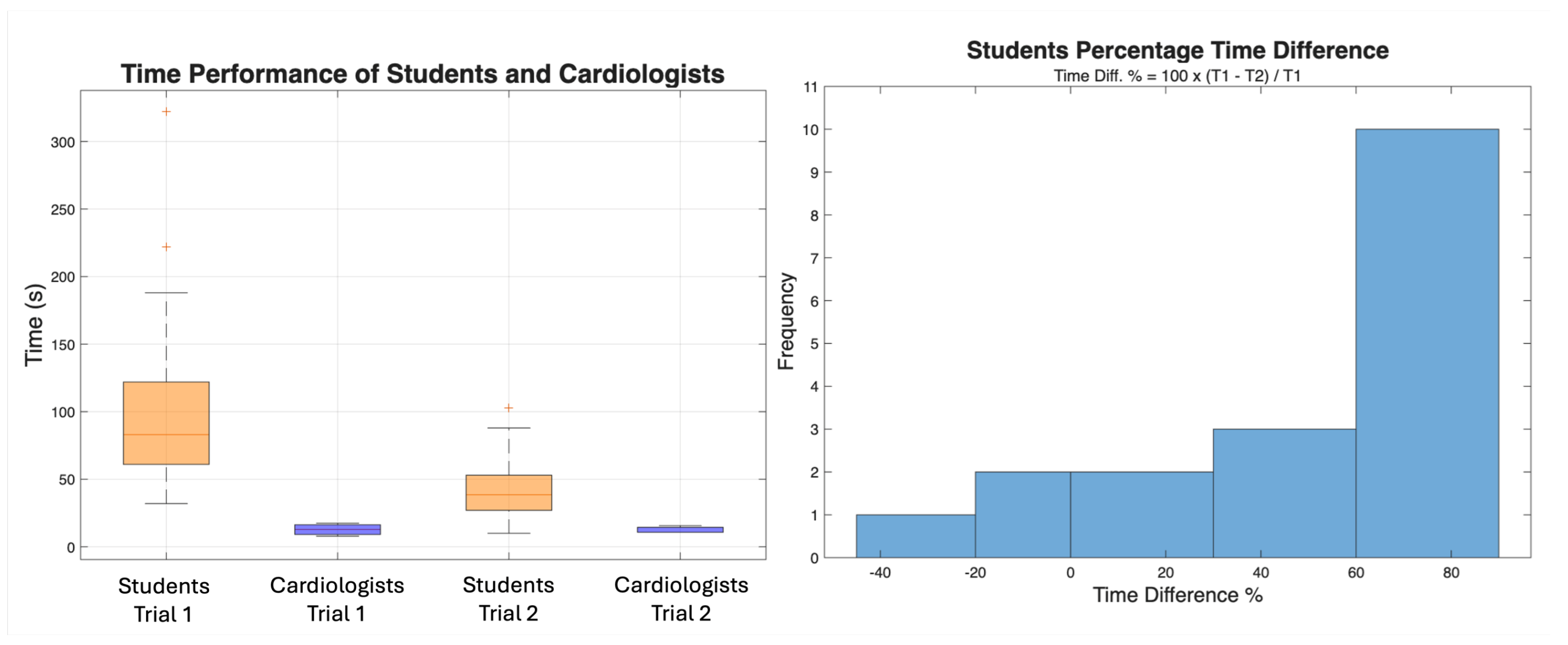
There was no statistically significant difference between the time improvements experienced by students using the Ballater Medical probes and the TeleMed probe (p = 0.3688), so the results were grouped together. As visible in Figure 7, only three students took longer on the second trial (negative time difference). Over 50% of students reduced their time by over 60%. The average time performance among students in Trial 1 was 107 ± 73 s and 44 ± 27 s in Trial 2. The median time performance among students in Trials 1 and 2 were 83 s and 39 s, respectively. The interquartile range among students in Trial 1 was 61–122 s and 27–53 s in Trial 2. The students improved their time by 47.21% on average; the t-test showed that this was a statistically significant change (p = 0.0001) and the Cohen’s d value showed a this was a strong improvement (d = 1.171).
Figure 7.
The box plots on the left show the time performance by students and cardiologists on Trial 1 and Trial 2 for the cardiologists and the students. The cardiologists were always faster and remained consistent in their time performance. The students were slower than the clinicians in the timed trials and improved their time after practising. The histogram on the right shows the percentage time difference observed in the students. It can be seen that 10/18 students reduced their time by 60% or more, 5/18 students improved by 0–60%, and 3/18 did not reduce their time on the second trial.
The cardiologists serving as the control all had less than 10 s of difference in their two trials. The average time performance among cardiologists in Trial 1 was 13 ± 5 s and 12 ± 3 s in Trial 2. The median time performance among cardiologists in Trials 1 and 2 were 13 s and 11 s, respectively. The interquartile range among cardiologists in Trial 1 was 9–16 s and 11–14 s in Trial 2. The t-test shows there was no statistically signifiant change among the cardiologists’ performance between Trials 1 and 2 (p = 0.9203).
Cardiologists were significantly quicker than students before (13 ± 5 s, p < 0.0001) and after training (12 ± 3 s, p = 0.0001). The mean time difference recorded by students (63 ± 76 s) was significantly higher than that observed among cardiologists (0 ± 6 s, p = 0.0029).
3.3. Survey-Based Evaluation
Question 1 had very varied answers with only two people having intermediate experience in ultrasound (Figure 8). Question 2 had an average rating of 4.4 ± 0.6, with 29 people either agreeing or strongly agreeing (Figure 8). Similarly, question 3 received an average rating of 4.2 ± 0.7, 27 people either agreeing or strongly agreeing (Figure 8). The fourth question, assessing tactile feel, received an average rating of 4.3 ± 0.7 (Figure 8). Answers to question 5 report that all participants would use this phantom for training. In response to question 6, most educators thought the phantom could be used at all levels (14/31) or before and whilst training on patients (13/31). Only 3 educators thought this phantom training would be better only before patient training. One participant out of 31 responded “Never” to question 6; however, they answered “strongly agree” on questions 2–5, meaning they think it was highly effective and would “strongly agree” with its use in training. Answers to question 7 were varied with an average score of 2.2 ± 1.1, equating a price range of £100–£500. Figure 8 depicts all answers to the questionnaire as a series of histograms.
Figure 8.
This figure shows the distribution of the answers to the educator’s questionnaire as histograms, showing the distribution of the scores for every field of assessment.
A number of educators also provided additional feedback via the optional comments boxes in the questionnaire. One anatomist commented: “Great tactile feel—anatomy was accurate. Nice touch to be able to aspirate fluid (meant to be blood)”. Multiple people with experience level 5 suggested improving realism by rendering the artery incompressible and adding a vessel wall. It was noted by one that having multiple layers in the skin, fat, and muscle layer would improve visual realism in the ultrasound images. Two anaesthesiologists suggested adding the Femoral Nerve to enable nerve block training or teaching to avoid it during UFVA. A nursing instructor noted that having a range of vessel spacing could result in different levels of challenge for trainees. Lastly, a lecturer in medicine suggested investigating the number of punctures the phantom can undertake using different tool sizes, including sheaths, introducers, and guidewires.
4. Discussion
The design process for this system was relatively basic and can be replicated on free online CAD software programs. The materials and techniques utilised to prepare the moulds are widely accessible to researchers and engineers, and the costs presented by this initial step (£36.82 and two days of labour) can be reached by most centres. Low-cost vascular phantoms on the markets and in the literature are often limited to simple vascular structures composed of single separate vessel cavities [10,11,12,16,17,18,19,20,21]; arguably this is conducted to maintain the moulds’ simplicity and avoid damaging the phantom whilst demoulding. The soft silicone vessel inserts complied to the shape of the phantom as they were being pulled out, enabling the creation of fully connected bifurcating vessel-shaped cavities in the PVCP rubber and allowed for easy, damage-free removal of the inserts after the material had solidified. This technique allows users to create complex vascular cavities without advanced multi-component mould designs or sacrificial mould parts. Instead of using complex pump systems or sealed fluid-filled cavities, this design submerged the phantom to fill the vessel cavities in with fluid. These principles can be easily adopted by the community to design new phantoms with complex vasculature. The process of printing with thin layers, sanding, and then coating the plastic mould in epoxy resin provided the final silicone moulds with a smooth surface finish, minimising the number of bubbles forming during casting. To further reduce bubbles on the surface of the phantom, future endeavours may attempt heating the rubber moulds to an intermediate temperature (e.g., 100 °C) and coating them in oil before pouring in the PVCP. This study utilised the moulds three times to produce three phantoms; however, it did not stress test nor evaluate the reusability of the moulds over time. The manufacturing process of heating, degassing, and casting the PVC presents more challenges, as it requires the use of a fume extractor hood and a vacuum chamber. The fume extractor is necessary to ensure the safety of the user, while the vacuum is necessary to minimise air bubbles and thus improve the homogeneity of the phantom material. This equipment is common in research facilities; however, it is often lacking in training centres without engineering capabilities. The average rating to question 7 shows educators would be willing to spend approximately £100–500 to procure or prepare this phantom. The raw material cost to prepare one phantom (£7.87) was 10–60 times lower than the educators’ expectations: in a non-commercial scenario, this phantom would meet and even go beyond the educators’ cost expectations. Overall, the design and manufacturing processes are low-cost and simple to recreate with a number of ultrasound-compatible materials, requiring basic engineering knowledge; however, the equipment necessary to handle PVCP presents challenges to smaller, less equipped centres.
The literature-derived [22] phantom material produced reusable phantoms that resisted repeat use throughout the four months of studies. The phantom material displayed an OO hardness of 18.0 ± 2.2, which is consistent with existing soft tissue phantoms [40,41,42,43]. The survey also reports that the tactile feel of the phantom was realistic and helpful for UFVA training, further validating this aspect’s realism. The speed of sound in the phantom (1454 ± 15 m/s) is similar to that of fat tissue in the leg (1440 m/s) [44] and falls within range of those experienced in soft tissue (1430–1647 m/s) [45]. Water was used to fill the vessel cavities as it finds common application as an accessible and disposable surrogate for blood in ultrasound simulation [23]. Clinical ultrasound assumes an average sound speed of 1540 m/s [40], which matches our phantom material to 2 significant figures. The acoustic impedance of PVCP (1.66 ± 0.02 ) also fell within soft tissue range, closely matching the acoustic properties of cardiac muscle (1.67 ) [37], showing the phantom exhibited a realistic ultrasound appearance. Ultimately, the realistic acoustic properties of the rubber allows for the phantoms to appear geometrically coherent with their design when imaged. Perhaps a more detailed analysis of the acoustic properties of this phantom material could provide greater insight for the fabrication of standard test objects in future research. Alas, the scope of this study was to generally recreate a simplified version of the femoral vasculature; it could thus be argued that the appearance and acoustic properties were realistic enough for this simulation study. The vessels retained the original diameter in all three models used in the evaluation study; however, the use of soft silicone moulds can result in inconsistent geometry on larger-scale production. The phantom’s anatomy was designed with experienced physicians and regarded as “accurate” by an anatomist in the optional comments, so it is reasonable to argue that the hereby proposed phantom anatomy is realistic enough for UFVA training. The fidelity expressed in this phantom is comparable to that of common commercial block phantoms or the previously described venous access model from CAE Healthcare. Our phantom was designed to provide core training in UFVA; though some anatomical features were not incorporated, this limitation is common among low-cost training phantoms [10,11,12,14] and is normally mitigated by adjunct lectures and demonstrations. To enhance training efficacy and versatility, comments from the survey-based evaluation suggest the addition of the Femoral Nerve, different layers of soft tissue, and the vessel walls. Though adding a consistent vessel wall or tissue texture would prove complex, a Femoral Nerve or Inguinal Ligament could be integrated using techniques analogous to those employed to create the vessel cavities. As suggested in the comments, the addition of pressurised flow in the FA would allow users to experience Doppler ultrasound without the need for volunteers. The design hereby proposed offers open vessel cavities so that in future studies, pumps could be utilised to recreate haemodynamic flow and haemostatic pressure in the FA; however, this may increase the costs and complexity associated with this project. To provide a more complete training session and appreciate the relevant features of haemodynamic flow, training sessions can provide annotated video demonstrations, while the phantom can be used to exercise needle insertion to keep patients and volunteers safe. Alternatively to video demonstrations, students could be paired in teams to image each other’s or their own carotid arteries and experience Doppler imaging first hand. This phantom successfully recreated the appearance and behaviour of a commercial block phantom whilst adapting the design to recreate the vascular anatomy of the Femoral Triangle.
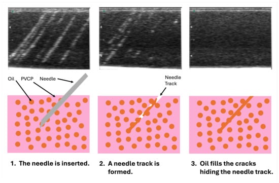
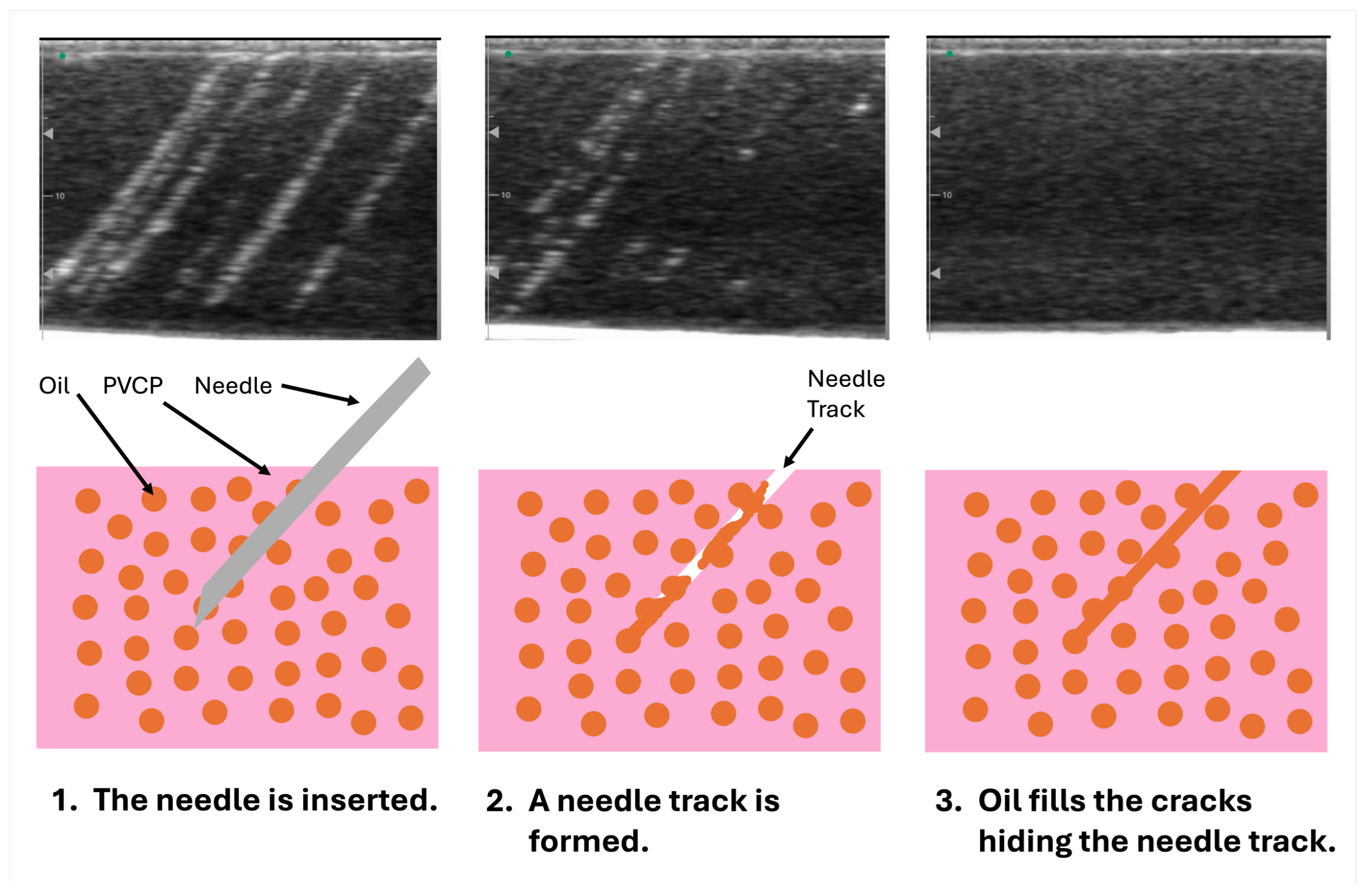
The surface of the phantom material releases mineral oil, resulting in an oily texture. When the phantom material is punctured, oil is released by the rubber, filling the gaps over time and masking the needle track when inspected with B-mode ultrasound. The healing properties of the phantom material are therefore visual, not mechanical. The healing process is graphically summarised in Figure 9. The phantom material healed the damage caused by 18 G and 20 G needle tracks within three hours, making it completely unapparent after a day; whereas all damage from 16 G needles remained noticeable even after a day had elapsed. This is consistent with commercially available BluePhantom models [14,15,46]. Multiple training exercises could be carried out throughout the day with 18+G needles, while tools larger than 18G are too disruptive for this phantom. This may arguably represent a limitation in some applications; however, as is often the case with simulation-based education or testing, there is always a trade-off between economic viability (in this case reusability) and realism. The 18 G needles are a common needle size to use for this application [5], so this should arguably not greatly impact the model’s training outcome or usability. Future studies may allow the phantom to support lesions from larger needles by increasing the oil content of the rubber matrix to speed-up the phantom’s healing time, or by employing a softer, more elastic PVCP which can better comply to the needle. To simultaneously visualise both vessels, UFVA is performed in the axial view, and needle tracks therefore appear as a small dot in the speckled background rather than a line. It could therefore be argued that the phantom can be utilised again after a day, or even three hours of healing. The difference in healing time experienced in this study may be attributed to the size of the needle tracks, and thus, the quantity of mineral oil necessary to fill them. Increments in oil content and rubber porosity may accelerate the rate of oil release and thus speed up the speed of recovery for this material, potentially enabling the recovery of larger lesions. Future investigations could assess whether dipping or flushing the needles in oil would have a significant impact on healing. None of the parallel needle tracks interconnected or tore the material sample during testing. Our tests show that the ultrasound phantom material could resist up to 1 parallel puncture per mm2 with bevelled needles sized at 16 +G. The area of interest in this phantom is of 2000 mm2, so in an ideal case scenario (such as robotic testing or software validation), the phantom could resist up to 2000 punctures costing only £0.0039 per puncture. In the case of teaching, improper technique and inconsistent puncture distribution on the area of interest would likely reduce the number of maximal punctures this material can sustain. Arguably, stress-testing the phantom using a large number of trainees and letting them assess when the material had healed enough would have provided more realistic limits to this phantom’s usability; however, the results may vary depending on the equipment, users’ experience, and manual dexterity.
Figure 9.
This figure illustrates the theorised process by which PVCP phantoms self-heal. When a needle is inserted a needle track is formed. Once the needle is removed, the emulsified oil seeps into the cracks, visually hiding the track.
The data collected during the conference focused on ultrasound-guided vascular access training, which attracted both medical educators and students. The setting, though advantageous for large-scale recruitment, resulted in a catch-as-catch-can convenience sample that limited detailed demographic profiling. In particular, the 18 medical students recruited came from training years 2–4 from the same institution. Though the data was only collected from undergraduate medical students with no practical experience in ultrasound, we acknowledge that their level of clinical exposure may vary based on numerous factors (such as course attendance, extra-curricular activities, etc.), resulting in a larger variance in the times collected. To mitigate this, students were allowed to familiarise themselves with ultrasound imaging before the timed trials. Additional baseline data on the participants’ previous experience may have been recorded; however, this might have compromised their anonymity. Collecting more personal data on clinical exposure whilst recruiting a larger number of trainee participants, would allow to analyse students cohorts based on their experience, and possibly narrowed some of the variance in time performance. Similarly, students in future studies could be exposed to multiple classes; however, due to the nature of our recruitment strategy and to maintain anonymity, this initial pilot study makes use of a single class of students. This study could also be repeated in different conferences to recruit participants form different medical backgrounds. Nonetheless, we believe that the students, educators, and clinicians provided a diverse sample that reflects the typical population targeted by such phantoms; future work will strive to capture vaster and controlled demographic details using a more sophisticated ethical structure of recruitment.
In the timed trials, students improved their time significantly (p = 0.0001), with the majority improving more than 60%. Cohen’s d (d = 1.171) further reinforced this point, showing a high magnitude of change was experienced between the two trials. The control group of cardiologists had shorter average times and showed less than 10 s difference between trials: this shows that their experience had a significant impact, further validating the phantom’s design. The large standard deviation in the student group’s first trial (±73 s) compared to the expert samples suggests that manual dexterity and prior exposure may still play a significant role in this task despite our attempts to mitigate this with the presentation and initial free practice. As well as improving their overall procedural time, the student group became more consistent in the second trial (±27 s). Despite the limited demographic detail among students, these findings robustly demonstrate that even among a heterogeneous group of beginner trainees—with potentially varied levels of clinical exposure—the phantom provided an effective practice/test environment. This resonates with the existing literature [8,9]. Further analysis should study the effects of long-term, repeated use of this phantom by inexperienced and experienced students such as Sabak M. et al. [8] and Oh E.J. et al. [9].
The timed-task and survey studies were useful in determining the effectiveness and uses for this phantom. Not all educators answering the questionnaire were experts in UFVA; however, we believe that this is more representative of the real-life use of phantoms such as this, as they can be used to simulate multiple training or research scenarios. Responses to questions 2, 3, 4, and 5, as well as the time improvement experienced by students, showcase the phantom’s training capabilities. Confidence and manual dexterity can be developed through the use of this phantom, resulting in a reduction in operational time. This is consistent with existing phantom studies in electrophysiology and ultrasound-guided vascular access training [8,9,16,47,48]. The educators’ feedback places this phantom as a “useful” UFVA training tool before and during training on patients. Experienced users may even want to use this model to revise or test new techniques before teaching a class or testing new equipment. Recent studies in autonomous ultrasound robotics have highlighted the importance of stable, reparable simulation environments for prototyping new image processing techniques, pattern recognition software, or hardware [49,50,51]. This phantom can find applications in experimenting and teaching techniques in cardiovascular, neurovascular, and radiological intervention, as well as anaesthesiology and endovascular resuscitation. Using differently sized/shaped vessel inserts, users could replicate pathological variations and paediatric patients to experience a variety of patient anatomies. Calcification, clots, or plaque could be recreated using a combination of micro-bubbles [49] or a paste made or gypsum, lipids, hydrogels, or hard resins [52]. Pathological variants and anatomical obstacles would provide a more layered and realistic testing approach both for teaching and testing new technologies. Although no direct comparisons were drawn against commercial systems, responses to the last question show how educators would be willing to spend £100 to £500 on average. Featuring a material cost of less than £10 and relatively simple manufacturing, this answer highlights how making reusable rubber phantoms in-house with these methods can be a viable low-cost solution for centres with manufacturing capabilities.
As displayed in Table 1, the proposed PVCP phantom occupies a middle ground between the common gelatine-based ultrasound models [20,21,24,25] and commercial block phantoms [10,11,12,13,14,15]. The PVCP and gelatin phantoms require time and basic equipment to manufacture in-house; however, this maintains relatively low material costs (<£50 [21,25]). Commercial models offer substantial convenience at prices ranging from £500 to £1000 per unit. Though the longevity of the PVCP and commercial simulators was not formally investigated, both are made of rubber and should thus exceed the shelf-life of gelatine hydrogel, which has been reported to degrade within 50 days [25]. Similarly to the PVCP rubber used in this study, commercial models can heal from 18–20 G punctures, although manufacturers do not directly state a maximum number of punctures. Unlike the other tissue-mimicking materials, gelatine hydrogel is not self-healing and relatively fragile; reports by A. Shah et al. [24] note that gelatine can tolerate a maximum of 30 vascular punctures. The vasculature observable in common block phantoms is composed of non-specific sealed vessel cavities that are prone to collecting air and cannot be connected to flow systems. Gelatine models often incorporate latex tubing or balloons to replicate vessel cavities [20,21,24,25], restricting them to non-bifurcating vessels that can be connected to flow systems. The PVCP phantom presented in this article offers more anatomical realism and offers open bifurcating vessel cavities that can be linked to pulsatile flow pumps.
Table 1.
A comparison between commercial block phantoms, the proposed PVCP phantom, and gelatin-based experimental models.
Table 1 graphically summarises the comparison between the proposed PVCP phantom, common gelatine-based ultrasound phantoms [20,21,24,25], and commercial ultrasound block phantoms. The PVCP phantom and gelatine phantom both require time and equipment to manufacture them, but present low material costs (<£50); the commercial alternatives can be easily purchased at scale; however, this requires a larger investment (£500–1000 per model). Though the longevity of the commercial phantom and the PVCP phantoms was not assessed, they are made of rubber and should outcompete the shelf life of fresh gelatine (up to 50 days [25]). Product reports [10,11,12,13,14,15] indicate that the commercial block phantoms can heal form 18–20 G needle punctures, which is comparable to the healing abilities of the proposed PVCP phantom; however, manufacturers do not directly state a maximum number of punctures. On the other hand, gelatine phantoms are not self-healing and have a limited number of uses (30 vascular punctures [24]). The vasculature observable in common block phantoms is made of non-specific sealed vessel cavities which are prone to collecting air bubbles and cannot be connected to flow systems. The gelatine phantoms tend to incorporate single latex tubes or balloons to replicate vessel cavities [20,21,24,25], limiting them to non-bifurcating structures that can be connected to flow systems. The PVCP phantom presented in this article features open bifurcating vessel cavities, which can be linked to a flow system.
Overall, this study offered a practical, customisable, and low-cost alternative to the commercial and experimental vascular access phantoms, but was subject to some limitations. The convenience sampling technique implemented in the evaluation allowed for the recruitment of over 50 participants; however, this volume of anonymous recruitment prevented us from tracking participants performance over multiple training sessions, limiting the level of insight achievable with this evaluation method. This study’s material evaluation lacks longevity data for the PVCP mixture and the moulds. The healing mechanism of the PVCP relies on visual healing, not mechanical healing, so the phantom will inevitably break after repeated use. Additionally, this study did not assess the maximal force or deformations that can be applied with the probe on the phantom; however, it could be argued that a force high enough to damage or significantly deform a PVC rubber block would likely hurt a patient. Though possible with the current design method, the model was not connected to haemodynamic flow pumps to minimise costs. This phantom maintained a simple anatomy, omitting tissue layers, vascular walls, and nerves.
Future developments should aim to enhance anatomical fidelity by incorporating the Femoral Nerve, soft tissue layers, and possibly the Saphenous Vein, and, if possible, vascular walls. The current design features open vessel cavities, so a pulsatile flow system could be integrated; by restricting outflow, the arterial channel could be pressurised to simulate a pressure differential between arterial and venous systems. Further investigations should also expand the student evaluation by repeating the timed trials across multiple attempts, tracking time performance and variance across the student cohort. With a higher level of ethical clearance, data could be collected from traceable volunteers to track their skill acquisition over a longer period of time, providing greater insight into the long-term effects of simulation-based training.
5. Conclusions
In the field of ultrasound simulation, a gap persists between the high costs of commercial phantoms and the fragility or anatomical simplicity of self-made models. Our study addresses this gap by combining a self-healing PVCP material [22] with an accessible, customisable manufacturing method to create reusable vascular phantoms capable of supporting complex anatomy at a cost of £7.87 in materials. The resulting model can withstand up to 1 puncture per mm, and material tests demonstrated reliable healing of 18–20 G needle punctures within 3 h, enabling users to run multiple experiments or training classes within the same day. Validation by student time trials and the educators’ feedback highlight how this simulation tool could be effectively used to practice manual dexterity, with procedural times decreasing from 107 s to 44 s after only 5 min of free practice. More broadly, this research offers an accessible and customisable approach to design and deploy tailored ultrasound models for numerous academic, industrial, and training applications. Like other self-made simulators [39,53], this PVCP phantom presents organisation with manufacturing capabilities the opportunity to develop bespoke test environments or training scenarios without relying on high-cost commercial models and lays the foundation for future developments in more anatomically advanced, task-specific ultrasound phantom deigns.
Supplementary Materials
The following supporting information can be downloaded at: https://www.mdpi.com/article/10.3390/app16020933/s1, Video S1: SDC 1—3 min Demonstartion Video.mov, PDF S2: SDC 2—Evaluation Questionnaire.pdf.
Author Contributions
Conceptualization, P.B.-R., C.S. and K.R.; methodology, C.S., J.D., L.L., S.S., I.B., G.C., J.S., F.P., C.B. (Camille Benoist), C.B. (Calum Byrne) and G.G.; validation, C.S., L.L., S.S., J.M.B. and S.E.W.; formal analysis, C.S. and J.D.; investigation, C.S. and N.S.F.; resources, K.R., P.B.-R., E.S. and R.J.H.; data curation, C.S.; writing—original draft preparation, C.S.; writing—review and editing, K.R., P.B.-R., R.J.H. and A.A.P.; supervision, K.R., P.B.-R., E.S. and R.J.H.; funding acquisition, K.R. and P.B.-R. All authors have read and agreed to the published version of the manuscript.
Funding
This work was supported by the Wellcome Centre for Medical Engineering at King’s College London [WT203148/Z/16/Z]; the British Heart Foundation (BHF) Centre of Excellence at King’s College London; the Department of Health and Social Care (DHSC) through the National Institute for Health and Care Research (NIHR) MedTech; and Vitro Diagnostic Co-operative (MIC) Award for Cardiovascular Diseases to Guy’s and St Thomas’ NHS Foundation Trust in partnership with King’s College London [MIC-2016-019]. This work was supported by funding from Caranx Medical, Nice, France. Dr. Steven E. Williams is supported by the British Heart Foundation [FS/20/26/34952].
Institutional Review Board Statement
The study was conducted in accordance with the Declaration of Helsinki, and the protocol was approved by the Ethics Committee of King’s College London (study references: MRSP-24/25-47887, MRSP-24/25-48196) on 19 March 2025 and 1 April 2025. These non-interventional studies did not make use of live or ex-vivo animal or human tissue, and all medical scans and images used were anonymised. A description of the research was provided to all potential volunteers. Informed consent was waived by the ethics committee for this minimal-risk educational study, with participant information provided and completion of the task/questionnaire taken as implied consent. No identifiable or personal information was collected for any participant to ensure anonymity.
Informed Consent Statement
Informed consent was obtained from all subjects involved in the study.
Data Availability Statement
The original contributions presented in this study are included in the article/Supplementary Materials. Further inquiries can be directed to the corresponding author.
Acknowledgments
We are grateful to the students and educators participating in the validation process of this research. We thank the technical teams and colleagues at King’s College London, Caranx Medical, and Ballater Medical for their support with throughout this study.
Conflicts of Interest
Authors Carlo Saija, Iklef Bechar, Giulio Cerruti, Jonas Smits, Flavie Przybylski, Camille Benoist, Eric Sejor, and Pierre Berthet-Ryne were employed by the company Caranx Medical. The remaining authors declare that this research was conducted in the absence of any commercial or financial relationships that could be construed as potential conflicts of interest. The funders had no role in the design of the study; in the collection, analyses, or interpretation of data; in the writing of the manuscript; or in the decision to publish the results.
Abbreviations
The following abbreviations are used in this manuscript:
| FVA | Femoral Vascular Access |
| CFA | Common Femoral Artery |
| CFV | Common Femoral Vein |
| FA | Femoral Artery |
| FV | Femoral Vein |
| UFVA | Ultrasound-Guided Femoral Vascular Access |
| PVCP | Polyvinyl Chloride Plastisol |
| CaCO3 | Calcium Carbonate (chalk powder) |
| PLA | Polylactic Acid |
| CAD | Computer-Aided Design |
| OO | Shore Hardness Scale (OO) |
References
- Basinger, H.; Hogg, J.P. Anatomy, Abdomen and Pelvis: Femoral Triangle. In StatPearls; Treasure Island, FL, USA, 2023. [Google Scholar]
- Hughes, P.; Scott, C.; Bodenham, A. Ultrasonography of the femoral vessels in the groin: Implications for vascular access. Anaesthesia 2000, 55, 1198–1202. [Google Scholar] [CrossRef] [Scilit]
- Castro, D.; Martin Lee, L.M.; Bhutta, B.S. Femoral Vein Central Venous Access. In StatPearls; Treasure Island, FL, USA, 2023. [Google Scholar] [PubMed]
- Franco-Sadud, R.; Schnobrich, D.; Matthews, B.K.; Candotti, C.; Abdel-Ghani, S.; Perez, M.G.; Rodgers, S.C.; Mader, M.J.; Haro, E.K.; Dancel, R.; et al. Recommendations on the Use of Ultrasound Guidance for Central and Peripheral Vascular Access in Adults: A Position Statement of the Society of Hospital Medicine. J. Hosp. Med. 2019, 14, 521–580. [Google Scholar] [CrossRef] [Scilit]
- Berry, C. How to Do Femoral Vein Cannulation, Ultrasound-Guided-Critical Care Medicine. MSD Manual Professional Edition. 2024. Available online: https://www.msdmanuals.com/en-gb/professional/critical-care-medicine/how-to-do-central-vascular-procedures/how-to-do-femoral-vein-cannulation,-ultrasound-guided (accessed on 4 February 2025).
- Wagener, M.; Boeddinghaus, J.; Gaemperli, O.; Räber, L.; Nietlispach, F.; Meier, P.; Muller, O.; Weilenmann, D.; Jeger, R. Trends in Coronary and Structural Heart Interventions in Switzerland over the Last 16 Years and Impact of COVID-19: Insights from the National Swiss PCI Survey. J. Clin. Med. 2022, 11, 7459. [Google Scholar] [CrossRef] [Scilit] [PubMed]
- Bhatnagar, P.; Wickramasinghe, K.; Wilkins, E.; Townsend, N. Trends in the epidemiology of cardiovascular disease in the UK. Heart 2016, 102, 1945–1952. [Google Scholar] [CrossRef] [Scilit] [PubMed]
- Sabak, M. Homemade Phantoms Improve Ultrasound-Guided Vein Cannulation Confidence and Procedural Performance on Patients. Turk. J. Trauma Emerg. Surg. 2022, 28, 1312–1316. [Google Scholar] [CrossRef] [Scilit]
- Oh, E.J.; Lee, J.H.; Kwon, E.J.; Min, J.J. Simulation-based training using a vessel phantom effectively improved first attempt success and dynamic needle-tip positioning ability for ultrasound-guided radial artery cannulation in real patients. PLoS ONE 2020, 15, e0234567. [Google Scholar] [CrossRef] [Scilit]
- Vascular Access Ultrasound Phantom: 4 Vein Bifurcated with Deep Vein. Available online: https://simandskills.co.uk/products/vascular-access-ultrasound-phantom-4-vein-bifurcated-with-deep-vein (accessed on 30 October 2024).
- Branched 4 Vessel Ultrasound Training Block Model. Available online: https://simandskills.co.uk/products/branched-4-vessel-ultrasound-training-block-model?pr_prod_strat=e5_desc&pr_rec_id=27019b57f&pr_rec_pid=6644917043369&pr_ref_pid=7641722552550&pr_seq=uniform (accessed on 30 October 2024).
- Regional Anaesthesia Ultrasound Training Block Model. Available online: https://simandskills.co.uk/products/regional-anaesthesia-ultrasound-training-block-model (accessed on 30 October 2024).
- Ultrasound Refill Fluid-Red-1 Litredefault Title. Available online: https://simandskills.co.uk/products/red-ultrasound-refill-fluid-blue-phantom?variant=39575558127785 (accessed on 30 October 2024).
- Roully, A. Leg Femoral and Saphenous Vein Venous Access Ultrasound Training Model with DVT Option. Available online: https://adam-rouilly.co.uk/product/abp067-leg-model-with-femoral-and-saphenous-vein-venous-access-for-ultrasound-training-no-dvt/ (accessed on 30 October 2024).
- Gen II Femoral Vascular Access and Regional Anaesthesia Ultrasound Training Model. Available online: https://simandskills.co.uk/products/gen-ii-femoral-vascular-access-and-regional-anesthesia-ultrasound-training-model?pr_prod_strat=e5_desc&pr_rec_id=eb747556b&pr_rec_pid=6904370659497&pr_ref_pid=6644441940137&pr_seq=uniform (accessed on 30 October 2024).
- Haiser, A.; Aydin, A.; Kunduzi, B.; Ahmed, K.; Dasgupta, P. A Systematic Review of Simulation-Based Training in Vascular Surgery. J. Surg. Res. 2022, 15, e0234567. [Google Scholar] [CrossRef] [Scilit]
- Abraham, S.V.; Melit, R.J.; Krishnan, S.V.; George, T.; Kunhahamed, M.O.; Kassyap, C.K.; Bhoi, S.; Sinha, T.P. Indigenously developed ultrasound phantom model versus a commercially available training model: Randomized double-blinded study to assess its utility to teach ultrasound guided vascular access in a controlled setting. J. Med. Ultrasound 2022, 30, 11. [Google Scholar] [CrossRef] [Scilit] [PubMed]
- Earle, M.; Portu, G.D.; DeVos, E. Agar ultrasound phantoms for low-cost training without refrigeration. Afr. J. Emerg. Med. 2016, 6, 18–23. [Google Scholar] [CrossRef] [Scilit]
- Richardson, C.; Bernard, S.; Dinh, V.A. A Cost-effective, Gelatin-Based Phantom Model for Learning Ultrasound-Guided Fine-Needle Aspiration Procedures of the Head and Neck. J. Med. Ultrasound 2015, 34, 1479–1484. [Google Scholar] [CrossRef] [Scilit]
- Kendall, J.L.; Faragher, J.P. Ultrasound-guided central venous access: A homemade phantom for simulation. Can. J. Emerg. Med. 2007, 9, 371–373. [Google Scholar] [CrossRef] [Scilit]
- Selame, L.A.; Risler, Z.; Zakaria, S.J.; Hughes, L.P.; Lewiss, R.E.; Kehm, K.; Goodsell, K.; Kalwani, R.; Mirsch, D.; Kluger, S.B.; et al. A comparison of homemade vascular access ultrasound phantom models for peripheral intravenous catheter insertion. J. Vasc. Access 2020, 22, 891–897. [Google Scholar] [CrossRef] [Scilit]
- Pepley, D.F.; Sonntag, C.C.; Prabhu, R.S.; Yovanoff, M.A.; Han, D.C.; Miller, S.R.; Moore, J.Z. Building Ultrasound Phantoms With Modified Polyvinyl Chloride: A Comparison of Needle Insertion Forces and Sonographic Appearance With Commercial and Traditional Simulation Materials. Simul. Healthc. J. Soc. Simul. Healthc. 2018, 13, 149–153. [Google Scholar] [CrossRef] [Scilit] [PubMed]
- Shrimal, P.; Thakur, N.; Gopinath, B.; Mishra, P.; Rajalekshmi, R.; Bhoi, S.; Aggarwal, P.; Jamshed, N.; Upadhyay, A. Comparing commercial versus low-cost gelatinous phantoms for ultrasound-guided needle tracking: A randomized crossover trial, among emergency medicine residents. Turk. J. Emerg. Med. 2024, 24, 103–110. [Google Scholar] [CrossRef] [Scilit] [PubMed]
- Shah, A. A Low-Cost, Reusable, Ballistic Gelatin Ultrasound Phantom for Simulation of Glenohumeral Intraarticular Injections. AEM Educ. Train. 2018, 2, 169–173. [Google Scholar] [CrossRef] [Scilit] [PubMed]
- Rathbun, K.M.; Harryman, C.F.; Re, A.T. Prolonging the Shelf Life of Homemade Gelatin Ultrasound Phantoms. J. Med. Ultrasound 2022, 30, 130–134. [Google Scholar] [CrossRef] [Scilit]
- Jakubowicz, I.; Yarahmadi, N.; Gevert, T. Effects of Accelerated and Natural Ageing on Plasticized Polyvinyl Chloride (PVC). Polym. Degrad. Stab. 1999, 66, 415–421. [Google Scholar] [CrossRef] [Scilit]
- Makris, K.; Langeveld, J.; Clemens, F. A review on the durability of PVC sewer pipes: Research vs. practice. Struct. Infrastruct. Eng. 2020, 16, 880–897. [Google Scholar] [CrossRef] [Scilit]
- Jawli, A.; Aldehani, W.; Nabi, G.; Huang, Z. Tissue-Mimicking Material Fabrication and Properties for Multiparametric Ultrasound Phantoms: A Systematic Review. Bioengineering 2024, 11, 620. [Google Scholar] [CrossRef] [Scilit]
- Ceh, D.; Peters, T.M.; Chen, E.C.S. Acoustic Characterization of Polyvinyl Chloride and Self-Healing Silicone as Phantom Materials. In Proceedings of the SPIE Medical Imaging, Orlando, FL, USA, 22–25 February 2015. [Google Scholar] [CrossRef] [Scilit]
- Mori, T.; Ishikawa, N.; Mimura, H. Bioequivalent and Non-Aqueous Polyurethane Gel for Ultrasound Phantom. J. Chem. Eng. Jpn. 2022, 55, 300–306. [Google Scholar] [CrossRef] [Scilit]
- Rodgers, J.L.; Jones, J.; Bolleddu, S.I.; Vanthenapalli, S.; Rodgers, L.E.; Shah, K.; Karia, K.; Panguluri, S.K. Cardiovascular Risks Associated with Gender and Aging. J. Cardiovasc. Dev. Dis. 2019, 6, 19. [Google Scholar] [CrossRef] [Scilit]
- Keiler, J.; Seidel, R.; Wree, A. The femoral vein diameter and its correlation with sex, age and body mass index—An anatomical parameter with clinical relevance. Phlebology 2019, 34, 58–69. [Google Scholar] [CrossRef] [Scilit]
- Sandgren, T.; Sonesson, B.; Ahlgren, Å.R.; Länne, T. The diameter of the common femoral artery in healthy human: Influence of sex, age, and body size. J. Vasc. Surg. 1999, 29, 503–510. [Google Scholar] [CrossRef] [Scilit] [PubMed]
- Shin, H.C.; Prager, R.; Gomersall, H.; Kingsbury, N.; Treece, G.; Gee, A. Estimation of Average Speed of Sound Using Deconvolution of Medical Ultrasound Data. Ultrasound Med. Biol. 2010, 36, 623–636. [Google Scholar] [CrossRef] [Scilit]
- Ruby, L.; Sanabria, S.J.; Saltybaeva, N.; Frauenfelder, T.; Alkadhi, H.; Rominger, M.B. Comparison of ultrasound speed-of-sound of the lower extremity and lumbar muscle assessed with computed tomography for muscle loss assessment. Medicine 2021, 100, e25947. [Google Scholar] [CrossRef] [Scilit] [PubMed]
- Ruby, L.; Sanabria, S.J.; Obrist, A.S.; Martini, K.; Forte, S.; Goksel, O.; Frauenfelder, T.; Kubik-Huch, R.A.; Rominger, M.B. Breast Density Assessment in Young Women with Ultrasound based on Speed of Sound: Influence of the Menstrual Cycle. Medicine 2019, 98, e16123. [Google Scholar] [CrossRef] [Scilit]
- Chen, P.; Pollet, A.M.A.O.; Panfilova, A.; Zhou, M.; Turco, S.; den Toonder, J.M.J.; Mischi, M. Acoustic Characterization of Tissue-Mimicking Materials for Ultrasound Perfusion Imaging Research. Ultrasound Med. Biol. 2022, 48, 124–142. [Google Scholar] [CrossRef] [Scilit]
- Black, K.J.L.; Pusic, M.V.; Harmidy, D.; McGillivray, D. Pediatric Intravenous Insertion in the Emergency Department. Pediatr. Emerg. Care 2005, 21, 707–711. [Google Scholar] [CrossRef] [Scilit]
- Saija, C.; Sabu, S.; Leung, L.; Lowe, E.; Noor Al-Bahrani, A.; Antonio, M.; Herridge, M.; Chowdhury, N.M.; Gibson, G.; Byrne, C.; et al. Evaluation of a Three-Dimensional Printed Interventional Simulator for Cardiac Ablation Therapy Training. Appl. Sci. 2024, 14, 8423. [Google Scholar] [CrossRef] [Scilit]
- Mencarelli, M.; Puggelli, L.; Virga, A.; Furferi, R.; Volpe, Y. Acoustic velocity and stability of tissue-mimicking echogenic materials for ultrasound training phantoms. J. Mater. Sci. 2024, 59, 6509–6524. [Google Scholar] [CrossRef] [Scilit]
- Leibinger, A.; Forte, A.J.; Tan, Z.; Oldfield, M.; Beyrau, F.; Dini, D.; Ferdinando, F. Soft Tissue Phantoms for Realistic Needle Insertion: A Comparative Study. Ann. Biomed. Eng. 2016, 44, 2442–2452. [Google Scholar] [CrossRef] [Scilit]
- Cafarelli, A.; Miloro, P.; Verbeni, A.; Carbone, M.; Menciassi, A. Speed of sound in rubber-based materials for ultrasonic phantoms. J. Ultrasound 2016, 19, 251–256. [Google Scholar] [CrossRef] [Scilit]
- Sparks, J.L.; Vavalle, N.A.; Kasting, K.E.; Long, B.; Tanaka, M.L.; Sanger, P.A.; Schnell, K.; Conner-Kerr, T.A. Use of Silicone Materials to Simulate Tissue Biomechanics as Related to Deep Tissue Injury. Adv. Skin Wound Care 2015, 28, 59–68. [Google Scholar] [CrossRef] [Scilit]
- Korta Martiartu, N.; Nakhostin, D.; Ruby, L.; Frauenfelder, T.; Rominger, M.B.; Sanabria, S.J. Speed of sound and shear wave speed for calf soft tissue composition and nonlinearity assessment. Quant. Imaging Med. Surg. 2021, 11, 4149–4161. [Google Scholar] [CrossRef] [Scilit] [PubMed]
- Benjamin, A.; Zubajlo, R.E.; Dhyani, M.; Samir, A.E.; Thomenius, K.E.; Grajo, J.R.; Anthony, B.W. Surgery for Obesity and Related Diseases: I. A Novel Approach to the Quantification of the Longitudinal Speed of Sound and Its Potential for Tissue Characterization. Ultrasound Med. Biol. 2018, 44, 2739–2748. [Google Scholar] [CrossRef] [Scilit] [PubMed]
- Midwestern University. Femoral Vessels and Nerves: Central Venous Access and Regional Anesthesia, BPF1400 Series, Instructions and User Guide. 2025. Available online: https://www.midwestern.edu/documents/bp-femoral-model-user-guide (accessed on 3 December 2025).
- De Ponti, R.; Marazzi, R.; Doni, L.A.; Tamborini, C.; Ghiringhelli, S.; Salerno-Uriarte, J.A. Simulator training reduces radiation exposure and improves trainees’ performance in placing electrophysiologic catheters during patient-based procedures. Heart Rhythm 2012, 9, 1280–1285. [Google Scholar] [CrossRef] [Scilit] [PubMed]
- De Ponti, R.; Marazzi, R.; Ghiringhelli, S.; Salerno-Uriarte, J.A.; Calkins, H.; Cheng, A. Superiority of Simulator-Based Training Compared With Conventional Training Methodologies in the Performance of Transseptal Catheterization. J. Am. Coll. Cardiol. 2011, 58, 359–363. [Google Scholar] [CrossRef] [Scilit]
- Lin, X.X.; Li, M.D.; Ruan, S.M.; Ke, W.P.; Zhang, H.R.; Huang, H.; Wu, S.H.; Cheng, M.Q.; Tong, W.J.; Hu, H.T.; et al. Autonomous robotic ultrasound scanning system: A key to enhancing image analysis reproducibility and observer consistency in ultrasound imaging. Front. Robot. AI 2025, 12, 1527686. [Google Scholar] [CrossRef] [Scilit]
- Ning, G.; Liang, H.; Zhang, X.; Liao, H. Inverse-Reinforcement-Learning-Based Robotic Ultrasound Active Compliance Control in Uncertain Environments. IEEE Trans. Ind. Electron. 2024, 71, 1686–1696. [Google Scholar] [CrossRef] [Scilit]
- Ning, G.; Liang, H.; Zhang, X.; Liao, H. Autonomous Robotic Ultrasound Vascular Imaging System With Decoupled Control Strategy for External-Vision-Free Environments. IEEE Trans. Biomed. Eng. 2023, 70, 3166–3177. [Google Scholar] [CrossRef] [Scilit]
- Sotiriou, M.; Yiannakou, M.; Damianou, C. Investigating atherosclerotic plaque phantoms for ultrasound therapy. J. Ultrasound 2022, 25, 709–720. [Google Scholar] [CrossRef] [Scilit] [PubMed]
- Wang, Z.; Saija, C.; Raison, N.; Aydin, A.; Xu, Z.; Zuo, K.; Rhode, K.; Pontiki, A. Low-Cost Male Urogenital Simulator for Penile Implant Surgery Training: A 3D Printing Approach. 3D Print. Med. 2025, 11, 1. [Google Scholar] [CrossRef] [Scilit] [PubMed]

Disclaimer/Publisher’s Note: The statements, opinions and data contained in all publications are solely those of the individual author(s) and contributor(s) and not of MDPI and/or the editor(s). MDPI and/or the editor(s) disclaim responsibility for any injury to people or property resulting from any ideas, methods, instructions or products referred to in the content. |
© 2026 by the authors. Licensee MDPI, Basel, Switzerland. This article is an open access article distributed under the terms and conditions of the Creative Commons Attribution (CC BY) license.