


Abstract
Parotid sialocele is a challenging complication following major salivary gland surgery. Traditional conservative management often fails, while revision surgery carries a high risk of iatrogenic facial nerve injury. This retrospective, single-center case series evaluated the clinical efficacy and safety of ultrasound-guided Botulinum Toxin Type A (BoNT-A) infiltration in eight consecutive patients presenting with post-surgical parotid sialoceles. The protocol comprised complete ultrasound-guided evacuative aspiration followed by intraglandular injections of OnabotulinumtoxinA into functional parenchyma using a standardized grid mapping technique. Dosage was individualized based on the sonographic estimation of the residual volume of glandular tissue. Post-procedure care included a soft diet, temporary avoidance of sialagogues, and strict avoidance of local massage for 24 h. Complete clinical and sonographic resolution was achieved in all 8 treated cases within a mean interval of 10 days post-injection. Follow-up at 3 months confirmed no recurrences or late-onset complications. No local or systemic adverse events were recorded. In this preliminary case series, ultrasound-guided intraglandular BoNT-A injection represented a safe and encouraging minimally invasive second-line option for post-surgical parotid sialoceles.
1. Introduction
Surgical resection of benign and malignant parotid gland tumors is associated with a well-documented risk of surgical-site complications.
Major salivary gland surgery, particularly parotidectomy, carries a recognized risk of post-operative morbidity despite advancements in surgical techniques and intraoperative nerve monitoring. Post-operative surgical-site complications occur in over 20% of cases, including surgical-site infection, hematoma, and transient facial nerve weakness, which occurs in up to 20–40% of cases depending on the extent of dissection [1,2]. Among these, post-surgical parotid sialocele and salivary fistula represent troublesome complications, with an estimated incidence ranging between 5% and 14% following partial or total parotidectomy, representing a primary cause of prolonged wound healing, local swelling, and patient distress [1].
A parotid sialocele typically arises when transected parenchymal ducts continuously leak saliva into the subcutaneous space [3]. Uncontrolled accumulation of saliva within the surgical bed builds progressive subcutaneous pressure, leading to painful swelling, skin-flap thinning, and potential wound breakdown. This complication is rare but debilitating, primarily resulting from penetrating facial trauma, mandibular fractures, or iatrogenic injuries during major salivary gland surgery. If left untreated, sialoceles can evolve into cutaneous salivary fistulae, chronic infections, or recurrent sialadenitis. Traditional management strategies, including serial blind needle aspirations, compressive bandaging, and systemic anticholinergic therapy, often require prolonged management, carry inconsistent outcomes and have high recurrence rates. Drug therapy may also be uncomfortable due to anticholinergic side effects (e.g., severe xerostomia, blurred vision, urinary retention). Conversely, open re-exploration of a recently operated surgical field remains an undesirable last resort due to the severe risk of permanent facial nerve damage [2].
Intraglandular infiltration of Botulinum Toxin Type A (BoNT-A) has emerged as a targeted pharmacological approach to achieve temporary chemical denervation of cholinergic parasympathetic terminals, halting active salivary secretion and allowing the structural healing of transected parenchymal ducts. While BoNT-A was historically administered using anatomical surface landmarks, landmark-guided injections may suffer from significant placement variability [4]. In modern head and neck management, high-resolution ultrasound (US) provides real-time visualization of soft tissue anatomy and needle-tip trajectory, significantly enhancing the safety and precision of therapeutic intraglandular injections in salivary pathologies. The clinical utility and superiority of real-time ultrasound guidance over landmark-based approaches are well documented across various parotid procedures, such as ultrasound-guided core needle biopsy (CNB), drooling, sialorrhea, and neurologic pathologies, where direct visualization guarantees high diagnostic precision while safeguarding adjacent delicate neurovascular structures [5,6,7,8,9,10,11]. Translating this real-time imaging precision to post-surgical complications allows for a clear visualization of the fluid cavity and precise, targeted delivery of therapeutic agents into the residual functional parenchyma without risking inadvertent puncture of nearby structures.
To overcome these limitations, intraglandular chemodenervation using Botulinum Toxin Type A (BoNT-A) has gained broad clinical acceptance across a range of orofacial and salivary disorders owing to its established safety and therapeutic efficacy [12,13]. By temporarily blocking presynaptic acetylcholine release at autonomic cholinergic terminals innervating the acini, BoNT-A halts active salivary secretion. Deprived of ongoing hydrostatic pressure, the sialocele cavity is allowed to collapse, promoting tissue adhesion and permanent fibrotic healing [14].
Despite the proven pharmacological efficacy of BoNT-A, conventional landmark-based injection techniques rely on external anatomical surface coordinates, which are notoriously unviable in a post-surgical parotid bed. Surgical resection, tissue distortion, and scarring significantly alter normal glandular contours, leading to high rates of off-target delivery. Blind injections risk either depositing the neurotoxin into non-functional fibrotic tissue, rendering the treatment ineffective, or causing unwanted diffusion into adjacent structures, such as the masseter muscle or the main trunk of the facial nerve, potentially inducing transient motor weakness.
While the efficacy of BoNT-A in salivary blockade is recognized, a lack of standardized procedural protocols regarding dosage distribution, grid spacing, and volume estimation in the specific context of post-parotidectomy sialoceles remains. To address this gap, this study describes a standardized, reproducible clinical workflow that combines individualized, parenchymal-volume-adjusted dosing, two-dimensional collection volume estimation with real-time ultrasound-guided grid mapping, to optimize parenchymal coverage in distorted post-surgical beds. The primary objective is to report the clinical outcomes, resolution timeline, and safety profile of this standardized ultrasound-guided BoNT-A protocol in patients with post-surgical parotid sialoceles who failed initial conservative management.
2. Materials and Methods
2.1. Patient Cohort and Study Design
This retrospective case series analyzed 8 consecutive patients who developed persistent post-surgical parotid sialoceles and were treated at our institution between January 2020 and June 2026. This study was conducted and reported in accordance with the Strengthening the Reporting of Observational Studies in Epidemiology (STROBE) statement guidelines [15]. To minimize selection bias, all consecutive eligible patients presenting with persistent post-surgical parotid sialocele during the study period were included. Although the final sample size (n = 8) reflects the relatively low incidence of this post-operative complication, it represents a complete, unselected consecutive cohort treated under a single standardized institutional protocol. The study was conducted in accordance with the ethical principles outlined in the Declaration of Helsinki and approved by the local ethics committee (prot. N° 115-25, 23 April 2024). Informed consent was obtained from all individual participants included in the study for both the off-label intraglandular administration of BoNT-A and the anonymized collection and publication of their clinical data.
Inclusion criteria were: (1) adult patients who developed a post-surgical parotid sialocele following parotidectomy or enucleo-resection; (2) sonographic confirmation of a well-defined fluid collection in the surgical bed; and (3) failure of short-term conservative management (7–15 days). Exclusion criteria were: (1) active wound infection or purulent parotitis and (2) known hypersensitivity to Botulinum Toxin Type A.
Given the retrospective observational nature of this study and the low incidence of post-parotidectomy sialocele, no formal a priori sample size or power calculation was performed; the sample size reflects all consecutive eligible patients managed at our institution during the study frame.
Prior to interventional treatment, all patients underwent an initial standardized conservative management for a window of 7 to 15 days. Conservative measures strictly comprised:
- Serial evacuative needle aspirations (performed 2 to 4 times per patient upon fluid reaccumulation).
- Continuous firm elasto-adhesive compression bandaging renewed every 24 h.
- Daily surgical wound care, inspection, and local dressing changes.
- Strict dietary restriction avoiding sialagogue foods (e.g., acidic or sour items) to minimize reflex gustatory salivary stimulation.
Conservative treatment failure was objectively defined as: (1) persistent or rapid re-accumulation of subcutaneous fluid requiring at least two evacuative needle aspirations within the conservative window; (2) absence of volumetric reduction on serial clinical/ultrasound examination; or (3) progressive skin-flap compromise/maceration due to local fluid pressure. Patients meeting these criteria after 7 to 15 days of strict conservative measures were classified as non-responders and scheduled for BoNT-A infiltration.
Systemic antisialagogue medications (such as atropine or scopolamine) were not administered in any patient to avoid systemic anticholinergic side effects. In every case where conservative therapy failed to arrest fluid production within the 7-to-15-day window, patients transitioned directly to ultrasound-guided BoNT-A infiltration.
Clinical and demographic data were retrospectively extracted from institutional electronic medical records and the dedicated sonographic imaging database. Collected variables included: age and gender, primary surgical procedure, time interval between surgery and sialocele onset (days), and pre-treatment cross-sectional collection dimensions (Dmax and Dmin in mm), calculated baseline sialocele volume (cc) and total administered OnabotulinumtoxinA dose (UI). Outcome variables were: time to complete clinical and sonographic resolution (days), occurrence of local or systemic adverse events (e.g., facial motor weakness, masseteric weakness, hematoma, infection, or xerostomia), and recurrence status at the 3-month follow-up. Data structuring and curation were performed using Microsoft Excel® (Microsoft Corp., Redmond, WA, USA).
2.2. Volumetric Estimation Formula
Ultrasonographic volumetric estimation of soft-tissue fluid collections is classically derived from the standard prolate ellipsoid model () [16]. However, post-parotidectomy sialoceles represent fluid collections constrained within a narrow interfascial space between the masseter muscle and the overlying skin flap. As such, they characteristically exhibit a flattened, non-spherical geometry heavily dictated by surrounding anatomical boundaries [16,17]. Applying the unadjusted 0.52 ellipsoidal multiplier in this setting would overestimate actual liquid volume. To address this issue, two-dimensional cross-sectional ultrasound measurements (Dmax, maximum longitudinal diameter; Dmin, maximum orthogonal transverse diameter, expressed in millimeters) were processed using an adjusted fusiform collection formula:
where V represents the estimated volume in cubic centimeters (“cc” or “mL”), and 0.35 serves as the empirical tissue-conformational correction coefficient formulated to account for the physical compression of the transverse axis within the subcutaneous tissue bed.
2.3. Intervention Protocol and Ultrasound-Guided Infiltration
The interventional protocol was standardized across all patients using real-time high-resolution ultrasound (US) guidance to maximize targeting accuracy and minimize collateral structure injury.
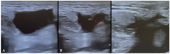
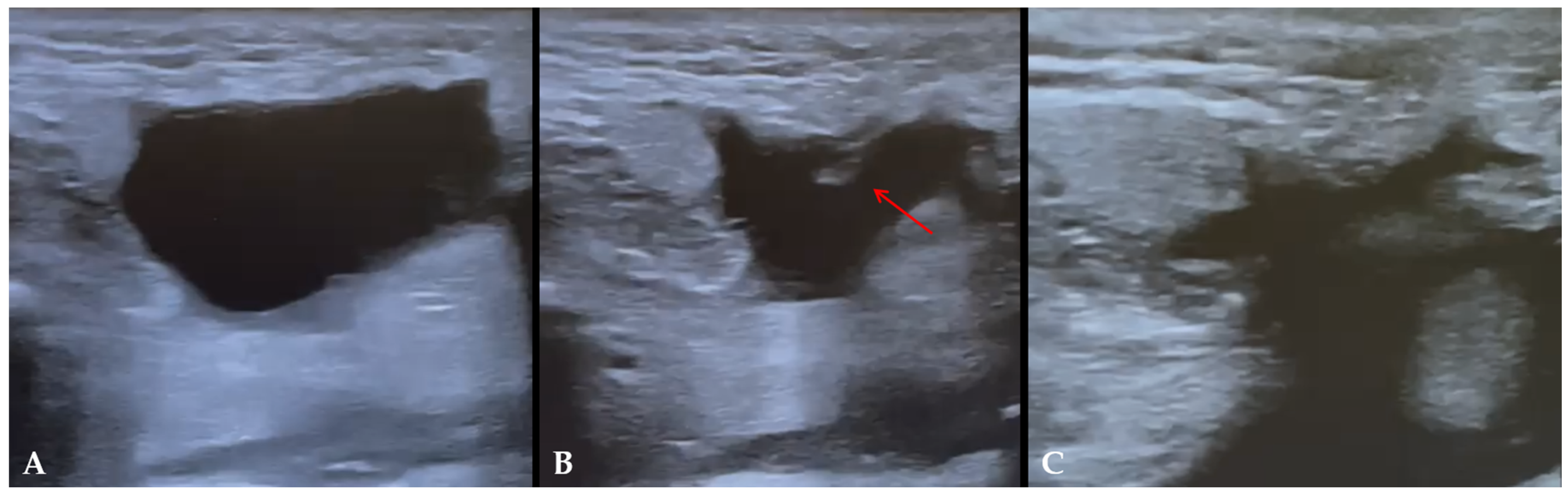
Prior to toxin infiltration, complete sonographically guided percutaneous aspiration of the sialocele contents was performed using a 21-gauge needle to decompress the cavity entirely. The aspirated fluid was evaluated for volume, color, and clarity displaying clear to serosanguinous salivary characteristics. Complete cavity evacuation was confirmed in real-time under US by visualizing the collapse of the fluid space and coaptation of the cavity walls (Figure 1).
Figure 1.
Real-time ultrasound-guided aspiration of a post-parotidectomy sialocele. (A) Pre-procedural ultrasound scan demonstrating well-defined, hypoechoic to anechoic fluid collection within the residual parotid bed. (B) Intra-procedural scan showing the needle tip (red arrow) within the collapsing fluid cavity during active aspiration. (C) Post-aspiration scan confirming complete evacuation and collapse of the sialocele cavity prior to intraglandular botulinum toxin injection.
OnabotulinumtoxinA (Botox®, Allergan, Irvine, CA, USA) was reconstituted with sterile preservative-free 0.9% sodium chloride solution (2.5 mL saline per 100 UI vial). Infiltration was delivered into the functional glandular parenchyma surrounding the collapsed cavity using a customized “grid mapping” approach. Under real-time high-resolution ultrasound guidance (in-plane technique), injections were performed using a 27-gauge, 38 mm needle. The multi-point grid mapping was standardized as follows: injection points were spaced approximately 1.0 to 1.5 cm apart across the residual functional parenchyma surrounding the collapsed cavity. The total administered dose ranged from 40 to 80 UI, customized based on the sonographic estimation of residual functional parenchymal volume. Toxin administration was structured as fractionated injections of 4 to 8 UI (0.1–0.2 mL) per site across 8 to 10 total injection points per gland. The injection sequence proceeded systematically across two depth planes: 4 to 5 deep parenchymal sites (adjacent to the deep fascial bed) followed by 4 to 5 superficial perimarginal sites across the parenchymal quadrants.
Needle-tip trajectory and insertion depth were continuously monitored under real-time US to ensure placement strictly within the hyperechoic functional acinar tissue, keeping needle depth superficial to the retromandibular vein and masseteric fascia to avoid deep neurovascular structures and minimize off-target diffusion toward the expected pathway of the main facial nerve trunk.
The rationale for utilizing a multi-point grid mapping technique lies in the pharmacokinetic diffusion properties of OnabotulinumtoxinA within glandular tissue. Because the therapeutic effective radius of a localized BoNT-A micro-injection is typically constrained to approximately 1.0–1.5 cm from the needle tip, relying on a single high-volume central depot risks uneven cholinergic blockade, incomplete parenchymal inactivation, and central fluid retention. Fractionating the calculated total dose into standardized aliquots distributed across the targeted sites creates an overlapping, uniform gradient of neurotoxin concentration throughout the remaining functioning acini, preventing high local concentration peaks, and therefore minimizing the risk of unwanted deep diffusion toward the main trunk of the facial nerve or the adjacent masseter muscle.
Following the procedure, patients received standardized post-injection care instructions: strict avoidance of local manual massage or pressure over the infiltrated parotid area for at least 24 h to prevent unwanted anatomical diffusion of the toxin, application of a light protective dressing for 24 h, adoption of a soft diet for 24 h, and temporary avoidance of sialagogue foods (e.g., acidic, citrus, or excessively sour foods) for 7 days to minimize reflex gustatory parasympathetic stimulation during the initial onset of the cholinergic block.
2.4. Follow-Up and Outcome Measures
Clinical and ultrasound evaluations were performed at baseline (T0, immediately prior to infiltration), and at standardized follow-up intervals: Day 7, Day 14, Day 30, and 3 months post-injection. The primary outcome measure was complete clinical resolution, defined as the total absence of palpable or sonographically detectable fluid collection without the need for further aspiration. Secondary outcome measures included treatment-related complications (e.g., facial motor weakness, masseter muscle weakness, local hematoma, infection, or systemic xerostomia).
2.5. Statistical Analysis
Continuous variables (age, onset time, collection dimensions, estimated volumes, and toxin dosage) were expressed as mean ± standard deviation (SD) and range as well as median and interquartile range (IQR; 25th–75th percentiles) given the sample size. Categorical variables are expressed as absolute frequencies and percentages. Descriptive statistical evaluations and database structuring were performed using Microsoft Excel® (Microsoft Corp., Redmond, WA, USA).
3. Results
The study cohort comprised 8 patients (5 males, 3 females, mean age 64.5 ± 13.0 years, median 67.0 years, IQR: 55.5–74.5). Sialocele onset occurred at a mean of 12 ± 2.6 days postoperatively (median 12.0 days, IQR: 10.0–14.3). Surgical etiologies included enucleo-resection (ER, n = 7) and subtotal parotidectomy (n = 1). A total dose ranging from 40 UI to 80 UI (mean 56.3 ± 11.9 UI, median 55.0 UI, IQR: 50.0–60.0) was administered. Baseline collection dimensions, calculated volumes, and individual BoNT-A doses are detailed in Table 1.
Table 1.
Baseline demographics, clinical characteristics, and BoNT-A treatment parameters. SD: Standard Deviation; ER: Enucleo-resection; Subtotal: Subtotal Parotidectomy; BTX: OnabotulinumtoxinA.
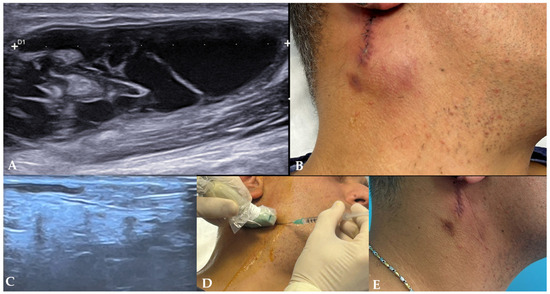
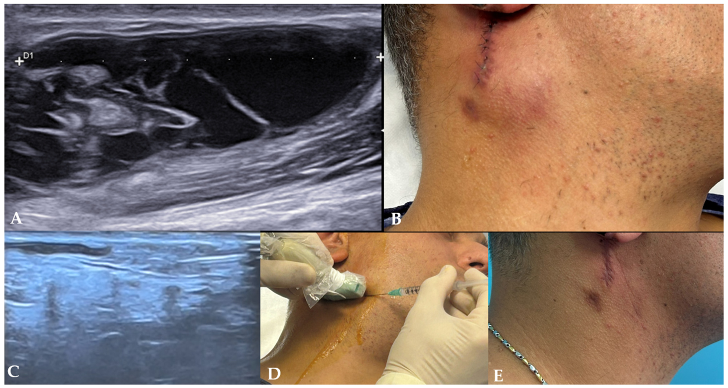
Complete clinical and sonographic resolution of the sialocele was achieved in 100% of cases (8/8) (Figure 2 and Figure 3).
Figure 2.
Clinical and ultrasonographic evaluations of a post-surgical parotid sialocele (Case 3). (A) Baseline ultrasound scan displaying a large hypoechoic fluid collection point measuring 43 mm in maximum longitudinal diameter (D1). (B) Pre-treatment clinical presentation demonstrating noticeable facial swelling in the right parotid region adjacent to the surgical scar. (C) Real-time intra-procedural ultrasound scan during percutaneous infiltration of Botulinum Toxin Type A into the residual functional parotid parenchyma. (D) Clinical procedural view of real-time ultrasound-guided intraglandular injection using an in-plane technique. (E) Clinical presentation at 3-month follow-up demonstrating complete resolution of the swelling with an optimal aesthetic result.
Figure 3.
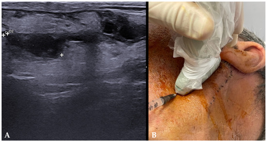
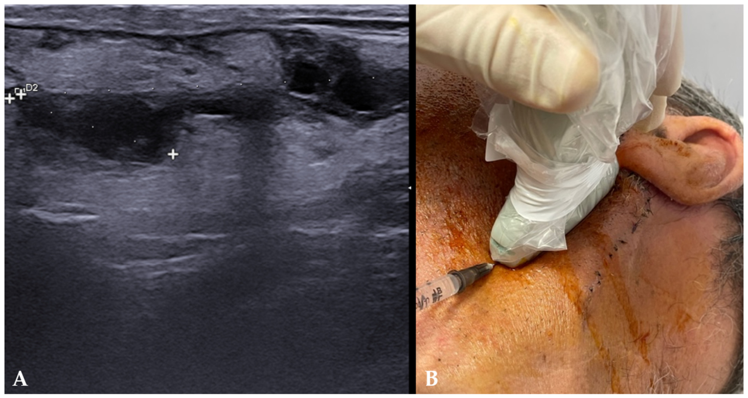
Ultrasonographic assessment and procedural setup for post-surgical sialocele management (Case 2). (A) High-resolution ultrasound scan displaying an elongated hypoechoic fluid cavity with calipers delineating maximum dimensions (D1: 19 mm; D2: 45 mm). (B) Clinical setup showing real-time transducer positioning and in-plane needle insertion adjacent to the post-parotidectomy surgical scar.
Following complete decompression and targeted BoNT-A infiltration, no re-accumulation of salivary fluid occurred. Because patient follow-up was conducted at discrete scheduled outpatient visits rather than through daily monitoring, complete regression was documented at the first follow-up assessment demonstrating total absence of fluid accumulation. Long-term sonographic monitoring at 3 months confirmed the complete absence of fluid recurrences or late-onset complications in all 8 patients.
No procedure-related local or systemic complications were recorded. Specifically, no patients developed transient or permanent facial nerve weakness, masseteric muscle weakness, local infection, sialadenitis, or symptomatic systemic xerostomia.
4. Discussion
Post-operative surgical-site complications remain a recognized challenge following parotid gland surgery. In a large cohort analysis evaluating parotidectomy for benign tumors, Henneman et al. reported an overall surgical site complication rate of 21%, with seromas, sialoceles, and salivary fistulae representing a substantial proportion of non-infectious wound sequelae [1]. Although typically categorized as low-grade events under the Clavien–Dindo classification system (Grade I or II), persistent post-surgical sialoceles cause notable patient morbidity, local swelling, pain, and the risk of overlying skin breakdown or secondary infection if unmanaged.
Traditionally, conservative management has relied on repeated needle aspiration, pressure dressings, anticholinergic pharmacotherapy, and strict dietary modification. However, these conventional approaches often yield slow recovery and are frequently complicated by systemic anticholinergic side effects (e.g., severe xerostomia, blurred vision, urinary retention, cardiac arrhythmias) or procedural failure requiring invasive open revision surgery. In our cohort, establishing a conservative management window of a maximum of 7 to 15 days before intervening with Botulinum Toxin Type A (BoNT-A) prevented long-term wound morbidity and avoided the transformation of sialoceles into chronic cutaneous salivary fistulae.
The biochemical foundation of BoNT-A efficacy relies on its precise interaction with the SNARE (Soluble NSF Attachment Protein Receptor) protein complex. Specifically, the toxin cleaves the SNAP-25 protein, an essential component required for the docking and fusion of acetylcholine (ACh) vesicles with the presynaptic membrane of parasympathetic post-ganglionic nerve terminals.
By chemically halting vesicle exocytosis, BoNT-A drives the local concentration of active ACh to near-zero values, inducing a temporary “functional silencing” of the glandular parenchyma [3]. As demonstrated in systematic reviews evaluating the histological and immunohistochemical effects of BoNT-A on salivary tissue, chemodenervation leads to a marked reduction in acinar immunoreactivity and temporary functional inhibition without causing acinar necrosis or permanent parenchymal destruction [18]. This neuro-glandular blockade shifts the parotid sialocele from an active, high-pressure fluid cavity into a static, stagnant fluid collection. Deprived of hydrostatic reinforcement, the fluid is rapidly reabsorbed, allowing the damaged margins of the parenchyma or ductal wall to collapse and undergo permanent cicatrization.
While our protocol exclusively utilized OnabotulinumtoxinA (BoNT-A), alternative neurotoxin serotypes, particularly Botulinum Toxin Type B (BoNT-B/RimabotulinumtoxinB), have also been described in autonomic salivary management [19]. Unlike BoNT-A, which selectively targets SNAP-25, BoNT-B exerts its enzymatic cleavage on vesicle-associated membrane protein (VAMP)/synaptobrevin, displaying a distinct affinity for autonomic parasympathetic terminals. Nevertheless, BoNT-A remains the preferred first-line agent in head and neck practice owing to its superior clinical duration of action, reduced pain upon local tissue injection, lower incidence of neutralizing antibody development, and well-documented safety profile with minimal risk of systemic anticholinergic dispersion.
The dosing strategy in our series was explicitly individualized based on the estimated volume of residual, functional glandular parenchyma that needed to be inactivated, rather than strictly on the volume of the accumulated fluid collection itself [3]. This physiological approach explains the absence of a direct linear relationship between the initial sialocele volume and the total units of toxin administered. For instance, a larger fluid collection area arising within a heavily resected gland with minimal remaining parenchyma required a relatively low overall dose (40 UI) to achieve complete secretory arrest. Conversely, patients with substantial functional glandular tissue surrounding the defect required higher total doses (up to 80 UI) distributed across the grid injection points to ensure complete cholinergic blockade across all active acinar units. In a systematic review evaluating botulinum toxin injection protocols in the maxillofacial region, Rauso et al. highlighted that distributing the toxin across multiple strategic points achieves predictable muscular and parenchymal coverage while minimizing the required dose per site [20]. Tailoring the dose to the residual secreting target avoids over-dosing depleted surgical beds while ensuring sufficient cholinergic blockade in larger remnants.
While early clinical protocols utilized anatomical surface landmarks for intraglandular BoNT-A injections, recent evidence strongly cautions against blind techniques [21]. Loens et al. demonstrated a significant spatial mismatch between landmark-based injection points and actual ultrasound-identified glandular centroids, with a mean horizontal deviation of approximately 21 mm in the parotid gland [4]. Furthermore, individual anatomical variations, driven by age, body mass index (BMI), and post-surgical tissue distortion, render landmark dependence highly unreliable.
In a clinical verification study evaluating 459 gland injections, Marks et al. [22] demonstrated that blind landmark-based needle placement reached the intended target in only 68% of parotid injections and a striking 32% of submandibular injections prior to ultrasound verification, with accuracy remaining unimproved regardless of practitioner seniority. In our protocol, real-time high-resolution ultrasound guidance provided a major safety and therapeutic advantage over surgical revision or blind techniques [22]. Re-exploring an acute surgical field compromised by dense inflammatory edema and granulation tissue carries an exceptionally high risk of iatrogenic trauma to the facial nerve branches. Ultrasound mapping ensured localized toxin dispersion strictly within the functional parotid parenchyma while avoiding placement near the facial nerve trunk/branches or deep diffusion into the masseter muscle.
The success of BoNT-A in managing post-surgical sialoceles aligns with its well-documented efficacy across a spectrum of salivary gland disorders. Systematic reviews in pediatric and adult populations demonstrate that intraglandular BoNT-A injections consistently reduce salivary flow and alleviate obstructive symptoms in chronic parotid sialadenitis, sialorrhea, and ductal stenosis with an outstanding safety profile [23,24]. Although novel alternative delivery routes, such as intraductal BoNT-A infusion via ductal cannulation, have been explored by Schwalje and Hoffman for intact ductal systems, direct percutaneous ultrasound-guided parenchymal infiltration remains the gold standard in post-parotidectomy sialoceles, where normal ductal continuity is frequently disrupted or transected [25].
Secondary chemodenervation remains the standard of care for established sialoceles. Nonetheless, recent clinical research has explored possible early prophylactic strategies. Lee et al. demonstrated in a prospective pilot study that intraoperative botulinum toxin injection into the residual parotid parenchyma during superficial partial parotidectomy significantly reduces postoperative salivary flow and the incidence of salivary complications without adverse effects [26]. Incorporating intraoperative or early post-surgical chemo-denervation could represent a promising paradigm shift in high-risk resections such as deep-lobe resections or cases with intraoperative ductal transection. Furthermore, future studies integrating contrast-enhanced ultrasound (CEUS) or elastography may further refine real-time parenchymal viability mapping, optimizing grid injection accuracy.
Beyond direct parenchymal infiltration, retrograde intraductal instillation of BoNT-A via Stensen’s duct may be an innovative alternative administration route. From a conceptual standpoint, delivering the neurotoxin retrograde through the ductal tree could allow homogeneous distribution across the functional acini while completely avoiding parenchymal needle punctures [27,28].
The main intrinsic limitation of BoNT-A therapy is its temporary duration of action, as axonal sprouting and SNAP-25 regeneration typically restore cholinergic transmission within 3 to 6 months as shown in several studies regarding drooling and sialorrhea [29,30,31]. However, in the context of post-surgical or traumatic sialoceles, this temporary window of salivary suppression (anhidrosis/asialia) aligns well with the pathophysiology of post-surgical sialoceles. Unlike chronic salivary disorders (such as neurogenic sialorrhea or chronic sialadenitis), which require long-term maintenance therapy, a post-surgical sialocele represents an acute structural disruption of the parenchymal ductal network. The temporary window of complete salivary suppression provided by BoNT-A drives the immediate collapse of the fluid cavity, enabling definitive fibrotic tissue adhesion and permanent anatomical scarring of the transected parenchymal ducts. Once complete structural sealing and cicatrization of the surgical bed are established, recurrence is clinically negligible even after full cholinergic recovery of the remaining glandular tissue. In our series, no recurrences or late fluid re-accumulations were observed at the 3-month evaluation. Nevertheless, from a methodological standpoint, extending follow-up to 12 months in future prospective studies will provide further empirical confirmation of long-term stability.
This study has several limitations that should be acknowledged. First, its retrospective single-center design and the small sample size (n = 8) limit the statistical power and generalizability of the findings. Second, the absence of a parallel control group, such as patients undergoing prolonged conservative treatment alone, placebo/sham injections, or surgical revision, precludes direct comparative conclusions and prevents us from completely ruling out natural resolution, confounding effects from preceding conservative measures, or selection bias. clinical and ultrasonographic evaluations were performed by the operating team without a blinded independent assessor, which may introduce observer bias. Finally, standardized patient-reported outcome measures (PROMs), such as validated pain scales or quality-of-life questionnaires, were not systematically recorded in the retrospective historical charts.
5. Conclusions
Ultrasound-guided intraglandular infiltration of Botulinum Toxin Type A showed promising clinical outcomes as a minimally invasive second-line strategy for post-surgical parotid sialoceles failing short-term conservative management. In our preliminary series, complete resolution was observed within 10 days post-injection without procedure-related complications. However, given the small sample size, retrospective nature and lack of a control arm, these findings cannot serve as the basis for clinical standards. Larger prospective, randomized, comparative trials incorporating standardized patient-reported outcome measures (PROMs) are necessary to confirm these preliminary observational results and establish formal clinical recommendations.
Author Contributions
Conceptualization, G.M. and G.L.G.; methodology, G.M., A.C. and G.L.G.; validation, A.M.R., A.B. and A.S.; formal analysis, G.M. and A.C.; investigation, G.M., A.M.R., A.B. and A.S.; data curation, G.M. and A.C.; writing—original draft preparation, G.M.; writing—review and editing, E.N.S. and G.L.G.; visualization, A.M.R. and A.B.; supervision, E.N.S. and G.L.G. All authors have read and agreed to the published version of the manuscript.
Funding
This research received no external funding.
Institutional Review Board Statement
The study was conducted in accordance with the Declaration of Helsinki and approved by the local ethics committee (AOU G Martino, Messina, Italy, protocol code 115-25, 23 April 2024).
Informed Consent Statement
Informed consent was obtained from all subjects involved in the study. Written informed consent has been obtained from the patients to publish this paper.
Data Availability Statement
Data is available upon reasonable request from the corresponding author.
Conflicts of Interest
The authors declare no conflicts of interest.
Abbreviations
The following abbreviations are used in this manuscript:
| BoNT-A | Botulinum toxin type A |
| BoNT-B | Botulinum toxin type B |
| US | Ultrasound |
| ER | Enucleo-resection |
| CNB | Core needle biopsy |
| ACh | Acetylcholine |
| SNARE | Soluble NSF attachment protein receptor |
| SNAP-25 | Synaptosomal-associated protein 25 |
| VAMP | Vesicle-associated membrane protein |
| SD | Standard deviation |
| STROBE | Strengthening the Reporting of Observational Studies in Epidemiology |
References
- Henneman, R.; Berger, D.M.S.; Karakullukcu, M.B.; Smeele, L.E.; Lohuis, P.; Jozwiak, K.; van der Hage, J.A.; Balm, A.J.M. Surgical site complications after parotid gland surgery for benign tumors in a centralized setting: A Clavien-Dindo class cohort analysis. Eur. J. Surg. Oncol. 2020, 46, 258–262. [Google Scholar] [CrossRef] [Scilit] [PubMed]
- Boschetti, C.E.; Lo Giudice, G.; Spuntarelli, C.; Apice, C.; Rauso, R.; Santagata, M.; Tartaro, G.; Colella, G. Kabat Rehabilitation in Facial Nerve Palsy after Parotid Gland Tumor Surgery: A Case-Control Study. Diagnostics 2022, 12, 565. [Google Scholar] [CrossRef] [Scilit] [PubMed]
- Lovato, A.; Restivo, D.A.; Ottaviano, G.; Marioni, G.; Marchese-Ragona, R. Botulinum toxin therapy: Functional silencing of salivary disorders. Acta Otorhinolaryngol. Ital. 2017, 37, 168–171. [Google Scholar] [CrossRef] [Scilit] [PubMed]
- Loens, S.; Bruggemann, N.; Steffen, A.; Baumer, T. Localization of Salivary Glands for Botulinum Toxin Treatment: Ultrasound Versus Landmark Guidance. Mov. Disord. Clin. Pract. 2020, 7, 194–198. [Google Scholar] [CrossRef] [Scilit] [PubMed]
- Lo Giudice, G.; Nastro Siniscalchi, E.; Arena, P.; Bottari, A.; Silipigni, S.; Calvo, A.; De Ponte, F.S.; Stagno, A. Diagnostic accuracy of ultrasound guided core needle biopsy in parotid gland neoplasms: A retrospective comparative study. J. Cranio-Maxillofac. Surg. 2025, 53, 2279–2287. [Google Scholar] [CrossRef] [Scilit] [PubMed]
- Abboud, W.A.; Nadel, S.; Hassin-Baer, S.; Arad, A.; Dobriyan, A.; Yahalom, R. Ultrasound-Guided Botulinum Toxin Injections into the Salivary Glands for the Treatment of Drooling. Isr. Med. Assoc. J. 2019, 21, 116–119. [Google Scholar] [PubMed]
- Barbero, P.; Busso, M.; Tinivella, M.; Artusi, C.A.; De Mercanti, S.; Cucci, A.; Veltri, A.; Avagnina, P.; Calvo, A.; Chio, A.; et al. Long-term follow-up of ultrasound-guided botulinum toxin-A injections for sialorrhea in neurological dysphagia. J. Neurol. 2015, 262, 2662–2667. [Google Scholar] [CrossRef] [Scilit] [PubMed]
- Jongerius, P.H.; Joosten, F.; Hoogen, F.J.; Gabreels, F.J.; Rotteveel, J.J. The treatment of drooling by ultrasound-guided intraglandular injections of botulinum toxin type A into the salivary glands. Laryngoscope 2003, 113, 107–111. [Google Scholar] [CrossRef] [Scilit] [PubMed]
- Lungren, M.P.; Halula, S.; Coyne, S.; Sidell, D.; Racadio, J.M.; Patel, M.N. Ultrasound-Guided Botulinum Toxin Type A Salivary Gland Injection in Children for Refractory Sialorrhea: 10-Year Experience at a Large Tertiary Children’s Hospital. Pediatr. Neurol. 2016, 54, 70–75. [Google Scholar] [CrossRef] [Scilit] [PubMed]
- Dogu, O.; Apaydin, D.; Sevim, S.; Talas, D.U.; Aral, M. Ultrasound-guided versus ‘blind’ intraparotid injections of botulinum toxin-A for the treatment of sialorrhoea in patients with Parkinson’s disease. Clin. Neurol. Neurosurg. 2004, 106, 93–96. [Google Scholar] [CrossRef] [Scilit] [PubMed]
- Shoval, H.A.; Antelis, E.; Hillman, A.; Wei, X.; Tan, P.; Alejandro, R.; Kim, H. Onabotulinum Toxin A Injections Into the Salivary Glands for Spinal Muscle Atrophy Type I: A Prospective Case Series of 4 Patients. Am. J. Phys. Med. Rehabil. 2018, 97, 873–878. [Google Scholar] [CrossRef] [Scilit] [PubMed]
- Serrera-Figallo, M.A.; Ruiz-de-Leon-Hernandez, G.; Torres-Lagares, D.; Castro-Araya, A.; Torres-Ferrerosa, O.; Hernandez-Pacheco, E.; Gutierrez-Perez, J.L. Use of Botulinum Toxin in Orofacial Clinical Practice. Toxins 2020, 12, 112. [Google Scholar] [CrossRef] [Scilit] [PubMed]
- Safarpour, D.; Jabbari, B. Botulinum Toxin Treatment for Cancer-Related Disorders: A Systematic Review. Toxins 2023, 15, 689. [Google Scholar] [CrossRef] [Scilit] [PubMed]
- Maharaj, S.; Mungul, S.; Laher, A. Botulinum toxin A is an effective therapeutic tool for the management of parotid sialocele and fistula: A systematic review. Laryngoscope Investig. Otolaryngol. 2020, 5, 37–45. [Google Scholar] [CrossRef] [Scilit] [PubMed]
- von Elm, E.; Altman, D.G.; Egger, M.; Pocock, S.J.; Gotzsche, P.C.; Vandenbroucke, J.P.; Initiative, S. The Strengthening the Reporting of Observational Studies in Epidemiology (STROBE) statement: Guidelines for reporting observational studies. J. Clin. Epidemiol. 2008, 61, 344–349. [Google Scholar] [CrossRef] [Scilit] [PubMed]
- Brunn, J.; Block, U.; Ruf, G.; Bos, I.; Kunze, W.P.; Scriba, P.C. Volumetric analysis of thyroid lobes by real-time ultrasound (author’s transl). Dtsch. Med. Wochenschr. 1981, 106, 1338–1340. [Google Scholar] [CrossRef] [Scilit] [PubMed]
- Wagner, J.M.; Rebik, K.; Spicer, P.J. Ultrasound of Soft Tissue Masses and Fluid Collections. Radiol. Clin. 2019, 57, 657–669. [Google Scholar] [CrossRef] [Scilit] [PubMed]
- Oliveira, J.B.; Evencio-Neto, J.; Baratella-Evencio, L. Histological and immunohistochemical findings of the action of botulinum toxin in salivary gland: Systematic review. Braz. J. Biol. 2017, 77, 251–259. [Google Scholar] [CrossRef] [Scilit] [PubMed]
- Dressler, D. Clinical applications of botulinum toxin. Curr. Opin. Microbiol. 2012, 15, 325–336. [Google Scholar] [CrossRef] [Scilit] [PubMed]
- Rauso, R.; Lo Giudice, G.; Tartaro, G.; Zerbinati, N.; Nicoletti, G.F.; Fragola, R. Botulinum toxin type A injections for masticatory muscles hypertrophy: A systematic review. J. Cranio-Maxillofac. Surg. 2022, 50, 7–18. [Google Scholar] [CrossRef] [Scilit] [PubMed]
- Fuster Torres, M.A.; Berini Aytes, L.; Gay Escoda, C. Salivary gland application of botulinum toxin for the treatment of sialorrhea. Med. Oral Patol. Oral Cir. Bucal 2007, 12, E511–E517. [Google Scholar] [PubMed]
- Marks, I.H.; Ridgley Vaidya, J.; Israel, O.; Nixon, P.; Sharma, R. Does injection of botulinum toxin to salivary glands require ultrasound guidance? Int. J. Pediatr. Otorhinolaryngol. 2024, 186, 112114. [Google Scholar] [CrossRef] [Scilit] [PubMed]
- Rodwell, K.; Edwards, P.; Ware, R.S.; Boyd, R. Salivary gland botulinum toxin injections for drooling in children with cerebral palsy and neurodevelopmental disability: A systematic review. Dev. Med. Child Neurol. 2012, 54, 977–987. [Google Scholar] [CrossRef] [Scilit] [PubMed]
- Strohl, M.P.; Chang, C.F.; Ryan, W.R.; Chang, J.L. Botulinum toxin for chronic parotid sialadenitis: A case series and systematic review. Laryngoscope Investig. Otolaryngol. 2021, 6, 404–413. [Google Scholar] [CrossRef] [Scilit] [PubMed]
- Schwalje, A.T.; Hoffman, H.T. Intraductal Salivary Gland Infusion With Botulinum Toxin. Laryngoscope Investig. Otolaryngol. 2019, 4, 520–525. [Google Scholar] [CrossRef] [Scilit] [PubMed]
- Lee, D.J.; Lee, Y.M.; Park, H.J.; Lee, J.W.; Cha, W. Intraoperative botulinum toxin injection for superficial partial parotidectomy: A prospective pilot study. Clin. Otolaryngol. 2021, 46, 998–1004. [Google Scholar] [CrossRef] [Scilit] [PubMed]
- Su, C.H.; Lee, K.S.; Tseng, T.M.; Tseng, H.; Ding, Y.F.; Koch, M.; Hung, S.H. Intraductal injection as an effective drug delivery route in the management of salivary gland diseases. Eur. Arch. Otorhinolaryngol. 2017, 274, 399–404. [Google Scholar] [CrossRef] [Scilit] [PubMed]
- Colella, G.; Lo Giudice, G.; De Luca, R.; Troiano, A.; Lo Faro, C.; Santillo, V.; Tartaro, G. Interventional sialendoscopy in parotidomegaly related to eating disorders. J. Eat. Disord. 2021, 9, 25. [Google Scholar] [CrossRef] [Scilit] [PubMed]
- Hattori, N.; Mukai, Y.; Nishikawa, N.; Hasegawa, K.; Tomiyama, M.; Kimura, Y.; Tsuboi, Y.; Takahashi, R.; Nakamura, R.; Izumi, Y.; et al. Efficacy and Safety of IncobotulinumtoxinA for Treatment of Sialorrhea: A Multicenter, Phase 3 Study in Japan. Mov. Disord. Clin. Pract. 2026, 13, 120–130. [Google Scholar] [CrossRef] [Scilit] [PubMed]
- Reid, S.M.; Johnstone, B.R.; Westbury, C.; Rawicki, B.; Reddihough, D.S. Randomized trial of botulinum toxin injections into the salivary glands to reduce drooling in children with neurological disorders. Dev. Med. Child Neurol. 2008, 50, 123–128. [Google Scholar] [CrossRef]
- Guidubaldi, A.; Fasano, A.; Ialongo, T.; Piano, C.; Pompili, M.; Masciana, R.; Siciliani, L.; Sabatelli, M.; Bentivoglio, A.R. Botulinum toxin A versus B in sialorrhea: A prospective, randomized, double-blind, crossover pilot study in patients with amyotrophic lateral sclerosis or Parkinson’s disease. Mov. Disord. 2011, 26, 313–319. [Google Scholar] [CrossRef] [Scilit] [PubMed]

Disclaimer/Publisher’s Note: The statements, opinions and data contained in all publications are solely those of the individual author(s) and contributor(s) and not of MDPI and/or the editor(s). MDPI and/or the editor(s) disclaim responsibility for any injury to people or property resulting from any ideas, methods, instructions or products referred to in the content. |
© 2026 by the authors. Licensee MDPI, Basel, Switzerland. This article is an open access article distributed under the terms and conditions of the Creative Commons Attribution (CC BY) license.