


Abstract
Objective. The main objective of this study was to examine the effects of transcutaneous auricular vagus nerve stimulation (taVNS) on psychological, sleep, and performance outcomes in powerlifters. Previous research has primarily focused on taVNS in clinical or healthy samples and showed improvements in psychological functioning, recovery, and sleep. Evidence for beneficial effects of taVNS in healthy athletes, however, is missing so far. Methods. Over the course of 4 weeks, 10 healthy adult powerlifters self-administered taVNS five times a week for 30 min after strength training or before bedtime, in an uncontrolled, single-arm pilot design without a sham or control condition. Measurements were taken at three time points: before the intervention phase (pre), immediately after the 4-week intervention phase (post) and 1 month after the post-assessment (follow-up). Pre, post, and follow-up measurements included various subjective psychological domains (recovery, stress, perceived stress, affect, memory, attention, anxiety, and well-being), objective sleep domains (nocturnal HRV, sleep score, sleep duration, and subjective sleep quality), and submaximal strength performance outcomes in squat, bench press, and deadlift. The small sample size required non-parametric testing. Additionally, a responder analysis was conducted to explore individual differences in outcome-specific responses. Results. Results showed nominally significant pre-post improvements (uncorrected for multiple comparisons) in positive affect and self-reported attentional difficulties, both revealing large effect sizes; however, given the absence of a control group and the lack of correction for multiple comparisons across multiple outcomes tested, these findings should be interpreted as exploratory and hypothesis-generating rather than confirmatory. The remaining psychological domains and sleep outcomes did not show meaningful changes. Regarding performance, only two of the ten participants provided complete baseline training data, so no reliable conclusions about a beneficial effect of taVNS on strength performance could be drawn. The responder analysis was consistent with the two nominally significant findings but did not reveal a clear overall pattern of taVNS effectiveness. Conclusions. The current study is the first to implement a 4-week taVNS intervention in strength training and demonstrate differences in domain-specific responsiveness to taVNS in athletes for psychological health and self-reported cognitive performance. These findings offer important insights into the feasibility and practical application of taVNS in sports settings. Further studies with larger, sham-controlled samples, a pre-registered primary outcome and a longer intervention period are needed to determine whether these preliminary, hypothesis-generating findings reflect genuine effects of taVNS.
1. Introduction
1.1. Vagus Nerve and Parasympathetic Activity
The vagus nerve (VN) is the longest cranial nerve in the human body and expands from the brainstem through the neck and thorax to the abdomen [1]. As the main component of the parasympathetic nervous system (PNS), its crucial role in regulating different bodily functions includes reducing the heart and respiration rate, blood pressure, stimulating digestion, immune responses, and mood [1,2]. As opposed to the sympathetic nervous system (SNS), which activates the “fight or flight” response during stressful situations, the PNS serves an inhibiting purpose (“rest and digest”), thus functioning as a “brake” for bodily functions [3,4].
Various studies highlight the association between PNS and psychological stress. Ziegler [5], for example, proposes that stress is associated with an increased sympathetic and a decreased parasympathetic activity. Another study indicates a stress-reducing effect through certain mind-body techniques that increase parasympathetic activity [6]. These findings highlight the notion that by deliberately targeting and increasing parasympathetic activity, psychological stress can be decreased.
Vagal tone is the main indicator for parasympathetic activity and reveals an individual’s autonomic flexibility, meaning the ability of the PNS to adapt to environmental changes by regulating arousal, respiration, heart rate, and attention [7]. Heart rate variability (HRV) is the most used measure to assess vagal tone and describes the variation in time intervals between consecutive heartbeats [8,9]. Higher vagal activity is positively associated with psychological well-being, including life satisfaction, and with higher levels of positive emotions and lower levels of negative emotions [3,7,10,11]. However, a higher HRV is not always best, and pathological conditions may be the cause [12]. Interestingly, one study suggests a non-linear relationship between vagal activity and well-being: individuals with moderate vagal activity may exhibit greater well-being than individuals with either low or high vagal activity [3]. Although there is no clear consensus on how parasympathetic vagal activity is linked to well-being or similar constructs, the literature suggests an important association.
A growing number of studies on HRV and vagal activity underline the central role of parasympathetic regulation in overall health [13], self-regulatory capacities, such as executive functioning and emotion regulation [14,15], attention and emotional processing [12], and resilience to stress [15]. While research on HRV and parasympathetic activity is mostly done in either clinical or healthy samples, studies specifically examining athletes in a sports context are scarce, with even fewer examining anaerobic and strength training. Notably, the PNS also plays an essential role in recovery due to its association with psychophysiological processes of self-regulation and self-modulation [16,17,18,19,20]. Therefore, HRV and parasympathetic activity are indices commonly used to assess physical recovery in athletes [21]. In one study, elite American track and field athletes were observed in regards to differences in HRV [22]. In this cross-sectional comparison, individuals were categorized into power/strength and endurance (aerobic) groups. Results showed significant differences in several HRV indices by sex, but not by training emphasis (power/strength vs. endurance). These findings suggest that HRV indices may not differ substantially between athletes with an aerobic versus an anaerobic training emphasis. This is relevant to the assumptions underlying the present study because HRV did not differ substantially by training modality in Berkoff et al. [22]. There is no clear a priori reason to expect powerlifters’ autonomic responsiveness to differ fundamentally from that of the endurance samples in which most prior taVNS research has been conducted. This finding was therefore taken as partial justification for extending current findings on taVNS and its parasympathetic modulation properties, deriving mainly from endurance samples in the sport context, to a strength-training population in the present study. Nevertheless, further strength training-specific research is needed to potentially offer novel insight in neuromodulating effects adjusted to the unique demands of anaerobic sports.
1.2. Vagus Nerve Stimulation
1.2.1. Forms of Vagus Nerve Stimulation
Vagus nerve stimulation (VNS) is a medical neuromodulation treatment that delivers electrical impulses to the VN [23]. VNS has been successfully used as an intervention for various medical conditions due to its ability to modulate neural activity and physiological responses [1,24]. VNS can be administered in different ways: an invasive and a non-invasive stimulation [25]. For invasive VNS (iVNS), a surgical implantation of a small pulse generator in the chest is needed, which is primarily used in drug-resistant epilepsy patients [26]. One form of non-invasive stimulation, which was used in the present study, is transcutaneous auricular vagus nerve stimulation (taVNS). taVNS requires no surgical implantation and is applied via an electrode to the left tragus of the outer ear, stimulating the auricular branch of the VN through electrical impulses [1,26]. In comparison, taVNS is a more cost-efficient and an easy-to-apply alternative with less side effects to iVNS [27].
1.2.2. Effects of VNS on Medical Conditions
Both iVNS and taVNS have demonstrated promising therapeutic effects across a range of medical conditions. For instance, they have shown positive effects in patients with Alzheimer’s disease [28] or tinnitus and tinnitus-evoked symptoms [29]. In various cardiac conditions, VNS has been reported to attenuate inflammation [30], reduce oxidative stress in patients with acute heart failure [31], lower blood pressure in patients with acute heart failure and hypertension [32,33], reduce postural tachycardia and atrial fibrillation burden [34,35], increase HRV [34,36], and positively influence cardiac physiology [37]. The latter demonstrate the cardioprotective abilities that different forms of VNS may have.
1.3. Effects of taVNS on Psychological Constructs
Besides its numerous uses in the medical field, taVNS has also shown various positive effects on psychological parameters. A recent study examined the effects of taVNS on cognition, anxiety, depression, sleep, and fatigue in patients suffering from long-COVID [38]. taVNS was administered twice a day for 30 min each for 10 consecutive days. Results demonstrated significant improvements in fluid cognition from baseline to post-intervention, with further enhancements observed at 1-month follow-up. Other cognitive subdomains also showed significant improvements, such as attention, processing speed, episodic memory, and working memory, showing moderate-to-large effect sizes. For anxiety, depression and sleep significant improvements were found at post and 1-month follow-up. Fatigue showed delayed response to taVNS, significantly improving only from baseline to 1-month follow-up. Another study examined taVNS in memory recall paired with reading in healthy individuals [39]. Results showed significant improvements in memory-based recall of a previously read text passage compared to controls, suggesting positive effects of taVNS in memory performance. Ferstl et al. [40] suggest that taVNS improved positive mood after effort exertion and further showed that improvements of motivation were greater when baseline scores of positive mood were lower, but no effects on negative mood were found. Bretherton et al. [36] also found improvements in mood parameters, including self-reported tension, depression, vigour, and mood disturbance, following 2 weeks of daily taVNS. More positive effects of taVNS on psychological constructs include significant improvements in well-being [27], depressive symptoms [41,42,43], anxiety [44], and some aspects of quality of life [36]. Recent studies also showed beneficial effects of taVNS in quality of life in patients with atrial fibrillation and heart failure [35,45] and in young healthy individuals by reducing acute stress levels [46]. A recent study by Dos Reis et al. [47] found that taVNS reduced perceived stress and stress levels. In contrast, Le Roy et al. [48] found a negative effect on perception of stress after repeated taVNS sessions, suggesting a nocebo effect of the intervention.
1.4. Effects of taVNS on Sleep
Recent findings suggest beneficial effects of taVNS on sleep measures [36,38]. More specifically, results showed improved sleep quality [47,48,49], improved sleep efficiency [48,50], increased total sleep time [49,50], and increased HRV [48,50,51]. Interestingly, Le Roy et al. [48] found that total sleep time decreased from pre- to post-intervention after repeated taVNS sessions, which may indicate the need to include potential individual or contextual factors, influencing the effect of taVNS. Overall, these findings are of great relevance to the present study as they may give an insight into potential effects of taVNS transferrable to the sports context. They offer a valid foundation for examining whether previously observed outcomes in healthy or clinical samples can also be observed in strength training athletes, specifically in powerlifters.
1.5. Effects of taVNS on Recovery and Performance in Sports
Recovery is an essential part for all athletes, with insufficient recovery leading to fatigue and reduced performance [52]. As mentioned previously, recovery is linked to parasympathetic activity, and so is performance [53]. It was found that autonomic nervous system activity increases during recovery times [19,54,55], thus suggesting a potential role for taVNS in enhancing these effects further. The literature on the effects of taVNS in athletes is very limited, with only one recent study by Çalι et al. [53] specifically focusing on sports performance in elite athletes. Importantly, participants in the study received one single session of taVNS for 20 min. Before and after treatment, sports performance was measured via isometric quadriceps muscle strength, heart rate, lower extremity balance, and grip strength. The authors concluded that a single session of taVNS does not significantly improve sports performance but found a moderate effect size in favour for the experimental group for quadriceps strength and anterior balance. The results raise the question whether a single application of taVNS may suffice for benefiting elite athletes. Another study focusing on taVNS in the sports context by Güçlüer et al. [56] investigated restoration of the parasympathetic activity in healthy males after aerobic exercise. Results indicated increases in parasympathetic activity following exercise, implying that taVNS can aid in accelerating sports recovery in healthy individuals. In line with these results is another study on recovery after aerobic exercise by Genç et al. [57] investigating post-COVID patients. The authors conclude that taVNS increased vagal activity in individuals with post-COVID syndrome, suggesting beneficial recovery after a session of aerobic exercise in non-athletes. Another study in sports revealed that taVNS could decrease fatigue and pain, as well as increase parasympathetic activity in healthy individuals, again underlining the beneficial effects of taVNS on recovery [58]. In spite of that, the authors did not detect a significant improvement in aerobic sports performance, which, however, is in line with Çalι et al. [53]. Importantly, Hatik et al. [58] implemented a 4-day taVNS intervention, while participants of the remaining studies received only a single session of taVNS [53,56,57].
1.6. The Present Study
Most research on taVNS either involves clinical populations or focuses exclusively on aerobic exercise, with no studies to date applying a 4-week taVNS intervention or investigating its effects in anaerobic sports. To address this gap, the current study aims to investigate taVNS in strength training athletes, specifically in powerlifters. In this study, we assessed whether a 4-week taVNS protocol can improve psychological, sleep and performance outcomes in powerlifters. Based on current literature findings of taVNS on psychological functioning, sleep, and sports performance, the following hypotheses were derived. We hypothesized that athletes would report better sports recovery and less stress, a reduction in perceived stress, more positive and less negative affect, improved cognitive performance in memory and attention, less anxiety, less competition anxiety, and a higher well-being compared to baseline. Furthermore, we expected a higher nocturnal HRV as an index for better recovery, and an increase over the course of the intervention. Regarding sleep measures, we hypothesized a higher sleep score, longer sleep duration, and better subjective sleep quality after the intervention. Additionally, we expected an increasing trend for all sleep outcomes over the course of the intervention. We further hypothesized improved strength performance by investigating submaximal strength in certain strength training exercises. Given the large number of outcomes assessed (more than 30 variables across the RESTQ-sport subscales, the PSS-10, the PANAS, the APSA, the STAI, the WAI-T, the WHO-5, and the sleep domains), no single primary outcome was pre-specified and no correction for multiple comparisons across outcomes was planned a priori. Consequently, and in the absence of a control or sham condition, this study is best characterized as an uncontrolled, exploratory pilot study, and all statistical analyses reported below should be interpreted as hypothesis-generating rather than confirmatory.
2. Methods
2.1. Participants
Male and female powerlifters (N = 10, four female), ranging in age from 20 to 34 years (M = 25.9 years, SD = 3.86 years) from strength sports clubs in Salzburg, Austria, voluntarily participated in the study. Inclusion criteria included at least 1 year of experience in strength training, training at least three times a week and training with a focus on the squat, bench press, and deadlift. Exclusion criteria included severing of the VN, metallic, or electronic implants (e.g., pacemaker, cochlea-implant), epilepsy, cardiac diseases (e.g., bradycardia), pregnancy, or the current intake of sleep medication. Exclusion criteria did not explicitly address current psychoactive medication use or diagnosed psychiatric conditions; this omission is acknowledged as a limitation given the psychological outcomes assessed in this study (see Section 4.1). Participants were recruited through flyers posted in powerlifting clubs and through direct, in-person outreach at the same clubs. Interested individuals completed a short online screening form assessing the inclusion and exclusion criteria stated above. Individuals who met all criteria were automatically forwarded to the baseline survey of the psychological parameters. All participants had at least 1 year of competitive powerlifting experience. All participants followed a structured powerlifting-specific program built around the three competition lifts (squat, bench press, and deadlift) as primary exercises. The training program was not prescribed or standardized by the study. Participants continued to follow the individualized program provided by their respective coaches, which they had already been using prior to the study and continued to use throughout its duration. No a priori sample-size calculation was conducted; the sample size of 10 was determined by feasibility constraints within the scope of an exploratory pilot study, as well as the limited number of taVNS devices available for the study. It should be noted that all participants were acquainted with the study coordinator (possible limitations will be discussed in the limitation section).
The study was approved by the ethics committee of the University of Salzburg, Austria, but was not preregistered in a public study registry. All participants were informed of potential risks or side effects, and written informed consent was obtained from all participants.
2.2. Procedure
The current study used a pre-post within-subject design with a 1-month follow-up (Figure 1). Initially, all participants completed an online survey collecting demographic information (name, age, sex, nationality, email address), training habits (inclusion criteria), and potential contraindications (exclusion criteria). When all criteria were met, participants proceeded to complete the survey assessing psychological measures. At the end of the survey, participants were required to send their training logs from the previous month or training block as a baseline for strength performance. After the survey, participants were handed out a sleep sensor and a taVNS device and given instructions on adequate usage. The sleep sensor was connected to the Sleep2 app (Nukkuaa GmbH, Wals-Siezenheim, Austria, https://www.sleep2.com/en/, accessed on 6 December 2024), which participants had to install to their own smartphones. Exact instructions on how to use the app were given. Participants then proceeded to wear the sleep sensor at home for 28 days during the intervention phase, and for 3 consecutive nights before (baseline), after (post-intervention), as well as for 3 nights at 1-month follow-up. The sleep sensor recorded nocturnal HRV, sleep score, and sleep duration. Additionally, each morning, subjective sleep quality was reported in the app for the corresponding time.
Figure 1.
Visual representation of the study design and sequence of measures.
During the 28-day intervention phase, participants self-administered taVNS five times per week for 30 min each session. Stimulation was applied to the tragus of the left ear, either after strength training sessions or before bedtime. Participants documented each stimulation session in a digital study protocol to ensure real-time tracking of compliance. Only the participant and the study coordinator had access to individual study protocols. The choice between administering stimulation after training or before bedtime was left to each participant’s personal preference and was not separately logged. Additionally, participants were instructed to maintain their daily living schedules throughout the entire study, such as sleeping and eating habits. Over the 4-week intervention, this amounted to a total of 20 scheduled sessions per participant (five per week). The digital study protocol served primarily as a compliance-monitoring instrument and was not planned as an outcome measure. Given the considerable participant burden already involved in the study (concurrent use of two devices, psychological questionnaires at three assessment time points, three separate strength-performance testing sessions, and nightly sleep tracking), protocol deviations beyond the number of completed sessions were not tracked separately, and no adverse events were spontaneously reported by participants.
At post-measurement, psychological outcomes again were measured via an online survey, and participants were reminded to wear the sleep sensor for further 3 nights. Additionally, participants had to complete strength performance training (post-intervention training), which included an individual warm-up, followed by the execution of a single repetition of each lift (squat, bench press, deadlift) at a rate of perceived exertion of 8 (RPE8). To determine the RPE8 single-repetition load, participants did not proceed directly from warm-up to a single attempt. Instead, they gradually increased the load across several sets until they reached a weight they could lift for one repetition while still perceiving they had approximately two repetitions in reserve, corresponding to an RPE of 8. A submaximal strength assessment at an RPE of 8 was chosen instead of testing for 1-repetition-maximum (1RM) to minimize disruption to athletes’ training prior to upcoming competitions while ensuring standardization and feasibility within the study context. Participants were advised to perform the post-intervention training under the supervision of their coach. The achieved strength values were then reported using an online survey. At 1-month follow-up, participants had to again fill out an online survey and complete the same strength performance training (follow-up training). At post-measurement and 1-month follow-up, participants were given a maximum of 3 days to complete the online survey and the strength performance training. Participants were encouraged to contact the study coordinator anytime they needed any assistance.
2.3. Materials
2.3.1. taVNS Device
The Parasym (Parasym Ltd., London, UK) device was used in the present study, transmitting mild electrical impulses to the VN with a frequency of 25 Hz and pulse width of 250 µs. Intensity levels were adjusted individually by the participants depending on their individual pain threshold. Participants were advised to select an intensity level just below their pain threshold. Participants should not feel any pain while stimulating but should feel a mild tingling sensation where the electrode is connected to the tragus. The device offered 45 fixed intensity levels to choose from. Because intensity was self-titrated to just below each participant’s individual pain threshold, the intensity actually applied varied between participants and was not logged centrally.
2.3.2. Sleep Sensor
The Polar Verity Sense sensor (Polar Electro Oy, Kempele, Finland) and the corresponding Sleep2 app (Nukkuaa GmbH, Wals-Siezenheim, Austria) were used to track sleep specific parameters, including nocturnal HRV, sleep score, sleep duration, and subjective sleep quality. The sensor was applied to the left upper arm before bedtime.
2.3.3. Psychological Outcomes
Sports Recovery and Stress. To assess the frequency of current stress and recovery the German version of the Recovery–Stress Questionnaires for Athletes (RESTQ-Sport) [59] was used. The questionnaire includes 19 scales with four items each (76 items in total), relating to the past 3 days and nights, plus one additional warm-up item that is not assigned to a scale and not included in scoring, resulting in 77 items administered overall. The RESTQ-S consists of seven overall stress scales (general stress, emotional stress, social stress, conflicts/pressure, fatigue, lack of energy, physical complaints), five overall recovery scales (success, social recovery, physical recovery, general well-being, sleep quality), three sport-specific stress scales (disturbed breaks, emotional exhaustion, injury), and four sport-specific recovery scales (being in shape, personal accomplishment, self-efficacy, self-regulation) [59]. A Likert-scale is used with values ranging from 0 (never) to 6 (always). High scores in the stress-related activity scales reflect intense subjective stress, whereas high scores in the recovery-related scales indicate good recovery.
Perceived Stress. Individual stress levels were assessed using the Perceived Stress Scale (PSS-10), originally developed by Cohen et al. [60], in its German version [61]. The questionnaire consists of 10 items asking how often an individual experienced certain feelings and thoughts over the past month. The German version maintains the item content and the 1-month reference period of the original, but it numbers its five-point response scale from 1 (never) to 5 (very often) rather than from 0 to 4 as in the original English scale [60]. Scoring followed the instructions of the German version [61]: the perceived helplessness subscale is formed by summing items 1, 2, 3, 6, 9, and 10, the perceived self-efficacy subscale by summing items 4, 5, 7, and 8, and the total score by adding the helplessness items to the reverse-scored self-efficacy items. Because of the 1–5 numbering, the possible range of this total score is 10–50 rather than the 0–40 obtained under the 0–4 numbering of the original scale. All PSS-10 analyses reported below, and all PSS-10 values reported in the descriptive statistics, are based on the mean item score (possible range 1–5). Because the mean item score is a strictly linear transformation of the total score, the two are rank-equivalent, and the non-parametric results reported below are identical for either scoring. Higher scores indicate higher levels of perceived stress.
Positive and Negative Affect. Positive and negative affect was assessed using the German version of the Positive and Negative Affect Schedule (PANAS) by Watson et al. [62]. The questionnaire includes 20 adjectives describing different feelings and emotions relating to the past 4 weeks. The two subscales Positive Affect and Negative Affect consist of 10 items each. A five-point Likert-scale is used with values ranging from 1 (not at all) to 5 (extremely). The Positive Affect subscale describes a pleasurable engagement with the environment, whereas the Negative Affect subscale reflects a general distress, including negative states like anger or anxiety. Higher scores in either of the subscales indicate more positive or negative affect [63].
Attention and Memory. For assessing self-reported memory and attention difficulties, the German version of the Attention Performance Self-Assessment (APSA) [64] was used. The questionnaire consists of 21 items relating to the past 4 weeks. Scores can be presented as a total score (total score cognitive performance) or as two subscales with nine items each: Prospective Memory Performance (memory) and Difficulty Maintaining Focused Attention (attention). Additionally, self-reported satisfaction with one’s cognitive performance is assessed. The answer format is a five-point Likert-scale ranging from 0 (never) to 4 (always). The APSA is a self-report instrument capturing participants’ subjective appraisal of their own everyday attention and memory functioning.
State and Trait Anxiety. State and trait anxiety was assessed using the German version of the State–Trait–Anxiety Inventory short version (STAI) [65]. State anxiety reflects a temporary emotional state of anxiety, which can differ and vary depending on the situation and time. Whereas trait anxiety describes a relatively stable personality trait and reflects the general anxiousness of an individual. In the short version, each scale includes 10 items on an eight-point Likert-scale with values ranging from 1 (not at all) to 8 (absolutely). Higher scores indicate higher state or trait anxiety [66].
Competition Anxiety. To measure dispositional competition anxiety, the Competition Anxiety Inventory–Trait (WAI-T) [67] was used. The questionnaire assesses the tendency as to which an individual reacts with anxiety in a competition situation perceived as threatening. The inventory consists of 14 items, with 12 assigned to three subscales: cognitive anxiety, somatic anxiety, and difficulty concentrating. These items are rated on a four-point Likert-scale ranging from 0 (not at all) to 3 (very). The remaining two items assessing whether participants perceive anxiety as performance-impairing (1) or performance-enhancing (5) were not used in the present study.
Mental Well-Being. The German version of the World Health Organization-Five Well-Being Index (WHO-5) [68] was used to measure mental well-being. It consists of five items relating to the past 2 weeks. Each item is rated on a six-point Likert scale with values ranging from 0 (never) to 5 (all the time). Higher scores indicate better mental well-being.
2.3.4. Sleep Outcomes
Nocturnal HRV. For nocturnal HRV, the standard deviation of the heart rate (SDHR) was used, which describes the dispersion of the intervals between consecutive heartbeats as computed by the Sleep2 application and is reported here in milliseconds (ms) [69]. The baseline HRV was calculated by averaging the SDHR from 3 nights prior to the intervention. The post-HRV metric was calculated by averaging the SDHR from the last 3 nights of the intervention phase, and the follow-up HRV metric was calculated averaging 3 nights at 1-month follow-up. The trend of the HRV during the course of the intervention was evaluated by averaging each of the 4 weeks. SDHR is a less commonly used HRV metric than standard time- or frequency-domain measures (e.g., SDNN, RMSSD, HF/LF ratio) and was adopted here because of the measurement options provided by the sleep sensor. Despite convincing first evidence being published in a preprint [70], no independent, peer-reviewed validation study could be identified for the Verity Sense sensor in combination with the Sleep2 application, for its proprietary sleep-score algorithm, or for the SDHR values derived from them; the sleep and nocturnal-HRV results reported here should therefore be regarded as device-specific.
Sleep Score. The sleep score is a metric that is weighted by three parameters: total sleep time (TST), sleep efficiency, and sleep continuity quality (i.e., the number of awakenings within the duration from the first to the last sleep epoch in minutes with a minimum duration of 2 continual wake minutes). The sleep score ranges from 0–100. A higher sleep score depicts better sleep. For the baseline sleep score, 3 nights prior to the intervention were averaged. For the post sleep score, the 3 last nights of the intervention phase were averaged, and for the follow-up sleep score, the 3 nights at 1-month follow-up were averaged. The trend of the sleep score during the course of the intervention was evaluated by averaging each of the 4 weeks.
Sleep Duration. Total sleep time (TST) is defined as the total duration of sleep within the duration from the first to the last sleep epoch in minutes and does not include wake times. For the baseline TST, 3 nights prior to the intervention were averaged. For the post-TST, the 3 last nights of the intervention phase were averaged, and for the follow-up TST, the 3 nights at 1-month follow-up were averaged. The trend of the sleep duration during the course of the intervention was evaluated by averaging each of the 4 weeks.
Subjective Sleep Quality. Subjective sleep quality was assessed using a sliding scale ranging from a 1–10 Likert-scale responding to the question, “How did you sleep tonight?”. A higher subjective sleep quality depicts better sleep. The trend of the subjective sleep quality during the course of the intervention was evaluated by averaging each of the four weeks.
2.3.5. Performance Outcomes
Percentual Strength Gain. To determine pre-intervention strength, participants’ training logs from the previous month or training block were reviewed. Weight load, repetition data, and RPE for each the squat, bench press, and deadlift from the (a) first and (b) heaviest training week were used to estimate the weight that would correspond to the 1-repetition at an individually perceived exertion level of 8 (RPE8) using a standardized 1-repetition-maximum calculator [71]. The (c) pre-intervention strength gain then was calculated as the percentual change between these two submaximal RPE8 values. The value for (d) post-intervention strength was assessed within 3 days following the intervention by conducting the strength performance training (post-intervention training). The value for (e) post-intervention strength gain for each lift was determined by calculating the percentual change of (b) the estimated 1-repetition at RPE8 of the heaviest training week and (d) the 1-repetition at RPE8 from the standardized training session. To isolate potential beneficial effects of the intervention beyond regular strength performance gains via strength training, the on-top effect was calculated by subtracting (c) the pre-intervention strength gain from (e) the post-intervention strength gain. This metric was intended to reflect any additional performance enhancements attributable to the taVNS rather than to strength training alone. This approach assumes that the historical gain rate observed during the prior training block (of unspecified duration) is a valid counterfactual for the gain that would have occurred over the 4-week intervention period without taVNS; this assumption is not empirically justified and represents a substantial conceptual limitation of the on-top-effect metric, which is why the resulting values are reported and interpreted only as descriptive case observations rather than as evidence for or against an effect of taVNS on performance.
For the follow-up-intervention strength, another strength performance training (follow-up training) was conducted the same way as for the post-intervention strength. This strength performance training was used to evaluate whether the strength performance observed at post-intervention was maintained after 1 month without further taVNS. The follow-up strength values were compared to the post-intervention strength values to assess potential changes over time.
2.4. Statistical Analyses
Data pre-processing and subsequent data analyses were carried out using IBM SPSS Statistics (Version 27.0, IBM Corp., Armonk, NY, USA) [72]. A one-way repeated-measures ANOVA had originally been planned to compare differences within subjects before and after the intervention, and at 1-month follow-up; however, this parametric test was not conducted, as the small sample size resulted in a violation of the normality assumption. Instead, the non-parametric Friedman’s ANOVA for dependent samples was used for all analyses reported below to compare differences between pre, post, and follow-up measures, unless indicated otherwise. Unless stated otherwise, all psychological analyses were based on N = 10; the number of participants included in each sleep-outcome analysis varied due to missing nights (detailed in the Results section). A level of p ≤ 0.050 was considered significant, and for better comparability, all values are expressed as mean ± SE or as median and interquartile range (IQR). When the omnibus test reached significance, the pairwise comparisons of Friedman’s ANOVA provided by the related-samples post-hoc test were used to determine which time-points differed. These report the test statistic T; that is, the difference in mean ranks between the two time points. Effect sizes for these comparisons were calculated as r = Z/√N, with Z taken from the Wilcoxon signed rank test and N referring to the number of observations entering the respective comparison. Effect sizes are reported for significant omnibus test findings and additionally for negative affect. Bonferroni correction was applied for pairwise comparisons to control for possible type I errors. This Bonferroni correction was applied only to the post-hoc pairwise comparisons conducted within a given domain once its omnibus Friedman test reached significance; it was not applied across the approximately 32 independent omnibus tests conducted across all psychological and sleep outcomes (reported in the Results section).
An AI-based language model (ChatGPT-4o, OpenAI, San Francisco, CA, USA) was used during the preparation of this manuscript to support the literature search and to refine the language of the text. All AI-generated suggestions were reviewed and edited by the authors.
3. Results
3.1. Descriptives
An overview of descriptive statistics for psychological and sleep outcomes is provided in table form. Descriptives include mean (SD) and median (IQR) for all time points. For psychological outcomes, minimum and maximum of attainable scores for each scale are provided for reference (Table 1). For sleep outcomes, minimum and maximum represent the values exhibited by participants (Table 2). Adherence to the stimulation protocol, which was recorded for compliance monitoring rather than as a planned outcome, was as follows: participants completed 189 of the 200 scheduled sessions (94.5%; median 20, range 15–20).
Table 1.
Descriptive statistics of psychological outcomes.
Table 2.
Descriptive statistics of sleep outcomes.
3.2. Analyses
3.2.1. Results for Psychological Outcomes
Sports Recovery and Stress. The outcome sports recovery and stress includes 19 subdomains. Among these, only the subdomain physical complaints seemed to change significantly: A significant overall change across all time points was observed, χ2 (2) = 6.06, p = 0.048. However, after Bonferroni-corrected post-hoc pairwise comparisons, physical complaints did not reach significance but showed a large effect size (r = 0.53). Descriptively, physical complaints appeared to increase from pre- (Mdn = 1.13) to post-intervention (Mdn = 1.63), T = 1.00, Bonferroni-adjusted p = 0.076, r = 0.53, indicating that the increase in physical complaints should be regarded as a descriptive trend rather than a statistically confirmed finding. No statistically significant changes were observed in the rest of the subdomains, which applies to the stress subscales, including general stress (p = 0.273), emotional stress (p = 0.687), social stress (p = 0.902), conflicts/pressure (p = 0.154), fatigue (p = 0.969), lack of energy (p = 0.971), disturbed breaks (p = 0.303), emotional exhaustion (p = 0.767) and injury (p = 0.656), as well as to the recovery subscales, including success (p = 0.905), social recovery (p = 0.828), physical recovery (p = 0.081), general well-being (p = 0.482), sleep quality (p = 0.892), being in shape (p = 0.895), personal accomplishment (p = 0.828), self-efficacy (p = 0.070), and self-regulation (p = 0.191).
Perceived Stress. Perceived stress did not significantly change after the intervention or at follow-up, χ2 (2) = 2.26, p = 0.323.
Positive and Negative Affect. Positive affect showed significant changes, χ2 (2) = 8.36, p = 0.015, with pairwise comparisons suggesting a significant increase from pre- (Mdn = 3.45) to post-intervention (Mdn = 3.65), T = 1.25, Bonferroni-adjusted p = 0.016, and showing a large effect, r = 0.71 (see Figure 2). Negative affect did not significantly change after the intervention or at 1-month follow-up, χ2 (2) = 0.17, p = 0.920, r = 0.13.
Figure 2.
Mean differences in positive affect at pre, post, and follow-up, error bars: 95% CI.
Attention and Memory. Self-reported difficulties in maintaining focused attention significantly changed after the intervention or at follow-up, χ2 (2) = 7.19, p = 0.027. Bonferroni-corrected pairwise comparisons suggested a significant decrease from pre- (Mdn = 1.89) to post-intervention (Mdn = 1.56), T = 1.15, Bonferroni-adjusted p = 0.030, r = 0.65, a large effect (Figure 3). Self-reported difficulties in prospective memory performance did not significantly change after the intervention or at follow-up, χ2 (2) = 1.08, p = 0.584. No significant changes were observed in the self-reported total score of cognitive performance (p = 0.174) or in self-reported overall satisfaction with one’s own cognitive performance (p = 0.180).
Figure 3.
Mean differences in difficulties in maintaining focused attention performance at pre, post, and follow-up, error bars: 95% CI.
Anxiety and Competition Anxiety. No significant changes were observed for either trait or state anxiety (p = 0.670 and p = 0.442, respectively), nor for any subdomains of competition anxiety, including somatic competition anxiety (p = 0.509), cognitive competition anxiety (p = 0.703), and difficulty concentrating (p = 0.432).
Mental Well-Being. Mental well-being did not significantly change after the intervention or at follow-up, χ2 (2) = 2.11, p = 0.349. For an exhaustive overview of all test statistics and p-values, please refer to Table 3.
Table 3.
Friedman’s ANOVA for psychological outcomes comparing pre, post, and 1-month follow-up.
3.2.2. Results for Sleep Outcomes
Results showed no significant changes in any of the sleep outcomes, including nocturnal HRV (p = 0.651, Figure 4), sleep score (p = 0.368, Figure 5), sleep duration (p = 0.276, Figure 6), and subjective sleep quality (p = 0.661, Figure 7). To assess potential changes over the course of the intervention, weekly averages were compared for each sleep outcome. However, no significant differences were observed between the 4 intervention weeks for nocturnal HRV (p = 0.516), sleep score (p = 0.472), sleep duration (p = 0.696) and subjective sleep quality (p = 0.974). Visually, the same trend is noticeable for all sleep outcomes, with a small increase from week 1 to week 2, followed by a gradual decrease throughout weeks 3 and 4. For reference, only a visual representation for nocturnal HRV is presented in Figure 8. For a comprehensive overview of test statistics and p-values, refer to Table 4.
Figure 4.
Mean differences in nocturnal heart rate variability. HRV = heart rate variability in milliseconds, error bars: 95% CI.
Figure 5.
Mean differences in sleep score ranging from 0–100, error bars: 95% CI.
Figure 6.
Mean differences in sleep duration in minutes, error bars: 95% CI.
Figure 7.
Mean differences in subjective sleep quality ranging from 1–10, error bars: 95% CI.
Figure 8.
Change in mean nocturnal HRV over the course of the intervention. HRV = heart rate variability in milliseconds, error bars: 95% CI.
Table 4.
Friedman’s ANOVA for sleep outcomes comparing pre, post and 1-month follow-up.
Table 5.
Number of missing nights for each participant.
Most participants tracked their sleep data as instructed and only missed a few nights over the overall study protocol. One participant missed 7 nights during the intervention period, one participant missed 3 nights during the post-assessment, and two participants missed 3 nights at follow-up. As each assessment (pre, post, follow-up) was based on the average of 3 nights, no post-assessment could be calculated for one participant, and no follow-up-assessment for two participants. For an overview of the number of missed nights for each participant, see Table 5.
3.2.3. Results for Performance Outcomes
Only two out of ten participants provided all the necessary data (weight load, repetition data, RPE) in their training logs to determine a baseline for strength performance. Because a reliable baseline could be established for only these two individuals, the following values are reported strictly as descriptive case observations and are not intended to support any inferential conclusion about an effect of taVNS on strength performance. At post-intervention, participant 1 showed an on-top-effect with increases of 11.36% in squats, 13.35% in bench press and 9.81% in deadlift. These improvements were maintained at 1-month follow-up. Participant 2 showed decreases at post-intervention of −6.12% in squats, −9% in bench press and −1.47% in deadlift. At 1-month follow-up, the same participant showed an increase in squat performance of 10%, a further decline in bench press of −7.14% and no change in deadlift performance, compared to post-intervention (see Table 6).
Table 6.
Overview of strength values, on-top-effects and percentual strength gain.
3.3. Responder Analysis
An exploratory responder analysis was conducted for psychological and sleep outcomes in order to gain a better understanding of individual variability in response to the intervention and to determine whether individual changes present a meaningful pattern [73,74]. Responders were classified as such when they showed a change from pre to post, or pre to follow-up exceeding a predefined, distribution-based threshold, referred to for convenience as a minimal clinically important difference (MCID). Based on literature recommendations, a distribution-based approach was used, calculating the MCID for each domain as 0.5× standard deviation of the change score (Δscore) [75]. As this MCID was derived from the standard deviation of change scores within the present small sample (N = 10) itself, rather than from an independently validated clinical benchmark, it should be understood as a descriptive, distribution-based indicator of relative change and not as a validated clinically important difference; the responder analysis as a whole is accordingly reported as exploratory and descriptive.
For the subscales of sports recovery and stress, responder rates from pre- to post-intervention varied across domains. Being in shape, physical complaints and fatigue showed response rates of 0–10%, indicating little to no meaningful changes in most participants. The highest responder rate was observed for self-regulation, with 60% of participants reaching the MCID threshold. Most domains revealed response rates of 20–40%, including general stress, conflicts/pressure, physical recovery, disturbed breaks, emotional stress, social stress, social recovery, sleep quality, emotional exhaustion, injury, lack of energy, success, personal accomplishment, and self-efficacy. Interestingly, self-efficacy showed a delayed response with 80% of participants reaching the threshold at 1-month follow-up, followed by self-regulation showing the same rate of responses at post-intervention (60%). Half of participants reached the meaningful clinical threshold for general well-being from pre to post. As for pre to 1-month follow-up, again a broad range of response patterns was observed. General stress, emotional stress, social stress, fatigue, lack of energy, social recovery, disturbed breaks, emotional exhaustion, injury, and being in shape exhibited responder rates of 20–40%. For success, physical recovery, general well-being, and personal accomplishment 50% of participants exhibited meaningful changes at 1-month follow-up. Only 10% showed meaningful changes in conflicts/pressure, physical complaints, and sleep quality.
From pre to post, response rates for perceived stress, positive affect, and self-reported attention were high with 60%, 70%, and 90% responders, respectively. At 1-month follow-up, positive affect and self-reported attention maintained a high number of responders (60%), while only 40% of participants reached the threshold for perceived stress. Forty-to-fifty percent of participants showed a meaningful response for self-reported satisfaction with one’s cognitive performance, state anxiety, and difficulty concentrating at competitions at post, with state anxiety and difficulty concentrating at competitions maintaining its response rates for follow-up (40%). For self-reported satisfaction with one’s cognitive performance, the response rate dropped to 30%. Responder analysis for negative affect, memory, the self-reported total score of cognitive performance, trait anxiety, somatic anxiety, cognitive anxiety, and mental well-being revealed response rates of ≤40%, for both post-intervention and 1-month follow-up. For visual representation of differences in response patterns, a few selected domains are presented, see positive affect (Figure 9), trait anxiety (Figure 10), fatigue (Figure 11), and physical recovery (Figure 12).
Figure 9.
Simple line chart for positive affect. Each line representing a participant.
Figure 10.
Simple line chart for trait anxiety. Each line representing a participant.
Figure 11.
Simple line chart for fatigue. Each line representing a participant.
Figure 12.
Simple line chart for physical recovery. Each line representing a participant.
Regarding sleep outcomes, 10–20% of participants exhibited meaningful changes in nocturnal HRV, sleep score, and sleep duration at post-intervention and follow-up. At post-intervention, subjective sleep quality showed 40% of responders, which dropped to 30% at 1-month follow-up. For an exhaustive overview of responder rates and the MCID thresholds for each domain, see Table 7 for post-intervention and Table 8 for follow-up.
Table 7.
Responder at post-intervention for psychological and sleep outcomes.
Table 8.
Responder at follow-up for psychological and sleep outcomes.
Visual representations of individual trends for all sleep outcomes over the course of the 4-week intervention can be seen in Figure 13 for nocturnal HRV, in Figure 14 for sleep score, in Figure 15 for sleep duration, and in Figure 16 for subjective sleep quality.
Figure 13.
Simple line chart for HRV over the course of 4 intervention weeks. Each line represents a participant. HRV = heart rate variability in milliseconds.
Figure 14.
Simple line chart for sleep score over the course of 4 intervention weeks. Each line representing a participant. Sleep score ranges from 0–100.
Figure 15.
Simple line chart for sleep duration over the course of 4 intervention weeks. Each line representing a participant. Sleep duration is given as total sleep time in minutes.
Figure 16.
Simple line chart for subjective sleep quality over the course of 4 intervention weeks. Each line representing a participant. Sleep quality ranges from 1–10.
4. Discussion
The present study is the first to examine a 4-week taVNS intervention in the context of strength training. The main objective was to assess the effects of taVNS on psychological, sleep, and performance outcomes in powerlifters.
Results showed two nominally significant, uncorrected changes among the many psychological outcomes assessed, specifically in positive affect and self-reported attention (difficulties in maintaining focused attention) after 4 weeks of daily taVNS. These improvements showed large effect sizes (r = 0.71 and r = 0.65, respectively); however, given the absence of a control group and the lack of correction for multiple comparisons, these should be interpreted cautiously as preliminary rather than as confirmation of a practically relevant effect. This effect was not maintained at 1-month follow-up. No significant improvements were observed in sleep outcomes, including nocturnal HRV, sleep score, sleep duration and subjective sleep quality, or in performance outcomes, neither post-intervention nor at follow-up. An explorative responder analysis revealed no distinct pattern of taVNS effectiveness across psychological or sleep outcomes. The analysis, however, showed higher response rates for positive affect and self-reported attention, highlighting the statistical significance from the main results and possibly indicating that these domains may be more sensitive to taVNS than others.
Psychological Outcomes. The present results on positive affect are in line with current literature on the effects of taVNS on affect and mood. In healthy participants, improved positive mood after a mood challenge coupled with taVNS was found, suggesting taVNS might aid in mood recovery [40]. Also, complementing present findings are results by Kok and Fredrickson [7], showing vagal tone to be associated with reciprocal prediction of positive emotions and feelings of connectedness. These findings emphasize the role of parasympathetic activity in positive affect and possible improvements to positive emotions via deliberate stimulation of the vagus nerve. Moreover, the observed improvement showed a large effect size, further underlining the relevance of the current finding. Additionally, the responder analysis revealed that more than two-thirds of participants exhibited a clinically meaningful increase in score, further highlighting a possible effect of taVNS on positive affect. However, given the nature of the present study, these findings should be interpreted with caution. Interestingly, a decline was found for negative affect, but it did not reach significance and showed a small effect size, corresponding to previous research by Ferstl et al. [40]. The authors found similar results observing a non-significant improvement in negative mood induced via taVNS.
A possible explanation for the current finding may be the following: At baseline, participants reported moderate levels of positive affect and low levels of negative affect with limited variance in their ratings. This restricted range reduces statistical power and leaves little room for measurable improvement. The scores also suggest a sample that was mentally healthy and emotionally balanced, so that the little affective distress present offered only a narrow window for change. Against this background, it is notable that a change emerged for positive but not for negative affect. Previous research has shown that positive affect responds more readily to interventions targeting positive affect than negative affect does to interventions targeting negative affect [76,77], which offers one possible account of this asymmetry.
These findings might explain the difference in significance for positive and negative affect in the current study. Positive affect may be more susceptible for change, while negative affect may either require a prolonged time of intervention for improvements to be seen or it may not be as sensitive to the intervention as positive affect, thus showing no significant change. A phenomenon notable to state is the fading affect bias (FAB), which may influence the ability to accurately remember past affect. Research on the FAB suggests that emotions associated with negative memories are fading much more rapidly than emotions linked to positive memories [78]. The FAB indicates that negative affect is “forgotten” more easily, thus leaving participants with more positive emotions at the present. As for the current study, the FAB might lead participants to better remember positive emotions from the last 4 weeks than negative ones. However, it is also important to discuss potential confounding effects due to social desirability. Participants may have anticipated certain positive outcomes from taVNS, which could have influenced them to rate positive affect more favorably in the post-assessment. Furthermore, as most participants were acquaintances of the study coordinator, this relationship might have increased their tendency to report higher positive scores in an effort to meet alleged expectations. Overall, while these patterns are consistent with a possible beneficial effect of taVNS on positive affect in powerlifters, this finding stems from an uncontrolled, single-group pilot design and cannot rule out placebo, expectancy, or social-desirability effects; it should therefore be regarded as preliminary until confirmed in a sham-controlled trial.
The observed significant reduction in self-reported attentional difficulties is in line with previous research by Zheng et al. [38], who demonstrated a significant improvement in the subdomain attention following taVNS. An important methodological difference to note is that the current study evaluated attention as a subjective self-report measure, whereas Zheng et al. [38] used an objective attention test, the Flanker Inhibitory Control and Attention Test. Additionally, the sample in the present study consisted of healthy athletes, while in Zheng et al. [38], participants suffered from long-COVID and related symptoms. Patients with long-COVID may suffer from greater cognitive impairments than healthy individuals. Notably, like Zheng et al. [38], most other prior studies used objective measures to assess the effects of taVNS on cognition (including attention), like the Serial Visual Presentation Task [79], the Acoustic and Visual Oddball Paradigm [80,81], and the Concept Shifting Task [82]. However, these studies all conceptualized attention differently from each other, and only Jacobs et al. [82] administered taVNS to the tragus, as in the present study. These methodological differences should be taken into consideration when comparing results related to attention. Overall, Ridgewell et al. [83] states that the results across studies on attention are mixed, and no consistent significant effect of taVNS on attention has been found. Interestingly, the current study’s responder analysis showed that 90% of participants demonstrated a meaningful decline in attentional difficulties and the improvement showed a large effect size. While these results are consistent with a possible beneficial effect of taVNS on attention, this uncontrolled pilot finding relies on subjective self-report and should be replicated using objective attention measures and a sham-controlled design before firmer conclusions can be drawn.
The subscale physical complaints of sports recovery and stress reflects participants’ self-reported perceptions of somatic discomfort or physical unwellness, including headaches. At baseline, participants reported low-to-moderate levels of physical complaints, indicating a generally healthy sample. The results showed an overall significant change in physical complaints across the intervention phase. However, post-hoc analyses demonstrated a non-significant increase from baseline to post-intervention, which indicated that physical complaints increased after the intervention but with no statistical significance. This finding is further supported by the results of the responder analysis, which showed that only 10% of participants demonstrated a meaningful improvement in physical complaints. This finding contradicts previous studies on taVNS, which demonstrate an overall positive effect on stress-related recovery and well-being [6,27,38]. The present result is also in contrast to the findings of a systematic review by Henssen et al. [84] that shows that taVNS is moderately effective in terms of headache disorders.
The current finding may not be due to a limited effectiveness of taVNS in this domain, but due to other factors: A possible explanation for the observed increase in physical complaints might involve the timing of the self-report. At the post-assessment, the self-report measure referred to the experiences over the last 3 days. Within those 3 days, participants had to undergo a relatively exhausting strength training as part of the strength performance assessment. The self-report and the training were both conducted close in time; thus, the strength training may have influenced participants’ perception of physical complaints, unwellness or potential tension headaches. This timing effect may have inflated physical complaints in the post-assessment, independent of actual effects of taVNS. The same reasoning could plausibly extend to other domains assessed at the identical post-intervention time point, such as fatigue and physical recovery, and should be borne in mind when interpreting those outcomes as well, even though they did not reach significance. The large effect size (r = 0.53) further emphasizes that the increase in physical complaints was substantial, although potentially made by these confounding factors rather than a direct effect of the intervention itself. Furthermore, most participants were in preparation for upcoming competitions, which may impact their individual perception for physical tension or discomfort. General training stress and weight load may increase stress levels and affect individuals experience of physical complaints.
Although self-efficacy showed no significant improvements at post or follow-up, it interestingly showed a delayed response with 80% of participants exhibiting meaningful change at 1-month follow-up. Given the absence of a directly comparable prior study in a healthy, non-invasively stimulated population, we do not attempt an interpretation of this pattern here. Other psychological outcomes failed to reach significance level and demonstrated a mixed pattern of change, with no consistent trend towards improvement or deterioration, indicating that some individuals or some domains may be more susceptible to taVNS than others.
Sleep Outcomes. The non-significant findings in sleep outcomes contradict current literature findings showing a significant improvement following taVNS in subjective sleep quality, longer sleep duration, and increases in sleep efficiency in patients with insomnia [49,50,85,86]. It is important to note, that in the present study, the sleep score was included because it incorporates sleep efficiency as one of its components. Other studies also highlight the benefits of taVNS on sleep in long-COVID patients at post-intervention and 1-month follow-up [38], as well as in in healthy individuals [36]. With regards to HRV, recent findings suggest an increase in nocturnal HRV in patients with insomnia after taVNS [50,85] and in patients with sleep bruxism [87]. Increases in HRV were also found in healthy individuals after taVNS treatment [51,88]. The differences in results compared to the present study may possibly be explained in at least two ways: Most studies focus on the effects of taVNS in a clinical sample, as opposed to a healthy one. The healthy, generally fit, and sporty sample in the present study may not benefit the same way from taVNS intervention as a clinical sample who might display more adverse and negative symptoms in general.
Another explanation can be made by looking at the operationalization of sleep constructs. There are different assessment metrics for HRV; primarily, time-domain values and frequency-domain values are used [12]. The previously stated studies used time-domain measures, like SDNN or RMSSD [50,85], and frequency-domain measures, such as high-/low-frequency (HF) and LF/HF ratio [50,51,85,88]. The present study could not use these measurements because the sleep tracker was limited in its measurements, so a rather uncommon metric for HRV was used, the SDHR [69,89]. It is important to note that the “gold standard” for clinical HRV assessment are other metrics stated above [69], as well as 24 h HRV recordings, because their predictive power is greater than short-term measurements [12]. In the current study, only nocturnal HRV during sleep was tracked since there were limitations to produce 24 h recordings due to the study concept and feasibility for participants. Thus, the chosen metric and the sample may have influenced the non-significant sleep results. Another interesting observation was the slight increase in nocturnal HRV from week 1 to week 2, followed by a gradual decrease up to week 4, as shown in Figure 8.
Interestingly, the number of applications, duration, and device settings of taVNS interventions vary greatly throughout the literature, reflecting the current lack of standardized protocols. Intervention durations in recent studies range from long-term applications such as 1 h daily for 6 months [35] to a single session of taVNS lasting only 15–25 min [36,53,57]. The present study occupies a middle ground with a duration of 4 weeks and an application frequency of five times per week, each lasting for 30 min. The current design provides sufficient experimental control and ecological feasibility, simultaneously providing adequate exposure to stimulation and practicability for participants. Additionally, it is important to consider that athletes may not benefit as much from single-use taVNS, but they may benefit more if used repeatedly and over time, as Çalι et al. [53] could not find enhancements in sports performance after a single session. The current study, therefore, implemented a 4-week intervention phase instead of a single-session.
Despite the heterogeneity in duration and application frequency, the literature shows more unity regarding stimulation settings like pulse frequency and width. Most settings range from 10 Hz and 100 µs [56,57,88] to 30 Hz and 500 µs [36,44,51,57]. Although no consensus on “optimal” stimulation settings exists in the current literature, there are relatively commonly used parameters, which is important for general comparability between results. The present study’s settings of 25 Hz and 250 µs fall well within this commonly used range.
As for the results of the responder analysis, it is important to note that low response rates do not necessarily reflect a deterioration in the measured outcomes. While a decline may be one possible explanation, it is also likely that no substantial change occurred or that the change was too small to exceed the threshold required for classifying as clinically meaningful. Overall, the responder results do not reveal a clear picture regarding the effects of taVNS, but they are consistent with the statistically significant findings of the main analysis, suggesting improvements in positive affect and self-reported attention. This consistency may suggest some domains may be more sensitive to changes via taVNS than others and offer insight into which parameters may benefit more from taVNS. However, given the nature of the study, further confirming research is needed to rule out the possibility of chance of these results.
Performance Outcomes. The current study is one of the first to assess performance in the context of strength training and offers a novel design for submaximal strength assessment. It is important to note that only two out of ten participants completed all necessary data to calculate possible performance outcomes. Thus, the following discussions are interpreted as descriptive case observations with limited validity in terms of taVNS effectiveness. Participant 1 showed improvements in performance following taVNS, with on-top-effects between 9.81% and 13.35% and maintained strength values at 1-month follow-up. Whereas participant 2 showed a contrary development, with a deterioration in performance between −1.47% and −9% at post-intervention, but at follow-up, the same participant showed a 10% increase in squat gains, further decline in bench press gains, and no changes in deadlift. Because of the inconsistent pattern of results, the small sample (N = 2), and the limited validity of the performance–gain conceptualization, firm conclusions cannot be drawn. This conceptualization should be used to generate hypotheses for future research rather than to provide a validated measure of performance gains in the present study.
It is notable to also state different factors that may influence the results, such as individual recovery status at the time of the post- and follow-up training, individual training periodization, and the proximity to upcoming competitions. The latter is of great importance for discussion in this context, since training load and subjective exertion rate typically increase over the course of a competition preparation, thus enhancing stress and impeding recovery in athletes. To counteract these challenges, athletes may require longer stimulation periods, different stimulation parameters, or a longer stimulation duration to achieve improvements. A further point to consider in this context may be the short period between baseline and post-intervention measurements of strength values. Four weeks may not be enough time for powerlifters to see noticeable increases in strength, specifically if there is no distinct hypertrophy or maximal strength phase [90]. Another limitation that is critical to discuss is the assessment of baseline strength. In the current study, the baseline was calculated based on the participants’ training protocols and a percentual gain from week 1 and week 4/heaviest week was calculated. These calculations are not based on standardized strength assessments and offer only an approximation of the actual strength prior to the intervention. It is important to note that declining strength values between week 1 and week 4 do not necessarily mean a decline in overall strength performance, but that could be due to other factors such as sickness, slight injury, or fatigue. However, a decline from week 1 to week 4 affects the percentual baseline strength gain in the current study and may in further consequence distort the on-top-effect, making it look like participants had a vast beneficial effect on strength.
This approach, however, was chosen because it allowed for more practical feasibility within the framework of the thesis and additional stress or test days for participants could be limited. Future research should aim for assessing strength at three different time points instead of two and should use the same training protocol for each time point. Possibly due to the limitations of the study protocol, no consistent performance-enhancing effect of taVNS in powerlifters’ submaximal strength performance was found. As for the present study, 4 weeks between pre and post measurement may have not been enough to detect possible strength changes. To allow for meaningful assessments of strength development, future studies are advised to implement an extended evaluation timeframe of at least 6 weeks between pre and post measures. Recent literature recommends 6–12 weeks of strength training for powerlifters to achieve meaningful increases in 1RM in squat und bench press, while for the deadlift, the results are less clear [90,91]. Regarding adherence to the stimulation protocol, it is important to note that the descriptive numbers stated in Section 3.1. were not subject to further analysis. The stimulation sessions were logged for compliance monitoring only and are therefore reported descriptively and not regarding a possible dose–response relationship. Lastly, verbal feedback was obtained from all participants regarding their overall impression of the study implementation, their experience with handling the taVNS device, and their assessment of the study design in terms of feasibility. This feedback was informal and was not collected in a standardized or anonymous format; it was therefore not analyzed systematically and is reported here for descriptive purposes only. Given that participants provided this feedback directly to the investigator, it is particularly susceptible to social desirability bias, and the consistently positive tone should not be interpreted as evidence for the efficacy or usefulness of the intervention itself. With these caveats in mind, participants reported positive impressions of the stimulation device, its handling, and the study design. Some participants noted that they initially needed time to adjust to using two separate devices (taVNS device and sleep sensor) but described that usage eventually became part of their daily routine. This observation may inform future protocol design: relying solely on the taVNS device could reduce participant burden and procedural complexity.
4.1. Limitations
The present study has some limitations that need acknowledgement. First, due to the small sample size, the generalizability of the findings is limited. The current sample size may have been insufficient to identify small but potentially meaningful changes, which may have increased the likelihood of a Type II error (i.e., failing to detect an existing effect). With regards to the sample characteristics themself, the conclusions may not be extended to athletes from a different sport, such as endurance or team sports. Different methodological assessments may be required to account for sport-specific differences. Another limitation involves the absence of a control group receiving a sham or placebo stimulation, which may reduce the internal validity of the study. Without a control group, the observed improvements in positive affect and self-reported attention may be difficult to attribute to taVNS or to placebo effects and participants’ expectations of the intervention. Considering that only two out of a wide range of assessed outcomes showed significant changes, the findings may reflect chance effects rather than robust treatment responses. This raises concerns about the general validity of the results and highlights the need for careful interpretation given the study’s underpowered design. A further limitation concerns the use of self-report measures for assessing psychological constructs. Self-reported data are subject to response biases, such as social desirability effects and recall or moderacy biases. This applies with particular force to the cognitive outcomes. No objective cognitive task was administered in the present study, so the observed reduction in attentional difficulties reflects a change in participants’ subjective appraisal of their own everyday attention and cannot be equated with objectively measured cognitive performance. This finding should therefore be understood as a change in subjective experience, which would need to be corroborated by objective cognitive testing in future sham-controlled studies. In this context, what also needs addressing is the social desirability effect in terms of the acquaintance between participants and the study coordinator. Most participants were acquainted either as training partners or through shared competition experience within the comparatively small Austrian powerlifting community. Such pre-existing acquaintance is likely to intensify social desirability effects. Data were collected anonymously and without the study coordinator present, but these safeguards cannot rule out the bias entirely. The findings should therefore be interpreted with caution and replicated using independent sampling.
Further, the study design did not monitor or control potential confounding variables, including training volume, training intensity, sleep time, or inconsistent use of the taVNS device. To account for inconsistent taVNS usage, participants had to keep track of stimulation times in the digital study protocol. However, it cannot be guaranteed that the entries were accurate or made consistently. The other factors may influence recovery, mood, well-being, sleep, or performance measures and conceal true effects of taVNS. In addition, the exclusion criteria did not screen for current psychoactive medication use or diagnosed psychiatric conditions, which is a further limitation given that part of the outcomes assessed were psychological. Stimulation intensity was self-titrated by each participant to just below their individual pain threshold and was not logged centrally, so the intensity actually applied varied between participants and could not be examined as a source of variation. Likewise, the decision to stimulate after training or before bedtime was left to personal preference and was not separately recorded, which leaves the timing of the intervention as a further unmonitored confound. A further limitation concerns the sleep measures: no independent validation could be identified for the sleep sensor in combination with the Sleep2 application, for its sleep-score algorithm, or for the SDHR metric, so the sleep and nocturnal-HRV results should be treated as device-specific and are not directly comparable with studies using polysomnography or standard time- or frequency-domain HRV metrics. Finally, the informal verbal feedback reported above was neither standardised nor collected anonymously and should be interpreted with the same caution as the acquaintance-related social-desirability bias; it should not be taken as independent corroborating evidence for the usefulness of the intervention. Lastly, methodological limitations of the responder analysis need to be addressed, too. Although, the threshold for clinical relevance is based on literature recommendations [75], the MCID is an arbitrary cutoff value [74]. The results may vary depending on how a cutoff value is determined, so the outcomes must be interpreted with caution [73].
4.2. Practical Implications
Despite these limitations, the present study is the first to implement a 4-week taVNS intervention in powerlifters and to demonstrate that such a protocol is practically feasible within athletes’ regular training routines. Notwithstanding mixed results across most domains, the observed changes in positive affect and self-reported attention, together with informal and non-standardized verbal feedback from participants that is subject to the same social-desirability concerns noted above, are not sufficient to support a recommendation for the practical use of taVNS to enhance performance, psychological functioning, or sleep in athletes at this stage. Given that taVNS devices are non-invasive, easy to apply, and relatively low-cost, they remain an interesting candidate for further sham-controlled research in athletic populations, but conclusions about their effectiveness as a training or recovery tool would be premature based on the present pilot data.
4.3. Conclusions
Given the still-developing understanding of taVNS and its mechanisms, the present study contributes to the growing field of neuromodulation by offering a structured and replicable design. The results reveal domain-specific patterns of change: the largest, uncorrected changes were observed for positive affect and self-reported attention, whereas no discernible pattern of change emerged for other psychological functioning domains, sleep parameters or strength performance.
While previous research has mostly focused on clinical populations or aerobic sports contexts with healthy individuals, the current study expands the range of potential new applications of taVNS in sports by applying it to strength athletes in a performance-oriented environment. The present study, thus, addresses a significant gap in the current literature. Given the pilot design and lack of a control condition, these patterns should be treated as preliminary and hypothesis-generating rather than as established effects of taVNS. Moreover, future studies should include larger, sham-controlled samples to determine whether the present patterns reflect genuine effects and to enhance generalizability across sports.
Overall, the present study demonstrates the feasibility of a novel taVNS protocol in sports and performance-related contexts and provides a foundation for future, adequately powered, sham-controlled investigations into its potential effects, rather than establishing evidence of benefit at this stage.
Author Contributions
Conceptualization, Methodology, Formal Analysis, Investigation, Resources, Data Curation, Writing—Original Draft Preparation, Writing—Review and Editing, Visualization, S.S. and F.R.; Supervision, F.R.; Project Administration, F.R. All authors have read and agreed to the published version of the manuscript.
Funding
This research received no external funding.
Institutional Review Board Statement
The study was conducted in accordance with the Declaration of Helsinki and approved by the Institutional Review Board of the University of Salzburg (protocol code EK-GZ 58/2024 10 January 2025).
Informed Consent Statement
Informed consent was obtained from all subjects involved in the study.
Data Availability Statement
The materials for data acquisition, datasets supporting the results, as well as further visual representations of remaining outcomes of the current paper, are available from the corresponding author on reasonable request.
Acknowledgments
In accordance with the guidelines of major academic publishers and the American Psychological Association (APA), this manuscript transparently acknowledges the use of AI-based language models (ChatGPT-4o, OpenAI, San Francisco, CA, USA) during its preparation. The tool was employed to support the literature search and to refine the language of the manuscript. It contributed to identifying relevant sources, as well as improving the clarity and readability of the text. All AI-generated suggestions were carefully reviewed and edited by the author to ensure accuracy, originality, and compliance with scholarly standards.
Conflicts of Interest
The authors declare no conflict of interest.
References
- Breit, S.; Kupferberg, A.; Rogler, G.; Hasler, G. Vagus nerve as modulator of the brain–gut axis in psychiatric and inflammatory disorders. Front. Psychiatry 2018, 9, 44. [Google Scholar] [CrossRef] [Scilit] [PubMed]
- Wehrwein, E.A.; Orer, H.S.; Barman, S.M. Overview of the anatomy, physiology, and pharmacology of the autonomic nervous system. Compr. Physiol. 2016, 6, 1239–1278. [Google Scholar] [CrossRef] [Scilit]
- Kogan, A.; Gruber, J.; Shallcross, A.J.; Ford, B.Q.; Mauss, I.B. Too much of a good thing? Cardiac vagal tone’s nonlinear relationship with well-being. Emotion 2013, 13, 599. [Google Scholar] [CrossRef] [Scilit] [PubMed]
- Tindle, J.; Tadi, P. Neuroanatomy, parasympathetic nervous system. In StatPearls; StatPearls Publishing: Treasure Island, FL, USA, 2022. Available online: https://www.ncbi.nlm.nih.gov/books/NBK553141/ (accessed on 6 December 2024).
- Ziegler, M.G. Psychological stress and the autonomic nervous system. In Primer on the Autonomic Nervous System, 3rd ed.; Elsevier: Amsterdam, The Netherlands, 2012; pp. 291–293. [Google Scholar]
- Chin, M.S.; Kales, S.N. Understanding mind–body disciplines: A pilot study of paced breathing and dynamic muscle contraction on autonomic nervous system reactivity. Stress Health 2019, 35, 542–548. [Google Scholar] [CrossRef] [Scilit] [PubMed]
- Kok, B.E.; Fredrickson, B.L. Upward spirals of the heart: Autonomic flexibility, as indexed by vagal tone, reciprocally and prospectively predicts positive emotions and social connectedness. Biol. Psychol. 2010, 85, 432–436. [Google Scholar] [CrossRef] [Scilit] [PubMed]
- Berntson, G.G.; Thomas Bigger, J., Jr.; Eckberg, D.L.; Grossman, P.; Kaufmann, P.G.; Malik, M.; Nagaraja, H.N.; Porges, S.W.; Saul, J.P.; Stone, P.H.; et al. Heart rate variability: Origins, methods, and interpretive caveats. Psychophysiology 1997, 34, 623–648. [Google Scholar] [CrossRef] [Scilit] [PubMed]
- Laborde, S.; Mosley, E.; Thayer, J.F. Heart rate variability and cardiac vagal tone in psychophysiological research–recommendations for experiment planning, data analysis, and data reporting. Front. Psychol. 2017, 8, 213. [Google Scholar] [CrossRef] [Scilit] [PubMed]
- Rottenberg, J. Cardiac vagal control in depression: A critical analysis. Biol. Psychol. 2007, 74, 200–211. [Google Scholar] [CrossRef] [Scilit] [PubMed]
- Oveis, C.; Cohen, A.B.; Gruber, J.; Shiota, M.N.; Haidt, J.; Keltner, D. Resting respiratory sinus arrhythmia is associated with tonic positive emotionality. Emotion 2009, 9, 265. [Google Scholar] [CrossRef] [Scilit] [PubMed]
- Shaffer, F.; Ginsberg, J.P. An overview of heart rate variability metrics and norms. Front. Public Health 2017, 5, 258. [Google Scholar] [CrossRef] [Scilit] [PubMed]
- McCraty, R.; Shaffer, F. Heart rate variability: New perspectives on physiological mechanisms, assessment of self-regulatory capacity, and health risk. Glob. Adv. Health Med. 2015, 4, 46–61. [Google Scholar] [CrossRef] [Scilit] [PubMed]
- Holzman, J.B.; Bridgett, D.J. Heart rate variability indices as bio-markers of top-down self-regulatory mechanisms: A meta-analytic review. Neurosci. Biobehav. Rev. 2017, 74, 233–255. [Google Scholar] [CrossRef] [Scilit] [PubMed]
- Perna, G.; Riva, A.; Defillo, A.; Sangiorgio, E.; Nobile, M.; Caldirola, D. Heart rate variability: Can it serve as a marker of mental health resilience? J. Affect. Disord. 2020, 263, 754–761. [Google Scholar] [CrossRef] [Scilit] [PubMed]
- Laborde, S.; Mosley, E.; Mertgen, A. Vagal tank theory: The three rs of cardiac vagal control functioning–resting, reactivity, and recovery. Front. Neurosci. 2018, 12, 458. [Google Scholar] [CrossRef] [Scilit] [PubMed]
- Mosley, E.; Laborde, S. A scoping review of heart rate variability in sport and exercise psychology. Int. Rev. Sport Exerc. Psychol. 2024, 17, 773–847. [Google Scholar] [CrossRef] [Scilit]
- Smith, R.; Thayer, J.F.; Khalsa, S.S.; Lane, R.D. The hierarchical basis of neurovisceral integration. Neurosci. Biobehav. Rev. 2017, 75, 274–296. [Google Scholar] [CrossRef] [Scilit] [PubMed]
- Stanley, J.; Peake, J.M.; Buchheit, M. Cardiac parasympathetic reactivation following exercise: Implications for training prescription. Sports Med. 2013, 43, 1259–1277. [Google Scholar] [CrossRef] [Scilit] [PubMed]
- Thayer, J.F.; Hansen, A.L.; Saus-Rose, E.; Johnsen, B.H. Heart rate variability, prefrontal neural function, and cognitive performance: The neurovisceral integration perspective on self-regulation, adaptation, and health. Ann. Behav. Med. 2009, 37, 141–153. [Google Scholar] [CrossRef] [Scilit] [PubMed]
- Laborde, S.; Wanders, J.; Mosley, E.; Javelle, F. Influence of physical post-exercise recovery techniques on vagally-mediated heart rate variability: A systematic review and meta-analysis. Clin. Physiol. Funct. Imaging 2024, 44, 14–35. [Google Scholar] [CrossRef] [Scilit] [PubMed]
- Berkoff, D.J.; Cairns, C.B.; Sanchez, L.D.; Moorman, C.T., III. Heart rate variability in elite American track-and-field athletes. J. Strength Cond. Res. 2007, 21, 227–231. [Google Scholar] [CrossRef] [Scilit] [PubMed]
- Howland, R.H. Vagus nerve stimulation. Curr. Behav. Neurosci. Rep. 2014, 1, 64–73. [Google Scholar] [CrossRef] [Scilit] [PubMed]
- Milby, A.H.; Halpern, C.H.; Baltuch, G.H. Vagus nerve stimulation for epilepsy and depression. Neurotherapeutics 2008, 5, 75–85. [Google Scholar] [CrossRef] [Scilit] [PubMed]
- Ben-Menachem, E.; Revesz, D.; Simon, B.; Silberstein, S. Surgically implanted and non-invasive vagus nerve stimulation: A review of efficacy, safety and tolerability. Eur. J. Neurol. 2015, 22, 1260–1268. [Google Scholar] [CrossRef] [Scilit] [PubMed]
- Ellrich, J. Transcutaneous auricular vagus nerve stimulation. J. Clin. Neurophysiol. 2019, 36, 437–442. [Google Scholar] [CrossRef] [Scilit] [PubMed]
- Kraus, T.; Hösl, K.; Kiess, O.; Schanze, A.; Kornhuber, J.; Forster, C. BOLD fMRI deactivation of limbic and temporal brain structures and mood enhancing effect by transcutaneous vagus nerve stimulation. J. Neural Transm. 2007, 114, 1485–1493. [Google Scholar] [CrossRef] [Scilit] [PubMed]
- Merrill, C.A.; Jonsson, M.A.; Minthon, L.; Ejnell, H.; Silander, H.C.S.; Blennow, K.; Karlsson, M.; Nordlund, A.; Rolstad, S.; Warkentin, S.; et al. Vagus nerve stimulation in patients with Alzheimer’s disease: Additional follow-up results of a pilot study through 1 year. J. Clin. Psychiatry 2006, 67, 1171–1178. [Google Scholar] [PubMed]
- Fernandez-Hernando, D.; Fernández-de-Las-Peñas, C.; Machado-Martin, A.; Angulo-Diaz-Parreno, S.; Garcia-Esteo, F.J.; Mesa-Jimenez, J.A. Effects of non-invasive Neuromodulation of the Vagus nerve for management of Tinnitus: A systematic review with meta-analysis. J. Clin. Med. 2023, 12, 3673. [Google Scholar] [CrossRef] [Scilit] [PubMed]
- De Herdt, V.; Bogaert, S.; Bracke, K.R.; Raedt, R.; De Vos, M.; Vonck, K.; Boon, P. Effects of vagus nerve stimulation on pro-and anti-inflammatory cytokine induction in patients with refractory epilepsy. J. Neuroimmunol. 2009, 214, 104–108. [Google Scholar] [CrossRef] [Scilit] [PubMed]
- Dasari, T.W.; Chakraborty, P.; Mukli, P.; Akhtar, K.; Yabluchanskiy, A.; Cunningham, M.W.; Csiszar, A.; Po, S.S. Noninvasive low-level tragus stimulation attenuates inflammation and oxidative stress in acute heart failure. Clin. Auton. Res. 2023, 33, 767–775. [Google Scholar] [CrossRef] [Scilit] [PubMed]
- Mbikyo, M.B.; Wang, A.; Ma, Q.; Miao, L.; Cui, N.; Yang, Y.; Fu, H.; Sun, Y.; Li, Z. Low-Level Tragus Stimulation Attenuates Blood Pressure in Young Individuals with Hypertension: Results from a Small-Scale Single-Blind Controlled Randomized Clinical Trial. J. Am. Heart Assoc. 2024, 13, e032269. [Google Scholar] [CrossRef] [Scilit] [PubMed]
- Nagai, M.; Dote, K.; Kato, M.; Sasaki, S.; Oda, N.; Po, S.S.; Dasari, T.W. Blood pressure variability after non-invasive low-level tragus stimulation in acute heart failure. J. Cardiovasc. Transl. Res. 2024, 17, 1347–1352. [Google Scholar] [CrossRef] [Scilit] [PubMed]
- Stavrakis, S.; Chakraborty, P.; Farhat, K.; Whyte, S.; Morris, L.; Abideen Asad, Z.U.; Karfonta, B.; Anjum, J.; Matlock, H.G.; Cai, X.; et al. Noninvasive vagus nerve stimulation in postural tachycardia syndrome: A randomized clinical trial. Clin. Electrophysiol. 2024, 10, 346–355. [Google Scholar]
- Stavrakis, S.; Stoner, J.A.; Humphrey, M.B.; Morris, L.; Filiberti, A.; Reynolds, J.C.; Elkholey, K.; Javed, I.; Twidale, N.; Riha, P.; et al. TREAT AF (transcutaneous electrical Vagus nerve stimulation to suppress atrial fibrillation) a randomized clinical trial. Clin. Electrophysiol. 2020, 6, 282–291. [Google Scholar]
- Bretherton, B.; Atkinson, L.; Murray, A.; Clancy, J.; Deuchars, S.; Deuchars, J. Effects of transcutaneous vagus nerve stimulation in individuals aged 55 years or above: Potential benefits of daily stimulation. Aging (Albany NY) 2019, 11, 4836. [Google Scholar] [CrossRef] [Scilit] [PubMed]
- Elamin, A.B.A.; Forsat, K.; Senok, S.S.; Goswami, N. Vagus nerve stimulation and its cardioprotective abilities: A systematic review. J. Clin. Med. 2023, 12, 1717. [Google Scholar] [CrossRef] [Scilit] [PubMed]
- Zheng, Z.S.; Simonian, N.; Wang, J.; Rosario, E.R. Transcutaneous vagus nerve stimulation improves Long COVID symptoms in a female cohort: A pilot study. Front. Neurol. 2024, 15, 1393371. [Google Scholar] [CrossRef] [Scilit] [PubMed]
- Thakkar, V.J.; Richardson, Z.A.; Dang, A.; Centanni, T.M. The effect of non-invasive vagus nerve stimulation on memory recall in reading: A pilot study. Behav. Brain Res. 2023, 438, 114164. [Google Scholar] [CrossRef] [Scilit] [PubMed]
- Ferstl, M.; Teckentrup, V.; Lin, W.M.; Kräutlein, F.; Kühnel, A.; Klaus, J.; Walter, M.; Kroemer, N.B. Non-invasive vagus nerve stimulation boosts mood recovery after effort exertion. Psychol. Med. 2022, 52, 3029–3039. [Google Scholar] [CrossRef] [Scilit] [PubMed]
- Liu, J.; Fang, J.; Wang, Z.; Rong, P.; Hong, Y.; Fan, Y.; Wang, X.; Park, J.; Jin, Y.; Liu, C.; et al. Transcutaneous vagus nerve stimulation modulates amygdala functional connectivity in patients with depression. J. Affect. Disord. 2016, 205, 319–326. [Google Scholar] [CrossRef] [Scilit] [PubMed]
- Tu, Y.; Fang, J.; Cao, J.; Wang, Z.; Park, J.; Jorgenson, K.; Lang, C.; Liu, J.; Zhang, G.; Zhao, Y.; et al. A distinct biomarker of continuous transcutaneous vagus nerve stimulation treatment in major depressive disorder. Brain Stimul. 2018, 11, 501–508. [Google Scholar] [CrossRef] [Scilit] [PubMed]
- Wu, C.; Liu, P.; Fu, H.; Chen, W.; Cui, S.; Lu, L.; Tang, C. Transcutaneous auricular vagus nerve stimulation in treating major depressive disorder: A systematic review and meta-analysis. Medicine 2018, 97, e13845. [Google Scholar] [PubMed]
- Ferreira, L.M.A.; Brites, R.; Fraião, G.; Pereira, G.; Fernandes, H.; de Brito, J.A.A.; Pereira Generoso, L.; Maziero Capello, M.G.; Pereira, G.S.; Scoz, R.D.; et al. Transcutaneous auricular vagus nerve stimulation modulates masseter muscle activity, pain perception, and anxiety levels in university students: A double-blind, randomized, controlled clinical trial. Front. Integr. Neurosci. 2024, 18, 1422312. [Google Scholar] [CrossRef] [Scilit] [PubMed]
- Stavrakis, S.; Elkholey, K.; Morris, L.; Niewiadomska, M.; Asad, Z.U.A.; Humphrey, M.B. Neuromodulation of inflammation to treat heart failure with preserved ejection fraction: A pilot randomized clinical trial. J. Am. Heart Assoc. 2022, 11, e023582. [Google Scholar] [CrossRef] [Scilit] [PubMed]
- Sanchez-Perez, J.A.; Gazi, A.H.; Rahman, F.N.; Seith, A.; Saks, G.; Sundararaj, S.; Erbrick, R.; Harrison, A.B.; Nichols, C.J.; Modak, M.; et al. Transcutaneous auricular vagus nerve stimulation and median nerve stimulation reduce acute stress in young healthy adults: A single-blind sham-controlled crossover study. Front. Neurosci. 2023, 17, 1213982. [Google Scholar] [CrossRef] [Scilit] [PubMed]
- Dos Reis, L.D.; Pereira Generoso, L.; Pereira, G.S.; Teixeira Barú, J.P.D.S.; Candido, N.L.; Maziero Capello, M.G.; de Castro, R.O.M.; Cardoso, E.J.R.; Scoz, R.D.; Ferreira, L.M.A.; et al. Effects of multisession prefrontal cortex tDCS or taVNS on stress, perceived stress and sleep quality: A double-blind, randomized controlled study. Front. Psychol. 2024, 15, 1343413. [Google Scholar] [CrossRef] [Scilit] [PubMed]
- Le Roy, B.; Martin-Krumm, C.; Gille, A.; Jacob, S.; Vigier, C.; Laborde, S.; Claverie, D.; Besnard, S.; Trousselard, M. Evaluation of taVNS for extreme environments: An exploration study of health benefits and stress operationality. Front. Neurol. 2023, 14, 1286919. [Google Scholar] [CrossRef] [Scilit] [PubMed]
- Zhang, S.; He, J.K.; Meng, H.; Zhao, B.; Zhao, Y.N.; Wang, Y.; Li, S.Y.; Wang, L.; Wu, M.Z.; Chen, Y.; et al. Effects of transcutaneous auricular vagus nerve stimulation on brain functional connectivity of medial prefrontal cortex in patients with primary insomnia. Anat. Rec. 2021, 304, 2426–2435. [Google Scholar] [CrossRef] [Scilit] [PubMed]
- Zhao, Y.N.; Li, S.Y.; Li, S.X.; Jiao, Y.; Wang, Y.; Zhang, S.; He, J.K.; Chen, Y.; Rong, P.J. Effect of transcutaneous auricular vagus nerve stimulation on nocturnal autonomic nervous function in primary insomnia patients. Zhongguo Zhen Jiu = Chin. Acupunct. Moxibustion 2022, 42, 619–622. [Google Scholar] [CrossRef] [Scilit] [PubMed]
- Clancy, J.A.; Mary, D.A.; Witte, K.K.; Greenwood, J.P.; Deuchars, S.A.; Deuchars, J. Non-invasive vagus nerve stimulation in healthy humans reduces sympathetic nerve activity. Brain Stimul. 2014, 7, 871–877. [Google Scholar] [CrossRef] [Scilit] [PubMed]
- Skorski, S.; Mujika, I.; Bosquet, L.; Meeusen, R.; Coutts, A.J.; Meyer, T. The temporal relationship between exercise, recovery processes, and changes in performance. Int. J. Sports Physiol. Perform. 2019, 14, 1015–1021. [Google Scholar] [CrossRef] [Scilit] [PubMed]
- Çalı, A.; Özden, A.V.; Ceylan, İ. Effects of a single session of noninvasive auricular vagus nerve stimulation on sports performance in elite athletes: An open-label randomized controlled trial. Expert Rev. Med. Devices 2024, 21, 231–237. [Google Scholar] [CrossRef] [Scilit] [PubMed]
- Garet, M.; Tournaire, N.; Roche, F.; Laurent, R.; Lacour, J.R.; Barthélémy, J.C.; Pichot, V. Individual interdependence between nocturnal ANS activity and performance in swimmers. Med. Sci. Sports Exerc. 2004, 36, 2112–2118. [Google Scholar] [CrossRef] [Scilit] [PubMed]
- Chen, J.L.; Yeh, D.P.; Lee, J.P.; Chen, C.Y.; Huang, C.Y.; Lee, S.D.; Chen, C.C.; Kuo, T.B.; Kao, C.L.; Kuo, C.H. Parasympathetic nervous activity mirrors recovery status in weightlifting performance after training. J. Strength Cond. Res. 2011, 25, 1546–1552. [Google Scholar] [CrossRef] [Scilit] [PubMed]
- Güçlüer, E.Ö.; Ural, İ.H.; Özden, A.V.; Alptekin, H.K.; Hatık, S.H. Effects of Auricular Vagus Nerve Stimulation on Cardio-Respiratory Functions After Aerobic Exercise. Int. J. Disabil. Sports Health Sci. 2024, 7, 494–501. [Google Scholar] [CrossRef] [Scilit]
- Genç, H.; Tahmaz, T.; Demircioğlu, G. The impact of non-invasive vagus nerve stimulation on recovery following aerobic exercise in individuals with post-COVID syndrome: A randomized controlled clinical trial. Eurasian J. Pulmonol. 2024, 26, 189–195. [Google Scholar] [CrossRef] [Scilit]
- Hatik, S.H.; Arslan, M.; Demirbilek, Ö.; Özden, A.V. The effect of transcutaneous auricular vagus nerve stimulation on cycling ergometry and recovery in healthy young individuals. Brain Behav. 2023, 13, e3332. [Google Scholar] [CrossRef] [Scilit] [PubMed]
- Kellmann, M.; Kallus, K.W. Recovery-stress questionnaire for athletes. In The Recovery-Stress Questionnaires; Routledge: London, UK, 2001; pp. 76–136. [Google Scholar]
- Cohen, S.; Kamarck, T.; Mermelstein, R. A global measure of perceived stress. J. Health Soc. Behav. 1983, 24, 385–396. [Google Scholar] [CrossRef] [Scilit]
- Schneider, E.E.; Schönfelder, S.; Wolf, M.; Wessa, M. The perceived stress scale: A self-report instrument to capture stress in clinical and nonclinical individuals. manuscript submitted for publication.
- Watson, D.; Clark, L.A.; Tellegen, A. Development and validation of brief measures of positive and negative affect: The PANAS scales. J. Personal. Soc. Psychol. 1988, 54, 1063. [Google Scholar] [CrossRef] [Scilit] [PubMed]
- Breyer, B.; Bluemke, M. Deutsche Version der Positive and Negative Affect Schedule PANAS (GESIS Panel); Zusammenstellung Sozialwissenschaftlicher Items und Skalen (ZIS): Mannheim, Germany, 2016. [Google Scholar]
- Bankstahl, U.S.; Görtelmeyer, R. APSA—Attention and Performance Self-Assessment, Deutsche Fassung; Merz Pharmaceuticals: Frankfurt am Main, Germany, 2013. [Google Scholar]
- Laux, L.; Glanzmann, P.; Schaffner, P.; Spielberger, C.D. Das State-Trait-Angstinventar (STAI): Theoretische Grundlagen und Handanweisung; Beltz: Weinheim, Germany, 1981. [Google Scholar]
- Grimm, J. State-Trait-Anxiety Inventory nach Spielberger: Deutsche Lang- und Kurzversion; MF-Working Paper 2009/02; Methodenforum der Universität Wien: Vienna, Austria, 2009. [Google Scholar]
- Brand, R.; Ehrlenspiel, F.; Graf, K. Wettkampf-Angst-Inventar (WAI): Manual zur Komprehensiven Eingangsdiagnostik von Wettkampfangst, Wettkampfängstlichkeit und Angstbewältigungsmodus im Sport; Bundesinstitut für Sportwissenschaft: Bonn, Germany, 2009. [Google Scholar]
- Brähler, E.; Mühlan, H.; Albani, C.; Schmidt, S. Teststatistische Prüfung und Normierung der deutschen Versionen des EUROHIS-QOL Lebensqualität-Index und des WHO-5 Wohlbefindens-Index. Diagnostica 2007, 53, 83–96. [Google Scholar] [CrossRef] [Scilit]
- Levin, C.J.; Swoap, S.J. The impact of deep breathing and alternate nostril breathing on heart rate variability: A human physiology laboratory. Adv. Physiol. Educ. 2019, 43, 270–276. [Google Scholar] [CrossRef] [Scilit] [PubMed]
- Topalidis, P.I.; Kogler, L.; Mitterer, C.; Hinterberger, A.; Baron, S.; Horst, R.T.; Schabus, M. Beyond the Hype? A Standardised Real-World Evaluation of Consumer Sleep Trackers (CST) in Extracting Sleep. PsyArXiv 2025. preprint. [Google Scholar]
- Supakraft. RPE 1RM Rechner. 2025. Available online: https://www.supakraft.at/pages/rpe-1rm-rechner (accessed on 13 August 2026).
- IBM Corp. IBM SPSS Statistics for Windows, Version 27.0; IBM Corp.: Armonk, NY, USA, 2021.
- Cook, C.E.; Bejarano, G.; Reneker, J.; Vigotsky, A.D.; Riddle, D.L. Responder analyses: A methodological mess. J. Orthop. Sports Phys. Ther. 2023, 53, 652–654. [Google Scholar] [PubMed]
- Snapinn, S.M.; Jiang, Q. Responder analyses and the assessment of a clinically relevant treatment effect. Trials 2007, 8, 31. [Google Scholar] [CrossRef] [Scilit] [PubMed]
- Wright, A.; Hannon, J.; Hegedus, E.J.; Kavchak, A.E. Clinimetrics corner: A closer look at the minimal clinically important difference (MCID). J. Man. Manip. Ther. 2012, 20, 160–166. [Google Scholar] [CrossRef] [Scilit] [PubMed]
- Craske, M.G.; Meuret, A.E.; Echiverri-Cohen, A.; Rosenfield, D.; Ritz, T. Positive affect treatment targets reward sensitivity: A randomized controlled trial. J. Consult. Clin. Psychol. 2023, 91, 350. [Google Scholar] [CrossRef] [Scilit] [PubMed]
- Craske, M.G.; Meuret, A.E.; Ritz, T.; Treanor, M.; Dour, H.; Rosenfield, D. Positive affect treatment for depression and anxiety: A randomized clinical trial for a core feature of anhedonia. J. Consult. Clin. Psychol. 2019, 87, 457. [Google Scholar] [CrossRef] [Scilit] [PubMed]
- Walker, W.R.; Skowronski, J.J. The fading affect bias: But what the hell is it for? Appl. Cogn. Psychol. 2009, 23, 1122–1136. [Google Scholar] [CrossRef] [Scilit]
- Maraver, M.J.; Steenbergen, L.; Hossein, R.; Actis-Grosso, R.; Ricciardelli, P.; Hommel, B.; Colzato, L.S. Transcutaneous vagus nerve stimulation modulates attentional resource deployment towards social cues. Neuropsychologia 2020, 143, 107465. [Google Scholar] [CrossRef] [Scilit] [PubMed]
- Rufener, K.S.; Geyer, U.; Janitzky, K.; Heinze, H.-J.; Zaehle, T. Modulating auditory selective attention by non-invasive brain stimulation: Differential effects of transcutaneous vagal nerve stimulation and transcranial random noise stimulation. Eur. J. Neurosci. 2018, 48, 2301–2309. [Google Scholar] [CrossRef] [Scilit] [PubMed]
- Ventura-Bort, C.; Wirkner, J.; Genheimer, H.; Wendt, J.; Hamm, A.O.; Weymar, M. Effects of transcutaneous vagus nerve stimulation (tVNS) on the P300 and alpha-amylase level: A pilot study. Front. Hum. Neurosci. 2018, 12, 202. [Google Scholar] [CrossRef] [Scilit] [PubMed]
- Jacobs, H.I.; Riphagen, J.M.; Razat, C.M.; Wiese, S.; Sack, A.T. Transcutaneous vagus nerve stimulation boosts associative memory in older individuals. Neurobiol. Aging 2015, 36, 1860–1867. [Google Scholar] [CrossRef] [Scilit] [PubMed]
- Ridgewell, C.; Heaton, K.J.; Hildebrandt, A.; Couse, J.; Leeder, T.; Neumeier, W.H. The effects of transcutaneous auricular vagal nerve stimulation on cognition in healthy individuals: A meta-analysis. Neuropsychology 2021, 35, 352. [Google Scholar] [CrossRef] [Scilit] [PubMed]
- Henssen, D.J.H.A.; Derks, B.; van Doorn, M.; Verhoogt, N.; Van Cappellen van Walsum, A.M.; Staats, P.; Vissers, K. Vagus nerve stimulation for primary headache disorders: An anatomical review to explain a clinical phenomenon. Cephalalgia 2019, 39, 1180–1194. [Google Scholar] [CrossRef] [Scilit] [PubMed]
- Wu, X.; Zhang, Y.; Luo, W.T.; Mai, R.R.; Hou, X.Y.; Xia, Z.Q.; Xu, B.Y.; Liu, B. Brain functional mechanisms determining the efficacy of transcutaneous auricular vagus nerve stimulation in primary insomnia. Front. Neurosci. 2021, 15, 609640. [Google Scholar] [CrossRef] [Scilit] [PubMed]
- Jiao, Y.; Guo, X.; Luo, M.; Li, S.; Liu, A.; Zhao, Y.; Zhao, B.; Wang, D.; Li, Z.; Zheng, X.; et al. Effect of transcutaneous vagus nerve stimulation at auricular concha for insomnia: A randomized clinical trial. Evid.-Based Complement. Altern. Med. 2020, 2020, 6049891. [Google Scholar] [CrossRef] [Scilit] [PubMed]
- Polini, F.; Budai, R. Multimodal transcutaneous auricular vagus nerve stimulation: An option in the treatment of sleep bruxism in a “polyvagal” context. CRANIO® 2024, 42, 779–787. [Google Scholar] [PubMed]
- Costa, T.; Smith, M.B.; Cave, M.H.; Ahmed, M.S.; Wang, Y.; Baker, M.R.; Watson, S.; McAllister-Williams, R.H. Effects of transauricular vagus nerve stimulation on heart rate variability: Wearable sensor data in healthy volunteers. BJPsych Open 2023, 9, S4–S5. [Google Scholar] [CrossRef] [Scilit]
- Chattopadhyay, S. The importance of time-domain HRV analysis in cardiac health prediction. Ser. Cardiol. Res. 2022, 4, 19–23. [Google Scholar] [CrossRef] [Scilit]
- Androulakis-Korakakis, P.; Michalopoulos, N.; Fisher, J.P.; Keogh, J.; Loenneke, J.P.; Helms, E.; Wolf, M.; Nuckols, G.; Steele, J. The minimum effective training dose required for 1RM strength in powerlifters. Front. Sports Act. Living 2021, 3, 713655. [Google Scholar] [CrossRef] [Scilit] [PubMed]
- Androulakis-Korakakis, P.; Fisher, J.P.; Steele, J. The minimum effective training dose required to increase 1RM strength in resistance-trained men: A systematic review and meta-analysis. Sports Med. 2020, 50, 751–765. [Google Scholar] [CrossRef] [Scilit] [PubMed]
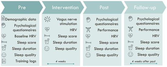
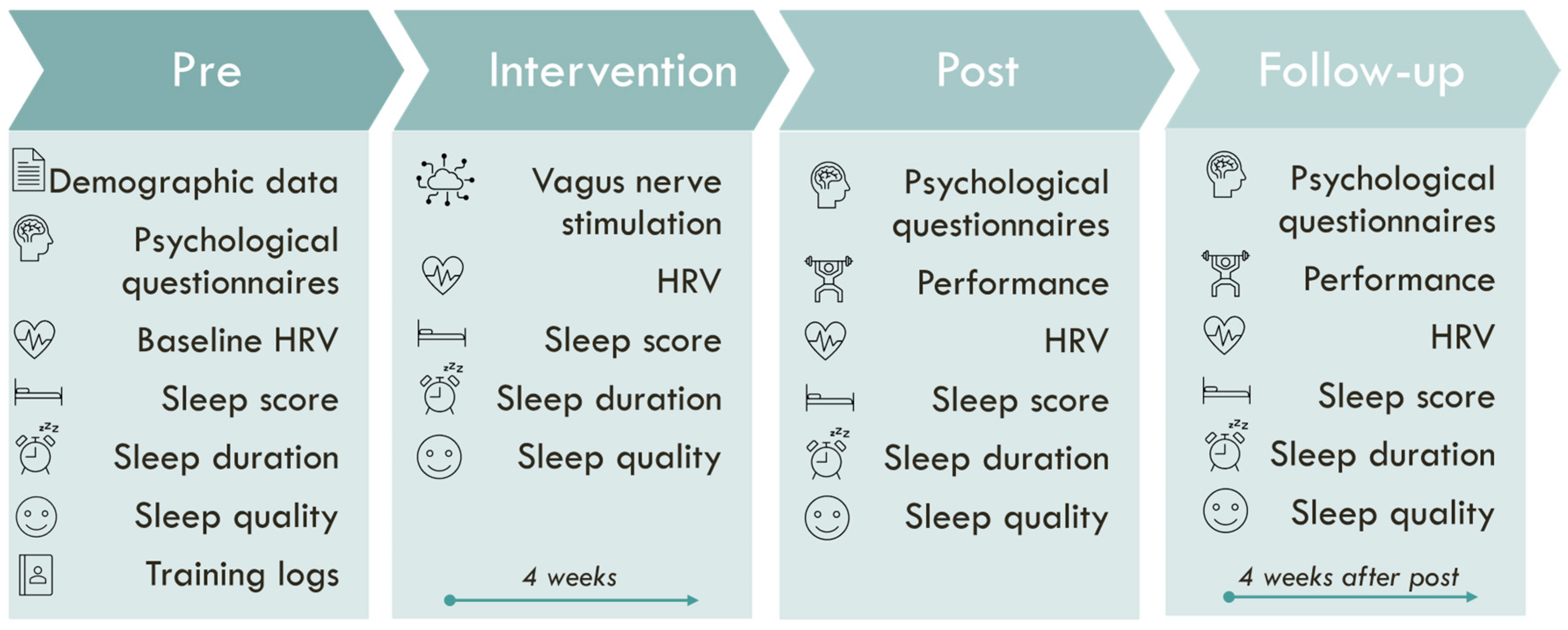
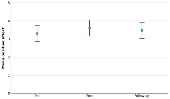
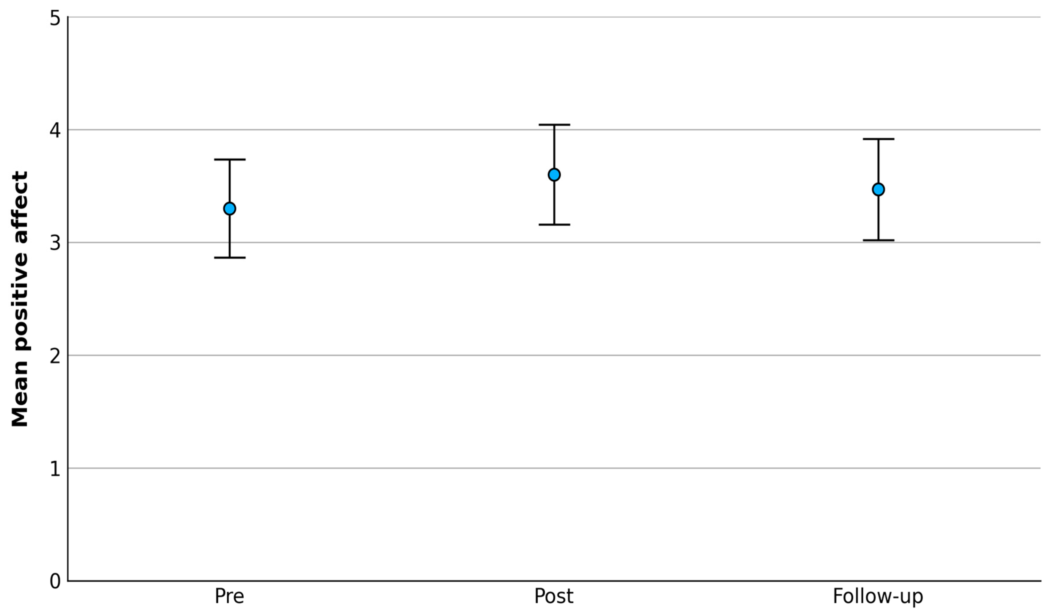
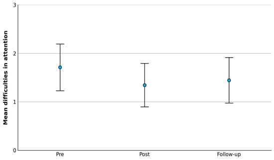
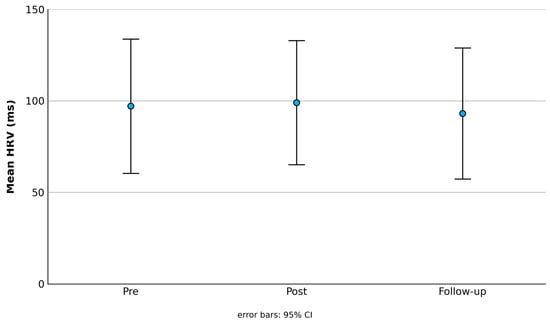
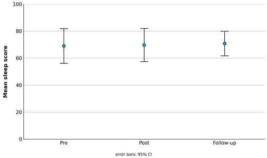
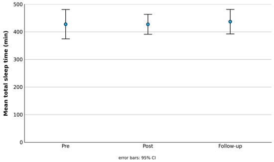
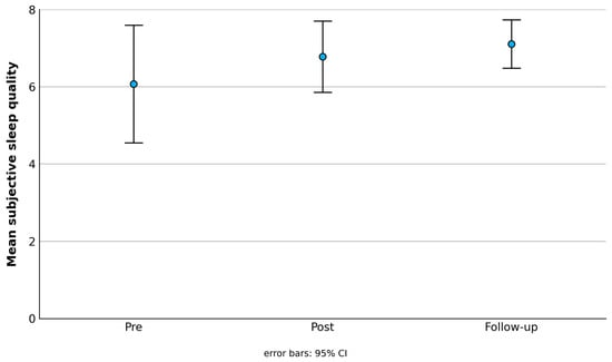
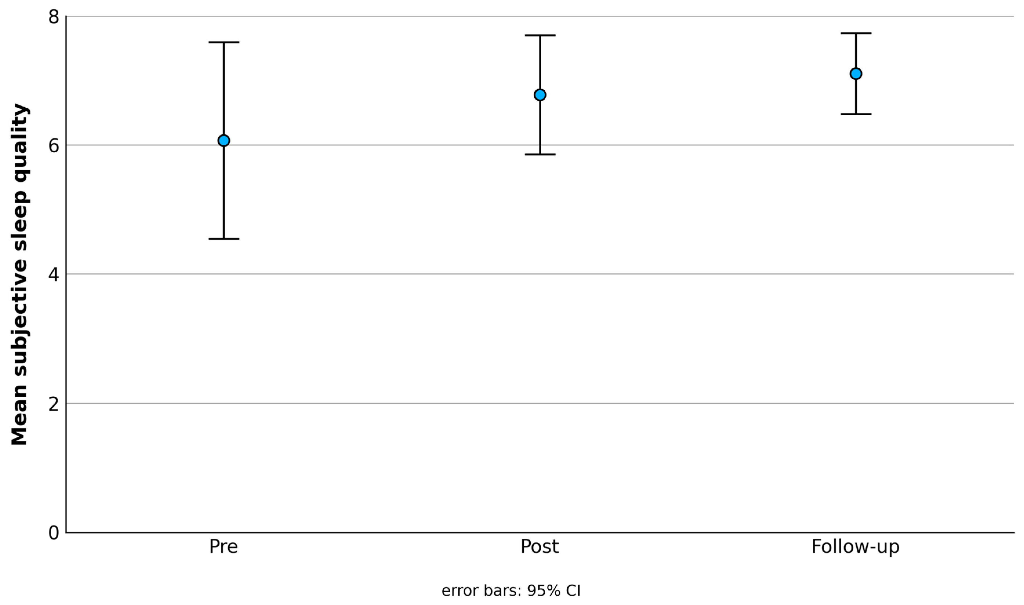
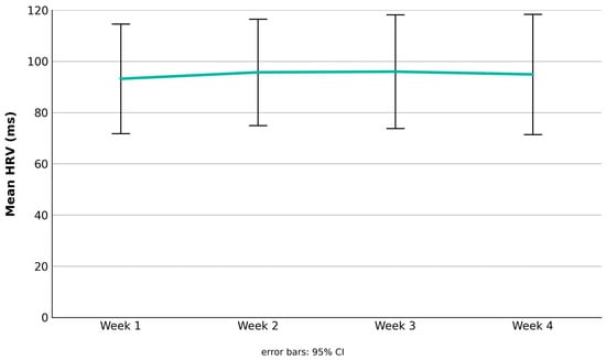
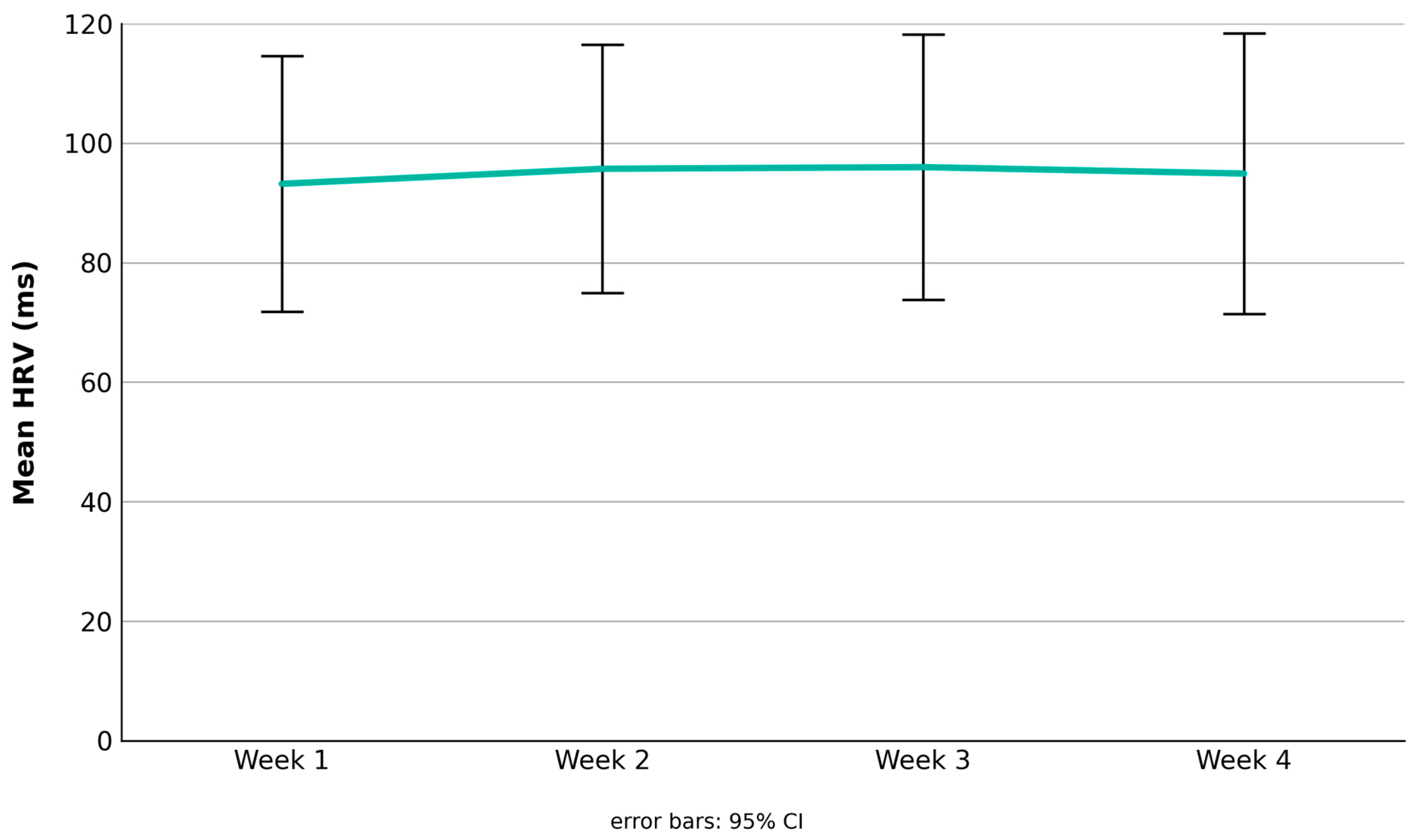
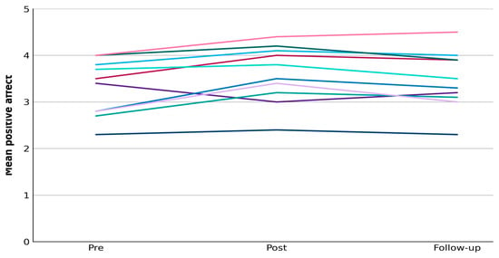
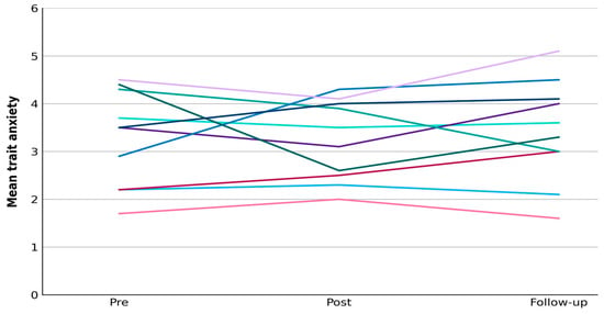
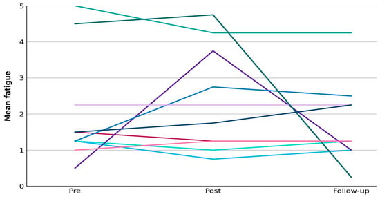
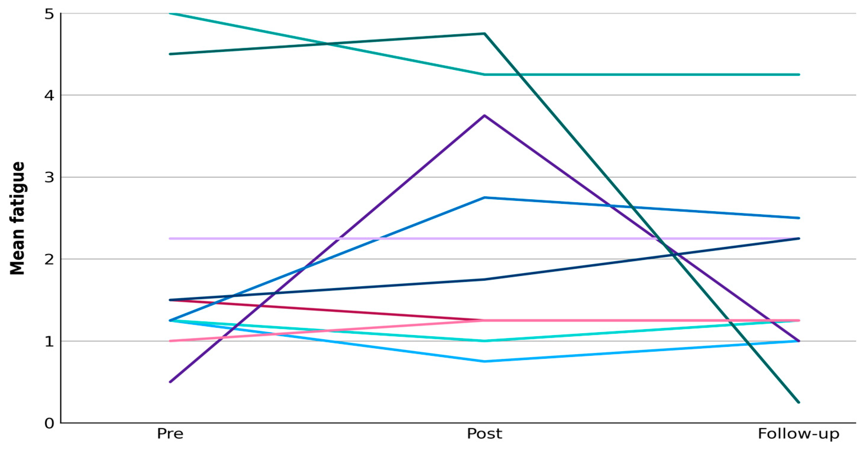
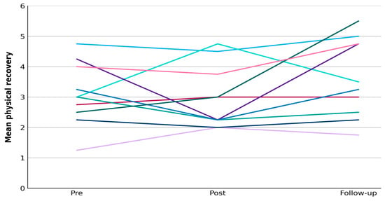
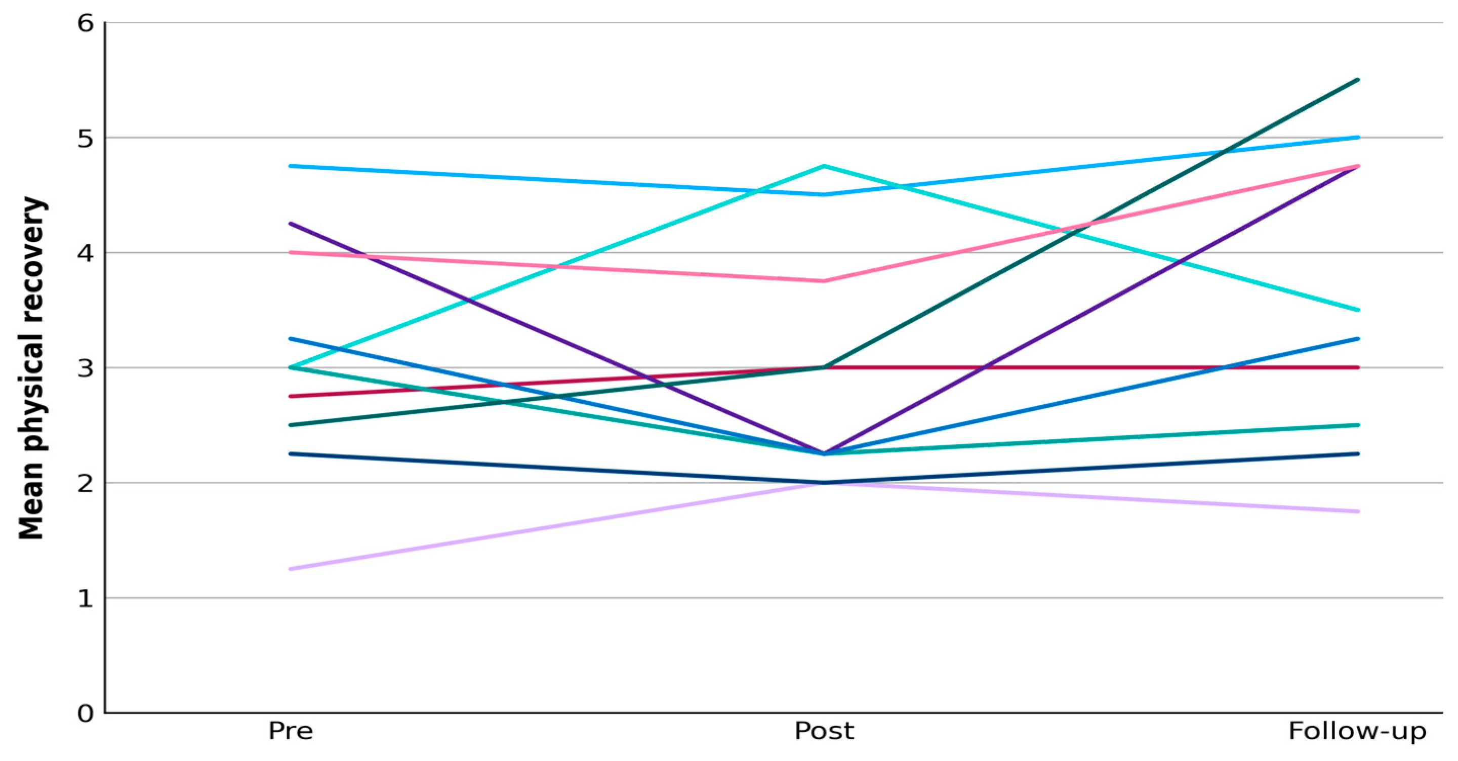
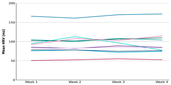
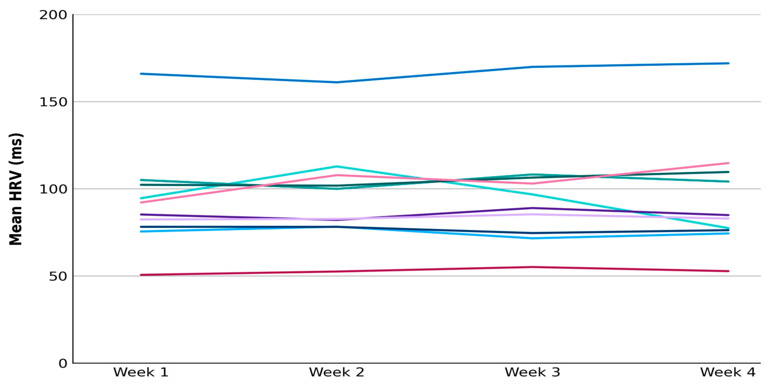
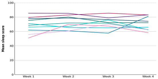
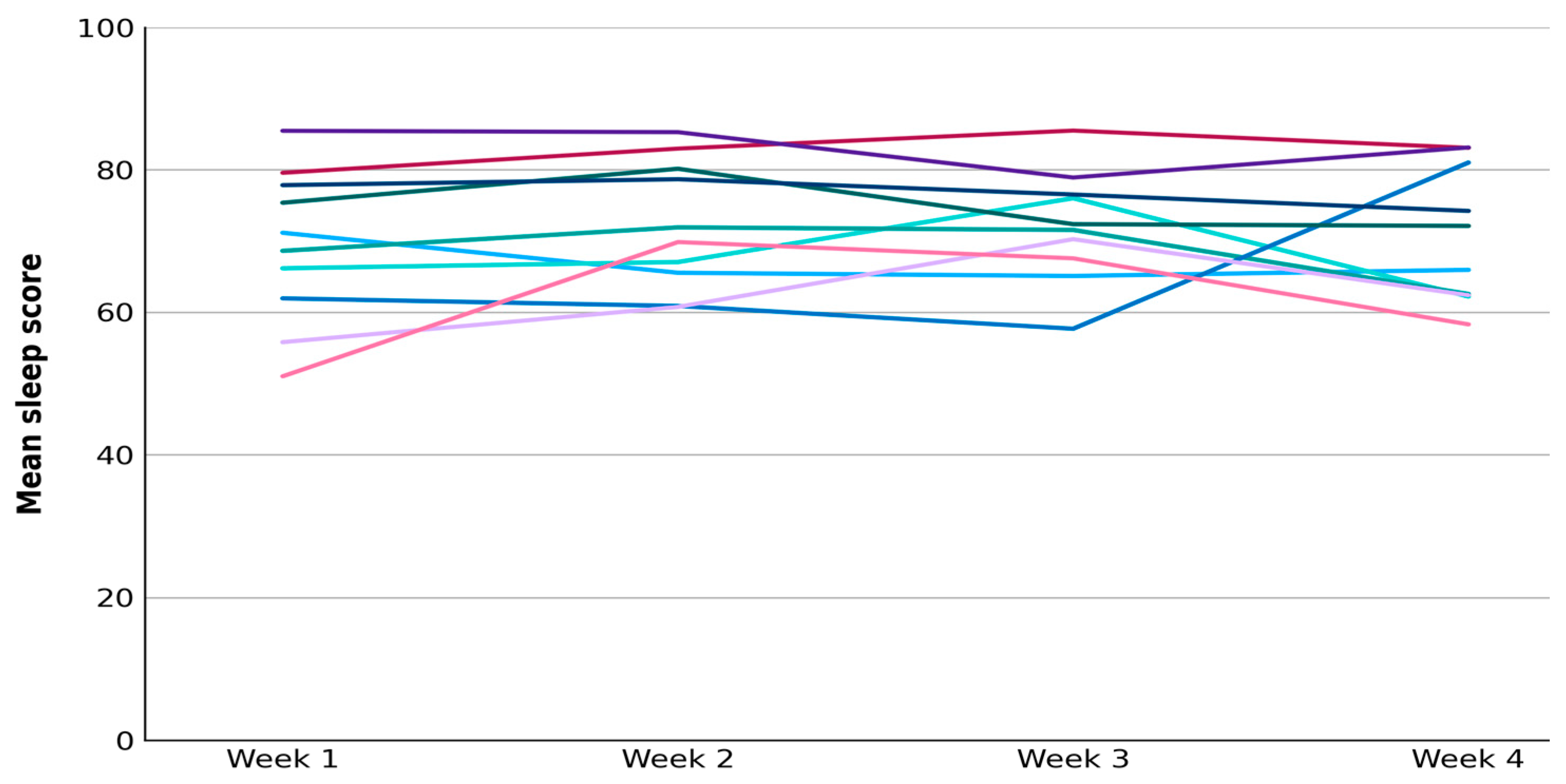
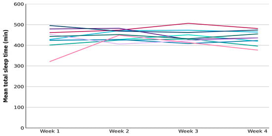
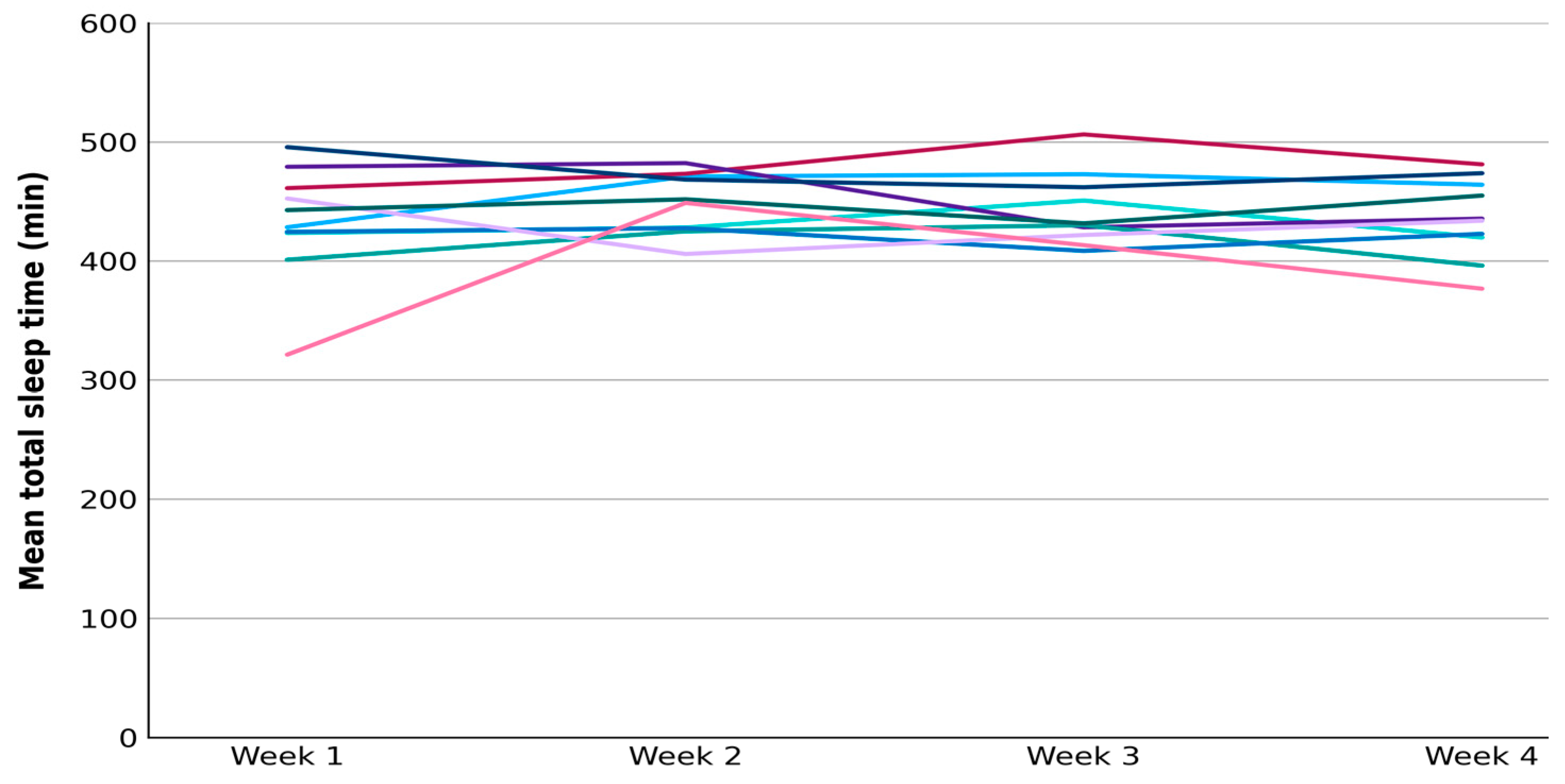
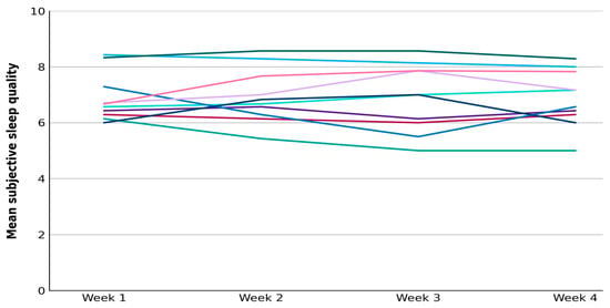
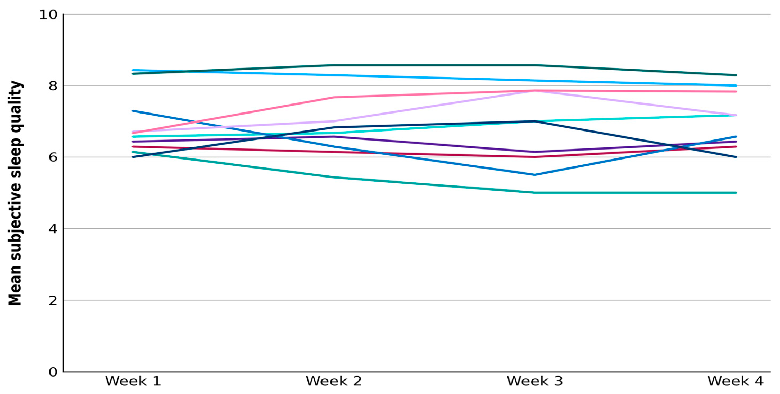

Disclaimer/Publisher’s Note: The statements, opinions and data contained in all publications are solely those of the individual author(s) and contributor(s) and not of MDPI and/or the editor(s). MDPI and/or the editor(s) disclaim responsibility for any injury to people or property resulting from any ideas, methods, instructions or products referred to in the content. |
© 2026 by the authors. Licensee MDPI, Basel, Switzerland. This article is an open access article distributed under the terms and conditions of the Creative Commons Attribution (CC BY) license.