


Abstract
Magnetic resonance imaging (MRI)-based prediction of multiple sclerosis (MS) progression depends on how imaging data are prepared, represented, modelled, and evaluated. This systematic review synthesized artificial intelligence (AI) methods across the MRI-to-prediction pipeline. PubMed, Scopus, Web of Science, and Google Scholar were searched for studies published from 2010 to March 2026. Eligible studies included patients with MS or clinically isolated syndrome, used brain MRI directly or as the source of predictors, evaluated future progression-related outcomes, and reported quantitative predictive performance. Thirty-seven studies were included and synthesized narratively because of methodological and outcome heterogeneity. Prediction targets included CIS-to-MS/CDMS conversion, future MRI disease activity, PIRA/PIRMA, disability worsening or confirmed progression, future EDSS or disability status, and RRMS-to-SPMS conversion. Lesion morphology and location, radiomics, tissue atrophy, longitudinal imaging changes, and multimodal variables provided useful prognostic information. Classical machine learning (ML) remained effective for structured biomarkers, while deep learning (DL) enabled direct image-based modelling; neither approach was uniformly superior. PROBAST + AI assessment rated model Development as High concern in 35 of 37 studies and Unclear in two, Evaluation as High risk of bias in all 37 studies, and Applicability as Low concern in 32, Unclear in two, and High in three. Overall, MRI-based AI shows promising prognostic potential, while stronger data separation, calibration, transparent reporting, and independent validation are needed to support reproducible clinical translation.
1. Introduction
Multiple sclerosis is a chronic immune-mediated disorder of the central nervous system characterized by demyelination and neuroaxonal injury, leading to heterogeneous neurological deficits and long-term disability [1,2]. Prognosis is difficult at the individual level because disease trajectories vary widely across phenotypes and over time, yet early identification of patients at higher risk of progression is important for treatment optimization and monitoring strategies [2]. MS is commonly described across clinical phenotypes including clinically isolated syndrome (CIS), relapsing remitting MS (RRMS), secondary progressive MS (SPMS), and primary progressive MS (PPMS), with RRMS representing most initial presentations and many patients transitioning to SPMS over the disease course [3,4]. Beyond physical disability, MS may also involve cognitive impairment, although the present review focuses primarily on MRI-based prediction of disease conversion, disability progression, relapse-independent progression, and future lesion activity [5].
MRI is central to MS diagnosis and longitudinal monitoring because it sensitively detects focal inflammatory lesions and supports evidence of dissemination in space and time [6]. Conventional sequences such as T2-W and FLAIR identify white matter (WM) hyperintensities that form the radiological foundation of MS assessment, and lesion burden and lesion distribution are associated with future outcomes [6,7]. However, conventional lesion measures correlate only moderately with clinical disability, often described as the clinico-radiological paradox, highlighting that lesion count/volume alone cannot fully explain progression and disability accumulation [8]. Recent MS imaging syntheses emphasize that MRI prognostication increasingly depends on extracting richer quantitative information from lesions and normal-appearing tissue, while also requiring careful standardization to support reliable interpretation across scanners and centers [9].
As MRI datasets become larger and more complex, AI methods are increasingly used to convert raw images into quantitative markers and predictions. Rather than being applied only at the end of analysis, AI can contribute at several stages of the MRI workflow. For example, deep learning has shown that lesion activity, such as enhancing lesions, can be predicted from non-contrast MRI in clinically relevant MS applications [10]. ML methods can operate on structured radiomic descriptors extracted from multi sequence MRI to classify MS lesion activity [11]. These developments motivate a review structure that follows the MRI analysis pipeline itself. Choices made early in the workflow, such as preprocessing, harmonization, segmentation and feature representation, directly influence what downstream predictive models can learn and how well they generalize.
A key practical challenge is that MRI derived biomarkers and AI models are highly sensitive to variability in acquisition and analysis, and multi-center studies face differences in scanners, vendors, protocols and longitudinal scanner changes that can introduce non biological variability and lead to inconsistent biomarker estimates and spurious associations with clinical outcomes if not addressed [12]. Consensus efforts in the MS imaging community therefore emphasize that harmonized acquisition and standardized post processing are essential prerequisites for using quantitative MRI biomarkers, and any AI models built on them, as clinically credible tools [12]. At the quantitative analysis stage, DL-based automated MS lesion segmentation offers a scalable alternative to manual delineation; however, segmentation accuracy and robustness to domain shift remain critical factors governing the reliability of derived quantitative imaging biomarkers [13]. Beyond lesion volume, richer feature representations such as lesion geometry, spatial distribution, and radiomics/texture features are designed to capture subtle, disease relevant microstructural patterns that are not discernible through routine MRI reading, and recent multicenter evidence indicates that FLAIR-based radiomics can contribute to predicting disability worsening in MS, particularly when variability in image quality and spatial resolution is carefully addressed [14]. Together, these findings support the idea that the type of MRI-derived representation (e.g., voxel-level images, masks, engineered radiomics, longitudinal change maps) strongly influences the modelling strategy that is feasible and the prediction task that can be addressed.
Several reviews have already summarized the application of ML and DL methods in MS. These include reviews that organize studies according to broad clinical application domains, such as diagnosis, disease progression, and treatment response [15], reviews that categorize MRI-based prognostic modelling according to specific outcome measures, including conversion from CIS, Expanded Disability Status Scale (EDSS)-related disability, cognitive outcomes, motor impairment, and disease activity [16], and reviews that focus specifically on DL approaches for automated MS lesion segmentation [13].
While these reviews provide valuable overviews of the field, models are typically grouped according to task category or algorithmic approach, which can obscure how individual methodological choices relate to one another across the modelling process. In practice, successful clinical translation depends on the coherence of the entire methodological workflow, encompassing MRI acquisition and standardization, preprocessing and harmonization, biomarker extraction and representation, predictive modelling, and rigorous evaluation and validation. Explicit consideration of this end-to-end process is therefore essential for understanding the strengths, limitations, and translational potential of ML-based approaches in MS.
Accordingly, this review contributes a pipeline-oriented synthesis of AI methods across the MRI-to-prediction workflow for MS progression. We structure the literature according to the functional role of AI within the workflow: (i) MRI data preparation and preprocessing (including harmonization and quality control), (ii) imaging biomarker extraction and representation (lesion/tissue quantification, spatial and morphological characterization, radiomics, representation learning), and (iii) modelling strategies that map these MRI-derived representations to clinically meaningful progression endpoints.
This review examines progression-related prediction in four clinically relevant settings: conversion from CIS to MS or clinically definite MS (CDMS), transition from RRMS to SPMS, relapse-independent disability progression including PIRA-related outcomes, and future inflammatory activity indicated by new or enlarging lesions on follow-up MRI. These settings represent different stages and manifestations of disease evolution. They have important implications for prognosis, treatment planning, and long-term monitoring. Prediction of CIS conversion focuses on identifying individuals who are likely to develop clinically established MS at an early stage. Disability progression and PIRA-related outcomes reflect the accumulation of neurological impairment over time. New or enlarging lesions serve as MRI markers of ongoing inflammatory activity. The transition from RRMS to SPMS was also considered because it represents a clinically important milestone in disease progression. However, direct AI-based prediction of this transition was rarely investigated in the eligible studies, highlighting an important gap in the current literature. By comparing AI approaches across these progression settings, this review evaluates the potential of MRI-derived information to support risk stratification and to guide more individualized monitoring and management strategies. By linking MRI-derived feature representations with modelling strategies and prediction tasks, this pipeline-based framework helps identify which model types are commonly used for specific outcomes, where limitations in generalizability arise, and which stages of the workflow may constrain clinical translation.
2. Materials and Methods
2.1. Reporting Guideline and Literature Search
This systematic review was conducted and reported in accordance with the Preferred Reporting Items for Systematic Reviews and Meta-Analyses (PRISMA) 2020 statement [17]. The review was not prospectively registered, and no separate review protocol was prepared.
A structured literature search was conducted in PubMed, Scopus, Web of Science, and Google Scholar to identify studies applying AI, ML, or DL to brain MRI for the prediction of MS progression-related outcomes. The search covered publications from 2010 to March 2026. Search terms represented four main concepts: (i) MS and CIS; (ii) MRI and related imaging modalities; (iii) AI, ML, DL, neural networks, and radiomics; and (iv) progression-related outcomes, including conversion, disability progression, EDSS, progression independent of relapse activity (PIRA), disease activity, and new or enlarging lesions.
Search syntax was adapted to the requirements of each database. The PubMed strategy used Medical Subject Headings (MeSH) and title/abstract terms, while Scopus and Web of Science used database-specific field searching and Boolean operators. Google Scholar was used as an additional search source using the terms MS progression prediction + AI + DL + ML methods. The complete search strings, search limits, dates of searching, and numbers of records retrieved from each source are provided in Supplementary Table S1.
Previously published reviews on AI, MRI, and MS progression were consulted for background and contextual discussion only and were not used as an additional source for study identification or inclusion. All studies considered for inclusion were identified through the four documented database searches (PubMed, Scopus, Web of Science, and Google Scholar), and no records were added through citation searching, websites, organizations, or other review-based searching.
2.2. Eligibility Criteria
Studies were eligible if they: (i) included human participants with MS or CIS; (ii) performed patient-level prediction of a future MS-related outcome; (iii) used brain MRI directly as model input or as the source of predictors in an evaluated prediction model; (iv) applied AI, ML, or DL either for outcome prediction or for generating MRI-derived predictors used in an evaluated multivariable prediction model; (v) used MRI alone or combined with clinical, demographic, treatment, cerebrospinal fluid, electrophysiological, laboratory, or other variables; (vi) ensured the predictor-assessment period preceded the outcome-prediction period; and (vii) reported quantitative predictive performance measures, such as AUC/AUROC, C-index, accuracy, sensitivity, specificity, precision, F1 score, Brier score, MAE, or RMSE.
Prediction was not limited to baseline data. Longitudinal predictors were eligible if all predictor information was available before the start of the outcome-prediction period. Studies were excluded if predictor information was obtained during or after outcome assessment, or if the same follow-up MRI was used both as a model input and to define the outcome at that time.
Eligible outcomes included conversion from CIS to MS or CDMS; future physical disability status, worsening, or confirmed progression; PIRA, progression independent of relapse and MRI activity (PIRMA), or related relapse-independent progression outcomes; future patient-level radiological activity, including occurrence, count, or burden of new or enlarging T2 lesions or gadolinium-enhancing lesions; and transition from RRMS to SPMS.
Formal diagnostic or disability composite outcomes were eligible if they represented a prespecified progression endpoint or if all components fell within eligible outcome categories. Generic disease-activity composites combining heterogeneous clinical and radiological events were excluded when an ineligible component could independently determine the outcome and eligible components were not separately extractable. Relapse alone, cognitive decline alone, and treatment response alone were ineligible unless a separately extractable eligible progression-related outcome was also reported.
Studies limited to cross-sectional diagnosis, disease-subtype classification, current-status classification, association analysis without prediction, or lesion detection/segmentation without future patient-level prediction were excluded. Voxel-, lesion-, or region-level prediction without a patient-level outcome was also excluded. MRI used only for participant selection, outcome definition, preprocessing, segmentation, or descriptive analysis was insufficient unless the resulting MRI-derived information was included in an eligible future-outcome prediction model.
Eligible publications were full-length original research articles published in English as peer-reviewed journal papers or full conference/workshop proceedings papers between January 2010 and March 2026. Reviews, meta-analyses, editorials, commentaries, protocols, letters lacking complete original-study information, case reports or case series, animal studies, non-original book chapters, and conference abstracts or posters without a full report were excluded. Full original research papers published as chapters in peer-reviewed conference proceedings volumes were eligible.
2.3. Study Selection
All retrieved records were imported into Rayyan, which was used for duplicate management and screening. A total of 1532 records were identified: 326 from PubMed, 665 from Scopus, 520 from Web of Science, and 21 from Google Scholar. After removal of 662 duplicate records, 870 records remained for title and abstract screening.
Title/abstract screening and full-text eligibility assessment were initially performed by the first author (U.V.) using the predefined eligibility criteria. No automated tool was used to make study-eligibility decisions. The final set of included studies and the full-text exclusion decisions were independently verified by a second author (A.W.). Any uncertainties identified during verification were discussed and resolved by consensus.
Following title and abstract screening, 805 records were excluded, and 65 reports were sought for full-text retrieval. Two reports could not be retrieved, leaving 63 reports for full-text eligibility assessment. Twenty-six reports were excluded after full-text assessment, resulting in 37 studies included in the systematic review. The study-selection process and full-text exclusion categories are presented in the PRISMA 2020 flow diagram Figure 1.
Figure 1.
PRISMA 2020 flow diagram of database searching and study selection.
All studies included in the final review were represented in the documented database-search records and corresponding PRISMA flow diagram; no included studies were introduced from a separate or parallel review.
2.4. Data Extraction
Data were extracted using a predefined structured framework. For each eligible study, the following information was recorded: author and reference; cohort, study design, follow-up and DMT exposure; prediction outcome, definition and horizon; input data; MRI preprocessing and segmentation; feature extraction and quantification; data processing and validation; model or architecture; performance metrics; and key MRI predictors/contribution. MRI variables were distinguished as quantitative or image-derived features and categorical radiological variables, and whether MRI variables were retained in the final prediction model was recorded where reported. Information was extracted as reported in the original publications. Where information was unavailable, it was recorded as “not reported” or “unclear” rather than inferred. Initial data extraction was performed by the first author (U.V.). The principal data extracted for the final 37 included studies were independently verified by a second author (A.W.).
2.5. Methodological Quality, Risk of Bias, and Applicability Assessment
Methodological quality, risk of bias, and applicability were assessed using PROBAST + AI and its accompanying Explanation and Elaboration guidance [18]. For each study, relevant information on participants, predictors, outcome, sample size, missing data, class imbalance, feature selection, model development, validation, and performance reporting were extracted from the article and available Supplementary Material and assessed against the PROBAST + AI signaling questions.
PROBAST + AI assesses model Development quality (Dev) and model Evaluation risk of bias (Eval) separately across four domains: participants and data sources (D1), predictors (D2), outcome (D3), and analysis (D4). Development focuses on how appropriately the model was constructed, including sample size, predictor handling, missing data, class imbalance, and overfitting control. Evaluation focuses on how reliably performance was assessed, including evaluation sample size, data leakage, resampling procedures, and performance assessment. Applicability (App) was assessed separately for D1-D3 according to the match between the study and the review question.
Signaling questions were rated as Yes, Probably Yes, Probably No, No, No Information, or Not Applicable, and overall judgements as Low, Unclear, or High. Unreported information was not inferred. Materially different models, outcomes, prediction horizons, redevelopment procedures, or validation stages were assessed separately where required. Detailed PROBAST + AI assessments were conducted for the included studies and distinct model or evaluation stages, together with a study-level summary of overall judgements by MS progression-prediction outcome category.
2.6. Data Synthesis
Given the heterogeneity in MS phenotype, cohort characteristics, progression outcomes, MRI inputs, prediction horizons, feature representations, modelling approaches, and validation strategies, the findings were synthesized narratively and in structured tables rather than by meta-analysis. Studies were compared across the MRI analysis pipeline, including image preparation and preprocessing, biomarker extraction and representation, predictive modelling, and evaluation, with emphasis on the relationships among MRI-derived information, model design, progression outcome, and validation strategy. For consistency, AUC/AUROC values reported as percentages were converted to the 0–1 scale.
3. AI Methods for MRI Data Preparation and Preprocessing
MRI-based AI studies in MS increasingly rely on multicenter and longitudinal datasets, but scanner- and site-related differences introduce substantial non-biological variability. Variations in vendor, field strength, pulse sequence, software version, and acquisition protocol can reduce model robustness and obscure true disease-related signals when data are pooled without harmonization. For this reason, harmonization and quality-controlled preprocessing are now recognized as essential prerequisites for reliable quantitative MRI biomarkers in multicenter MS research [12].
3.1. Image Denoising and Artifact Correction
Brain MRI images are commonly affected by noise and motion artifacts that can reduce the quality of downstream analyses [19]. In MS MRI, typical artifacts include motion artifacts [20,21], susceptibility distortions [22], gradient nonlinearity [23], and radio-frequency inhomogeneities [24]. Each of these artifacts can significantly impact downstream analyses [19]. DL methods have shown strong performance in addressing both noise and motion artifacts, often outperforming classical filtering approaches by learning to separate signal from noise in a brain anatomy-aware manner [25].
For denoising, Kidoh et al. tested a DL-based reconstruction (dDLR) approach for brain MRI and reported statistically higher PSNR and SSIM than comparator convolutional neural networks (CNN) denoisers in experiments, along with improved qualitative assessments; in a volunteer study across multiple sequences (including FLAIR and MPRAGE), dDLR-processed lower-averaging acquisitions (NAQ2) achieved significantly better SSIM/PSNR than unprocessed NAQ2 and was qualitatively comparable to or better than higher-averaging acquisitions (NAQ5) for most criteria. This supports the idea that AI denoising can be used upstream to improve the quality of inputs that feed segmentation and feature extraction [26].
For motion artifacts, Pawar et al. evaluated a DL motion-correction method on clinical T1-W MPRAGE images identified as motion degraded, reporting reductions in ghosting/ringing/blurring and significant improvements in visual grading scores across multiple neuroanatomical regions; they also reported that a portion of repeated scans could be avoided, indicating operational relevance for clinical-scale datasets [27]. The proposed two stage multi loss MC Net combines novel hybrid L1 loss in the first stage to suppress global artifacts with a Total Variation (TV) term in the second stage to refine boundaries, delivering higher peak signal to noise ratio (PSNR)/structural similarity index measure (SSIM) than single stage L1 or L1 + TV models while preserving image context [28]. Finally, a multiscale fully convolutional network estimates motion artifacts from large amounts of simulated motion corrupted data, subtracts the artifact only estimate from the input, and produces corrected images with significantly reduced artifacts [29].
For ringing artifacts, Zhang et al. developed a CNN approach to reduce Gibbs-ringing artifacts; they reported that their method removed ringing without noticeable smoothing and improved quantitative similarity to fully sampled references (e.g., root-mean-square error (RMSE)/PSNR/SSIM) relative to existing methods, illustrating how AI can correct a concrete reconstruction-related artifact before downstream analysis [30].
3.2. Bias Field Correction and Intensity Normalization
Bias field (intensity inhomogeneity) and inconsistent intensity scaling are long-recognized challenges in MRI because raw MR intensities are not standardized physical units, and they can vary across acquisitions even for the same tissue class [31]. These issues matter directly for ML/DL because many segmentation and feature extraction methods depend on intensity distributions and tissue contrast.
For bias field correction, the widely used N4ITK method is an extension/improvement of the classical normalization (N3) algorithm and introduces algorithmic modifications (including B-spline fitting/optimization changes) to improve bias field correction performance. N4 remains a common upstream step in contemporary MRI pipelines (including neuroimaging and MS contexts) because it reduces non-biological intensity nonuniformity that otherwise confounds tissue/lesion separation [32].
For intensity normalization, Shinohara et al. show that conventional MRI is measured in arbitrary units that are not comparable across subjects or visits, introduce formal “statistical principles of image normalization” (SPIN), and propose tissue-based “white stripe” methods that match the mean and variance of normal appearing white matter (NAWM) across scans. Across thousands of ADNI, AIBL, and MS scans, principled normalization greatly reduces between scan tissue intensity variability and prevents spurious grey matter (GM) loss from naive histogram matching, making robust normalization essential in large multi-site studies [31].
Critically for MS-specific longitudinal workflows, Roy et al. propose a 4D longitudinal intensity normalization method “in the presence of MS lesions,” explicitly aiming to yield “stable and consistent longitudinal segmentations” and positioning the method as a preprocessing step usable with any segmentation method; they model intensity variability linked to lesion dynamics while keeping lesion information unaffected. This paper provides direct justification for putting intensity normalization explicitly in the preprocessing stage of MS MRI pipelines rather than treating it as an incidental detail [33].
Finally, more general tissue-based standardization approaches, as highlighted by De Nunzio et al., emphasize that the lack of fixed tissue-specific intensity affects post-processing tasks such as automatic segmentation and classification, reinforcing the automatic link between normalization choices and downstream ML/DL stability [34].
3.3. MRI Harmonization Across Scanners and Sites
A central preprocessing challenge in MS is harmonizing MRI acquisition, analysis, and storage across sites. A harmonization-focused MS review from the MAGNIMS Study Group explicitly motivates harmonization because cooperative, large-scale datasets are needed to develop diagnostic and prognostic biomarkers, yet differences in acquisition parameters, image analysis, and data storage across sites can introduce bias and represent a substantial constraint; the review surveys state-of-the-art harmonization developments and provides recommendations [12].
This problem is not theoretical: harmonization studies show that scanner effects can be large enough to hinder inference and even generate spurious findings. ComBat-based methods model additive and multiplicative scanner effects and, in both cross-sectional and longitudinal extensions, have been shown to remove unwanted site variability, increase power to detect biological effects, and improve type I error control compared with unharmonized data or simple scanner covariate adjustment [35,36]. In line with this, recent MS progression work has extracted radiomics features from WML and NAWM using PyRadiomics, harmonized them with Longitudinal ComBat, and then applied recursive feature elimination before training ML models to predict EDSS worsening from multi-center FLAIR MRI, further illustrating that harmonization of derived MRI biomarkers is becoming standard practice prior to modelling in longitudinal multi scanner MS studies [14].
Deep learning can also be used for image-level or contrast-level harmonization. Dewey et al. propose DeepHarmony, a U-Net-based approach trained with a small overlap cohort scanned with two protocols; they report improved consistency of volume quantification across scanning protocols and show in an MS longitudinal dataset that atrophy calculations were substantially affected by protocol change, with those differences substantially reduced after DeepHarmony-based harmonization. This directly supports the pipeline claim that harmonization affects the reliability of derived quantitative biomarkers (atrophy/volumetrics) in MS [37].
3.4. Image Registration and Spatial Alignment
Registration and spatial alignment are essential in MS MRI pipelines because (i) multi-sequence analysis requires co-registration across contrasts, and (ii) longitudinal progression endpoints (new/enlarging lesions, atrophy trajectories, change maps) rely on consistent within-subject spatial correspondence across time [38].
Classical registration frameworks such as ANTs model spatial alignment as an optimization over a transformation (rigid, affine, elastic, or diffeomorphic) and a similarity metric (e.g., mean squared difference, cross correlation, mutual information), often within a multi resolution scheme. Symmetric Normalization (SyN) in ANTs estimates diffeomorphic mappings between images by integrating a regularized velocity field in a symmetric fashion, and underpins groupwise template building (SyGN) and downstream tasks such as atlas-based labelling and cortical thickness analysis [39]. In many recent MS DL prognostic studies, however, registration is implemented using conventional affine normalization to standard templates (e.g., FLIRT to MNI 152) primarily to ensure comparable anatomical locations and voxel wise correspondence across patients and scanners, rather than as a methodological focus in itself [40].
Balakrishnan et al. introduce VoxelMorph, a CNN-based framework for deformable registration, reporting that the unsupervised model’s accuracy is comparable to state-of-the-art methods while operating orders of magnitude faster, and that auxiliary segmentation information can further improve registration performance [41]. This complements the more traditional affine normalization used in MS progression-prediction pipelines, where PD/T2, T1 and lesion masks are skull-stripped, intensity-normalized and then affinely registered to a common or MNI152 space before being passed to CNNs for disease-worsening or conversion prediction [42].
An MS-specific longitudinal lesion-change pipeline illustrates the role of alignment and related preprocessing. In a fully automatic method for segmenting changes in FLAIR-hyperintense WML on serial MS MRI, the authors describe preprocessing steps including intrasubject registration and bias field correction/intensity scaling prior to voxel-level analysis of intensity changes in lesions and NAWM-based statistical testing. This provides direct evidence that longitudinal MS endpoints require explicit alignment as a preprocessing stage [38].
3.5. AI-Assisted Quality Control
Quality control (QC) is an important preliminary step in MRI analysis because low-quality scans can bias subsequent processing and analysis. Esteban et al. addressed this problem with MRI Quality Control tool (MRIQC), which extracts image quality metrics, generates visual reports, and applies a binary accept/exclude classifier to support more objective and scalable QC. However, although MRIQC achieved about 76% accuracy on unseen-site data, its performance remained affected by site effects, showing that generalization across acquisition centers is still challenging [43]. Samani et al. extended this direction in diffusion MRI through QC-Automator, a CNN-based transfer-learning framework that detects specific artifacts such as motion, multiband interleaving, ghosting, susceptibility, herringbone, and chemical shift [44]. Together, these studies show that AI-assisted QC can reduce reliance on manual inspection, but robust cross-site and cross-protocol performance remains a key requirement for reliable downstream neuroimaging analysis.
3.6. Domain Adaptation and Harmonization Networks for Cross-Site Variability
Cross-site variability is a major challenge in MS lesion segmentation because differences across scanners, sites, and imaging protocols can reduce model generalization to new domains. Ackaouy et al. addressed this through Seg-JDOT, an unsupervised domain adaptation framework based on optimal transport that aligns source and target representations without requiring manual target-domain annotations. They also showed that domain shift persists even after image standardization, suggesting that preprocessing alone cannot fully remove cross-site bias [45]. Consistent with this, Zeng et al. noted that current MS lesion adaptation methods mainly rely on feature adaptation and suggested that future work should combine both image-level and feature-level adaptation [13]. Therefore, domain adaptation and harmonization-oriented networks are likely to be important for building MS imaging models that remain reliable across centers.
4. AI Methods for Imaging Biomarker Extraction
Imaging biomarker extraction transforms pre-processed MRI into quantitative features for prediction. This includes automated lesion/tissue segmentation to compute lesion volumes and brain atrophy; characterization of lesion morphology and spatial distribution; high-dimensional radiomic/texture descriptors; and temporal change maps from longitudinal scans. Classical pipelines (e.g., FreeSurfer/SPM tissue segmentation, LST/BIANCA lesion segmentation) and DL models (U-Net, nnU-Net) have been used. Each method has limitations (e.g., segmentation errors for small/unclear lesions, variability across scanners). Radiomics features capture sub-resolution patterns but require standardized inputs. Learned representations (deep features) promise richer biomarkers but also demand large data or pretraining. In all cases, consistent extraction under domain shift is a key challenge.
4.1. Lesion Detection and Segmentation
Automated lesion segmentation tools differ in both their input requirements and their underlying modelling strategies, and several automatic MRI-based methods have been developed to address these challenges. A comparison of commonly used segmentation methods, their required inputs, preprocessing steps, and extracted outputs is provided in Table 1. MSmetrix is a fully automated unsupervised method that uses 3D T1-W and FLAIR MRI in a probabilistic framework to detect WM lesions as an outlier to normal brain while simultaneously segmenting GM, WM, and CSF, and it does not require training data [46]. Lesion-TOADS is a fully automatic atlas-based method that uses multichannel MRI together with topological and statistical atlases to delineate MS lesions while segmenting the brain into its major structures [47]. BIANCA is a fully automated supervised method based on the k-nearest neighbor (k-NN) algorithm; it is described as multimodal and allows incorporation of spatial information and local spatial intensity averaging [48]. BIANCA-MS was developed from the original BIANCA specifically for MS lesion segmentation and introduced two key modifications: a harmonized setting tested across different MRI protocols and a cleaning step intended to improve consistency between automated and manual segmentations [49]. The authors further reported that BIANCA-MS outperformed other available tools and showed comparable performance across different scanning protocols, modality sets, and image resolutions, supporting its use under heterogeneous imaging conditions [49].
The Lesion Segmentation Tool (LST) for SPM provides two lesion-segmentation algorithms: the lesion growth algorithm (LGA), which requires T1 plus FLAIR, and the lesion prediction algorithm (LPA), which requires FLAIR only; the toolbox also includes a longitudinal pipeline [50]. More recent DL methods include LST-AI, which is presented as an advanced DL-based extension of LST built as an ensemble of three 3D U-Nets [51]. Another CNN-based method is nicMSlesions, which is based on a cascade of two 3D patch-wise CNN, where the first network is designed to be more sensitive to candidate lesion voxels and the second to reduce false positives from the first stage [52]. Automated MS lesion-segmentation methods therefore encompass probabilistic, atlas-based, k-NN-based, and CNN-based approaches. However, robustness across heterogeneous imaging protocols remains an important requirement for broader clinical application.
4.2. Tissue Segmentation and Structural Quantification
Structural quantification in progression-focused MS studies has been performed using automated morphometric tools; for example, regional cortical and deep GM atrophy in CIS was assessed using FreeSurfer to derive progression-relevant tissue measures [53]. Beyond lesion masks, tissue segmentation and structural quantification provide volumetric markers of diffuse neurodegeneration that complement focal lesion-based assessment in MS progression research [54]. In CIS, selective GM atrophy has shown prognostic value, with atrophy of the superior frontal gyrus, thalamus, and cerebellum identified as independent predictors of conversion to MS [53]. Early longitudinal CIS work further showed that global brain volume loss and GM volume loss occur within the first year, and that brain volume loss predicted conversion to MS [55]. In RRMS, tissue-compartment measures also retain prognostic relevance, as the GM:NAWM ratio at diagnosis has been reported to predict both disability progression and conversion to SPMS [56]. At a regional level, deep GM degeneration, particularly thalamic atrophy, appears especially informative, with multicenter evidence showing that deep GM volume loss drives disability worsening, while both baseline thalamic atrophy and more recent AI-measured thalamic atrophy have been associated with later disability progression [57,58,59]. Together, tissue segmentation and structural quantification enable objective assessment of global and regional neurodegeneration, particularly GM and deep-GM damage, and provide complementary information beyond conventional lesion-based measures.
4.3. Spatial and Morphological Lesion Characterization
Several studies have shown that automated structural characterization of lesions provides prognostic information beyond lesion count alone. In CIS, shape-based features extracted from automated lesion masks were used to quantify lesion morphology, and important predictors included mean lesion volume, minimum sphericity, and minimum surface-volume-ratio, with converters showing larger and less round lesions than non-converters [60]. Beyond simple geometric descriptors, lesion geometry has also been incorporated into Support Vector Machine (SVM)-based prediction models for conversion from CIS to CDMS, supporting the value of structural lesion characterization in early prognostic modelling [61]. Spatial lesion distribution has also shown predictive relevance. In a recent explainable ML study, periventricular and infratentorial lesion locations were identified among the significant imaging variables associated with CIS-to-CDMS conversion [62]. These findings indicate that lesion geometry and spatial distribution provide prognostic information beyond conventional measures of lesion burden alone. Representative FLAIR MRI and lesion-mask visualizations illustrating lesion segmentation and spatial characterization are shown in Figure 2. The examples demonstrate how visible white-matter lesions can be represented as segmentation masks and further characterized according to their anatomical distribution, including periventricular, cortex-contacting, and infratentorial locations.
Figure 2.
Representative FLAIR MRI and lesion-mask visualizations illustrating MS lesion segmentation and spatial characterization. (A) Healthy/control FLAIR MRI; (B) MS FLAIR MRI showing visible white-matter lesions; (C) green indicates the white-matter lesion segmentation mask; (D) magenta indicates the periventricular (ventricle-contacting) lesion mask; (E) blue indicates the cortex-contacting lesion mask; (F) light pink indicates the infratentorial lesion mask; and (G) shows the combined spatial lesion-distribution map. These examples illustrate how conventional MRI appearance can be converted into lesion masks and spatially characterized lesion representations for subsequent quantitative imaging analysis and progression modelling. The representative MRI data were obtained from the access-controlled “MRI dataset for susceptibility-based radiomic feature extraction in healthy controls and patients with multiple sclerosis” hosted on Zenodo [63]. The lesion-mask visualizations were generated by the authors from the MRI data.
4.4. Radiomics and Feature Engineering
Radiomics and feature engineering aim to extract large numbers of quantitative features from medical images to characterize image phenotypes beyond visual inspection. In the radiomics workflow described by Lambin et al., the process begins with high-quality standardized imaging, followed by definition of the region of interest through automated segmentation or expert delineation, after which quantitative imaging features are extracted from the defined region. These features include descriptors of shape and texture [64]. In ML-based MS progression prediction, radiomics and feature engineering transform MRI data into explicit handcrafted predictors, whereas DL approaches derive feature representations implicitly within the network architecture.
The extracted features are generally grouped into several major classes. First-order features describe the distribution of voxel intensities within the segmented region. Shape features quantify geometric properties such as size, compactness, elongation, and surface characteristics, and are derived from the segmentation mask rather than from grey-level values. Texture features describe spatial relationships and heterogeneity patterns using matrices such as the grey-level co-occurrence matrix (GLCM), grey-level run length matrix (GLRLM), grey-level size zone matrix (GLSZM), neighboring grey tone difference matrix (NGTDM), and grey-level dependence matrix (GLDM) [65].
Radiomics studies often generate very large numbers of features, making feature engineering necessary to improve robustness and reduce redundancy. This process commonly includes reproducibility filtering, correlation reduction, and selection of the most informative features before model building. To promote consistency across studies, the Image Biomarker Standardization Initiative (IBSI) established standardized definitions, benchmark values, and reporting guidance for radiomic features and radiomics workflows [66].
Reporting of radiomics standardization was heterogeneous across the included studies. Khan et al. reported the use of PyRadiomics 2.20 and stated that image discretization was aligned with IBSI recommendations [14]. In contrast, other studies described reproducibility or standardization procedures without explicitly reporting IBSI compliance. Peng et al. and Shi et al. assessed feature reproducibility using intraclass correlation coefficients [67,68], while Elhaie et al. reported the PyRadiomics extension in 3D Slicer together with standardized resampling and fixed-bin-width discretization [69]. However, explicit verification of extracted features against IBSI reference values was not consistently reported. This variation limits direct comparison of radiomic features across studies and reinforces the need for more complete reporting of feature-extraction settings and standardization procedures.
Radiomic and engineered features therefore provide quantitative representations of image intensity, morphology, and heterogeneity beyond conventional lesion burden; however, their reproducibility and cross-study comparability depend on standardized extraction and transparent reporting.
4.5. Representation Learning from MRI
In DL approaches, baseline MRI scans are used directly as model inputs, and prognostic imaging biomarkers are computationally derived as latent feature representations within the network instead of being manually specified as handcrafted features. Tousignant et al. used multimodal MRI volumes in an end-to-end 3D CNN framework for future disability progression prediction [70], while Storelli et al. showed that baseline T1-W and T2-W MRI can support prediction of clinical and cognitive worsening without explicit handcrafted feature extraction [40]. Taloni et al. further showed that ResNet50-based analysis of 3D T1-W MRI identified informative frontal GM regions [71], and Coll et al. extended this paradigm to survival modelling of PIRA using EfficientNet-derived features from routine MRI at the first demyelinating event [72]. Accordingly, these approaches derive progression-relevant imaging representations directly from MRI rather than relying on predefined handcrafted biomarkers.
Table 1.
Comparison of automatic MRI lesion segmentation algorithms for multiple sclerosis, including required inputs, preprocessing workflows, and extracted lesion-related outputs.
5. AI Methods for Modelling MRI Features and Predicting Clinical Outcomes
AI methods for MS progression prediction use either explicit MRI-derived biomarkers or MRI directly to model future clinical outcomes. MRI-based ML reviews consistently describe this literature as using imaging variables such as lesion measures and tissue-volume markers, often together with demographic and clinical covariates, for tasks including CIS-to-MS conversion, EDSS-related disability prediction, and other progression outcomes [16,62].
5.1. Classical Machine Learning Approaches
Classical ML is well suited to settings in which MRI information is represented as structured input variables rather than raw images. In this setting, the model operates on predefined biomarkers such as lesion geometry, lesion location, lesion burden, and GM-based measures, usually combined with baseline demographic or clinical data [61]. In CIS-to-CDMS prediction, SVMs and ensemble tree-based models are among the most frequently applied approaches. Bendfeldt et al. [61] used SVMs with lesion geometry metrics derived from Minkowski functionals together with global GM volume information and concluded that both lesion geometry and GM-based information can aid prediction of conversion to clinically definite MS [61].
More recently, Rasouli et al. [62] developed an explainable eXtreme Gradient Boosting (XGBoost) model using demographic, clinical, and imaging variables and identified age, gender, schooling, motor symptoms, infratentorial lesions, periventricular lesions, oligoclonal bands in CSF, and lesion/symptom type as the most influential predictors of CIS-to-CDMS conversion [62]. A separate 2024 comparative CIS study benchmarked CatBoost, XGBoost, LightGBM, random forest, SVM, and logistic regression on the same public CIS cohort, showing that boosted-tree and other feature-based classifiers remain competitive when heterogeneous structured inputs are used [73]. Earlier work by Zhao et al. [74] showed that SVMs incorporating short-term clinical and brain MRI data outperformed logistic regression for predicting MS disease course in that study [74].
Classical ML therefore provides a suitable framework for modelling pre-extracted and interpretable MRI biomarkers, particularly when these features are integrated with clinical or demographic covariates. SVM, XGBoost, and related ensemble methods are among the commonly applied approaches in progression-focused studies.
5.2. Deep Learning Models for End-to-End Prediction
DL models for end-to-end prediction use MRI directly as model input, allowing prognostic representations to be learned automatically within the network rather than being manually predefined as handcrafted biomarkers. Tousignant et al. [70] introduced the first automatic end-to-end DL framework for predicting future disability progression from baseline multimodal brain MRI in MS. Their model used a 3D CNN with parallel convolutional pathways to learn multi-scale MRI patterns directly from the images, and prediction improved when lesion label masks were included as supplementary inputs. The study also showed that uncertainty estimates derived from Monte Carlo dropout were useful for identifying less reliable predictions and scans that might require closer review [70].
Subsequent studies extended this strategy to other progression-related outcomes. Storelli et al. [40] developed a CNN-based model using baseline T1-W and T2-W MRI to predict clinical worsening, cognitive deterioration, or both at 2 years in a multicenter MS cohort [40]. Taloni et al. [71] likewise used baseline 3D T1-W MRI for disability progression prediction and found that the most informative predictive regions were located mainly in the frontal GM, showing that DL can derive anatomically meaningful prognostic patterns directly from structural MRI [71].
A recent application of end-to-end DL is the prediction of PIRA following a first demyelinating event. Coll et al. [72] trained an EfficientNet-based DL survival model on routine baseline T1-W and T2-FLAIR MRI to estimate the cumulative probability of progression independent of relapse activity (PIRA). Their study further showed that the DL-derived PIRA risk score improved a conventional age-adjusted Cox model, indicating that end-to-end MRI features added prognostic value beyond classical covariates. Interpretability analysis also highlighted the frontoparietal cortex as an important region in the learned prediction [72].
Evidence from broader systematic evaluations remains more conservative. A 2025 systematic review and meta-analysis concluded that MRI-driven AI models predict MS disability progression with only moderate accuracy, and that DL has shown only marginal, non-significant improvement over strong classical baselines, while external validation and calibration remain limited across studies [75].
End-to-end DL provides a direct framework for MRI-based prognosis in MS; however, reported performance gains over carefully engineered feature-based models remain modest. The literature remains dominated by established CNN architecture, indicating a need for further evaluation of alternative DL designs and their robustness across independent datasets.
5.3. Multimodal and Hybrid Models
Multimodal and hybrid models combine MRI-derived quantitative features and/or categorical radiological variables with clinical, demographic, biochemical, or other variables to improve MS progression prediction, rather than relying on imaging alone [76,77]. In progression-focused studies, multimodal models commonly integrate routine MRI lesion descriptors, including lesion location, lesion type, new lesion activity, and contrast enhancement, with demographic and clinical variables and, in some studies, CSF biomarkers. Rasouli et al. [62] used XGBoost on demographic, clinical, and imaging data, and identified periventricular lesions, infratentorial lesions, and CSF oligoclonal bands among the key contributors to CIS-to-CDMS prediction [62].
Poretto et al. [76] similarly integrated demographic, clinical, biochemical, and routine MRI-derived radiological variables, including lesion number, lesion localization, new lesions, and contrast enhancement, for early PIRA prediction [76]. A related comparative CIS study expanded the multimodal feature space by combining MRI indicators, CSF measures, and clinical and environmental-history variables [78].
Yoo et al. [77] employed a modelling strategy that combined deep-learned lesion-pattern representations with user-defined MRI and clinical features for CIS-to-CDMS prediction. The final model integrated CNN-derived latent imaging features with explicitly engineered radiological and clinical variables [77].
Multimodal modelling enables MRI-derived information to be evaluated alongside patient-level clinical and demographic characteristics. According to the reviewed studies, these models commonly combined lesion descriptors with clinical, demographic, and selected biological variables. Some approaches further integrated deep-learned imaging representations with explicitly engineered radiological and clinical predictors. This extends multimodal modelling beyond conventional structured feature combinations and may capture complementary information from both learned and handcrafted features.
5.4. Longitudinal and Temporal Modelling
Longitudinal and temporal modelling aims to capture disease evolution across time rather than relying only on a single baseline scan. In the studies selected here, temporal information is introduced mainly in two ways: (i) through direct comparison of baseline and follow-up MRI to detect new or evolving lesion activity, and (ii) through forecasting future lesion outcomes from baseline imaging and later follow-up labels [79,80].
A direct paired-image comparison strategy is exemplified by Salem et al. (2022) [80]. Their method uses baseline and follow-up 3D T2-FLAIR scans together, first aligning them and then learning a deformation field that non-linearly registers the baseline image to the follow-up space. The network then uses the baseline image, follow-up image, and learned deformation field jointly to segment new lesions. The model therefore incorporates both spatial and appearance changes between scans rather than relying solely on voxel-wise intensity differences, supporting the detection of newly developed lesions [80].
Durso-Finley et al. (2022) [79] used baseline multi-sequence MRI to predict future new and enlarging T2 lesion counts on follow-up MRI across multiple treatments, thereby framing lesion activity prediction as a forecasting problem from baseline imaging [79]. A related but simpler temporal design appears in Afzal et al. (2018) [81], who used baseline and 1-year follow-up T2-W MRI in a LeNet-style CNN for CIS-to-MS prediction. The study used two time-point MRI inputs but did not describe a separate temporal module such as recurrence or explicit subtraction. Thus, temporal information was incorporated mainly through the inclusion of both scans as model input [81].
In Yılmaz Acar et al. (2022) [82], temporal modelling was approached as future disease-activity prediction using 3D-DWT features extracted from MRI. The study supports a feature-engineered forecasting strategy, rather than a direct baseline–follow-up change-detection framework [82]. Hechenberger et al. (2022) [5] is more appropriately considered a longitudinal prognostic study than a temporal AI model. The study related information processing speed, together with MRI measures such as lesion load and brain volumes, to later disability progression [5].
Across the selected studies, temporal information was represented primarily through serial MRI inputs or follow-up-defined lesion outcomes. These approaches demonstrate the potential of longitudinal information to support prediction of lesion activity and disease evolution beyond static baseline characterization.
6. Evaluation Strategies and Performance Metrics
The studies summarized in Table 2 evaluated predictive performance using a wide range of metrics, reflecting differences in prediction targets and model outputs. Across the table, the most common measures were accuracy, sensitivity, specificity, and AUC, especially for classification tasks such as CIS to CDMS conversion, lesion activity classification, and general progression prediction. This suggests that, despite differences in the clinical prediction tasks, most studies evaluated model performance using classification-based metrics.
A smaller subset of studies used metrics that were more closely matched to non-classification targets. Kırkbir et al. evaluated 5-year EDSS prediction with RMSE, which is more appropriate for continuous outcome prediction than accuracy or AUC [83]. Elliott et al. used hazard ratios, confidence intervals, and p values within Cox regression and related statistical models [84], showing that time to event or progression risk studies require different evaluation criteria from standard classification frameworks.
Table 2 also shows that evaluation metrics change when the target was lesion activity or lesion detection rather than patient level progression status. Yılmaz Acar et al. reported F1 score, precision, and recall for future disease activity prediction [82], while Salem et al. used F1, Dice, Positive Predictive Value per Lesion (PPVL), and sensitivity for new lesion detection [80]. Durso Finley et al. reported average precision for treatment specific prediction of future new/enlarging T2 lesion burden [79]. These examples show that when the task shifts from patient level outcome prediction to lesion level forecasting or segmentation style detection, overlap based and precision–recall type measures become more informative than accuracy alone.
In terms of validation design, cross-validation (CV) was the most common internal evaluation strategy, but its implementation varied widely across studies. Table 2 includes seven-fold CV, three-fold CV with bootstrapping, repeated stratified 10-fold CV, 10-fold CV, leave one out CV, 5-fold CV, and nested CV, depending on the study design and sample size. This variation makes direct numerical comparison across studies difficult, because reported performance depends not only on the model but also on the way training, validation, and testing were partitioned.
A more distinctive observation is that external validation remained relatively uncommon. Storelli et al. used an independent test set from a different scanner [40], and Coll et al. performed external validation on an independent cohort [72], which are particularly valuable strategies in MRI-based MS prediction because scanner and protocol differences can otherwise lead to over optimistic internal results. Compared with the larger number of studies relying only on internal cross-validation, these examples highlight that true generalizability testing is still limited.
Table 2 further shows that dataset imbalance was handled in several different ways. Some studies used class weighted loss, balanced minibatch sampling, or explicit class imbalance handling during modelling, while others addressed imbalance earlier by removing severely imbalanced variables or by using augmentation strategies such as rotations, reflections, flipping, scaling, and patch resampling. This suggests that imbalance was widely recognized as an important methodological issue, but no single correction strategy dominated across the reviewed studies.
Benchmarking was usually performed within individual studies by comparing several candidate models or feature sets on the same dataset. Table 2 includes comparisons among RF, XGBoost, decision tree, logistic regression, SVM, k NN, naive Bayes, Extra Trees, hist gradient boosting, and DL architectures, depending on the task. In other studies, benchmarking was done by comparing shape-based, intensity-based, and combined feature models [60], or by contrasting wavelet-based and non-wavelet approaches [82]. This indicates that benchmarking was mainly used to identify the strongest model within a study rather than to establish a universal benchmark across the field.
Reproducibility was addressed most clearly in radiomics-based studies. Radiomics-based studies applied ICC-based reproducibility filtering to retain only stable radiomic features before subsequent feature selection [67,68]. Elhaie et al. reduced redundancy through collinearity removal, RFECV, and an additional L1 regularization check [69]. These studies show that, when very large numbers of engineered imaging features are extracted, stability filtering and feature selection procedures become central to reproducible evaluation.
Overall, Table 2 shows that model assessment in MS prediction studies is highly heterogeneous, spanning classification, regression, survival, lesion detection, and treatment response metrics, with cross-validation as the dominant validation strategy and external validation still comparatively rare. It also shows that imbalance handling, within study benchmarking, and feature stability procedures are essential for interpreting reported performance, particularly when models rely on heterogeneous MRI derived biomarkers and high dimensional feature spaces.
Table 2.
Summary of the reviewed studies describing AI models, MRI features/variables, and MS progression prediction.
7. Emerging Directions in AI for MRI Analysis
Emerging AI methods are beginning to reshape MRI analysis in MS by moving beyond narrowly task-specific models toward reusable representations, collaborative training strategies, and more interpretable systems. This shift is especially relevant in MS, where scanner variability, modest cohort sizes, and the need for longitudinal monitoring have traditionally limited robustness and generalizability. Recent MS-focused reviews also note that many methods remain early-stage, but the field is clearly moving toward larger, more transferable, and clinically deployable frameworks [107].
One important direction is the rise of foundation models for medical imaging. In general terms, foundation models aim to learn broad reusable image representations that can later be adapted to multiple downstream tasks rather than retrained from scratch for each one. In MRI, Sun et al. reported a foundation model for motion correction, resolution enhancement, denoising, and harmonization, trained on a large and diverse set of images and shown to handle pathological brains including MS. Their model also improved downstream segmentation, registration, and diagnostic tasks, suggesting that foundation-style models may become valuable early-stage components in MS MRI pipelines as universal preprocessors or representation backbones [108]. A more recent example is BrainIAC, a general-purpose brain MRI foundation model trained with contrastive self-supervised learning and validated across seven downstream applications, showing how large pretraining quantities can support transferable latent representations for later task adaptation [109].
Closely related to this is self-supervised and contrastive learning, which reduce dependence on expensive manual labels by learning useful representations from unlabeled images. A systematic review of self-supervised learning in medical imaging found that self-supervised pretraining generally improves performance over purely supervised training and grouped current methods into contrastive, generative, innate-relationship, and self-prediction strategies [110]. This is particularly relevant to MS, where lesion labels, progression labels, and harmonized multicenter annotations are costly to obtain [111]. The BrainIAC study is also important in this context because it shows how contrastive SSL can be scaled to large brain MRI collections and then adapted to downstream tasks, providing a concrete example of how self-supervised learning and foundation modelling are beginning to converge [109].
Another key direction is federated learning and privacy-preserving AI. In MS, Hindawi et al. demonstrated a real-world, multicenter federated learning framework for lesion segmentation using nnU-Net, trained across three sites and 512 MRI cases without sharing raw patient data. Their study is important not only because it showed technical feasibility, but also because it addressed the practical regulatory and governance barriers that often prevent centralized pooling of MS MRI data. For future progression modelling, this kind of distributed training is likely to be increasingly important, particularly when multi-site robustness is needed [112].
Explainable AI is also emerging as a central requirement for clinical translation. In progression modelling, Campanioni et al. used baseline MRI and SHAP-based analysis to predict MS trajectory descriptors while identifying which features most strongly influenced model output [102]. At the segmentation level, Spagnolo et al. proposed instance-level explanation maps for MS lesion segmentation by extending SmoothGrad and Grad-CAM++ and showed that the model relied more on FLAIR than MPRAGE, with lesion-border context contributing to detection [113]. These studies are useful because they move beyond generic black box prediction and begin to connect AI decisions back to anatomically and clinically interpretable MRI patterns.
Large-scale and multi-center MRI resources are another major driver of progress. MindGlide showed how DL can repurpose large routine MRI archives by learning to extract brain region and WM lesion volumes from single contrast MRI; it was trained on 4247 scans from 2934 MS patients across 592 scanners and externally validated on 14,952 scans from 1001 patients [114]. At a broader disease-modelling level, Eshaghi et al. used MRI-based unsupervised ML to identify three data-driven MS subtypes from a training cohort of 6322 patients, with an independent validation cohort of 3068 patients, and showed that these subtypes differed in disability progression and treatment response [115]. Together, these examples show that larger and more heterogeneous MRI collections are not only improving validation but are also enabling new biological and prognostic questions to be addressed.
Overall, these emerging directions suggest that the next phase of AI for MS MRI analysis will move beyond isolated single-task models toward more robust and clinically useful frameworks. For MS specifically, the most important near-term advances are likely to come from integrating these developments across the full MRI pipeline, from preprocessing and biomarker extraction to prognosis and treatment-response modelling.
8. Discussion
The central finding of this review is that MRI-based prediction of MS progression should be considered as a complete analytical pipeline rather than as the performance of a single algorithm. Image quality, spatial alignment, intensity standardization, harmonization, segmentation, feature construction, data processing, and outcome modelling are closely connected. Errors or variability introduced at an earlier stage may affect lesion or tissue measurements and can subsequently influence the predictive model. This issue is particularly relevant in multicenter and longitudinal studies, where differences in scanners, acquisition protocols, and image-processing procedures may resemble disease-related change. Reported model performance should therefore be interpreted in relation to the complete MRI and modelling workflow rather than the final classifier or neural network alone.
The reviewed studies also demonstrate that the prognostic information contained in MRI extends beyond conventional lesion count and total lesion volume. These measures remain useful indicators of inflammatory lesion burden, but lesion shape and spatial distribution provided additional information for predicting conversion from CIS to MS or CDMS [60,61,62]. Grey-matter and deep-grey-matter measures also contributed to prediction across CIS conversion, PIRA/disability progression, and RRMS-to-SPMS transition [85,91,93,96,104,105]. Radiomic and other high-dimensional MRI features contributed to disability-worsening prediction [14,94,95], while learned MRI representations also supported future lesion-activity and disability prediction [40,70,72,79,82,90]. These findings are consistent with the clinico-radiological paradox, because lesions with similar total burden may differ in their anatomical location, structural characteristics, and clinical effect [8]. The selection of imaging biomarkers should therefore be related to the intended prediction outcome rather than based only on increasing the number of available imaging features.
The evidence did not show that increasing model complexity consistently improved prediction. Classical ML approaches remained effective when MRI information was represented by structured and interpretable variables, including lesion geometry, lesion burden, regional measures, radiomics, and combined imaging and clinical variables. DL approaches were more directly suited to settings in which prognostic representations were learned directly from MRI volumes or other high-dimensional imaging inputs [40,70,72,79,90,101]. These approaches address different modelling requirements and should not be compared only according to the highest reported AUC or accuracy. More informative comparisons require evaluation on similar populations and outcomes and comparison with simpler clinical or lesion-based models. This is particularly important for multimodal models because improvements in predictive performance may partly arise from age, baseline disability, treatment information, or other clinical variables rather than from MRI alone.
A major challenge was the heterogeneity of outcomes described as MS progression. The included studies addressed six main prediction settings: CIS-to-MS or CDMS conversion, future patient-level lesion or MRI disease activity, PIRA/PIRMA, disability worsening or confirmed progression, future EDSS or disability status, and RRMS-to-SPMS transition. These outcomes represent different aspects of disease evolution and are not directly interchangeable. Formal composite endpoints also differed in purpose. Yoo et al. used CIS-to-MS conversion according to the McDonald diagnostic criteria [89], whereas Shen et al. used 24-week confirmed composite disability progression based on EDSS, T25FW, and 9HPT [96]. These predefined diagnostic or disability composites were distinguished from generic non-separable disease-activity composites when determining eligibility. Even within disability-related studies, operational definitions differed substantially. Poretto et al. used non-confirmed PIRA at Month 36 [76], Ianniello et al. required PIRA to be confirmed after 24 weeks and sustained for at least 12 months [92], and Cagol et al. used relapse-independent EDSS worsening confirmed for at least six months [93]. In contrast, Aerts et al. evaluated unconfirmed EDSS worsening [94]. These differences in confirmation requirements, relapse independence, and follow-up duration limit direct comparison of model performance across studies. Direct AI-based prediction of RRMS-to-SPMS transition remained uncommon and was identified in Rocca et al. [104], Pisani et al. [105], and Seccia et al. [106]. Consequently, similar performance measures may represent substantially different prediction tasks, emphasizing the need for consistent outcome definitions and prediction horizons.
At the level of the included studies, validation design remained one of the most important methodological limitations. Cross-validation was widely used, but its implementation differed considerably across studies. Table 2 also shows that feature selection, imputation, preprocessing, hyperparameter selection, and other data-dependent procedures were not always clearly confined to the training data. In some studies, predictor screening or feature selection was performed before cross-validation or before internal evaluation. In others, the relationship between preprocessing, model selection, and the reported validation procedure was unclear. These practices can lead to optimistic performance estimates because information from the evaluation data may indirectly influence model development. Patient-level separation and clear placement of all data-dependent procedures within the training process are therefore essential when evaluating MRI-based progression models.
External validation was also limited compared with internal evaluation. Several studies used independent cohorts or second-centre datasets, but many relied entirely on cross-validation, internal holdout testing, or out-of-bag evaluation. The methodological information summarized in Table 2 further indicates that some studies repeated model development or feature selection in an independent cohort rather than applying a completely fixed model. Such studies provide useful evidence of reproducibility, but they do not provide the same evidence as direct evaluation of a previously developed model in an independent population. Furthermore, reductions in performance were observed in some external evaluations, illustrating the challenge of maintaining predictive performance across scanners, sites, and populations. Generalizability therefore remains insufficiently established for many of the reviewed approaches.
Differences in study populations and treatment exposure provide an additional source of heterogeneity. The included studies ranged from clinical-trial and registry populations to single-centre observational cohorts, and DMT exposure was handled inconsistently. Some studies included treatment-related variables in the prediction model, whereas others included treated populations without explicitly modelling treatment effects, and several studies did not report treatment information. This issue is particularly relevant to PIRA-related prediction. Poretto et al. incorporated treatment lag and DMT efficacy among the candidate predictors [76], whereas treatment use was not incorporated into the prediction model in Coll et al. [72]. Differences in DMT exposure and treatment changes may therefore influence subsequent progression trajectories and complicate interpretation of the independent contribution of MRI biomarkers. The available reporting also provided limited evidence for assessing generalizability across pediatric populations and across racially or ethnically diverse groups. These factors should be considered when determining whether a model developed in one cohort can be applied to a broader MS population.
Some included publications also analyzed overlapping patient populations, which should be considered when interpreting the overall evidence. Aksoy et al., Haouam and Benmalek, and Rasouli et al. were based on the same or substantially overlapping Mexican CIS source cohort, although the analytic samples, preprocessing, and modelling approaches differed between reports [62,78,86]. Similarly, Yoo et al. (2016) and Yoo et al. (2017) analyzed the same 140-patient cohort, with the later study extending the earlier model by incorporating additional clinical and MRI variables [77,89]. These studies were therefore treated as separate modelling reports but not as independent patient cohorts or independent replications of predictive performance. Consequently, concordant findings across these publications should not be interpreted as equivalent to replication across independently recruited populations, and participant numbers from overlapping reports should not be summed as unique evidence.
Sample size and the number of progression events also require careful consideration. Cohort sizes varied markedly across the reviewed studies, ranging from 40 patients in Yılmaz Acar et al. [82] to 1817 participants in Durso-Finley et al. [79]. Such differences limit direct comparison of reported predictive performance across studies. Several studies also evaluated large numbers of imaging features or complex models despite relatively small numbers of patients or progression events. A large total cohort does not necessarily provide strong model-development evidence when the outcome event is uncommon, and small event numbers may increase uncertainty in performance estimates. More generally, limited event numbers combined with multiple predictors, feature-selection procedures, or model configurations may produce unstable internal estimates that require evaluation in larger independent cohorts.
The evaluation measures reported across studies were similarly heterogeneous. Accuracy, sensitivity, specificity, and AUC were frequently used for classification outcomes, whereas regression and survival models required different measures. Roca et al. evaluated continuous two-year EDSS prediction using MSE [101], while Cagol et al. [93] and Coll et al. [72] used concordance-based measures for time-to-PIRA prediction. Calibration was reported much less frequently. A model may distinguish higher-risk from lower-risk patients while still providing inaccurate estimates of individual risk. The absence of calibration, confidence intervals, or other measures of uncertainty in many studies therefore limits interpretation of the clinical reliability of reported performance. This limitation is particularly important in studies with small event numbers or substantial class imbalance.
Reproducibility of imaging features also remains important. The included radiomics studies demonstrated different levels of reporting for feature extraction, preprocessing, feature reduction, harmonization, and standardization [14,92,93]. Explicit IBSI-related reporting was not consistent across the included progression-prediction studies. The use of PyRadiomics alone should not be interpreted as evidence of complete standardization. More consistent reporting of feature-extraction settings and reproducibility procedures would improve comparison of radiomic findings across studies. Similarly, lesion masks and tissue measures should not automatically be considered error-free predictors, because segmentation performance may vary across scanners and methods [13,49,52]. The reliability of downstream prediction therefore remains dependent on the stability of imaging biomarkers entering the model.
Interpretability methods such as feature-importance analysis, SHAP, regional relevance maps, and saliency approaches provided useful information about the imaging and clinical variables contributing to prediction [62,72,84,93,100]. However, identification of an influential feature or anatomical region does not by itself demonstrate that the association is biologically stable or clinically generalizable. Interpretability findings should therefore be considered together with validation design, cohort size, imaging reproducibility, and consistency across independent populations. This is particularly relevant when high-dimensional imaging features are reduced to a small number of apparently important predictors.
8.1. Methodological Quality, Risk of Bias, and Applicability of the Prediction Evidence
The PROBAST + AI assessment provides an additional perspective on the prediction findings synthesized in this review. The detailed model- and evaluation-stage assessments are presented in Table 3, and the corresponding study-level summary across the six MS progression-prediction categories is shown in Table 4. Among the 37 included studies, model Development was rated as High methodological concern in 35 studies and Unclear in two, whereas Evaluation was rated as High risk of bias in all 37 studies. Applicability showed a substantially different pattern, with Low concern in 32 studies, Unclear concern in two, and High concern in three. These findings should not be interpreted as indicating that the reported prediction results lack value. Several studies demonstrated encouraging discrimination and showed that MRI-derived information can contribute to prediction of future MS-related outcomes. Rather, the PROBAST + AI findings indicate the methodological conditions that influence how confidently those performance estimates can be expected to reproduce in new patients.
Development, Evaluation, and Applicability address different aspects of the evidence. Development reflects how appropriately a model was constructed, Evaluation reflects the reliability of its performance assessment, and Applicability reflects alignment with the review question. This distinction is important because a clinically relevant model may still require stronger development or evaluation procedures.
Several practices observed across the reviewed studies provide a useful foundation for future research. Predictor information was commonly collected before the future outcome, MRI or MRI-derived variables were incorporated directly into prediction, and many studies used cross-validation, holdout testing, out-of-bag evaluation, or independent cohorts rather than reporting training performance alone. Regularisation, dropout, early stopping, patient-level data separation, nested cross-validation, multicentre datasets, and independent-cohort assessment were also used in parts of the literature. These are important strengths. The assessment nevertheless shows that a Low-concern Development or Low-risk Evaluation judgement generally requires these safeguards to be applied consistently across the complete modelling pipeline, rather than relying on one methodological strength in isolation.
The clearest opportunities for improvement were concentrated in the analysis and evaluation process. Future studies should plan the sample size and number of outcome events in relation to the number of candidate predictors and model complexity. They should also document participant exclusions and missing-data handling and maintain strict patient-level separation between model development and evaluation. Importantly, all data-dependent operations, including imputation, normalization or harmonization, feature selection, predictor screening, and hyperparameter optimization, should be fitted using the training data only. When resampling is used, these procedures should be repeated within each training fold rather than performed once before cross-validation. The reviewed literature already recognizes this issue: feature selection, imputation, preprocessing, and hyperparameter selection were not always clearly confined to the training data.
Class imbalance also requires careful treatment. Oversampling, undersampling, SMOTE, balanced bootstrapping, or class weighting can be useful during model development, particularly for uncommon progression outcomes. Future studies should clearly report where these procedures are applied and evaluate the final model under the outcome distribution expected in the target population. Where imbalance correction changes predicted probabilities, recalibration should also be considered. Similarly, external validation should ideally evaluate a fully specified and locked model in an independent population without repeating feature selection, tuning, or model fitting. Redevelopment in a second cohort provides useful evidence of reproducibility but should be distinguished from validation of the original fixed model. This distinction was relevant in the reviewed literature, where some independent cohorts were used for renewed model development rather than direct testing of a previously fixed model.
A particularly important gap was the completeness of performance evaluation. Discrimination and classification measures such as AUC, C-index, accuracy, sensitivity, specificity, precision, and F1 score were commonly reported, whereas calibration was much less consistently assessed. For individualized prognosis, these properties are complementary: discrimination describes how well a model separates patients with different outcomes, whereas calibration determines whether predicted risks agree with observed risks. Future studies should therefore report calibration together with discrimination, appropriate uncertainty estimates, and, where clinically relevant, measures of clinical utility. Predictor- and outcome-assessment independence should also be reported more explicitly. Where a variable used as a predictor is closely related to, or contributes to, the definition of the later outcome, its role should be clearly justified because this can affect the independence of prediction and outcome assessment.
Overall, the PROBAST + AI assessment indicates that the current evidence is highly relevant to MRI-based MS progression prediction, as reflected by the predominantly Low applicability concern. The main opportunity is therefore to strengthen how promising models are developed and evaluated so that their reported predictive value can be established more confidently across independent populations. More rigorous and transparent validation will help translate the prognostic information already demonstrated in this field into reproducible and clinically useful prediction tools.
8.2. Limitations
This systematic review has some limitations. The included studies were heterogeneous in study population, MRI representation, prediction outcome and definition, follow-up duration, modelling approach, and reported performance measures. This heterogeneity limited direct comparison between studies and prevented meaningful quantitative pooling of predictive performance. Methodological reporting was also incomplete in several primary studies, particularly for preprocessing and feature selection within validation procedures, missing-data handling, class-imbalance management, calibration, treatment exposure, and external validation. These reporting gaps also affected the PROBAST + AI assessment; where the information required for a signalling question was not reported in the article or available Supplementary Material, it was recorded as No Information rather than inferred. Therefore, some uncertainty in the assessment reflects limitations in reporting as well as limitations in study design. Although PROBAST + AI provides structured criteria and guidance, some domain-level and overall judgements require reviewer interpretation; consequently, individual ratings in Table 3 and Table 4 may vary to some extent between assessors. This potential variability was considered when interpreting the overall assessment findings. Finally, the review was not prospectively registered, and no separate review protocol was prepared.
Table 3.
Detailed PROBAST + AI assessment of model development quality, model evaluation risk of bias, and applicability for the included studies and distinct model or evaluation stages.
Table 4.
Study-level summary of PROBAST + AI overall judgements for the studies presented in Table 3, by MS progression-prediction outcome category.
9. Conclusions
This review shows that MRI-based prediction of MS progression is best considered as an end-to-end measurement and modelling pipeline rather than as a comparison of algorithms alone. Across the 37 included studies, prognostic information was derived from lesion geometry and spatial distribution, radiomics, tissue atrophy, longitudinal imaging changes, and multimodal combinations of imaging and clinical variables. Classical ML and DL each showed value depending on MRI representation, prediction outcome, and study design. PROBAST + AI assessment rated model Development as High concern in 35 studies and Unclear in two, Evaluation as High risk of bias in all 37 studies, and Applicability as Low concern in 32, Unclear in two, and High in three. This pattern indicates that most studies addressed populations, MRI predictors, and progression outcomes relevant to the review question, while stronger methodological safeguards are needed to establish reproducible predictive performance. Future studies should prioritize adequately sized multicenter cohorts, training-only data-dependent processing, transparent handling of missing data and class imbalance, calibration and uncertainty assessment, and independent evaluation of locked models. These steps will help translate the promising MRI-based prognostic evidence into more reliable tools for individualized MS risk assessment and monitoring.
Supplementary Materials
The following supporting information can be downloaded at: https://www.mdpi.com/article/10.3390/app16188955/s1, Table S1: Complete Literature Search Strategies.
Author Contributions
U.V.: Writing—review & editing, Writing—original draft, Methodology, Investigation, Formal analysis, Data curation, Conceptualization. A.W.: Writing—review & editing, Supervision, Formal analysis, Conceptualization. W.S.: Writing—review & editing, Supervision, Formal analysis, Conceptualization. E.K.: Writing—review & editing, Formal analysis, Conceptualization. H.V.D.-M.: Funding acquisition, Writing—review & editing, Supervision, Formal analysis, Conceptualization. S.H.: Writing—review & editing, Supervision, Formal analysis, Conceptualization. C.S.: Writing—review & editing, Formal analysis, Data curation, Conceptualization. All authors have read and agreed to the published version of the manuscript.
Funding
This work was supported by the Vision Research Foundation, Health Research Council of New Zealand’s project 21/144, the Marsden Fund Project 22-UOA-120, Royal Society Catalyst: Seeding General Project 25-UOA-035-CSG, the MedTech Research Translator—Research Acceleration Programme (RAP) Stage I Project (3735793), the Hebei Provincial Science and Technology Program—International Science and Technology Cooperation Project (25292001D), the Kānoa Regional Development Unit New Zealand and New Zealand Multiple Sclerosis Research Trust.
Institutional Review Board Statement
Not applicable.
Informed Consent Statement
Not applicable.
Data Availability Statement
No new data were created or analyzed in this study. Data sharing is not applicable to this article.
Conflicts of Interest
The authors declare no conflicts of interest.
Abbreviations
The following abbreviations are used in this manuscript:
| 2D | Two-dimensional | MEP | Motor-Evoked Potential |
| 3D | Three-dimensional | MICE | Multiple Imputation By Chained Equations |
| 9HPT | Nine-Hole Peg Test | MLP | Multilayer Perceptron |
| AI | Artificial Intelligence | MNI | Montreal Neurological Institute |
| ANTs | Advanced Normalization Tools | MPRAGE | Magnetization-Prepared Rapid Gradient-Echo |
| App | Applicability to the Review Question | MRI | Magnetic Resonance Imaging |
| AUC | Area Under the Curve | MRIQC | MRI Quality Control |
| AUROC | Area Under the Receiver Operating Characteristic Curve | MS | Multiple Sclerosis |
| BET | Brain Extraction Tool | MSE | Mean Squared Error |
| BIANCA | Brain Intensity AbNormality Classification Algorithm | N3 | Nonparametric Nonuniform Intensity Normalization |
| BIANCA-MS | BIANCA for Multiple Sclerosis | NAWM | Normal-Appearing White Matter |
| BPF | Brain Parenchymal Fraction | NEDA-3 | No Evidence of Disease Activity-3 |
| CART | Classification and Regression Tree | NE-T2 | New/Enlarging T2 Lesions |
| CATE | Conditional Average Treatment Effect | NGTDM | Neighboring Grey Tone Difference Matrix |
| CCDP24 | 24-Week Confirmed Composite Disability Progression | NPV | Negative Predictive Value |
| CDA | Confirmed Disability Accumulation | OCB | Oligoclonal Bands |
| CDMS | Clinically Definite MS | OCT | Optical Coherence Tomography |
| CI | Confidence Interval | OOB | Out-of-Bag |
| CIS | Clinically Isolated Syndrome | PCA | Principal Component Analysis |
| CNN | Convolutional Neural Networks | PD | Proton Density |
| CSF | Cerebrospinal Fluid | PDDS | Patient-Determined Disease Steps |
| CV | Cross-Validation | PIRA | Progression Independent of Relapse Activity |
| D1 | Participants And Data Sources | PIRMA | Progression Independent of Relapse and MRI Activity |
| D2 | Predictors | PPMS | Primary Progressive MS |
| D3 | Outcome | PPV | Positive Predictive Value |
| D4 | Analysis | PPVL | Positive Predictive Value per Lesion |
| DBM | Deformation-Based Morphometry | PR-AUC | Area Under the Precision–Recall Curve |
| Dev | Model Development Quality | PRISMA | Preferred Reporting Items for Systematic Reviews and Meta-Analyses |
| DGM | Deep Grey Matter | PSNR | Peak Signal-to-Noise Ratio |
| dDLR | DL-Based Reconstruction | QC | Quality Control |
| DIR | Double Inversion Recovery | qMRI | Quantitative Magnetic Resonance Imaging |
| DIS | Dissemination in Space | RBF | Radial Basis Function |
| DL | Deep Learning | ReLU | Rectified Linear Unit |
| DMD | Disease-Modifying Drug | RF | Random Forest |
| DMT | Disease-Modifying Therapy | RFE | Recursive Feature Elimination |
| DOR | Diagnostic Odds Ratio | RFECV | Recursive Feature Elimination with Cross-Validation |
| EDSS | Expanded Disability Status Scale | RMSE | Root-Mean-Square Error |
| EHR | Electronic Health Record | ROC | Receiver Operating Characteristic |
| EPTS | Evoked-Potential Time Series | ROC-AUC | Area Under the Receiver Operating Characteristic Curve |
| Eval | Model Evaluation Risk of Bias | ROI | Region of Interest |
| FLAIR | Fluid-Attenuated Inversion Recovery | RRMS | Relapsing Remitting MS |
| FSL | FMRIB Software Library | RSF | Random Survival Forest |
| GLCM | Grey-Level Co-Occurrence Matrix | SD | Standard Deviation |
| GLDM | Grey-Level Dependence Matrix | SDMT | Symbol Digit Modalities Test |
| GLRLM | Grey-Level Run Length Matrix | SEL | Slowly Expanding Lesion |
| GLSZM | Grey-Level Size Zone Matrix | sGFAP | Serum Glial Fibrillary Acidic Protein |
| GM | Grey Matter | SHAP | SHapley Additive exPlanations |
| H | High concern/risk | SMOTE | Synthetic Minority Over-sampling Technique |
| HR | Hazard Ratio | sNfL | Serum Neurofilament Light Chain |
| HD-BET | High-Definition Brain Extraction Tool | SPIN | Statistical Principles of Image Normalization |
| IBSI | Image Biomarker Standardization Initiative | SPMS | Secondary Progressive MS |
| ICA | Independent Component Analysis | SP-RiSc | Secondary Progressive Risk Score |
| ICC | Intraclass Correlation Coefficient | SSIM | Structural Similarity Index Measure |
| ICBM | International Consortium for Brain Mapping | SuStaIn | Subtype and Staging Inference |
| KFS | Kurtzke Functional Systems Score | SVC | Support Vector Classifier |
| k-NN | K-Nearest Neighbor | SVM | Support Vector Machine |
| L | Low concern/risk | SyN | Symmetric Normalization |
| LCModel | Linear Combination Model | T25FW | Timed 25-Foot Walk |
| LGA | Lesion Growth Algorithm | T25WT | Timed 25-Foot Walk Test |
| LASSO | Least Absolute Shrinkage and Selection Operator | tARCS | Total Audio Recorded Cognitive Screen score |
| LSTM | Long Short-Term Memory | TV | Total-Variation |
| LoR | Logistic Regression | U | Unclear concern/risk |
| LST | Lesion Segmentation Tool | UMAP | Uniform Manifold Approximation and Projection |
| MAE | Mean Absolute Error | VFT | Verbal Fluency Test |
| MALF | Multi-Atlas Label Fusion | WML | White-Matter Lesion |
| MCC | Matthews Correlation Coefficient | WM | White Matter |
| MCMC | Markov Chain Monte Carlo | XGBoost | eXtreme Gradient Boosting |
References
- Ghasemi, N.; Razavi, S.; Nikzad, E. Multiple Sclerosis: Pathogenesis, Symptoms, Diagnoses and Cell-Based Therapy. Cell J. 2017, 19, 1–10. [Google Scholar] [CrossRef] [Scilit] [PubMed]
- Reeve, K.; On, B.I.; Havla, J.; Burns, J.; Gosteli-Peter, M.A.; Alabsawi, A.; Alayash, Z.; Götschi, A.; Seibold, H.; Mansmann, U.; et al. Prognostic models for predicting clinical disease progression, worsening and activity in people with multiple sclerosis. Cochrane Database Syst. Rev. 2023, 9, CD013606. [Google Scholar] [CrossRef] [Scilit] [PubMed]
- Katz Sand, I. Classification, diagnosis, and differential diagnosis of multiple sclerosis. Curr. Opin. Neurol. 2015, 28, 193. [Google Scholar] [CrossRef] [Scilit] [PubMed]
- Oh, J.; Alikhani, K.; Bruno, T.; Devonshire, V.; Giacomini, P.S.; Giuliani, F.; Nakhaipour, H.R.; Schecter, R.; Larochelle, C. Diagnosis and Management of Secondary-Progressive Multiple Sclerosis: Time for Change. Neurodegener. Dis. Manag. 2019, 9, 301–317. [Google Scholar] [CrossRef] [Scilit] [PubMed]
- Hechenberger, S.; Helmlinger, B.; Ropele, S.; Pirpamer, L.; Bachmaier, G.; Damulina, A.; Pichler, A.; Khalil, M.; Enzinger, C.; Pinter, D. Information processing speed as a prognostic marker of physical impairment and progression in patients with multiple sclerosis. Mult. Scler. Relat. Disord. 2022, 57, 103353. [Google Scholar] [CrossRef] [Scilit] [PubMed]
- Pretorius, P.M.; Quaghebeur, G. The Role of MRI in the Diagnosis of MS. Clin. Radiol. 2003, 58, 434–448. [Google Scholar] [CrossRef] [Scilit] [PubMed]
- Deloire, M.S.A.; Ruet, A.; Hamel, D.; Bonnet, M.; Dousset, V.; Brochet, B. MRI predictors of cognitive outcome in early multiple sclerosis. Neurology 2011, 76, 1161–1167. [Google Scholar] [CrossRef] [Scilit] [PubMed]
- Barkhof, F. The clinico-radiological paradox in multiple sclerosis revisited. Curr. Opin. Neurol. 2002, 15, 239. [Google Scholar] [CrossRef] [Scilit] [PubMed]
- Rocca, M.A.; Preziosa, P.; Barkhof, F.; Brownlee, W.; Calabrese, M.; De Stefano, N.; Granziera, C.; Ropele, S.; Toosy, A.T.; Vidal-Jordana, À.; et al. Current and future role of MRI in the diagnosis and prognosis of multiple sclerosis. Lancet Reg. Health Eur. 2024, 44, 100978. [Google Scholar] [CrossRef] [Scilit] [PubMed]
- Narayana, P.A.; Coronado, I.; Sujit, S.J.; Wolinsky, J.S.; Lublin, F.D.; Gabr, R.E. Deep Learning for Predicting Enhancing Lesions in Multiple Sclerosis from Noncontrast MRI. Radiology 2020, 294, 398–404. [Google Scholar] [CrossRef] [Scilit] [PubMed]
- Elhaie, M.; Shahbazi-Gahrouei, D. Sequence-level machine learning assessment of MRI protocols in classifying multiple Sclerosis lesions. J. Radiat. Res. Appl. Sci. 2025, 18, 101860. [Google Scholar] [CrossRef] [Scilit]
- De Stefano, N.; Battaglini, M.; Pareto, D.; Cortese, R.; Zhang, J.; Oesingmann, N.; Prados, F.; Rocca, M.A.; Valsasina, P.; Vrenken, H.; et al. MAGNIMS recommendations for harmonization of MRI data in MS multicenter studies. Neuroimage Clin. 2022, 34, 102972. [Google Scholar] [CrossRef] [Scilit] [PubMed]
- Zeng, C.; Gu, L.; Liu, Z.; Zhao, S. Review of Deep Learning Approaches for the Segmentation of Multiple Sclerosis Lesions on Brain MRI. Front. Neuroinform 2020, 14, 610967. [Google Scholar] [CrossRef] [Scilit] [PubMed]
- Khan, H.; Woodruff, H.C.; Giraldo, D.L.; Werthen-Brabants, L.; Mali, S.A.; Amirrajab, S.; De Brouwer, E.; Popescu, V.; Van Wijmeersch, B.; Gerlach, O.; et al. Leveraging hand-crafted radiomics on multicenter FLAIR MRI for predicting disability worsening in people with multiple sclerosis. Front. Neurosci. 2025, 19, 1610401. [Google Scholar] [CrossRef] [Scilit] [PubMed]
- Vázquez-Marrufo, M.; Sarrias-Arrabal, E.; García-Torres, M.; Martín-Clemente, R.; Izquierdo, G. A systematic review of the application of machine-learning algorithms in multiple sclerosis. Neurología 2023, 38, 577–590. [Google Scholar] [CrossRef] [Scilit] [PubMed]
- Yousef, H.; Malagurski Tortei, B.; Castiglione, F. Predicting multiple sclerosis disease progression and outcomes with machine learning and MRI-based biomarkers: A review. J. Neurol. 2024, 271, 6543–6572. [Google Scholar] [CrossRef] [Scilit] [PubMed]
- Page, M.J.; McKenzie, J.E.; Bossuyt, P.M.; Boutron, I.; Hoffmann, T.C.; Mulrow, C.D.; Shamseer, L.; Tetzlaff, J.M.; Akl, E.A.; Brennan, S.E.; et al. The PRISMA 2020 statement: An updated guideline for reporting systematic reviews. BMJ 2021, 372, n71. [Google Scholar] [CrossRef] [Scilit] [PubMed]
- Moons, K.G.M.; Damen, J.A.A.; Kaul, T.; Hooft, L.; Navarro, C.A.; Dhiman, P.; Beam, A.L.; Calster, B.V.; Celi, L.A.; Denaxas, S.; et al. PROBAST+AI: An updated quality, risk of bias, and applicability assessment tool for prediction models using regression or artificial intelligence methods. BMJ 2025, 388, e082505. [Google Scholar] [CrossRef] [Scilit] [PubMed]
- Zhang, L.; Wu, M.; Wang, L.; Steffens, D.C.; Potter, G.G.; Liu, M. Iterative Learning for Joint Image Denoising and Motion Artifact Correction of 3D Brain MRI. arXiv 2024, arXiv:2403.08162. [Google Scholar] [CrossRef] [Scilit]
- Havsteen, I.; Ohlhues, A.; Madsen, K.H.; Nybing, J.D.; Christensen, H.; Christensen, A. Are Movement Artifacts in Magnetic Resonance Imaging a Real Problem?—A Narrative Review. Front. Neurol. 2017, 8, 232. [Google Scholar] [CrossRef] [Scilit] [PubMed]
- Liu, J.; Beck, E.S.; Filippini, S.; van Gelderen, P.; de Zwart, J.A.; Norato, G.; Sati, P.; Al-Louzi, O.; Kolb, H.; Donadieu, M.; et al. Navigator-Guided Motion and B0 Correction of T2*-weighted MRI Improves Multiple Sclerosis Cortical Lesion Detection. Investig. Radiol. 2021, 56, 409–416. [Google Scholar] [CrossRef] [Scilit] [PubMed]
- Begnoche, J.P.; Schilling, K.G.; Boyd, B.D.; Cai, L.Y.; Taylor, W.D.; Landman, B.A. EPI susceptibility correction introduces significant differences far from local areas of high distortion. Magn. Reson. Imaging 2022, 92, 1–9. [Google Scholar] [CrossRef] [Scilit] [PubMed]
- Yarach, U.; Luengviriya, C.; Danishad, K.A.; Stucht, D.; Godenschweger, F.; Schulze, P.; Speck, O. Correction of Gradient Nonlinearity Artifacts in Prospective Motion Correction for 7T MRI. Magn. Reson. Med. 2015, 73, 1562–1569. [Google Scholar] [CrossRef] [Scilit] [PubMed]
- Helms, G.; Dathe, H.; Kallenberg, K.; Dechent, P. High-resolution maps of magnetization transfer with inherent correction for RF inhomogeneity and T1 relaxation obtained from 3D FLASH MRI. Magn. Reson. Med. 2008, 60, 1396–1407. [Google Scholar] [CrossRef] [Scilit] [PubMed]
- Muro, I.; Isoiwa, T.; Shibukawa, S.; Usui, K.; Otsuka, Y. Using Deep Learning to Simultaneously Reduce Noise and Motion Artifacts in Brain MR Imaging. Magn. Reson. Med. Sci. 2025, 24, mp.2024-0098. [Google Scholar] [CrossRef] [Scilit] [PubMed]
- Kidoh, M.; Shinoda, K.; Kitajima, M.; Isogawa, K.; Nambu, M.; Uetani, H.; Morita, K.; Nakaura, T.; Tateishi, M.; Yamashita, Y.; et al. Deep Learning Based Noise Reduction for Brain MR Imaging: Tests on Phantoms and Healthy Volunteers. Magn. Reson. Med. Sci. 2020, 19, 195–206. [Google Scholar] [CrossRef] [Scilit] [PubMed]
- Pawar, K.; Chen, Z.; Seah, J.; Law, M.; Close, T.; Egan, G. Clinical utility of deep learning motion correction for T1 weighted MPRAGE MR images. Eur. J. Radiol. 2020, 133, 109384. [Google Scholar] [CrossRef] [Scilit] [PubMed]
- Zhang, L.; Wang, X.; Rawson, M.; Balan, R.; Herskovits, E.H.; Melhem, E.R.; Chang, L.; Wang, Z.; Ernst, T. Motion Correction for Brain MRI Using Deep Learning and a Novel Hybrid Loss Function. Algorithms 2024, 17, 215. [Google Scholar] [CrossRef] [Scilit]
- Sommer, K.; Saalbach, A.; Brosch, T.; Hall, C.; Cross, N.M.; Andre, J.B. Correction of Motion Artifacts Using a Multiscale Fully Convolutional Neural Network. Am. J. Neuroradiol. 2020, 41, 416–423. [Google Scholar] [CrossRef] [Scilit] [PubMed]
- Zhang, Q.; Ruan, G.; Yang, W.; Liu, Y.; Zhao, K.; Feng, Q.; Chen, W.; Wu, E.X.; Feng, Y. MRI Gibbs-ringing artifact reduction by means of machine learning using convolutional neural networks. Magn. Reson. Med. 2019, 82, 2133–2145. [Google Scholar] [CrossRef] [Scilit] [PubMed]
- Shinohara, R.T.; Sweeney, E.M.; Goldsmith, J.; Shiee, N.; Mateen, F.J.; Calabresi, P.A.; Jarso, S.; Pham, D.L.; Reich, D.S.; Crainiceanu, C.M.; et al. Statistical normalization techniques for magnetic resonance imaging. Neuroimage Clin. 2014, 6, 9–19. [Google Scholar] [CrossRef] [Scilit] [PubMed]
- Tustison, N.J.; Avants, B.B.; Cook, P.A.; Zheng, Y.; Egan, A.; Yushkevich, P.A.; Gee, J.C. N4ITK: Improved N3 bias correction. IEEE Trans. Med. Imaging 2010, 29, 1310–1320. [Google Scholar] [CrossRef] [Scilit] [PubMed]
- Roy, S.; Carass, A.; Shiee, N.; Pham, D.L.; Calabresi, P.; Reich, D.; Prince, J.L. Longitudinal intensity normalization in the presence of multiple sclerosis lesions. Proc. IEEE Int. Symp. Biomed. Imaging 2013, 1384–1387. [Google Scholar] [CrossRef] [Scilit] [PubMed]
- De Nunzio, G.; Cataldo, R.; Carlà, A. Robust Intensity Standardization in Brain Magnetic Resonance Images. J. Digit. Imaging 2015, 28, 727–737. [Google Scholar] [CrossRef] [Scilit] [PubMed]
- Fortin, J.-P.; Cullen, N.; Sheline, Y.I.; Taylor, W.D.; Aselcioglu, I.; Cook, P.A.; Adams, P.; Cooper, C.; Fava, M.; McGrath, P.J.; et al. Harmonization of cortical thickness measurements across scanners and sites. Neuroimage 2018, 167, 104–120. [Google Scholar] [CrossRef] [Scilit] [PubMed]
- Beer, J.C.; Tustison, N.J.; Cook, P.A.; Davatzikos, C.; Sheline, Y.I.; Shinohara, R.T.; Linn, K.A. Alzheimer’s Disease Neuroimaging Initiative Longitudinal ComBat: A method for harmonizing longitudinal multi-scanner imaging data. Neuroimage 2020, 220, 117129. [Google Scholar] [CrossRef] [Scilit] [PubMed]
- Dewey, B.E.; Zhao, C.; Reinhold, J.C.; Carass, A.; Fitzgerald, K.C.; Sotirchos, E.S.; Saidha, S.; Oh, J.; Pham, D.L.; Calabresi, P.A.; et al. DeepHarmony: A deep learning approach to contrast harmonization across scanner changes. Magn. Reson. Imaging 2019, 64, 160–170. [Google Scholar] [CrossRef] [Scilit] [PubMed]
- Schmidt, P.; Pongratz, V.; Küster, P.; Meier, D.; Wuerfel, J.; Lukas, C.; Bellenberg, B.; Zipp, F.; Groppa, S.; Sämann, P.G.; et al. Automated segmentation of changes in FLAIR-hyperintense white matter lesions in multiple sclerosis on serial magnetic resonance imaging. Neuroimage Clin. 2019, 23, 101849. [Google Scholar] [CrossRef] [Scilit] [PubMed]
- Avants, B.B.; Tustison, N.J.; Song, G.; Cook, P.A.; Klein, A.; Gee, J.C. A reproducible evaluation of ANTs similarity metric performance in brain image registration. Neuroimage 2011, 54, 2033–2044. [Google Scholar] [CrossRef] [Scilit] [PubMed]
- Storelli, L.; Azzimonti, M.; Gueye, M.; Vizzino, C.; Preziosa, P.; Tedeschi, G.; De Stefano, N.; Pantano, P.; Filippi, M.; Rocca, M.A. A Deep Learning Approach to Predicting Disease Progression in Multiple Sclerosis Using Magnetic Resonance Imaging. Investig. Radiol. 2022, 57, 423. [Google Scholar] [CrossRef] [Scilit] [PubMed]
- Balakrishnan, G.; Zhao, A.; Sabuncu, M.R.; Guttag, J.; Dalca, A.V. VoxelMorph: A Learning Framework for Deformable Medical Image Registration. IEEE Trans. Med. Imaging 2019, 38, 1788–1800. [Google Scholar] [CrossRef] [Scilit] [PubMed]
- Tousignant Duran, A. Prediction of Future Multiple Sclerosis Disease Progression Using Deep Learning Analysis of MRI Data. Available online: https://escholarship.mcgill.ca/concern/theses/td96k7322 (accessed on 22 February 2026).
- Esteban, O.; Birman, D.; Schaer, M.; Koyejo, O.O.; Poldrack, R.A.; Gorgolewski, K.J. MRIQC: Advancing the automatic prediction of image quality in MRI from unseen sites. PLoS ONE 2017, 12, e0184661. [Google Scholar] [CrossRef] [Scilit] [PubMed]
- Samani, Z.R.; Alappatt, J.A.; Parker, D.; Ismail, A.A.O.; Verma, R. QC-Automator: Deep Learning-Based Automated Quality Control for Diffusion MR Images. Front. Neurosci. 2020, 13, 1456. [Google Scholar] [CrossRef] [Scilit] [PubMed]
- Ackaouy, A.; Courty, N.; Vallée, E.; Commowick, O.; Barillot, C.; Galassi, F. Unsupervised Domain Adaptation with Optimal Transport in Multi-Site Segmentation of Multiple Sclerosis Lesions from MRI Data. Front. Comput. Neurosci. 2020, 14, 19. [Google Scholar] [CrossRef] [Scilit] [PubMed]
- Jain, S.; Sima, D.M.; Ribbens, A.; Cambron, M.; Maertens, A.; Van Hecke, W.; De Mey, J.; Barkhof, F.; Steenwijk, M.D.; Daams, M.; et al. Automatic segmentation and volumetry of multiple sclerosis brain lesions from MR images. NeuroImage Clin. 2015, 8, 367–375. [Google Scholar] [CrossRef] [Scilit] [PubMed]
- Shiee, N.; Bazin, P.-L.; Ozturk, A.; Reich, D.S.; Calabresi, P.A.; Pham, D.L. A topology-preserving approach to the segmentation of brain images with multiple sclerosis lesions. Neuroimage 2010, 49, 1524–1535. [Google Scholar] [CrossRef] [Scilit] [PubMed]
- Griffanti, L.; Zamboni, G.; Khan, A.; Li, L.; Bonifacio, G.; Sundaresan, V.; Schulz, U.G.; Kuker, W.; Battaglini, M.; Rothwell, P.M.; et al. BIANCA (Brain Intensity AbNormality Classification Algorithm): A new tool for automated segmentation of white matter hyperintensities. NeuroImage 2016, 141, 191–205. [Google Scholar] [CrossRef] [Scilit] [PubMed]
- Gentile, G.; Jenkinson, M.; Griffanti, L.; Luchetti, L.; Leoncini, M.; Inderyas, M.; Mortilla, M.; Cortese, R.; De Stefano, N.; Battaglini, M. BIANCA-MS: An optimized tool for automated multiple sclerosis lesion segmentation. Human. Brain Mapp. 2023, 44, 4893–4913. [Google Scholar] [CrossRef] [Scilit] [PubMed]
- Schmidt, P.; Wink, L. LST: A lesion segmentation tool for SPM. Manual/Documentation for Version 3.0.0, 2019. Available online: https://www.statistical-modelling.de/LST_documentation.pdf (accessed on 5 September 2026).
- Wiltgen, T.; McGinnis, J.; Schlaeger, S.; Kofler, F.; Voon, C.; Berthele, A.; Bischl, D.; Grundl, L.; Will, N.; Metz, M.; et al. LST-AI: A deep learning ensemble for accurate MS lesion segmentation. NeuroImage Clin. 2024, 42, 103611. [Google Scholar] [CrossRef] [Scilit] [PubMed]
- Valverde, S.; Cabezas, M.; Roura, E.; González-Villà, S.; Pareto, D.; Vilanova, J.C.; Ramió-Torrentà, L.; Rovira, À.; Oliver, A.; Lladó, X. Improving automated multiple sclerosis lesion segmentation with a cascaded 3D convolutional neural network approach. Neuroimage 2017, 155, 159–168. [Google Scholar] [CrossRef] [Scilit] [PubMed]
- Calabrese, M.; Rinaldi, F.; Mattisi, I.; Bernardi, V.; Favaretto, A.; Perini, P.; Gallo, P. The predictive value of gray matter atrophy in clinically isolated syndromes. Neurology 2011, 77, 257–263. [Google Scholar] [CrossRef] [Scilit] [PubMed]
- Lomer, N.B.; Asalemi, K.A.; Saberi, A.; Sarlak, K. Predictors of multiple sclerosis progression: A systematic review of conventional magnetic resonance imaging studies. PLoS ONE 2024, 19, e0300415. [Google Scholar] [CrossRef] [Scilit] [PubMed]
- Pérez-Miralles, F.; Sastre-Garriga, J.; Tintoré, M.; Arrambide, G.; Nos, C.; Perkal, H.; Río, J.; Edo, M.C.; Horga, A.; Castilló, J.; et al. Clinical impact of early brain atrophy in clinically isolated syndromes. Mult. Scler. 2013, 19, 1878–1886. [Google Scholar] [CrossRef] [Scilit] [PubMed]
- Moccia, M.; Quarantelli, M.; Lanzillo, R.; Cocozza, S.; Carotenuto, A.; Carotenuto, B.; Alfano, B.; Prinster, A.; Triassi, M.; Nardone, A.; et al. Grey:white matter ratio at diagnosis and the risk of 10-year multiple sclerosis progression. Eur. J. Neurol. 2017, 24, 195–204. [Google Scholar] [CrossRef] [Scilit] [PubMed]
- Eshaghi, A.; Prados, F.; Brownlee, W.J.; Altmann, D.R.; Tur, C.; Cardoso, M.J.; De Angelis, F.; van de Pavert, S.H.; Cawley, N.; De Stefano, N.; et al. Deep gray matter volume loss drives disability worsening in multiple sclerosis. Ann. Neurol. 2018, 83, 210–222. [Google Scholar] [CrossRef] [Scilit] [PubMed]
- Hänninen, K.; Viitala, M.; Paavilainen, T.; Karhu, J.O.; Rinne, J.; Koikkalainen, J.; Lötjönen, J.; Soilu-Hänninen, M. Thalamic Atrophy Predicts 5-Year Disability Progression in Multiple Sclerosis. Front. Neurol. 2020, 11, 606. [Google Scholar] [CrossRef] [Scilit] [PubMed]
- Zivadinov, R.; Bergsland, N.; Jakimovski, D.; Weinstock-Guttman, B.; Benedict, R.H.B.; Riolo, J.; Silva, D.; Dwyer, M.G. DeepGRAI Registry Study group Thalamic atrophy measured by artificial intelligence in a multicentre clinical routine real-word study is associated with disability progression. J. Neurol. Neurosurg. Psychiatry 2022, 93, 1128–1136. [Google Scholar] [CrossRef] [Scilit] [PubMed]
- Zhang, H.; Alberts, E.; Pongratz, V.; Mühlau, M.; Zimmer, C.; Wiestler, B.; Eichinger, P. Predicting conversion from clinically isolated syndrome to multiple sclerosis–An imaging-based machine learning approach. NeuroImage Clin. 2019, 21, 101593. [Google Scholar] [CrossRef] [Scilit] [PubMed]
- Bendfeldt, K.; Taschler, B.; Gaetano, L.; Madoerin, P.; Kuster, P.; Mueller-Lenke, N.; Amann, M.; Vrenken, H.; Wottschel, V.; Barkhof, F.; et al. MRI-based prediction of conversion from clinically isolated syndrome to clinically definite multiple sclerosis using SVM and lesion geometry. Brain Imaging Behav. 2019, 13, 1361–1374. [Google Scholar] [CrossRef] [Scilit] [PubMed]
- Rasouli, S.; Dakkali, M.S.; Azarbad, R.; Ghazvini, A.; Asani, M.; Mirzaasgari, Z.; Arish, M. Predicting the conversion from clinically isolated syndrome to multiple sclerosis: An explainable machine learning approach. Mult. Scler. Relat. Disord. 2024, 86, 105614. [Google Scholar] [CrossRef] [Scilit] [PubMed]
- Invitation to Join ‘MRI_Dataset_Susceptibility-Based_Radiomic_Features’|Zenodo. Available online: https://zenodo.org/me/requests/c98a2a15-348d-4643-90d5-f3370a4fa4c4 (accessed on 20 August 2026).
- Lambin, P.; Rios-Velazquez, E.; Leijenaar, R.; Carvalho, S.; van Stiphout, R.G.P.M.; Granton, P.; Zegers, C.M.L.; Gillies, R.; Boellard, R.; Dekker, A.; et al. Radiomics: Extracting more information from medical images using advanced feature analysis. Eur. J. Cancer 2012, 48, 441–446. [Google Scholar] [CrossRef] [Scilit] [PubMed]
- Zwanenburg, A.; Leger, S.; Vallières, M.; Löck, S. Image biomarker standardisation initiative. Radiology 2020, 295, 328–338. [Google Scholar] [CrossRef] [Scilit] [PubMed]
- IBSI. IBSI—Image Biomarker Standardisation Initiative. Available online: https://theibsi.github.io/ (accessed on 24 March 2026).
- Peng, Y.; Zheng, Y.; Tan, Z.; Liu, J.; Xiang, Y.; Liu, H.; Dai, L.; Xie, Y.; Wang, J.; Zeng, C.; et al. Prediction of unenhanced lesion evolution in multiple sclerosis using radiomics-based models: A machine learning approach. Mult. Scler. Relat. Disord. 2021, 53, 102989. [Google Scholar] [CrossRef] [Scilit] [PubMed]
- Shi, Z.; Ma, Y.; Ding, S.; Yan, Z.; Zhu, Q.; Xiong, H.; Li, C.; Xu, Y.; Tan, Z.; Yin, F.; et al. Radiomics derived from T2-FLAIR: The value of 2- and 3-classification tasks for different lesions in multiple sclerosis. Quant. Imaging Med. Surg. 2024, 14, 2049–2059. [Google Scholar] [CrossRef] [Scilit] [PubMed]
- Elhaie, M.; Etemadifar, M.; Adariani, A.R.; Khorasani, A.; Shahbazi-Gahrouei, D. Machine learning-based classification of multiple sclerosis lesion activity using multi-sequence MRI radiomics: A complete analysis of T1, T2, FLAIR, DWI, and SWI features. Pol. J. Radiol. 2025, 90, e394–e403. [Google Scholar] [CrossRef] [Scilit] [PubMed]
- Tousignant, A.; Lemaître, P.; Precup, D.; Arnold, D.L.; Arbel, T. Prediction of Disease Progression in Multiple Sclerosis Patients using Deep Learning Analysis of MRI Data. In Proceedings of the 2nd International Conference on Medical Imaging with Deep Learning; PMLR: Cambridge, MA, USA, 2019; pp. 483–492. Available online: https://proceedings.mlr.press/v102/tousignant19a.html (accessed on 22 August 2025).
- Taloni, A.; Farrelly, F.A.; Pontillo, G.; Petsas, N.; Giannì, C.; Ruggieri, S.; Petracca, M.; Brunetti, A.; Pozzilli, C.; Pantano, P.; et al. Evaluation of Disability Progression in Multiple Sclerosis via Magnetic-Resonance-Based Deep Learning Techniques. Int. J. Mol. Sci. 2022, 23, 10651. [Google Scholar] [CrossRef] [Scilit] [PubMed]
- Coll, L.; Pareto, D.; Aparicio-Serrano, F.; Otero-Romero, S.; Cobo-Calvo, A.; Reinders, E.; Alberich, M.; Arévalo, M.J.; Arrambide, G.; Auger, C.; et al. Deep learning to predict progression independent of relapse activity at a first demyelinating event. Brain Commun. 2025, 7, fcaf243. [Google Scholar] [CrossRef] [Scilit] [PubMed]
- Luu, M.S.K.; Tuchinov, B.N.; Prokaeva, A.I.; Korobko, D.S.; Malkova, N.A.; Tulupov, A.A. Discovering predictive features of multiple sclerosis from clinically isolated syndrome with machine learning. Artif. Intell. Health 2024, 1, 107. [Google Scholar] [CrossRef] [Scilit]
- Zhao, Y.; Healy, B.C.; Rotstein, D.; Guttmann, C.R.G.; Bakshi, R.; Weiner, H.L.; Brodley, C.E.; Chitnis, T. Exploration of machine learning techniques in predicting multiple sclerosis disease course. PLoS ONE 2017, 12, e0174866. [Google Scholar] [CrossRef] [Scilit] [PubMed]
- Houshi, S.; Khodakarami, Z.; Shaygannejad, A.; Khosravi, F.; Shaygannejad, V. AI-powered disease progression prediction in multiple sclerosis using magnetic resonance imaging: A systematic review and meta-analysis. Neurol. Res. 2025, 48, 419–433. [Google Scholar] [CrossRef] [Scilit] [PubMed]
- Poretto, V.; Endrizzi, W.; Betti, M.; Bovo, S.; Bellinvia, A.; Ragni, F.; Lapucci, C.; Moroni, M.; Marangoni, S.; Portaccio, E.; et al. Machine Learning Analysis Applied to Prediction of Early Progression Independent of Relapse Activity in Multiple Sclerosis Patients. Eur. J. Neurol. 2025, 32, e70417. [Google Scholar] [CrossRef] [Scilit] [PubMed]
- Yoo, Y.; Tang, L.W.; Brosch, T.; Li, D.K.B.; Metz, L.; Traboulsee, A.; Tam, R. Deep Learning of Brain Lesion Patterns for Predicting Future Disease Activity in Patients with Early Symptoms of Multiple Sclerosis. In Deep Learning and Data Labeling for Medical Applications; Carneiro, G., Mateus, D., Peter, L., Bradley, A., Tavares, J.M.R.S., Belagiannis, V., Papa, J.P., Nascimento, J.C., Loog, M., Lu, Z., et al., Eds.; Springer International Publishing: Cham, Switzerland, 2016; pp. 86–94. [Google Scholar] [CrossRef] [Scilit]
- Haouam, K.-D.; Benmalek, M. Machine Learning Algorithms for Early Prediction of Multiple Sclerosis Progression: A Comparative Study. Adv. Artif. Intell. Mach. Learn. 2024, 4, 2027–2051. [Google Scholar] [CrossRef] [Scilit]
- Durso-Finley, J.; Falet, J.-P.; Nichyporuk, B.; Douglas, A.; Arbel, T. Personalized Prediction of Future Lesion Activity and Treatment Effect in Multiple Sclerosis from Baseline MRI. In Proceedings of the 5th International Conference on Medical Imaging with Deep Learning; PMLR: Cambridge, MA, USA, 2022; pp. 387–406. Available online: https://proceedings.mlr.press/v172/durso-finley22a.html (accessed on 10 March 2026).
- Salem, M.; Ryan, M.A.; Oliver, A.; Hussain, K.F.; Lladó, X. Improving the detection of new lesions in multiple sclerosis with a cascaded 3D fully convolutional neural network approach. Front. Neurosci. 2022, 16, 1007619. [Google Scholar] [CrossRef] [Scilit] [PubMed]
- Afzal, H.M.R.; Luo, S.; Ramadan, S.; Lechner-Scott, J.; Li, J. Automatic Prediction of the Conversion of Clinically Isolated Syndrome to Multiple Sclerosis Using Deep Learning. In Proceedings of the 2018 2nd International Conference on Video and Image Processing, ICVIP ’18; Association for Computing Machinery: New York, NY, USA, 2018; pp. 231–235. [Google Scholar] [CrossRef] [Scilit]
- Yılmaz Acar, Z.; Başçiftçi, F.; Ekmekci, A.H. Future activity prediction of multiple sclerosis with 3D MRI using 3D discrete wavelet transform. Biomed. Signal Process. Control 2022, 78, 103940. [Google Scholar] [CrossRef] [Scilit]
- Kırkbir, İ.B.; Kurt, B.; Boz, C.; Terzi, M.; Sarı, A. Predicting 5-Year EDSS in Multiple Sclerosis with LSTM Networks: A Deep Learning Approach to Disease Progression. J. Clin. Neurosci. 2025, 136, 111218. [Google Scholar] [CrossRef] [Scilit] [PubMed]
- Elliott, C.; Belachew, S.; Wolinsky, J.S.; Hauser, S.L.; Kappos, L.; Barkhof, F.; Bernasconi, C.; Fecker, J.; Model, F.; Wei, W.; et al. Chronic white matter lesion activity predicts clinical progression in primary progressive multiple sclerosis. Brain 2019, 142, 2787–2799. [Google Scholar] [CrossRef] [Scilit] [PubMed]
- Tayyab, M.; Metz, L.M.; Li, D.K.B.; Kolind, S.; Carruthers, R.; Traboulsee, A.; Tam, R.C. Accounting for uncertainty in training data to improve machine learning performance in predicting new disease activity in early multiple sclerosis. Front. Neurol. 2023, 14, 1165267. [Google Scholar] [CrossRef] [Scilit] [PubMed]
- Aksoy, S.; Demircioglu, P.; Bogrekci, I. SeruNet-MS: A Two-Stage Interpretable Framework for Multiple Sclerosis Risk Prediction with SHAP-Based Explainability. Neurol. Int. 2025, 17, 151. [Google Scholar] [CrossRef] [Scilit] [PubMed]
- Wottschel, V.; Chard, D.T.; Enzinger, C.; Filippi, M.; Frederiksen, J.L.; Gasperini, C.; Giorgio, A.; Rocca, M.A.; Rovira, A.; De Stefano, N.; et al. SVM recursive feature elimination analyses of structural brain MRI predicts near-term relapses in patients with clinically isolated syndromes suggestive of multiple sclerosis. NeuroImage Clin. 2019, 24, 102011. [Google Scholar] [CrossRef] [Scilit] [PubMed]
- Wottschel, V.; Alexander, D.C.; Kwok, P.P.; Chard, D.T.; Stromillo, M.L.; De Stefano, N.; Thompson, A.J.; Miller, D.H.; Ciccarelli, O. Predicting outcome in clinically isolated syndrome using machine learning. NeuroImage Clin. 2015, 7, 281–287. [Google Scholar] [CrossRef] [Scilit] [PubMed]
- Yoo, Y.; Tang, L.Y.W.; Li, D.K.B.; Metz, L.; Kolind, S.; Traboulsee, A.L.; Tam, R.C. Deep learning of brain lesion patterns and user-defined clinical and MRI features for predicting conversion to multiple sclerosis from clinically isolated syndrome. Comput. Methods Biomech. Biomed. Eng. Imaging Vis. 2019, 7, 250–259. [Google Scholar] [CrossRef] [Scilit]
- Sepahvand, N.M.; Hassner, T.; Arnold, D.L.; Arbel, T. CNN Prediction of Future Disease Activity for Multiple Sclerosis Patients from Baseline MRI and Lesion Labels. In Proceedings of the Brainlesion: Glioma, Multiple Sclerosis, Stroke and Traumatic Brain Injuries; Crimi, A., Bakas, S., Kuijf, H., Keyvan, F., Reyes, M., van Walsum, T., Eds.; Springer International Publishing: Cham, Switzerland, 2019; pp. 57–69. [Google Scholar] [CrossRef] [Scilit]
- Marastoni, D.; Colato, E.; Foschi, M.; Tamanti, A.; Ziccardi, S.; Eccher, C.; Crescenzo, F.; Bajrami, A.; Schiavi, G.M.; Camera, V.; et al. Intrathecal Inflammatory Profile and Gray Matter Damage Predict Progression Independent of Relapse Activity in Early Multiple Sclerosis. Neurol. Neuroimmunol. Neuroinflamm 2025, 12, e200399. [Google Scholar] [CrossRef] [Scilit] [PubMed]
- Ianniello, A.; Barbuti, E.; Capobianco, M.F.; Tranfa, M.; Miele, C.; Ruggieri, S.; Pontillo, G.; Cocozza, S.; Pantano, P.; Pozzilli, C.; et al. Predictors of short-term, relapse-independent progression in multiple sclerosis: A machine learning approach based on clinical data and conventional MRI-derived features. J. Neurol. Sci. 2026, 483, 125846. [Google Scholar] [CrossRef] [Scilit] [PubMed]
- Assessing the Relative Importance of Imaging and Serum Biomarkers in Capturing Disability, Cognitive Impairment, and Clinical Progression in Multiple Sclerosis-Cagol-2026-Advanced Science-Wiley Online Library. Available online: https://advanced.onlinelibrary.wiley.com/doi/10.1002/advs.202512946 (accessed on 12 August 2026).
- Aerts, S.; Werthen-Brabants, L.; Khan, H.; Giraldo, D.L.; De Brouwer, E.; Geys, L.; Popescu, V.; Sijbers, J.; Woodruff, H.C.; Dhaene, T.; et al. Combining magnetic resonance imaging and evoked potentials enhances machine learning prediction of multiple sclerosis disability worsening. Front. Immunol. 2026, 17, 1625837. [Google Scholar] [CrossRef] [Scilit] [PubMed]
- Wang, Z.; Li, S.; Liu, J.; Li, Y.; Tang, M.; Wang, J.; Ren, W.; Guo, J.; Shao, Z.; Jiang, J.; et al. Nomograms integrating cortical morphometric metrics with 3D multi-parametric MRI radiomics for predicting disability progression and cognitive worsening in relapsing–remitting multiple sclerosis: A multi-center validation study. J. Neurol. 2025, 272, 792. [Google Scholar] [CrossRef] [Scilit] [PubMed]
- Shen, A.A.; McLoughlin, A.; Vernon, Z.; Lin, J.; Carano, R.A.D.; Bickel, P.J.; Song, Z.; Huang, H. Assessing the role of volumetric brain information in multiple sclerosis progression. Comput. Struct. Biotechnol. J. 2025, 27, 2014–2033. [Google Scholar] [CrossRef] [Scilit] [PubMed]
- Bejarano, B.; Bianco, M.; Gonzalez-Moron, D.; Sepulcre, J.; Goñi, J.; Arcocha, J.; Soto, O.; Carro, U.D.; Comi, G.; Leocani, L.; et al. Computational classifiers for predicting the short-term course of Multiple sclerosis. BMC Neurol. 2011, 11, 67. [Google Scholar] [CrossRef] [Scilit] [PubMed]
- Zhao, Y.; Wang, T.; Bove, R.; Cree, B.; Henry, R.; Lokhande, H.; Polgar-Turcsanyi, M.; Anderson, M.; Bakshi, R.; Weiner, H.L.; et al. Ensemble learning predicts multiple sclerosis disease course in the SUMMIT study. npj Digit. Med. 2020, 3, 135. [Google Scholar] [CrossRef] [Scilit] [PubMed]
- Andorra, M.; Freire, A.; Zubizarreta, I.; de Rosbo, N.K.; Bos, S.D.; Rinas, M.; Høgestøl, E.A.; de Rodez Benavent, S.A.; Berge, T.; Brune-Ingebretse, S.; et al. Predicting disease severity in multiple sclerosis using multimodal data and machine learning. J. Neurol. 2024, 271, 1133–1149. [Google Scholar] [CrossRef] [Scilit] [PubMed]
- Pellegrini, F.; Copetti, M.; Sormani, M.P.; Bovis, F.; de Moor, C.; Debray, T.P.; Kieseier, B.C. Predicting disability progression in multiple sclerosis: Insights from advanced statistical modeling. Mult. Scler. 2020, 26, 1828–1836. [Google Scholar] [CrossRef] [Scilit] [PubMed]
- Roca, P.; Attye, A.; Colas, L.; Tucholka, A.; Rubini, P.; Cackowski, S.; Ding, J.; Budzik, J.-F.; Renard, F.; Doyle, S.; et al. Artificial intelligence to predict clinical disability in patients with multiple sclerosis using FLAIR MRI. Diagn. Interv. Imaging 2020, 101, 795–802. [Google Scholar] [CrossRef] [Scilit] [PubMed]
- Campanioni, S.; Veiga, C.; Prieto-González, J.M.; González-Nóvoa, J.A.; Busto, L.; Martinez, C.; Alberte-Woodward, M.; García De Soto, J.; Pouso-Diz, J.; Fernández Ceballos, M.D.L.Á.; et al. Explainable machine learning on baseline MRI predicts multiple sclerosis trajectory descriptors. PLoS ONE 2024, 19, e0306999. [Google Scholar] [CrossRef] [Scilit] [PubMed]
- Al-iedani, O.; Lea, S.; Alshehri, A.; Maltby, V.E.; Saugbjerg, B.; Ramadan, S.; Lea, R.; Lechner-Scott, J. Multi-modal neuroimaging signatures predict cognitive decline in multiple sclerosis: A 5-year longitudinal study. Mult. Scler. Relat. Disord. 2024, 81, 105379. [Google Scholar] [CrossRef] [Scilit] [PubMed]
- Rocca, M.A.; Valsasina, P.; Meani, A.; Pagani, E.; Cordani, C.; Cervellin, C.; Filippi, M. Network Damage Predicts Clinical Worsening in Multiple Sclerosis: A 6.4-Year Study. Neurol. Neuroimmunol. Neuroinflamm 2021, 8, e1006. [Google Scholar] [CrossRef] [Scilit] [PubMed]
- Pisani, A.I.; Scalfari, A.; Crescenzo, F.; Romualdi, C.; Calabrese, M. A novel prognostic score to assess the risk of progression in relapsing−remitting multiple sclerosis patients. Eur. J. Neurol. 2021, 28, 2503–2512. [Google Scholar] [CrossRef] [Scilit] [PubMed]
- Seccia, R.; Gammelli, D.; Dominici, F.; Romano, S.; Landi, A.C.; Salvetti, M.; Tacchella, A.; Zaccaria, A.; Crisanti, A.; Grassi, F.; et al. Considering patient clinical history impacts performance of machine learning models in predicting course of multiple sclerosis. PLoS ONE 2020, 15, e0230219. [Google Scholar] [CrossRef] [Scilit] [PubMed]
- Falet, J.-P.R.; Nobile, S.; Szpindel, A.; Barile, B.; Kumar, A.; Durso-Finley, J.; Arbel, T.; Arnold, D.L. The role of AI for MRI-analysis in multiple sclerosis—A brief overview. Front. Artif. Intell. 2025, 8, 1478068. [Google Scholar] [CrossRef] [Scilit] [PubMed]
- Sun, Y.; Wang, L.; Li, G.; Lin, W.; Wang, L. A foundation model for enhancing magnetic resonance images and downstream segmentation, registration and diagnostic tasks. Nat. Biomed. Eng. 2025, 9, 521–538. [Google Scholar] [CrossRef] [Scilit] [PubMed]
- Tak, D.; Garomsa, B.A.; Zapaishchykova, A.; Chaunzwa, T.L.; Climent Pardo, J.C.; Ye, Z.; Zielke, J.; Ravipati, Y.; Pai, S.; Vajapeyam, S.; et al. A generalizable foundation model for analysis of human brain MRI. Nat. Neurosci. 2026, 29, 945–956. [Google Scholar] [CrossRef] [Scilit] [PubMed]
- Huang, S.-C.; Pareek, A.; Jensen, M.; Lungren, M.P.; Yeung, S.; Chaudhari, A.S. Self-supervised learning for medical image classification: A systematic review and implementation guidelines. npj Digit. Med. 2023, 6, 74. [Google Scholar] [CrossRef] [Scilit] [PubMed]
- Bai, L.; Wang, D.; Wang, H.; Barnett, M.; Cabezas, M.; Cai, W.; Calamante, F.; Kyle, K.; Liu, D.; Ly, L.; et al. Improving multiple sclerosis lesion segmentation across clinical sites: A federated learning approach with noise-resilient training. Artif. Intell. Med. 2024, 152, 102872. [Google Scholar] [CrossRef] [Scilit] [PubMed]
- Hindawi, S.; Szubstarski, B.; Boernert, E.; Tackenberg, B.; Wuerfel, J. Federated learning for lesion segmentation in multiple sclerosis: A real-world multi-center feasibility study. Front. Neurol. 2025, 16, 1620469. [Google Scholar] [CrossRef] [Scilit] [PubMed]
- Spagnolo, F.; Molchanova, N.; Cuadra, M.B.; Ocampo-Pineda, M.; Melie-Garcia, L.; Granziera, C.; Andrearczyk, V.; Depeursinge, A. Instance-level quantitative saliency in multiple sclerosis lesion segmentation. Sci. Rep. 2026, 16, 6917. [Google Scholar] [CrossRef] [Scilit] [PubMed]
- Goebl, P.; Wingrove, J.; Abdelmannan, O.; Brito Vega, B.; Stutters, J.; Ramos, S.D.G.; Kenway, O.; Rossor, T.; Wassmer, E.; Arnold, D.L.; et al. Enabling new insights from old scans by repurposing clinical MRI archives for multiple sclerosis research. Nat. Commun. 2025, 16, 3149. [Google Scholar] [CrossRef] [Scilit] [PubMed]
- Eshaghi, A.; Young, A.L.; Wijeratne, P.A.; Prados, F.; Arnold, D.L.; Narayanan, S.; Guttmann, C.R.G.; Barkhof, F.; Alexander, D.C.; Thompson, A.J.; et al. Identifying multiple sclerosis subtypes using unsupervised machine learning and MRI data. Nat. Commun. 2021, 12, 2078. [Google Scholar] [CrossRef] [Scilit] [PubMed]
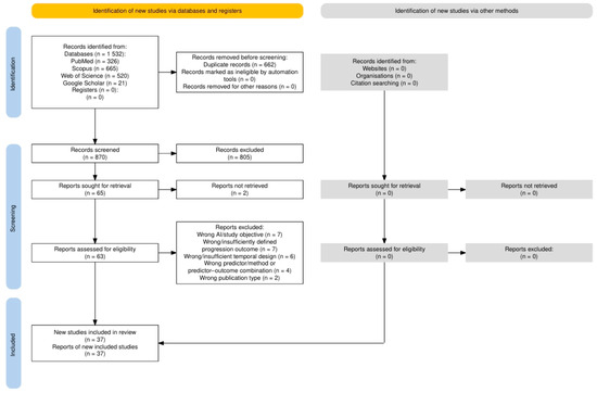
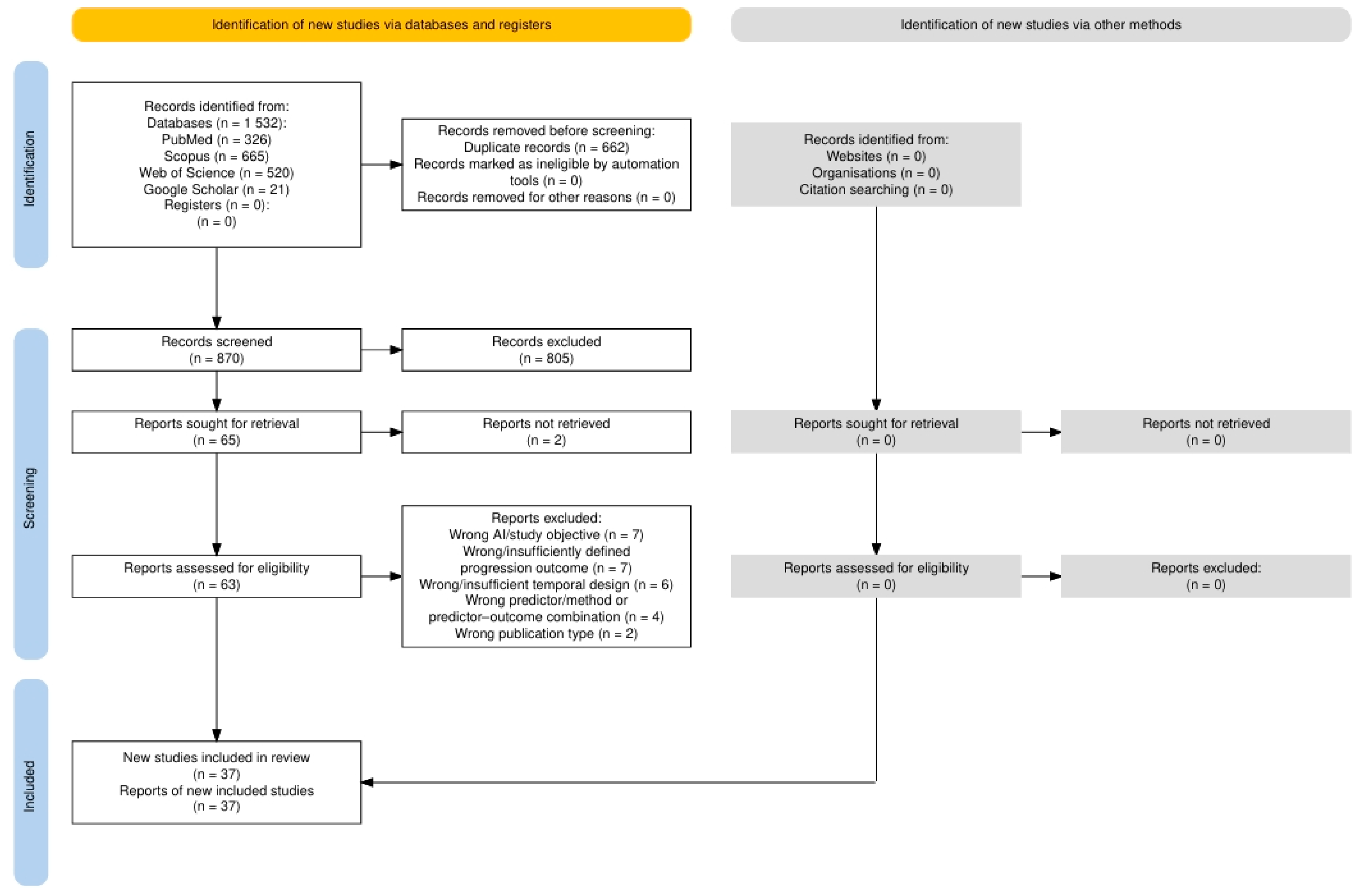
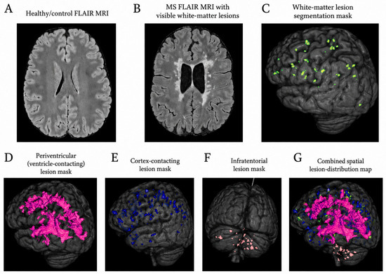
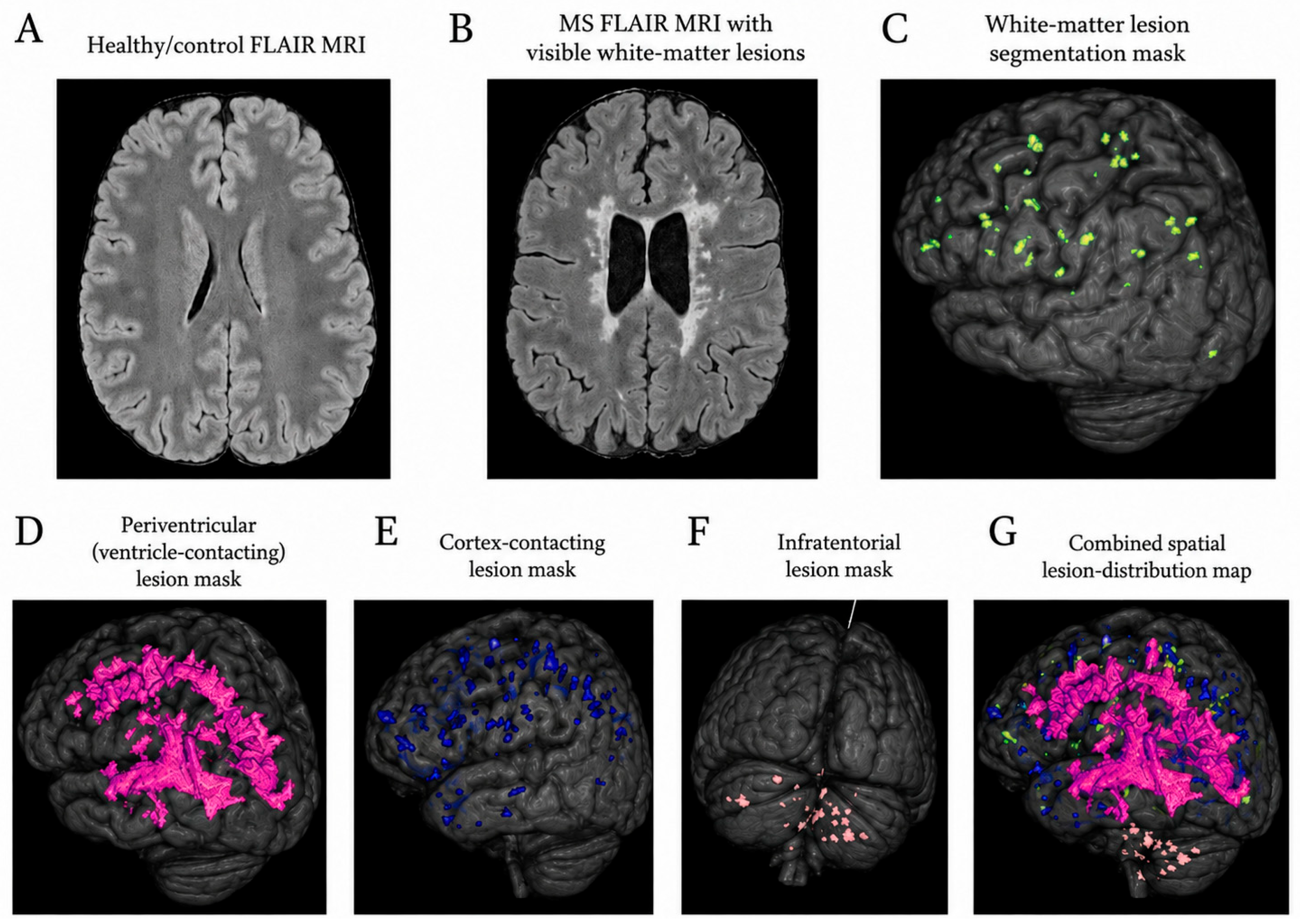
Disclaimer/Publisher’s Note: The statements, opinions and data contained in all publications are solely those of the individual author(s) and contributor(s) and not of MDPI and/or the editor(s). MDPI and/or the editor(s) disclaim responsibility for any injury to people or property resulting from any ideas, methods, instructions or products referred to in the content. |
© 2026 by the authors. Licensee MDPI, Basel, Switzerland. This article is an open access article distributed under the terms and conditions of the Creative Commons Attribution (CC BY) license.