


Featured Application
This study provides practical guidance for veterinary diagnostic laboratories regarding agreement between the Mindray BC-60R analyzer and manual leukocyte differentials in rabbits and identifying situations in which microscopic blood smear evaluation remains essential.
Abstract
This study evaluated the performance of the Mindray BC-60R differential WBC count (AD) in comparison with a manual method (MD) in rabbits. Diagnostic blood samples were analyzed within 1 h of collection, and MD was performed by counting 100 leukocytes. Method comparison was first performed in the complete dataset (n = 27) and subsequently in a morphologically restricted dataset (n = 23), after separating four samples with major leukocyte morphological abnormalities associated with abnormal scattergram patterns. In the morphologically restricted dataset, Spearman’s rank correlation between AD and MD was excellent for heterophils (r = 0.94, p < 0.05) and lymphocytes (r = 0.92, p < 0.05) and moderate for monocytes (r = 0.71, p < 0.05) and basophils (r = 0.71, p < 0.05), but not significant for eosinophils, due to insufficient spread of values. Passing–Bablok regression revealed no statistically significant constant or proportional bias for heterophils, lymphocytes, or monocytes, whereas basophils showed both. The Bland–Altman analysis showed a significant underestimation of heterophils and overestimation of lymphocytes and basophils, while monocyte and eosinophil biases were not significant. Abnormal scattergram patterns were indicative of morphologically abnormal leukocytes. The Mindray BC-60R analyzer showed good agreement with the manual differential for the major leukocyte populations, particularly heterophils and lymphocytes, in samples without major morphological abnormalities. Evaluation of blood smear in rabbits is required to identify the atypical cell population on a scatterplot.
1. Introduction
Rabbits are kept worldwide in multiple contexts, including as companion animals, laboratory models, and production animals, with each sector presenting specific health and welfare challenges. Health and welfare concerns, including infectious disease, digestive disorders, and stress-related conditions, require objective diagnostic tools to support clinical assessment and monitoring [1]. Hematologic evaluation represents a fundamental component of such diagnostic workups, providing essential information on inflammatory, infectious, and systemic conditions. In the companion animal sector, pet rabbit ownership has grown substantially, and welfare awareness among owners is evolving. However, preventive healthcare measures, including vaccination and early disease detection, are not consistently applied, and veterinary consultation often occurs only after clinical signs develop [2]. Therefore, increasingly precise and accurate laboratory and diagnostic methods are needed.
The Mindray BC-60R VET (Mindray, Shenzhen, China) is a recently introduced automated hematology analyzer that utilizes laser-based flow cytometry technology. Its evaluation in veterinary medicine was first reported in 2024 in dogs and cats [3]. The instrument software includes species-specific settings for multiple domestic and exotic species: dogs, cats, horses, cattle, sheep, goats, llamas, alpacas, rabbits, rats, mice, ferrets, pigs, pandas, monkeys. Its predecessor (BC-5000Vet, Mindray, Shenzhen, China) demonstrated good analytical performance in dogs and cats [4]. Although species-dependent differences have been reported between these species [3], to the authors’ knowledge, the performance of the BC-60R has not previously been evaluated in rabbits.
Given the distinct characteristics of rabbit leukocytes, species-specific validation is necessary. The predominant leukocyte population in rabbits consists of lymphocytes, followed by heterophils, the rabbit equivalent of neutrophils. In Romanowsky-stained blood smears, the secondary granules of rabbit heterophils stain pink, in contrast to the pale blue cytoplasm typically observed in canine and feline neutrophils [5]. These hematological particularities may influence leukocyte classification in automated analyzers, potentially affecting analytical performance.
The aim of the present study was to evaluate the analytical performance of the Mindray BC-60R VET hematology analyzer in differentiating leukocyte types in rabbits, using manual microscopic differential counts as the reference method.
2. Materials and Methods
This was a retrospective study of blood samples collected from client-owned rabbits (n = 31) presented to the New Companion Animals Clinic, Faculty of Veterinary Medicine, Cluj-Napoca, between September 2024 and October 2025. The rabbits represented different ages, sexes, and breeds, and presented with a range of clinical conditions, including gastrointestinal, inflammatory, neoplastic, and neurological disorders (Appendix A, Table A1). No clinically healthy rabbits were included, as all samples originated from clinical patients presented for diagnostic evaluation. Approximately 0.5 mL of whole blood was collected from either the marginal auricular vein or the lateral saphenous vein into 2 mL K3-EDTA tubes for routine hematological evaluation and diagnostic purposes. In critically ill or dehydrated rabbits, when venipuncture was limited, blood samples collected in lithium heparin tubes for biochemical analysis were also considered for hematological evaluation, provided that leukocyte morphology was adequately preserved. Written informed consent for the use of anonymized clinical data for research purposes was routinely obtained from all owners at the time of presentation, in accordance with the standard institutional procedures of the Faculty of Veterinary Medicine, Cluj-Napoca. The study employs a retrospective observational design and is based exclusively on clinical data and hematological results obtained as a part of the routine activities of the hematology laboratory. All data were anonymized prior to analysis, animal owners provided consent for the use of the data for research purposes in accordance with institutional procedures, and no personal or sensitive identifying information was collected or processed. Ethical review and approval were waived for this study because the research did not involve human participants, live animal experimentation, or any procedures requiring ethical oversight.
Complete blood counts (CBCs) were performed on a BC-60R hematology analyzer (Mindray, Shenzhen, China) within 1 h of sampling, although a small number of samples were analyzed within 4 h. Blood smears were prepared during the same time interval. Mindray BC-60R is a new laser-based hematology analyzer that performs leukocyte enumeration and differential in a DIFF channel. In this channel, erythrocytes are lysed, and a fluorochrome dye (asymmetric cyanine fluorochrome) selectively binds to intracellular nucleic acids (DNA and RNA) of leukocytes. A five-part differential (neutrophils/heterophils, lymphocytes, monocytes, eosinophils, and basophils) is obtained through fluorescence-based flow cytometry. Cell populations are differentiated based on fluorescence intensity (FL), reflecting the relative nucleic acid content, and side scatter (SS), indicating cytoplasmic granularity and structural complexity.
For each sample, a single manual differential count of 100 leukocytes was performed by one observer on a quick May–Grunwald–Giemsa-stained blood smear (Bio-Optica, Milano, Italy). The observer was blinded to Mindray BC-60R results. In our laboratory, automated results showing discrepancies with microscopic findings are corrected when appropriate. Leukocyte scattergrams were visually inspected for overlapping cell populations and abnormal cell clusters. Scattergram abnormalities were interpreted in conjunction with the corresponding microscopic leukocyte morphology. Method comparison was first performed using the complete dataset (n = 27). A complementary analysis was subsequently performed using a morphologically restricted dataset (n = 23), after separating four samples with major leukocyte morphological abnormalities associated with abnormal scattergram patterns, consistent with an approach previously described in veterinary analyzer validation [6].
Samples were excluded from the study if any of the following criteria were met: 1—visible clots in samples; 2—poor-quality slides (extensive WBC lysis, trapping of WBC in platelet aggregates).
Statistical analyses were performed using MedCalc (version 19.2.6, MedCalc Software Ltd., Mariakerke, Belgium). Graphs were prepared using GraphPad Prism (version 11.0.2, GraphPad Software, Boston, MA, USA). Spearman’s rank correlation coefficients were calculated to evaluate the correlation between automated and manual differential counts. Passing–Bablok regression was used to assess constant and proportional between-method bias, whereas Bland–Altman analysis was used to estimate mean bias and 95% limits of agreement. p < 0.05 was considered statistically significant.
3. Results
3.1. General Characteristics of Population
A total of 31 blood samples were initially included in the study. Three samples were excluded because of visible clot formation and one because of marked hemolysis, leaving 27 samples for statistical analysis. Among these, two samples had been collected in heparin tubes for biochemical testing because the patients were critically ill and dehydrated; however, as leukocyte morphology was well preserved, these samples were considered suitable and were retained for the hematological analysis.
The study population included 27 rabbits with different pathologies, with a slightly higher proportion of males (56%, 15/27) compared to females (44%, 12/27). The mean age was 4.5 years (range: 2 months to 8 years). The most represented breed was Lionhead (30%, 8/27), followed by mixed breed (26%, 7/27) and dwarf rabbits (19%, 5/27).
The clinical causes are presented in Appendix A, Table A1. Hematological disorders include leukocytosis (3), leukopenia (2), heterophilia (7), heteropenia (1), lymphocytosis (3), lymphopenia (11), monocytosis (1), eosinophilia (1), left shift (1), mild and moderate toxic changes (4), severe left shift and toxic changes (3), immature leukocytes (1), anemia (2), polycythemia (3), nucleated red blood cells (nRBCs) (2), thrombocytosis (2), and platelet clumps (16), of which 9 caused false thrombocytopenia.
Normally distributed data were observed for heterophils (both analyzer and manual method), lymphocytes (analyzer), and eosinophils (analyzer). Non-normal distribution was observed for lymphocytes, monocytes, eosinophils, and basophils in the manual method, as well as for monocytes and basophils measured by the analyzer. For the BC-60R analyzer, mean (±SD) values were 53.3 ± 21.1 for heterophils, 32.8 ± 20.6 for lymphocytes, and 0.9 ± 0.6 for eosinophils. Median (range) values for monocytes and basophils were 5.0% (3.9–7.5%) and 3.3% (1.9–4.1%), respectively. For the manual differential count, the mean (±SD) value for heterophils was 61.7 ± 21.8. Median (range) values were 26% (17.8–38.2%) for lymphocytes, 4% (3–6%) for monocytes, 1% (1%) for eosinophils, and 1% (0–1.5%) for basophils.
3.2. Method Comparison of Complete Dataset
The correlation between analyzer differential (AD) and manual differential (MD) was moderate for heterophils (r = 0.79, p < 0.05), lymphocytes (r = 0.78, p < 0.05), monocytes (r = 0.79, p < 0.05), and basophils (r = 0.77, p < 0.05) and low and not statistically significant for eosinophils (r = 0.36, p > 0.05).
Passing–Bablok regression analysis revealed no statistically significant constant or proportional bias for heterophils. Lymphocytes showed a significant constant bias, while monocytes and basophils demonstrated significant proportional bias, the latter being more pronounced. Eosinophils showed no evidence of constant or proportional bias (Table 1).
Table 1.
Results of the Passing–Bablok and Bland–Altman analyses comparing the differential WBC counts obtained by the Mindray BC-60R and the manual method in the complete dataset (n = 27).
Bland–Altman analyses identified a statistically significant negative bias of −8.35 for heterophils. No statistically significant bias was observed for lymphocytes (1.81), monocytes (2.65), eosinophils (−0.23), or basophils (4.38). Wide 95% limits of agreement were observed for heterophils, lymphocytes, monocytes, and basophils, whereas eosinophils exhibited comparatively narrower limits of agreement (Table 1).
A sensitivity analysis excluding the two samples collected in lithium-heparin tubes (n = 25) produced comparable correlation coefficients, Passing–Bablok regression parameters, and Bland–Altman bias and limits of agreement, indicating that their inclusion did not materially influence the method-comparison results (Appendix A, Table A2).
3.3. Description of Samples with Major Leukocyte Morphological Abnormalities
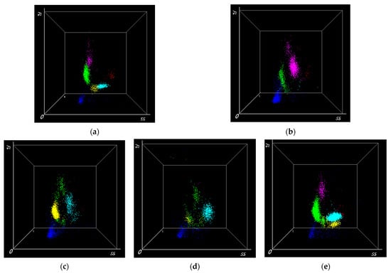
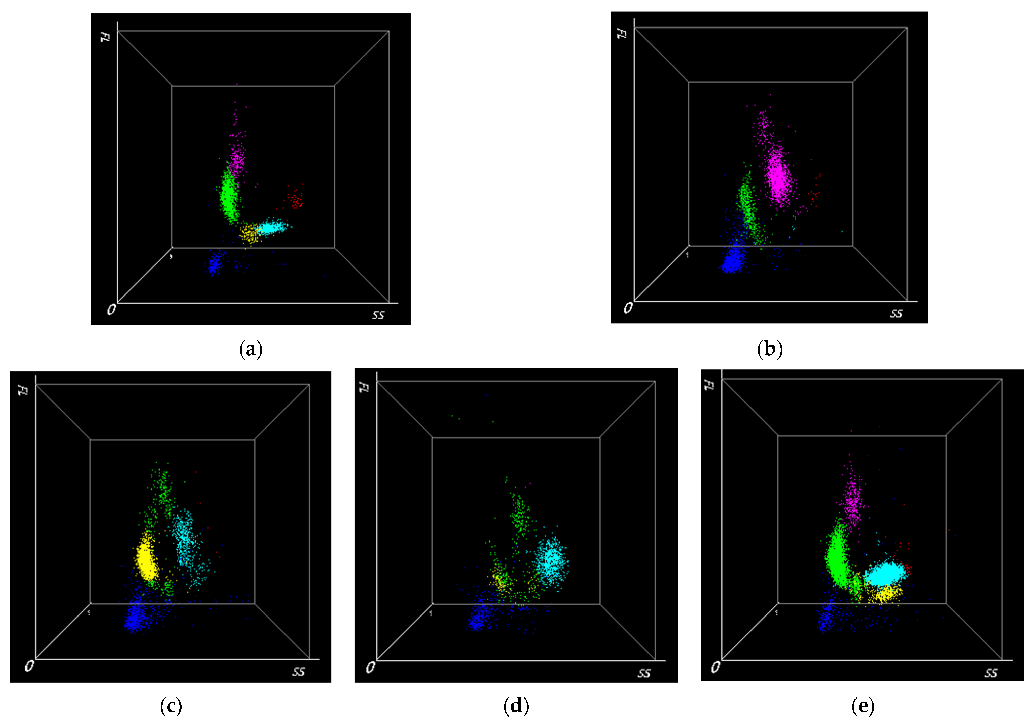
Scattergram evaluation identified four cases with major leukocyte morphological abnormalities associated with abnormal scattergram patterns, which were subsequently separated from the complementary morphologically restricted analysis. In first case, a rightward displacement of the monocyte cluster was observed, characterized by a marked increase in side scatter and expansion of the population area due to variable fluorescence intensity (Figure 1b). In the second case, the basophil cluster was displaced to the left, showing low side scatter and low-to-moderate fluorescence intensity. The lymphocyte population appeared split into two distinct clusters: one located between the basophil and heterophil clusters, characterized by moderate side scatter and low fluorescence intensity, and a second cluster positioned above this population, with moderate side scatter and high fluorescence intensity. The neutrophil cluster was displaced upward (high fluorescence intensity) (Figure 1c). In the third case, the lymphocyte cluster appeared divided into two populations. The first population was shifted upward and to the right, showing high fluorescence intensity and moderate side scatter. The second population was dispersed along the X-axis, with low-to-high side scatter and variable fluorescence intensity. The basophil cluster was displaced to the left and upward, showing low side scatter and low-to-moderate fluorescence intensity. The neutrophil cluster was displaced upward, showing moderate-to-high fluorescence intensity (Figure 1d). In the fourth case, an additional lymphocyte-like population was identified to the left of the heterophil cluster, characterized by moderate side scatter and low fluorescence intensity (Figure 1e).
Figure 1.
Scattergrams of samples with major leukocyte morphological abnormalities from BC-60R. X-axis: side scatter (SS)—intracellular complexity; Y-axis: intensity of fluorescence (FL)—content of nucleic acid; ● green—lymphocyte population; ● light-blue—heterophils; ● purple—monocytes; ● red—eosinophils; ● yellow—basophils; ● deep-blue—particles assigned to the ghost region are dark blue and are excluded from the leukocyte differential calculation. (a) Representative scattergram showing clear separation of the leukocyte populations. (b) Immature heterophils with severe toxic changes were misclassified as monocytes (Figure 2b). (c) Heterophils with marked toxic changes, especially toxic granulation, were most likely counted as basophils (Figure 2c). (d) Immature heterophils with marked toxic changes were misclassified as lymphocytes and heterophil cloud shifted to the right (Figure 2f). (e) Atypical immature cells were classified as lymphocytes (Figure 2d,e).
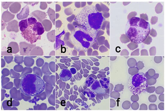
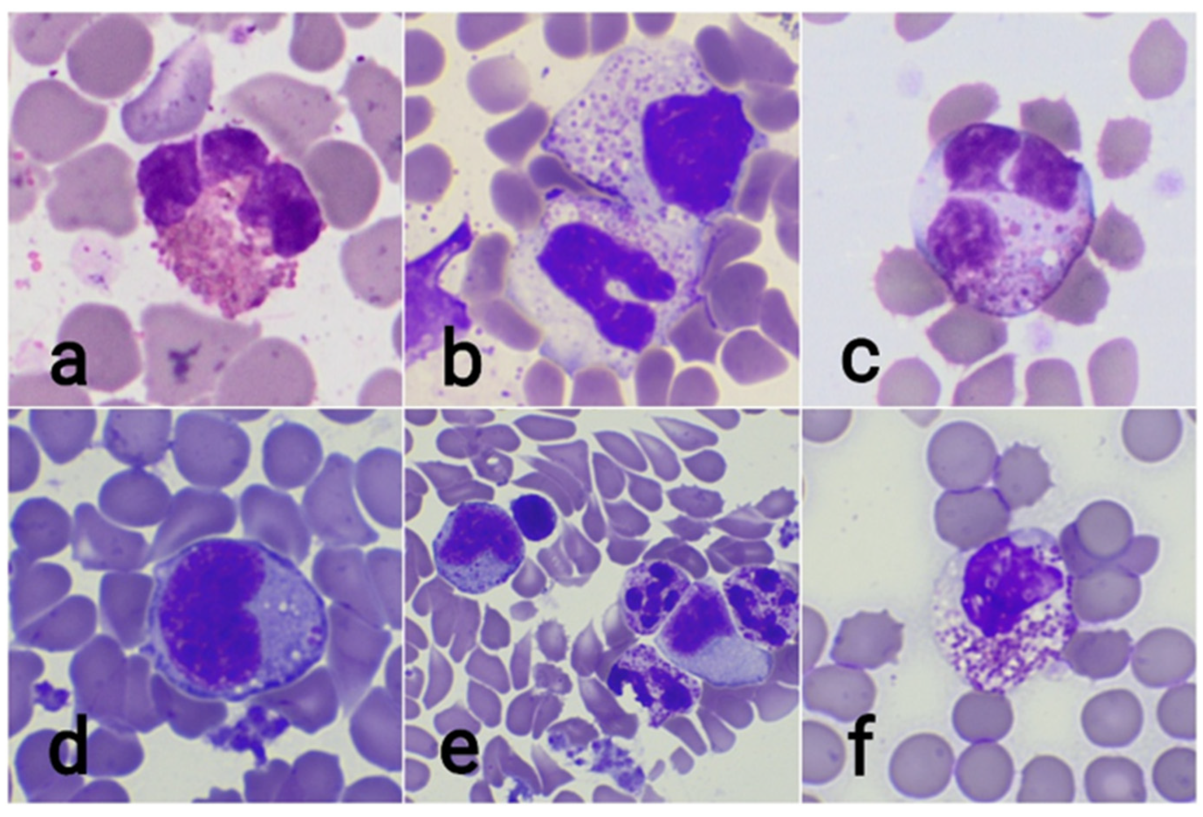
These abnormal scattergram patterns were associated with marked cytological abnormalities on blood smear evaluation. The evaluation of the first three cases revealed severe toxic changes, including basophilic cytoplasm, intracytoplasmic vacuolation, abnormal granulation, and left shift (Figure 2b,c,f). In the fourth case, round cells larger than lymphocytes, occasionally containing pink–magenta granules, were observed (Figure 2d,e). The patient was diagnosed with thymoma.
Figure 2.
Morphology of normal and abnormal heterophils. (a) Heterophil in a healthy rabbit. (b) Lower—band heterophil with marked toxic changes (basophilic cytoplasm, less granulation); upper—metamyelocyte. (c) Markedly toxic mature heterophils: cytoplasmic basophilia, less granulation than normal, and toxic granulation. (d) Immature cells, most likely metamyelocytes. (e) Left—metamyelocytes (deep-blue cytoplasm and pinkish granules and reniform nucleus); right—myeloblast (moderate amount of basophilic cytoplasm, oval nucleus). (f) Markedly toxic metamyelocytes: cytoplasmic basophilia, vacuolization, and less granulation than normal. May–Grunwald–Giemsa stain, ×1000.
Method agreement was first evaluated in the complete dataset (n = 27). A complementary analysis was then performed using a morphologically restricted dataset (n = 23), after separating four samples with major leukocyte morphological abnormalities associated with abnormal scattergram patterns.
3.4. Method Comparison of Morphologically Restricted Dataset
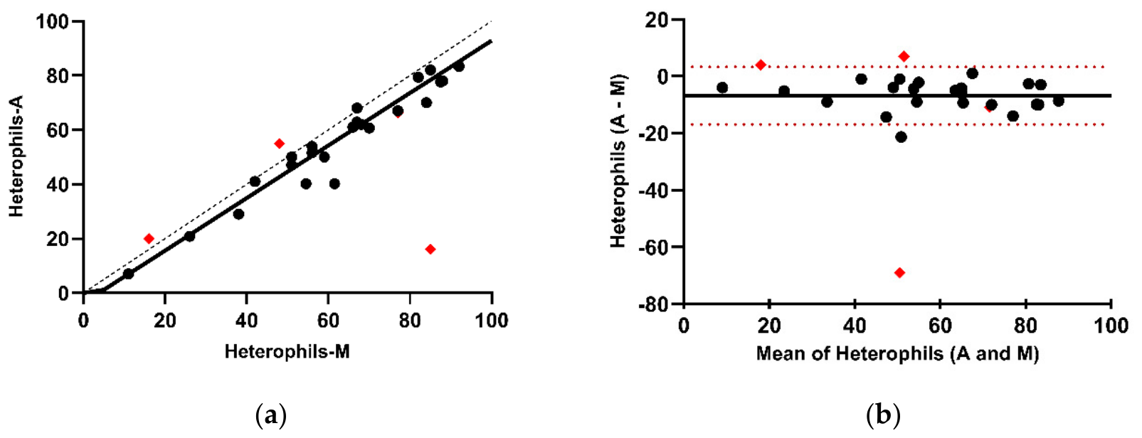
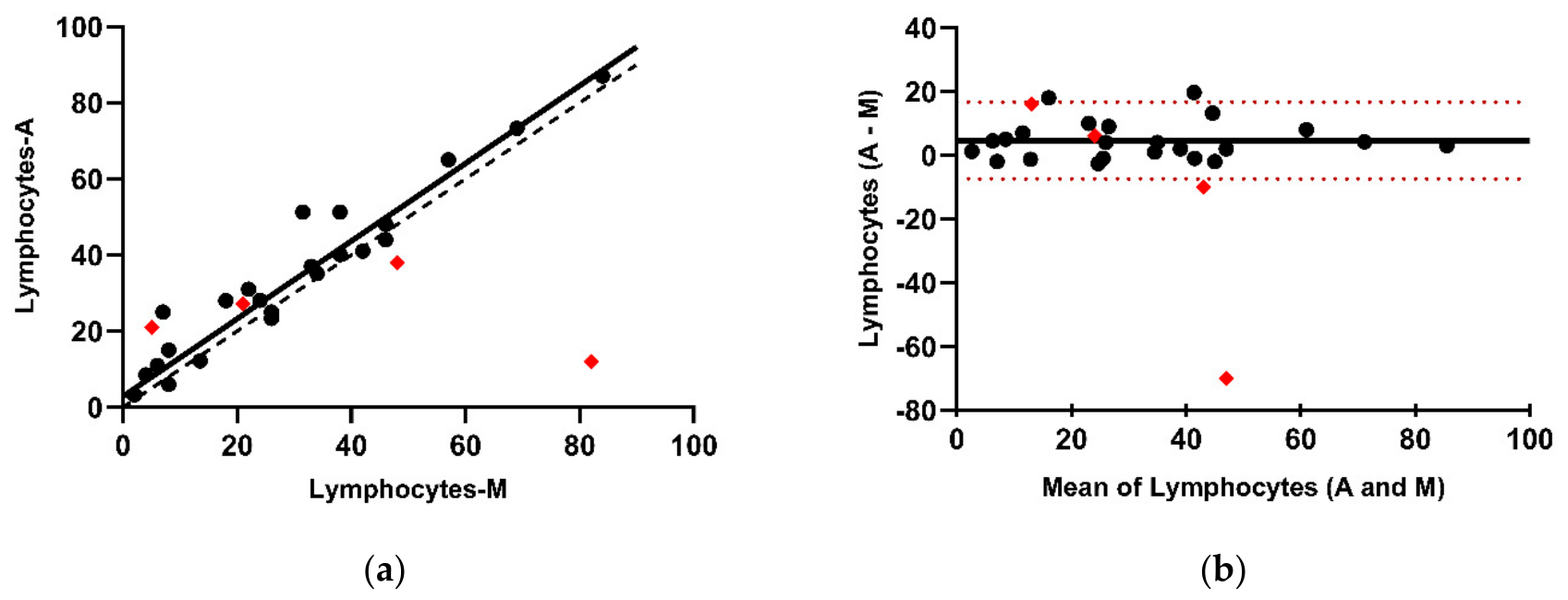
In the morphologically restricted dataset, correlation between AD and MD improved to excellent for heterophils (r = 0.94, p < 0.05) and lymphocytes (r = 0.92, p < 0.05), and remained moderate for monocytes (r = 0.71, p < 0.05) and basophils (r = 0.71, p < 0.05), while the correlation for eosinophils remained low and was not statistically significant (r = 0.29, p > 0.05).
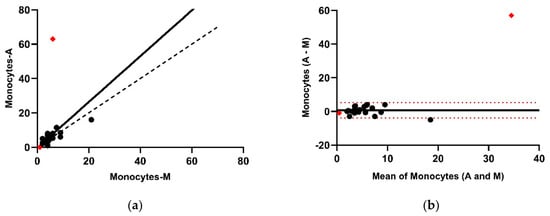
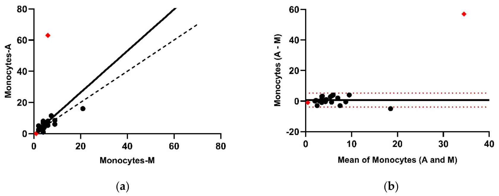
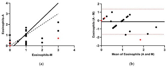
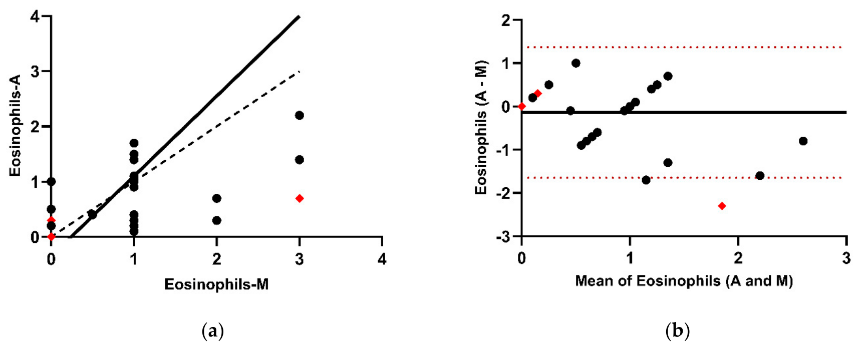
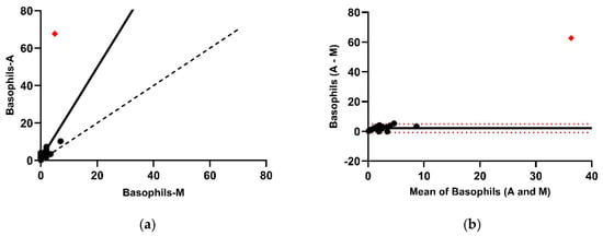
Passing–Bablok regression revealed no statistically significant constant or proportional bias for heterophils, lymphocytes, monocytes, or eosinophils. In contrast, basophils demonstrated both significant constant and proportional bias (Table 2, Figure 3a, Figure 4a, Figure 5a, Figure 6a and Figure 7a).
Table 2.
Results of the Passing–Bablok and Bland–Altman analyses comparing the differential WBC counts obtained by the Mindray BC-60R and the manual method in the morphologically restricted dataset (n = 23).
Figure 3.
Passing–Bablok regression analysis and Bland–Altman plot for heterophil percentages obtained by BC-60R compared to the manual method in the morphologically restricted dataset (n = 23). (a) The black, dashed diagonal line in the Passing–Bablok regression analysis plot is the line of identity, and the black line is the calculated line of regression. Black circles represent samples included in the regression analysis; red diamonds represent the four samples with major leukocyte morphological abnormalities that were separated from the morphologically restricted dataset. (b) In the Bland–Altman plot, the difference between the 0 and the black line indicates the bias of the Mindray BC-60R minus the manual differential counts. The 95% CIs of the calculated bias are represented by the 2 red dashed lines.
Figure 4.
Passing–Bablok regression analysis and Bland–Altman plot for lymphocyte percentages obtained by BC-60R compared to the manual method in the morphologically restricted dataset (n = 23). (a) The black, dashed diagonal line in the Passing–Bablok regression analysis plot is the line of identity, and the black line is the calculated line of regression. Black circles represent samples included in the regression analysis; red diamonds represent the four samples with major leukocyte morphological abnormalities that were separated from the morphologically restricted dataset. (b) In the Bland–Altman plot, the difference between the 0 and the black line indicates the bias of the Mindray BC-60R minus the manual differential counts. The 95% CIs of the calculated bias are represented by the 2 red dashed lines.
Figure 5.
Passing–Bablok regression analysis and Bland–Altman plot for monocyte percentages obtained by BC-60R compared to the manual method in the morphologically restricted dataset (n = 23). (a) The black, dashed diagonal line in the Passing–Bablok regression analysis plot is the line of identity, and the black line is the calculated line of regression. Black circles represent samples included in the regression analysis; red diamonds represent the four samples with major leukocyte morphological abnormalities that were separated from the morphologically restricted dataset. (b) In the Bland–Altman plot, the difference between the 0 and the black line indicates the bias of the Mindray BC-60R minus the manual differential counts. The 95% CIs of the calculated bias are represented by the 2 red dashed lines.
Figure 6.
Passing–Bablok regression analysis and Bland–Altman plot for eosinophil percentages obtained by Mindray BC-60R compared to the manual method in the morphologically restricted dataset (n = 23). (a) The black, dashed diagonal line in the Passing–Bablok regression analysis plot is the line of identity, and the black line is the calculated line of regression. Black circles represent samples included in the regression analysis; red diamonds represent the four samples with major leukocyte morphological abnormalities that were separated from the morphologically restricted dataset. (b) In the Bland–Altman plot, the difference between the 0 and the black line indicates the bias of the Mindray BC-60R minus the manual differential counts. The 95% CIs of the calculated bias are represented by the 2 red dashed lines.
Figure 7.
Passing–Bablok regression analysis and Bland–Altman plot for basophil percentages obtained by Mindray BC-60R compared to the manual method in the morphologically restricted dataset (n = 23). (a) The black, dashed diagonal line in the Passing–Bablok regression analysis plot is the line of identity, and the black line is the calculated line of regression. Black circles represent samples included in the regression analysis; red diamonds represent the four samples with major leukocyte morphological abnormalities that were separated from the morphologically restricted dataset. (b) In the Bland–Altman plot, the difference between the 0 and the black line indicates the bias of the Mindray BC-60R minus the manual differential counts. The 95% CIs of the calculated bias are represented by the 2 red dashed lines.
Bland–Altman analysis showed a statistically significant negative bias for heterophils (−6.8; 95% CI −9.0 to −4.6; p < 0.0001) and significant positive biases for lymphocytes (4.6; 95% CI 2.0 to 7.3; p = 0.0015) and basophils (2.1; 95% CI 1.4 to 2.7; p < 0.0001). No statistically significant bias was observed for monocytes (0.7; p = 0.155) or eosinophils (−0.1; p = 0.381) (Figure 3b, Figure 4b, Figure 5b, Figure 6b and Figure 7b). The 95% limits of agreement narrowed across all leukocyte subsets in the morphologically restricted dataset (Table 2).
4. Discussion
The current study demonstrates the agreement between the BC-60R automated leukocyte differential and manual differential counts in rabbits. In addition, abnormal scattergram patterns were associated with marked morphological abnormalities, including left shift, severe toxic changes, and immature leukocytes, emphasizing the importance of microscopic smear review.
Preanalytical factors may significantly influence hematological results; therefore, only fresh samples analyzed within 4 h of collection were included, and samples with visible clots were excluded. Hemolysis and platelet clumping are among the most common preanalytical errors affecting hematology analyzers. Marked hemolysis can decrease hematocrit (HCT) and red blood cell (RBC) counts and may spuriously increase mean corpuscular hemoglobin concentration (MCHC), whereas clot formation can reduce platelet counts and, if substantial, both red blood cell and leukocyte counts [7]. Additionally, leukocytes may become entrapped in platelet aggregates or be affected by storage conditions. Monocytes appear to be particularly sensitive to storage, showing significant decreases after 24 h, likely due to cellular deterioration [8]. A total of 27 samples were included in the study. Two samples collected in lithium-heparin tubes for biochemical analysis were also included because leukocyte morphology was adequately preserved for differential evaluation. Heparin has not been shown to significantly affect total leukocyte counts or differentials in dogs and horses, although in cats it has been associated with lower monocyte and lymphocyte counts compared with EDTA [9]. In the present study, a sensitivity analysis excluding these two samples yielded comparable Spearman correlation coefficients, Passing–Bablok regression parameters, and Bland–Altman bias and limits of agreement, indicating that their inclusion did not materially influence the method-comparison results (Appendix A, Table A2). Nevertheless, an anticoagulant-related effect on individual samples cannot be excluded, and further studies are required to specifically evaluate the effects of different anticoagulants on hematological parameters in rabbits.
Heterophils and lymphocytes showed a moderate correlation between the Mindray BC-60R analyzer and the manual differential count. Passing–Bablok regression revealed a significant constant bias for lymphocytes, while no significant bias was identified for heterophils. Bland–Altman analysis demonstrated a negative bias for heterophils (−8.35) and a positive bias for lymphocytes (1.81). In the morphologically restricted dataset, higher correlation coefficients were observed for both cell types. No significant constant or proportional bias was detected by Passing–Bablok regression, while Bland–Altman analysis showed a reduced negative bias for heterophils (−6.8) and an increased positive bias for lymphocytes (4.6). Similar findings have been reported in previous studies. A study evaluating the ADVIA 2120 analyzer in rabbits demonstrated good agreement between automated and manual leukocyte differentials, both in the entire dataset and in the subset excluding samples with left shift and toxic changes [10]. Likewise, a large study using Sysmex analyzers in healthy (n = 120) and diseased (n = 505) rabbits reported good to excellent correlations for heterophils (r = 0.89 and 0.94) and lymphocytes (r = 0.91 and 0.92), respectively [11]. The observed positive bias for lymphocytes may be partially explained by limitations of the manual differential count, which can underestimate lymphocyte numbers due to the relatively low number of cells evaluated (100 leukocytes), thereby increasing counting variability. Heterophils and lymphocytes represent the predominant leukocyte populations in adult rabbits, with an approximate heterophil-to-lymphocyte (H:L) ratio of 45:45 under physiological conditions. This ratio is known to change in response to stress and inflammatory processes, making accurate differentiation of these cell types clinically relevant [5].
Monocytes, eosinophils, and basophils are low-abundance leukocyte populations, and their results showed greater variability compared with more prevalent cell types such as heterophils and lymphocytes. This observation is consistent with previous studies demonstrating reduced analytical performance for low-frequency leukocyte populations in veterinary hematology analyzers [12]. The moderate agreement observed for monocytes may also be influenced by limitations of the manual differential count. Monocytes, being among the largest circulating leukocytes, tend to migrate toward the margins and feathered edge of the blood smear during slide preparation. As these regions are not always systematically evaluated during microscopic examination, manual counts may underestimate the true monocyte proportion. In addition, overlap in scatter characteristics between monocytes and other leukocyte populations may further contribute to reduced analytical agreement [13].
The low correlation observed for eosinophils can be attributed primarily to their low circulating numbers. Similar findings have been reported in rabbits using both the ADVIA 2120 [10] and Sysmex analyzers [11], particularly in clinically affected animals, whereas a moderate correlation has been described in healthy rabbits. In contrast, species-related differences have been reported in the literature. In cats, eosinophil counts show good agreement between automated and manual methods when analyzed using both ADVIA and Sysmex analyzers. In dogs, correlation ranges from moderate (ADVIA) to good (Sysmex), whereas in horses, eosinophils generally demonstrate moderate agreement across both platforms [9].
A moderate correlation was observed for basophils, with both constant and proportional bias identified in the morphologically restricted dataset. Although basophils are present in very low numbers in most domestic species such as dogs and cats, rabbits typically exhibit higher baseline proportions, reaching up to approximately 5% in healthy individuals [5]. Automated hematology analyzers generally show limited performance in identifying basophils in dogs and cats; however, ADVIA analyzers can more reliably detect this population due to their unique cytochemical properties, as basophils are resistant to lysis in the BASO channel [14]. Accordingly, good agreement between automated and manual methods has been reported in rabbits using ADVIA systems [10]. In contrast, the results obtained in the present study are comparable to those reported for Sysmex analyzers, which, similarly to the BC-60R, rely on flow cytometric principles for leukocyte differentiation. In these systems, leukocytes are permeabilized and stained with fluorescent dyes, and classification is based on a combination of fluorescence intensity (reflecting nucleic acid content) and side scatter signals (reflecting cellular granularity) [11]. These methodological characteristics may contribute to the observed variability in basophil identification.
Scatter plot evaluation was performed for all samples. Four samples lacking clear separation of leukocyte populations or showing atypical cell clusters were analyzed as clinically relevant discrepant cases and were not included in the complementary morphologically restricted dataset. Specifically, three samples showed severe toxic changes and left shift associated with abnormal scattergram patterns, while a fourth sample contained an atypical immature cell population. In a validation study of the Sysmex analyzer in dogs, cats, and horses, incomplete or inaccurate differential counts were associated with samples showing left shift and toxic changes [6]. In such cases, increased RNA content in immature or toxic neutrophils leads to enhanced fluorescence signals, causing an upward shift in scattergrams and overlap with lymphocyte clusters. This phenomenon may result in misclassification or incomplete leukocyte differentiation. Similarly, neutrophil misclassification has been reported with the ProCyte Dx, where neutrophils were displaced into the lymphocyte or monocyte regions of the scattergram, resulting in erroneous classification [15].
Interestingly, in the present study, in one case, heterophils with severe toxic changes (Figure 2c) were misclassified as basophils rather than lymphocytes or monocytes. In another case, heterophils with toxic changes and immature forms were misclassified as lymphocytes and basophils. Misclassification of toxic heterophils as basophils differs from previous reports in dogs and cats; however, the mechanisms underlying this misclassification could not be determined in the present study. Furthermore, leukocyte classification based solely on scatter properties has been shown to be feasible in rabbits, with high accuracy for granulocytes and lymphocytes; however, the close proximity of gating regions, particularly between monocytes and large lymphocytes, may lead to misclassification in automated systems [13]. These limitations are inherent to flow cytometry-based techniques, as cells are classified according to their optical properties, which can be altered by activation, disease, or cellular immaturity [16]. These findings highlight the importance of scattergram evaluation and manual smear review in samples with suspected abnormal leukocyte populations. Further studies incorporating additional cell-characterization methods, such as cytochemical staining or immunophenotyping, may allow more definitive identification of atypical or immature leukocyte populations and help determine which cell types are associated with specific patterns of automated misclassification. Additional investigation is also warranted to determine the factors contributing to the constant and proportional bias observed for basophils.
Most veterinary hematology analyzers were initially developed for human medicine and subsequently adapted for use in animals. A validation study conducted on its predecessor, the BC-5000, which included 853 samples and followed ICSH and CLSI guidelines, demonstrated excellent accuracy for leukocyte differentiation [17]. Furthermore, studies in dogs and cats have shown that this type of analyzer is suitable for routine hematological analysis, including in patients with various hematologic abnormalities [4].
The present study has several limitations that should be acknowledged. First, no a priori sample-size calculation was performed because of the retrospective nature of the study and the limited number of eligible rabbit samples available during the study period. The relatively small sample size may limit the statistical robustness of the analysis, as method-comparison studies generally recommend a minimum of 40 samples to ensure robust evaluation of agreement [18]. However, the number of samples included in the present study was above the minimum suggested by CLSI for verification of performance studies, where approximately 20 specimens may be sufficient for an initial evaluation, provided that the samples span the clinically relevant measurement range [19]. The reduced sample size of the morphologically restricted dataset (n = 23) further increases the uncertainty of the Passing–Bablok regression estimates and Bland–Altman limits of agreement, particularly for low-frequency leukocyte populations. Therefore, the present results should be interpreted as preliminary evidence. Further validation in larger populations with diverse clinical and hematological abnormalities is warranted.
Second, manual differential counts were performed by a single observer due to the retrospective nature of the study. Although this approach ensured internal consistency, neither intra-observer variability through replicate counts nor inter-observer variability through independent assessment could be evaluated. Inter-observer variability is a recognized source of imprecision in manual leukocyte differentiation [20]. In addition, the inherent imprecision of manual leukocyte differential counts represents an important limitation. Differential counts follow a binomial distribution, and their variability is determined by both the proportion of the cell population and the finite number of cells evaluated. Consequently, low-frequency leukocyte populations such as eosinophils, basophils and, to a lesser extent, monocytes, exhibit substantially higher variability, particularly when based on counts of 100 cells, which may contribute to apparent discrepancies between manual and automated methods [21]. Therefore, the present findings should be interpreted as agreement between the automated and manual differential methods rather than as an assessment of absolute analytical accuracy, as the manual differential itself is not an error-free reference standard. Larger manual differential counts, replicate counts, and independent evaluation by additional experienced observers should be considered in future studies to reduce the uncertainty associated with the comparative microscopic method.
5. Conclusions
Under the evaluated conditions, the Mindray BC-60R showed good agreement with the manual differential for heterophils and lymphocytes, particularly in samples without major morphological abnormalities. However, agreement varied among cell types, with persistent underestimation of heterophils and constant and proportional bias for basophils. Samples with major morphological abnormalities were associated with greater automated–manual discrepancies. Therefore, scattergrams showing poor separation or overlap of leukocyte populations or atypical cell clusters, particularly when associated with marked toxic or immature leukocytes, should prompt microscopic blood smear evaluation.
Author Contributions
Conceptualization, I.M.M.; methodology, I.M.M. and P.J.O.; validation, I.M.M. and P.J.O.; formal analysis, I.M.M. and P.J.O.; investigation, I.M.M., M.C.T., M.A.O., L.B., A.I.M.-D., O.S. and B.S.; resources, M.C.T., M.A.O., L.B., A.I.M.-D., O.S., B.S. and I.M.; data curation, I.M.M., M.C.T. and M.A.O.; writing—original draft preparation, I.M.M.; writing—review and editing, I.M.M., M.C.T., A.I.M.-D., O.S., B.S., P.J.O. and I.M.; visualization, I.M.M. and P.J.O.; supervision, P.J.O. and I.M.; project administration, I.M.M. and I.M. All authors have read and agreed to the published version of the manuscript.
Funding
This research received no external funding.
Institutional Review Board Statement
The study employs a retrospective observational design and is based exclusively on clinical data and hematological results obtained as a part of the routine activities of the hematology laboratory. All data were anonymized prior to analysis, and animal owners provided consent for the use of the data for research purposes in accordance with institutional procedures, and no personal or sensitive identifying information was collected or processed. Ethical review and approval were waived for this study because the research did not involve human participants, live animal experimentation, or any procedures requiring ethical oversight.
Data Availability Statement
The data related to this study are available and can be obtained by contacting the corresponding author.
Acknowledgments
Parts of this study were presented at the ESVCP Congress in Bristol, United Kingdom, on 1–4 October 2025. A special thank you is extended to the staff of the New Companion Animal Clinic for their constant support and for always being receptive and open to collaboration throughout this study. I.M.M. also gratefully acknowledges O’Brien for his guidance throughout this study and in earlier academic work.
Conflicts of Interest
The authors declare no conflicts of interest.
Abbreviations
The following abbreviations are used in this manuscript:
| CBC | Complete blood count |
| AD | Analyzer differential |
| MD | Manual method |
Appendix A
Table A1.
Distribution of underlying clinical conditions in the study population.
Table A2.
Sensitivity analysis assessing the influence of the two lithium-heparin samples on the comparison between automated and manual leukocyte differential counts (n = 25).
References
- Fels, M.; Benato, L.; Whittaker, A. Health and Welfare of Rabbits (on a Farm, in the Laboratory or as a Pet). Front. Vet. Sci. 2024, 11, 1522420. [Google Scholar] [CrossRef] [Scilit] [PubMed]
- Boglárka, S.; Bodnar, K.; Marta, P.; Katalin, S. Study of Animal Welfare Aspects Among Pet Rabbit Owners. Lucr. Stiint. Ser. I 2019, 21, 108–112. [Google Scholar]
- Cheng, X. Comparison of Mindray BC-60R Vet with Sysmex XT-2000iV Analyzer Using Total Observed Error, Total Allowable Error, and QC Rules Defined in the American Society for Veterinary Clinical Pathology (ASVCP) Guidelines. Ph.D. Thesis, University of Veterinary Medicine Budapes, Budapest, Hungary, 2024. [Google Scholar]
- Thongsahuan, S.; Fonghoi, L.; Kaewfai, S.; Srinoun, K. Precision and Accuracy of the Mindray BC-5000Vet Hematology Analyzer for Canine and Feline Blood. Vet. Clin. Pathol. 2020, 49, 207–216. [Google Scholar] [CrossRef] [Scilit] [PubMed]
- Campbell, T.W.; Grant, K.R. Exotic Animal Hematology and Cytology, 5th ed.; John Wiley & Sons: Hoboken, NJ, USA, 2022; ISBN 9781119660231. [Google Scholar]
- Lilliehöök, I.; Tvedten, H. Validation of the Sysmex XT-2000iV Hematology System for Dogs, Cats, and Horses. II. Differential Leukocyte Counts. Vet. Clin. Pathol. 2009, 38, 175–182. [Google Scholar] [CrossRef] [Scilit] [PubMed]
- DeNicola, D.B. Advances in Hematology Analyzers. Top. Companion Anim. Med. 2011, 26, 377–386. [Google Scholar] [CrossRef] [Scilit] [PubMed]
- Ameri, M.; Schnaars, H.A.; Sibley, J.R.; Honor, D.J. Stability of Hematologic Analytes in Monkey, Rabbit, Rat, and Mouse Blood Stored at 4 °C in EDTA Using the ADVIA 120 Hematology Analyzer. Vet. Clin. Pathol. 2011, 40, 188–193. [Google Scholar] [CrossRef] [Scilit] [PubMed]
- Bauer, N.; Nakagawa, J.; Dunker, C.; Failing, K.; Moritz, A. Evaluation of the Automated Hematology Analyzer Sysmex XT-2000iVTM Compared to the ADVIA® 2120 for Its Use in Dogs, Cats, and Horses. Part II: Accuracy of Leukocyte Differential and Reticulocyte Count, Impact of Anticoagulant and Sample Aging. J. Vet. Diagn. Investig. 2012, 24, 74–89. [Google Scholar] [CrossRef] [Scilit] [PubMed]
- Oikonomidis, I.L.; Milne, E.; Piccinelli, C. Differential White Blood Cell Counts in Rabbits: A Comparison of the Advia 2120 and a Manual Method. J. Vet. Diagn. Investig. 2021, 33, 670–676. [Google Scholar] [CrossRef] [Scilit] [PubMed]
- Weiß, J. Veränderungen des Weißen Blutbildes bei Kaninchen (Oryctolagus cuniculus) mit Verschiedenen Erkrankungen. Ph.D. Thesis, Freie Universität Berlin, Berlin, Germany, 2019. [Google Scholar]
- Rishniw, M.; Pion, P.D. Evaluation of Performance of Veterinary In-Clinic Hematology Analyzers. Vet. Clin. Pathol. 2016, 45, 604–614. [Google Scholar] [CrossRef] [Scilit] [PubMed]
- Munga, A.; Beqiraj, D.; Brecchia, G.; Postoli, R.; Mencchetti, L.; Ozuni, E.; Agradi, S.; Zalla, P.; Castrica, M.; Muça, G.; et al. Identification of Rabbit Main Leukocyte Populations Based on Scatter Properties: A Flow Cytometric Approach. Int. J. Vet. Sci. 2023, 12, 887–891. [Google Scholar] [CrossRef] [Scilit] [PubMed]
- Lilliehöök, I.; Tvedten, H.W. Errors in Basophil Enumeration with 3 Veterinary Hematology Systems and Observations on Occurrence of Basophils in Dogs. Vet. Clin. Pathol. 2011, 40, 450–458. [Google Scholar] [CrossRef] [Scilit] [PubMed]
- Bergstrand, E.; Tvedten, H.W.; Lilliehöök, I. Detection of Frequent Neutrophil Misclassification by the ProCyte Dx in Sick Dogs and How to Avoid It. J. Small Anim. Pract. 2022, 63, 603–608. [Google Scholar] [CrossRef] [Scilit] [PubMed]
- Davis, W.C.; Hamilton, M.J. Use of Flow Cytometry to Develop and Characterize a Set of Monoclonal Antibodies Specific for Rabbit Leukocyte Differentiation Molecules. J. Vet. Sci. 2008, 9, 51–66. [Google Scholar] [CrossRef] [Scilit] [PubMed]
- Xiang, D.; Yue, J.; Lan, Y.; Sha, C.; Ren, S.; Li, Y.; Li, M.; Wang, C. Evaluation of Mindray BC-5000 Hematology Analyzer: A New Miniature 5-Part WBC Differential Instrument. Int. J. Lab. Hematol. 2015, 37, 597–605. [Google Scholar] [CrossRef] [Scilit] [PubMed]
- Jensen, A.L.; Kjelgaard-Hansen, M. Method Comparison in the Clinical Laboratory. Vet. Clin. Pathol. 2006, 35, 276–286. [Google Scholar] [CrossRef] [Scilit] [PubMed]
- Carey, R.; Anderson, F.; George, H.; Hartmann, A.; Janzen, V.; Kallner, A.; Levine, J.; Schiffgens, J.; Srinivasan, A.; Tholen, D. User Verification of Performance for Precision and Trueness; Approved Guideline—Second Edition; CLSI Document EP15-A2; Clinical and Laboratory Standards Institute: Wayne, PA, USA, 2006; Volume 25. [Google Scholar]
- Fuentes-Arderiu, X.; García-Panyella, M.; Dot-Bach, D. Between-Examiner Reproducibility in Manual Differential Leukocyte Counting. Accredit. Qual. Assur. 2007, 12, 643–645. [Google Scholar] [CrossRef] [Scilit]
- Peng, C. Recommendations for Methods and Criteria for Evaluating the Imprecision of Leukocyte (WBC) Differential Count (Proportional). J. Appl. Lab. Med. 2025, 10, 1333–1346. [Google Scholar] [CrossRef] [Scilit] [PubMed]

Disclaimer/Publisher’s Note: The statements, opinions and data contained in all publications are solely those of the individual author(s) and contributor(s) and not of MDPI and/or the editor(s). MDPI and/or the editor(s) disclaim responsibility for any injury to people or property resulting from any ideas, methods, instructions or products referred to in the content. |
© 2026 by the authors. Licensee MDPI, Basel, Switzerland. This article is an open access article distributed under the terms and conditions of the Creative Commons Attribution (CC BY) license.