


Abstract
The synuclein family, comprising α-, β-, and γ-synucleins, consists of highly conserved, intrinsically disordered proteins sharing substantial structural homology and exhibiting multifunctional roles in membrane interactions, vesicle trafficking, lipid homeostasis, and cellular signalling. Synucleins are enriched at presynaptic terminals, where they regulate synaptic vesicle dynamics and neurotransmission by contributing to vesicle clustering, trafficking, and the availability of vesicles for release. Aberrant expression, misfolding, and aggregation of synucleins are associated with several human diseases, particularly neurodegenerative disorders and certain cancers, highlighting their biological and clinical relevance. The primary aim of this review was to provide a focused overview of current knowledge regarding the physiological presynaptic functions of synucleins. By concentrating on this relatively underexplored aspect of synuclein biology, we sought to highlight their roles in synaptic transmission and presynaptic regulation, thereby complementing the extensive literature devoted to their pathological significance. Here, the recent findings on the contributions of α-, β-, and γ-synucleins to synaptic vesicle organization, trafficking and neurotransmitter release were summarized, and their emerging roles in maintaining presynaptic homeostasis were discussed. In addition, we consider how disruption of these physiological functions may contribute to synaptic dysfunction and the development of disease.
1. Introduction
The synuclein family comprises three small, soluble neuronal proteins: α-synuclein, β-synuclein, and γ-synuclein, encoded by the SNCA, SNCB, and SNCG genes, respectively. Synucleins share significant sequence homology and are highly expressed in the nervous system, and accumulating evidence suggests that they perform both overlapping and distinct roles in neuronal physiology [1]. Structurally, synucleins are intrinsically disordered proteins capable of adopting multiple conformations depending on post-translational modifications, protein–protein interactions, and binding to lipid membranes.
A major focus of synuclein research has been α-synuclein due to its strong association with neurodegenerative disease. Misfolded, aggregated, and fibrillated α-synuclein is the principal component of large intracellular inclusions, the histopathological hallmark of Parkinson’s disease, dementia with Lewy bodies, multiple system atrophy, and certain other, less common neurological disorders [2]. These final products of α-synuclein aggregation, along with highly toxic intermediate products of this process, are not merely by-products of the pathological process but are actively involved in neuronal dysfunction through synaptic impairment, mitochondrial stress, and propagation of toxic protein species [3,4]. Although not as prominently as for α-synuclein, malfunctions of two other members of the family have been linked with neurodegenerative diseases [5,6,7].
However, synucleins are not inherently pathological proteins. Under physiological conditions, they contribute to maintaining synaptic efficiency [8,9], particularly in dopaminergic neurons where they regulate vesicle cycling, neurotransmitter release probability, and dopamine homeostasis [10,11].
Among synucleins, α-synuclein is the best-characterized presynaptic protein and the family member most closely associated with synaptic vesicle biology. Under physiological conditions, α-synuclein exists in a dynamic equilibrium between membrane-bound α-helical multimers and largely unstructured cytosolic monomers [12]. In its membrane-associated state, α-synuclein interacts with synaptic vesicles and contributes to the regulation of presynaptic function [12]. Despite substantial progress in the field, its precise molecular role remains incompletely understood, and both facilitatory and inhibitory effects on neurotransmitter release have been reported depending on the experimental context. Conversely, SNCA mutations, gene multiplications, altered post-translational modifications, and abnormal protein accumulation have been associated with impaired vesicle trafficking, disrupted membrane homeostasis, and progressive synaptic dysfunction [13]. Increasing evidence suggests that abnormalities of synaptic vesicle dynamics represent early pathogenic events that precede overt dopaminergic neuronal loss in Parkinson’s disease [14]. Importantly, these mechanisms are relevant not only to Parkinson’s disease but also to other synucleinopathies, including dementia with Lewy bodies and multiple system atrophy. For this reason, α-synuclein is being explored not only as a mechanistic driver of disease but also as a biomarker and therapeutic target [15]. These mechanistic findings were supported by clinical studies in patient cohorts: studies of cerebrospinal fluid (CSF) have demonstrated increased levels of α-synuclein protofibrils and seeding-competent α-synuclein oligomers in patients with Parkinson’s disease [16]. Although the diagnostic discrimination remains limited because of overlap with control populations, CSF α-synuclein oligomer levels have been associated with disease severity, suggesting potential prognostic value in addition to their role as biomarkers of synuclein pathology [17].
Despite extensive research into the pathological significance of synucleins, their physiological functions in synaptic vesicle dynamics and neurotransmission remain comparatively less integrated across the literature [14]. Therefore, the aim of this review is to summarize current knowledge regarding the roles of α-, β-, and γ-synucleins in the synaptic vesicle cycle and neurotransmission, with particular emphasis on their physiological functions at the presynaptic terminal. We further discuss the extent to which synuclein family members exhibit overlapping or distinct activities and consider how disruption of these physiological functions may contribute to the development of synucleinopathies.
2. Synuclein-Mediated Organization of Synaptic Vesicle Pools
α-Synuclein exhibits a strong affinity for highly curved lipid membranes, such as those found in synaptic vesicles. Early biochemical and biophysical studies established that α-synuclein binds selectively to acidic phospholipids and adopts an α-helical conformation upon membrane association [18,19,20]. This interaction is mediated by its N-terminal amphipathic region and is sensitive to membrane curvature and lipid composition. It has also been demonstrated that α-synuclein can modulate bilayer structure and stability, reinforcing its role as a membrane-modifying protein [21,22].
Upon membrane binding, α-synuclein undergoes a disorder-to-helix transition that facilitates association with highly curved membranes enriched in negatively charged phospholipids. Electrostatic interactions between the positively charged N-terminal region and anionic phospholipids, together with hydrophobic insertion into the lipid bilayer, stabilize membrane binding [23]. This preference for highly curved membranes is particularly relevant to synaptic vesicles, whose lipid composition and geometry provide an ideal environment for α-synuclein recruitment. Importantly, membrane-bound α-synuclein exhibits cooperative binding behaviour and can self-assemble into supramolecular structures on vesicle surfaces, suggesting that its physiological role extends beyond simple membrane association toward the organization of vesicle pools within presynaptic terminals [23].
More recent structural and cellular studies have confirmed that this curvature sensitivity enables α-synuclein to selectively associate with small vesicles and regulate their spatial organization within presynaptic terminals [24,25]. Functionally, α-synuclein promotes vesicle clustering, which may facilitate efficient neurotransmitter release by maintaining vesicles near active zones. Moreover, interaction with synaptic vesicles can affect the condensation state of α-synuclein, potentially promoting irreversible aggregation under pathological conditions [26]. Two other members of the synuclein family are also able to bind synaptic vesicles and modulate α-synuclein binding [27,28].
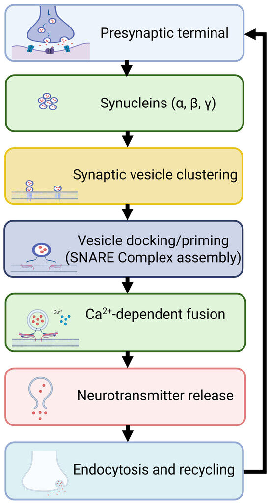
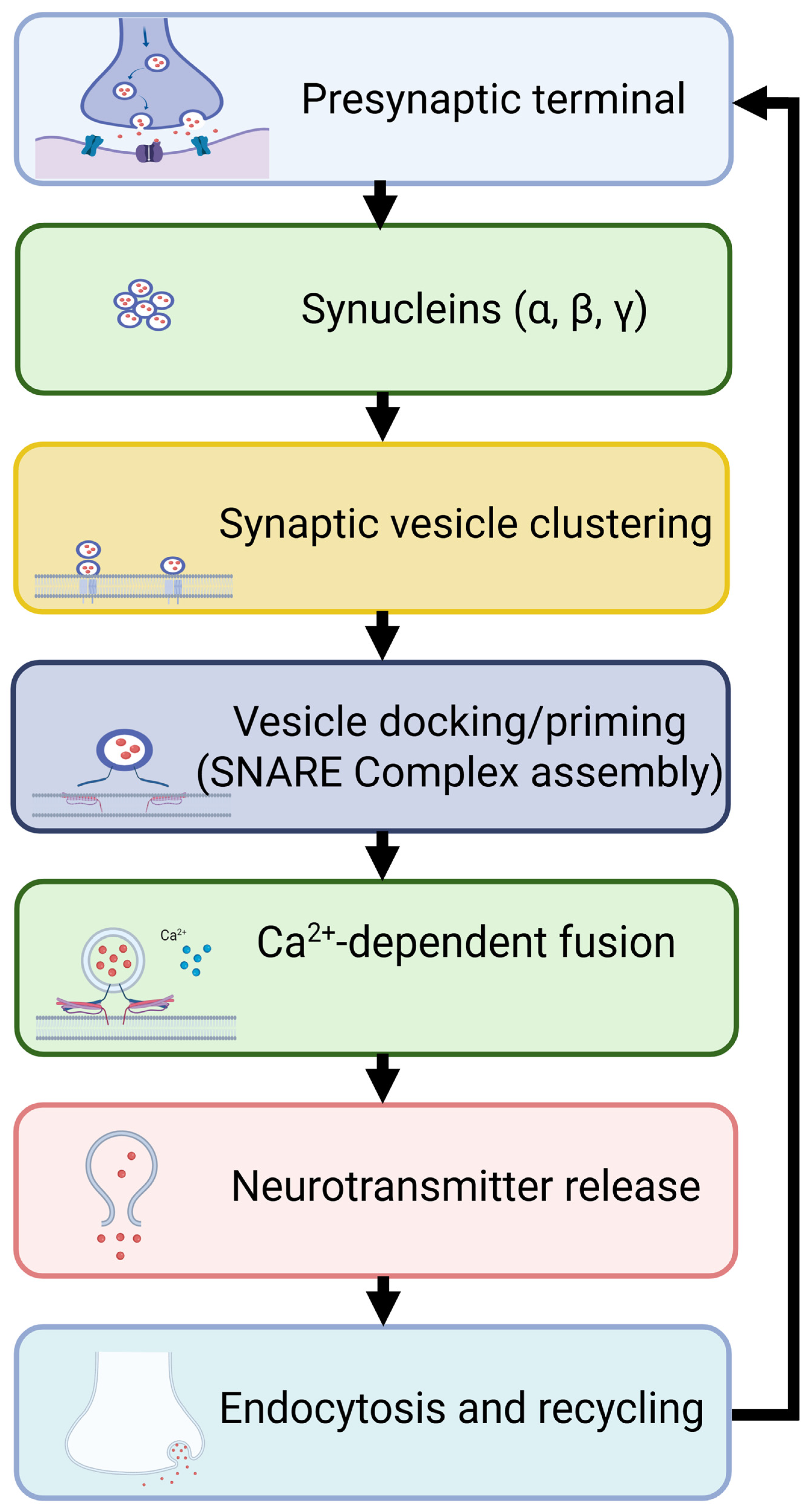
Synucleins play an important role in maintaining the balance between different vesicle pools, particularly the reserve pool and the readily releasable pool (Figure 1). Early genetic, biochemical, and physiological studies have shown that deletion of α-synuclein leads to impairments in dopaminergic neurotransmission and presynaptic function [29,30,31]. Complementary work demonstrated that α-synuclein regulates the size of synaptic vesicle pools and enhances neurotransmitter release under certain conditions [32,33].
Figure 1.
Synucleins in neurotransmission. α-, β-, and γ-synucleins are involved in the regulation of all key points of the synaptic vesicle cycle by promoting synaptic vesicle clustering, facilitating SNARE complex assembly during docking and priming, and supporting endocytosis and vesicle recycling. Altogether, synucleins contribute to efficient neurotransmitter release and the maintenance of presynaptic homeostasis. Created in Biorender. Lysikova, E. (2026) https://BioRender.com.
More recent experimental studies using genetic models have further demonstrated that the loss of α-synuclein or all three synucleins affects vesicle pool organization and dynamics. The absence of synucleins led to increased tethering of synaptic vesicles to the active zone, together with a reduction in short connector-mediated interlinking between vesicles. These ultrastructural alterations were accompanied by specific changes in protein phosphorylation in α/β/γ-triple synuclein knockout neurons [34,35]. Conversely, increased expression of α-synuclein can enhance vesicle mobilization but may also disrupt normal synaptic homeostasis [36,37,38]. Together, these findings indicate that α-synuclein acts as a fine-tuning regulator of presynaptic terminal architecture and vesicle availability, ensuring appropriate neurotransmitter release under varying physiological conditions. Although synucleins contribute to the organization and maintenance of synaptic vesicle pools, efficient neurotransmission depends on the precise coordination of subsequent stages of the synaptic vesicle cycle.
3. Synucleins in Vesicle Docking and Priming
Evidence from several experimental models indicates that α-synuclein participates not only in vesicle clustering but also in processes associated with vesicle docking, priming, fusion, and recycling, thereby contributing to the maintenance of synaptic efficiency [39,40].
The functional versatility of α-synuclein is closely linked to its intrinsically disordered structure. Comparative analyses of α-, β-, and γ-synucleins demonstrate that all family members exhibit extensive intrinsic disorder, which is an evolutionarily conserved feature that enables dynamic interactions with numerous protein and lipid partners [41]. Notably, the α-synuclein interactome is enriched in proteins involved in neurotransmission and the SV cycle, suggesting that structural flexibility is essential for its role as a presynaptic organizer [41].
Physiological concentrations of α-synuclein have been shown to support efficient synaptic transmission, whereas elevated α-synuclein levels impair synaptic vesicle cycling and reduce neurotransmitter release efficiency [42].
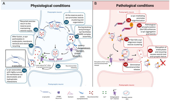
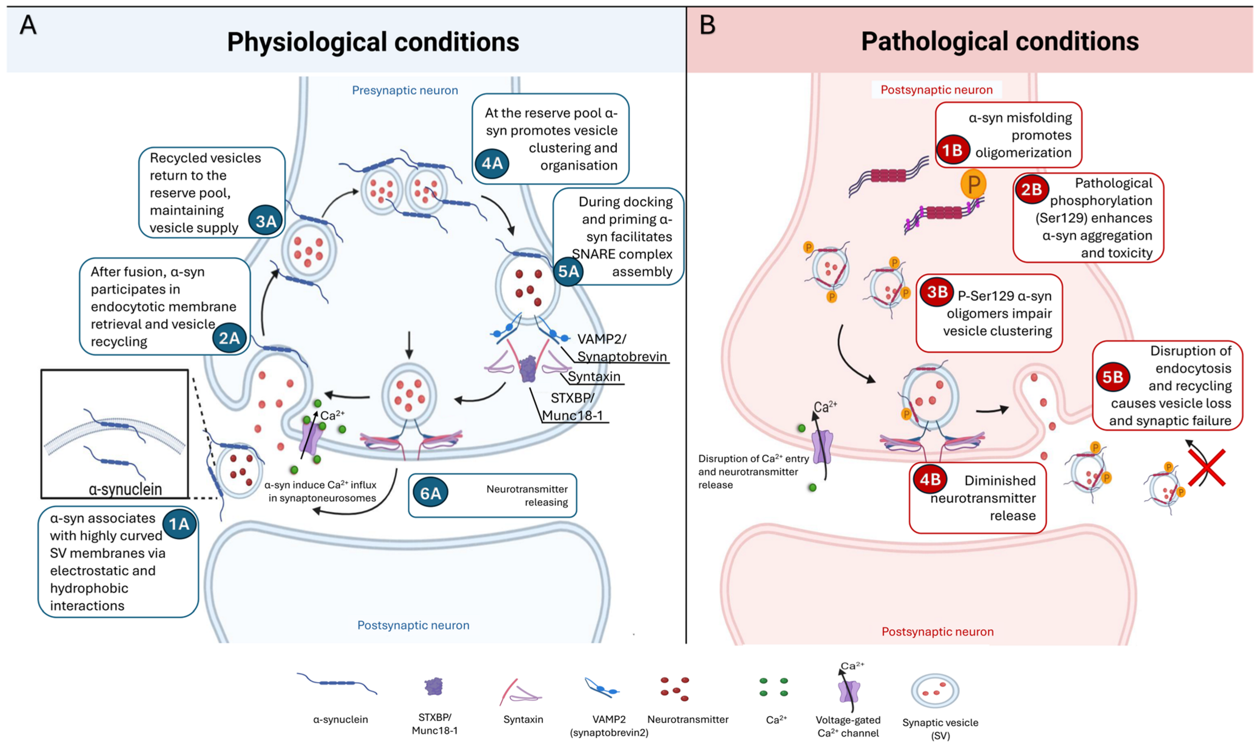
A substantial body of evidence now supports a role for α-synuclein in the regulation of exocytosis through the modulation of SNARE-complex assembly. α-Synuclein interacts with synaptobrevin-2/VAMP2 and facilitates the formation of functional SNARE complexes required for vesicle docking and fusion. This chaperone-like activity is believed to contribute to the efficiency and fidelity of neurotransmitter release. Recent proteomic and genetic studies have suggested a close relationship between α-synuclein and the presynaptic release machinery. In vivo proximity proteomics combined with CRISPR screening identified STXBP1 (Munc18-1), a key regulator of SNARE assembly, as a potential modifier of α-synuclein toxicity in dopaminergic neurons [43]. These preliminary findings further suggest that reduced STXBP1 expression may enhance neuronal vulnerability to α-synuclein, indicating that disturbances in SNARE-dependent vesicle fusion contribute to synaptic dysfunction to neurodegeneration [43] (Figure 2).
Figure 2.
Schematic representation of physiological and pathological roles of α-synuclein in the synaptic vesicle cycle. (A) Under physiological conditions, α-synuclein (α-syn) associates with highly curved synaptic vesicle (SV) membranes through electrostatic and hydrophobic interactions (1A). Following vesicle fusion, α-syn has been proposed to participate in endocytic membrane retrieval and vesicle recycling (2A), allowing recycled vesicles to return to the reserve pool and thus possibly maintain synaptic homeostasis (3A). At the reserve pool, α-syn promotes vesicle clustering and organization (4A). During docking and priming, α-syn has been proposed to facilitate SNARE complex assembly through interactions with VAMP2 (synaptobrevin-2) and associated proteins such as STXBP1/Munc18-1 (5A). α-syn also modulates calcium-dependent neurotransmitter release and contributes to efficient exocytosis (6A). (B) Under pathological conditions, elevated α-syn expression promotes misfolding and oligomerization (1B). Pathological phosphorylation at serine 129 (pS129) enhances α-syn aggregation and toxicity (2B). Excess pS129 α-syn oligomers induce abnormal vesicle microclustering and activity-dependent declustering of synaptic vesicles (3B). Disruption of docking/priming and exocytosis reduces Ca2+ entry and neurotransmitter release (4B), and impaired endocytosis leads to vesicle loss and synaptic failure (5B). α-syn, α-synuclein; SV, synaptic vesicle; SNARE, soluble NSF attachment receptor proteins; VAMP2, vesicle-associated membrane protein 2; STXBP1, syntaxin-binding protein 1; Ca2+, calcium ion; p- Ser129, phosphorylation at serine 129. Created in Biorender. Lysikova, E. (2026) https://BioRender.com.
Recent work has identified endocytic dysfunction as a central feature of several synucleinopathy models. Vidyadhara et al. demonstrated that GBA mutations, the strongest genetic risk factor for PD and DLB, induce transcriptional downregulation of genes involved in SV endocytosis and synapse assembly [44]. Importantly, these endocytic abnormalities were associated with cognitive impairment and occurred even in the absence of extensive α-synuclein pathology, suggesting that disruption of vesicle recycling can represent an independent driver of synaptic dysfunction [44].
Similarly, comprehensive analyses of presynaptic pathology in neurodegenerative disorders increasingly place endocytic failure among the earliest detectable abnormalities. Reviews focusing on SV trafficking emphasize that perturbations in vesicle retrieval, recycling, and reserve-pool maintenance occur before overt neurodegeneration in both PD and Alzheimer’s disease [40]. These findings support a model in which synaptic dysfunction emerges from cumulative defects across the entire vesicle cycle rather than from isolated disturbances in exocytosis alone.
The physiological activities of α-synuclein are strongly influenced by post-translational modifications. Among these, phosphorylation at serine 129 (pS129) is particularly important because it represents the dominant pathological species found in Lewy bodies and Lewy neurites (Figure 2).
Recent structural analyses have shown that pS129 stabilizes a more compact α-synuclein conformation, potentially altering both protein–protein and protein-membrane interactions [45]. Functional studies indicate that pS129 exhibits increased membrane affinity, enhanced oligomerization, and greater vesicle clustering capacity compared with wild-type protein [46]. However, these apparent gains of function ultimately result in impaired synaptic physiology. Excess pS129 causes activity-dependent defects in vesicle trafficking, including vesicle declustering, and the abnormal redistribution of recently endocytosed vesicles away from synaptic release sites [46]. Thus, phosphorylation appears to convert α-synuclein from a physiological organizer of vesicle pools into a pathological disruptor of SV dynamics.
Recent studies further suggest that α-synuclein functions at the intersection of lipid biology and metal homeostasis. Copper ions interact with both phosphatidylserine-containing membranes and α-synuclein, influencing membrane binding and vesicle association [47]. Synaptic vesicles may therefore participate in copper buffering and transport, while copper itself may regulate α-synuclein-mediated vesicle docking and fusion [47].
Under pathological conditions, oligomeric α-synuclein can directly damage membranes. Single-vesicle tracking experiments revealed that α-synuclein oligomers form dynamic membrane pores through a multistep process involving membrane recruitment, partial insertion, and pore maturation [48]. Such pore-forming activity could disrupt vesicle integrity, alter ion homeostasis, and exacerbate synaptic dysfunction independent of fibrillar aggregation.
Increasing evidence supports the view that presynaptic dysfunction represents one of the earliest pathogenic events in PD. Dopaminergic neurons appear particularly vulnerable because of their extensive axonal arborization, high energetic demands, and dependence on tightly regulated vesicle cycling [39]. Defects in dopamine release, vesicle recycling, and presynaptic trafficking emerge during prodromal stages of disease, often years before measurable neuronal loss [39].
Human-induced pluripotent stem cell-derived cortical neurons carrying SNCA triplications exhibit impaired SV cycling even in the absence of overt neurodegeneration, directly linking elevated α-synuclein expression to presynaptic dysfunction [42]. Likewise, proteomic analyses across multiple PD models reveal convergent alterations in synaptic proteins and vesicle trafficking pathways preceding neuronal death [43]. Together, these findings support a paradigm in which disruption of α-synuclein-mediated regulation of the SV cycle initiates a cascade of synaptic failure that ultimately culminates in neurodegeneration.
Importantly, many of the synaptic abnormalities associated with α-synuclein occur long before neuronal death, emphasizing the presynaptic terminal as a critical site of disease initiation. Future studies should focus on defining the molecular determinants that govern the transition from physiological to pathological α-synuclein states and identifying therapeutic strategies capable of restoring normal vesicle dynamics without disrupting essential synaptic functions. Such approaches may offer opportunities for disease modification during the earliest stages of Parkinson’s disease and related synucleinopathies.
Because these processes ultimately determine the efficiency and plasticity of synaptic transmission, alterations in α-synuclein function have profound consequences for neurotransmitter release and neuronal communication.
4. Involvement of Synucleins in Neurotransmitter Release and Vesicle Recycling
Following vesicle docking and priming, calcium-dependent membrane fusion, endocytosis, and synaptic vesicle recycling ensure sustained neurotransmission. Members of the synuclein family—α-, β-, and γ-synuclein—are highly enriched at presynaptic terminals and have emerged as important modulators of these processes. Although α-synuclein is best known for its pathological aggregation in Parkinson’s disease (PD) and other synucleinopathies, growing evidence indicates that synucleins are integral components of the presynaptic machinery that regulate neurotransmitter release under physiological conditions [12,49,50,51]. Accumulating evidence indicates that the functional consequences of synuclein activity differ substantially between neuronal populations. In particular, dopaminergic neurons appear to rely more heavily on synuclein-dependent regulation than many glutamatergic synapses. Conversely, alterations in synuclein expression, aggregation, or post-translational modification can profoundly disrupt neurotransmission and contribute to early synaptic dysfunction preceding neuronal degeneration [14,50,52].
The importance of α-synuclein in neurotransmission is further highlighted by studies demonstrating that loss of α-synuclein function leads to measurable defects in synaptic vesicle biology. Systematic vesicle-omics analyses in α-synuclein knockout mice have revealed significant alterations in vesicle composition, ultrastructure, and lipid organization, indicating that α-synuclein is required for maintaining normal presynaptic function and neurotransmitter release efficiency [53]. Structural studies suggest that interactions between the N-terminal and C-terminal regions of the protein generate electrostatic properties that facilitate membrane association and vesicle binding [54].
Experimental evidence supports a chaperone-like role of α-synuclein in stabilizing protein interactions necessary for efficient exocytosis [12,55]. This interaction enhances vesicle docking and fusion competence at presynaptic membranes, and this process is dependent on membrane lipid composition [56,57]. Structural and functional studies further suggest that α-synuclein promotes clustering of SNARE proteins, thereby optimizing neurotransmitter release without directly initiating membrane fusion [36,55,58].
Synucleins influence synaptic vesicle release probability by modulating fusion pore dynamics and vesicle availability. Increased α-synuclein expression accelerates the kinetics of individual exocytotic events, enhancing cargo release and decreasing fusion pore closure associated with “kiss-and-run” events, while also altering short-term synaptic plasticity [36]. These findings indicate that synuclein regulates exocytotic fusion pore dilation in a dose-dependent manner. Conversely, loss of synucleins leads to subtle but measurable impairments in synaptic transmission, indicating a regulatory rather than essential role [59,60]. Importantly, effects of synuclein are activity-dependent. During sustained or high-frequency stimulation, α-synuclein was shown to help maintain synaptic performance by regulating vesicle turnover and preventing depletion, but low-frequency stimulation leads to release depression [61]. These findings support the concept that synucleins stabilize neurotransmission under physiologically demanding conditions.
The functional role of synucleins varies across neuronal subtypes. The strongest evidence comes from dopaminergic neurons, where α-synuclein regulates dopamine vesicle trafficking, release dynamics, and synaptic plasticity [38]. Choi et al. demonstrated that α-synuclein expression is required for normal somatodendritic dopamine release within the substantia nigra, and that this effect is mediated through the modulation of L-type calcium channels (LTCCs) [62]. Loss of α-synuclein resulted in impaired dopamine release, reduced spike-frequency adaptation, diminished activity-dependent gene induction, and deficits in synaptic plasticity [62]. These findings indicate that α-synuclein contributes not only to vesicle exocytosis but also to broader activity-dependent signalling mechanisms that influence neuronal adaptation and circuit function [62]. Dysregulation of α-synuclein in these systems is closely associated with impaired dopamine handling and increased synaptic vulnerability. However, while complete absence of all three members of synuclein family substantially reduced SNARE complex assembly in total brain samples, no such effect was observed in samples from the striatum of triple synuclein knockout mice [55,60]. Moreover, the structural organization of striatal dopaminergic synapses of these mice was not different from those of wildtype mice [60], whereas significant differences were observed for CA3 hippocampal synapses [63]. These suggest different requirements for functional synucleins for different types of synapses.
The influence of α-synuclein on dopamine neurotransmission extends beyond exocytotic mechanisms. Studies using α-synuclein transgenic models demonstrate significant alterations in regional neurotransmitter metabolism and dopamine homeostasis, particularly within basal ganglia circuits [64]. Furthermore, therapeutic interventions that reduce α-synuclein accumulation restore dopamine levels and improve motor performance, supporting a close relationship between α-synuclein homeostasis and dopaminergic neurotransmission [64,65].
Calcium influx is the principal trigger of neurotransmitter release, and increasing evidence indicates that α-synuclein directly influences calcium-dependent exocytosis. While physiological α-synuclein contributes to normal coupling between calcium entry and vesicle fusion, pathological forms of the protein can profoundly alter this relationship.
Tomagra et al. demonstrated that extracellular α-synuclein oligomers selectively enhance Cav2.2 (N-type) calcium currents in substantia nigra dopaminergic neurons [66]. Increased calcium influx was associated with enhanced depolarization-evoked dopamine release and a higher frequency of quantal exocytotic events [66]. Despite promoting dopamine release, α-synuclein oligomers simultaneously reduced spontaneous neuronal firing, resulting in extracellular dopamine accumulation and intracellular calcium overload [66]. These findings illustrate how pathological α-synuclein species can initially produce apparent gains in neurotransmitter release, while simultaneously initiating mechanisms that promote neuronal vulnerability and degeneration [66].
Following calcium-dependent exocytosis, efficient neurotransmission requires rapid endocytosis and recycling of synaptic vesicles. The ability of α-synuclein to remodel lipid membranes, as demonstrated in earlier membrane interaction studies, supports a role in endocytosis and vesicle reformation following neurotransmitter release [19,21,22]. Recent evidence indicates that α-synuclein interacts with components of the endocytic machinery, facilitating efficient vesicle retrieval and maintaining synaptic fidelity [34]. A combination of optical imaging, electron microscopy, and slice electrophysiology demonstrated that synucleins play an important role in maintaining the fast kinetics of synaptic vesicle endocytosis. Significantly slowed endocytosis observed in neurons from synuclein-knockout mice could be rescued by the individual expression of α-, β-, or γ-synuclein, suggesting that all three members of the family are involved in the modulation of early stages of the endocytic branch of the synaptic vesicle recycling process [34,35]. Disruption of synuclein function can impair vesicle recycling, leading to synaptic dysfunction and reduced neurotransmission efficiency [67]. Beyond the proposed role in maintaining the spatial organization of synaptic vesicle pools, synucleins also influence subsequent stages of the synaptic vesicle cycle, including vesicle docking, priming, fusion, and membrane retrieval.
Evidence from non-neuronal secretory systems further supports a role for α-synuclein in regulating late stages of membrane fusion. In human sperm, α-synuclein enhances fusion-pore expansion during the acrosome reaction, indicating that modulation of membrane fusion dynamics may represent a conserved property of the protein [68].
Although most studies have focused on α-synuclein, β- and γ-synucleins also contribute to presynaptic function [51]. Functional analyses of knockout mice suggest that individual family members possess both overlapping and nonredundant roles in neural circuits [51]. In particular, γ-synuclein deficiency produces motor and behavioural abnormalities not observed following deletion of α- or β-synuclein alone, indicating specialized physiological functions [51]. These findings support the notion that neurotransmitter release is regulated by coordinated actions of all synuclein family members rather than α-synuclein alone [51].
The physiological role of α-synuclein contrasts sharply with the effects of its pathological forms. Misfolded, oligomeric, and aggregated α-synuclein species disrupt multiple stages of neurotransmission [14,50]. A growing body of evidence indicates that synaptic dysfunction emerges early in PD and related synucleinopathies, often preceding overt neuronal loss [14]. Ultrastructural, proteomic, and lipidomic analyses have revealed abnormalities in vesicle acidification, membrane lipid composition, and release-site architecture, collectively resulting in diminished presynaptic efficiency [14,53]. These observations support the concept that α-synuclein-mediated synaptic dysfunction represents an initiating event in PD pathogenesis rather than a secondary consequence of neurodegeneration [14].
Pathological accumulation of α-synuclein also interferes with physiological recycling mechanisms. Using a preformed fibril propagation model, Dimitrov et al. demonstrated that presynaptic accumulation of endogenous α-synuclein significantly prolongs vesicle endocytosis [52]. Mechanistically, α-synuclein accumulation promotes microtubule overassembly, leading to sequestration of dynamin, a key mediator of synaptic vesicle retrieval [52]. Impaired endocytosis compromises vesicle recycling and ultimately reduces the capacity of synapses to sustain neurotransmitter release during repetitive activity [52].
Furthermore, α-synuclein aggregates display cell-type-specific patterns of accumulation that may influence neurotransmitter systems differently. Kameda et al. demonstrated that neuronal vulnerability to α-synuclein aggregation varies according to the neurotransmitter phenotype, suggesting that neurotransmitter identity itself may influence synuclein pathology and propagation [69].
As discussed above, post-translational modifications strongly influence α-synuclein function, with phosphorylation at serine 129 (pS129) being particularly important because it represents the dominant pathological modification observed in Lewy pathology [49,50]. Beyond its effects on vesicle dynamics, α-synuclein hyperphosphorylation compromises neurotransmission by reducing the expression of proteins essential for synaptic function [70]. In enteric neurons, increased pS129 α-synuclein levels were associated with reduced expression of Synapsin-1, Synaptotagmin-1, VAMP2, and Syntaxin-1, leading to impaired neurotransmitter release [70]. Inhibition of α-synuclein hyperphosphorylation restored expression of these proteins and improved neuronal function [70]. Similarly, therapeutic approaches that reduce α-synuclein phosphorylation decrease pathological aggregation and improve dopamine neurotransmission in experimental PD models [65].
Accumulating evidence supports the concept that neurotransmitter release dysfunction is among the earliest manifestations of synucleinopathies. Synaptic abnormalities associated with α-synuclein arise before substantial neuronal loss and are increasingly recognized as primary contributors to disease progression [14,53]. Altered vesicle trafficking, disrupted calcium signaling, defective endocytosis, impaired dopamine release, and abnormal SNARE function collectively compromise synaptic communication and neuronal network activity [14,52,62,66].
Importantly, these alterations occur along a continuum. Physiological α-synuclein promotes neurotransmitter release through the regulation of vesicle dynamics and calcium-dependent exocytosis [14,49,62], whereas excessive accumulation, oligomerization, or pathological phosphorylation progressively convert α-synuclein into a mediator of synaptic failure [50,66,70]. This transition from physiological regulator to pathogenic disruptor of neurotransmission provides a mechanistic framework linking normal synaptic biology to the development of neurodegenerative disease.
Thus, synucleins, particularly α-synuclein, play essential roles in neurotransmitter release [14,49,50]. Their functions are especially important in dopaminergic neurons, where α-synuclein modulates dopamine release and activity-dependent neuronal plasticity [62,66]. However, pathological alterations in α-synuclein structure, abundance, or aggregation state, or post-translational modification, disrupt these physiological processes, leading to defective neurotransmission and early synaptic dysfunction [14,50,52,70]. The growing recognition that abnormalities in neurotransmitter release precede neurodegeneration has shifted attention toward the presynaptic terminal as a critical site of disease initiation and a promising target for therapeutic intervention in Parkinson’s disease and related synucleinopathies [14,53].
Current evidence discussed throughout this section demonstrates that α-synuclein exerts both physiological and pathological effects throughout the synaptic vesicle cycle, as summarized in Table 1.
Table 1.
Roles of α-synuclein at different stages of the synaptic vesicle cycle.
5. Common and Different Effects of Synuclein Family Members on Synaptic Vesicles
Synuclein family members exhibit both overlapping and distinct roles in neuronal function, reflecting a balance between redundancy and specialization (Table 2). A possibility of functional redundancy within the family was first proposed because of the high degree of amino acid sequence similarity, highly overlapping expression patterns, and predominant localization in neuronal presynaptic terminals, particularly for α-synuclein and β-synuclein. Early genetic studies supported this idea. Single synuclein mouse knockout models display subtle phenotypes, suggesting that remaining family members can at least partially compensate for the absent synuclein [29,30,63,71,72]. In double synuclein knockout mice, biochemical, morphological and physiological changes were slightly more pronounced [59,73,74]; however, in these models, increased expression of the remaining member of the family (i.e., γ-synuclein in α/β-double knockout and β-synuclein in α/γ-double knockout, as well as in α-single knockout) was observed [73,74,75], again suggesting functional compensation.
Not surprisingly, complete depletion of all three synucleins causes more profound effects on the biochemistry, morphology, and physiology of the knockout animal nervous system than depletion of one or two family members [60,63,76,77]. But changes, particularly the decline of synaptic function, develop mainly in ageing triple synuclein knockout mice [55,63,78], suggesting that one of synucleins’ normal functions is to counterbalance age-related deterioration of neuronal synapse physiology.
Each synuclein has a defined effect on the dopaminergic nigrostriatal system in ageing animals, as was revealed in a simultaneous study of mouse lines with all possible combinations of synuclein knockouts [59]. In the maintenance of the normal striatal content of dopamine, the main role belongs to α-synuclein, followed by β-synuclein, with no significant role for γ-synuclein. At the same time, β-synuclein, but not the two other synucleins, appeared to play a role in compensating for the age-related and nigrostriatal dopaminergic system-independent decline of animal balance and coordination in motor behavioural tests [78]. In a separate study, it was demonstrated that synucleins compete for binding sites on the surface of synaptic vesicles, and β-synuclein enhances the vesicular uptake of dopamine and structurally similar molecules, e.g., MPP+, by forming multiprotein complexes that enhance the efficiency of the VMAT2 monoamine transporter [28].
Table 2.
Comparative roles of α-, β-, and γ-Synucleins on synaptic vesicle functions.
Despite significant progress, key mechanistic questions remain unresolved, including the precise molecular interactions of synucleins, particularly on the surface of synaptic vesicles, and how their functions are coordinated within the presynaptic terminal. Improving our understanding in this area is essential, as it will not only clarify fundamental aspects of synaptic biology but also provide critical insights into how synaptic dysfunction contributes to the onset and progression of synucleinopathies.
6. Discussion
In this review, we selected the most well-documented experimental evidence demonstrating that synucleins function primarily as modulators rather than indispensable structural components of the presynaptic machinery. Their contribution becomes increasingly important during sustained neuronal activity, ageing and physiological stress, conditions under which subtle alterations in presynaptic organization may accumulate over time.
Although α-synuclein remains the most extensively studied family member because of its strong association with synucleinopathies, we also highlight the important contributions of β- and γ-synuclein to synaptic homeostasis [27,34,35]. Collectively, the data summarized in Table 2 indicate that all three members of the synuclein family share several fundamental properties such as membrane binding, presynaptic localisation, the regulation of synaptic vesicle dynamics, participation in neurotransmitter release, and partial functional redundancy. However, they differ substantially in the extent of their contribution to these processes. α-synuclein emerges as a dominant regulator of vesicle clustering and SNARE assembly and is strongly linked to synaptic pathology, while β-synuclein exerts weaker but partly compensatory and anti-aggregation effects. In contrast, γ-synuclein has less well-defined roles, with more restricted expression and weaker evidence for direct synaptic vesicle regulation.
Heteromerization of β- or γ-synuclein with α-synuclein reduces α-synuclein’s synaptic vesicle binding in a concentration-dependent manner, which supports the idea that the family cooperates by tuning α-synuclein’s presynaptic engagement rather than simply duplicating it [27]. Together with the overlap in their locations, membrane binding properties and effects on vesicle dynamics, these findings support the concept of partial functional redundancy within the family [29,63,71,72]. An earlier study on knockout mouse models also demonstrated that deletion of synucleins did not impair basic brain function or survival, indicating the presence of substantial compensatory capacity. Nevertheless, the reduction in striatal dopamine levels observed in α/β-double knockout mice compared to single knockouts highlights the physiological importance of cooperation among family members [30]. It was also reported that β-synuclein improves dopamine uptake in triple α/β/γ-synuclein-deficient synaptic vesicles [28].
At the same time, the distinct phenotypes observed in different combinations of knockout variants indicate that individual synucleins also possess specialized functions that cannot be fully compensated for by the remaining family members [78].
Provided data suggest that synucleins act as modulators rather than indispensable structural components of neurotransmission machinery [10,11]. We strongly support this emerging concept. The effects of synucleins might often be subtle under basal conditions but become increasingly important with prolonged neuronal activity, ageing or physiological stress. This concept explains why synuclein-deficient mice frequently display relatively mild phenotypes early in their life and then develop more pronounced synaptic and behavioural lesions with ageing or when another factor is applied as a trigger [78]. All these factors support the hypothesis that synucleins provide long-term stabilization of presynaptic function and protect synapses against activity-dependent or age-related decline [63].
The important relationship between the physiological and pathological functions of synucleins represents another important aspect in synaptic dynamics. Many molecular properties that are essential for normal synaptic activity, such as membrane binding [18,19,20,21,22], vesicle clustering [24,25], oligomerisation [4], and interactions with SNARE proteins [55], are also closely linked to abnormal aggregation. Therefore, neurodegeneration may arise not only from entirely novel toxic mechanisms but also because of dysregulation or exaggeration of normal physiological processes. Changes in protein concentration, impaired clearance, post-translational modifications, or compromised membrane lipid composition could shift synucleins from functional regulators toward aggregation-prone and toxic species [79,80].
The available evidence also highlights substantial synapse- and neuron-specific differences in synuclein function. Dopaminergic neurons appear particularly sensitive to α-synuclein dysfunction, consistent with the selective vulnerability observed in Parkinson’s disease. However, studies of hippocampal and cortical synapses indicate that the requirements for synuclein-dependent regulation vary considerably across neuronal populations [60,63]. Understanding the basis of this selective vulnerability remains one of the major unresolved questions in the field.
Despite considerable progress, several mechanistic issues remain incompletely understood. The precise structural organization of synucleins on synaptic vesicle membranes, the dynamics of their interactions with endocytic and exocytic proteins, and the extent of cooperation or competition between family members require further investigation. Advances in super-resolution microscopy, cryo-electron microscopy, and in vivo functional imaging are likely to provide important insights into these processes in the coming years.
7. Conclusions
Collectively, the available evidence indicates that synucleins function as dynamic modulators of presynaptic organization, enabling synapses to maintain efficient neurotransmission under varying physiological demands.
Understanding how these normal physiological functions become disrupted remains essential for explaining the transition from synaptic dysfunction to neurodegeneration and may identify new therapeutic targets for synucleinopathies.
Although α-synuclein has attracted the greatest attention because of its central role in neurodegenerative disease, β- and γ-synuclein also appear to participate in maintaining synaptic integrity and may compensate for one another under certain physiological conditions. Their ability to fine-tune vesicle pool organization, fusion pore dynamics, and endocytosis is particularly important during periods of intense neuronal activity and throughout ageing. At the same time, disruption of these normal physiological functions appears closely linked to the pathogenesis of synucleinopathies, where altered synuclein conformation and aggregation impair synaptic function and neuronal survival.
A deeper understanding of synuclein biology is therefore essential, not only for elucidating the fundamental mechanisms governing synaptic transmission but also for identifying therapeutic strategies aimed at preventing or reversing synaptic dysfunction in neurodegenerative disease. Future studies integrating molecular, structural, and physiological approaches will be crucial for clarifying how normal synuclein function transitions into pathology and how these processes may be selectively targeted in disease.
Author Contributions
Conceptualization, V.L.B. and N.N.; final writing, E.A.L., V.L.B. and N.N.; literature data collection and analysis, E.A.L., I.S.S., N.V.E., M.V.K. and M.S.K.; project administration, M.S.K. and M.V.K. All authors have read and agreed to the published version of the manuscript.
Funding
This work was supported by the Ministry of Science and Higher Education of the Russian Federation (the Federal Scientific–Technical Program for Genetic Technologies Development for 2019–2030, agreement no. 075-15-2025-558, FZWG-2026-0003), and by the IPAC RAS State Target (no. FFSG-2024-0023 to M.S.K).
Institutional Review Board Statement
Not applicable.
Informed Consent Statement
Not applicable.
Data Availability Statement
No new data were created or analyzed in this study. Data sharing is not applicable to this article.
Conflicts of Interest
The authors declare no conflicts of interest.
Abbreviations
The following abbreviations are used in this manuscript:
| VAMP2 | Vesicle associated membrane protein 2 |
| VMAT | Vesicular monoamine transporter |
| VMAT2 | Vesicular monoamine transporter 2 |
| MPP+ | 1-methyl-4-phenylpyridinium |
| PD | Parkinson’s disease |
| pS129 | serine-129-phosphorylated α-synuclein |
| SNARE | Soluble N-ethylmaleimide-sensitive fusion protein attachment protein receptor |
| STXBP1(Munc18-1) | Syntaxin-binding protein 1 |
| SV | Synaptic vesicle |
References
- Clayton, D.F.; George, J.M. Synucleins in synaptic plasticity and neurodegenerative disorders. J. Neurosci. Res. 1999, 58, 120–129. [Google Scholar] [CrossRef] [Scilit] [PubMed]
- Goedert, M.; Jakes, R.; Spillantini, M.G. The Synucleinopathies: Twenty Years On. J. Park. Dis. 2017, 7, S51–S69. [Google Scholar] [CrossRef] [Scilit] [PubMed]
- Uversky, V.N. Neuropathology, biochemistry, and biophysics of α-synuclein aggregation. J. Neurochem. 2007, 103, 17–37. [Google Scholar] [CrossRef] [Scilit] [PubMed]
- Winner, B.; Jappelli, R.; Maji, S.K.; Desplats, P.A.; Boyer, L.; Aigner, S.; Hetzer, C.; Loher, T.; Vilar, M.; Campioni, S.; et al. In vivo demonstration that α-synuclein oligomers are toxic. Proc. Natl. Acad. Sci. USA 2011, 108, 4194–4199. [Google Scholar] [CrossRef] [Scilit] [PubMed]
- Galvin, J.E.; Uryu, K.; Lee, V.M.; Trojanowski, J.Q. Axon pathology in Parkinson’s disease and Lewy body dementia hippocampus contains alpha-, beta-, and gamma-synuclein. Proc. Natl. Acad. Sci. USA 1999, 96, 13450–13455. [Google Scholar] [CrossRef] [Scilit] [PubMed]
- Fujita, M.; Sugama, S.; Sekiyama, K.; Sekigawa, A.; Tsukui, T.; Nakai, M.; Waragai, M.; Takenouchi, T.; Takamatsu, Y.; Wei, J.; et al. A β-synuclein mutation linked to dementia produces neurodegeneration when expressed in mouse brain. Nat. Commun. 2010, 1, 110. [Google Scholar] [CrossRef] [Scilit] [PubMed]
- Ohtake, H.; Limprasert, P.; Fan, Y.; Onodera, O.; Kakita, A.; Takahashi, H.; Bonner, L.T.; Tsuang, D.W.; Murray, I.V.; Lee, V.M.; et al. Beta-synuclein gene alterations in dementia with Lewy bodies. Neurology 2004, 63, 805–811. [Google Scholar] [CrossRef] [Scilit] [PubMed]
- Iwai, A.; Masliah, E.; Yoshimoto, M.; Ge, N.; Flanagan, L.; de Silva, H.A.; Kittel, A.; Saitoh, T. The precursor protein of non-A beta component of Alzheimer’s disease amyloid is a presynaptic protein of the central nervous system. Neuron 1995, 14, 467–475. [Google Scholar] [CrossRef] [Scilit] [PubMed]
- Nakajo, S.; Shioda, S.; Nakai, Y.; Nakaya, K. Localization of phosphoneuroprotein 14 (PNP 14) and its mRNA expression in rat brain determined by immunocytochemistry and in situ hybridization. Mol. Brain Res. 1994, 27, 81–86. [Google Scholar] [CrossRef] [Scilit] [PubMed]
- Huang, M.; Wang, B.; Li, X.; Fu, C.; Wang, C.; Kang, X. α-Synuclein: A Multifunctional Player in Exocytosis, Endocytosis, and Vesicle Recycling. Front. Neurosci. 2019, 13, 28. [Google Scholar] [CrossRef] [Scilit] [PubMed]
- Runwal, G.M.; Edwards, R.H. The role of α-synuclein in exocytosis. Exp. Neurol. 2024, 373, 114668. [Google Scholar] [CrossRef] [Scilit] [PubMed]
- Gao, V.; Briano, J.A.; Komer, L.E.; Burré, J. Functional and Pathological Effects of α-Synuclein on Synaptic SNARE Complexes. J. Mol. Biol. 2023, 435, 167714. [Google Scholar] [CrossRef] [Scilit] [PubMed]
- Fanning, S.; Selkoe, D.; Dettmer, U. Vesicle trafficking and lipid metabolism in synucleinopathy. Acta Neuropathol. 2021, 141, 491–510. [Google Scholar] [CrossRef] [Scilit] [PubMed]
- Treviño, M.; Guerra-Crespo, M.; Padilla-Godínez, F.J.; Ortega-Robles, E.; Arias-Carrión, O. Synaptic Vesicle Disruption in Parkinson’s Disease: Dual Roles of α-Synuclein and Emerging Therapeutic Targets. Brain Sci. 2025, 16, 7. [Google Scholar] [CrossRef] [Scilit] [PubMed]
- Xu, L.; Pu, J. Alpha-Synuclein in Parkinson’s Disease: From Pathogenetic Dysfunction to Potential Clinical Application. Park. Dis. 2016, 2016, 1720621. [Google Scholar] [CrossRef] [Scilit] [PubMed]
- von Euler Chelpin, M.; Söderberg, L.; Fälting, J.; Möller, C.; Giorgetti, M.; Constantinescu, R.; Blennow, K.; Zetterberg, H.; Höglund, K. Alpha-Synuclein Protofibrils in Cerebrospinal Fluid: A Potential Biomarker for Parkinson’s Disease. J. Park. Dis. 2020, 10, 1429–1442. [Google Scholar] [CrossRef] [Scilit] [PubMed]
- Majbour, N.; Aasly, J.; Abdi, I.; Ghanem, S.; Erskine, D.; van de Berg, W.; El-Agnaf, O. Disease-Associated α-Synuclein Aggregates as Biomarkers of Parkinson Disease Clinical Stage. Neurology 2022, 99, e2417–e2427. [Google Scholar] [CrossRef] [Scilit] [PubMed]
- Davidson, W.S.; Jonas, A.; Clayton, D.F.; George, J.M. Stabilization of α-synuclein secondary structure upon binding to synthetic membranes. J. Biol. Chem. 1998, 273, 9443–9449. [Google Scholar] [CrossRef] [Scilit] [PubMed]
- Jo, E.; McLaurin, J.; Yip, C.M.; St George-Hyslop, P.; Fraser, P.E. α-Synuclein membrane interactions and lipid specificity. J. Biol. Chem. 2000, 275, 34328–34334. [Google Scholar] [CrossRef] [Scilit] [PubMed]
- Ulmer, T.S.; Bax, A.; Cole, N.B.; Nussbaum, R.L. Structure and dynamics of micelle-bound human α-synuclein. J. Biol. Chem. 2005, 280, 9595–9603. [Google Scholar] [CrossRef] [Scilit] [PubMed]
- Westphal, C.H.; Chandra, S.S. Monomeric synucleins generate membrane curvature. J. Biol. Chem. 2013, 288, 1829–1840. [Google Scholar] [CrossRef] [Scilit] [PubMed]
- Zhu, M.; Li, J.; Fink, A.L. The association of α-synuclein with membranes affects bilayer structure, stability and fibril formation. J. Biol. Chem. 2003, 278, 40186–40197. [Google Scholar] [CrossRef] [Scilit] [PubMed]
- Makasewicz, K.; Linse, S.; Sparr, E. Interplay of α-synuclein with Lipid Membranes: Cooperative Adsorption, Membrane Remodeling and Coaggregation. JACS Au 2024, 4, 1250–1262. [Google Scholar] [CrossRef] [Scilit] [PubMed]
- Burré, J.; Sharma, M.; Südhof, T.C. α-Synuclein assembles into higher-order multimers upon membrane binding to promote SNARE complex formation. Proc. Natl. Acad. Sci. USA 2014, 111, E4274–E4283. [Google Scholar] [CrossRef] [Scilit] [PubMed]
- Fusco, G.; Pape, T.; Stephens, A.D.; Mahou, P.; Costa, A.R.; Kaminski, C.F.; Kaminski Schierle, G.S.; Vendruscolo, M.; Veglia, G.; Dobson, C.M.; et al. Structural basis of synaptic vesicle assembly promoted by α-synuclein. Nat. Commun. 2016, 7, 12563. [Google Scholar] [CrossRef] [Scilit] [PubMed]
- Li, X.; Liu, K.; Li, D.; Brunger, A.; Liu, C.; Burré, J.; Diao, J. α-Synuclein condensation in synaptic vesicle function and synucleinopathies. Trends Cell Biol. 2026, 36, 57–70. [Google Scholar] [CrossRef] [Scilit] [PubMed]
- Carnazza, K.E.; Komer, L.E.; Xie, Y.X.; Pineda, A.; Briano, J.A.; Gao, V.; Na, Y.; Ramlall, T.; Buchman, V.L.; Eliezer, D.; et al. Synaptic vesicle binding of α-synuclein is modulated by β- and γ-synucleins. Cell Rep. 2022, 39, 110675. [Google Scholar] [CrossRef] [Scilit] [PubMed]
- Ninkina, N.; Millership, S.J.; Peters, O.M.; Connor-Robson, N.; Chaprov, K.; Kopylov, A.T.; Montoya, A.; Kramer, H.; Withers, D.J.; Buchman, V.L. β-Synuclein potentiates synaptic vesicle dopamine uptake and rescues dopaminergic neurons from MPTP-induced death in the absence of other synucleins. J. Biol. Chem. 2021, 297, 101375. [Google Scholar] [CrossRef] [Scilit] [PubMed]
- Abeliovich, A.; Schmitz, Y.; Fariñas, I.; Choi-Lundberg, D.; Ho, W.H.; Castillo, P.E.; Shinsky, N.; Verdugo, J.M.; Armanini, M.; Ryan, A.; et al. Mice lacking α-synuclein display functional deficits in the nigrostriatal dopamine system. Neuron 2000, 25, 239–252. [Google Scholar] [CrossRef] [Scilit] [PubMed]
- Chandra, S.; Gallardo, G.; Fernández-Chacón, R.; Schlüter, O.M.; Südhof, T.C. Double-knockout mice for α- and β-synucleins: Effect on synaptic functions. Proc. Natl. Acad. Sci. USA 2004, 101, 14966–14971. [Google Scholar] [CrossRef] [Scilit] [PubMed]
- Yavich, L.; Tanila, H.; Vepsäläinen, S.; Jäkälä, P. Role of α-synuclein in presynaptic dopamine recruitment. J. Neurosci. 2004, 24, 11165–11170. [Google Scholar] [CrossRef] [Scilit] [PubMed]
- Murphy, D.D.; Rueter, S.M.; Trojanowski, J.Q.; Lee, V.M. Synucleins are developmentally expressed, and alpha-synuclein regulates the size of the presynaptic vesicular pool in primary hippocampal neurons. J. Neurosci. 2000, 20, 3214–3220. [Google Scholar] [CrossRef] [Scilit] [PubMed]
- Liu, S.; Ninan, I.; Antonova, I.; Battaglia, F.; Trinchese, F.; Narasanna, A.; Kolodilov, N.; Dauer, W.; Hawkins, R.D.; Arancio, O. α-Synuclein produces a long-lasting increase in neurotransmitter release. EMBO J. 2004, 23, 4506–4516. [Google Scholar] [CrossRef] [Scilit] [PubMed]
- Vargas, K.J.; Schrod, N.; Davis, T.; Fernandez-Busnadiego, R.; Taguchi, Y.V.; Laugks, U.; Lucic, V.; Chandra, S.S. Synucleins have multiple effects on presynaptic architecture. Cell Rep. 2017, 18, 161–173. [Google Scholar] [CrossRef] [Scilit] [PubMed]
- Vargas, K.J.; Makani, S.; Davis, T.; Westphal, C.H.; Castillo, P.E.; Chandra, S.S. Synucleins regulate the kinetics of synaptic vesicle endocytosis. J. Neurosci. 2014, 34, 9364–9376. [Google Scholar] [CrossRef] [Scilit] [PubMed]
- Logan, T.; Bendor, J.; Toupin, C.; Thorn, K.; Edwards, R.H. α-Synuclein promotes dilation of the exocytotic fusion pore. Nat. Neurosci. 2017, 20, 681–689. [Google Scholar] [CrossRef] [Scilit] [PubMed]
- Nemani, V.M.; Lu, W.; Berge, V.; Nakamura, K.; Onoa, B.; Lee, M.K.; Chaudhry, F.A.; Nicoll, R.A.; Edwards, R.H. Increased expression of α-synuclein reduces neurotransmitter release by inhibiting synaptic vesicle reclustering after endocytosis. Neuron 2010, 65, 66–79. [Google Scholar] [CrossRef] [Scilit] [PubMed]
- Sulzer, D.; Edwards, R.H. The physiological role of α-synuclein and its relationship to Parkinson’s disease. J. Neurochem. 2019, 150, 475–486. [Google Scholar] [CrossRef] [Scilit] [PubMed]
- Martino, E.D.; Maduro, S.; Luchicchi, A.; Wilhelmus, M.M.M.; Orefice, N.S. Distinctive characteristics of dopamine neuron release mechanisms: Insights into Parkinson’s Disease. Biomed. Pharmacother. 2026, 200, 119534. [Google Scholar] [CrossRef] [Scilit] [PubMed]
- Zhu, Y.; Zhao, L.; Li, Y.; Tian, M.; Liao, Y.; Huang, J.; Guo, P.; Xie, Y. Disruption of Synaptic Vesicle Trafficking in Alzheimer’s and Parkinson’s Disease: Mechanisms and Therapeutic Implication. Int. J. Mol. Sci. 2026, 27, 3089. [Google Scholar] [CrossRef] [Scilit] [PubMed]
- Venati, S.R.; Uversky, V.N. Exploring Intrinsic Disorder in Human Synucleins and Associated Proteins. Int. J. Mol. Sci. 2024, 25, 8399. [Google Scholar] [CrossRef] [Scilit] [PubMed]
- White, A.J.; Clark, K.A.; Alexander, K.D.; Ramalingam, N.; Young-Pearse, T.L.; Dettmer, U.; Selkoe, D.J.; Ho, G.P.H. A stem cell-based assay platform demonstrates alpha-synuclein dependent synaptic dysfunction in patient-derived cortical neurons. npj Park. Dis. 2024, 10, 107. [Google Scholar] [CrossRef] [Scilit] [PubMed]
- Shonai, D.; Kent, J.; Okafor, A.; Gao, Y.; Parameswaran, P.; Bustamante, E.; Ramani, B.; Diao, Y.; Kampmann, M.; Soderblom, E.; et al. In vivo Proximity & Spatial Proteomics with CRISPR Screening Identify STXBP1 as a Protective Modifier of α-synuclein Toxicity in Dopamine Neurons. bioRxiv 2026, preprint. [Google Scholar] [CrossRef] [Scilit] [PubMed]
- Vidyadhara, D.J.; Bäckström, D.; Chakraborty, R.; Ruan, J.; Park, J.M.; Mistry, P.K.; Chandra, S.S. Synaptic vesicle endocytosis deficits underlie cognitive dysfunction in mouse models of GBA-linked Parkinson’s disease and dementia with Lewy bodies. Nat. Commun. 2025, 16, 8484. [Google Scholar] [CrossRef] [Scilit] [PubMed]
- Dollar, A.N.; Webb, I.K. Cross-Linking and Covalent Labeling Mass Spectrometry Reveal Proteoform-Driven Conformational Changes in Alpha Synuclein. Anal. Chem. 2026, 98, 2765–2774. [Google Scholar] [CrossRef] [Scilit] [PubMed]
- Wallace, J.N.; Crockford, Z.C.; Román-Vendrell, C.; Brady, E.B.; Hoffmann, C.; Vargas, K.J.; Potcoava, M.; Wegman, M.E.; Alford, S.T.; Milovanovic, D.; et al. Excess phosphoserine-129 α-synuclein induces synaptic vesicle trafficking and declustering defects at a vertebrate synapse. Mol. Biol. Cell. 2024, 35, ar10. [Google Scholar] [CrossRef] [Scilit] [PubMed]
- Teng, X.; Stefaniak, E.; Willison, K.R.; Ying, L. Interplay between Copper, Phosphatidylserine, and α-Synuclein Suggests a Link between Copper Homeostasis and Synaptic Vesicle Cycling. ACS Chem. Neurosci. 2024, 15, 2884–2896. [Google Scholar] [CrossRef] [Scilit] [PubMed]
- Bröchner, B.V.; Zhang, X.; Nielsen, J.; Kjems, J.; Otzen, D.E.; Malle, M.G. Single-vesicle Tracking of α-Synuclein Oligomers Reveals Pore Formation by a Three-Stage Model. ACS Nano 2025, 19, 32108–32122. [Google Scholar] [CrossRef] [Scilit] [PubMed]
- Ranjan, P. Synaptic Vesicle Architecture Modulates α-Synuclein Conformation and Pathogenic Transitions in Parkinson’s Disease. Biosci. Rep. 2026, 46, BSR20260364. [Google Scholar] [CrossRef] [Scilit] [PubMed]
- Wang, M.; Xu, Y.; Wang, S.; Liang, J.; Cui, X.; Zhang, C.; Yang, J.; Sun, L.; Jin, M. Multifaceted performances of α-synuclein in health and neurological diseases. Neuroscience 2026, 600, 153–162. [Google Scholar] [CrossRef] [Scilit] [PubMed]
- Sukhanova, I.S.; Chaprov, K.D.; Morozova, O.A.; Ovchinnikov, R.K.; Kukharskaya, O.A.; Zalevskaya, V.N.; Yusupova, N.M.; Lugovskaya, A.A.; Ninkina, N.N.; Kukharsky, M.S. Distinct Role of γ-Synuclein in the Regulation of Motor Performance and Behavioral Responses in Mice. Biomedicines 2026, 14, 92. [Google Scholar] [CrossRef] [Scilit] [PubMed]
- Dimitrov, D.; Raja, S.; Noor, H.; Takahashi, T. Common Mechanism Underlying Synaptic Dysfunction Caused by Preformed Fibril-Induced Accumulation of α-Synuclein or Tau in a Culture Propagation Model. J. Neurosci. 2025, 45, e0394252025. [Google Scholar] [CrossRef] [Scilit] [PubMed]
- Gao, V.; Chlebowicz, J.; Gaskin, K.; Briano, J.A.; Komer, L.E.; Pineda, A.; Jhalani, S.; Ahmad, S.; Uwaifo, E.; Black, L.S.; et al. Synaptic vesicle-omics in mice captures signatures of aging and synucleinopathy. Nat. Commun. 2025, 16, 4079. [Google Scholar] [CrossRef] [Scilit] [PubMed]
- Onishi, N.; Mazzaferro, N.; Kunstelj, Š.; Alvarado, D.A.; Muller, A.M.; Vázquez, F.X. Molecular Dynamics Study of α-Synuclein Domain Deletion Mutant Monomers. Biomolecules 2025, 15, 1577. [Google Scholar] [CrossRef] [Scilit] [PubMed]
- Burré, J.; Sharma, M.; Südhof, T.C. α-Synuclein promotes SNARE-complex assembly in vivo and in vitro. Science 2010, 329, 1663–1667. [Google Scholar] [CrossRef] [Scilit] [PubMed]
- Man, W.K.; Tahirbegi, B.; Vrettas, M.D.; Preet, S.; Ying, L.; Vendruscolo, M.; De Simone, A.; Fusco, G. The docking of synaptic vesicles on the presynaptic membrane induced by alpha-synuclein is modulated by lipid composition. Nat. Commun. 2021, 12, 927. [Google Scholar] [CrossRef] [Scilit] [PubMed]
- Yoo, J.; Shin, Y.K.; Lee, N.K. The role of alpha-synuclein in SNARE-mediated synaptic vesicle fusion. J. Mol. Biol. 2023, 435, 167775. [Google Scholar] [CrossRef] [Scilit] [PubMed]
- Fouke, J.M.; Wegman, M.E.; Weber, S.A.; Brady, E.B.; Román-Vendrell, C.; Morgan, J.R. Synuclein regulates synaptic vesicle clustering and docking at a vertebrate synapse. Front. Cell Dev. Biol. 2021, 9, 774650. [Google Scholar] [CrossRef] [Scilit] [PubMed]
- Senior, S.L.; Ninkina, N.; Deacon, R.; Bannerman, D.; Buchman, V.L.; Cragg, S.J.; Wade-Martins, R. Increased striatal dopamine release and hyperdopaminergic-like behaviour in mice lacking both alpha-synuclein and gamma-synuclein. Eur. J. Neurosci. 2008, 27, 947–957. [Google Scholar] [CrossRef] [Scilit] [PubMed]
- Anwar, S.; Peters, O.; Millership, S.; Ninkina, N.; Doig, N.; Connor-Robson, N.; Threlfell, S.; Kooner, G.; Deacon, R.M.; Bannerman, D.M.; et al. Functional alterations to the nigrostriatal system in mice lacking all three members of the synuclein family. J. Neurosci. 2011, 31, 7264–7274. [Google Scholar] [CrossRef] [Scilit] [PubMed]
- Somayaji, M.; Cataldi, S.; Choi, S.J.; Edwards, R.H.; Mosharov, E.V.; Sulzer, D. A dual role for alpha-synuclein in facilitation and depression of dopamine release from substantia nigra neurons in vivo. Proc. Natl. Acad. Sci. USA 2020, 117, 32701–32710. [Google Scholar] [CrossRef] [Scilit] [PubMed]
- Choi, S.J.; Frank, A.; Ben-Johny, M.; Tang, G.; Bendig, J.; Fanourakis, S.; Hobson, B.D.; Ramalingam, N.; Dettmer, U.; Kanter, E.; et al. α-Synuclein expression is required for somatodendritic dopamine release and immediate early gene induction. Sci. Adv. 2026, 12, eady6978. [Google Scholar] [CrossRef] [Scilit] [PubMed]
- Greten-Harrison, B.; Polydoro, M.; Morimoto-Tomita, M.; Diao, L.; Williams, A.M.; Nie, E.H.; Makani, S.; Tian, N.; Castillo, P.E.; Buchman, V.L.; et al. αβγ-Synuclein triple knockout mice reveal age-dependent neuronal dysfunction. Proc. Natl. Acad. Sci. USA 2010, 107, 19573–19578. [Google Scholar] [CrossRef] [Scilit] [PubMed]
- Zheng, Q.; Lu, Y.; Yu, D.; Zhao, L.; Wang, Y.; Chen, P.; Lu, F.; Liu, S. Spatial metabolomics reveals the effects of Acanthopanax senticosus on region-specific alterations in neurotransmitters and metabolites levels in the brains of α-syn transgenic Parkinson’s disease model mice. Metab. Brain Dis. 2025, 40, 290. [Google Scholar] [CrossRef] [Scilit] [PubMed]
- Waheeb, T.S.; Abdulkader, M.A.; Ghareeb, D.A.; Moustafa, M.E. Neuroprotective efficacy of berberine and caffeine against rotenone-induced neuroinflammatory and oxidative disturbances associated with Parkinson’s disease via inhibiting α-synuclein aggregation and boosting dopamine release. Inflammopharmacology 2025, 33, 2129–2150. [Google Scholar] [CrossRef] [Scilit] [PubMed]
- Tomagra, G.; Battaglia, A.; Franchino, C.; Bonzano, S.; Picollo, F.; Chiantia, G.; de Iure, A.; Calabresi, P.; Picconi, B.; Carbone, E.; et al. α-Synuclein oligomers slow down action potential firing and enhance dopamine release by increasing Cav2.2 currents in midbrain dopaminergic neurons. J. Physiol. 2026, 604, 3094–3113. [Google Scholar] [CrossRef] [Scilit] [PubMed]
- Chlebowicz, M.A.; Ivanova, V.; Burré, J. New insights into synaptic vesicle dysfunction in Parkinson’s disease. J. Park. Dis. 2026, 13, 1877718X261431203. [Google Scholar] [CrossRef] [Scilit] [PubMed]
- Buzzatto, M.V.; Ibáñez, J.E.; Tomes, C.N. Cell-penetrating alpha-synuclein accelerates the late stages of the acrosome reaction in human sperm. Mol. Hum. Reprod. 2026, 32, gaag024. [Google Scholar] [CrossRef] [Scilit] [PubMed]
- Kameda, H.; Okamoto, S.; Ogasawara, K.; Okuzumi, A.; Hattori, N.; Hioki, H.; Koike, M. The extent of alpha-synuclein aggregate accumulation in neurons varies depending on the type of neurotransmitter they release. Sci. Rep. 2025, 15, 44530. [Google Scholar] [CrossRef] [Scilit] [PubMed]
- Jia, B.; Zhao, Y.; Ren, X.; Zhang, D.; Jia, H.; Wang, D.; Wang, L.; Li, J. Opioids induce constipation by prompting alpha-Synuclein hyperphosphorylation in the colonic myenteric plexus. Neurochem. Int. 2025, 190, 106036. [Google Scholar] [CrossRef] [Scilit] [PubMed]
- Cabin, D.E.; Shimazu, K.; Murphy, D.; Cole, N.B.; Gottschalk, W.; McIlwain, K.L.; Orrison, B.; Chen, A.; Ellis, C.E.; Paylor, R.; et al. Synaptic vesicle depletion correlates with attenuated synaptic responses to prolonged repetitive stimulation in mice lacking alpha-synuclein. J. Neurosci. 2002, 22, 8797–8807. [Google Scholar] [CrossRef] [Scilit] [PubMed]
- Dauer, W.; Kholodilov, N.; Vila, M.; Trillat, A.C.; Goodchild, R.; Larsen, K.E.; Staal, R.; Tieu, K.; Schmitz, Y.; Yuan, C.A.; et al. Resistance of alpha-synuclein null mice to the parkinsonian neurotoxin MPTP. Proc. Natl. Acad. Sci. USA 2002, 99, 14524–14529. [Google Scholar] [CrossRef] [Scilit] [PubMed]
- Robertson, D.C.; Schmidt, O.; Ninkina, N.; Jones, P.A.; Sharkey, J.; Buchman, V.L. Developmental loss and resistance to MPTP toxicity of dopaminergic neurones in substantia nigra pars compacta of gamma-synuclein, alpha-synuclein and double alpha/gamma-synuclein null mutant mice. J. Neurochem. 2004, 89, 1126–1136. [Google Scholar] [CrossRef] [Scilit] [PubMed]
- Chandra, S.; Gallardo, G.; Fernández-Chacón, R.; Schlüter, O.M.; Südhof, T.C. Alpha-synuclein cooperates with CSPalpha in preventing neurodegeneration. Cell 2005, 123, 383–396. [Google Scholar] [CrossRef] [Scilit] [PubMed]
- Thomas, B.; Mandir, A.S.; West, N.; Liu, Y.; Andrabi, S.A.; Stirling, W.; Dawson, V.L.; Dawson, T.M.; Lee, M.K. Resistance to MPTP-neurotoxicity in α-synuclein knockout mice is complemented by human α-synuclein and associated with increased β-synuclein and Akt activation. PLoS ONE 2011, 6, e16706. [Google Scholar] [CrossRef] [Scilit] [PubMed]
- Vorobyov, V.; Deev, A.; Sukhanova, I.; Morozova, O.; Oganesyan, Z.; Chaprov, K.; Buchman, V.L. Loss of the synuclein family members differentially affects baseline- and apomorphine-associated EEG determinants. Biomedicines 2022, 10, 3128. [Google Scholar] [CrossRef] [Scilit] [PubMed]
- Vorobyov, V.; Deev, A.; Chaprov, K.; Ninkina, N. Disruption of electroencephalogram coherence between cortex/striatum and midbrain dopaminergic regions. Biomedicines 2024, 12, 881. [Google Scholar] [CrossRef] [Scilit] [PubMed]
- Connor-Robson, N.; Peters, O.M.; Millership, S.; Ninkina, N.; Buchman, V.L. Combinational losses of synucleins reveal their differential requirements for compensating age-dependent alterations. Neurobiol. Aging 2016, 46, 107–112. [Google Scholar] [CrossRef] [Scilit] [PubMed]
- Park, H.; Kam, T.I.; Dawson, V.L.; Dawson, T.M. α-Synuclein pathology as a target in neurodegenerative diseases. Nat. Rev. Neurol. 2025, 21, 32–47. [Google Scholar] [CrossRef] [Scilit] [PubMed]
- Battis, K.; Xiang, W.; Winkler, J. The Bidirectional Interplay of α-Synuclein with Lipids in the Central Nervous System and Its Implications for the Pathogenesis of Parkinson’s Disease. Int. J. Mol. Sci. 2023, 24, 13270. [Google Scholar] [CrossRef] [Scilit] [PubMed] [PubMed Central]

Disclaimer/Publisher’s Note: The statements, opinions and data contained in all publications are solely those of the individual author(s) and contributor(s) and not of MDPI and/or the editor(s). MDPI and/or the editor(s) disclaim responsibility for any injury to people or property resulting from any ideas, methods, instructions or products referred to in the content. |
© 2026 by the authors. Licensee MDPI, Basel, Switzerland. This article is an open access article distributed under the terms and conditions of the Creative Commons Attribution (CC BY) license.