


Abstract
Transcranial magnetic stimulation-electroencephalography (TMS–EEG) biomarkers have recently become available as a means to obtain new understanding of the causal chains of neuronal signaling in the brain. This is a key piece in the puzzle of how the brain is organized and how it works. Using dMRI tractography, we can map the circuit beneath a chosen cortical target; TMS can then stimulate it, and EEG records responses that reflect—and may even be caused by—activity in that structural circuit. The chain of events after stimulus delivery can be observed and quantified using current neuroimaging and TMS–EEG technology, a matter of tremendous relevance on how to approach novel therapeutic approaches in clinical conditions. Herein, we elaborate upon a perspective of how groundbreaking multi-locus TMS (mTMS) technology associated with EEG and multimodal neuroimaging can be applied to modulate the flow dynamics of the glymphatic system (GS). The enhancement of the GS waste clearance functionality has been shown to improve significantly symptom severity in neurodegenerative disorders such as Alzheimer’s (AD) and Parkinson’s disease (PD) or long COVID. In this perspective paper, we consider that next-generation therapeutics using versatile technologies such as noninvasive neuromodulation and neuroimaging will provide important benefits in public health and in how society can address the management of these difficult-to-deal-with ailments more effectively.
1. Introduction
This perspective paper is based on the theoretical and logical assumption that a sequence of specific interventions on a dynamical system can lead to predictable outcomes and that this approach enables for control or guidance of the brain’s state. This approach capitalizes on the most advanced translational research and technological innovations facilitating the translation of knowledge from the experimental animal to the human and from the bench to the bedside. In what follows, we elaborate upon the theoretical reasoning of causal chains in the domain of neuronal signaling in the brain. We propose a specific example application using advanced transcranial magnetic stimulation-electroencephalography (TMS–EEG) and neuroimaging technologies for next-generation noninvasive neuromodulation in neurodegenerative and autoimmune disorders. The recently discovered glymphatic system (GS), a highly organized brain structure sustaining a fluid dynamic capacity, clears from the brain noxious metabolites and waste products. Remarkably, the GS evaded the attention of anatomists for more than four centuries, given that the lymphatic system, which is the GS equivalent in the rest of the body, was demonstrated in detail already in the 1600s [1] and has originally its roots in the ancient Greeks. The waste clearance function of the GS is essential for the well-being of the central nervous system (CNS); when this function is altered, the brain malfunctions. GS functionality is enhanced during sleep. Neuroimaging has enabled us to assess GS fluid dynamic functionality with a recently developed diffusion magnetic resonance imaging (dMRI) technique called diffusion tensor imaging along the perivascular space (DTI-ALPS). GS waste clearing function is seriously severed in several pathological conditions, such as neuroinflammation (e.g., long COVID) and in particular neurodegeneration such as Alzheimer’s disease (AD), resulting in the accumulation of amyloid beta, tau protein, and several other noxious or waste substances and metabolic by-products from the brain parenchyma, which progressively and increasingly damage and kill neurons and glia cells. Just recently, two studies [2,3] have demonstrated that non-invasive neuromodulation applied on specific cortical brain targets can enhance GS functionality and improve the severity of symptoms in AD as assessed using DTI-ALPS. Although remarkable and effective, these studies lack a mechanistic and hypothesis-driven approach, which is not unusual in traditional medical practice and science where an empirical approach is commonly used. The paucity of reports in this topic is a considerable limitation [4,5,6,7,8,9]. Herein, we propose how to bridge this gap with a series of steps in a hypothesis-driven logic of causality from brain circuits to clinical impact, in which we hypothesized a mechanistic aspect currently lacking in the literature. We refer to this model as causal chain from brain circuit structural–functional identity to biomarker discovery and clinical judgment. The mechanism of action proposed herein is via the autonomic nervous system modulating neurochemically the GS fluid dynamics as will be discussed later. We place this conceptual construct within the framework of modern neuroscience and propose a hypothesis-driven therapeutic approach for GS pathological alterations using TMS–EEG. In this framework, systems biology, biomarker discovery, and mechanisms of action are the essential elements. We propose that the integration of anti-inflammatory and neuromodulatory interventions guided by neuroimaging and TMS-EEG biomarkers may represent a personalized precision-medicine strategy for the treatment of GS dysfunction. Leveraging translational research and technological advances from preclinical models to clinical applications, this approach has the potential to improve therapeutic efficacy and enhance the management of GS-related disorders, including AD, PD, MS, and long COVID, thereby contributing to improved public health outcomes.
2. Relevant Sections
2.1. Brain Circuit TMS-EEG-Based Biomarkers Characterized by Neuroimaging in the Context of Modern Neuroscience and Personalized, Precision Medicine
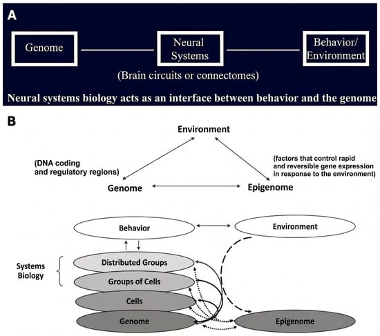
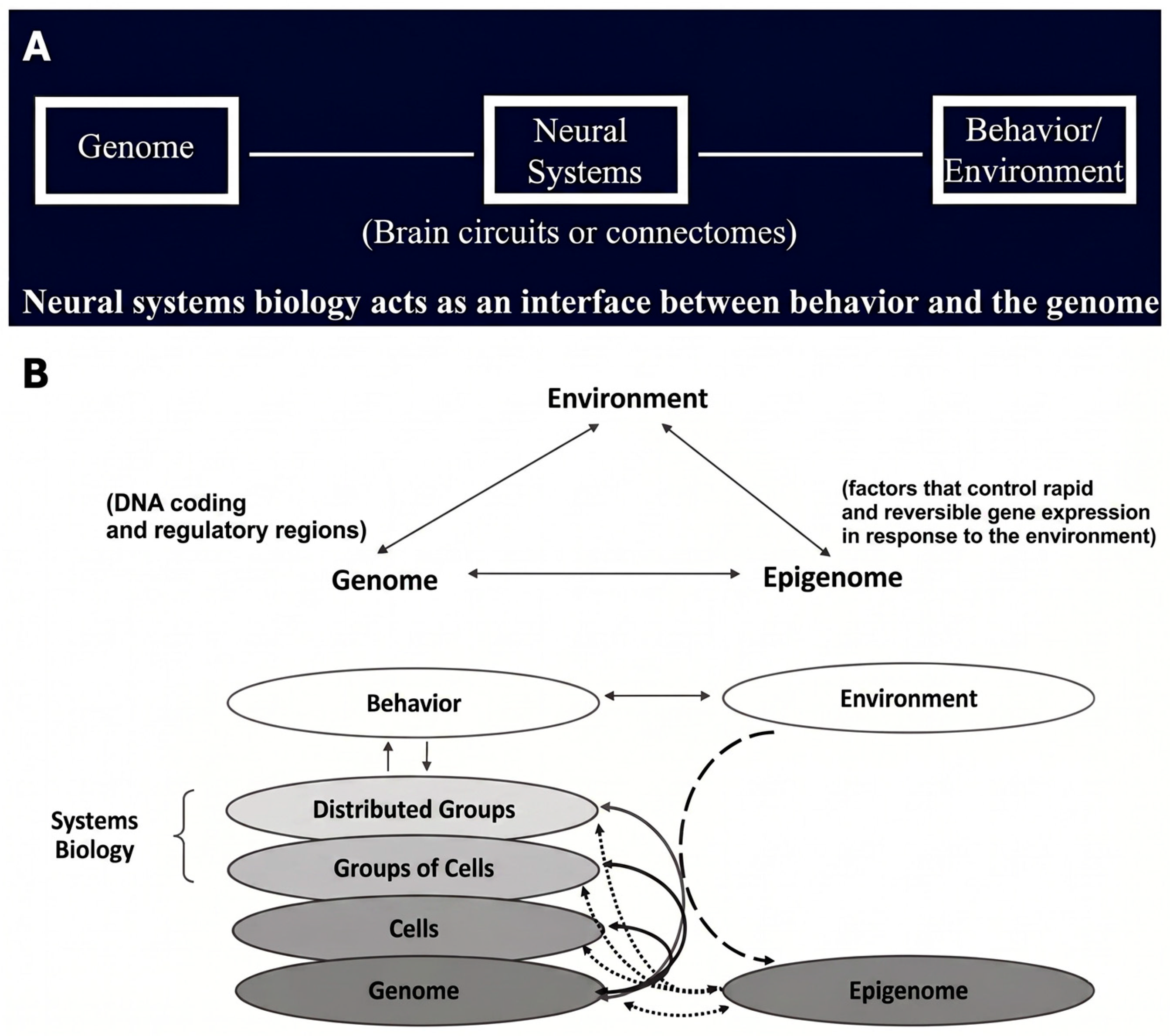
One of the essential goals in modern neuroscience is to reveal the neurobiological basis of behavior [10]. This concept has been espoused by the Research Domain Criteria (RDoC) approach since the 2000s and is changing the way we view and practice research, clinical neurology, and psychiatry (https://www.nimh.nih.gov/rdoc, (accessed on 8 June 2026)). In current neuroscience, this new framework is as important as the conceptual reasoning in modern medicine to therapeutically manipulate the microbiotic ecosystem in restoring an organism’s health [11]. Technically, the RDoC is a framework implemented as a dynamic matrix of domains of human functioning. Each one of these domains can be decomposed in several units of analysis, which are quantifiable. These units of analysis include genes, molecules, cells, circuits, physiology characteristics, self-reports, measures from paradigms and behavioral characteristics. The fact that all the units of analysis are measurable is critically important, because it allows RDoC to go beyond a qualitative approach and quantify human behavior, a topic of great importance and difficulty in current behavioral neuroscience. In this conceptual framework, the brain circuits are considered as hardwired units in the brain that underlie the diverse behaviors, which are carried out by an organism. At a broader epistemological level of biological understanding, brain circuits can be thought of as biomarkers acting as measurable biological indicators interfacing the genome and behavior as well as the environment [12,13,14,15,16,17] (Figure 1A,B).
Figure 1.
Neural circuits as an interface between the genome and behavior. (A) Schematic representation of neural systems as an intermediate level linking genomic influences with behavior and environmental interactions through brain circuits and functional connections. (B) Systems-biology model illustrating the reciprocal interactions among the genome, epigenome, environment, cellular organization, distributed neural networks, and behavior. Environmental factors may induce rapid and potentially reversible epigenetic changes, whereas neural activity and behavior can, in turn, influence gene-expression processes. Adapted with permission from Ref. [16]. 2006, Breiter et al.
Importantly, there is evidence that neural systems should be considered as putative endophenotypes in psychiatric disorders [18]. Within this theoretical framework of modern neuroscience and medicine, we hypothesize that interventions targeting the causal chains between genes and behavior will lead to more effective therapeutic outcomes in treating clinical conditions. In the continuum from genes to behavior, a causal chain is the series of stepwise transitions from one entity of the chain to the next in a causal fashion. In this approach, the brain circuits’ structural-functional identity is key in biomarker discovery and clinical judgement. In the process of determining a brain circuit structurally and functionally, the structural delineation and the electrophysiological response of this circuit are necessary requirements. In a recent study, Lioumis and colleagues (2025) showed that pre-SMA and SMA, two adjacent cortical areas with the same Brodmann cytoarchitectonic configuration in the mesial surface of each hemisphere, i.e., Brodmann area 6, exhibit distinct neurophysiological signatures shaped by their specific network characteristics—features that are rarely considered in clinical TMS targeting. That study indicated that when effecting cortical targets, they need to be selected based on their specific structural network characteristics and in combination with their neurophysiological identity [19]. Currently, this goal can be met using combined structural multimodal neuroimaging, namely T1W/T2W MRI and diffusion MRI (dMRI) tractography, to determine the specific brain circuit followed by TMS-EEG examination to record the specific electrophysiological response. Essentially, this approach allows us to stimulate a specific brain circuit based on its structural and functional identity and not just in terms of the topographic location or proximity of the cortical target zone of stimulation [19,20], a matter of great relevance in personalized treatments and precision neuropsychiatry. TMS–EEG biomarkers have recently emerged as a powerful approach for elucidating the causal dynamics of neuronal signaling in the brain [19]. Effective implementation of this method requires prior neuroimaging acquisition and analysis to inform and optimize subsequent TMS–EEG interventions. In conventional TMS neuromodulation, stimulation is typically delivered to a predetermined cortical site using predetermined pulse intensity and electric-field orientation. In contrast, our individualized precision approach begins by selecting the stimulation target based on functional MRI (fMRI), diffusion MRI (dMRI), or a combination of both. During stimulation, pulse parameters are then adaptively adjusted in real time, guided by concurrently recorded EEG responses. This allows for artifact minimization and ensures sufficiently robust TMS-evoked potentials by dynamically modifying stimulation location, orientation, and intensity, as previously demonstrated [21,22,23].
2.2. The Glymphatic System (GS): A Novel Target in Neuromodulation Therapeutics
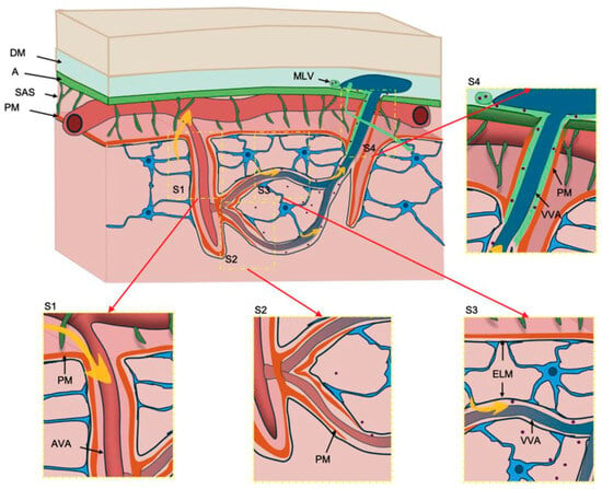
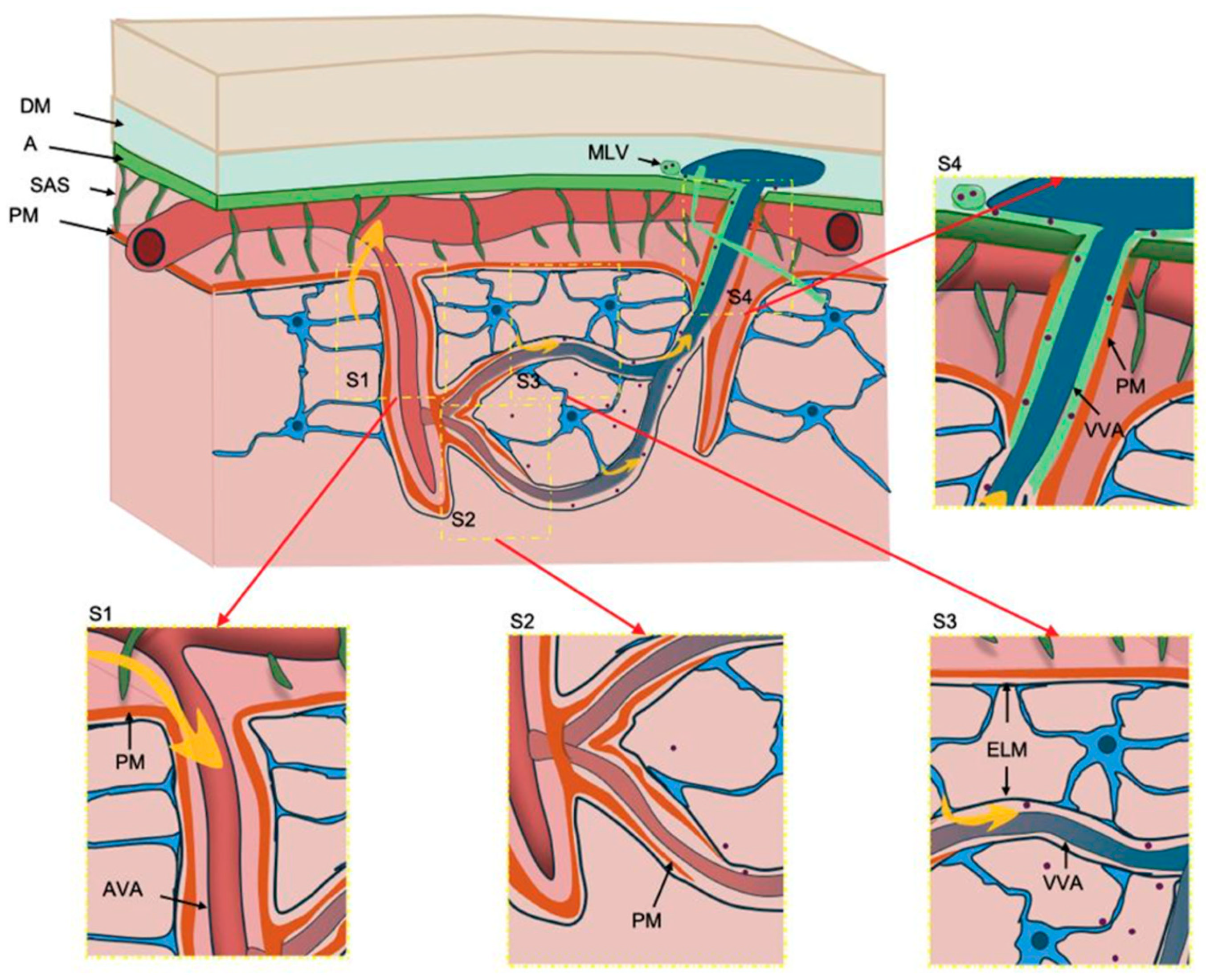
Next, we outline the structural and functional anatomy of the glymphatic system, its relevance in neurodegenerative and autoimmune disorders, and a hypothesis for a potential therapeutic approach in human GS dysfunctions. The GS is a structure whose principal function is to sustain a fluid dynamic capacity that enables the clearance of toxic metabolites and waste products. This continuous flow is driven by arterial pulsations and is portrayed by the influx of CSF from the subarachnoid space into the Virchow–Robin spaces and then to the brain parenchyma sustained by the astrocytic aquaporin-4 (AQP4) water channels en route to the perivenous spaces while transiting to the meningeal lymphatic vessels and, finally, to the cervical lymph nodes [24,25,26,27]. New insights have shed light into the roles of the glymphatic system’s four specific anatomical compartments (Figure 2) in fluid circulation and how they can be compromised in a selective way, given their distinct structural and functional roles in cerebrospinal fluid (CSF)–interstitial fluid (ISF) exchange, which results in impaired waste clearance, edema formation, and immune evasion in diverse pathological processes [28,29,30]. More specifically, the functional anatomy of the GS in its different parts would be as follows: S1 corresponds to periarterial vascular influx; cerebrospinal fluid (CSF) enters the periarterial spaces through the Virchow–Robin space, located between the arterial vascular adventitia (AVA) and the meningeal membrane (PM) [30,31,32]. S2 corresponds to the exchange of CSF and interstitial fluid where the PM becomes discontinuous, allowing CSF–interstitial fluid exchange mediated by astrocytic AQP4 channels [30,32,33]. S3 is the zone of perivascular venous parenchymal effluvium; glymphatic fluid flows along the perivenous spaces between the external limiting membrane (ELM) and the venous vascular adventitia (VVA) within the parenchyma [30,32,34]. S4 is where meningeal effluvium occurs; the flow continues in the meningeal compartment between the VVA and the PM towards the meningeal lymphatic vessels (MLV). AQP4 is relevant only in the parenchymal segments (S2–S3), but not in S1 or S4. Boxes S1–S4 highlight key anatomical features of each region [30] (Figure 2).
Figure 2.
The anatomy of the glymphatic system (GS) with its four compartments (i.e., S1, S2, S3, and S4). Abbreviations—A: arachnoid; AVA: arterial vascular adventitia; DM: dura mater; ELM: external limiting membrane; MLV: meningeal lymphatic vessel; PM: pia mater; SAS: subarachnoid space; VVA: venous vascular adventitia. See text for further details. Adapted with permission from Ref. [30]. 2025, Castañeyra-Ruiz et al.
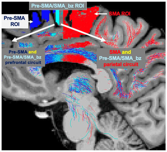
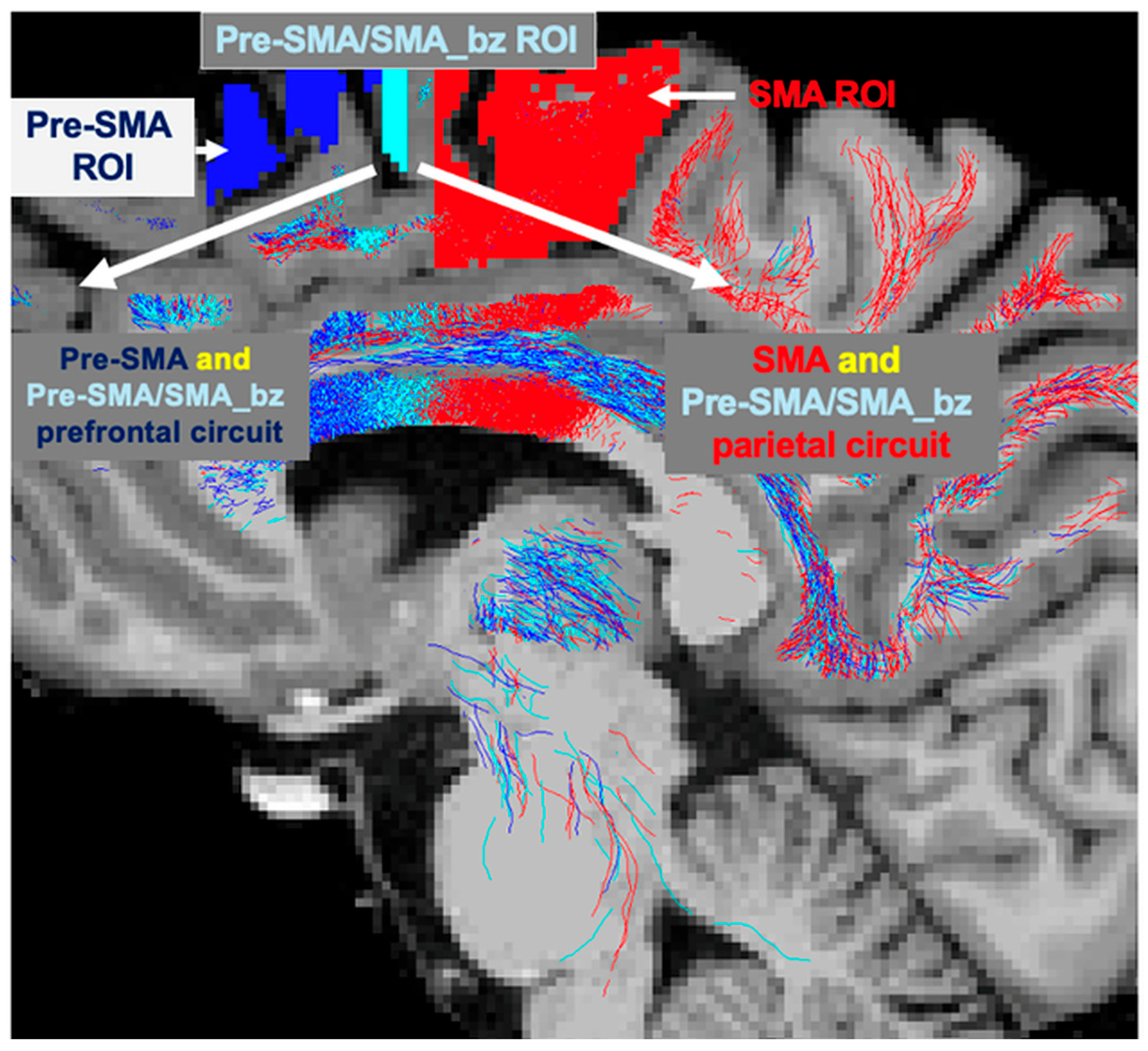
Given its vital role in clearance and removal of amyloid beta, tau protein and several other noxious or waste substances and metabolic by-products from the brain parenchyma as well as in immune surveillance [29], the study of the glymphatic system is considered critically important for developing treatments of neurodegenerative diseases such as Alzheimer’s (AD) or Parkinson’s disease (PD) as well as in autoimmune-related diseases such as long COVID. Especially, the GS is markedly boosted during restorative sleep [35] and by exercise. Recent advances in imaging have enabled the assessment of the clearance function of the glymphatic system using non-invasive tools. Neuroimaging for clinical glymphatic examinations in humans is now feasible, particularly using diffusion-imaging techniques, such as DTI-ALPS. This technique measures water diffusion along the perivascular space, a high DTI-ALPS index indicating an effective waste clearance through the glymphatic system [4,36]. Through a synthesis of non-invasive brain stimulation techniques such as TMS and neuroimaging, we can develop and implement neuromodulatory strategies with unprecedented precision, enabling individualized treatment plans for glymphatic system. Most TMS work on glymphatic function has been done in mice using animal models. Reviewing the scant literature available on this topic, TMS can be applied effectively to enhance glymphatic activity also in humans. More specifically, by applying TMS over the left parietal cortex, a therapeutic effect has been demonstrated recently, characterized by memory improvement, in older adults with mild cognitive impairment [2]. Importantly, Sudman and colleagues (2025) reported memory improvements correlated with high indices in DTI-ALPS, indicating a significant improvement in waste removal and, thus, in increased waste clearance efficiency in these subjects [2]. Another study by Gao and colleagues (2025) also showed improved waste removal by the glymphatic system as assessed by DTI-ALPS by applying repetitive TMS (rTMS) in patients with insomnia disorder [3]. Whereas Sundman et al. stimulated the parietal cortex and, consequently, the underlying parietal circuit, Gao et al. stimulated the DLPFC and the underlying prefrontal circuit. Given that both target circuits produced an improvement in GS function, it seems logical to expect that a simultaneous stimulation of both circuits would enhance their beneficial effects on the glymphatic waste clearance function. This could be achieved by stimulating the Pre-SMA/SMA_border zone cortex (Pre-SMA/SMA_bz), a cortical region with the unique property of being associated with both prefrontal and the parietal circuits (Figure 3), thus enabling us to affect both circuits simultaneously when stimulated by TMS [19].
Figure 3.
The Pre-SMA (dark blue), SMA (red) cortical ROIs and their border zone (bz) ROI Pre-SMA/SMA_bz (light blue) are shown on both panels. The Pre-SMA prefrontal circuit (dark blue) and the Pre-SMA/SMA_bz prefrontal circuit (light blue) as well as the SMA parietal circuit (red) and the Pre-SMA/SMA_bz parietal circuit (light blue) are shown in both panels. It should be noticed that the Pre-SMA/SMA_bz (light blue) ROI has the unique property to be connected with both, the prefrontal and the parietal circuits, thus sharing the connectivity profiles of the Pre-SMA and SMA ROIs. When we stimulate the Pre-SMA/SMA_bz with TMS we stimulate both circuits and not just one of them, enabling the brain to act as an integrated ensemble. Adapted with permission from Ref. [19]. 2025, Lioumis et al.
In a recent study, we demonstrated that different regions within the Pre-SMA and SMA show several different structural connectomes, each one of which is associated with a distinct neurophysiological response. Importantly, the Pre-SMA/SMA_bz has an underlying structural connectome, which shares both prefrontal and parietal characteristics [19].
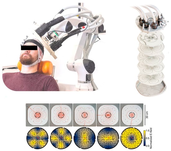
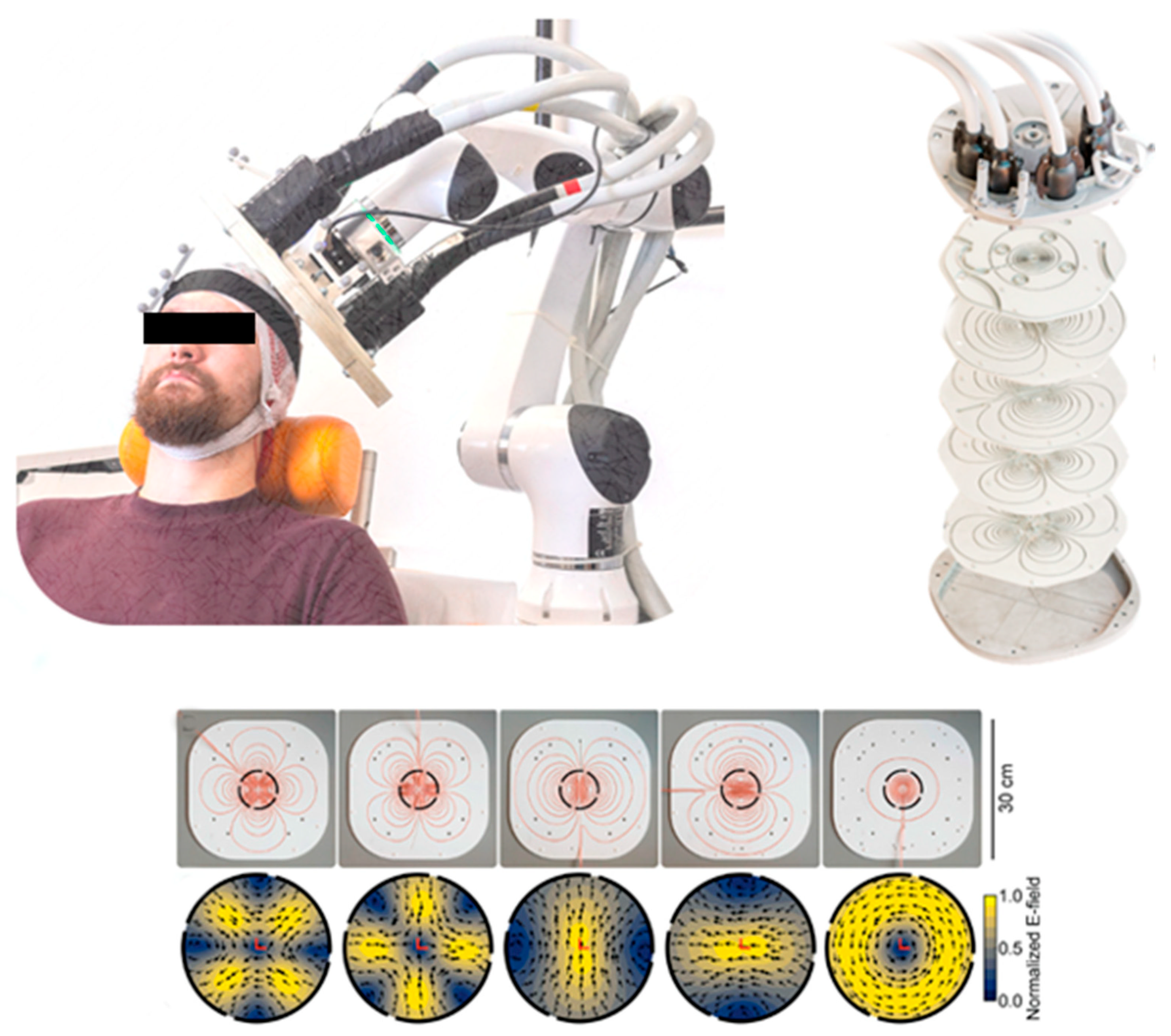
Thus, it is expected that the simultaneous neuromodulation of prefrontal and parietal circuits via the Pre-SMA/SMA_bz target would enhance glymphatic activity. Furthermore, with the novel multi-locus TMS (mTMS), we expect to increase treatment effectiveness. This is due to groundbreaking multi-channel mTMS design, which allows simultaneous stimulation of different cortical locations with different electric-field orientations and distributions [37] (Figure 4).
Figure 4.
The multi-locus transcranial magnetic stimulation (mTMS) technology. Adapted with permission from Ref. [37]. 2022, Nieminen et al.
We envision that the utilization of multimodal neuroimaging characterization of brain circuits by T1W/T2W MRI and dMRI tractography combined with mTMS-EEG application will guarantee specificity with respect to target engagement in GS noninvasive neuromodulatory treatments. This can be based on the determination of the structural and neurophysiological identity of the target and thus not only on the topographic location of the cortical target zone of stimulation as is routinely done [19,20]. Remarkably, by making possible the engagement of structurally and physiologically characterized specific brain circuits [19] capable of modulating the glymphatic system functionality, we could achieve a next-generation versatile noninvasive mTMS neuromodulatory therapy applicable in neurodegenerative disorders such as AD and PD as well as in other clinical conditions with impaired immune homeostasis such as long COVID.
2.3. Treating the Glymphatic System with Next-Generation Versatile Personalized Treatments in Neurodegenerative and Autoimmune-Mediated Disorders
Herein, we address a perspective of a next-generation versatile personalized treatment in GS dysfunction, using noninvasive mTMS-EEG neuromodulation and multimodal neuroimaging. By enhancing GS flow dynamics and its waste clearance capabilities, this therapeutic approach is expected to be an important asset in an array of clinical conditions. Prominent among these conditions are neurodegenerative diseases, such as AD and PD as well as autoimmune-mediated disorders such as long COVID.
Mechanistic aspects of glymphatic system treatment using TMS TMS could possibly influence the GS flow dynamics by cerebrospinal fluid (CSF) flow modulation, affecting its vasomotility and pulsatility. Given the well-established view of cortical-autonomic influences, particularly by the prefrontal cortex, this mechanistic effect could be carried out by prefrontal or parietal cortico-limbic and cortico-subcortical circuits stimulated by TMS at the neuronal and axonal origins or terminations of prefrontal and parietal targets [2,3,38]. Ultimately, these circuits could affect autonomic centers regulating hypothalamic and brainstem nuclei regulating sympathetic and parasympathetic release of neurotransmitters. The innervation of the GS is not a traditional direct “innervation” but rather, technically speaking, a neurochemical regulation. Thus, the functions of the GS are primarily affected by sympathetic and parasympathetic neurochemical regulation, during the sleep–wake cycle, aging, genetic factors, and body posture. The GS shows weak activity during wakefulness, while its activity increases during sleep and anesthesia [39], particularly via the sympathetic system, in which the locus coeruleus plays a significant role through norepinephrine as a neurochemical regulator. High sympathetic stimulation reduces glymphatic activity, while decreased sympathetic stimulation increases it, facilitating the exchange of CSF and ISF to clear waste metabolites. Thus, low norepinephrine concentrations allow astrocytes to relax and perivascular spaces to increase, facilitating CSF flow [40,41,42]. The cephalic parasympathetic nervous system (primarily via the facial and glossopharyngeal nerves, and to a lesser extent the vagus nerve) does not directly innervate the glymphatic system, but it can also exert neurochemical influence, producing cerebral vasodilation by modulating the pulsatility of intracranial arteries with variations in vascular caliber that would favor changes in the flow of cerebrospinal fluid (CSF) and interstitial fluid within the glymphatic system. Therefore, the parasympathetic nervous system facilitates glymphatic flow [39]. In essence, the parasympathetic nervous system enhances glymphatic function, being more active during sleep, while the sympathetic nervous system reduces glymphatic function, being more active during wakefulness.
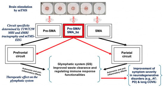
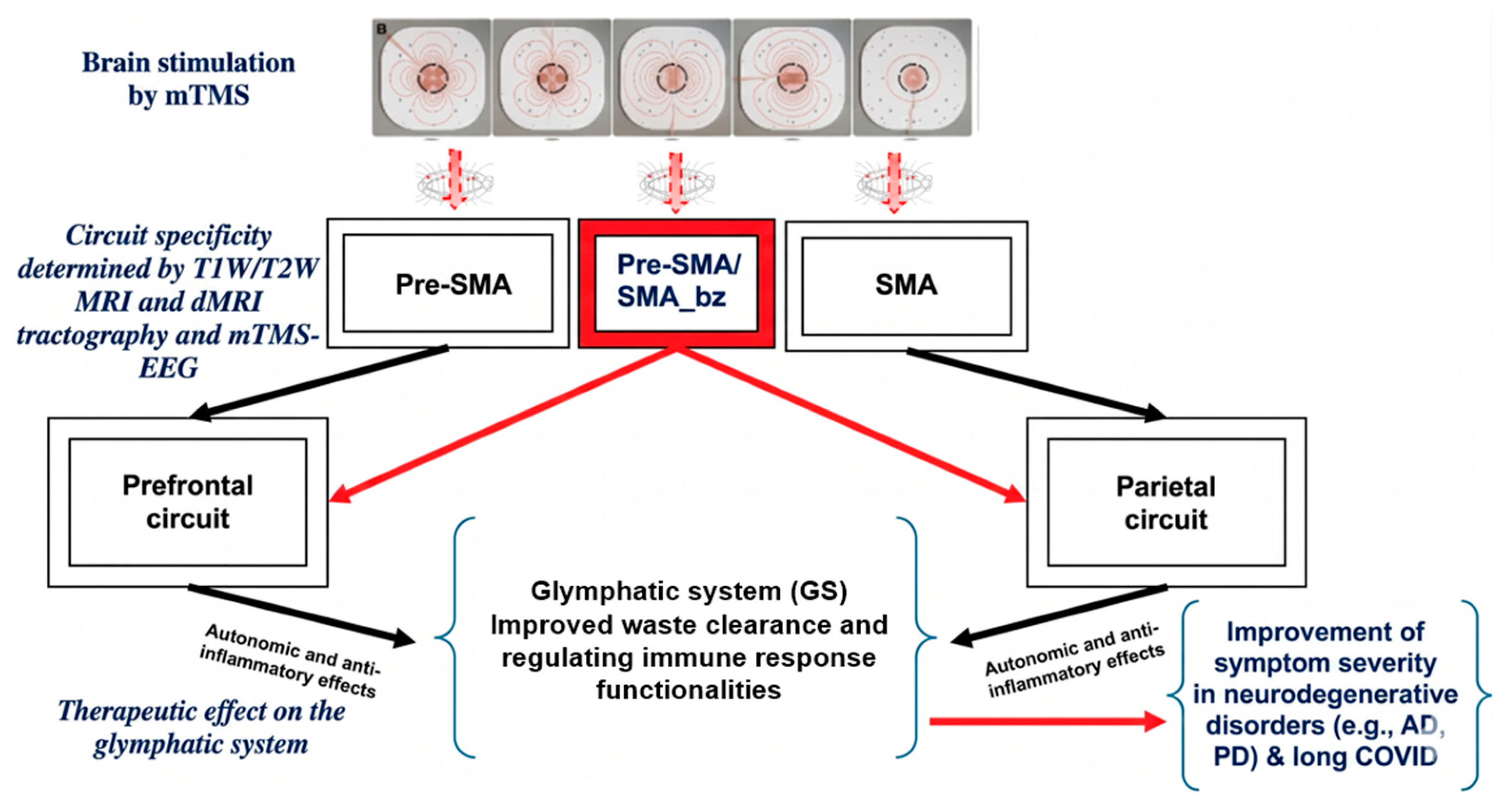
Innovative aspects of the proposed approach In this essay, we introduce a theoretical perspective following a series of steps in a logic of causality from brain circuits to clinical impact, which we refer to as causal chain from brain circuits structural–functional identity to biomarker discovery and clinical judgement. In this framework, systems biology, biomarker discovery, and mechanisms of action are the essential elements. This approach introduces several key innovations. (1) It introduces a systems neuroscience perspective to experimental therapeutics for the GS. Most work on GS has used preclinical models and focused on lifestyle interventions, such as sleep and exercise. (2) The current perspective focuses on a network-level approach of the GS that enables a mechanistic understanding of stimulation effects that cannot be inferred from behavioral endpoints alone. The only existing TMS study that has examined glymphatic function in older adults has emphasized behavioral outcomes [2]. (3) The system’s neuroscience approach moves beyond traditional models by implementing neuromodulatory strategies with unprecedented precision, enabling personalized treatment plans. The proposed approach that determines the target of stimulation based on the structurally and neurophysiologically defined circuit by combined multimodal neuroimaging and TMS-EEG in each individual patient, emphasizes the value for personalized treatments in line with NIH priorities towards individualized and precision medicine (Figure 5).
Figure 5.
Neuroimaging-guided neuromodulation in neurodegenerative and autoimmune-mediated disorders—A versatile next-generation therapeutic approach of the glymphatic system (GS) using multi-locus transcranial magnetic stimulation (mTMS)-EEG.
2.4. Limitations of the Proposed Approach and Future Studies
The proposed approach has several limitations, most notably the lack of direct experimental evidence demonstrating the effects of TMS on the autonomic nervous system, particularly on the locus coeruleus (LC) or its associated connectome. Although the hypothesis that TMS modulates glymphatic system (GS) flow dynamics is supported by a solid neuroanatomical rationale, this mechanism remains speculative. In contrast, the DTI-ALPS index is a well-established correlate of GS flow efficiency, with substantial evidence supporting its validity [4,5,6,7,8,9]. As such, it can be reliably employed as a primary outcome measure of GS dynamics, as follows: higher index values reflect increased and more efficient glymphatic waste clearance. Furthermore, a growing body of evidence suggests that TMS exerts beneficial effects on GS function, as well as across multiple disease conditions, including Alzheimer’s disease (AD), Parkinson’s disease (PD), multiple sclerosis (MS), and long COVID [43,44]. Therefore, combining TMS as a therapeutic intervention with the DTI-ALPS index as a measure of GS efficiency represents a logical and promising strategy. This approach may be particularly relevant for neurodegenerative and autoimmune disorders in which glymphatic dysfunction plays a role. Future studies are needed to elucidate the effects of TMS on the autonomic nervous system. In particular, investigations should focus on its impact on the sympathetic system, the locus coeruleus (LC), and the LC connectome, given their critical roles in the regulation of arousal. Such work represents a logical next step toward clarifying the mechanisms by which TMS may influence glymphatic system (GS) flow dynamics. Furthermore, substantial heterogeneity across patients, disease stages, stimulation protocols, neuroimaging acquisition methods, and individual variability may affect the reproducibility and generalizability of the proposed framework. Thus, the proposed approach is now presented as a future-oriented framework rather than an immediately deployable clinical protocol. Finally, we did not incorporate real-time tractography during TMS, as our approach relies on a multimodal neuroimaging workflow conducted before TMS delivery. Future advances may help overcome current practical constraints of this approach, including the increased session duration and its impact on patient compliance.
2.5. Clinical Relevance of the Glymphatic System Therapeutics and Its Cost-Benefit for Public Health and Society
Experimental and clinical studies show that sleep fragmentation, chronic insomnia, and sleep-disordered breathing (e.g., obstructive sleep apnea), reduce considerably glymphatic influx and clearance efficiency by disrupting slow-wave sleep, altering adrenergic downregulation and modifying intracranial pressure dynamics. These sleep-related disturbances not only disable the removal of neurotoxic metabolites, including amyloid-β and phosphorylated tau, but also compromise neuroimmune homeostasis, fostering a chronic pro-inflammatory state, which increases vulnerability in developing neurodegenerative conditions. Protracted impaired glymphatic function due to insufficient or non-restorative sleep may lead to accelerated cognitive decline and neuroinflammation. Moreover, the essential role of the GS in brain waste clearance and its relationship with neuroimmune mechanisms, including immune surveillance and neuroinflammation, impacts substantially neurodegenerative disease pathophysiology. TMS has been successfully used recently in GS treatment, targeting particularly the DLPFC and parietal cortex [2,3]. In Alzheimer’s disease, Aβ- and tau- protein accumulation, sleep abnormalities, and AQP4 depolarization change glymphatic clearance [45,46,47]. Recently, Sundman and colleagues (2025) investigated changes in the DTI analysis along the perivascular space (DTI-ALPS) index following theta-burst stimulation (TBS) in older adults with mild cognitive impairment (MCI) [2]. Furthermore, glymphatic dysfunction has also been demonstrated in Parkinson’s disease [48,49], Huntington’s disease (HD), amyotrophic lateral sclerosis (ALS), stroke, traumatic brain injury (TBI) and idiopathic normal pressure hydrocephalus (iNPH), and glioblastoma (GBM), as well as pathologies related to inflammation levels such as long COVID [30,34,45,46,50,51,52,53,54,55]. Moreover, current neuroimaging is a potential avenue for assessing GS fluid dynamics, in normality as well as in clinical conditions [2]. Importantly, targeting the GS offers a biologically plausible strategy to slow Alzheimer’s disease and other types of dementia as well as autoimmune-mediated disorders such as long COVID. Quantitative data, such as sleep physiology studies and neuroimaging/TMS-EEG biomarkers, support measurable benefits; delaying health decline by even a few months can save tens of thousands USD/EUR per patient [56,57]. This is a matter of great importance in public health, given that Europe has about 9.1 million people with dementia in the EU27 (2025) (approximately 12.1 million including non-EU countries) [58], while the United States has approximately 5.6 million cases [59] with substantial economic burden estimated at US$439 billion in Europe (approximately US$31,000 per patient) [56] and projected at US$781 billion in the US (approximately US$140,000 per patient) [60]. Enhancing glymphatic clearance (e.g., via sleep optimization, neuromodulation, or pharmacological/biological interventions) may slow neurodegeneration, and even modest delays in clinical progression, such as a 3-month postponement of institutionalization, can yield significant savings (approximately US$19,100 per patient; >US$1 billion overall) with an estimated about 10:1 return on investment [57], with comparable benefits observed in Europe. Policymakers should recognize that improving sleep and glymphatic function, alongside developing personalized, noninvasive neuromodulation treatments, is both scientifically supported and potentially cost-effective. These approaches deserve greater investment in clinical research and public health initiatives aimed at enhancing sleep quality and brain health. At the same time, achieving high-quality medical care while avoiding unnecessary healthcare costs depends heavily on sound clinical judgment. A physician’s clinical judgment combines medical knowledge and experience with critical thinking and intuition to guide diagnosis, treatment decisions, prognosis, and effective communication with patients. Recent work has emphasized that the integration of neuroscientific data into the clinical interview enhances the capacity of expert clinical judgment to support precision psychiatry, both in psychotherapy and neuromodulation settings [61]. In TMS, large real-world registries and mechanistic reviews show that treatment response depends on individual patient characteristics, stimulation parameters, and brain state, underscoring the need for clinician-guided, individualized evaluation of TMS candidates and protocols rather than rigid algorithmic selection [62,63]. Accordingly, the role and application of clinical judgment have assumed heightened significance within personalized medicine and precision neuropsychiatry approaches.
3. Conclusions
This perspective paper is based on a logic of causality from brain circuits to clinical impact, which we refer to as causal chain from brain circuit structural–functional identity to biomarker discovery and clinical judgement. We place this conceptual construct within the framework of modern neuroscience and propose a hypothesis-driven therapeutic approach for glymphatic system (GS) pathological alterations using TMS–EEG in the schema of personalized and precision medicine. The glymphatic system (GS) is essential for removing metabolic waste from the brain and supporting immune function. Research increasingly shows a strong connection between the glymphatic and neuroimmune systems. Efficient glymphatic activity helps clear harmful proteins and reduce inflammation, while dysfunction contributes to toxic protein buildup and chronic inflammation associated with disorders such as Alzheimer’s disease, Parkinson’s disease, multiple sclerosis, and long COVID. This relationship works in both directions, as persistent neuroinflammation can further impair glymphatic flow, creating a cycle that worsens disease progression. Understanding this interaction has led to growing interest in therapies aimed at improving glymphatic function, including lifestyle interventions like better sleep and exercise, as well as advanced approaches such as multi-locus TMS and vascular-targeted treatments. Overall, targeting the glymphatic system may provide new ways to reduce symptoms and slow the progression of neurodegenerative and autoimmune-related disorders. Herein, we propose that therapeutic models integrating the synergistic anti-inflammatory and neuromodulatory effects of neuroimaging-guided/TMS-EEG biomarkers may constitute a versatile next-generation, personalized, and precision-medicine therapeutic approach for the treatment of human GS alterations. By thoughtfully applying advanced translational research and technological innovations—from animal models to human studies, and from bench to bedside—we can develop new treatments that significantly improve public health by enabling more effective management of all challenging conditions associated with GS dysfunctions.
Funding
This research was funded by [R01MH112748, R01AG042512, K24MH116366, R01MH132610, R01MH125860, and R21NS136960, R01NS125307].
Institutional Review Board Statement
The study was conducted in accordance with the Declaration of Helsinki, and approved by the Institutional Review Board (or Ethics Committee) of Massachusetts General Hospital and Harvard Medical School, Boston, MA, USA.
Informed Consent Statement
Not applicable.
Data Availability Statement
No new data were created or analyzed in this study. Data sharing is not applicable to this article.
Acknowledgments
We would like to acknowledge the following grants for their support: R01MH112748 (NM), R01AG042512 (NM), K24MH116366, R01MH132610, R01MH125860 (NM), and R21NS136960 (NM), R01NS125307 (NM). We would like to thank Lluis Serra Majem for hosting the “International Special Winter think tank Seminar” on “Causal Chains in Research and Clinical Neuroscience: From Brain Circuits to Clinical Judgement and its Cost-Benefit Impact on Public Health and Society”, which took place in Las Palmas of Gran Canaria, Spain, on 21 February 2026, the contents of which are reflected in this perspective paper.
Conflicts of Interest
The authors declare no conflicts of interest.
References
- Park, J.; Riva, M.A. Gaspare Aselli (1581–1625) and Lacteis Venis: Four Centuries from the Discovery of Lymphatic System. Am. Surg. 2023, 89, 2325–2328. [Google Scholar] [PubMed]
- Sundman, M.H.; Liu, Y.; Chen, N.-K.; Chou, Y.-H. The Glymphatic System as a Therapeutic Target: TMS-Induced Modulation in Older Adults. Front. Aging Neurosci. 2025, 17, 1597311. [Google Scholar] [CrossRef] [Scilit] [PubMed]
- Gao, D.; Zhang, Z.; Feng, P.; Zhou, L.; Geng, Z.; Li, C.; Zhu, Y.; Yang, H. The Change of MRI Indexes of Brain Glymphatic Function and Sleep Status before and after Repeated Transcranial Magnetic Stimulation in Insomnia Disorder Patients. Front. Neurosci. 2025, 19, 1545885. [Google Scholar] [CrossRef] [Scilit] [PubMed]
- Taoka, T.; Masutani, Y.; Kawai, H.; Nakane, T.; Matsuoka, K.; Yasuno, F.; Kishimoto, T.; Naganawa, S. Evaluation of Glymphatic System Activity with the Diffusion MR Technique: Diffusion Tensor Image Analysis along the Perivascular Space (DTI-ALPS) in Alzheimer’s Disease Cases. Jpn. J. Radiol. 2017, 35, 172–178. [Google Scholar] [PubMed]
- Steward, C.E.; Venkatraman, V.K.; Lui, E.; Malpas, C.B.; Ellis, K.A.; Cyarto, E.V.; Vivash, L.; O’Brien, T.J.; Velakoulis, D.; Ames, D.; et al. Assessment of the DTI-ALPS Parameter along the Perivascular Space in Older Adults at Risk of Dementia. J. Neuroimaging 2021, 31, 569–578. [Google Scholar] [PubMed]
- Shen, T.; Yue, Y.; Ba, F.; He, T.; Tang, X.; Hu, X.; Pu, J.; Huang, C.; Lv, W.; Zhang, B.; et al. Diffusion along Perivascular Spaces as Marker for Impairment of Glymphatic System in Parkinson’s Disease. npj Park. Dis. 2022, 8, 174. [Google Scholar] [CrossRef] [Scilit]
- Zhang, Y.; Zhang, R.; Ye, Y.; Wang, S.; Jiaerken, Y.; Hong, H.; Li, K.; Zeng, Q.; Luo, X.; Xu, X.; et al. The Influence of Demographics and Vascular Risk Factors on Glymphatic Function Measured by Diffusion along Perivascular Space. Front. Aging Neurosci. 2021, 13, 693787. [Google Scholar] [CrossRef] [Scilit] [PubMed]
- Ringstad, G. Glymphatic Imaging: A Critical Look at the DTI-ALPS Index. Neuroradiology 2024, 66, 157–160. [Google Scholar] [CrossRef] [Scilit] [PubMed]
- Taoka, T.; Ito, R.; Nakamichi, R.; Nakane, T.; Kawai, H.; Naganawa, S. Diffusion Tensor Image Analysis ALong the Perivascular Space (DTI-ALPS): Revisiting the Meaning and Significance of the Method. Magn. Reson. Med. Sci. 2024, 23, 268–290. [Google Scholar] [CrossRef] [Scilit] [PubMed]
- Cowan, W.M.; Harter, D.H.; Kandel, E.R. The Emergence of Modern Neuroscience: Some Implications for Neurology and Psychiatry. Annu. Rev. Neurosci. 2000, 23, 343–391. [Google Scholar] [CrossRef] [Scilit] [PubMed]
- Whipps, J.M.; Lewis, K.; Cooke, R.C. Mycoparasitism and Plant Disease Control. In Fungi in Biological Control Systems; Manchester University Press: Manchester, UK, 1988; pp. 161–187. [Google Scholar]
- Gottesman, I.I.; Gould, T.D. The Endophenotype Concept in Psychiatry: Etymology and Strategic Intentions. Am. J. Psychiatry 2003, 160, 636–645. [Google Scholar] [CrossRef] [Scilit] [PubMed]
- Meyer-Lindenberg, A.; Weinberger, D.R. Intermediate Phenotypes and Genetic Mechanisms of Psychiatric Disorders. Nat. Rev. Neurosci. 2006, 7, 818–827. [Google Scholar] [CrossRef] [Scilit] [PubMed]
- Hyman, S.E.; Nestler, E.J. The Molecular Foundations of Psychiatry; American Psychiatric Press: Washington, DC, USA, 1993; ISBN 9780880483537. [Google Scholar]
- Makris, N.; Biederman, J.; Monuteaux, M.C.; Seidman, L.J. Towards Conceptualizing a Neural Systems-Based Anatomy of Attention-Deficit/Hyperactivity Disorder. Dev. Neurosci. 2009, 31, 36–49. [Google Scholar] [PubMed]
- Breiter, H.C.; Gasic, G.P.; Makris, N. Imaging the Neural Systems for Motivated Behavior and Their Dysfunction in Neuropsychiatric Illness. In Complex Systems Science in Biomedicine; Deisboeck, T.S., Kresh, J.Y., Eds.; Springer US: Boston, MA, USA, 2006; pp. 763–810. ISBN 9780387335322. [Google Scholar]
- John, B.; Lewis, K.R. Chromosome Variability and Geographic Distribution in Insects. Science 1966, 152, 711–721. [Google Scholar] [CrossRef] [Scilit] [PubMed]
- Makris, N. The Role of Modern Neuroimaging in Neuro-Psychoanalysis and the Visualization of Brain Circuits–Could Anger Be Neuro-Modulated? (Dynamische Psychiatrie, University of Heidelberg); Mattes Verlag GmbH: Heidelberg, Germany, 2020. [Google Scholar]
- Lioumis, P.; Roine, T.; Granö, I.; Aydogan, D.B.; Ukharova, E.; Souza, V.H.; Kičić, D.; Ilmoniemi, R.J.; Makris, N. Optimization of TMS Target Engagement: Current State and Future Perspectives. Front. Neurosci. 2025, 19, 1517228. [Google Scholar] [CrossRef] [Scilit] [PubMed]
- Kasthuri, N.; Hayworth, K.J.; Berger, D.R.; Schalek, R.L.; Conchello, J.A.; Knowles-Barley, S.; Lee, D.; Vázquez-Reina, A.; Kaynig, V.; Jones, T.R.; et al. Saturated Reconstruction of a Volume of Neocortex. Cell 2015, 162, 648–661. [Google Scholar] [CrossRef] [Scilit] [PubMed]
- Casarotto, S.; Fecchio, M.; Rosanova, M.; Varone, G.; D’Ambrosio, S.; Sarasso, S.; Pigorini, A.; Russo, S.; Comanducci, A.; Ilmoniemi, R.J.; et al. The Rt-TEP Tool: Real-Time Visualization of TMS-Evoked Potentials to Maximize Cortical Activation and Minimize Artifacts. J. Neurosci. Methods 2022, 370, 109486. [Google Scholar] [PubMed]
- Lioumis, P.; Rosanova, M. The Role of Neuronavigation in TMS-EEG Studies: Current Applications and Future Perspectives. J. Neurosci. Methods 2022, 380, 109677. [Google Scholar] [PubMed]
- Ukharova, E.; Sathyan, S.; Granö, I.; O’Meeghan, I.; Ahola, O.; Kainulainen, N.; Laurinoja, J.; Partanen, P.; Aydogan, D.B.; Ilmoniemi, R.J.; et al. Neuroimaging-Guided TMS-EEG for Real-Time Cortical Network Mapping. J. Vis. Exp. 2025, 220, e67339. [Google Scholar] [CrossRef] [Scilit] [PubMed]
- Iliff, J.J.; Wang, M.; Liao, Y.; Plogg, B.A.; Peng, W.; Gundersen, G.A.; Benveniste, H.; Vates, G.E.; Deane, R.; Goldman, S.A.; et al. A Paravascular Pathway Facilitates CSF Flow through the Brain Parenchyma and the Clearance of Interstitial Solutes, Including Amyloid β. Sci. Transl. Med. 2012, 4, 147ra111. [Google Scholar] [CrossRef] [Scilit] [PubMed]
- Iliff, J.J.; Lee, H.; Yu, M.; Feng, T.; Logan, J.; Nedergaard, M.; Benveniste, H. Brain-Wide Pathway for Waste Clearance Captured by Contrast-Enhanced MRI. J. Clin. Investig. 2013, 123, 1299–1309. [Google Scholar] [PubMed]
- Iliff, J.J.; Wang, M.; Zeppenfeld, D.M.; Venkataraman, A.; Plog, B.A.; Liao, Y.; Deane, R.; Nedergaard, M. Cerebral Arterial Pulsation Drives Paravascular CSF-Interstitial Fluid Exchange in the Murine Brain. J. Neurosci. 2013, 33, 18190–18199. [Google Scholar] [PubMed]
- Benveniste, H.; Liu, X.; Koundal, S.; Sanggaard, S.; Lee, H.; Wardlaw, J. The Glymphatic System and Waste Clearance with Brain Aging: A Review. Gerontology 2019, 65, 106–119. [Google Scholar] [PubMed]
- Iliff, J.J.; Chen, M.J.; Plog, B.A.; Zeppenfeld, D.M.; Soltero, M.; Yang, L.; Singh, I.; Deane, R.; Nedergaard, M. Impairment of Glymphatic Pathway Function Promotes Tau Pathology after Traumatic Brain Injury. J. Neurosci. 2014, 34, 16180–16193. [Google Scholar] [CrossRef] [Scilit] [PubMed]
- Kipnis, J.; Benveniste, H.; Eichmann, A.; Thomas, J.-L.; Reich, D.S.; Lewis, L.D.; Tsai, L.-H.; Drieu, A.; Bakker, E.N.T.P.; Kelley, D.H.; et al. Resolving the Mysteries of Brain Clearance and Immune Surveillance. Neuron 2025, 113, 3908–3923. [Google Scholar] [CrossRef] [Scilit] [PubMed]
- Castañeyra-Ruiz, L.; González-Marrero, I.; García-Abad, L.H.; Gonzalez-Arnay, E.; Camacho, M.; Carmona-Calero, E.M.; Lee, S.; Tran, C.T.-Q.; Hanak, B.W.; Muhonen, M.; et al. Aquaporin-4 in Glioblastoma: A Nexus of Glymphatic Dysfunction, Edema, Immune Evasion, and Treatment Resistance. Front. Cell. Neurosci. 2025, 19, 1685491. [Google Scholar] [CrossRef] [Scilit] [PubMed]
- Hablitz, L.M.; Nedergaard, M. The Glymphatic System: A Novel Component of Fundamental Neurobiology. J. Neurosci. 2021, 41, 7698–7711. [Google Scholar] [CrossRef] [Scilit] [PubMed]
- Xu, D.; Zhou, J.; Mei, H.; Li, H.; Sun, W.; Xu, H. Impediment of Cerebrospinal Fluid Drainage through Glymphatic System in Glioma. Front. Oncol. 2021, 11, 790821. [Google Scholar] [PubMed]
- Gomolka, R.S.; Hablitz, L.M.; Mestre, H.; Giannetto, M.; Du, T.; Hauglund, N.L.; Xie, L.; Peng, W.; Martinez, P.M.; Nedergaard, M.; et al. Loss of Aquaporin-4 Results in Glymphatic System Dysfunction via Brain-Wide Interstitial Fluid Stagnation. eLife 2023, 12, e82232. [Google Scholar] [CrossRef] [Scilit] [PubMed]
- Rasmussen, M.K.; Mestre, H.; Nedergaard, M. The Glymphatic Pathway in Neurological Disorders. Lancet Neurol. 2018, 17, 1016–1024. [Google Scholar] [CrossRef] [Scilit] [PubMed]
- Xie, L.; Kang, H.; Xu, Q.; Chen, M.J.; Liao, Y.; Thiyagarajan, M.; O’Donnell, J.; Christensen, D.J.; Nicholson, C.; Iliff, J.J.; et al. Sleep Drives Metabolite Clearance from the Adult Brain. Science 2013, 342, 373–377. [Google Scholar] [CrossRef] [Scilit] [PubMed]
- Boyd, E.D.; Kaur, J.; Ding, G.; Chopp, M.; Jiang, Q. Clinical Magnetic Resonance Imaging Evaluation of Glymphatic Function. NMR Biomed. 2024, 37, e5132. [Google Scholar] [CrossRef] [Scilit] [PubMed]
- Nieminen, J.O.; Sinisalo, H.; Souza, V.H.; Malmi, M.; Yuryev, M.; Tervo, A.E.; Stenroos, M.; Milardovich, D.; Korhonen, J.T.; Koponen, L.M.; et al. Multi-locus transcranial magnetic stimulation system for electronically targeted brain stimulation. Brain Stimul. 2022, 15, 116–124. [Google Scholar] [CrossRef] [Scilit] [PubMed]
- Siebner, H.R.; Funke, K.; Aberra, A.S.; Antal, A.; Bestmann, S.; Chen, R.; Classen, J.; Davare, M.; Di Lazzaro, V.; Fox, P.T.; et al. Transcranial Magnetic Stimulation of the Brain: What Is Stimulated? A Consensus and Critical Position Paper. Clin. Neurophysiol. 2022, 140, 59–97. [Google Scholar] [CrossRef] [Scilit] [PubMed]
- Nycz, B.; Mandera, M. The Features of the Glymphatic System. Auton. Neurosci. 2021, 232, 102774. [Google Scholar] [CrossRef] [Scilit] [PubMed]
- Hauglund, N.L.; Andersen, M.; Tokarska, K.; Radovanovic, T.; Kjaerby, C.; Sørensen, F.L.; Bojarowska, Z.; Untiet, V.; Ballestero, S.B.; Kolmos, M.G.; et al. Norepinephrine-Mediated Slow Vasomotion Drives Glymphatic Clearance during Sleep. Cell 2025, 188, 606–622.e17. [Google Scholar] [CrossRef] [Scilit] [PubMed]
- Wang, X.; Huang, P.; Haacke, E.M.; Wu, P.; Zhang, X.; Zhang, H.; Cheng, Z.; Tang, R.; Liu, F.; Liu, Y.; et al. MRI Index of Glymphatic System Mediates the Influence of Locus Coeruleus on Cognition in Parkinson’s Disease. Park. Relat. Disord. 2024, 123, 106558. [Google Scholar]
- Falgàs, N.; Tort-Colet, N.; Martín-Sobrino, I.; Mayà, G.; Peña-González, M.; Rudilosso, S.; Gaig, C.; Bosch, B.; Arqueros, A.; Pérez-Millan, A.; et al. Relationship between Locus Coeruleus and Slow-Wave Sleep in Aging and Alzheimer’s Disease. Alzheimers. Dement. 2026, 22, e71183. [Google Scholar] [PubMed]
- Alghanimy, A.; Work, L.M.; Holmes, W.M. The Glymphatic System and Multiple Sclerosis: An Evolving Connection. Mult. Scler. Relat. Disord. 2024, 83, 105456. [Google Scholar] [CrossRef] [Scilit] [PubMed]
- Chaganti, J.R.; Talekar, T.K.; Brew, B.J. Asymmetrical Glymphatic Dysfunction in Patients with Long Covid Associated Neurocognitive Impairment- Correlation with BBB Disruption. BMC Neurol. 2025, 25, 112. [Google Scholar] [CrossRef] [Scilit] [PubMed]
- Tang, M.; Diao, F.; Dong, Y.; Lin, J.; Wang, T.; Yang, L.; Lai, X.; Chen, X.; Zuo, J.; Xu, J.; et al. Systemic Inflammatory Response Index Mediates the Association between Stroke and Hypertension: A Cross-Sectional Study from NHANES 2001 to 2016. Front. Neurol. 2025, 16, 1603241. [Google Scholar] [CrossRef] [Scilit] [PubMed]
- Zhang, D.; Li, X.; Li, B. Glymphatic System Dysfunction in Central Nervous System Diseases and Mood Disorders. Front. Aging Neurosci. 2022, 14, 873697. [Google Scholar] [CrossRef] [Scilit] [PubMed]
- Yang, C.-C.; Huang, K.-Y.; Hsu, J.-L.; Hu, C.-J.; Lu, Y.-H.; Kuan, Y.-C. Effects of Intermittent Theta-Burst Stimulation on Cognition and Glymphatic System Activity in Mild Cognitive Impairment and Very Mild Alzheimer’s Disease: A Randomized Controlled Trial. J. Neuroeng. Rehabil. 2025, 22, 195. [Google Scholar] [PubMed]
- Lin, R.; Cai, G.; Chen, Y.; Zheng, J.; Wang, S.; Xiao, H.; Ye, Q.; Xue, Y.; Jiang, R. Association of Glymphatic System Function with Peripheral Inflammation and Motor Symptoms in Parkinson’s Disease. npj Park. Dis. 2025, 11, 62. [Google Scholar] [CrossRef] [Scilit]
- Rajai Firouzabadi, S.; Mohammadi, I.; Aghajanian, S.; Aarabi, A.; Oraee, S.; Soltani, S.M.; Mehrpour, A.; Salari, M. Glymphatic Dysfunction in Parkinson’s Disease: A Systematic Review and Meta-Analysis of Neuroimaging Studies. Mov. Disord. Clin. Pract. 2026, 13, 331–343. [Google Scholar] [PubMed]
- Noell, S.; Ritz, R.; Wolburg-Buchholz, K.; Wolburg, H.; Fallier-Becker, P. An Allograft Glioma Model Reveals the Dependence of Aquaporin-4 Expression on the Brain Microenvironment. PLoS ONE 2012, 7, e36555. [Google Scholar] [CrossRef] [Scilit] [PubMed]
- Gao, Y.; Liu, K.; Zhu, J. Glymphatic System: An Emerging Therapeutic Approach for Neurological Disorders. Front. Mol. Neurosci. 2023, 16, 1138769. [Google Scholar] [CrossRef] [Scilit] [PubMed]
- Gao, M.; Liu, Z.; Zang, H.; Wu, X.; Yan, Y.; Lin, H.; Yuan, J.; Liu, T.; Zhou, Y.; Liu, J. A Histopathologic Correlation Study Evaluating Glymphatic Function in Brain Tumors by Multiparametric MRI. Clin. Cancer Res. 2024, 30, 4876–4886. [Google Scholar] [CrossRef] [Scilit] [PubMed]
- Scalia, G.; Ferini, G.; Silven, M.P.; Noto, M.; Costanzo, R.; Maugeri, R.; Iacopino, D.G.; Zagardo, V.; Viola, A.; Nicoletti, G.F.; et al. Alterations of the Glymphatic System in Glioblastomas: A Systematic Review. Anticancer Res. 2024, 44, 3223–3230. [Google Scholar] [CrossRef] [Scilit] [PubMed]
- Mueller, S.M.; McFarland White, K.; Fass, S.B.; Chen, S.; Shi, Z.; Ge, X.; Engelbach, J.A.; Gaines, S.H.; Bice, A.R.; Vasek, M.J.; et al. Evaluation of Gliovascular Functions of AQP4 Readthrough Isoforms. Front. Cell. Neurosci. 2023, 17, 1272391. [Google Scholar] [CrossRef] [Scilit] [PubMed]
- Mou, K.; Chen, M.; Mao, Q.; Wang, P.; Ni, R.; Xia, X.; Liu, Y. AQP-4 in Peritumoral Edematous Tissue Is Correlated with the Degree of Glioma and with Expression of VEGF and HIF-Alpha. J. Neurooncol. 2010, 100, 375–383. [Google Scholar] [CrossRef] [Scilit] [PubMed]
- Alzheimer’s Association. 2025 Alzheimer’s Disease Facts and Figures. Alzheimers Dement. 2025, 21, e70235. [Google Scholar]
- Gilligan, A.M.; Malone, D.C.; Warholak, T.L.; Armstrong, E.P. Predictors of Hospitalization and Institutionalization in Medicaid Patient Populations with Alzheimer’s Disease. Adv. Alzheimer. Dis. 2013, 2, 74–82. [Google Scholar]
- Kang, S.S.; Meijer, E. Editorial: Economic Burden of Dementia in Europe. Nat. Rev. Neurol. 2022, 18, 662–663. [Google Scholar]
- Zissimopoulos, J.; Goldman, D.; Tysinger, B. The Cost of Dementia in 2025. Available online: https://schaeffer.usc.edu/research/the-cost-of-dementia-in-2025 (accessed on 30 March 2026).
- Bintener, C.; Bradshaw, A.C.; Duffner, L.; Miller, O. Alzheimer Europe Prevalence of Dementia in Europe 2025; Alzheimer Europe: Luxembourg, 2026. [Google Scholar]
- Pallanti, S. The Role of Neurosciences in Clinical Interviewing. Psychother. Psychosom. 2024, 93, 282–284. [Google Scholar] [CrossRef] [Scilit] [PubMed]
- Sackeim, H.A.; Aaronson, S.T.; Carpenter, L.L.; Hutton, T.M.; Mina, M.; Pages, K.; Verdoliva, S.; West, W.S. Clinical Outcomes in a Large Registry of Patients with Major Depressive Disorder Treated with Transcranial Magnetic Stimulation. J. Affect. Disord. 2020, 277, 65–74. [Google Scholar] [CrossRef] [Scilit] [PubMed]
- Sack, A.T.; Paneva, J.; Küthe, T.; Dijkstra, E.; Zwienenberg, L.; Arns, M.; Schuhmann, T. Target Engagement and Brain State Dependence of Transcranial Magnetic Stimulation: Implications for Clinical Practice. Biol. Psychiatry 2024, 95, 536–544. [Google Scholar] [PubMed]
Disclaimer/Publisher’s Note: The statements, opinions and data contained in all publications are solely those of the individual author(s) and contributor(s) and not of MDPI and/or the editor(s). MDPI and/or the editor(s) disclaim responsibility for any injury to people or property resulting from any ideas, methods, instructions or products referred to in the content. |
© 2026 by the authors. Licensee MDPI, Basel, Switzerland. This article is an open access article distributed under the terms and conditions of the Creative Commons Attribution (CC BY) license.