


Abstract
Wearable electrocardiogram (ECG) monitoring enables continuous cardiovascular assessment, yet signals acquired in ambulatory environments are inevitably corrupted by baseline wander, electrode motion artifacts, and muscle interference, which obscure diagnostically critical waveform features. Existing deep learning denoisers rely on heuristic attention mechanisms and time-domain-only losses, lacking principled control over what information the network retains or discards. To address this limitation, we propose EFIB-Net, an information bottleneck-guided multi-resolution network for robust ECG denoising. The framework introduces two complementary components: an efficient frequency-guided attention module that derives temporal attention weights directly from the energy distribution of parallel multi-resolution convolutional branches, requiring only four learnable parameters while providing physically interpretable feature selection that naturally highlights QRS complexes, and a variational information bottleneck constraint at the encoder–decoder bottleneck that forces the latent representation to retain only reconstruction-relevant information and discard noise, guided by a spectral–temporal composite loss. To the best of our knowledge, we are among the first to explicitly introduce the information bottleneck principle into deep-learning-based ECG signal denoising. Experiments on the MIT-BIH Arrhythmia Database show that EFIB-Net outperforms ten traditional and deep learning baselines across four standard metrics—signal-to-noise ratio (SNR), root mean square error, percentage root-mean-square difference, and correlation coefficient; at an input SNR of −5 dB it reaches dB output SNR, surpassing the strongest attention-based competitor by 1.77 dB () while using only 0.45 M parameters and 10.8 ms inference latency per segment; downstream evaluation further demonstrates that the denoised signals achieve 99.18% R-peak detection sensitivity and 91.26% heartbeat classification F1-score, both within approximately one percentage point of the clean-signal upper bound, making it practical for real-time cardiac monitoring on resource-constrained wearable devices. Zero-shot cross-database evaluation on the QT Database further confirms generalizability, with only 0.54 dB degradation without retraining.
1. Introduction
Continuous monitoring of cardiac electrical activity is essential for the early diagnosis and management of cardiovascular diseases [1]. Portable electrocardiogram (ECG) devices have become increasingly popular for long-term ambulatory monitoring owing to their noninvasive nature and ease of use [2]. Recent advances in consumer wearable technology have further accelerated this trend: the evaluation of ECG waveform accuracy in wrist-worn devices [3] and large-scale clinical trials using consumer smartwatches for atrial fibrillation screening [4] have demonstrated that continuous cardiac monitoring outside the clinic is both technically feasible and clinically valuable. Unlike clinical-grade recording systems, however, wearable devices operate in unconstrained environments where the acquired signals are inevitably degraded by various physiological and environmental interferences. The resulting signal distortion can obscure diagnostically critical waveform components [5], reduce the reliability of automated analysis algorithms, and ultimately compromise clinical decision-making. Recent advances in lightweight deep learning architectures have demonstrated the feasibility of deploying ECG analysis models directly on edge devices [6], further motivating the design of compact denoising networks that can operate under strict computational budgets.
Reliable ECG denoising is not merely a signal processing exercise but a prerequisite for accurate automated cardiac diagnosis. Deep learning models have been shown to achieve cardiologist-level performance in arrhythmia detection from single-lead ECG recordings [7], and foundation vision transformer models can substantially improve diagnostic accuracy across a broad range of cardiac conditions [8]. Multi-label classification frameworks have further enabled simultaneous identification of multiple cardiac abnormalities from standard 12-lead recordings [9]. However, the diagnostic performance of all such systems degrades substantially when the input ECG is corrupted by noise: waveform distortion can alter P-QRS-T morphology, shift ST-segment levels, and modify interval durations, leading to missed detections or false alarms. Therefore, developing robust and efficient ECG denoising methods that faithfully preserve diagnostically critical morphological features—while remaining compatible with resource-constrained wearable platforms—is an important and open research problem.
Three types of artifacts are particularly common in ambulatory ECG recordings [10]. Baseline wander (BW) arises primarily from respiration and body movement, manifesting as a slow, low-frequency drift that distorts the isoelectric line and can mimic ST-segment changes. Electrode motion (EM) artifacts result from unstable skin-electrode contact during physical activity, producing abrupt transient disturbances that overlap spectrally with the QRS complex. Muscle artifacts (MA), generated by skeletal electromyographic activity, introduce broadband high-frequency interference that can mask the fine morphology of P waves and T waves [11]. Because these three noise sources occupy different and often overlapping frequency bands, no single filtering strategy can address all of them simultaneously, which poses a fundamental challenge for ECG signal recovery. A comprehensive review of preprocessing and denoising techniques for ECG and magnetocardiography signals further illustrates the breadth and complexity of this challenge [12].
A large body of work has addressed ECG denoising through both traditional and learning-based methods (see Section 2 for a detailed review). Traditional approaches based on wavelet transforms [13], empirical mode decomposition [14], and variational mode decomposition [15] have achieved encouraging results on relatively stationary noise but rely heavily on manual hyperparameter selection and are sensitive to nonstationary artifacts. Deep learning denoisers, including fully convolutional autoencoders [16], adversarial networks [17], and Transformer-based architectures [18], have demonstrated improved adaptability through data-driven noise suppression, yet most are optimized solely with time-domain mean squared error (MSE), which tends to oversmooth the reconstructed signal and attenuate high-frequency details such as QRS peaks. More recently, attention mechanisms such as the convolutional block attention module (CBAM) [19] and efficient channel attention [20] have been integrated into denoising architectures to selectively emphasize diagnostically relevant features, and frequency-domain loss functions based on Fourier [21] and wavelet [22] representations have been proposed to preserve spectral fidelity. Despite this progress, two fundamental limitations persist across current architectures. First, existing attention modules derive their weights through heuristic operations such as global pooling followed by a multilayer perceptron (MLP), lacking a principled information-theoretic basis for determining which features carry diagnostic information and which carry noise, leaving the attention mechanism vulnerable to noise-dominated activations in very low signal-to-noise ratio (SNR) regimes. Second, while frequency-domain losses constrain the spectral properties of the output signal, they impose no explicit constraint on the internal latent representation of the network; the encoder may simultaneously encode both signal and noise information into the bottleneck, and the decoder must rely entirely on the reconstruction loss to disentangle them. From an information-theoretic perspective, both issues stem from a common root: the absence of explicit control over the amount and type of information flowing through the network’s intermediate representations.
The information bottleneck (IB) principle [23] offers a theoretically grounded framework for precisely this type of problem. The IB objective seeks a compressed representation Z that maximizes the mutual information with the target variable while minimizing with the input, thereby retaining only the information relevant for reconstruction and discarding noise. Alemi et al. [24] reformulated IB as a tractable variational objective, termed variational information bottleneck (VIB), enabling end-to-end training with deep neural networks. While IB-inspired ideas have been applied to biomedical signal classification [25], to the best of our knowledge, we are among the first to explicitly introduce IB into deep-learning-based ECG signal denoising.
Motivated by these observations, we propose EFIB-Net, an information bottleneck-guided multi-resolution network for ECG denoising. EFIB-Net addresses the two identified limitations through two complementary and independently motivated components. For efficient feature selection, we design an efficient frequency-guided attention (EFGA) module that derives temporal attention weights directly from the energy distribution of parallel multi-resolution convolutional branches, requiring only four learnable parameters while providing physically interpretable attention. For principled information compression, we introduce a VIB constraint at the network bottleneck that forces the latent representation to retain only reconstruction-relevant information, providing a theoretically grounded mechanism for noise suppression that is complementary to the spectral–temporal composite loss applied at the output. Comprehensive experiments on the MIT-BIH Arrhythmia Database demonstrate that EFIB-Net outperforms ten baselines across standard denoising metrics, and—critically—downstream evaluation confirms that the denoised signals preserve the waveform fidelity required for reliable R-peak detection and heartbeat classification, while zero-shot cross-database evaluation on the QT Database demonstrates that these improvements generalize beyond the training distribution.
The remainder of this paper is organized as follows. Section 2 reviews related work. Section 3 describes the proposed method. Section 4 presents the experimental results and analysis. Section 5 discusses the findings and their implications. Section 6 concludes the paper and summarizes the main contributions.
2. Related Work
2.1. Traditional Signal Processing Approaches
Wavelet transform-based methods decompose the signal into multiresolution subbands and apply coefficient thresholding to suppress noise. Kumar et al. [13] proposed a stationary wavelet transform denoising scheme that mitigates the translation sensitivity of the standard discrete wavelet transform (DWT), while Prashar et al. [26] designed a dual-tree complex wavelet transform framework to further improve shift invariance. Despite their effectiveness on relatively stationary signals, wavelet methods rely heavily on the manual selection of basis functions and thresholding strategies, limiting their adaptability to nonstationary artifacts [27]. Empirical mode decomposition and its extensions, such as complete ensemble empirical mode decomposition with adaptive noise (CEEMDAN) [14], offer a data-driven alternative but are prone to mode mixing and endpoint instability. Variational mode decomposition (VMD) and hybrid strategies combining VMD with empirical wavelet transform (EWT) [15,28] alleviate mode mixing to some extent but increase computational complexity without fully resolving hyperparameter sensitivity. A comprehensive review by Jia et al. [12] further illustrates the breadth and complexity of these challenges.
2.2. Deep Learning Denoisers
Over the past decade, deep neural networks have been increasingly applied to ECG denoising. Chiang et al. [16] introduced a fully convolutional denoising autoencoder (FCN-DAE) with competitive performance across multiple noise types. Das and Sahana [29] extended the autoencoder framework with deeper architectures, and Lin et al. [30] proposed a disentangled autoencoder that separates signal and noise representations in the latent space. Shaheen et al. [31] designed a fully gated denoising autoencoder with morphology-aware losses, while Wang et al. [18] proposed a convolutional Transformer framework with adaptive parametric ReLU. Adversarial training strategies have also been explored: Hou et al. [17] proposed an adversarial denoising convolutional neural network (CNN), and Mvuh et al. [32] applied a multichannel encoder–decoder generative adversarial network (GAN) to fetal ECG denoising. Hybrid residual architectures have further enhanced feature representation [33]. A recent systematic review [34] provides a comprehensive overview of deep learning architectures applied to ECG analysis. Despite encouraging results, most architectures are optimized solely with time-domain mean squared error (MSE), which tends to oversmooth the reconstructed signal, and many require substantial computational resources that hinder deployment on wearable hardware.
2.3. Attention Mechanisms and Frequency-Domain Losses
Recent work has pursued two largely independent lines of improvement. On the feature selection front, Chorney et al. [19] introduced CBAM into a convolutional autoencoder (CDAE-BAM), outperforming eight existing denoisers. Singh and Sharma [20] proposed ACDAE with efficient channel attention (ECA) on skip connections. Di Paolo and Castro [35] demonstrated attention-guided feature selection for heartbeat classification, and Ramadhan et al. [36] combined sequential and channel attention with successful edge device deployment. On the optimization front, Wang and Li [21] proposed TF-TransUNet1D with a hybrid time-frequency loss, and Chen et al. [22] designed a diagnostic-wise hybrid loss based on wavelet decomposition. Although both attention-driven feature reweighting and spectral fidelity constraints contribute measurable improvements, each line of work has so far been pursued in isolation, and none provides principled information-theoretic control over the latent representation. The present work bridges this gap by integrating frequency-guided attention with information bottleneck regularization in a unified framework.
3. Proposed Method
3.1. Problem Formulation and Architecture Overview
In ambulatory ECG monitoring, the observed signal can be modeled as an additive mixture of the underlying clean cardiac signal and nonstationary noise:
where denotes the noisy observation, the clean ECG signal, and the composite noise comprising BW, EM, and MA components. The goal of ECG denoising is to recover an estimate that is as close to as possible while preserving diagnostically relevant morphological features.
From an information-theoretic perspective, this task can be reformulated as learning a latent representation Z that retains maximal information about the clean signal while discarding noise-related content. Formally, the IB principle seeks to optimize the trade-off:
where denotes mutual information and controls the degree of compression. A larger enforces a more aggressive bottleneck, forcing the representation to retain only the most essential signal information. This formulation motivates the design of EFIB-Net, whose architecture is illustrated in Figure 1.
Figure 1.
Overall architecture of the proposed EFIB-Net. The noisy ECG input first passes through a multi-resolution decomposition layer (MRDL) with parallel convolutional branches, followed by efficient frequency-guided attention (EFGA). The refined features are then processed by a U-Net encoder–decoder with a variational information bottleneck (VIB) at the latent layer. A global residual connection produces the final denoised output.
As shown in Figure 1, EFIB-Net adopts an encoder–decoder backbone augmented with two core modules. Given a noisy ECG segment , the MRDL first extracts multi-scale features through parallel convolutional branches with kernel sizes of 3, 11, and 31. These features are then reweighted by the EFGA module, which derives temporal attention directly from the energy distribution of each branch. The attention-refined features are fed into a U-Net encoder that progressively downsamples the representation via max-pooling layers. At the deepest level, a VIB layer compresses the latent code by parameterizing it as a stochastic variable, enforcing information-theoretic compression. The U-Net decoder then reconstructs the noise estimate through transposed convolutions and skip connections from corresponding encoder levels. Finally, a global residual connection yields the denoised output:
where is the noise estimate produced by the network. This residual learning formulation simplifies the mapping that the network must learn, as ECG noise typically exhibits less structured patterns than the signal itself. Table 1 summarizes the detailed layer configuration of EFIB-Net.
Table 1.
Layer configuration of EFIB-Net. The input segment length is ; channel widths are , , ; and the VIB latent dimensionality is .
3.2. Multi-Resolution Decomposition Layer
ECG signals exhibit diagnostically relevant features across a wide range of temporal scales. Narrow QRS complexes occupy approximately 80–120 ms, corresponding to 29–43 samples at a 360 Hz sampling rate, while slower components such as BW and respiratory modulation extend over hundreds of milliseconds. A single fixed-size convolutional kernel cannot efficiently capture both fine-grained local details and broad contextual trends. Inspired by multiresolution analysis in wavelet theory, the MRDL employs three parallel one-dimensional convolutional branches with kernel sizes of 3, 11, and 31 to decompose the input into feature maps at different temporal resolutions:
where denotes a one-dimensional convolution with kernel size k, BN is batch normalization, and ReLU is the rectified linear unit activation. Each branch uses appropriate zero-padding to preserve the temporal length L. The small kernel () captures high-frequency details such as sharp R-peak transitions and subtle notches, analogous to detail coefficients in wavelet decomposition. The medium kernel () targets intermediate-scale structures including QRS complex morphology and T-wave boundaries. The large kernel () models low-frequency trends and baseline drift, similar to approximation coefficients.
The three feature maps are concatenated along the channel dimension and fused through a convolution:
where denotes channel-wise concatenation and is a pointwise convolution that projects the concatenated features back to channels. Because the projection weights are learned end-to-end, the relative contribution of each resolution branch is adapted to the noise characteristics present in the training data rather than prescribed by a fixed rule.
3.3. Efficient Frequency-Guided Attention
Existing attention modules such as CBAM and Squeeze-and-Excitation (SE) rely on global pooling followed by an MLP to generate channel and spatial attention weights. While effective, this approach introduces considerable additional parameters and lacks a direct connection to the physical content of the features being weighted. In contrast, the MRDL branches already encode frequency-specific information: the branch responds predominantly to high-frequency energy, while the branch captures low-frequency energy. The EFGA module exploits this observation by deriving temporal attention weights directly from the energy distribution across branches, thereby avoiding redundant parameterization.
Specifically, for each branch k, the instantaneous energy at time step t is computed by averaging the squared activations across channels:
The temporal attention weight is then obtained through a learnable linear combination of these energy signals followed by a sigmoid activation:
where and b are the only learnable parameters of EFGA, and denotes the sigmoid function. The attention weight is broadcast across all channels and applied element-wise to the fused feature map:
where ⊙ denotes element-wise multiplication.
EFGA introduces only four scalar parameters, in contrast to the thousands typically required by CBAM or SE. Despite this extreme compactness, the mechanism is physically interpretable: time steps dominated by high-frequency energy (e.g., QRS regions) receive higher attention because the coefficient amplifies their contribution, while quiescent segments with predominantly low-frequency content are relatively suppressed. This design choice aligns with the clinical observation that sharp waveform transitions carry the most diagnostic value and are the most vulnerable to noise corruption.
3.4. Encoder–Decoder with Variational Information Bottleneck
The attention-refined feature map is processed by a symmetric U-Net encoder–decoder. The encoder consists of stacked convolutional blocks, each comprising a one-dimensional convolution with kernel size 3, batch normalization, and ReLU activation, followed by a max-pooling layer with stride 2 for temporal downsampling. At each encoder level, the feature map is stored for subsequent skip connections. The decoder mirrors the encoder, using transposed convolutions for upsampling and concatenating the corresponding encoder features via skip connections to recover fine-grained temporal details.
At the deepest level of the encoder, a VIB layer is inserted to impose an explicit information-theoretic constraint on the latent representation. Rather than passing the encoder output h directly to the decoder, VIB parameterizes the latent variable Z as a conditional Gaussian distribution:
where and are produced by two parallel fully connected (FC) layers applied to h. During training, Z is sampled via the reparameterization trick:
During inference, only the mean is used, yielding deterministic predictions. The VIB regularization term is the Kullback–Leibler (KL) divergence between the learned posterior and a standard Gaussian prior :
where d is the dimensionality of Z. Minimizing encourages the encoder to produce a compact latent code in which noise-related dimensions are pushed toward the uninformative prior, while signal-related dimensions are preserved to minimize reconstruction error. The hyperparameter in the total loss (see Section 3.5) controls the strength of this compression: a larger enforces stronger bottleneck pressure, removing more noise at the potential cost of signal fidelity. The operating value of is therefore itself a critical design choice; its effect on performance and latent sparsity is characterized experimentally in Section 4.4.
3.5. Spectral–Temporal Composite Loss
The total training objective integrates four complementary terms:
where , , and are weighting coefficients that balance the relative contributions of the four terms. The first two terms operate in the time domain. The mean squared error penalizes the average squared deviation between the denoised output and the ground truth s, providing a strong gradient signal for global amplitude recovery. The L1 term further penalizes absolute errors, which promotes the sharpness of high-amplitude transitions such as R peaks by reducing sensitivity to outliers that would otherwise be oversmoothed by alone.
The spectral loss constrains frequency-domain fidelity by computing the L1 distance between the magnitude spectra of the predicted and target signals:
where denotes the discrete Fourier transform and the element-wise magnitude. This term prevents the network from converging to trivially smooth solutions that minimize time-domain error at the expense of spectral distortion. In particular, QRS complexes carry most of their energy in the 10–25 Hz band, and preserving this band is essential for reliable downstream arrhythmia detection. Without an explicit spectral constraint, networks trained solely with time-domain objectives tend to attenuate this band, producing pseudo-Gibbs oscillations near sharp waveform transitions. The final term , derived from the VIB formulation in Section 3.4, regularizes the latent representation from an information-theoretic perspective rather than from the output space.
Figure 2 illustrates the complementary roles of the four loss components. Panel (a) depicts the time-domain terms, which penalize point-wise amplitude differences between and ; the shaded region between the two waveforms visualizes the instantaneous residual that and jointly minimize. Panel (b) illustrates the spectral term, which aligns the magnitude spectra and preserves QRS-band energy by penalizing the gap within the diagnostically critical frequency range. Panel (c) visualizes the effect of the KL term on the latent code: after information-theoretic compression, only a small subset of latent dimensions retains substantial information about the clean signal (red bars), while the remaining dimensions are driven toward the uninformative prior (gray bars). The three panels together make the role of each term explicit: time-domain and spectral terms constrain the output of the network, whereas the KL term operates at the level of the internal representation.
Figure 2.
Conceptual illustration of the spectral–temporal composite loss. (a) Time-domain losses penalize point-wise amplitude differences between the prediction and the ground truth ; the shaded region denotes the residual . (b) Spectral loss aligns the magnitude spectra and , preserving QRS-band (10–25 Hz) energy and preventing oversmoothing. (c) KL term compresses the latent representation so that only signal-relevant dimensions (red) retain substantial information, while noise-related dimensions (gray) are driven toward the uninformative prior . The three terms jointly implement the information bottleneck trade-off in a tractable and end-to-end differentiable manner.
Within the IB framework, these four loss components serve complementary roles that can be mapped directly onto the mutual information terms in the IB objective. The time-domain terms ( and ) and the spectral term () jointly maximize the mutual information by enforcing reconstruction fidelity in both the temporal and spectral domains, corresponding to Panels (a) and (b) of Figure 2. In contrast, the KL term minimizes by compressing the latent representation toward an uninformative prior and discarding noise-related information, corresponding to Panel (c). Together, they implement the IB trade-off of Equation (2) in a tractable and end-to-end differentiable manner, with the hyperparameters and controlling the relative strength of spectral fidelity and information compression, respectively.
3.6. Algorithm Summary
The complete training and inference procedures of EFIB-Net are summarized in Algorithm 1.
| Algorithm 1 EFIB-Net: Training and Inference |
|
4. Results and Analysis
4.1. Experimental Setup
The proposed EFIB-Net was evaluated using two publicly available databases from PhysioNet: the Massachusetts Institute of Technology–Beth Israel Hospital (MIT-BIH) Arrhythmia Database, which supplies the clean ECG recordings, and the MIT-BIH Noise Stress Test Database (NSTDB), which supplies the noise recordings used to corrupt them. PhysioNet is an open-access repository of physiological signal databases maintained by the MIT Laboratory for Computational Physiology (https://physionet.org/ (accessed on 15 June 2026)). Clean ECG signals were obtained from the MIT-BIH Arrhythmia Database [37], which contains 48 half-hour segments of two-channel ambulatory ECG recordings from 47 subjects. The signals were digitized at 360 Hz with 11-bit resolution over a 10 mV range. This database includes a wide variety of pathological waveforms, such as premature ventricular contractions and bundle branch blocks, making it suitable for evaluating denoising robustness under clinically relevant conditions. Noise signals were sourced from the NSTDB [38], which provides calibrated recordings of BW, EM, and MA collected from physically active volunteers using standard Holter devices. It is important to note that these noise recordings are real physiological artifacts captured from human subjects rather than synthetically generated signals (e.g., white noise or pink noise). BW noise is characterized by low-frequency spectral content concentrated below 0.5 Hz, EM artifacts exhibit transient, non-stationary bursts with energy primarily in the 1–10 Hz range overlapping with the QRS spectrum, and MA noise has a broadband spectrum extending from approximately 5 Hz to beyond 50 Hz, heavily overlapping with the clinically relevant ECG frequency bands.
Noisy ECG signals were generated dynamically using an additive noise model. For each clean ECG segment of length 1024 samples (approximately 2.84 s at 360 Hz), a noise segment of equal length was randomly cropped from the NSTDB recordings and scaled to achieve a target input SNR. Five input SNR levels were considered: −5, 0, 5, 10, and 15 dB, spanning conditions from intense physical activity to relatively quiet resting states. A negative input SNR indicates that the noise power exceeds the signal power; for example, at −5 dB the noise power is approximately 3.16 times greater than the signal power (), representing an extremely challenging scenario that commonly arises during vigorous physical activity in ambulatory monitoring. Four noise configurations were tested: BW only, EM only, MA only, and a mixed condition in which BW and MA were combined with equal power. The dataset was split at the subject level to prevent data leakage: 33 subjects were used for training, 7 for validation, and 7 for testing (corresponding approximately to 70%/15%/15% of the database). The held-out test set comprises 7 subjects and yields 8421 segments of length 1024 (equivalent to roughly 6.65 h of ECG).
All experiments were conducted using PyTorch 2.0 with Python 3.10 and CUDA 11.8 on a workstation equipped with an Intel Core i9 processor and an NVIDIA RTX 3090 GPU (24 GB VRAM). The key software dependencies include NumPy 1.24, SciPy 1.11, and scikit-learn 1.3 for data preprocessing and statistical analysis. The network was trained for 500 epochs using the AdamW optimizer with an initial learning rate of and a cosine annealing scheduler. The mini-batch size was set to 64. All convolutional and fully connected layers were initialized using Kaiming (He) uniform initialization. Batch normalization layers used a momentum of and . No dropout was applied in the final architecture, as the VIB regularization already provides an information-theoretic form of capacity control; this design choice was validated during preliminary experiments in which adding dropout (rates –) to the encoder yielded no additional benefit and slightly degraded output SNR. Gradient clipping was applied with a maximum -norm of to stabilize training during the early epochs when the KL term can produce large gradients. The loss weighting coefficients were set to , , and based on preliminary validation experiments; the sensitivity of the model to the choice of is analysed in detail in Section 4.4. Early stopping with a patience of 30 epochs was applied to prevent overfitting; in practice training converged between 210 and 260 epochs across seeds.
To quantitatively assess denoising performance, four standard metrics were adopted. The output signal-to-noise ratio measures the ratio of signal power to residual noise power in the reconstructed signal:
where is the clean reference and is the denoised output. The root mean square error (RMSE) quantifies the average reconstruction error:
The percentage root-mean-square difference (PRD) is a widely adopted, amplitude-normalized reconstruction distortion metric in the ECG denoising literature [19,20]. It is defined as
Unlike RMSE, PRD normalizes the reconstruction error by the total energy of the clean signal, which makes it less sensitive to overall amplitude scaling and therefore comparable across subjects with heterogeneous baseline amplitudes. Lower PRD values indicate better denoising quality. Finally, the correlation coefficient (CC) evaluates morphological similarity between the denoised and clean signals:
where and denote the mean values of the clean and denoised signals, respectively. Higher SNR and CC values, and lower RMSE and PRD values, indicate better denoising quality.
All metrics are reported as the mean ± one standard deviation across the seven test subjects: for each subject, the per-segment metric values are first averaged to obtain a subject-level estimate, and the mean and standard deviation are then computed over the seven subject-level estimates. Given the small subject-level sample size () and the fact that subject-level metrics are not guaranteed to follow a Gaussian distribution, parametric tests such as the paired t-test can be unreliable. We therefore adopt the two-sided Wilcoxon signed-rank test on the subject-level scores to assess the statistical reliability of the observed performance margin between EFIB-Net and the strongest baselines. With paired samples, the exact two-sided Wilcoxon test can achieve a minimum p-value of ; we therefore report significance at the and levels rather than smaller thresholds. In addition to p-values, we report Cohen’s as an effect-size indicator for the key comparisons.
Ten baseline methods spanning three categories were included for comparison. The traditional signal processing group comprised DWT with Symlet-8 soft thresholding, CEEMDAN [14], and VMD-EWT [15]. The deep learning group without attention included a denoising autoencoder (DAE), FCN-DAE [16], 1D-CNN, a bidirectional long short-term memory network (BiLSTM), and a vanilla U-Net trained with MSE loss. The attention-based deep learning group included ACDAE [20] and CDAE-BAM [19]. A detailed summary of all ten baseline methods—including their full names, categories, core algorithmic principles, and original references—is provided in Table 2. All deep learning baselines were trained from scratch on the same data splits with identical preprocessing, and their hyperparameters were tuned via grid search on the validation set to ensure a fair comparison.
Table 2.
Summary of the ten baseline denoising methods compared in this study.
4.2. Comparison with Baseline Methods
Table 3 consolidates the quantitative denoising results of all methods under mixed noise (BW + MA) across the five input SNR levels and all four evaluation metrics. The pattern to look for is vertical: within every input SNR level, EFIB-Net holds the best value in all four columns, and its margin over the strongest baseline is widest at the lowest input SNR. Among the traditional approaches, DWT achieved the lowest performance at every SNR level, with an output SNR of only dB at the most challenging −5 dB condition. CEEMDAN and VMD-EWT improved upon DWT by adaptively decomposing the signal into intrinsic modes, yet their output SNR at −5 dB remained below 3 dB, indicating that decomposition-based strategies struggle to separate strong overlapping artifacts from the cardiac signal.
Table 3.
Quantitative comparison of all denoising methods under mixed noise (BW + MA), consolidating the four evaluation metrics (output SNR, RMSE, PRD, CC) across the five input SNR levels into a single table. Within each input SNR level, methods are grouped as traditional, attention-free deep learning, attention-based deep learning, and the proposed model; values are mean ± standard deviation over the seven test subjects, with bold marking the best result in each column.
The deep learning methods without attention mechanisms exhibited a clear improvement over traditional approaches. The basic DAE achieved dB at −5 dB input SNR, roughly doubling the output of DWT. FCN-DAE and 1D-CNN delivered further gains through fully convolutional architectures, while BiLSTM benefited from its ability to model temporal dependencies in both forward and backward directions. The vanilla U-Net, which serves as the architectural backbone of EFIB-Net, reached dB, providing the starting point for the ablation analysis presented in Section 3.3. Despite these advances, all methods in this group remained below 6 dB at −5 dB input, suggesting that standard encoder–decoder or recurrent architectures without explicit feature selection mechanisms are insufficient for separating strong overlapping artifacts from the cardiac signal. It is worth noting that, although most methods preserve their relative ranking across SNR levels owing to subject-level averaging, minor crossovers between comparable models are observed: for example, BiLSTM is slightly inferior to U-Net at −5 dB but surpasses it at 0–15 dB, and FCN-DAE overtakes BiLSTM at 10 and 15 dB. This behaviour is consistent with the fact that recurrent temporal modelling provides the largest relative benefit at intermediate SNR, whereas fully convolutional denoisers become more competitive when the signal-to-noise ratio is already favourable.
Introducing attention mechanisms yielded a notable step forward. ACDAE, which applies ECA on skip connections, achieved dB at −5 dB input, a 0.78 dB gain over the vanilla U-Net. CDAE-BAM further improved this to dB by combining channel and temporal attention through CBAM, confirming that adaptive feature reweighting is beneficial for ECG denoising. However, these attention-based models still rely on heuristic pooling-MLP pathways to generate attention weights, and they do not impose any constraint on the information content of the latent representation.
EFIB-Net achieved the highest performance across all four metrics and all SNR levels. At the most severe −5 dB condition, the proposed method attained an output SNR of dB, surpassing the best attention-based competitor (CDAE-BAM) by 1.77 dB and outperforming the traditional DWT baseline by 6.60 dB. The RMSE was reduced to mV, less than half of the DWT value ( mV), the PRD was reduced to (a 25.7% relative reduction against CDAE-BAM’s ), and the CC reached , indicating strong morphological preservation even under extreme noise. Two-sided Wilcoxon signed-rank tests on the subject-level scores () confirmed that the improvement of EFIB-Net over CDAE-BAM is statistically significant for every metric at every SNR level (the signed-rank statistic reaches the maximum of in all twenty comparisons, corresponding to the exact two-sided p-value of ; ). The associated effect sizes are large: at −5 dB, Cohen’s is for SNR, for RMSE, for PRD, and for CC, indicating that the observed margins are not only statistically robust but also practically meaningful relative to the subject-level variability. The improvement of EFIB-Net over ACDAE is significant at the same level. As the input SNR increased, EFIB-Net maintained its lead: at 15 dB, it achieved an output SNR of dB, an RMSE of mV, a PRD of , and a CC of .
Figure 3 illustrates these trends across the full SNR range, with error bars corresponding to one subject-level standard deviation. Three observations are noteworthy. First, EFIB-Net consistently occupies the top position in the output SNR and CC curves and the bottom position in the RMSE curve, demonstrating stable superiority regardless of noise severity, and the associated error bars for EFIB-Net are the tightest among all methods, reflecting not only the highest mean performance but also the smallest inter-subject variability. Second, the performance gap between EFIB-Net and all baselines is largest at low input SNRs and gradually narrows at higher SNRs. This behavior is consistent with the IB hypothesis: the VIB constraint provides the greatest benefit when the input contains a large proportion of noise-related information that must be actively compressed, whereas at high SNRs the signal-to-noise separation is already relatively straightforward. Third, the traditional methods (shown as dashed gray lines) form a distinct lower cluster, while the deep learning methods (solid blue lines) form a middle cluster, and EFIB-Net (solid red) stands alone at the top with non-overlapping error bars against the strongest baseline in the to 5 dB range, clearly demonstrating the combined benefit of EFGA and VIB over existing approaches.
Figure 3.
Quantitative comparison across input SNR levels: (a) output SNR (the gray dashed diagonal indicates the identity line where output SNR equals input SNR; all methods lie above this line, confirming positive denoising gain), (b) RMSE, and (c) CC. Error bars denote one subject-level standard deviation across the seven test subjects. Traditional methods (dashed gray), deep learning baselines (solid blue), and EFIB-Net (solid red) are visually grouped. EFIB-Net achieves the best mean performance at every operating point, with non-overlapping error bars against the strongest baseline in the low-SNR regime.
4.3. Ablation Study
To assess the contribution of each proposed component, five model variants were evaluated on the test set under the most challenging noise condition (input SNR = −5 dB, mixed BW + MA noise). Starting from a vanilla U-Net trained with MSE loss as the baseline, each component was added incrementally: the multi-resolution decomposition layer (MRDL), the efficient frequency-guided attention (EFGA), and the variational information bottleneck (VIB). The full EFIB-Net additionally enables the spectral–temporal composite loss (Section 3.5) on top of the VIB-augmented variant. Figure 4 summarizes the results.
Figure 4.
Performance contribution of each component at −5 dB input SNR: (a) output SNR, (b) RMSE, (c) CC. Error bars denote one subject-level standard deviation. The progressive improvement confirms that MRDL, EFGA, and VIB each provide independent and complementary gains.
The MRDL module delivered the single largest improvement, raising the output SNR from to dB (+1.31 dB, Wilcoxon signed-rank , ) and reducing the RMSE from to mV. This result confirms that parallel multi-resolution convolution branches are more effective than fixed-kernel feature extraction for ECG signals, which contain diagnostically relevant components spanning a wide range of temporal scales. By simultaneously capturing sharp QRS transitions through the small kernel () and slow baseline trends through the large kernel (), the MRDL provides the encoder with richer and more structured input representations than a single convolutional layer.
Adding EFGA on top of MRDL yielded a further 0.80 dB gain in output SNR (from 6.45 to dB, Wilcoxon , ) and improved the CC from 0.790 to 0.850. This improvement demonstrates that the frequency-guided attention mechanism successfully identifies and emphasizes time steps with high diagnostic value. Notably, this gain was achieved with only four learnable scalar parameters, confirming that the multi-resolution branch energies already encode sufficient information for effective temporal attention without requiring the heavier pooling-MLP pathway used by CBAM.
The introduction of VIB at the bottleneck brought the output SNR to dB (+0.23 dB) and the RMSE to mV. Although the marginal SNR gain is modest and the Wilcoxon test at this step is at the boundary of detectable significance with subjects (the sign is consistent across all subjects with a small effect size of ), the VIB constraint plays a more significant role in improving the quality of the latent representation rather than directly boosting the output metric, as further analyzed in Section 4.4 and Section 4.6. The CC improved from 0.850 to 0.860 and, notably, the subject-level standard deviation of the SNR decreased monotonically along the ablation sequence (from 0.35 for the baseline U-Net to 0.31 for the VIB-augmented model), indicating improved cross-subject stability. The relatively small incremental SNR gain at this stage is expected: the MRDL and EFGA have already removed a substantial portion of the noise before the signal reaches the bottleneck, leaving less redundant information for VIB to compress.
The full EFIB-Net, which combines all three components with the spectral–temporal composite loss, achieved the best overall performance: dB output SNR, mV RMSE, and CC. The transition from the VIB-only variant to the full model (+0.64 dB, Wilcoxon , ) demonstrates that the spectral loss and the VIB regularization are synergistic: the spectral term constrains the frequency-domain fidelity of the output, while the VIB term constrains the information content of the latent code, and together they suppress both time-domain and spectral-domain residual artifacts more effectively than either mechanism alone. Across the full ablation sequence, the cumulative improvement from the baseline U-Net to the complete EFIB-Net amounts to 2.98 dB in output SNR, a 37.7% reduction in RMSE, and a 0.175 increase in CC.
4.4. Sensitivity to the VIB Compression Coefficient
The VIB coefficient in Equation (12) governs the trade-off between reconstruction fidelity () and information compression () and is therefore the single most important hyperparameter introduced by our framework. To characterize the model’s sensitivity to this choice, we trained seven variants of the full EFIB-Net in which was swept across four orders of magnitude, , while all other settings were held identical to the main experiment (input SNR dB, mixed noise). The variant with corresponds to removing the KL term entirely, reducing the bottleneck to a deterministic fully connected layer. For each variant we additionally recorded the number of active latent dimensions, defined as the number of dimensions whose per-dimension KL divergence exceeds a threshold of after training; this quantity serves as a direct empirical proxy for the effective capacity used by the encoder. Results are summarized in Table 4 and visualized in Figure 5.
Table 4.
Sensitivity of EFIB-Net to the VIB compression coefficient at input SNR dB under mixed noise. Results are reported as mean ± standard deviation across seven test subjects. “Active dims” refers to the number of latent dimensions with (out of ). Bold row marks the operating point adopted in the main experiments.
Figure 5.
Sensitivity of EFIB-Net to the VIB compression coefficient . (a) Output SNR at input SNR dB as a function of ; error bars denote one subject-level standard deviation. Performance follows an inverted-U shape with a peak at . (b) Number of active latent dimensions (out of ) as a function of , exhibiting monotonic compression.
Three observations emerge from these experiments. First, Figure 5a reveals a clear inverted-U relationship: performance improves monotonically as grows from 0 to , peaks at the operating point adopted in the main experiments, and degrades progressively thereafter. The drop beyond reflects the classical IB trade-off—excessive compression discards not only noise-related content but also diagnostically relevant signal components, damaging reconstruction fidelity. At the extreme of , only a single latent dimension remains active and the SNR collapses to dB, below even the vanilla U-Net baseline. Second, the benefit of VIB regularization is robust to moderate misspecification of : across the range , the output SNR stays within dB of the peak, and all four settings in this range outperform the variant without VIB by at least dB. This suggests that practitioners can safely adopt values within roughly one order of magnitude of without extensive tuning. Third, as shown in Figure 5b, the number of active latent dimensions decreases monotonically with , falling from 32 at to 8 at the optimal and ultimately to 1 at . The operating point therefore corresponds to an effective 4× compression of the latent code ( active dimensions), which is consistent with the sparse per-dimension KL divergence pattern analyzed in Section 4.6, where only eight dimensions retain high KL values while the remaining dimensions collapse toward the uninformative prior, and provides the empirical basis for the claim that VIB performs representation-level denoising before the decoder begins reconstruction.
4.5. Noise-Type-Specific Evaluation
To investigate how different artifact types affect denoising performance, all methods were evaluated separately under four noise configurations at an input SNR of −5 dB: BW only, EM only, MA only, and mixed (BW + MA). Figure 6 presents the output SNR for each method under each condition.
Figure 6.
Output SNR at −5 dB input SNR under four noise configurations. EFIB-Net achieves the highest performance across all noise types, with the largest absolute gains observed under EM conditions.
All methods achieved their best performance under BW contamination and their worst under MA. This ordering is expected: BW is concentrated in a narrow low-frequency band below 0.5 Hz and is therefore relatively easy to isolate from the cardiac signal, whereas MA spans a broad frequency range that heavily overlaps with the QRS complex spectrum, making separation considerably more difficult. EM artifacts fall between these two extremes, as they produce transient disturbances with both low- and mid-frequency content.
EFIB-Net consistently outperformed all baselines across all four noise types. Under BW, it reached 10.25 dB, exceeding the best competitor CDAE-BAM (8.50 dB) by 1.75 dB. This advantage can be attributed to the large-kernel branch (k = 31) in the MRDL, which effectively captures the slow drift characteristic of BW, combined with the VIB constraint that prevents the encoder from encoding this predictable low-frequency pattern into the latent representation. Under EM artifacts, EFIB-Net achieved 7.80 dB compared with 5.95 dB for CDAE-BAM, yielding the largest margin among all noise types (+1.85 dB). EM artifacts are particularly challenging because their transient nature can mislead attention mechanisms into treating them as diagnostically relevant events; the information-theoretic compression of VIB mitigates this by forcing the representation to retain only temporally consistent signal components. Under MA, EFIB-Net attained 7.15 dB versus 5.50 dB for CDAE-BAM (+1.65 dB). Although the absolute gain is slightly smaller than under EM, the relative improvement remains substantial given the inherent difficulty of broadband noise removal. Under mixed noise, EFIB-Net achieved 8.12 dB, consistent with the results reported in Table 3.
The traditional methods exhibited a pronounced sensitivity to noise type. DWT achieved 3.20 dB under BW but dropped to only 0.85 dB under MA, a degradation of 2.35 dB. In contrast, EFIB-Net showed a more graceful decline from 10.25 dB (BW) to 7.15 dB (MA), a difference of 3.10 dB across a much higher absolute performance range. This robustness across noise types supports the claim that the multi-resolution architecture, combined with the information bottleneck constraint, enables EFIB-Net to adapt its internal representations to the spectral characteristics of different artifacts rather than relying on a fixed filtering strategy.
4.6. Visual and Interpretability Analysis
Beyond quantitative metrics, visual inspection of the denoised waveforms is essential to assess whether clinically relevant morphological features are faithfully preserved. Figure 7 compares the reconstruction results for a representative ECG segment contaminated by mixed noise at an input SNR of −5 dB. As shown in Figure 7b, EFIB-Net closely tracks the ground-truth signal: the QRS complex morphology is well preserved, the R-peak amplitude is accurately restored, and the isoelectric baseline remains stable. In contrast, the DWT output in Figure 7c exhibits noticeable amplitude attenuation, residual baseline fluctuation, and pseudo-Gibbs oscillations near sharp R-peak transitions, which could increase the risk of false positives in automated arrhythmia detection. The reconstruction error plot in Figure 7d further highlights this difference: the EFIB-Net residual is tightly concentrated around zero, whereas the DWT residual shows systematic errors with larger magnitude and structured patterns, particularly at QRS locations.
Figure 7.
Waveform comparison at −5 dB input SNR: (a) noisy input (gray) versus ground truth (black); (b) EFIB-Net output; (c) DWT output; (d) reconstruction error of both methods. EFIB-Net preserves R-peak amplitude and baseline stability, while DWT introduces amplitude attenuation and pseudo-Gibbs oscillations.
A key advantage of EFIB-Net over black-box denoising models is the interpretability provided by the EFGA module. Figure 8 visualizes the internal operation of EFGA on a clean ECG segment. The three-branch energy profiles in Figure 8b reveal how the MRDL decomposes the signal across temporal scales: the high-frequency energy (red) responds sharply and exclusively to QRS complexes, the mid-frequency energy (blue) captures broader QRS and T-wave activity, and the low-frequency energy (green) reflects the overall signal envelope including P and T waves. The resulting EFGA attention weight in Figure 8c exhibits clear peaks aligned with the QRS locations, confirming that the learned linear combination of branch energies naturally assigns the highest attention to the most diagnostically critical and noise-vulnerable regions. Between heartbeats, settles to a moderate baseline level rather than dropping to zero, ensuring that lower-amplitude features such as P waves and T waves are not entirely suppressed. This behavior is achieved with only four learnable parameters and requires no explicit QRS detection or annotation, emerging purely from the interaction between multi-resolution feature extraction and the downstream reconstruction objective.
Figure 8.
EFGA attention visualization: (a) clean ECG signal with QRS locations marked; (b) normalized energy profiles from the three MRDL branches; (c) learned attention weight showing peaks at QRS regions. The attention mechanism is physically interpretable and requires only four scalar parameters.
To illustrate the effect of the VIB constraint on the latent representation, Figure 9 compares the per-dimension KL divergence of the bottleneck code with and without VIB regularization. Without VIB (Figure 9a), all latent dimensions carry substantial information, as evidenced by a smoothly decaying KL profile. This means the encoder distributes both signal and noise information across the entire latent space, leaving the decoder to separate them during reconstruction. With VIB (Figure 9b, ), the KL distribution exhibits a clear binary structure: approximately eight dimensions retain high KL values (red), indicating that they encode essential signal information and deviate significantly from the uninformative prior, while the remaining dimensions collapse to near-zero KL (gray), meaning they have been effectively compressed toward the prior and carry negligible information. This sparse activation pattern—consistent with the “8 active dimensions” reported for in Table 4—confirms that the VIB constraint forces the network to concentrate diagnostically relevant information into a compact subset of latent dimensions while actively discarding noise-related content, providing a principled and automatic mechanism for signal-noise separation at the representation level.
Figure 9.
Per-dimension KL divergence of the bottleneck representation: (a) without VIB, all dimensions carry information indiscriminately; (b) with VIB (), only a few signal-relevant dimensions (red) remain active while noise-related dimensions (gray) are compressed toward the prior.
4.7. Computational Efficiency
For deployment on resource-constrained wearable platforms, computational cost is as important as denoising quality. Table 5 compares the model size, computational complexity, and inference latency of all deep learning methods evaluated in this study. Traditional signal processing methods (DWT, CEEMDAN, VMD-EWT) are excluded from this comparison as they do not involve learnable parameters, though it is worth noting that iterative decomposition methods such as CEEMDAN and VMD typically require several hundred milliseconds per segment due to their sequential nature.
Table 5.
Computational efficiency comparison of deep learning methods. Inference time is measured on a single CPU core for a 1024-point ECG segment. Bold values indicate the best among all methods.
EFIB-Net achieves the smallest model size (0.45 M parameters) and the lowest computational cost (58.6 M floating-point operations (FLOPs)) among all compared deep learning methods, while simultaneously delivering the highest denoising performance. Compared with CDAE-BAM, which is the strongest baseline in terms of output SNR, EFIB-Net uses 75.7% fewer parameters (0.45 M versus 1.85 M) and requires 76.1% fewer FLOPs (58.6 M versus 245.7 M), yet improves the output SNR by 1.77 dB at −5 dB input. This efficiency advantage stems from two design choices: the EFGA module replaces the parameter-heavy CBAM with a four-parameter energy-based attention mechanism, and the VIB bottleneck compresses the latent representation into a compact code (only 8 active dimensions at , as reported in Table 4), enabling narrower decoder layers.
The inference latency of EFIB-Net is 10.8 ms per 1024-point segment on a single CPU core, well below the typical real-time threshold of 500 ms for continuous ECG monitoring. This is approximately 3.3× faster than CDAE-BAM (35.8 ms) and 5.4× faster than BiLSTM (58.3 ms). The feedforward architecture of EFIB-Net, free from recurrent loops or iterative decomposition steps, ensures that inference time scales linearly with segment length and is amenable to further acceleration through model quantization or hardware-specific optimization. These results confirm that EFIB-Net provides a favorable trade-off between denoising quality and computational efficiency, making it a practical candidate for real-time cardiac monitoring on edge devices.
4.8. Downstream Cardiac Diagnostic Evaluation
Standard denoising metrics (SNR, RMSE, PRD, CC) quantify signal-level reconstruction quality but do not directly measure the extent to which denoised signals preserve clinically actionable information. To bridge this gap, we evaluated all methods on two downstream cardiac diagnostic tasks: R-peak detection and heartbeat classification. Both tasks use the same seven-subject test set and the most challenging noise condition (−5 dB input SNR, mixed BW + MA noise).
R-peak detection. The Pan–Tompkins algorithm [39]—the most widely adopted real-time QRS detector in clinical ECG systems—was applied to the denoised output of each method. Detected R-peak locations were compared against the cardiologist-annotated beat positions provided with the MIT-BIH Arrhythmia Database using a 150 ms tolerance window, which is the standard matching criterion in the literature [37]. Two metrics were computed: sensitivity , which measures the proportion of true beats correctly detected, and positive predictivity , which measures the proportion of detections that correspond to true beats.
Heartbeat classification. Following the evaluation protocol recommended by the Association for the Advancement of Medical Instrumentation (AAMI) [40], each detected heartbeat was classified into five categories: normal or bundle branch block (N), supraventricular ectopic (S), ventricular ectopic (V), fusion (F), and unknown or paced (Q). A lightweight one-dimensional CNN classifier (three convolutional blocks followed by two fully connected layers, 0.12 M parameters) was pre-trained on clean ECG segments from the 33 training subjects until convergence. During evaluation, the denoised signals from the seven test subjects were segmented around the annotated R-peak positions and fed into this frozen classifier without any fine-tuning, so that classification differences across denoising methods reflect purely the quality of the denoised waveform rather than classifier adaptation. Overall accuracy (Acc) and macro-averaged F1-score were reported.
Table 6 presents the results. The comparison to focus on is each method’s distance from the two reference rows (clean and no-denoising): the closer a method’s four values are to the clean-signal row, the more diagnostic information it preserves. On the clean (noise-free) ECG, the Pan–Tompkins detector achieved Se and +P , consistent with established benchmarks [39]. Without any denoising, the severe noise at −5 dB drastically degraded detection performance to Se and +P , confirming that noise-corrupted signals pose a critical challenge for automated cardiac analysis.
Table 6.
Downstream cardiac diagnostic performance at −5 dB input SNR under mixed noise (BW + MA). R-peak detection: sensitivity (Se) and positive predictivity (+P) using the Pan–Tompkins algorithm. Heartbeat classification: overall accuracy (Acc) and macro-averaged F1-score using a pre-trained lightweight 1D-CNN classifier. All values are reported as percentages (%). Bold values indicate the best result among denoising methods.
Among the denoising methods, the ranking in downstream diagnostic tasks closely mirrors the ranking in signal-level metrics, providing cross-validated evidence that the reconstruction quality improvements reported in Table 3 translate into tangible clinical benefits. EFIB-Net achieved Se and +P for R-peak detection, approaching the clean-signal benchmark (Se , +P ) and substantially outperforming the strongest baseline CDAE-BAM (Se , +P ). This means that EFIB-Net denoising reduces the R-peak miss rate from 2.88% (CDAE-BAM) to 0.82%, a 2.8-fold reduction that directly impacts the reliability of heart rate and rhythm monitoring in ambulatory settings.
For heartbeat classification, EFIB-Net achieved an overall accuracy of 96.53% and a macro-averaged F1-score of 91.26%, compared with 94.27% accuracy and 87.35% F1 for CDAE-BAM. The 3.91 percentage-point improvement in F1 is particularly noteworthy because the macro-averaged F1 is sensitive to the performance on minority classes (S, V, F), which are precisely the abnormal heartbeat categories most relevant to clinical diagnosis. The gap between EFIB-Net and the clean-signal upper bound (Acc: 96.53% vs. 97.18%; F1: 91.26% vs. 92.47%) is less than one percentage point, indicating that EFIB-Net denoising recovers nearly all of the diagnostic information that noise corruption destroys. In contrast, the traditional DWT method retained only 85.64% accuracy and 73.38% F1, representing a loss of more than 11 percentage points in accuracy relative to the clean signal.
These results provide direct experimental evidence that EFIB-Net denoising preserves the waveform morphology required for reliable automated cardiac diagnosis, addressing the question of whether noise reduction by the proposed procedure can safeguard the identification of functional abnormalities associated with cardiac dysfunction.
4.9. Cross-Database Generalization Evaluation
To investigate the generalizability of the proposed approach beyond a single recording environment and patient cohort, we performed a cross-database evaluation on the QT Database [41], a separate PhysioNet resource containing 105 fifteen-minute two-lead ECG recordings from 105 subjects with a broader range of morphological variations. Although the MIT-BIH Arrhythmia Database used throughout Section 4.1, Section 4.2, Section 4.3, Section 4.4, Section 4.5, Section 4.6, Section 4.7 and Section 4.8 is the most widely adopted benchmark in ECG denoising research [37], evaluating on an independent database with different recording conditions provides stronger evidence that the learned denoising behaviour transfers to practical settings. Critically, the EFIB-Net model used in this experiment is identical to the one trained on MIT-BIH—no retraining, fine-tuning, or domain adaptation was performed—so that the results reflect pure zero-shot generalization to unseen recording conditions.
Experimental setup. From the QT Database, the first lead of each record was extracted and segmented into non-overlapping 1024-point windows at the native 250 Hz sampling rate. Clean segments were selected by discarding windows that contained annotation gaps or visually apparent signal loss. Synthetic noise was injected using the same NSTDB-based protocol described in Section 4.1: BW, EM, and MA noise were mixed and scaled to achieve input SNR levels of , 0, 5, and 10 dB. The pre-trained EFIB-Net and the three strongest deep learning baselines (CDAE-BAM, U-Net, BiLSTM) were applied to the corrupted signals without any modification, and the same four metrics (SNR, RMSE, PRD, CC) were computed.
Table 7 summarizes the cross-database results. EFIB-Net achieved an output SNR of dB at −5 dB input on the QT Database, compared with dB on the MIT-BIH test set (Table 3), representing a degradation of only 0.54 dB despite the complete absence of QT Database signals during training. This degradation is substantially smaller than that observed for the baseline methods: BiLSTM, U-Net, and CDAE-BAM suffered drops of 0.74, 0.72, and 0.69 dB, respectively. Consequently, the performance gap between EFIB-Net and the strongest baseline actually widened on the unseen database—from 1.75 dB on MIT-BIH to 1.90 dB on the QT Database at −5 dB input—indicating that the proposed architecture generalizes more robustly than conventional attention-based denoisers.
Table 7.
Cross-database generalization results on the QT Database under mixed noise (BW + MA). All models were trained exclusively on the MIT-BIH Arrhythmia Database and applied without retraining or fine-tuning. ΔSNR denotes the degradation in output SNR relative to the within-database (MIT-BIH) result reported in Table 3. Bold values indicate the best result among the four methods at each SNR level.
The same trend held across all input SNR levels. At 0, 5, and 10 dB input, EFIB-Net’s cross-database degradation was 0.51, 0.47, and 0.44 dB, respectively, while the corresponding average degradation of the three baselines was 0.70, 0.67, and 0.66 dB. The narrowing of the degradation gap at higher SNR is consistent with the observation that distribution shift has a more pronounced effect under severe noise, where the model must rely more heavily on learned structural priors. The CC values on the QT Database followed a similar pattern: EFIB-Net maintained a CC of at −5 dB input, compared with for CDAE-BAM, preserving the relative advantage observed on the MIT-BIH benchmark.
These results support the claim that the multi-resolution feature extraction, frequency-guided attention, and information bottleneck compression learned by EFIB-Net capture general structural properties of ECG signals—such as QRS morphology, spectral energy distribution, and noise statistics—rather than dataset-specific artifacts. From an information-theoretic perspective, the VIB constraint encourages the encoder to retain only the most compact and general representation of the clean signal, inherently discouraging overfitting to the idiosyncrasies of any particular recording environment. The zero-shot generalization capability is particularly relevant for practical deployment, where the model must process signals from diverse patient populations and recording devices without per-device retraining.
5. Discussion
The results presented in this study demonstrate that introducing information-theoretic principles into ECG denoising yields benefits that go beyond incremental metric improvements. By treating denoising as an information compression problem rather than a pure regression task, EFIB-Net addresses two longstanding limitations in the field. First, the EFGA module shows that effective attention does not require complex parameterized pathways; when the upstream feature extractor already encodes frequency-specific information, attention weights can be derived directly from physical signal properties with as few as four learnable parameters. This finding challenges the prevailing trend of adopting increasingly heavy attention modules and suggests that domain-aware architectural design can achieve better efficiency-performance trade-offs than generic components. Second, the VIB constraint demonstrates that explicit control over the information content of the latent representation provides a principled mechanism for signal-noise separation that is complementary to output-level losses. The sparse latent activation pattern observed in Figure 9, together with the -sweep in Table 4 and Figure 5, confirms that VIB forces the network to concentrate diagnostic information into a compact subset of dimensions, effectively performing representation-level denoising before the decoder even begins reconstruction, and that this benefit is robust to moderate misspecification of the compression coefficient. Together, these two components establish a new design paradigm for physiological signal denoising: lightweight, interpretable, and grounded in information theory.
The downstream diagnostic evaluation presented in Section 4.8 provides direct evidence that the signal-level improvements achieved by EFIB-Net translate into clinically meaningful benefits. The R-peak detection sensitivity of 99.18% at −5 dB input SNR approaches the clean-signal benchmark and substantially exceeds all baselines, confirming that the denoised waveforms preserve the sharp morphological transitions on which QRS detectors rely. More importantly, the heartbeat classification results demonstrate that the denoised signals retain sufficient morphological fidelity for discriminating between normal and pathological heartbeat categories, with a macro-averaged F1-score of 91.26%—less than 1.2 percentage points below the clean-signal upper bound. This is consistent with the high correlation coefficient ( at −5 dB) reported in Table 3, and provides a rigorous empirical link between reconstruction quality and diagnostic utility. From an information-theoretic perspective, the VIB constraint explicitly maximizes the mutual information between the latent representation and the clean signal, providing a principled guarantee that diagnostically relevant features—P-QRS-T morphology, relative amplitudes, and inter-wave timing intervals—are retained during noise suppression rather than discarded alongside the noise.
The cross-database evaluation in Section 4.9 addresses a further concern regarding the generalizability of the proposed approach. By applying the MIT-BIH-trained EFIB-Net model directly to QT Database recordings without any retraining or domain adaptation, we demonstrate that the learned denoising representations capture general structural properties of ECG signals rather than dataset-specific idiosyncrasies. Notably, EFIB-Net exhibited the smallest cross-database degradation among all compared methods (0.54 dB versus 0.69–0.74 dB for the baselines at −5 dB input), and the performance gap between EFIB-Net and the strongest baseline actually widened on the unseen database. This robustness can be attributed to the VIB constraint, which encourages the encoder to learn a maximally compressed representation that retains only the most general and informative features of the clean signal, naturally discouraging overfitting to the recording characteristics of any single database.
Several limitations of the current study should be acknowledged. First, the model is trained in a supervised manner using synthetic noise mixtures generated from the NSTDB; although this protocol is standard in the literature, the noise statistics may differ from those encountered in real-world wearable recordings. Extending EFIB-Net to semi-supervised or self-supervised settings would reduce dependence on paired clean-noisy data and improve generalizability to unseen artifact patterns. Second, the evaluation is limited to single-lead ECG; multi-lead extensions could exploit inter-lead redundancy within the IB framework to further improve denoising quality. Third, deploying EFIB-Net on ultra-low-power microcontrollers requires model compression techniques such as INT8 quantization and structured pruning, which are facilitated by the already compact architecture but have not yet been validated. Fourth, although the cross-database evaluation in Section 4.9 demonstrates that EFIB-Net generalizes from the MIT-BIH Arrhythmia Database (360 Hz) to the QT Database (250 Hz) without retraining, both databases were recorded using clinical-grade equipment in hospital settings. Modern consumer wearable devices employ different electrode materials, form factors, and analog front-ends that may produce artifact characteristics distinct from those present in clinical recordings. Validating EFIB-Net on contemporary wearable ECG datasets—such as those collected from smartwatches or chest-strap monitors in free-living conditions—would further strengthen the evidence for real-world clinical applicability. Finally, the EFGA and VIB modules are not specific to ECG signals; applying them to other quasi-periodic physiological signals—such as photoplethysmography for pulse waveform recovery, electroencephalography for brain–computer interface preprocessing [42], respiratory waveforms for sleep monitoring, and phonocardiography for heart sound analysis—represents a promising avenue for broader impact in wearable health monitoring.
Several research questions also remain open and merit further investigation. First, although the -sweep experiment demonstrates the robustness of EFIB-Net to moderate variations in the VIB compression coefficient, developing an adaptive mechanism that automatically adjusts based on the estimated input noise level could further enhance performance across heterogeneous operating conditions. Second, the current framework treats each ECG segment independently; incorporating inter-segment temporal context through recurrent or Transformer-based extensions may improve denoising continuity at segment boundaries. Third, while the downstream evaluation in Section 4.8 demonstrates that EFIB-Net preserves the waveform fidelity required for R-peak detection and heartbeat classification, extending this analysis to additional clinical tasks—such as ST-segment change detection sensitivity, heart rate variability analysis, and 12-lead multi-label diagnosis—would further strengthen the evidence for clinical translation. Finally, investigating the theoretical relationship between the optimal value and the statistical properties of the noise distribution could yield principled guidelines for configuring the VIB constraint without empirical tuning.
6. Conclusions
This paper proposed EFIB-Net, an information bottleneck-guided multi-resolution network for robust ECG denoising in wearable monitoring applications. The framework introduces two complementary components: an efficient frequency-guided attention module that derives temporal attention weights directly from multi-resolution branch energies using only four learnable parameters, and a variational information bottleneck constraint that forces the latent representation to retain only diagnostically relevant information while discarding noise. Comprehensive experiments on the MIT-BIH Arrhythmia Database under diverse noise conditions demonstrated that EFIB-Net consistently outperforms ten baseline methods spanning traditional signal processing and deep learning approaches across four standard evaluation metrics. At an input SNR of −5 dB with mixed artifacts, the proposed method achieved an output SNR of dB, an RMSE of mV, a PRD of , and a correlation coefficient of , surpassing the strongest attention-based competitor by 1.77 dB while using 75.7% fewer parameters, with all margins confirmed to be statistically significant by two-sided Wilcoxon signed-rank tests () with large effect sizes ( for all four metrics at −5 dB). Critically, downstream cardiac diagnostic evaluation showed that EFIB-Net denoising preserves the waveform fidelity required for reliable automated analysis: R-peak detection sensitivity reached 99.18% and heartbeat classification F1-score reached 91.26%—both within approximately one percentage point of the clean-signal upper bound—providing direct evidence that the proposed noise reduction procedure safeguards the identification of functional abnormalities associated with cardiac dysfunction. Zero-shot cross-database evaluation on the QT Database further confirmed that EFIB-Net generalizes beyond the training distribution, retaining an output SNR of 7.58 dB at −5 dB input with only 0.54 dB degradation—the smallest drop among all compared methods—demonstrating that the learned representations capture general ECG signal structure rather than dataset-specific characteristics. Ablation studies and a dedicated -sensitivity analysis confirmed that each component provides independent and complementary gains and that the VIB compression strength is stable across roughly one order of magnitude around the optimal value; visual analysis demonstrated that the learned attention weights are physically interpretable, naturally highlighting QRS complexes without explicit annotations. With an inference latency of 10.8 ms per segment on a single CPU core and a model size of only 0.45 M parameters, EFIB-Net offers a practical balance between denoising quality and computational efficiency for real-time cardiac monitoring on resource-constrained edge devices.
In summary, the main contributions of this work are as follows:
- We propose EFIB-Net, which, to the best of our knowledge, is among the first to explicitly introduce the information bottleneck principle into deep-learning-based ECG signal denoising, providing a principled mechanism for signal–noise separation at the representation level.
- We design an efficient frequency-guided attention (EFGA) module that derives physically interpretable temporal attention weights from multi-resolution branch energies with only four learnable parameters, demonstrating that domain-aware design can outperform heavyweight generic attention modules.
- We introduce a variational information bottleneck (VIB) constraint at the latent bottleneck that forces the network to discard noise-related information while retaining diagnostically relevant content, and we provide comprehensive experimental validation of its compression behavior through -sensitivity analysis and latent sparsity visualization.
- We establish through downstream R-peak detection and heartbeat classification experiments that the denoised signals preserve the morphological fidelity required for reliable automated cardiac diagnosis, bridging the gap between signal-level reconstruction metrics and clinical utility.
- We demonstrate through zero-shot cross-database evaluation on the QT Database that the proposed framework generalizes beyond the training distribution, with the smallest performance degradation among all compared methods, confirming that the information bottleneck constraint encourages the learning of general ECG signal representations.
Looking forward, extending EFIB-Net to multi-lead recordings, validating on contemporary wearable ECG datasets, and deploying on ultra-low-power microcontrollers represent important next steps toward clinical translation.
Author Contributions
Conceptualization, M.M. and C.L.; methodology, M.M. and C.L.; software, M.M. and J.C.; validation, C.L., Y.M. and J.C.; formal analysis, M.M.; investigation, M.M. and Y.M.; resources, L.Z.; data curation, M.M. and J.C.; writing—original draft preparation, M.M. and C.L.; writing—review and editing, L.Z. and Y.M.; visualization, M.M.; supervision, L.Z.; project administration, L.Z.; funding acquisition, L.Z. All authors have read and agreed to the published version of the manuscript.
Funding
This research received no external funding.
Institutional Review Board Statement
Not applicable.
Informed Consent Statement
Not applicable.
Data Availability Statement
The ECG signals and noise recordings used in this study are publicly available from PhysioNet: the MIT-BIH Arrhythmia Database (https://physionet.org/content/mitdb/ (accessed on 15 June 2026)), the MIT-BIH Noise Stress Test Database (https://physionet.org/content/nstdb/ (accessed on 15 June 2026)), and the QT Database (https://physionet.org/content/qtdb/ (accessed on 15 June 2026)).
Acknowledgments
During the preparation of this manuscript, the authors used ChatGPT (OpenAI, GPT-4o) for the purposes of manuscript typesetting assistance. The authors have reviewed and edited the output and take full responsibility for the content of this publication.
Conflicts of Interest
The authors declare no conflicts of interest.
Abbreviations
The following abbreviations are used in this manuscript:
| 1D-CNN | One-dimensional convolutional neural network |
| AAMI | Association for the Advancement of Medical Instrumentation |
| Acc | Accuracy |
| ACDAE | Attention-based convolutional denoising autoencoder |
| BiLSTM | Bidirectional long short-term memory |
| BN | Batch normalization |
| BW | Baseline wander |
| CBAM | Convolutional block attention module |
| CC | Correlation coefficient |
| CDAE-BAM | Convolutional denoising autoencoder with block attention module |
| CEEMDAN | Complete ensemble empirical mode decomposition with adaptive noise |
| CNN | Convolutional neural network |
| DAE | Denoising autoencoder |
| DWT | Discrete wavelet transform |
| ECA | Efficient channel attention |
| ECG | Electrocardiogram |
| EFGA | Efficient frequency-guided attention |
| EFIB-Net | Efficient frequency-guided information-bottleneck network |
| EM | Electrode motion |
| EWT | Empirical wavelet transform |
| FC | Fully connected |
| FCN-DAE | Fully convolutional denoising autoencoder |
| FLOPs | Floating-point operations |
| GAN | Generative adversarial network |
| IB | Information bottleneck |
| KL | Kullback–Leibler |
| MA | Muscle artifact |
| MIT-BIH | Massachusetts Institute of Technology–Beth Israel Hospital |
| MLP | Multilayer perceptron |
| MRDL | Multi-resolution decomposition layer |
| MSE | Mean squared error |
| NSTDB | Noise Stress Test Database |
| +P | Positive predictivity |
| PRD | Percentage root-mean-square difference |
| QRS | QRS complex (the Q, R, and S deflections of the ECG) |
| ReLU | Rectified linear unit |
| RMSE | Root mean square error |
| Se | Sensitivity |
| SE | Squeeze-and-Excitation |
| SNR | Signal-to-noise ratio |
| U-Net | U-shaped encoder–decoder network |
| VIB | Variational information bottleneck |
| VMD | Variational mode decomposition |
References
- Moreno-Sánchez, P.A.; García-Isla, G.; Corino, V.D.; Vehkaoja, A.; Brukamp, K.; Van Gils, M.; Mainardi, L. ECG-based data-driven solutions for diagnosis and prognosis of cardiovascular diseases: A systematic review. Comput. Biol. Med. 2024, 172, 108235. [Google Scholar] [PubMed]
- Sirtoli, V.G.; Liamini, M.; Lins, L.T.; Lessard-Tremblay, M.; Cowan, G.E.R.; Zednik, R.J.; Gagnon, G. Removal of motion artifacts in capacitive electrocardiogram acquisition: A review. IEEE Trans. Biomed. Circuits Syst. 2023, 17, 394–412. [Google Scholar] [CrossRef] [Scilit] [PubMed]
- Escrivães, I.; Lopes, D.; Vilaça, J.L.; Varela-Lema, L.; Morais, P. Evaluation of ECG waveform accuracy in the CardioBAN wearable device: An initial analysis. Appl. Sci. 2025, 15, 13143. [Google Scholar] [CrossRef] [Scilit]
- Perez, M.V.; Mahaffey, K.W.; Hedlin, H.; Rumsfeld, J.S.; Garcia, A.; Ferris, T.; Balasubramanian, V.; Russo, A.M.; Rajmane, A.; Cheung, L.; et al. Large-scale assessment of a smartwatch to identify atrial fibrillation. N. Engl. J. Med. 2019, 381, 1909–1917. [Google Scholar] [CrossRef] [Scilit] [PubMed]
- Lee, R.; Kang, B.; Kim, D.; Kim, D. Correlation based dynamic time warping for ECG waveform. Appl. Sci. 2026, 16, 2369. [Google Scholar] [CrossRef] [Scilit]
- Gracy, G.A.; Pravin, S.C. A lightweight residual dilated temporal transformer block for ECG classification on edge devices. Sci. Rep. 2026, 16, 35531. [Google Scholar] [CrossRef] [Scilit] [PubMed]
- Hannun, A.Y.; Rajpurkar, P.; Haber, M.; Sahani, N.; Tuzcu, E.M.; Topol, E.J.; Ng, A.Y. Cardiologist-level arrhythmia detection and classification in ambulatory electrocardiograms using a deep neural network. Nat. Med. 2019, 25, 65–69. [Google Scholar] [CrossRef] [Scilit] [PubMed]
- Vaid, A.; Jiang, J.; Sawant, A.; Lerakis, S.; Argulian, E.; Ahuja, Y.; Lampert, J.; Charney, A.; Greenspan, H.; Narula, J.; et al. A foundational vision transformer improves diagnostic performance for electrocardiograms. npj Digit. Med. 2023, 6, 108. [Google Scholar] [CrossRef] [Scilit] [PubMed]
- Suh, J.; Kim, J.; Lee, E.; Kim, J.; Hwang, D.; Park, J.; Lee, J.; Park, J.; Moon, S.-Y.; Kim, Y.; et al. Learning ECG representations for multi-label classification of cardiac abnormalities. In Proceedings of the 2021 Computing in Cardiology (CinC), Brno, Czech Republic, 13–15 September 2021; IEEE: Piscataway, NJ, USA, 2021; Volume 48, pp. 1–4. [Google Scholar]
- Shen, J.J.; Li, X.; Wang, Y.H.; Li, Y.; Bian, J.; Zhu, X.J.; He, X.Y.; Li, J.M. Anti-motion interference electrocardiograph monitoring system: A review. IEEE Sens. J. 2024, 24, 15727–15747. [Google Scholar] [CrossRef] [Scilit]
- Yadav, N.K.; Dhawan, A.; Tiwari, M.; Jha, S.K. Multistage cascaded LMS adaptive FIR filter and its application to multiple artifacts removal from ECG. IETE J. Res. 2025, 71, 2294–2311. [Google Scholar] [CrossRef] [Scilit]
- Jia, Y.; Pei, H.; Liang, J.; Zhou, Y.; Yang, Y.; Cui, Y.; Xiang, M. Preprocessing and denoising techniques for electrocardiography and magnetocardiography: A review. Bioengineering 2024, 11, 1109. [Google Scholar] [CrossRef] [Scilit] [PubMed]
- Kumar, A.; Tomar, H.; Mehla, V.K.; Komaragiri, R.; Kumar, M. Stationary wavelet transform based ECG signal denoising method. ISA Trans. 2021, 114, 251–262. [Google Scholar] [CrossRef] [Scilit] [PubMed]
- Yao, L.P.; Pan, Z.L. A new method based CEEMDAN for removal of baseline wander and powerline interference in ECG signals. Optik 2020, 223, 165566. [Google Scholar] [CrossRef] [Scilit]
- Mir, H.Y.; Singh, O. Power-line interference and baseline wander elimination in ECG using VMD and EWT. Comput. Methods Biomech. Biomed. Eng. 2024, 27, 2111–2130. [Google Scholar]
- Chiang, H.-T.; Hsieh, Y.-Y.; Fu, S.-W.; Hung, K.-H.; Tsao, Y.; Chien, S.-Y. Noise reduction in ECG signals using fully convolutional denoising autoencoders. IEEE Access 2019, 7, 60806–60813. [Google Scholar] [CrossRef] [Scilit]
- Hou, Y.R.; Liu, R.X.; Shu, M.L.; Chen, C.F. An ECG denoising method based on adversarial denoising convolutional neural network. Biomed. Signal Process. Control 2023, 84, 104964. [Google Scholar] [CrossRef] [Scilit]
- Wang, J.; Pei, S.; Yang, Y.; Wang, H. Convolutional transformer-driven robust electrocardiogram signal denoising framework with adaptive parametric ReLU. Math. Biosci. Eng. 2024, 21, 4286–4308. [Google Scholar] [CrossRef] [Scilit] [PubMed]
- Chorney, W.; Wang, H.; He, L.; Lee, S.; Fan, L. Convolutional block attention autoencoder for denoising electrocardiograms. Biomed. Signal Process. Control 2023, 86, 105242. [Google Scholar] [CrossRef] [Scilit]
- Singh, P.; Sharma, A. Attention-based convolutional denoising autoencoder for two-lead ECG denoising and arrhythmia classification. IEEE Trans. Instrum. Meas. 2022, 71, 4007710. [Google Scholar]
- Wang, S.; Li, L. TF-TransUNet1D: Time-frequency guided transformer U-Net for robust ECG denoising in digital twin. arXiv 2025, arXiv:2508.20398. [Google Scholar]
- Chen, M.; Li, Y.; Du, M.; Shi, Y.; Shi, W.; Wei, S. Diagnostic-wise hybrid loss for ECG signals reconstruction based on wavelet decomposition. IEEE Trans. Instrum. Meas. 2025, 74, 2529312. [Google Scholar] [CrossRef] [Scilit]
- Tishby, N.; Pereira, F.C.; Bialek, W. The information bottleneck method. arXiv 2000, arXiv:physics/0004057. [Google Scholar]
- Alemi, A.A.; Fischer, I.; Dillon, J.V.; Murphy, K. Deep variational information bottleneck. In Proceedings of the 5th International Conference on Learning Representations (ICLR), Toulon, France, 24–26 April 2017. [Google Scholar]
- Wu, K.; E, S.; Yang, N.; Zhang, A.; Yan, X.; Mu, C.; Song, Y. A novel approach to enhancing biomedical signal recognition via hybrid high-order information bottleneck driven spiking neural networks. Neural Netw. 2024, 183, 106976. [Google Scholar] [CrossRef] [Scilit] [PubMed]
- Prashar, N.; Sood, M.; Jain, S. Design and implementation of a robust noise removal system in ECG signals using dual-tree complex wavelet transform. Biomed. Signal Process. Control 2021, 63, 102212. [Google Scholar] [CrossRef] [Scilit]
- Ahmed, A.F. An efficient wavelet thresholding strategy and robust shrinkage approach for de-noising ECG signal. Int. J. Imaging Syst. Technol. 2024, 34, e23009. [Google Scholar]
- Jenkal, W.; Latif, R.; Laaboubi, M. ECG signal denoising using an improved hybrid DWT-ADTF approach. Cardiovasc. Eng. Technol. 2024, 15, 77–94. [Google Scholar] [PubMed]
- Das, M.; Sahana, B.C. A deep-learning-based auto encoder-decoder model for denoising electrocardiogram signals. IETE J. Res. 2025, 71, 326–340. [Google Scholar]
- Lin, H.; Liu, R.; Liu, Z. ECG signal denoising method based on disentangled autoencoder. Electronics 2023, 12, 1606. [Google Scholar] [CrossRef] [Scilit]
- Shaheen, A.; Ye, L.; Karunaratne, C.; Seppänen, T. Fully-gated denoising auto-encoder for artifact reduction in ECG signals. Sensors 2025, 25, 801. [Google Scholar] [PubMed]
- Mvuh, F.L.; Ko’a, C.; Bodo, B. Multichannel high noise level ECG denoising based on adversarial deep learning. Sci. Rep. 2024, 14, 801. [Google Scholar] [CrossRef] [Scilit] [PubMed]
- Qi, T.; Zhang, H.; Zhao, H.; Shen, C.; Liu, X. Research on ECG signal classification based on hybrid residual network. Appl. Sci. 2024, 14, 11202. [Google Scholar] [CrossRef] [Scilit]
- Xiao, Q.; Lee, K.; Mokhtar, S.A.; Ismail, I.; Pauzi, A.L.B.M.; Zhang, Q.; Lim, P.Y. Deep learning-based ECG arrhythmia classification: A systematic review. Appl. Sci. 2023, 13, 4964. [Google Scholar] [CrossRef] [Scilit]
- Di Paolo, Í.F.; Castro, A.R. Intra- and interpatient ECG heartbeat classification based on multimodal convolutional neural networks with an adaptive attention mechanism. Appl. Sci. 2024, 14, 9307. [Google Scholar] [CrossRef] [Scilit]
- Ramadhan, A.; Kushirayati, S.; Aurellia, S.; Ramadhan, M.; Hunafa, M.; Rachmadi, M.; Mantau, A.; Nurmaini, S.; Mandala, S.; Jatmiko, W. Convolutional autoencoder with sequential and channel attention for robust ECG signal denoising with edge device implementation. IEEE Access 2025, 13, 54407–54422. [Google Scholar] [CrossRef] [Scilit]
- Moody, G.B.; Mark, R.G. The impact of the MIT-BIH arrhythmia database. IEEE Eng. Med. Biol. Mag. 2001, 20, 45–50. [Google Scholar] [CrossRef] [Scilit] [PubMed]
- Moody, G.B.; Muldrow, W.; Mark, R.G. A noise stress test for arrhythmia detectors. Comput. Cardiol. 1984, 11, 381–384. [Google Scholar]
- Pan, J.; Tompkins, W.J. A real-time QRS detection algorithm. IEEE Trans. Biomed. Eng. 1985, BME-32, 230–236. [Google Scholar] [CrossRef] [Scilit] [PubMed]
- de Chazal, P.; O’Dwyer, M.; Reilly, R.B. Automatic classification of heartbeats using ECG morphology and heartbeat interval features. IEEE Trans. Biomed. Eng. 2004, 51, 1196–1206. [Google Scholar] [CrossRef] [Scilit] [PubMed]
- Laguna, P.; Mark, R.G.; Goldberger, A.L.; Moody, G.B. A database for evaluation of algorithms for measurement of QT and other waveform intervals in the ECG. Comput. Cardiol. 1997, 24, 673–676. [Google Scholar]
- Olejarczyk, E.; Sobieszek, A.; Assenza, G. Automatic detection of the EEG spike–wave patterns in epilepsy: Evaluation of the effects of transcranial current stimulation therapy. Int. J. Mol. Sci. 2024, 25, 9122. [Google Scholar] [CrossRef] [Scilit] [PubMed]
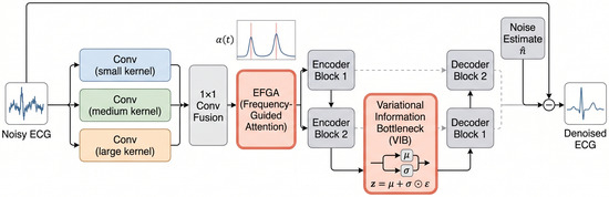
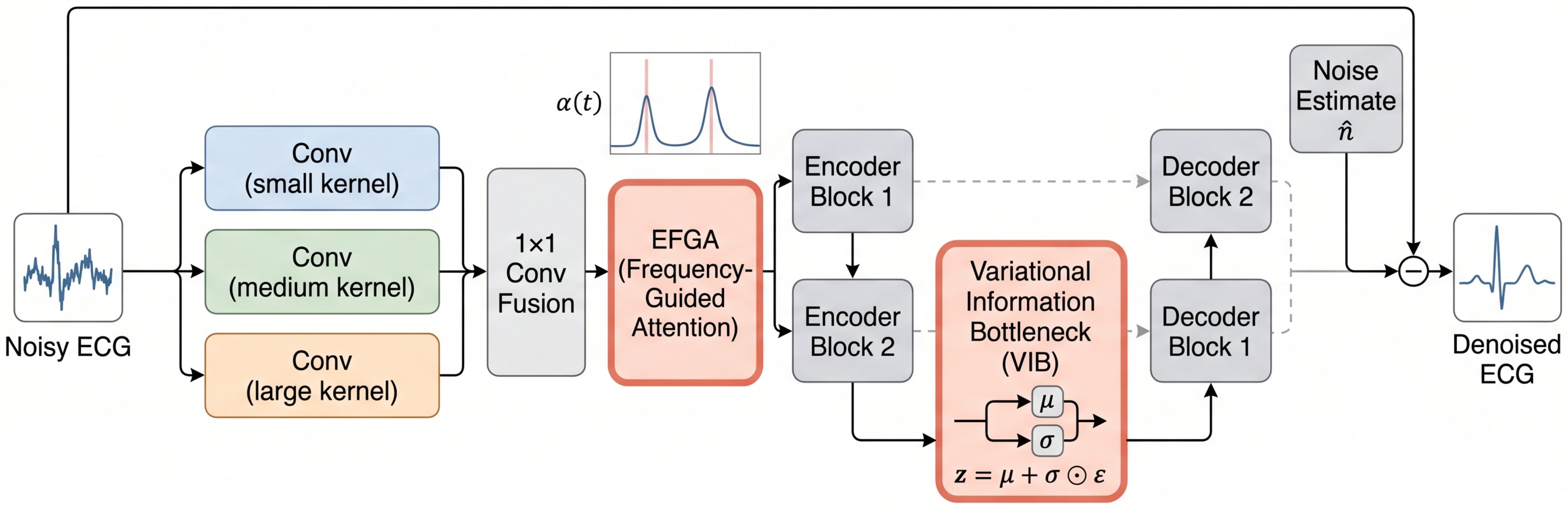
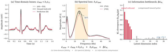
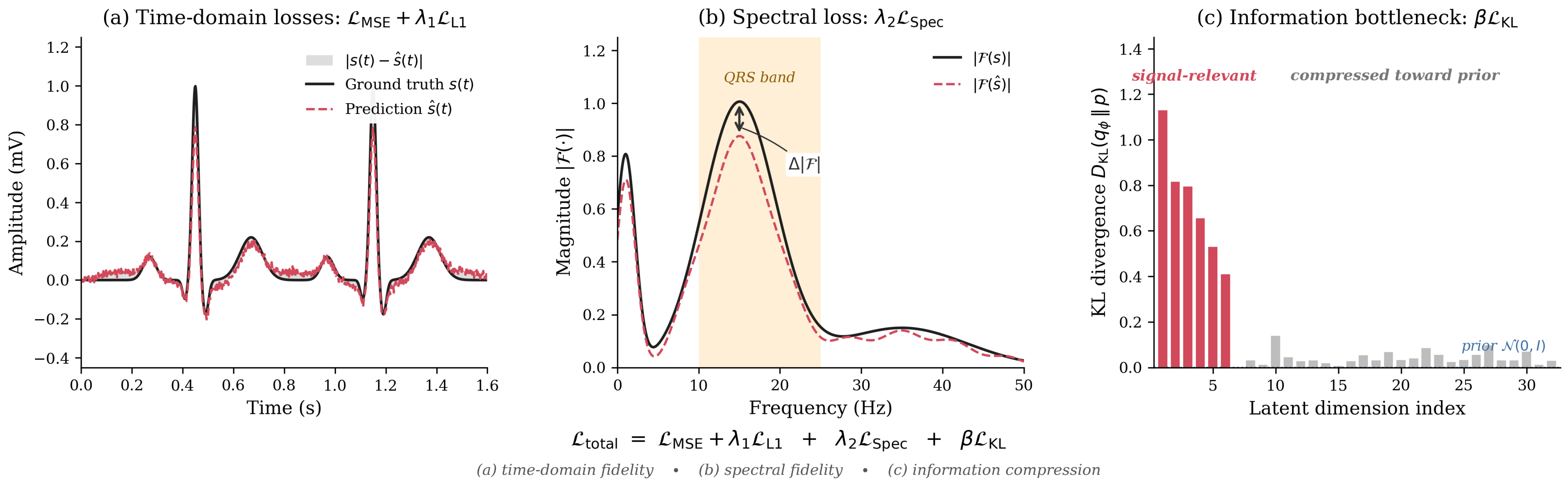
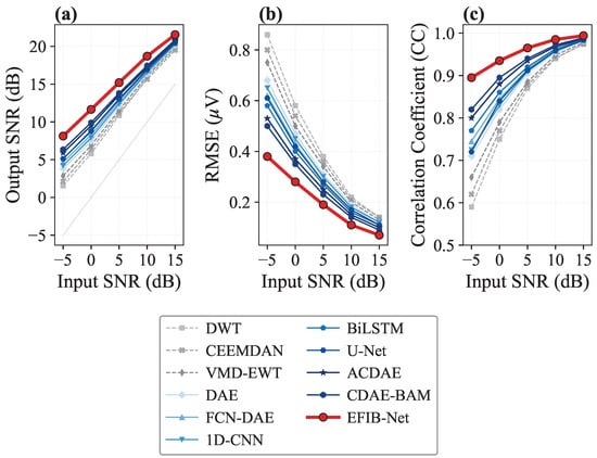
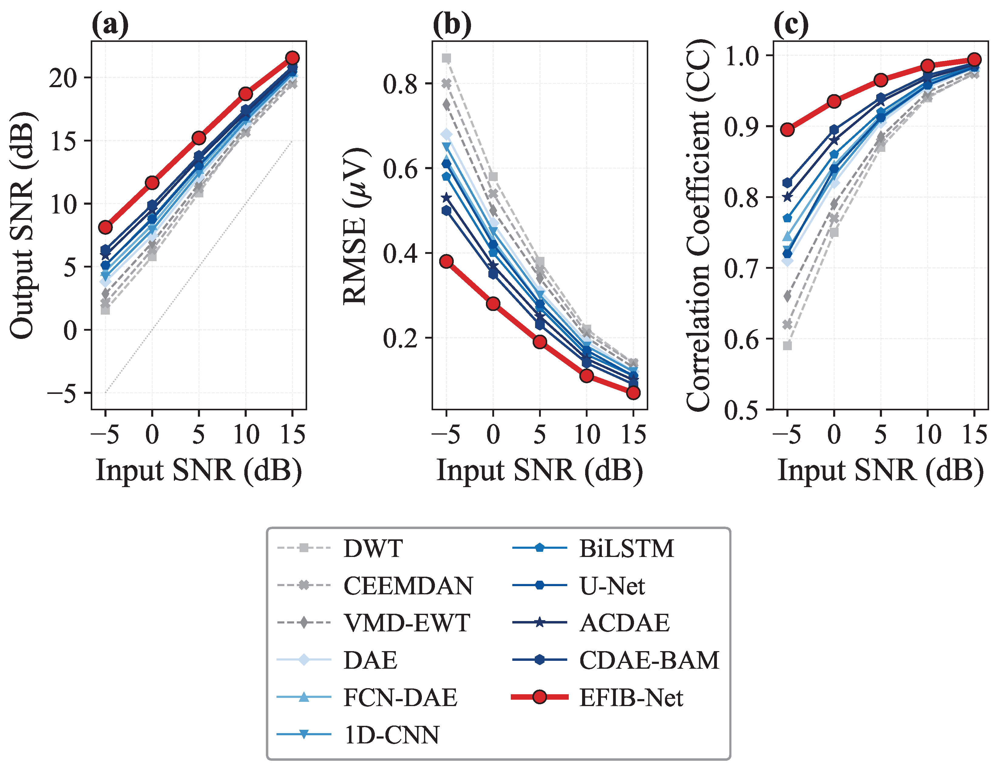
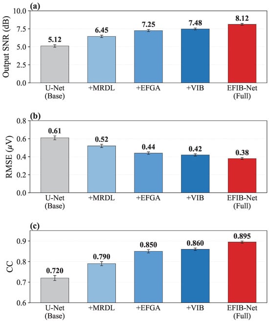
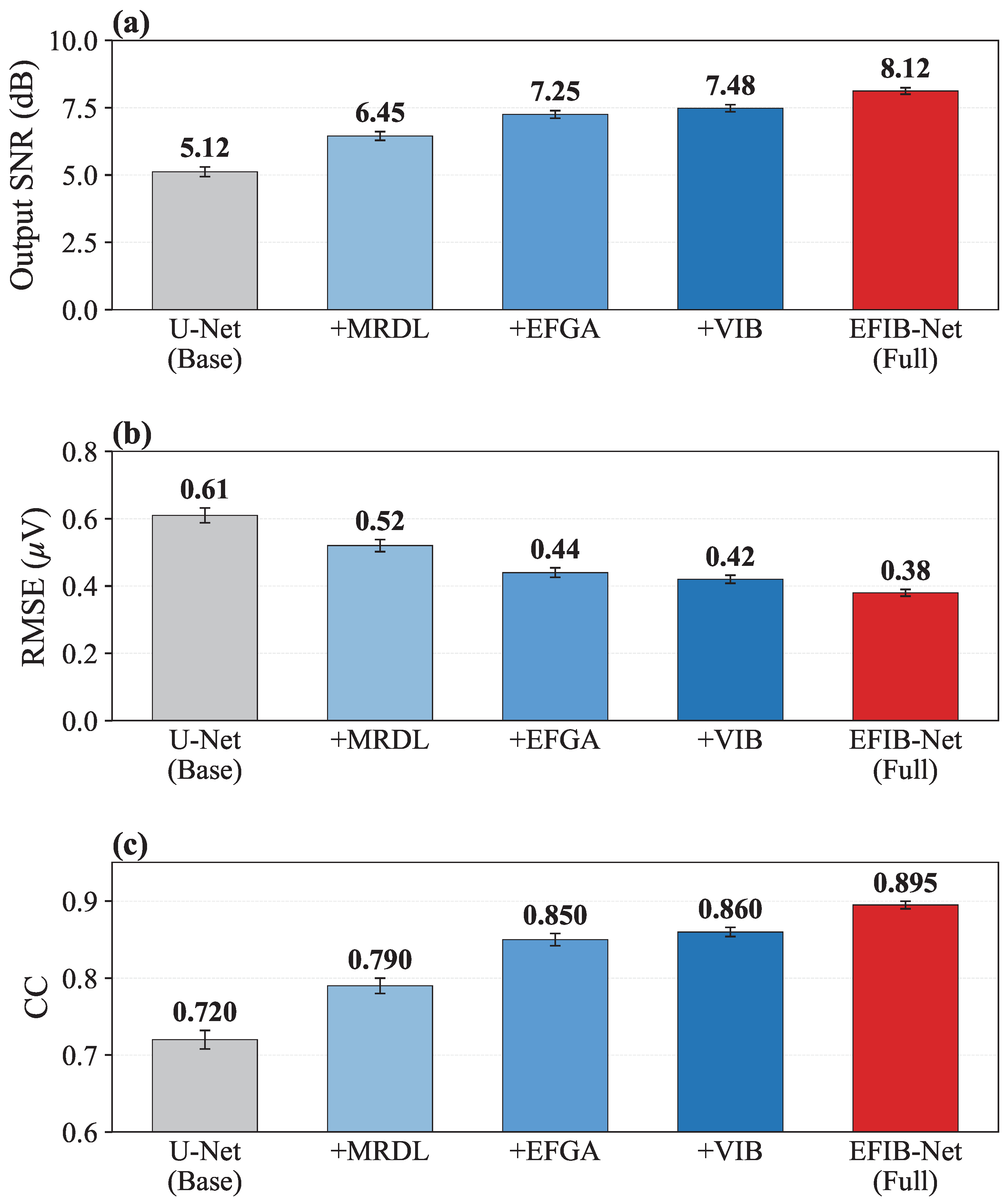
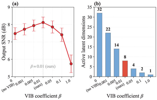
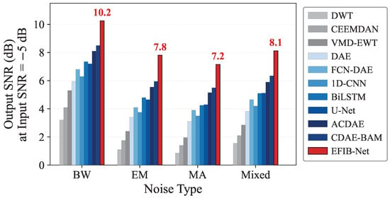
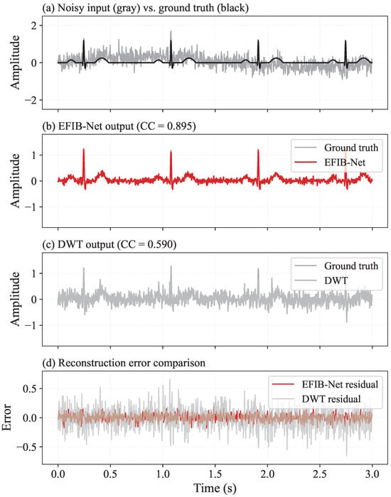
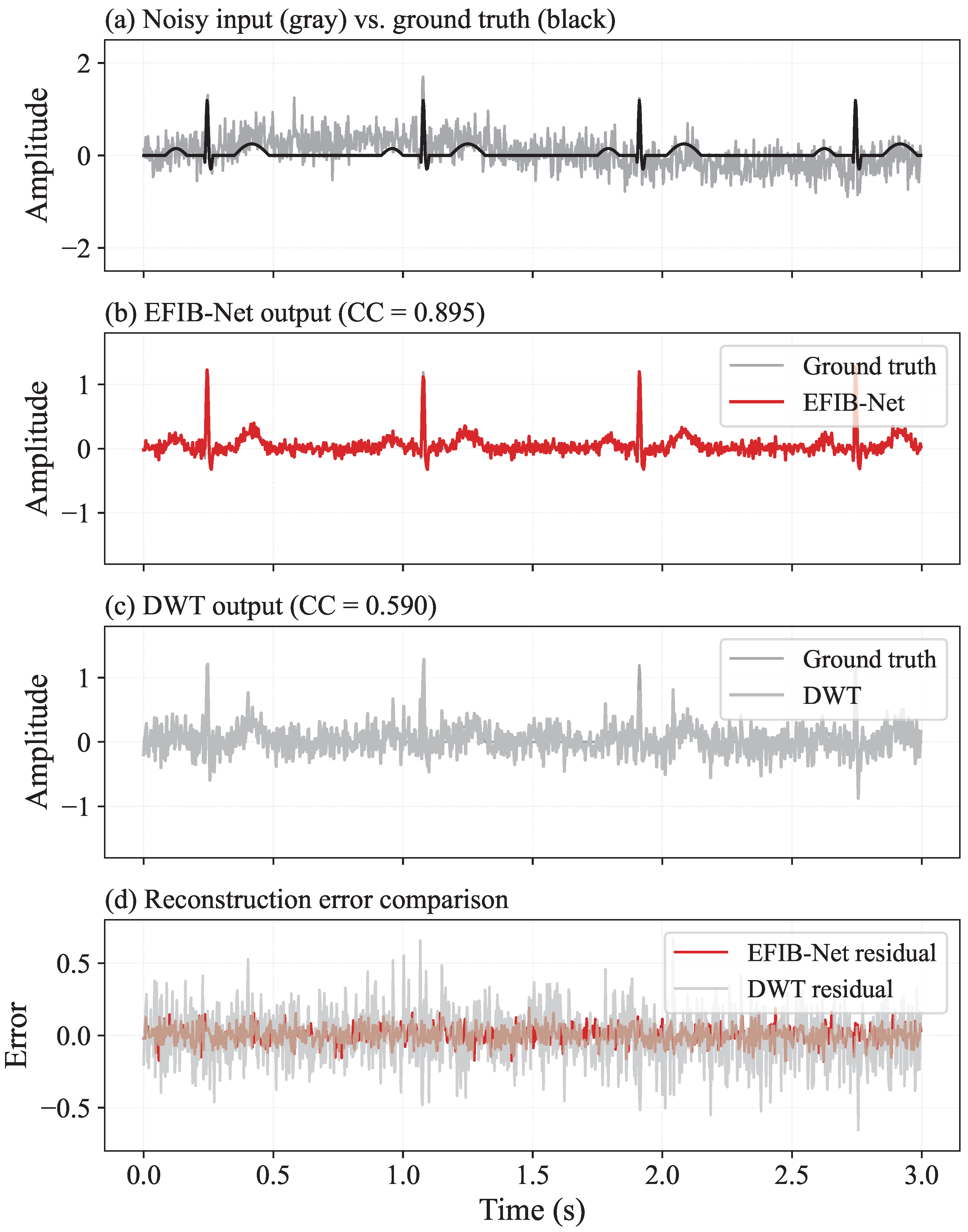
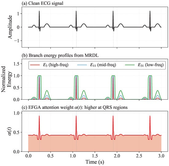
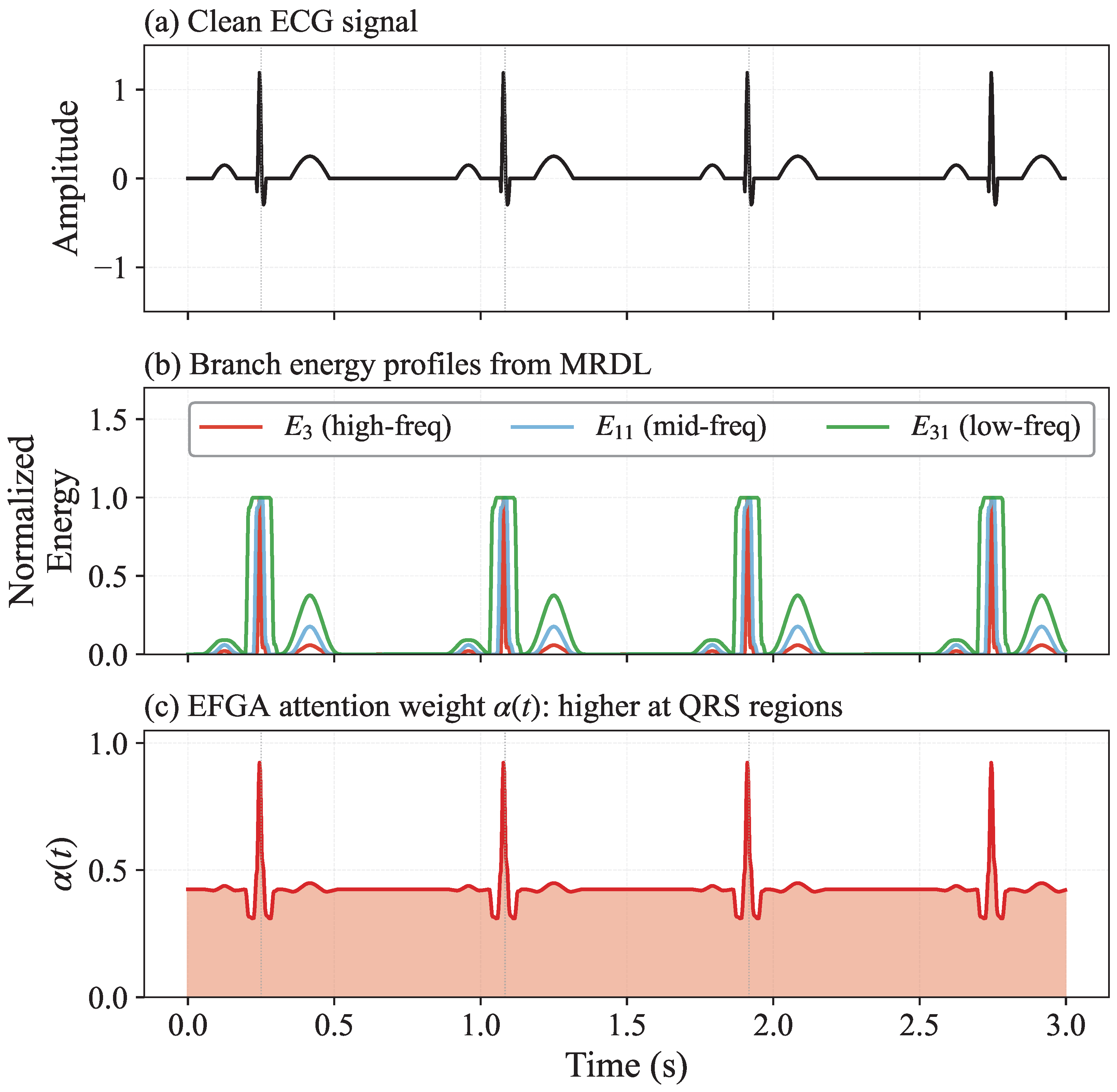
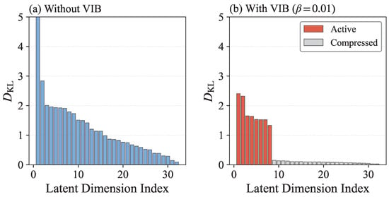
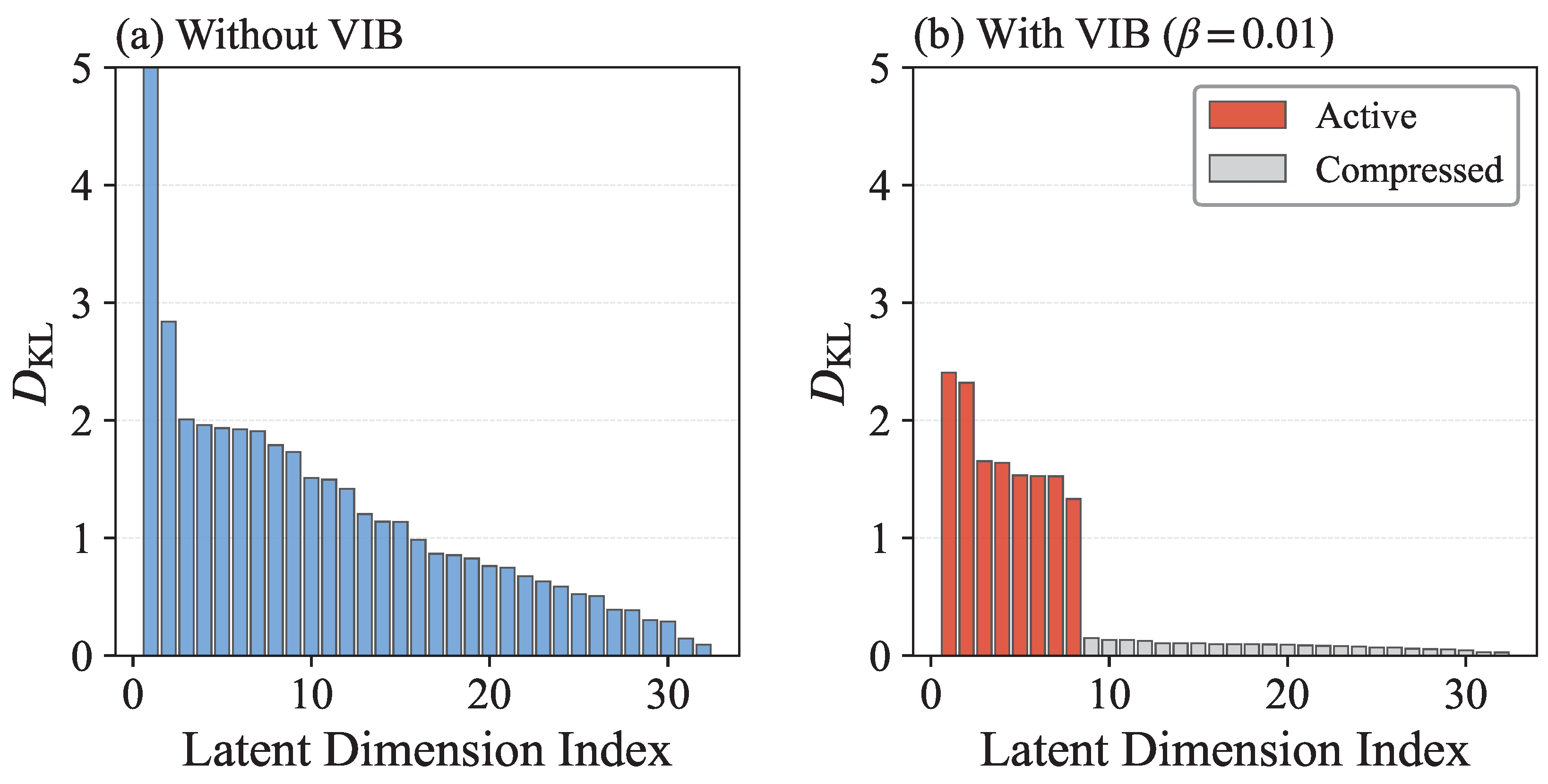

Disclaimer/Publisher’s Note: The statements, opinions and data contained in all publications are solely those of the individual author(s) and contributor(s) and not of MDPI and/or the editor(s). MDPI and/or the editor(s) disclaim responsibility for any injury to people or property resulting from any ideas, methods, instructions or products referred to in the content. |
© 2026 by the authors. Licensee MDPI, Basel, Switzerland. This article is an open access article distributed under the terms and conditions of the Creative Commons Attribution (CC BY) license.