


Abstract
Background: Rapid palatal expansion (RPE) is the gold standard for maxillary deficiency in growing patients. Conventional soldered expanders often present challenges in adaptation, chairside procedures and bond stability. This study compares traditional RPEs with customized CAD (Computer Aided Design)/CAM (Computer Aided Manufacturing) expanders regarding clinical efficiency and patient experience. Methods: Thirty growing patients (mean age: 9.2 ± 1.1 years) were allocated to two groups: traditional RPEs (n = 15) and customized CAD/CAM RPEs (n = 15). Outcomes included bond failures, chairside time (from initial try into cementation) and short-term patient-reported discomfort via a 10-point Visual Analogue Scale (VAS) 24 h after appliance placement. Significance was set at p < 0.05. Results: The CAD/CAM group showed significantly fewer bond failures (0.2 ± 0.4 vs. 1.1 ± 0.8; p < 0.05) and shorter chairside time (12.4 ± 2.1 min vs. 24.6 ± 3.8 min; p < 0.001). Patient discomfort was also significantly lower in the CAD/CAM group (VAS: 4.1 ± 1.0 vs. 6.3 ± 1.2; p < 0.05). Conclusions: Within the limitations of this pilot exploratory non-randomized study, customized CAD/CAM RPEs were associated with fewer bond failures, shorter chairside application time, and lower short-term discomfort at 24 h compared with traditional appliances. These preliminary findings should be interpreted with caution and confirmed by larger randomized controlled studies.
1. Introduction
Transverse maxillary deficiency is a common dentoskeletal problem in growing patients and is frequently associated with posterior crossbite, dental crowding, a narrow maxillary arch and functional mandibular displacement. If left un-treated, it may adversely affect occlusal development and craniofacial growth [1,2,3,4]. In this context, RPE is widely regarded as the treatment of choice for correcting maxillary transverse discrepancies during growth. The prevalence of transverse discrepancies in mixed dentition has been reported to range from 8% to 22%, depending on the population studied and diagnostic criteria adopted [5,6,7,8,9].
The rationale for RPE is based on the opening of the midpalate suture and the resulting increase in maxillary width, accompanied by dentoalveolar adaptation. When treatment is performed in growing subjects, orthopedic effects are generally more favorable because of the greater skeletal responsiveness of the maxillary sutures. For this reason, RPE has long represented a cornerstone of interceptive orthodontics [10,11,12,13,14,15].
Conventional expanders, whether banded or bonded, have demonstrated clinical effectiveness over time; however, their fabrication and delivery still rely on several laboratory and chairside steps that may affect appliance precision and overall efficiency [16,17,18,19,20]. Traditional workflows usually include impression taking, cast preparation, band selection, soldering procedures, appliance finishing and intraoral adjustment before cementation. Each of these phases may introduce inaccuracies that compromise appliance fit and increase the need for chairside corrections [21,22,23,24].
From a clinical perspective, inadequate adaptation of the expander may affect appliance stability. Bond failures during the active expansion phase represent a relevant complication, as they may interrupt treatment, require additional appointments, prolong therapy and reduce patient compliance. Repeated recementation may also increase the biological and clinical burden of treatment [25,26,27,28,29,30].
Another clinically relevant issue is the time required for appliance delivery. In young patients, lengthy chairside procedures may reduce cooperation and increase discomfort and stress [31,32,33,34]. Therefore, reducing insertion and cementation time is an important objective, not only for improving workflow efficiency, but also for enhancing the overall patient experience.
The progressive integration of digital technologies into orthodontics has introduced new possibilities for appliance design and manufacturing [35,36,37,38]. Intraoral scanning, three-dimensional planning, and CAD/CAM production enable the fabrication of customized expanders designed according to the individual anatomy of each patient. Compared with conventional appliances, digitally manufactured expanders may offer improved fit, greater reproducibility and reduced need for intraoral adjustment at delivery [39,40,41,42,43,44,45].
A more accurate adaptation of the appliance may also provide biomechanical and clinical advantages. Better fitting components could improve stability during activation, reduce stress at the cementation interfaces and limit the occurrence of detachment or debonding. In addition, customized morphology and smoother surfaces may contribute to im-proved tolerability, particularly during the first days after placement, when discomfort is usually greatest [46,47,48].
Although CAD/CAM expanders are becoming increasingly popular in contemporary orthodontics, the available evidence on clinically meaningful outcomes remains limited [49,50,51,52]. Most published studies have primarily focused on digital accuracy and technical feasibility, whereas fewer investigations have evaluated practical parameters directly affecting daily clinical management, such as bond stability, chairside efficiency and patient-reported discomfort [53,54,55].
In clinical practice, the success of rapid palatal expansion does not depend exclusively on the orthopedic effect obtained at the midpalatal suture, but also on the stability, precision, and tolerability of the appliance throughout the active expansion phase. Even when conventional expanders are effective from a therapeutic perspective, their fabrication involves multiple analog steps that may influence the final intraoral fit. Minor discrepancies occurring during impression taking, cast pouring, band adaptation, soldering, or finishing may require chairside adjustments at delivery and may increase the risk of stress concentration at the cemented interfaces. These aspects are particularly relevant in growing patients, in whom treatment cooperation, chairside tolerance, and appointment duration represent important determinants of clinical efficiency. A poorly adapted appliance may cause discomfort, occlusal interference, soft-tissue irritation, or reduced stability during screw activation. Consequently, even small improvements in appliance adaptation and insertion procedures may have a meaningful impact on everyday orthodontic management. Within this context, customized CAD/CAM expanders may offer a potential clinical advantage because they are designed on the individual digital anatomy of the patient. The virtual design process allows more controlled positioning of the framework, more accurate adaptation to anchorage teeth, and a reduction in manual laboratory steps. From a theoretical point of view, this may translate into fewer intraoral adjustments, shorter delivery time, improved retention, and better patient-reported tolerability. However, these potential advantages require clinical validation through studies evaluating practical outcomes that are directly relevant to clinicians and patients.
Therefore, the aim of the present study was to compare traditional soldered RPEs with fully customized CAD/CAM RPEs in growing patients. The comparison focused on three clinically relevant outcomes: bond failures during the expansion phase, chairside application time and patient reported discomfort at 24 h after appliance placement [56,57,58,59,60,61].
2. Materials and Methods
2.1. Study Design and Ethical Considerations
This study was designed as a pilot exploratory, single-center, prospective, non-randomized comparative clinical study conducted in growing patients requiring rapid palatal expansion as part of their interceptive orthodontic treatment. The study was carried out at the Orthodontic Department of the University of L’Aquila, Italy, between November 2024 and November 2025. Data collection was completed within this period, and the present manuscript does not represent an interim analysis. The protocol complied with the principles of the Declaration of Helsinki and with institutional ethical standards. Ethical approval was obtained from the Institutional Review Board of the University of Bari “Aldo Moro” (Prot. No. 0029116, 23 March 2023; Study code: EAPAS). Written informed consent was obtained from the parents or legal guardians of all participants before enrollment. Assent was also obtained from the children when appropriate for age and level of under-standing. No a priori sample size calculation was performed. Therefore, the present investigation should be considered exploratory and hypothesis-generating and the findings should be interpreted with caution. The study was conceived to evaluate short-term clinical performance rather than skeletal or dentoalveolar treatment effects. For this reason, the selected outcomes were directly related to appliance delivery and early clinical management. Bond failures were chosen as an indicator of appliance retention and stability during the active expansion phase; chairside application time was selected as a measure of clinical workflow efficiency; and patient-reported discomfort at 24 h was used to assess early tolerability immediately after appliance placement. These outcomes were considered particularly relevant for pediatric orthodontic patients, in whom reduced appointment duration and improved appliance comfort may positively influence cooperation and treatment acceptance.
2.2. Study Sample
A total of 30 growing patients with transverse maxillary deficiency were prospectively enrolled. All patients required RPE as part of their interceptive orthodontic treatment. Patients were prospectively recruited according to predefined eligibility criteria. Allocation to the treatment group was based on the type of expander clinically prescribed and appliance availability at the time of recruitment; therefore, allocation was non-randomized.
At baseline, standard orthodontic records were obtained for all participants and included age, sex, dentition stage, type of posterior crossbite, anchorage teeth and clinical diagnosis of transverse maxillary deficiency.
After confirmation of eligibility and treatment planning, participants were allocated to one of the two study groups according to the type of expander prescribed for clinical use at the time of recruitment and appliance availability. During enrollment, efforts were made to achieve the greatest possible baseline comparability between groups with respect to demographic and clinical characteristics. Therefore, allocation was prospective but non-randomized.
The sample was divided as follows:
- Group T (Traditional RPEs): 15 patients treated with conventional soldered expanders.
- Group C (Customized RPEs): 15 patients treated with fully customized CAD/CAM expanders.
Because of the nature of the intervention, blinding of the operator and the participants was not feasible. To reduce operator related variability, all appliances were inserted by the same experienced orthodontist using the same clinical protocol.
Inclusion criteria:
- -
- Mixed dentition stage.
- -
- Skeletal transverse maxillary deficiency diagnosed clinically and on dental casts.
- -
- Unilateral or bilateral posterior crossbite.
- -
- No previous orthodontic treatment.
Exclusion criteria:
- -
- Craniofacial syndromes or congenital malformations.
- -
- Systemic diseases affecting bone metabolism.
- -
- Poor oral hygiene or active periodontal disease.
- -
- Lack of parental consent.
During enrollment, efforts were made to obtain clinically comparable groups with respect to demographic and clinical characteristics. However, complete patient-level baseline data were not available for a formal retrospective statistical comparison between groups.
2.3. Traditional RPEs: Group T
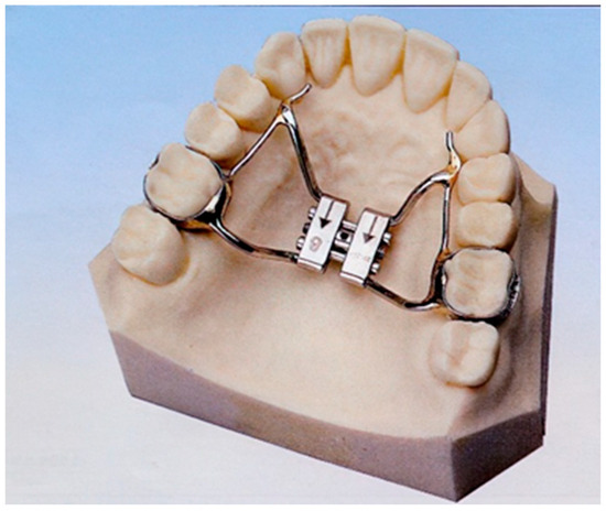
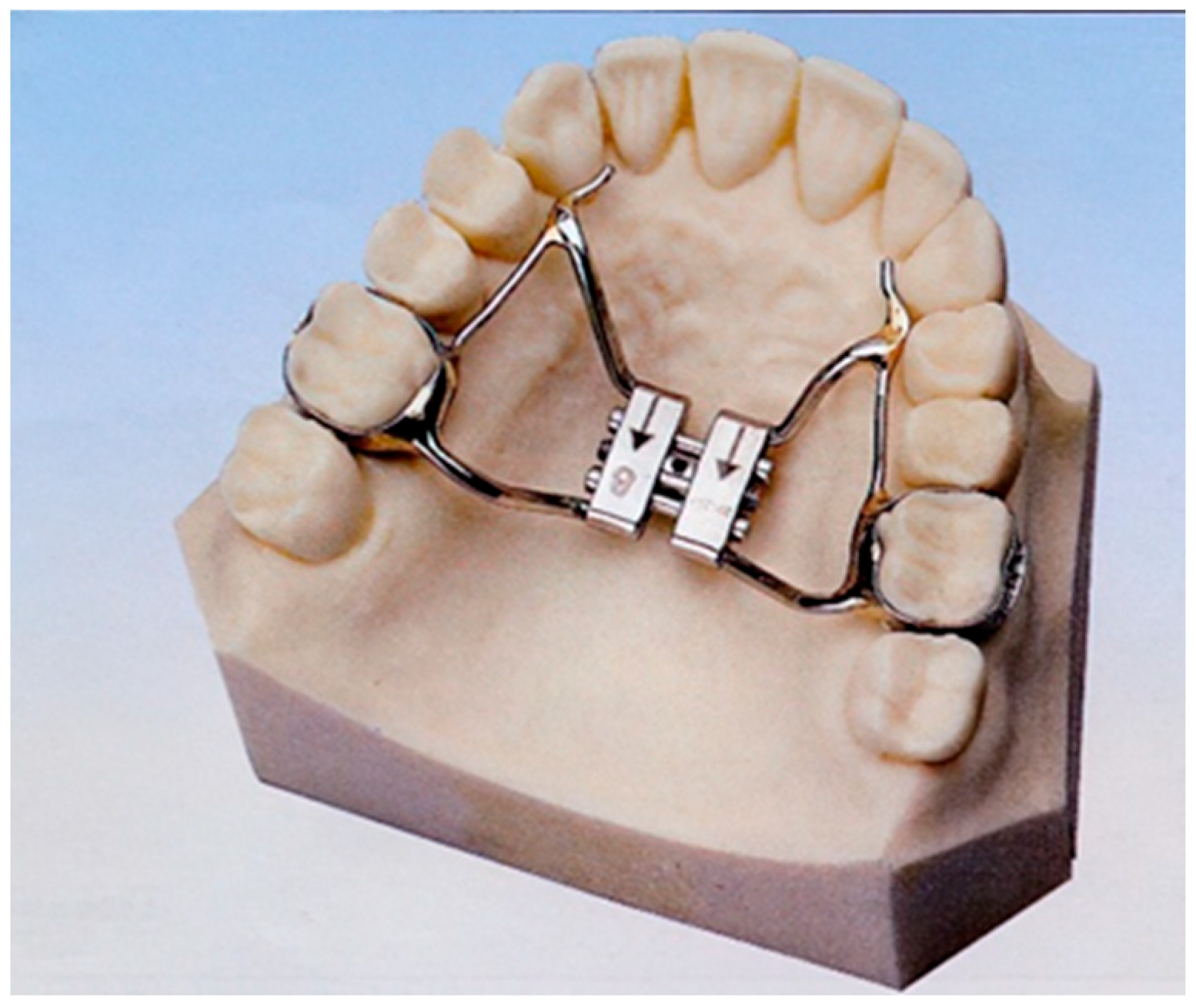
For patients in Group T, maxillary impressions were taken using conventional alginate material and poured in dental stone. Stainless steel bands were selected and adapted to the maxillary first permanent molars or deciduous second molars, according to dental eruption stage. The expander framework and expansion screw were then assembled and soldered by a dental technician according to standard laboratory procedures. The appliance was subsequently finished, polished and checked before clinical delivery (Figure 1). Before cementation, the conventional appliance was evaluated intraorally to verify complete seating of the bands, passive fit of the framework, and absence of premature occlusal contacts. When necessary, minor chairside adjustments were performed to improve adaptation before final cementation. These adjustments were included in the recorded chairside application time, as they represent a clinically relevant component of the conventional delivery workflow.
Figure 1.
Traditional RPEs.
2.4. Customized CAD/CAM RPEs: Group C
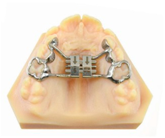
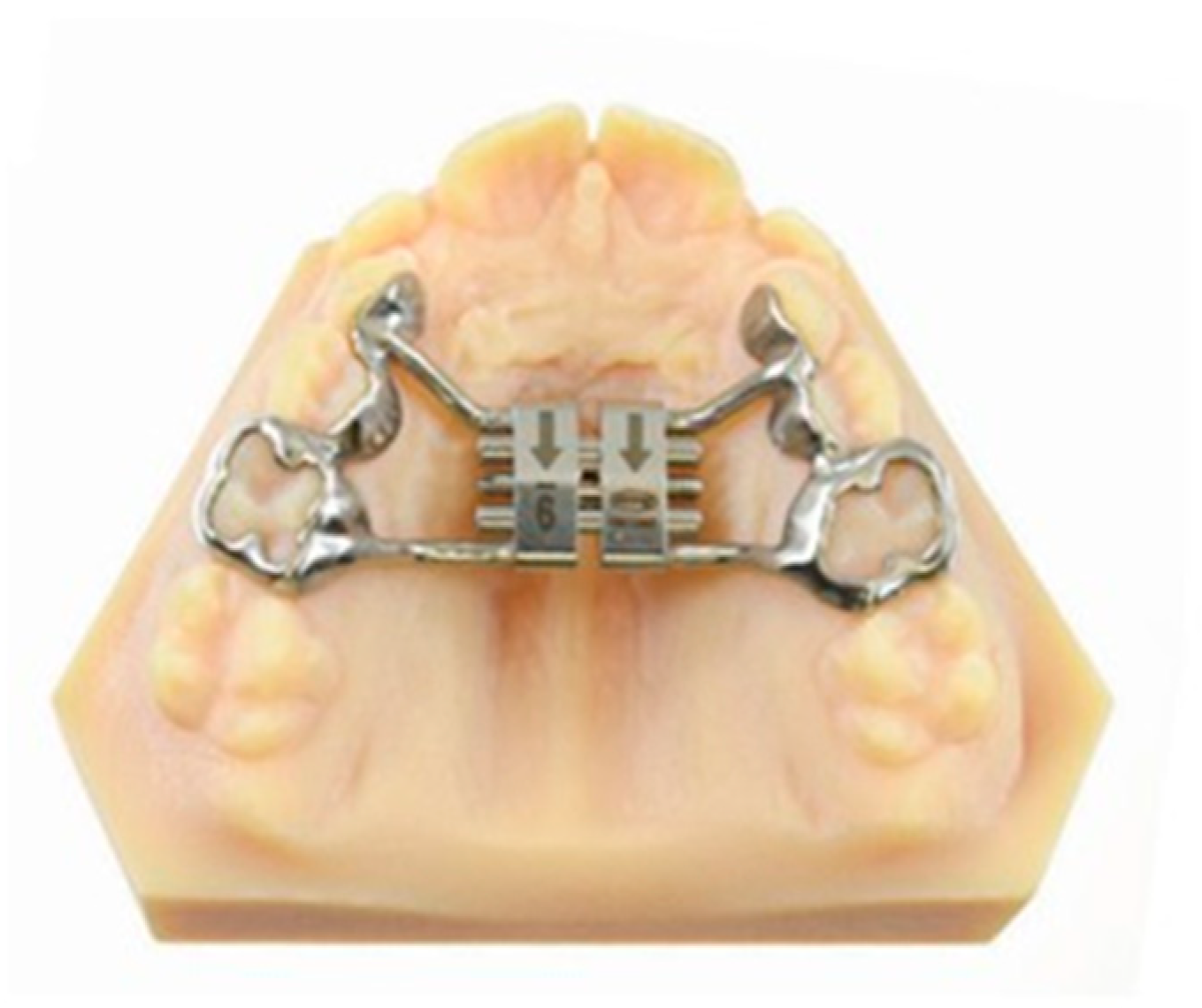
For patients in Group C, digital impressions of the maxillary arch were obtained using an intraoral scanner. The ex-panders were designed using dedicated CAD software, allowing three-dimensional visualization and virtual adaptation of bands and framework to each patient’s dental anatomy. The appliances were then manufactured using a CAM workflow and were delivered ready for clinical insertion (Figure 2). The digital workflow was intended to minimize manual adaptation procedures and to improve the correspondence between the appliance and the patient-specific dental anatomy. During virtual planning, the extension of the framework and the adaptation of the anchorage components were designed to obtain stable support while avoiding unnecessary bulk and potential soft-tissue interference. At clinical delivery, the customized appliances were checked for passive seating and correct positioning in the same manner as the traditional expanders. Any additional adjustment required before cementation was included in the chairside application time.
Figure 2.
CAD/CAM RPEs.
2.5. Clinical Procedure
Before cementation, each appliance was checked intraorally for passive fit, correct seating on the anchorage teeth and absence of occlusal interferences. Cementation was performed using glass ionomer cement according to the manufacturer’s instructions.
Chairside application time was recorded with a stopwatch and defined as the time elapsed from the initial try-in of the appliance to completion of cementation.
Following appliance delivery, parents or caregivers received standardized oral hygiene and activation instructions. The activation protocol was the same for both groups and consisted of two quarter-turns per day (0.5 mm/day) until the planned clinical endpoint was reached. Overcorrection was clinically defined as correction of the posterior crossbite until the palatal cusps of the maxillary posterior teeth contacted or slightly overpassed the buccal cusps of the mandibular posterior teeth. Patients were reviewed weekly during the active phase of expansion.
To minimize variability in appliance delivery and outcome recording, all appliances were inserted and all clinical outcomes were recorded by the same experienced orthodontist. Since only one operator was involved, inter-observer reliability could not be assessed and no formal inter-examiner calibration was performed. The same cementation material and the same clinical insertion sequence were used for both groups. Eligibility criteria were identical for both groups and all participants were managed within the same clinical setting. Owing to the nature of the intervention, neither operator nor patients could be blinded.
2.6. Outcome Measures
Three main outcomes were evaluated:
- (1)
- Bond failures: were defined as any partial or complete loss of retention involving a band or any cemented component of the appliance that required recementation, repair or replacement during the active expansion phase. The total number of bond failures per patient was recorded from appliance insertion to completion of active expansion. Partial and complete debonding events were counted equally when they required clinical intervention. Recementation, repair, or replacement was considered part of the management of the same bond failure event. This definition was adopted to ensure that only clinically relevant failures were recorded. Minor findings that did not require intervention, such as small marginal cement defects without loss of retention, were not counted as bond failures. Conversely, any event requiring interruption of the planned expansion protocol or an additional clinical procedure was considered a bond failure, irrespective of whether the appliance was partially or completely detached.
- (2)
- Chairside time was measured in minutes, from the initial appliance try-in to final cementation.
- (3)
- Short-term patient-reported discomfort: Patient-reported discomfort was assessed using a 10-point VAS, where 0 indicated no discomfort and 10 indicated maximum discomfort. VAS scores were collected at 24 h after appliance insertion using standardized age-appropriate verbal instructions, with parental or caregiver support when needed. The 24 h time point was selected because it represents the early post-placement phase, during which discomfort related to appliance bulk, occlusal interference, pressure sensation, and soft-tissue adaptation is usually more evident. Patients and caregivers were instructed to report the perceived discomfort associated with the appliance rather than pain related to unrelated oral conditions. Although the VAS is a simple and widely used tool, the results were interpreted cautiously because younger patients may require parental support to understand and complete the scale.
2.7. Statistical Analysis
Descriptive statistics were calculated for all study variables. Continuous data are presented as mean ± standard deviation (SD). The distribution of continuous variables was assessed using the Shapiro–Wilk test.
Between group comparisons for chairside application time and patient reported discomfort (VAS scores at 24 h) were performed using independent samples tests. For normally distributed variables, the independent-samples t-test was used; otherwise, the Mann–Whitney U test was applied. Bond failures were analyzed as count data and between group differences using the Mann–Whitney U test because of the limited range and non-normal distribution of this variable. All tests were two-sided and the level of significance was set at p < 0.05. Exact p-values were reported whenever available. However, because only aggregated descriptive data were available at the revision stage, exact p-values and 95% confidence intervals could not be calculated retrospectively for all outcomes. Given the limited sample size, the results should be interpreted with caution. Because of the limited sample size, the exploratory nature of the study and the absence of complete patient-level raw data, no multivariable regression analysis was performed. No adjustment for multiple comparisons was applied because of the exploratory nature of the study.
3. Results
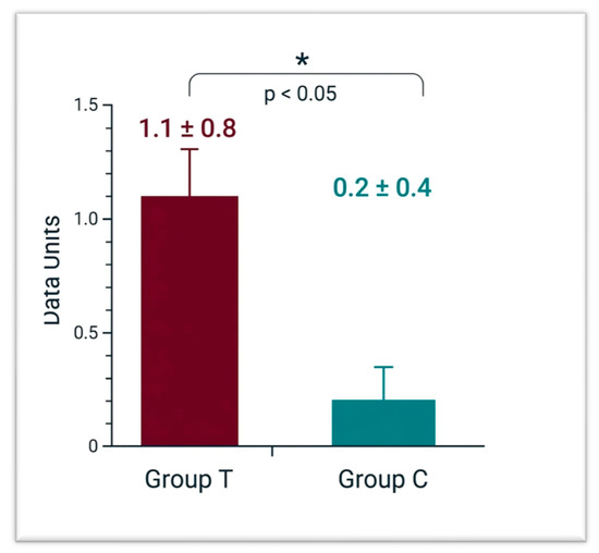
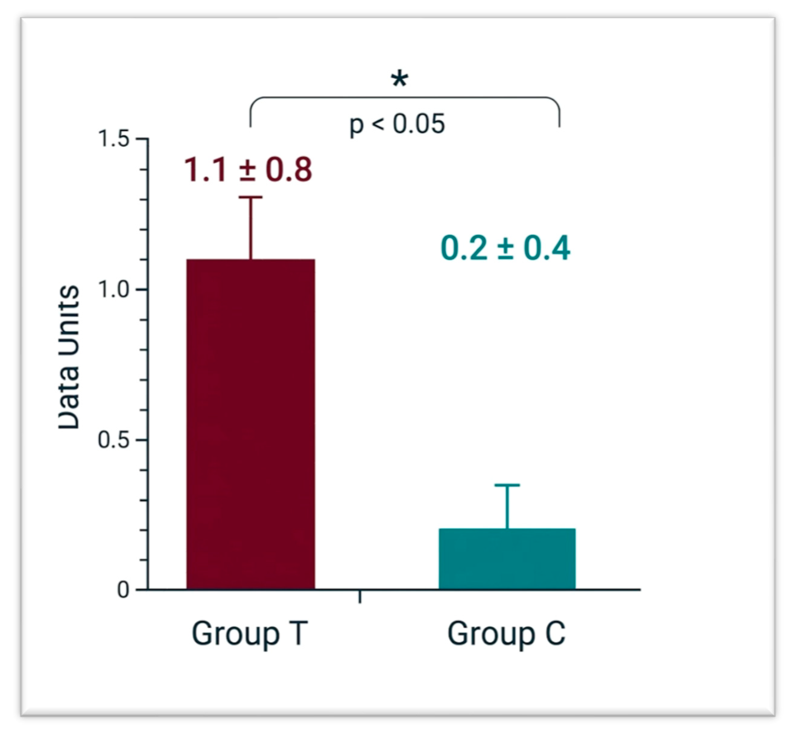
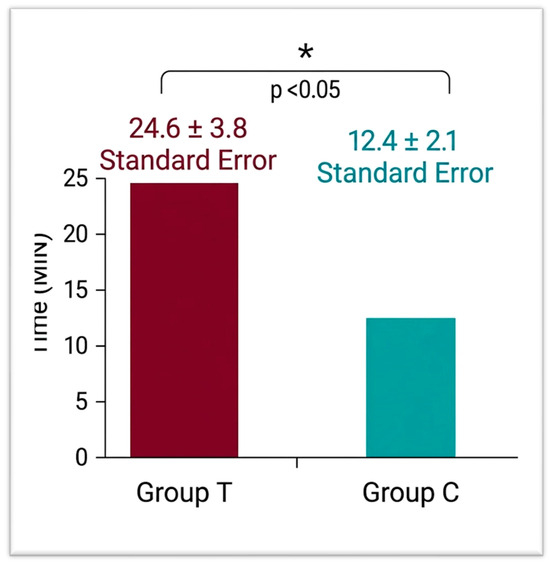
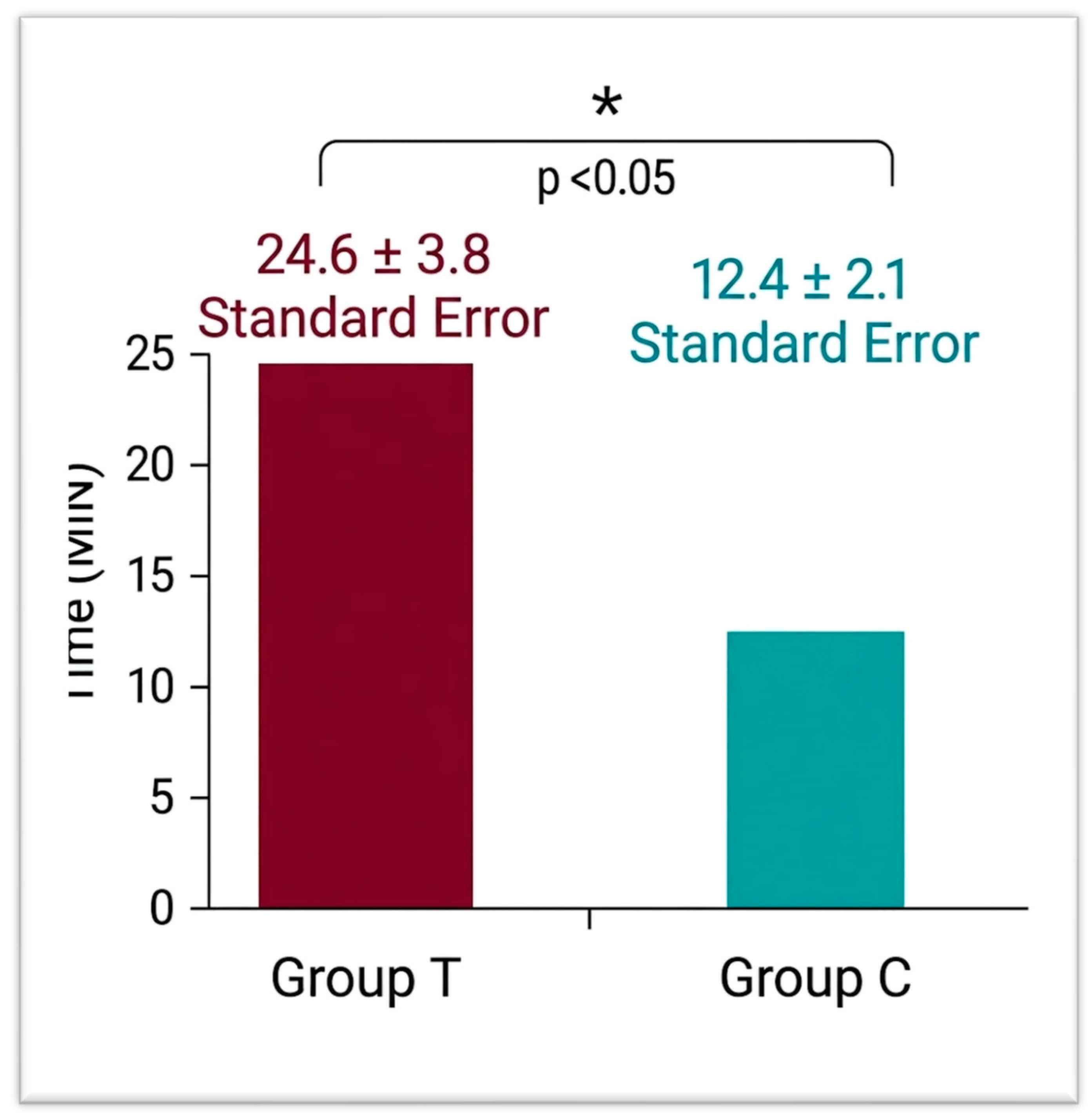
The study sample consisted of 30 growing patients, including 16 males and 14 females, with a mean age of 9.2 ± 1.1 years. The customized CAD/CAM expander group showed significantly fewer bond failures than the traditional expander group (0.2 ± 0.4 vs. 1.1 ± 0.8) (Figure 3). Individual patient-level distributions, ranges, and quartiles of bond failures were not available for retrospective reporting. Chairside application time was also significantly shorter in the customized group, with mean delivery time nearly halved compared with that of the traditional group (12.4 ± 2.1 min. vs. 24.6 ± 3.8 min.) (Figure 4).
Figure 3.
Bond failures per patient (mean ± SD) in traditional versus CAD/CAM customized RPEs (p < 0.05).
Figure 4.
Chairside application time (min; mean ± SD) from appliance try-in to ce mentation in traditional versus CAD/CAM customized RPEs (p < 0.001).
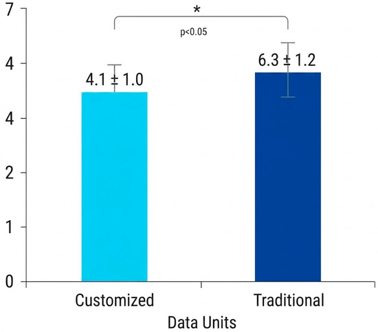
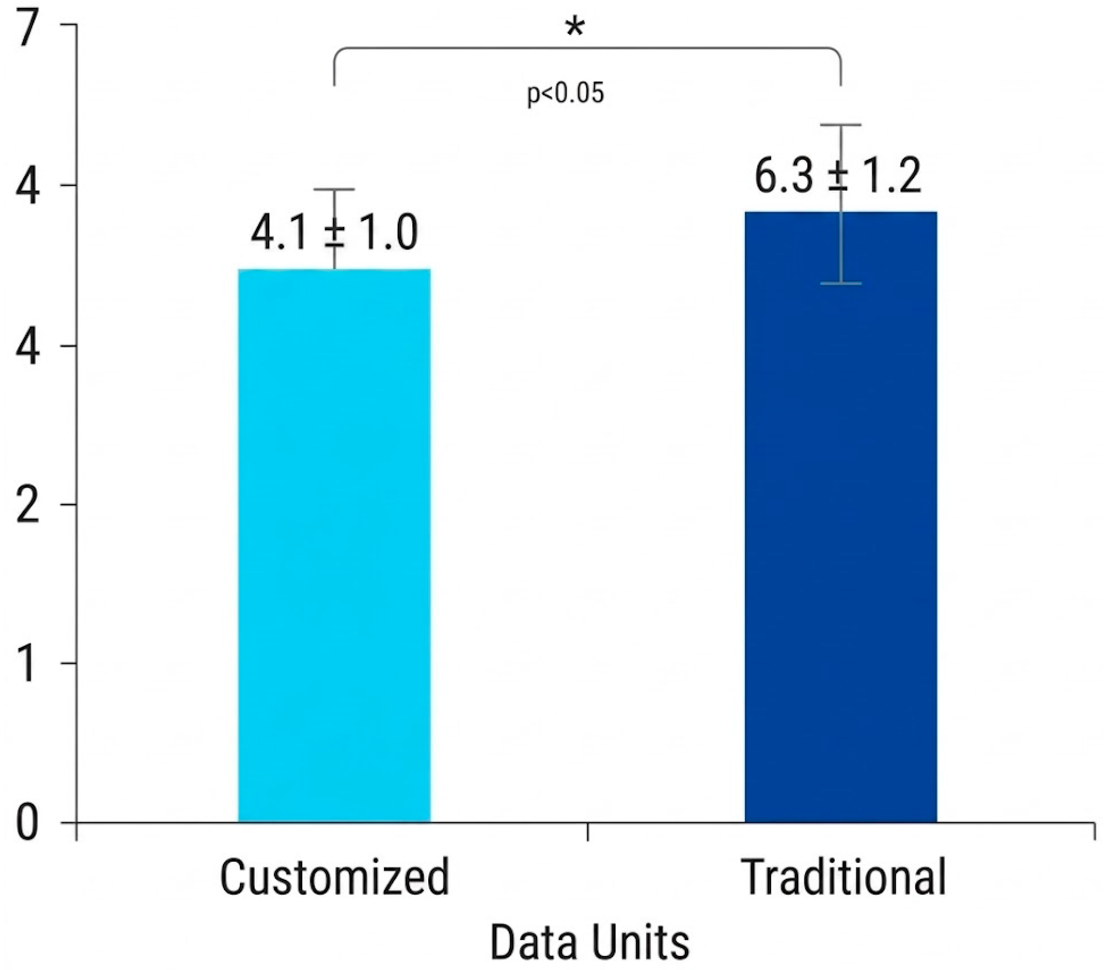
Regarding patient discomfort, values at 24 h were significantly lower in the customized group than in the traditional group (VAS 4.1 ± 1.0 vs. 6.3 ± 1.2) (Figure 5).
Figure 5.
Patient-reported discomfort at 24 h (VAS 0-10; mean ± SD) in customized versus traditional RPEs (p < 0.05).
Overall, the three evaluated outcomes consistently favored the customized CAD/CAM group. The mean number of bond failures per patient was lower in the customized group, suggesting improved appliance stability during the active expansion phase. Chairside application time was reduced by approximately 12 min in the CAD/CAM group compared with the traditional group, corresponding to an almost 50% reduction in delivery time. In addition, the lower VAS score recorded at 24 h suggests that customized appliances may be better tolerated during the early post-placement period.
From a clinical perspective, these differences may be relevant even in a small exploratory sample. Fewer bond failures may reduce the need for emergency appointments, recementation procedures, and interruptions of the expansion protocol. Similarly, shorter chairside time may be advantageous in growing patients, who may have limited tolerance for prolonged procedures. Finally, reduced short-term discomfort may contribute to improved acceptance of the appliance during the initial adaptation period. However, because of the pilot nature of the study and the absence of complete patient-level data for additional analyses, these findings should be interpreted as preliminary.
4. Discussion
The present prospective non-randomized comparative study showed that fully customized CAD/CAM RPEs were as-sociated with fewer bond failures, shorter chairside application time and lower patient reported discomfort at 24 h after insertion than traditional soldered expanders [62,63,64,65]. These findings are consistent with the ongoing transition toward digital orthodontic workflows, in which CAD/CAM design and additive manufacturing allow the fabrication of increasingly individualized appliances with potential advantages in terms of fit, efficiency and clinical reproducibility [66,67,68]. Recent reviews have emphasized that one of the main advantages of these technologies is the possibility of fabricating highly individualized intraoral devices while reducing dependence on conventional laboratory procedures and improving overall workflow management [69,70,71,72,73].
At present, direct comparative evidence between conventional tooth-borne RPEs and fully customized CAD/CAM tooth-borne expanders remains scarce. Drago et al. investigated a digitally designed tooth borne expander, but their comparison concerned different anchorage units rather than a digital versus conventional design [74,75,76,77]. More recently, Pozzan et al. compared a customized Haas inspired RPEs with a conventional Haas expander, but their outcomes were limited to three-dimensional palatal changes and did not include bond failures, chairside delivery time or patient reported tolerability [46,78,79,80]. In parallel, recent reviews suggest that much of the contemporary digital expansion literature is increasingly centered on digitally planned MARPE (Miniscrew Assisted Rapid Palatal Expansion) rather than on customized tooth borne RPEs in growing patients [81,82,83,84,85]. Therefore, the present study addresses a clinically relevant gap by focusing on outcomes that directly affect every day orthodontic practice [86,87,88,89,90]. The present findings should be interpreted in the context of a broader clinical shift from standardized orthodontic appliances toward individualized digital devices. In interceptive orthodontics, appliance customization may be particularly valuable because anatomical variability, mixed dentition characteristics, and patient cooperation can strongly influence treatment management. Unlike conventional expanders, which require several analog fabrication steps and may need chairside adaptation, CAD/CAM appliances are designed to reproduce patient-specific morphology from the beginning of the workflow. This may reduce the cumulative errors associated with impression materials, dental casts, manual band adaptation, and soldering procedures. Although the present study was not designed to evaluate the accuracy of the manufacturing process, the clinical outcomes indirectly support the potential relevance of improved adaptation. Reduced bond failures, shorter chairside time, and lower discomfort may all be related to better correspondence between the appliance and the supporting dental structures. Therefore, the clinical benefit of CAD/CAM expanders may not be limited to digital innovation itself, but may derive from the possibility of producing a more reproducible and anatomically adapted appliance.
The lower number of bond failures observed in the customized group may plausibly be explained by the more accurate adaptation of the appliance to the patient’s dental anatomy [91,92,93,94,95,96]. A better fit may reduce insertion related stress, limit the need for intraoral adjustments and improve stability during the active phase of expansion. Although robust comparative evidence on debonding outcomes is still lacking, the currently available literature supports the concept that individualized appliance design may improve adaptation and biomechanical control [97,98,99,100,101,102,103]. This aspect is important because bond failure during rapid palatal expansion is not only a mechanical complication, but also a factor that can affect treatment continuity. Detachment or partial loss of retention may require unscheduled appointments, temporary suspension of activation, appliance repair, or recementation. These events may increase chairside burden for the clinician and may reduce confidence and compliance in young patients and their caregivers. Therefore, even a modest reduction in bond failures may have practical relevance in routine orthodontic care. In the present study, the lower number of failures in the customized group may be associated with a more passive and homogeneous fit of the appliance at the time of cementation. A better adaptation may distribute functional and activation-related forces more evenly across the anchorage units and may reduce localized stress at the cement interface. However, this explanation remains hypothetical because no biomechanical analysis or quantitative fit assessment was performed.
The marked reduction in chairside application time observed in the CAD/CAM group is also clinically relevant, particularly in pediatric orthodontics, where long procedures may negatively affect cooperation. Although the observed reduction of approximately 12 min may appear modest in absolute terms, it may be clinically meaningful in pediatric orthodontic practice, where shorter delivery appointments can improve cooperation, reduce fatigue and stress, and increase overall chairside efficiency. The reduction in chairside time observed in the CAD/CAM group may also reflect a simplification of the clinical delivery phase. Traditional expanders often require verification of band seating, correction of minor discrepancies, removal of interferences, and adaptation of the framework before cementation. In contrast, a customized appliance delivered after digital planning may require fewer intraoral corrections. This difference is particularly relevant in pediatric patients, for whom prolonged appointments may increase fatigue, anxiety, and movement during clinical procedures. From an organizational perspective, shorter appliance delivery may improve scheduling efficiency and reduce clinical workload. Although the present study did not perform a cost-effectiveness analysis, reduced chairside time and fewer repair appointments could partially compensate for the higher laboratory or digital production costs associated with CAD/CAM appliances. Future studies should specifically investigate this aspect through economic and workflow analyses. Likewise, the lower discomfort reported at 24 h may reflect better appliance adaptation and reduced occlusal or soft tissue interference during the initial phase of treatment [104,105,106,107,108,109]. Although the present study evaluated discomfort after appliance placement rather than during impression taking, pediatric studies consistently report that digital procedures are bet-ter accepted than conventional impressions and may improve comfort and patient preference [110,111,112,113,114,115,116]. This broader evidence supports the hypothesis that a digital workflow may contribute to a more favorable overall treatment experience [117,118,119,120]. The difference in patient-reported discomfort may be explained by several factors. Customized appliances may present a more accurate adaptation to tooth morphology and a more controlled extension of the framework, potentially reducing pressure areas, occlusal interferences, and soft-tissue irritation. Moreover, a shorter and smoother delivery procedure may positively influence the patient’s early perception of the appliance. In children, the initial experience with an orthodontic device can affect cooperation, acceptance, and family perception of treatment complexity. Nevertheless, discomfort was assessed only at 24 h after placement. This time point captures early tolerability but does not provide information on discomfort during the entire activation phase or after completion of expansion. For this reason, the present findings should be considered preliminary and should be confirmed using repeated patient-reported outcome measures over multiple time points.
The present study has several limitations. First, the sample size was limited, and no a priori sample size calculation was performed; therefore, the study should be considered exploratory and hypothesis-generating. In addition, the availability of aggregated data only prevented the retrospective calculation of exact p-values and 95% confidence intervals for all outcomes, thereby limiting the precision of statistical interpretation. Furthermore, no correction for multiple comparisons was applied; therefore, the possibility of type I error cannot be excluded. Second, group allocation was non-randomized, and residual selection bias cannot be excluded. Third, although efforts were made during recruitment to obtain clinically comparable groups, complete patient-level baseline data were not available for a formal statistical comparison between groups. Therefore, residual confounding related to unmeasured baseline demographic or clinical differences cannot be excluded. Fourth, all clinical outcomes were recorded by the same operator; therefore, inter-observer reliability could not be assessed, and no independent blinded assessment was performed. Finally, only short-term clinical outcomes were evaluated, and no long-term skeletal, dentoalveolar, or stability-related outcomes were assessed [77]. Although VAS instructions were adapted to the age of the patients, the use of a pediatric-specific validated scale, such as the Wong-Baker FACES scale, would have strengthened the assessment of discomfort. Patient-reported discomfort was assessed only at 24 h after appliance placement; therefore, no conclusions can be drawn regarding discomfort during the subsequent active expansion phase.
Future prospective studies with larger samples and longer follow-up are needed to determine whether the short-term advantages observed with customized CAD/CAM expanders translate into superior long term clinical performance.
5. Conclusions
Within the limitations of this prospective non-randomized comparative study, fully customized CAD/CAM RPEs were associated with fewer bond failures, shorter chairside application time and improved patient comfort at 24 h after appliance placement. However, due to the limited sample size and the exploratory non-randomized design, these findings should be interpreted with caution and confirmed by larger randomized controlled studies.
Another limitation is that the study evaluated clinical management outcomes rather than biological treatment effects. No three-dimensional skeletal, dentoalveolar, or periodontal measurements were performed; therefore, the present results cannot determine whether customized CAD/CAM expanders produce different orthopedic or dentoalveolar effects compared with traditional appliances. In addition, patient-reported discomfort was assessed using a single VAS measurement at 24 h, without repeated follow-up evaluations during the active expansion phase. Future randomized controlled trials should include larger samples, standardized baseline comparisons, blinded outcome assessment when feasible, patient-level statistical analysis, longer follow-up, and combined evaluation of clinical, skeletal, dentoalveolar, periodontal, patient-reported, and economic outcomes.
Author Contributions
Conceptualization: S.S. and L.E.S.; methodology: G.D. and S.S.; software: J.J. and F.R.; validation: G.M. and A.D.I.; formal analysis: A.M.I. and F.I.; investigation: F.C. and C.C.; resources: G.D. and L.E.S.; data curation: C.C. and F.C.; writing—original draft preparation: S.S., L.E.S., C.C., F.C. and F.I.; writing—review and editing: A.M.I., G.M., J.J., F.R., A.D.I. and G.D.; visualization: A.M.I. and F.I.; supervision: S.S., F.I. and G.D.; project administration: J.J. and F.R. All authors have read and agreed to the published version of the manuscript.
Funding
This research received no external funding.
Institutional Review Board Statement
The study was conducted in accordance with the Declaration of Helsinki and approved by the Institutional Review Board of the University of Bari “Aldo Moro” (Prot. No. 0029116, 23 March 2023; Study code: EAPAS).
Informed Consent Statement
Written informed consent has been obtained from the patient(s) to publish this paper.
Data Availability Statement
The data presented in this study are available from the corresponding author upon reasonable request. The data are not publicly available because of privacy and ethical restrictions.
Conflicts of Interest
The authors declare no conflicts of interest.
Abbreviations
The following abbreviations are used in this manuscript:
| CAD | Computer-Aided Design |
| CAM | Computer-Aided Manufacturing |
| MARPE | Miniscrew-Assisted Rapid Palatal Expansion |
| RPE | Rapid Palatal Expansion |
| RPEs | Rapid Palatal Expanders |
| SD | Standard Deviation |
| VAS | Visual Analogue Scale |
References
- Jedliński, M.; Mazur, M.; Grocholewicz, K.; Janiszewska-Olszowska, J. 3D Scanners in Orthodontics-Current Knowledge and Future Perspectives-A Systematic Review. Int. J. Environ. Res. Public Health 2021, 18, 1121. [Google Scholar] [CrossRef] [Scilit] [PubMed]
- Paľovčík, M.; Tomášik, J.; Zsoldos, M.; Thurzo, A. 3D-Printed Accessories and Auxiliaries in Orthodontic Treatment. Appl. Sci. 2025, 15, 78. [Google Scholar] [CrossRef] [Scilit]
- Koulivand, S.; Ghodsi, S.; Siadat, H.; Alikhasi, M. A Clinical Comparison of Digital and Conventional Impression Techniques Regarding Finish Line Locations and Impression Time. J. Esthet. Restor. Dent. 2020, 32, 236–243. [Google Scholar] [CrossRef] [Scilit] [PubMed]
- Moss, J.P. Rapid Expansion. Int. J. Orthod. 1976, 14, 15–19. [Google Scholar] [PubMed]
- Kuhr, F.; Schmidt, A.; Rehmann, P.; Wöstmann, B. A New Method for Assessing the Accuracy of Full Arch Impressions in Patients. J. Dent. 2016, 55, 68–74. [Google Scholar] [CrossRef] [Scilit] [PubMed]
- Cozzani, M.; Antonini, S.; Lupini, D.; Decesari, D.; Anelli, F.; Doldo, T. A New Proposal: A Digital Flow for the Construction of a Haas-Inspired Rapid Maxillary Expander (HIRME). Materials 2020, 13, 2898. [Google Scholar] [CrossRef] [Scilit] [PubMed]
- Avashia, Y.; Bittar, P.; Suresh, V.; Powers, D.B. A Novel Approach for the Management and Prevention of Self-Induced Masticatory Lingual Trauma in the Neurologically Injured Patient. Craniomaxillofac. Trauma Reconstr. 2018, 11, 242–248. [Google Scholar] [CrossRef] [Scilit] [PubMed]
- Saccomanno, S.; Martini, C.; D’Alatri, L.; Farina, S.; Grippaudo, C. A Specific Protocol of Myo-Functional Therapy in Children with Down Syndrome. A Pilot Study. Eur. J. Paediatr. Dent. 2018, 19, 243–246. [Google Scholar] [CrossRef] [Scilit] [PubMed]
- Alpiner, M.L.; Beaver, H.A. Criteria for Rapid Maxillary Expansion. J. Mich. State Dent. Assoc. 1971, 53, 39–42. [Google Scholar] [PubMed]
- Ender, A.; Mehl, A. Accuracy of Complete-Arch Dental Impressions: A New Method of Measuring Trueness and Precision. J. Prosthet. Dent. 2013, 109, 121–128. [Google Scholar] [CrossRef] [Scilit] [PubMed]
- Schmidt, A.; Klussmann, L.; Wöstmann, B.; Schlenz, M.A. Accuracy of Digital and Conventional Full-Arch Impressions in Patients: An Update. J. Clin. Med. 2020, 9, 688. [Google Scholar] [CrossRef] [Scilit] [PubMed]
- Seelbach, P.; Brueckel, C.; Wöstmann, B. Accuracy of Digital and Conventional Impression Techniques and Workflow. Clin. Oral Investig. 2013, 17, 1759–1764. [Google Scholar] [CrossRef] [Scilit] [PubMed]
- Afrashtehfar, K.I.; Alnakeb, N.A.; Assery, M.K.M. Accuracy of intraoral scanners versus traditional impressions: A rapid umbrella review. J. Evid.-Based Dent. Pract. 2022, 22, 101719. [Google Scholar] [CrossRef] [Scilit] [PubMed]
- Abduo, J.; Elseyoufi, M. Accuracy of Intraoral Scanners: A Systematic Review of Influencing Factors. Eur. J. Prosthodont. Restor. Dent. 2018, 26, 101–121. [Google Scholar] [CrossRef] [Scilit] [PubMed]
- Revilla-León, M.; Özcan, M. Additive Manufacturing Technologies Used for Processing Polymers: Current Status and Potential Application in Prosthetic Dentistry. J. Prosthodont. 2019, 28, 146–158. [Google Scholar] [CrossRef] [Scilit] [PubMed]
- Gonçalves, F.M.; Taveira, K.V.M.; Araujo, C.M.D.; Ravazzi, G.M.N.C.; Guariza Filho, O.; Zeigelboim, B.S.; Santos, R.S.; Stechman Neto, J. Association between Atypical Swallowing and Malocclusions: A Systematic Review. Dent. Press J. Orthod. 2022, 27, e2221285. [Google Scholar] [CrossRef] [Scilit] [PubMed]
- Li, J.; Yu, T.-T.; Yan, H.-C.; Qiao, Y.-Q.; Wang, L.-C.; Zhang, T.; Li, Q.; Zhou, Y.-H.; Liu, D.-W. T Cells Participate in Bone Remodeling during the Rapid Palatal Expansion. FASEB J. 2020, 34, 15327–15337. [Google Scholar] [CrossRef] [Scilit] [PubMed]
- Liu, P.; Jiao, D.; Wang, X.; Liu, J.; Martin, D.; Guo, J. Changes in Maxillary Width and Up-per Airway Spaces in Young Adults after Surgically Assisted Rapid Palatal Expansion with Surgically Facilitated Orthodontic Therapy. Oral Surg. Oral Med. Oral Pathol. Oral Radiol. 2019, 127, 381–386. [Google Scholar] [CrossRef] [Scilit] [PubMed]
- Dipalma, G.; Inchingolo, A.D.; Pezzolla, C.; Sardano, R.; Trilli, I.; Di Venere, D.; Inch-ingolo, F.; Palermo, A.; Inchingolo, A.M. The Association Between Temporomandibular Disorders and Tinnitus: Evidence and Therapeutic Perspectives from a Systematic Review. J. Clin. Med. 2025, 14, 881. [Google Scholar] [CrossRef] [Scilit] [PubMed]
- Grippaudo, C.; Paolantonio, E.G.; Antonini, G.; Saulle, R.; La Torre, G.; Deli, R. Associa-tion between Oral Habits, Mouth Breathing and Malocclusion. Acta Otorhinolaryngol. Ital. 2016, 36, 386–394. [Google Scholar] [CrossRef] [Scilit] [PubMed]
- Wang, C.; Xiang, X.; Mao, Q.; Liu, C. CAD/CAM Design and 3D Printing of a Personalised Rapid Palatal Expander for Maxillary Transverse Deficiency. J. Pak. Med. Assoc. 2024, 74, 153–157. [Google Scholar] [CrossRef] [Scilit] [PubMed]
- Dipalma, G.; Inchingolo, A.M.; Casamassima, L.; Nardelli, P.; Ciccarese, D.; De Sena, P.; Inchingolo, F.; Palermo, A.; Severino, M.; Maspero, C.M.N.; et al. Effectiveness of Dental Restorative Materials in the Atraumatic Treatment of Carious Primary Teeth in Pediatric Dentistry: A Systematic Review. Children 2025, 12, 511. [Google Scholar] [CrossRef] [Scilit] [PubMed]
- Villa, M.P.; Evangelisti, M.; Martella, S.; Barreto, M.; Del Pozzo, M. Can Myofunctional Therapy Increase Tongue Tone and Reduce Symptoms in Children with Sleep-Disordered Breathing? Sleep Breath. 2017, 21, 1025–1032. [Google Scholar] [CrossRef] [Scilit] [PubMed]
- Zaruba, M.; Mehl, A. Chairside Systems: A Current Review. Int. J. Comput. Dent. 2017, 20, 123–149. [Google Scholar] [PubMed]
- Georgieva, M.; Emiliyanov, E.; Yordanova, G. Changes and Correlations Between the Width and Height of the Hard Palate During Rapid Maxillary Expansion with a Printed Tooth-Borne Expander. Healthcare 2025, 13, 2756. [Google Scholar] [CrossRef] [Scilit] [PubMed]
- Džakula, A.; Lukačević, I.; Lovrenčić; Mujkić, A. Children as Complex Patients. Croat. Med. J. 2024, 65, 70–72. [Google Scholar] [CrossRef] [Scilit] [PubMed]
- Serrano-Velasco, D.; Martín-Vacas, A.; Cintora-López, P.; Paz-Cortés, M.M.; Aragoneses, J.M. Comparative Analysis of the Comfort of Children and Adolescents in Digital and Con-ventional Full-Arch Impression Methods: A Crossover Randomized Trial. Children 2024, 11, 190. [Google Scholar] [CrossRef] [Scilit] [PubMed]
- Sun, Z.; Hueni, S.; Tee, B.C.; Kim, H. Mechanical Strain at Alveolar Bone and Circummaxillary Sutures during Acute Rapid Palatal Expansion. Am. J. Orthod. Dentofac. Orthop. 2011, 139, e219–e228. [Google Scholar] [CrossRef] [Scilit] [PubMed]
- Mehta, S.; Wang, D.; Upadhyay, M.; Vich, M.L.; Yadav, S. Long-Term Effects on Alveolar Bone with Bone-Anchored and Tooth-Anchored Rapid Palatal Expansion. Am. J. Orthod. Dentofac. Orthop. 2022, 161, 519–528. [Google Scholar] [CrossRef] [Scilit] [PubMed]
- Angelieri, F.; Cevidanes, L.H.S.; Franchi, L.; Gonçalves, J.R.; Benavides, E.; McNamara, J.A. Midpalatal Suture Maturation: Classification Method for Individual Assessment before Rapid Maxillary Expansion. Am. J. Orthod. Dentofac. Orthop. 2013, 144, 759–769. [Google Scholar] [CrossRef] [Scilit] [PubMed]
- Dipalma, G.; Inchingolo, A.D.; Inchingolo, A.M.; Piras, F.; Carpentiere, V.; Garofoli, G.; Azzollini, D.; Campanelli, M.; Paduanelli, G.; Palermo, A.; et al. Artificial Intelligence and Its Clinical Applications in Orthodontics: A Systematic Review. Diagnostics 2023, 13, 3677. [Google Scholar] [CrossRef] [Scilit] [PubMed]
- Wei, D.N.; Mi, Y.L.; Feng, J.N.; Ren, J. Different rapid maxillary expansion methods in the treatment of adult patients with obstructive sleep apnea hypopnea syndrome. Chin. J. Stomatol. 2023, 58, 196–200. [Google Scholar] [CrossRef] [Scilit] [PubMed]
- Gracco, A.; Malaguti, A.; Lombardo, L.; Mazzoli, A.; Raffaeli, R. Palatal Volume Following Rapid Maxillary Expansion in Mixed Dentition. Angle Orthod. 2010, 80, 153–159. [Google Scholar] [CrossRef] [Scilit] [PubMed]
- Bavaresco, D.; Von Meusel, L.D.Z.; Franco, A.; Makeeva, I.; Paranhos, L.R.; Cericato, G.O. Morphology of the Palatal Rugae before and after Orthodontic Treatment with and without Rapid Maxillary Expansion and Dental Extractions. Indian J. Dent. Res. 2020, 31, 241–246. [Google Scholar] [CrossRef] [Scilit] [PubMed]
- Saadeh, M.; Macari, A.; Haddad, R.; Ghafari, J. Instability of Palatal Rugae Following Rapid Maxillary Expansion. Eur. J. Orthod. 2017, 39, 474–481. [Google Scholar] [CrossRef] [Scilit] [PubMed]
- Abad-Coronel, C.; Ruiz, D.; Quelal, M.Á.; Estrada, D.; Mena Córdova, N.; Aliaga, P. Comparative Evaluation of Conventional and Digital Workflow Impressions for Implant-Supported Restorations. Dent. J. 2026, 14, 120. [Google Scholar] [CrossRef] [Scilit] [PubMed]
- Bosoni, C.; Nieri, M.; Franceschi, D.; Souki, B.Q.; Franchi, L.; Giuntini, V. Comparison between Digital and Conventional Impression Techniques in Children on Preference, Time and Comfort: A Crossover Randomized Controlled Trial. Orthod. Craniofacial Res. 2023, 26, 585–590. [Google Scholar] [CrossRef] [Scilit] [PubMed]
- Tohme, H.; Lawand, G.; Chmielewska, M.; Makhzoume, J. Comparison between Stereophotogrammetric, Digital, and Conventional Impression Techniques in Implant-Supported Fixed Complete Arch Prostheses: An in Vitro Study. J. Prosthet. Dent. 2023, 129, 354–362. [Google Scholar] [CrossRef] [Scilit] [PubMed]
- Inchingolo, A.M.; Patano, A.; De Santis, M.; Del Vecchio, G.; Ferrante, L.; Morolla, R.; Pezzolla, C.; Sardano, R.; Dongiovanni, L.; Inchingolo, F.; et al. Comparison of Different Types of Palatal Expanders: Scoping Review. Children 2023, 10, 1258. [Google Scholar] [CrossRef] [Scilit] [PubMed]
- Burzynski, J.A.; Firestone, A.R.; Beck, F.M.; Fields, H.W., Jr.; Deguchi, T. Comparison of Digital Intraoral Scanners and Alginate Impressions: Time and Patient Satisfaction. Am. J. Orthod. Dentofac. Orthop. 2018, 153, 534–541. [Google Scholar] [CrossRef] [Scilit] [PubMed]
- Mangano, A.; Beretta, M.; Luongo, G.; Mangano, C.; Mangano, F. Conventional Vs Digital Impressions: Acceptability, Treatment Comfort and Stress Among Young Orthodontic Patients. Open Dent. J. 2018, 12, 118–124. [Google Scholar] [CrossRef] [Scilit] [PubMed]
- Bahammam, H.A. Conventional vs. Digital Impression: Comfort Level, Preferences, and Acceptance of Treatment Time among Orthodontic Patients. Open Dent. J. 2022, 16, e187421062208181. [Google Scholar] [CrossRef] [Scilit]
- Pozzan, L.; Lupini, D.; Bertoli, A.; Tripodi, D.; Primožič, J.; Contardo, L. Customized HIRME vs Haas Expander: A 3-Dimensional Volumetric and Surface Evaluation. Semin. Orthod. 2025, 31, 192–196. [Google Scholar] [CrossRef] [Scilit]
- Bezabih, S.; Fantaye, W.; Tesfaye, M. Dental Anxiety: Prevalence and Associated Factors, among Children Who Visited Jimma University Specialized Hospital Dental Clinic. Ethiop. Med. J. 2013, 51, 115–121. [Google Scholar] [PubMed]
- Battista, G.; Tredanari, A.; Annarumma, F. Design and Manufacturing of a Fully Digital Palatal Expander without Physical Models. J. Clin. Orthod. 2020, 54, 445–450. [Google Scholar] [PubMed]
- Ferati, K.; Bexheti-Ferati, A.; Palermo, A.; Pezzolla, C.; Trilli, I.; Sardano, R.; Latini, G.; Inchingolo, A.D.; Inchingolo, A.M.; Malcangi, G.; et al. Diagnosis and Orthodontic Treatment of Obstructive Sleep Apnea Syndrome Children—A Systematic Review. Diagnostics 2024, 14, 289. [Google Scholar] [CrossRef] [Scilit] [PubMed]
- Bilello, G.; Currò, G.; Messina, P.; Scardina, G. Rapid Palatal Expansion: The Role of Microcirculation. Minerva Stomatol. 2015, 64, 155–165. [Google Scholar] [PubMed]
- Yoon, A.; Abdelwahab, M.; Bockow, R.; Vakili, A.; Lovell, K.; Chang, I.; Ganguly, R.; Liu, S.Y.-C.; Kushida, C.; Hong, C. Impact of Rapid Palatal Expansion on the Size of Adenoids and Tonsils in Children. Sleep Med. 2022, 92, 96–102. [Google Scholar] [CrossRef] [Scilit] [PubMed]
- Kinzinger, G.S.M.; Lisson, J.A.; Buschhoff, C.; Hourfar, J.; Korbmacher-Steiner, H. Impact of Rapid Maxillary Expansion on Palatal Morphology at Different Dentition Stages. Clin. Oral Investig. 2022, 26, 4715–4725. [Google Scholar] [CrossRef] [Scilit] [PubMed]
- Lecocq, G. Digital Impression-Taking: Fundamentals and Benefits in Orthodontics. Int. Orthod. 2016, 14, 184–194. [Google Scholar] [CrossRef] [Scilit] [PubMed]
- Yilmaz, H.; Aydin, M.N. Digital versus Conventional Impression Method in Children: Comfort, Preference and Time. Int. J. Paediatr. Dent. 2019, 29, 728–735. [Google Scholar] [CrossRef] [Scilit] [PubMed]
- Jang, W.; Choi, Y.J.; Cha, J.-Y.; Kang, Y.G.; Kim, E.; Kim, K.-H. Three-Dimensional Eval-uation of Dentopalatal Changes after Rapid Maxillary Expansion in Growing Children. Am. J. Orthod. Dentofac. Orthop. 2024, 165, 103–113. [Google Scholar] [CrossRef] [Scilit] [PubMed]
- Garcovich, D.; Aiuto, R.; Lorenzo, A.A.; Bouserhal, J.; Dioguardi, M.; Martin, M.A. Digital Versus Conventional Impression Techniques in Children: A Systematic Review and Meta-Analysis of Efficiency, Comfort and Patient Preference. Int. J. Paediatr. Dent. 2026, 36, 254–264. [Google Scholar] [CrossRef] [Scilit] [PubMed]
- Papaspyridakos, P.; Vazouras, K.; Chen, Y.-W.; Kotina, E.; Natto, Z.; Kang, K.; Chochlidakis, K. Digital vs Conventional Implant Impressions: A Systematic Review and Meta-Analysis. J. Prosthodont. 2020, 29, 660–678. [Google Scholar] [CrossRef] [Scilit] [PubMed]
- Gurani, S.F.; Di Carlo, G.; Cattaneo, P.M.; Thorn, J.J.; Pinholt, E.M. Effect of Head and Tongue Posture on the Pharyngeal Airway Dimensions and Morphology in Three-Dimensional Imaging: A Systematic Review. J. Oral Maxillofac. Res. 2016, 7, e1. [Google Scholar] [CrossRef] [Scilit] [PubMed]
- Coloccia, G.; Inchingolo, A.D.; Inchingolo, A.M.; Malcangi, G.; Montenegro, V.; Patano, A.; Marinelli, G.; Laudadio, C.; Limongelli, L.; Di Venere, D.; et al. Effectiveness of Dental and Maxillary Transverse Changes in Tooth-Borne, Bone-Borne, and Hybrid Palatal Expansion through Cone-Beam Tomography: A Systematic Review of the Literature. Medicina 2021, 57, 288. [Google Scholar] [CrossRef] [Scilit] [PubMed]
- Ghoneim, A.M.; Dowidar, K.M.L.; Talaat, D.M. Effectiveness of Digital Versus Conven-tional Impression on Discomfort and Dental Anxiety in Preschool Children (Randomised Controlled Clinical Trial). Int. J. Paediatr. Dent. 2026, 1–9. [Google Scholar] [CrossRef] [Scilit] [PubMed]
- Guimarães, K.C.; Drager, L.F.; Genta, P.R.; Marcondes, B.F.; Lorenzi-Filho, G. Effects of Oropharyngeal Exercises on Patients with Moderate Obstructive Sleep Apnea Syndrome. Am. J. Respir. Crit. Care Med. 2009, 179, 962–966. [Google Scholar] [CrossRef] [Scilit] [PubMed]
- Farronato, M.; Farronato, D.; Giannì, A.B.; Inchingolo, F.; Nucci, L.; Tartaglia, G.M.; Maspero, C. Effects on Muscular Activity after Surgically Assisted Rapid Palatal Expansion: A Prospective Observational Study. Bioengineering 2022, 9, 361. [Google Scholar] [CrossRef] [Scilit] [PubMed]
- Inchingolo, A.D.; Inchingolo, A.M.; Ciocia, C.; Calò, F.; Savastano, S.; Inchingolo, F.; Palermo, A.; Giudice, G.; Di Venere, D.; Marinelli, G.; et al. Efficacy of Mandibular Advancement Devices in the Treatment of Mild to Moderate Obstructive Sleep Apnea: A Systematic Review. Int. J. Transl. Med. 2025, 5, 49. [Google Scholar] [CrossRef] [Scilit]
- Arkadia, T.; Kanazawa, M.; Iwaki, M.; Suzuki, T.; Minakuchi, S. Evaluating the Influence of Ambient Light on Scanning Trueness, Precision, and Time of Intra Oral Scanner. J. Prosthodont. Res. 2018, 62, 324–329. [Google Scholar] [CrossRef] [Scilit] [PubMed]
- Vitai, V.; Németh, A.; Sólyom, E.; Czumbel, L.M.; Szabó, B.; Fazekas, R.; Gerber, G.; Hegyi, P.; Hermann, P.; Borbély, J. Evaluation of the Accuracy of Intraoral Scanners for Complete-Arch Scanning: A Systematic Review and Network Meta-Analysis. J. Dent. 2023, 137, 104636. [Google Scholar] [CrossRef] [Scilit] [PubMed]
- Sevillano, M.G.C.; Kemmoku, D.T.; Noritomi, P.Y.; Fernandes, L.Q.P.; Capelli Junior, J.; Quintão, C. New Highlights on Effects of Rapid Palatal Expansion on the Skull Base: A Finite Element Analysis Study. Dent. Press J. Orthod. 2021, 26, e2120162. [Google Scholar] [CrossRef] [Scilit] [PubMed]
- Maggiordomo, A.; Farronato, M.; Tartaglia, G.; Tarini, M. A Method Based on 3D Affine Alignment for the Quantification of Palatal Expansion. PLoS ONE 2022, 17, e0278301. [Google Scholar] [CrossRef] [Scilit] [PubMed]
- Oliva, G.; Huanca Ghislanzoni, L.; Dalessandri, D.; Silvestrini-Biavati, A.; Ugolini, A. Palatal Changes in Crossbite Patients Treated with Rapid Maxillary Expansion vs Untreated Ones: A Geometric Morphometric Study. Orthod. Craniofacial. Res. 2020, 23, 439–444. [Google Scholar] [CrossRef] [Scilit] [PubMed]
- Crincoli, V.; Inchingolo, A.D.; Marinelli, G.; Lagioia, R.; Bassi, P.; Ciocia, C.; Calò, F.; Deodato, R.; Marsella, G.; Inchingolo, F.; et al. Evaluation of the Possible Correlation Between Dental Occlusion and Craniomandibular Disorders by Means of Teethan® Electromyography: Clinical-Observational Study on 20 Patients. J. Clin. Med. 2025, 14, 5508. [Google Scholar] [CrossRef] [Scilit] [PubMed]
- de Felício, C.M.; Folha, G.A.; Ferreira, C.L.P.; Medeiros, A.P.M. Expanded Protocol of Oro-facial Myofunctional Evaluation with Scores: Validity and Reliability. Int. J. Pediatr. Otorhinolaryngol. 2010, 74, 1230–1239. [Google Scholar] [CrossRef] [Scilit] [PubMed]
- Gan, Y.; Huang, J.; Li, D.; Xu, H.; Han, S.; Zhu, H.; Wei, Z.; Cai, Z. Gag Reflex Manage-ment in Dental Practice: A Bibliometric Analysis from 2000 to 2025. Front. Oral Health 2025, 6, 1666164. [Google Scholar] [CrossRef] [Scilit] [PubMed]
- Winkler, C.H.; Bjelopavlovic, M.; Lehmann, K.M.; Petrowski, K.; Irmscher, L.; Berth, H. Impact of Dental Anxiety on Dental Care Routine and Oral-Health-Related Quality of Life in a German Adult Population—A Cross-Sectional Study. J. Clin. Med. 2023, 12, 5291. [Google Scholar] [CrossRef] [Scilit] [PubMed]
- Dipalma, G.; Marinelli, G.; Bassi, P.; Lagioia, R.; Calò, F.; Cavino, M.; Inchingolo, F.; Vinjolli, F.; Bordea, I.R.; Minervini, G.; et al. Impact of Functional Therapy on Skeletal Structures and Airways in Patients with Class II Malocclusion: Comparison of Treatment in Prepubertal and Pubertal Phases. Life 2025, 15, 1144. [Google Scholar] [CrossRef] [Scilit] [PubMed]
- Yu, C.-M.; Lin, W.-C.; Peng, C.-Y.; Lee, C.-C.; Lin, C.-C. Impact of Maneuverability Constraints on Intraoral Scanner Performance. Diagnostics 2026, 16, 501. [Google Scholar] [CrossRef] [Scilit] [PubMed]
- Ender, A.; Attin, T.; Mehl, A. In Vivo Precision of Conventional and Digital Methods of Obtaining Complete-Arch Dental Impressions. J. Prosthet. Dent. 2016, 115, 313–320. [Google Scholar] [CrossRef] [Scilit] [PubMed]
- Resende, C.C.D.; Barbosa, T.A.Q.; Moura, G.F.; Tavares, L.d.N.; Rizzante, F.A.P.; George, F.M.; das Neves, F.D.; Mendonça, G. Influence of Operator Experience, Scanner Type, and Scan Size on 3D Scans. J. Prosthet. Dent. 2021, 125, 294–299. [Google Scholar] [CrossRef] [Scilit] [PubMed]
- Drago, S.; Campobasso, A.; Battista, G.; De Mari, A.; Menini, M.; Migliorati, M. Torque Changes of Anchorage Units in Preadolescent Patients Treated with a Digitally Designed Tooth-Borne Expander Anchored to Deciduous vs. Permanent Molars. Eur. J. Paediatr. Dent. 2023, 24, 297–303. [Google Scholar] [CrossRef] [Scilit] [PubMed]
- Revilla-León, M.; Lanis, A.; Yilmaz, B.; Kois, J.C.; Gallucci, G.O. Intraoral Digital Implant Scans: Parameters to Improve Accuracy. J. Prosthodont. 2023, 32, 150–164. [Google Scholar] [CrossRef] [Scilit] [PubMed]
- Muçolli, G.; Nikç, F.; Muçolli, G. Intraoral scanners and conventional im-pressions: A literature review. Georgian Med. News 2024, 94–99. [Google Scholar] [PubMed]
- Mangano, F.; Gandolfi, A.; Luongo, G.; Logozzo, S. Intraoral Scanners in Dentistry: A Re-view of the Current Literature. BMC Oral Health 2017, 17, 149. [Google Scholar] [CrossRef] [Scilit] [PubMed]
- Zimmermann, M.; Mehl, A.; Mörmann, W.H.; Reich, S. Intraoral Scanning Systems—A Cur-rent Overview. Int. J. Comput. Dent. 2015, 18, 101–129. [Google Scholar] [PubMed]
- Róth, I.; Czigola, A.; Joós-Kovács, G.L.; Dalos, M.; Hermann, P.; Borbély, J. Learning Curve of Digital Intraoral Scanning—An in Vivo Study. BMC Oral Health 2020, 20, 287. [Google Scholar] [CrossRef] [Scilit] [PubMed]
- Boyd, K.L.; Saccomanno, S.; Coceani Paskay Hv, L.; Quinzi, V.; Marzo, G. Maldevelopment of the Cranio-Facial-Respiratory Complex: A Darwinian Perspective. Eur. J. Paediatr. Dent. 2021, 22, 225–229. [Google Scholar] [CrossRef] [Scilit] [PubMed]
- Gelb, M.; Montrose, J.; Paglia, L.; Saccomanno, S.; Quinzi, V.; Marzo, G. Myofunctional Therapy Part 2: Prevention of Dentofacial Disorders. Eur. J. Paediatr. Dent. 2021, 22, 163–167. [Google Scholar] [CrossRef] [Scilit] [PubMed]
- Botzer, E.; Quinzi, V.; Salvati, S.E.; Coceani Paskay, L.; Saccomanno, S. Myofunctional Therapy Part 3: Tongue Function and Breastfeeding as Precursor of Oronasal Functions. Eur. J. Paediatr. Dent. 2021, 22, 248–250. [Google Scholar] [CrossRef] [Scilit] [PubMed]
- Camacho, M.; Certal, V.; Abdullatif, J.; Zaghi, S.; Ruoff, C.M.; Capasso, R.; Kushida, C.A. Myofunctional Therapy to Treat Obstructive Sleep Apnea: A Systematic Review and Meta-Analysis. Sleep 2015, 38, 669–675. [Google Scholar] [CrossRef] [Scilit] [PubMed]
- Boyd, K.; Saccomanno, S.; Lewis, C.J.; Coceani Paskay, L.; Quinzi, V.; Marzo, G. Myofunctional Therapy. Part 1: Culture, Industrialisation and the Shrinking Human Face. Eur. J. Paediatr. Dent. 2021, 22, 80–81. [Google Scholar] [CrossRef] [Scilit] [PubMed]
- Cuccia, A.M.; Lotti, M.; Caradonna, D. Oral Breathing and Head Posture. Angle Orthod. 2008, 78, 77–82. [Google Scholar] [CrossRef] [Scilit] [PubMed]
- Feștilă, D.; Ciobotaru, C.D.; Suciu, T.; Olteanu, C.D.; Ghergie, M. Oral Breathing Effects on Malocclusions and Mandibular Posture: Complex Consequences on Dentofacial Development in Pediatric Orthodontics. Children 2025, 12, 72. [Google Scholar] [CrossRef] [Scilit] [PubMed]
- Washington, S.C.; Ray, J. Orofacial Myofunctional Assessments in Adults with Malocclusion: A Scoping Review. Int. J. Orofac. Myol. Myofunctional Ther. 2021, 47, 22–31. [Google Scholar] [CrossRef] [Scilit]
- Saba, E.S.; Kim, H.; Huynh, P.; Jiang, N. Orofacial Myofunctional Therapy for Obstructive Sleep Apnea: A Systematic Review and Meta-Analysis. Laryngoscope 2024, 134, 480–495. [Google Scholar] [CrossRef] [Scilit] [PubMed]
- Inchingolo, A.D.; Inchingolo, A.M.; Campanelli, M.; Carpentiere, V.; de Ruvo, E.; Ferrante, L.; Palermo, A.; Inchingolo, F.; Dipalma, G. Orthodontic Treatment in Patients with Atypical Swallowing and Malocclusion: A Systematic Review. J. Clin. Pediatr. Dent. 2024, 48, 14–26. [Google Scholar] [CrossRef] [Scilit] [PubMed]
- Inchingolo, A.M.; Inchingolo, A.D.; Trilli, I.; Ferrante, L.; Di Noia, A.; de Ruvo, E.; Palermo, A.; Inchingolo, F.; Dipalma, G. Orthopedic Devices for Skeletal Class III Malocclusion Treatment in Growing Patients: A Comparative Effectiveness Systematic Review. J. Clin. Med. 2024, 13, 7141. [Google Scholar] [CrossRef] [Scilit] [PubMed]
- Christopoulou, I.; Kaklamanos, E.G.; A Makrygiannakis, M.; Bitsanis, I.; I Tsolakis, A. Patient-Reported Experiences and Preferences with Intraoral Scanners: A Systematic Review. Eur. J. Orthod. 2021, 44, 56–65. [Google Scholar] [CrossRef] [Scilit] [PubMed]
- Saccomanno, S.; Antonini, G.; D’Alatri, L.; D’Angelantonio, M.; Fiorita, A.; Deli, R. Patients Treated with Orthodontic-Myofunctional Therapeutic Protocol. Eur. J. Paediatr. Dent. 2012, 13, 241–243. [Google Scholar] [PubMed]
- Huang, Y.-S.; Guilleminault, C. Pediatric Obstructive Sleep Apnea and the Critical Role of Oral-Facial Growth: Evidences. Front. Neurol. 2012, 3, 184. [Google Scholar] [CrossRef] [Scilit] [PubMed]
- Tzur-Gadassi, L.; Hevroni, A.; Gross, M.; Davidovich, E. Pediatric obstructive sleep apnea—An orthodontic perspective. Refuat Ha-Peh Veha-Shinayim 2014, 31, 48–58. [Google Scholar]
- Benkirane, L.; Aghoutan, H.; Alaoui, N.; Atifi, F.; El Quars, F.; Elarabi, S. Perceptions of Young Patients During Digital and Conventional Impression-Taking: A Comparative Semi-Experimental Randomized Study. Cureus 2025, 17, e93729. [Google Scholar] [CrossRef] [Scilit] [PubMed]
- Flügge, T.V.; Schlager, S.; Nelson, K.; Nahles, S.; Metzger, M.C. Precision of Intraoral Digi-tal Dental Impressions with iTero and Extraoral Digitization with the iTero and a Model Scanner. Am. J. Orthod. Dentofac. Orthop. 2013, 144, 471–478. [Google Scholar] [CrossRef] [Scilit] [PubMed]
- Saccomanno, S.; Di Tullio, A.; D’Alatri, L.; Grippaudo, C. Proposal for a Myofunctional Therapy Protocol in Case of Altered Lingual Frenulum. A Pilot Study. Eur. J. Paediatr. Dent. 2019, 20, 67–72. [Google Scholar] [CrossRef] [Scilit] [PubMed]
- Sailer, I.; Mühlemann, S.; Fehmer, V.; Hämmerle, C.H.F.; Benic, G.I. Randomized Con-trolled Clinical Trial of Digital and Conventional Workflows for the Fabrication of Zirconia-Ceramic Fixed Partial Dentures. Part I: Time Efficiency of Complete-Arch Digital Scans versus Conventional Impressions. J. Prosthet. Dent. 2019, 121, 69–75. [Google Scholar] [CrossRef] [Scilit] [PubMed]
- Michel, M.C.F.B. Rapid Maxillary Expansion in Child with Obstructive Sleep Apnea. Available online: https://pt.ohi-s.com/articles-videos/rapid-maxillary-expansion-in-pediatric-patients-with-obstructive-sleep-apnea-an-umbrella-review/ (accessed on 17 April 2026).
- Villa, M.P.; Malagola, C.; Pagani, J.; Montesano, M.; Rizzoli, A.; Guilleminault, C.; Ronchetti, R. Rapid Maxillary Expansion in Children with Obstructive Sleep Apnea Syndrome: 12-Month Follow-Up. Sleep Med. 2007, 8, 128–134. [Google Scholar] [CrossRef] [Scilit] [PubMed]
- Inchingolo, A.D.; Ferrara, I.; Viapiano, F.; Netti, A.; Campanelli, M.; Buongiorno, S.; Latini, G.; Carpentiere, V.; Ciocia, A.M.; Ceci, S.; et al. Rapid Maxillary Expansion on the Adolescent Patient: Systematic Review and Case Report. Children 2022, 9, 1046. [Google Scholar] [CrossRef] [Scilit] [PubMed]
- Inchingolo, A.D.; Laforgia, A.; Inchingolo, A.M.; Latini, G.; Pezzolla, C.; Nardelli, P.; Palermo, A.; Inchingolo, F.; Malcangi, G.; Dipalma, G. Rapid Palate Expansion’s Impact on Nasal Breathing: A Systematic Review. Int. J. Pediatr. Otorhinolaryngol. 2025, 190, 112248. [Google Scholar] [CrossRef] [Scilit] [PubMed]
- Logozzo, S.; Zanetti, E.M.; Franceschini, G.; Kilpelä, A.; Mäkynen, A. Recent Advances in Dental Optics—Part I: 3D Intraoral Scanners for Restorative Dentistry. Opt. Lasers Eng. 2014, 54, 203–221. [Google Scholar] [CrossRef] [Scilit]
- Quinzi, V.; Nota, A.; Caggiati, E.; Saccomanno, S.; Marzo, G.; Tecco, S. Short-Term Effects of a Myofunctional Appliance on Atypical Swallowing and Lip Strength: A Prospective Study. J. Clin. Med. 2020, 9, 2652. [Google Scholar] [CrossRef] [Scilit] [PubMed]
- Stefanucci, M.; Stefanucci, N.; Ralli, L.; Spuntarelli, M. Stomatognathic System and the Impact of the Tongue on Postural Development during Childhood: A Systematic Review. Ann. Stomatol. 2025, 16, 398–415. [Google Scholar] [CrossRef] [Scilit]
- Schott, T.C.; Arsalan, R.; Weimer, K. Students’ Perspectives on the Use of Digital versus Conventional Dental Impression Techniques in Orthodontics. BMC Med. Educ. 2019, 19, 81. [Google Scholar] [CrossRef] [Scilit] [PubMed]
- Viet, H.; Marya, A.; Apuzzo, F.d.; Nucci, L. The Clinical Applications and Outcomes of Digital MARPE in Orthodontics: A Scoping Review. Semin. Orthod. 2025, 31, 299–309. [Google Scholar] [CrossRef] [Scilit]
- Harari, D.; Redlich, M.; Miri, S.; Hamud, T.; Gross, M. The Effect of Mouth Breathing versus Nasal Breathing on Dentofacial and Craniofacial Development in Orthodontic Patients. Laryngoscope 2010, 120, 2089–2093. [Google Scholar] [CrossRef] [Scilit] [PubMed]
- Al Ibrahim, I.; Keeling, A.; Osnes, C. The Effect of Operator Scanning Speed on the True-ness and Precision of Full-Arch Digital Impressions Captured in Vitro Using an Intraoral Scanner. J. Osseointegr. 2021, 13, S265–S270. [Google Scholar] [CrossRef]
- Schmalzl, J.; Róth, I.; Borbély, J.; Hermann, P.; Vecsei, B. The Impact of Software Updates on Accuracy of Intraoral Scanners. BMC Oral Health 2023, 23, 219. [Google Scholar] [CrossRef] [Scilit] [PubMed]
- Humphris, G.M.; Morrison, T.; Lindsay, S.J. The Modified Dental Anxiety Scale: Validation and United Kingdom Norms. Community Dent. Health 1995, 12, 143–150. [Google Scholar] [PubMed]
- Saccomanno, S.; Saran, S.; Vanella, V.; Mastrapasqua, R.F.; Raffaelli, L.; Levrini, L. The Potential of Digital Impression in Orthodontics. Dent. J. 2022, 10, 147. [Google Scholar] [CrossRef] [Scilit] [PubMed]
- Lee, W.Y.; Okeson, J.P.; Lindroth, J. The Relationship between Forward Head Posture and Temporomandibular Disorders. J. Orofac. Pain 1995, 9, 161–167. [Google Scholar] [PubMed]
- von Elm, E.; Altman, D.G.; Egger, M.; Pocock, S.J.; Gøtzsche, P.C.; Vandenbroucke, J.P.; STROBE Initiative. The Strengthening the Reporting of Observational Studies in Epidemiology (STROBE) Statement: Guidelines for Reporting Observational Studies. PLoS Med. 2007, 4, e296. [Google Scholar] [CrossRef] [Scilit] [PubMed]
- Burhardt, L.; Livas, C.; Kerdijk, W.; van der Meer, W.J.; Ren, Y. Treatment Comfort, Time Perception, and Preference for Conventional and Digital Impression Techniques: A Comparative Study in Young Patients. Am. J. Orthod. Dentofac. Orthop. 2016, 150, 261–267. [Google Scholar] [CrossRef] [Scilit] [PubMed]
- Sirkhot, A.I.; Musani, S.; Godil, A.; Shaikh, M.; Attarwala, T.; Sarguroh, A.; Naik, R. Trueness and Precision of Different Intraoral Scanners for Shade Assessment under Variable Light Conditions-a Cross-Sectional Study. Digit. Dent. J. 2025, 2, 100024. [Google Scholar] [CrossRef] [Scilit]
- Mangano, F.G.; Veronesi, G.; Hauschild, U.; Mijiritsky, E.; Mangano, C. Trueness and Precision of Four Intraoral Scanners in Oral Implantology: A Comparative in Vitro Study. PLoS ONE 2016, 11, e0163107. [Google Scholar] [CrossRef] [Scilit] [PubMed]
- World Medical Association. World Medical Association Declaration of Helsinki: Ethical Principles for Medical Research Involving Human Subjects. JAMA 2013, 310, 2191–2194. [Google Scholar] [CrossRef] [Scilit] [PubMed]
- Homsi, K.; Ramachandran, V.; Del Campo, D.M.; Del Campo, L.M.; Kusnoto, B.; At-sawasuwan, P.; Viana, G.; Oubaidin, M.; Allareddy, V.; Elnagar, M.H. The Use of Teleortho-dontics during the COVID-19 Pandemic and beyond–Perspectives of Patients and Providers. BMC Oral Health 2023, 23, 490. [Google Scholar] [CrossRef] [Scilit] [PubMed]
- Scoppa, F.; Saccomanno, S.; Bianco, G.; Pirino, A. Tongue Posture, Tongue Movements, Swallowing, and Cerebral Areas Activation: A Functional Magnetic Resonance. Appl. Sci. 2020, 10, 6027. [Google Scholar] [CrossRef] [Scilit]
Disclaimer/Publisher’s Note: The statements, opinions and data contained in all publications are solely those of the individual author(s) and contributor(s) and not of MDPI and/or the editor(s). MDPI and/or the editor(s) disclaim responsibility for any injury to people or property resulting from any ideas, methods, instructions or products referred to in the content. |
© 2026 by the authors. Licensee MDPI, Basel, Switzerland. This article is an open access article distributed under the terms and conditions of the Creative Commons Attribution (CC BY) license.