


Featured Application
This study may help guide the selection of intraoral scanners for implant-supported digital impression workflows across different scanning scenarios.
Abstract
Accurate transfer of implant position is essential for implant-supported prosthodontic workflows. This in vitro study compared the trueness and precision of five intraoral scanners in single crown, three-unit fixed partial denture, and full-arch implant-supported scanning scenarios using root mean square (RMS) deviation analysis. Two maxillary resin models, representing partially dentulous and fully edentulous conditions, were fabricated through a CAD/CAM and 3D-printing workflow with implant analogs and scan bodies. Reference datasets were obtained with an InEos X5 desktop scanner, and each intraoral scanner was used to perform 10 scans per scenario. After standardized scenario-specific trimming, datasets were analyzed in Geomagic Control X. Statistical analysis included two-way analysis of variance and follow-up one-way analysis of variance with Tukey post hoc comparisons using Bonferroni-adjusted thresholds. Trueness was affected by scanner type (p < 0.001) and scenario (p < 0.001), without interaction (p = 0.096). Precision was affected by scanner type (p = 0.012), scenario (p = 0.004), and their interaction (p < 0.001). iTero Lumina and Helios 600 showed lower trueness deviations, whereas Trios 5 showed greater deviations, especially in full-arch scans. Scanner selection and scan extent should therefore be considered when interpreting surface-based RMS accuracy in implant-supported digital scans.
1. Introduction
For many years, dental implants have enhanced the treatment of individuals who are partially or totally edentulous. Although implant-supported dental prostheses have proven to be a reliable long-term treatment option, their success largely depends on the accuracy of the impression, as an imprecise capture may fail to record the exact implant positions and their spatial relationships with adjacent oral structures, such as teeth, alveolar crests, and soft tissues [1]. Conventional impression techniques have long been regarded as the standard approach for implant impressions; however, their accuracy may be influenced by material-related factors such as elastic recovery, stiffness or flexibility, dimensional stability, polymerization shrinkage, hydrophilicity, polymerization kinetics, or tear resistance, and their interaction with the impression technique itself [2].
Prosthodontics has changed significantly since the introduction of digital impressions and computer-aided design and computer-aided manufacturing (CAD/CAM). Virtual treatment planning and simulation, combining digital impressions with three-dimensional (3D) imaging, have enhanced the precision and predictability of prosthesis fabrication. The clinical benefits of digital impressions include increased operator efficiency and reduced chair time and number of patient visits, leading to enhanced comfort for patients and clinicians when compared with conventional impression techniques [3,4,5,6].
Intraoral scanners (IOSs) capture a sequence of images from the patient’s dental arches using optical technologies such as structured light and/or laser-based systems, allowing the scanned surfaces to be reconstructed in 3D. The acquired data are processed by dedicated software to generate a virtual mesh representation of the dental arches [7,8]. However, the way this surface data is captured differs among manufacturers. Current IOSs may rely on different imaging principles, including video capture, confocal microscopy, triangulation, structured light projection, interferometry, wave sampling, or laser-based acquisition. These differences in optical design and image-capturing strategy can influence the clarity, continuity, and overall quality of the generated digital model [9,10]. IOSs, now used routinely, enable the fabrication of implant-supported restorations through a fully digital workflow. Data recorded using a scan body are transferred to CAD software for designing the prosthetic restorations, which are directly sent for fabrication [11]. The accuracy of each step within this digital workflow is therefore critical to the success of the treatment. Consequently, a thorough understanding of the performance of different IOSs and the influencing factors of scanning technique and outcome is required to optimize their accuracy [12].
Accurate digital impressions are essential for fabricating restorations that fit implants correctly. According to ISO 5725, accuracy is a general term that includes two complementary components: trueness and precision. Trueness refers to the closeness of agreement between the means of repeated measurements and the true or accepted reference value, and therefore reflects systematic deviation from the reference. Precision refers to the closeness of agreement among repeated measurements obtained under specified conditions, and therefore reflects repeatability and random variation [13]. The accuracy of intraoral scanning is influenced by numerous clinical and technical parameters. These include implant depth, ambient lighting conditions, the presence of moisture in the scanned area, the file format utilized during the design process (e.g., standard tessellation language (STL) or other specific formats), and differences in optical equipment. The assessment of digital impression accuracy differs from conventional cast evaluation. While conventional casts are often assessed using linear distances between selected reference points, digital datasets can be analyzed by superimposing three-dimensional surface data onto a reference model with dedicated metrology software. Therefore, 3D reverse engineering software is frequently used to provide detailed surface-based accuracy assessments [14]. Other important factors include the movement of the tongue and cheeks, the length of the edentulous ridge, the quantity and shape of keratinized gingiva, the number, position, and angulation of implants, as well as the unique features of scan bodies. These factors may contribute to cumulative distortion throughout the digital impression process when they adversely impact scan quality [15,16,17].
The accuracy of IOSs differs across different devices and also depends on the scanned area, whether partial or full arch [18,19]. However, comparative data evaluating multiple IOSs under different clinically relevant implant-supported digital scanning scenarios remain limited.
Accordingly, this in vitro study aimed to evaluate and compare the surface-based trueness and precision of five different IOSs in digital impressions for single crown (SC), three-unit fixed partial denture (FPD), and full-arch (FA) implant-supported digital scanning scenarios using root mean square (RMS) deviation analysis.
The null hypothesis was that, under the standardized scanning strategy applied in this study, no significant differences would be observed in surface-based RMS trueness and precision among the different IOSs across the evaluated implant-supported scanning scenarios.
2. Materials and Methods
2.1. Study Model Preparation
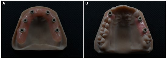
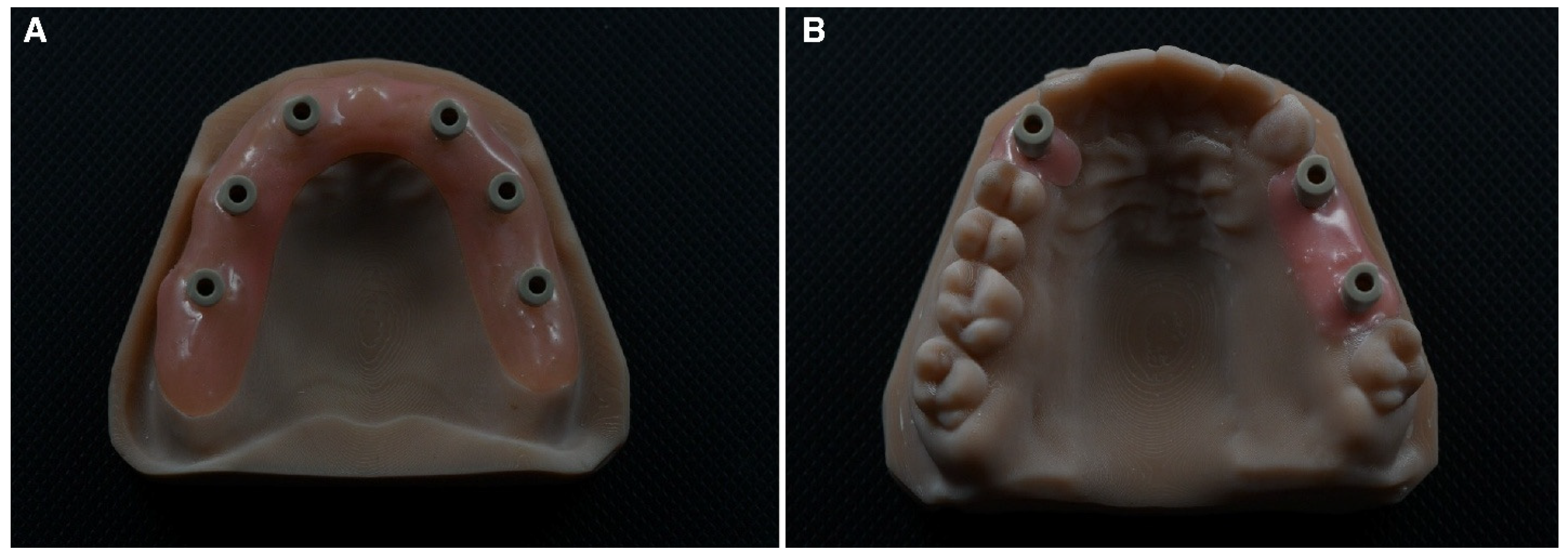
Two different maxillary sample models (fully edentulous and dentulous) were designed using CAD software (Exocad DentalCAD, v3.2 ‘Elefsina’; Exocad GmbH, Darmstadt, Germany). Implant sites were digitally prepared on the models to simulate different clinically relevant implant-supported scenarios. On the fully edentulous model, implant sites corresponding to teeth 12, 22, 14, 24, 16, and 26 were prepared to simulate a full-arch implant scenario. In the dentulous model, an implant site corresponding to tooth 13 was prepared to simulate a single-unit implant scenario. Additionally, teeth 24, 25, and 26 were included, with implant sites prepared at teeth 24 and 26 to simulate a three-unit implant fixed partial denture scenario. The models were printed using 3D Printer (Asiga MAX UV; Asiga, Sydney, NSW, Australia) with resin material (Alias model precise resin, Dokuz Kimya, İstanbul, Turkey). Implant sites corresponding to the digitally grooved regions were prepared using a parallelometer (Rotaks-Dent, Istanbul, Turkey) equipped with a micromotor and hard bur to ensure parallelism. Digital implant analogs (T6 32204; Nucleoss, Menderes/Izmir, Turkey) were then placed and fixed within the resin models under parallelometer guidance to simulate the clinical implant positions. High-precision non-reflective polyether-ether-ketone (PEEK) scan bodies (T6, 32898; Nucleoss, Menderes/Izmir, Turkey) were screwed onto the implant analogs and the models were scanned using a laboratory scanner (Autoscan DS-Mix; Shining 3D, Hangzhou, China). The acquired data were transferred to the same CAD software, where the digital gingival tissues were designed for the models, and new resin models were printed with spaces for the digital implant analogs and pink resin gingiva (Alias gingiva, Dokuz Kimya, İstanbul, Turkey). The implant analogs were then fixed to the models using screws, and the corresponding scan bodies were screwed to the implant analogs (Figure 1).
Figure 1.
Master resin models used in the study. (A) Edentulous maxillary model with scan bodies in teeth number 12, 22, 14, 24, 16, and 26 to simulate a full-arch (FA) scenario. (B) Partially dentulous maxillary model with a scan body in tooth number 13 to simulate a single unit (SC) implant scenario, and two scan bodies in teeth number 24 and 26 to simulate a fixed partial denture (FPD) implant scenario.
2.2. Reference Dataset Generation
The two final master resin models with the scan bodies in position were scanned ten times for each model using a desktop scanner CEREC InEos X5 (Sirona Dental System, Bensheim, Germany), which has been reported in a previous study to provide high accuracy, with values of approximately 2.8 μm [20]. These scans were subsequently imported into a reverse engineering software (Geomagic Design X software 2024.3.2, 3D Systems, Rock Hill, SC, USA) and trimmed by using a preconfigured template cutting tool (in order to always reproduce the same cuts). The partially dentulous models were divided into two parts to represent two different scenarios.
As a result, 10 preconfigured cuts corresponding to each scanning scenario—SC, FPD, and FA—were obtained. The superimposition of the 10 meshes prepared for each scenario was performed using reverse engineering software, and one reference model was generated for each scenario. The three scenario-specific reference meshes were saved as STL files.
2.3. Intraoral Scanner Acquisition Protocol
The resin models were scanned using five IOSs (Trios 5, 3Shape, Copenhagen, Denmark; iTero Lumina, Align Technology, Santa Clara, CA, USA; Trios 4, 3Shape, Copenhagen, Denmark; CS 3600, Carestream Dental, Atlanta, GA, USA; Helios 600 Eighteeth, Changzhou, China), each with varying characteristics shown in Table 1.
Table 1.
Characteristics of the IOSs used.
The sample size calculation was performed by considering the between-group comparison results reported in the reference study [21]. Power analysis software (G*Power 3.1.9.2; Heinrich-Heine-Universität Düsseldorf, Düsseldorf, Germany) was used for the power analysis. The significance level (α) was set at 5%, and the test power (1−β) was set at 80%. Because five different IOS groups were compared, the calculation was performed using a one-way ANOVA model. Based on similar variables in the reference study, the effect size was accepted as f = 0.40. According to these parameters, the minimum total sample size was calculated as 50. Thus, 10 scans were captured for each of the three scenarios (SC, FPD, FA) with each of the IOSs. All models were completely scanned using a standardized zig-zag scanning strategy, proceeding from the buccal to occlusal and palatal surfaces, with continuous overlap movements to ensure detailed data acquisition of teeth and scan bodies. All scans were performed under a standardized environment in room conditions with a temperature of 22 °C and 45% relative humidity. All IOSs were calibrated before usage for scanning according to the manufacturer’s recommendations. All scanning procedures were performed by one operator (M.N.).
2.4. Three-Dimensional Surface Deviation Analysis
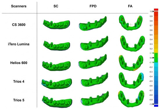
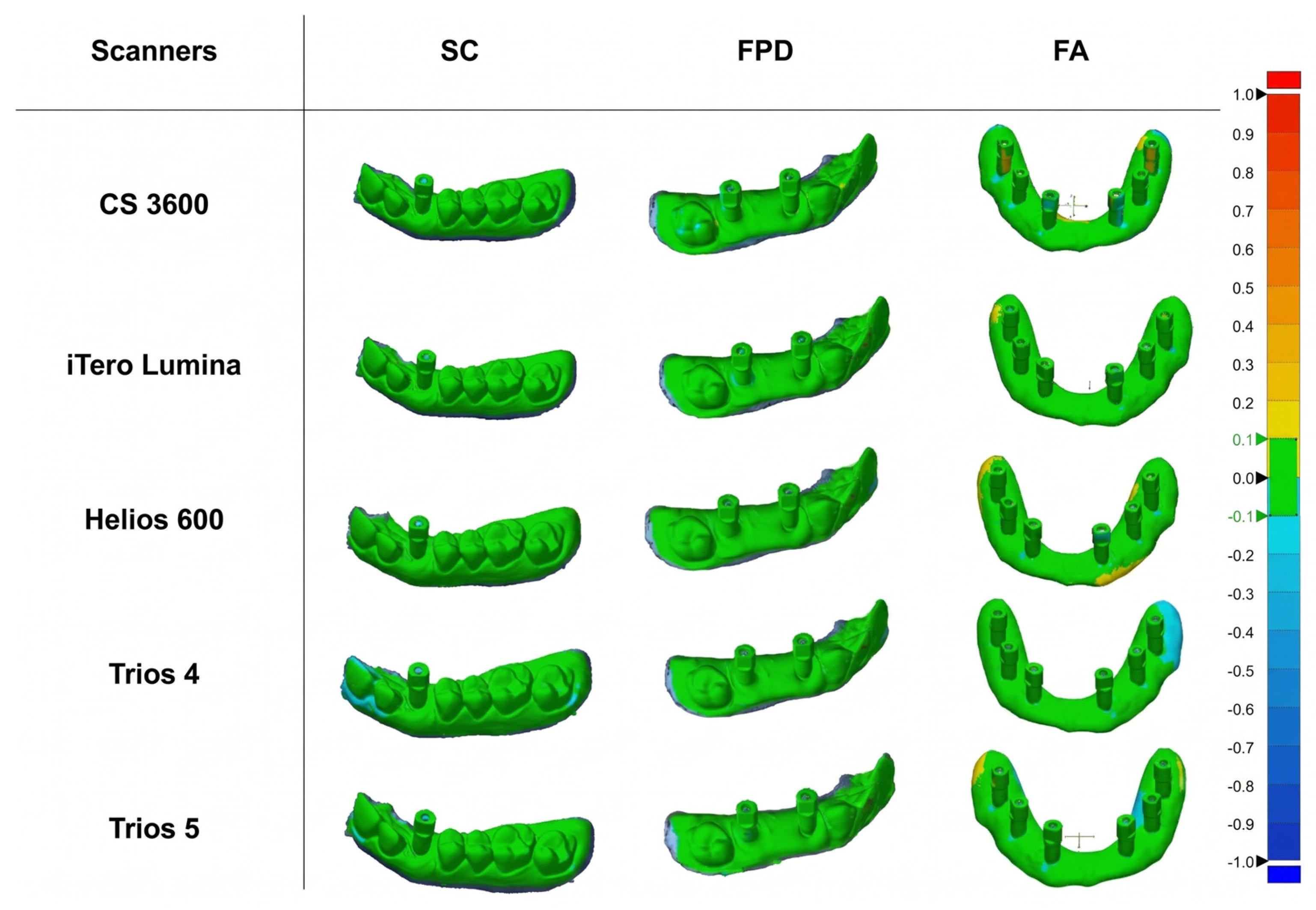
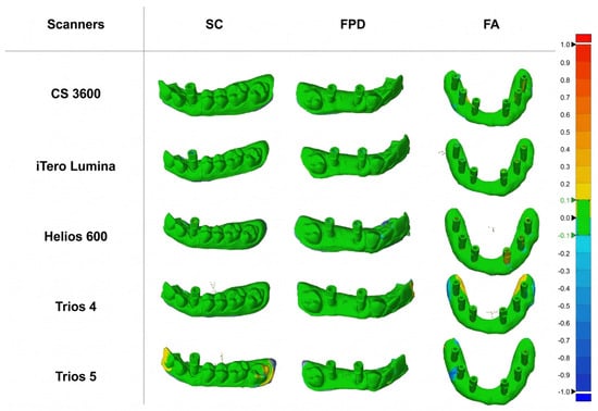
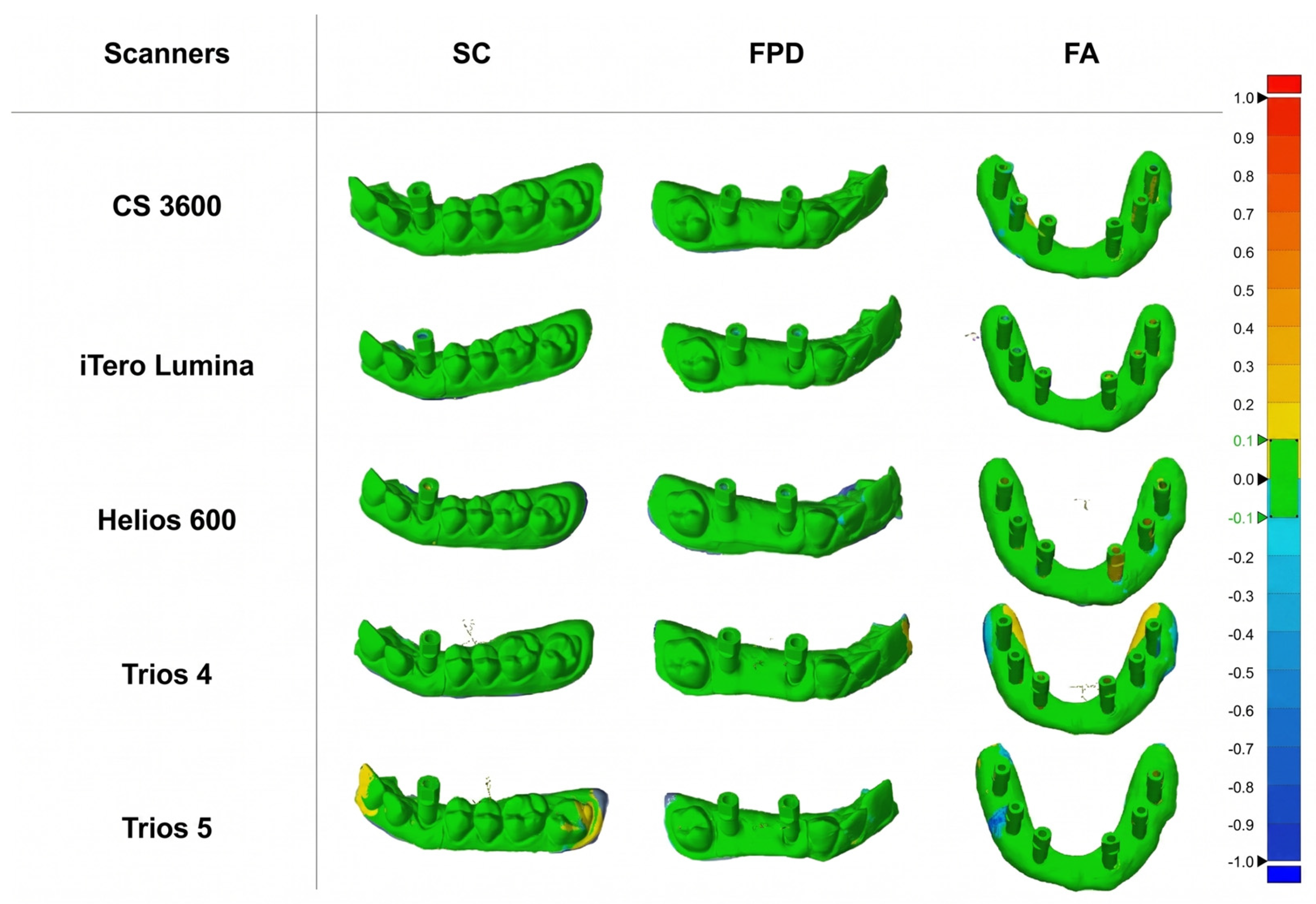
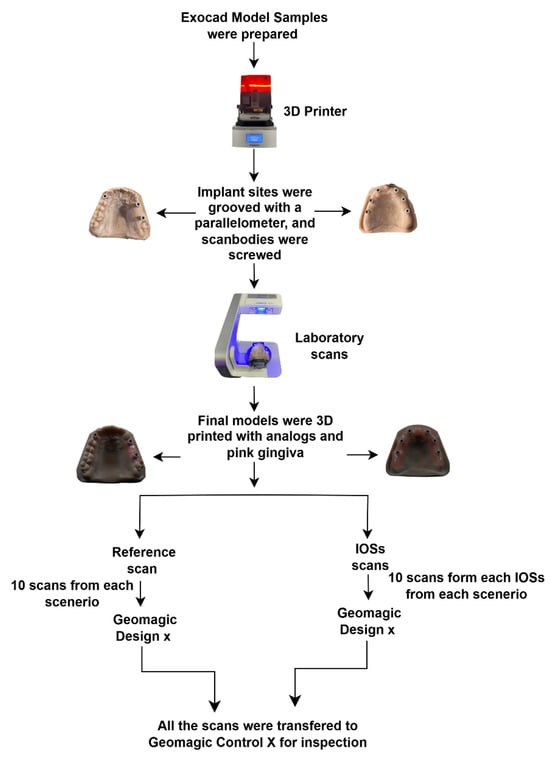
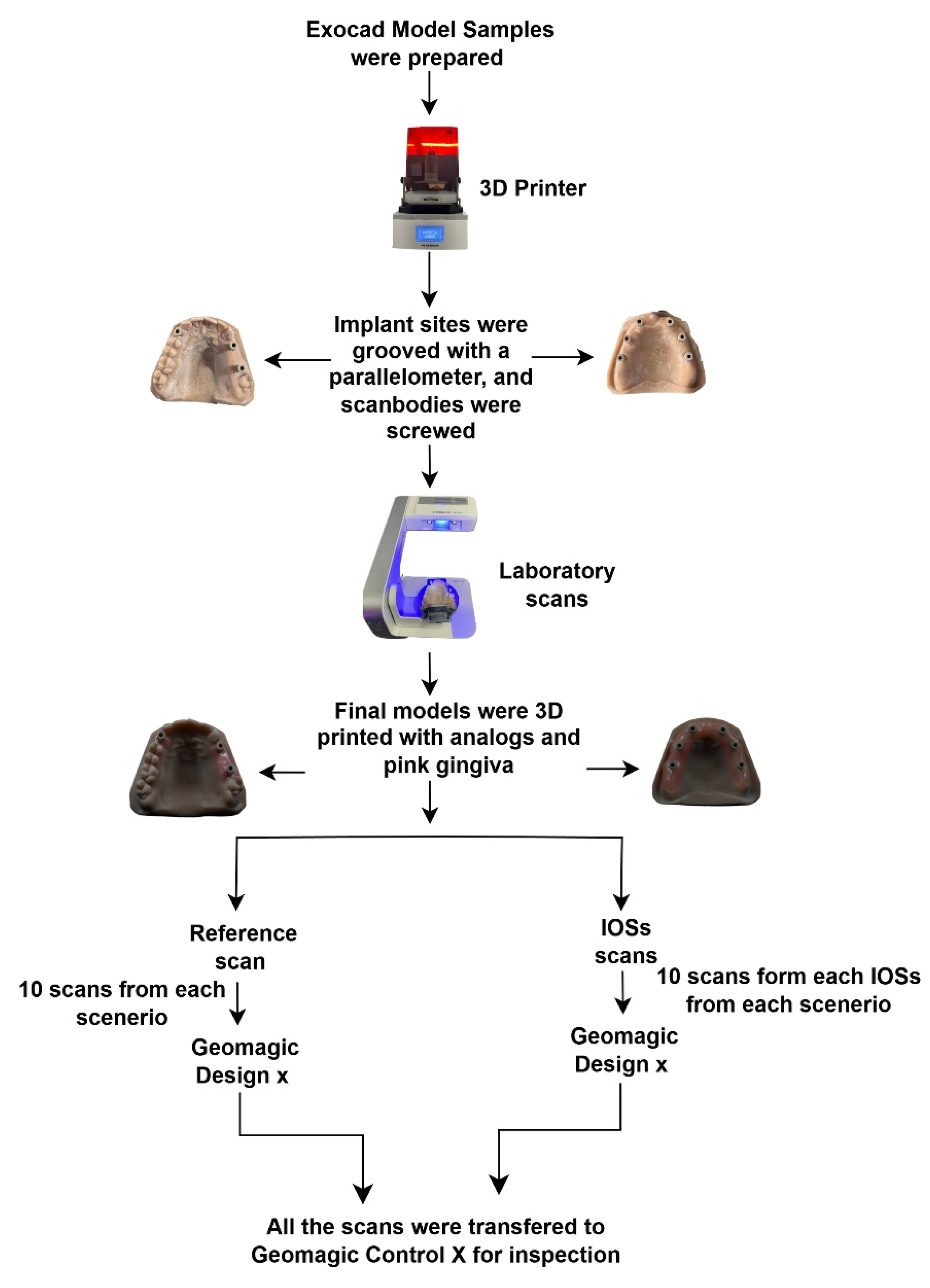
All the models scanned with different IOSs were also imported into and cut by the reverse engineering software Geomagic Design X using a preconfigured template cutting tool to reproduce the same cuts. The partially dentulous models that were scanned by IOSs were also divided into two parts to represent two different scenarios. This resulted in 150 scans, divided into 50 scans for each scenario, with 10 scans from each IOS. The same standardized scenario-specific trimming boundaries used for the RMs were applied to all IOS datasets. Accordingly, the SC dataset included the scan body at tooth position 13 together with the surrounding partially dentulous model surfaces within the predefined cut, whereas the FPD dataset included the scan bodies at tooth positions 24 and 26 together with the surrounding partially dentulous model surfaces within the predefined cut. Therefore, the SC and FPD datasets represented predefined surface-based scanning scenarios rather than only scan body regions. All the 150 scans and their corresponding three RMs were imported into a 3D analysis software (Geomagic Control X software 2024.1.0, 3D Systems, Rock Hill, SC, USA) for inspection. The scans were first superimposed using initial pre-alignment with global registration to standardize orientation across all datasets, followed by a best-fit alignment algorithm to minimize spatial deviation relative to the reference geometry. For trueness assessment, all scans acquired using the different IOSs were superimposed onto their corresponding RMs. For precision assessment, the scan with the lowest RMS deviation within each scanner and scenario group was selected as the internal reference dataset. The remaining scans from the same scanner and scenario were then superimposed onto this internal reference, and relative RMS deviations were calculated. The RMS analysis was performed on the complete scenario-specific surface dataset after standardized trimming. Therefore, the evaluated areas included the scan bodies together with the surrounding teeth and/or simulated soft-tissue areas present within each predefined scenario. Each report included RMS deviation values and 3D color-deviation maps (scale −1.0 to +1.0 mm). Green regions represented areas within the defined tolerance band (±0.1 mm), while blue (−1.0 mm) and red (+1.0 mm) indicated negative and positive deviations (Figure 2 and Figure 3). The workflow of the study is summarized in the chart in Figure 4.
Figure 2.
Surface deviation maps showing RMS trueness assessment of five IOSs across the three predefined scenario-specific datasets (SC, FPD, and FA). The color scale ranged from −1.0 to +1.0 mm. Green regions define a tolerance band ±0.1 mm, while blue −1.0 mm and red +1.0 mm indicate negative and positive deviations, respectively.
Figure 3.
Surface deviation maps showing RMS precision assessment of five IOSs across the three predefined scenario-specific datasets (SC, FPD, and FA). The color scale ranged from −1.0 to +1.0 mm. Green regions define a tolerance band ±0.1 mm, while blue −1.0 mm and red +1.0 mm indicate negative and positive deviations, respectively.
Figure 4.
Workflow summarizing the study steps.
2.5. Statistical Analysis
Statistical analyses were performed using IBM SPSS Statistics version 28.0 (IBM Corp., Armonk, NY, USA). Normality assumptions were assessed using the Shapiro–Wilk test. Homogeneity of variances across scanner groups and scanning scenarios was evaluated using Levene’s test. Non-significant Levene’s test results (p > 0.05) indicated that the assumption of equal variances was satisfied, supporting the use of parametric analyses.
The primary analysis consisted of two-way analysis of variance (ANOVA) models evaluating the main effects of scanner type and scanning scenario, as well as their interaction effects, on trueness and precision measurements. When significant omnibus effects were identified, one-way ANOVA followed by Tukey’s honestly significant difference (HSD) post hoc tests was performed as follow-up simple-effect analyses to determine pairwise differences among scanners within each scenario and among scenarios within each scanner.
To control the family-wise Type I error rate arising from multiple pairwise comparisons, Bonferroni-adjusted significance thresholds were applied. For comparisons among the three scanning scenarios, statistical significance was set at p < 0.016 (0.05/3), whereas for comparisons among the five scanners, significance was set at p < 0.005 (0.05/10).
Statistical significance was set at p < 0.05 for all omnibus analyses. Effect sizes were reported using partial eta squared (η2), which represents the proportion of variance in the dependent variable attributable to a given factor after controlling for the effects of other factors in the model. According to commonly accepted guidelines, η2 values of approximately 0.01, 0.06, and 0.14 indicate small, medium, and large effect sizes, respectively.
3. Results
3.1. Surface-Based RMS Trueness Analysis
Two-way ANOVA indicated statistically significant differences in RMS trueness values according to both scanner type (F(4) = 16.488, p < 0.001, η2 = 0.328) and scenario type (F(2) = 17.318, p < 0.001, η2 = 0.204). The scanner × scenario interaction was not statistically significant (F(8) = 1.735, p = 0.096), indicating that differences between scanners were generally consistent across scenarios, as shown in Table 2.
Table 2.
Two-Way ANOVA for RMS Trueness values.
RMS trueness values for the five scanners across the three scanning scenarios are given in Table 3, where lower RMS values indicate the higher trueness. One-way ANOVA showed statistically significant differences among scanners in the SC (p < 0.001) and FA (p < 0.001) scenarios, as well as a limited significant difference in the FPD scenario (p = 0.047).
Table 3.
Comparison of surface-based RMS Trueness values (mm).
In the FPD scenario, RMS trueness values ranged from 0.08 ± 0.05 mm (iTero Lumina) to 0.23 ± 0.08 mm (Trios 5). The remaining scanners (CS 3600, Helios 600, and Trios 4) showed intermediate RMS trueness values. Post hoc pairwise comparisons revealed no statistically significant differences among these scanners (p > 0.005).
In the SC scenario, iTero Lumina, Helios 600, and Trios 4 demonstrated the lower RMS trueness values, followed by CS 3600 and Trios 5. Post hoc analysis indicated that significant differences were found between Helios 600 and Trios 5, CS 3600 and iTero Lumina, iTero Lumina and Trios 5, and Trios 4 and Trios 5 (p < 0.005).
In the FA scenario, Trios 4 and Trios 5 exhibited significantly higher RMS trueness values compared to iTero Lumina and Helios 600, which demonstrated significantly lower RMS trueness values (<0.10 mm). CS 3600 showed no statistically significant differences compared with other scanners. Post hoc analysis indicated that significant differences were found between Helios 600 and Trios 4, Helios 600 and Trios 5, Trios 4 and iTero Lumina, and Trios 5 and iTero Lumina (p < 0.005).
According to post hoc analyses, a significant difference was found between FPD and SC, and between FPD and FA for iTero Lumina (p < 0.016). A significant difference was found between FPD and SC for Helios 600 (p < 0.016). A significant difference was found between FPD and FA, and between SC and FA for Trios 4 (p < 0.016).
3.2. Surface-Based RMS Precision Analysis
Two-way ANOVA indicated statistically significant differences in RMS precision values according to both scanner type (F(4) = 3.377, p = 0.012, η2 = 0.101) and scenario type (F(2) = 5.824, p = 0.004, η2 = 0.088). The scanner × scenario interaction was also significant (F(8) = 3.765, p < 0.001, η2 = 0.201), indicating that the repeatability of each scanner varied depending on the scanning scenario, as shown in Table 4.
Table 4.
Two-Way ANOVA for RMS Precision values.
RMS precision comparisons are summarized in Table 5, where lower RMS values indicate the higher precision. One-way ANOVA revealed significant differences among scanners in the FPD (p = 0.009) and FA (p < 0.001) scenarios, whereas no significant difference was observed in the SC scenario (p = 0.349).
Table 5.
Comparison of surface-based RMS Precision values (mm).
In the FPD scenario, post hoc analysis revealed a statistically significant difference only between CS 3600 and iTero Lumina, with iTero Lumina demonstrating lower RMS precision values (0.04 ± 0.02 mm). No significant differences were detected among the remaining scanners. In the SC scenario, no statistically significant differences in RMS precision values were observed among scanners. RMS precision values ranged from (0.08 ± 0.04 mm) for Trios 4 and Trios 5 to (0.14 ± 0.08 mm) for iTero Lumina.
In the FA scenario, post hoc analysis revealed statistically significant differences between iTero Lumina and Trios 5. iTero Lumina showed lower RMS precision value (0.05 ± 0.01 mm) compared with Trios 5. Additionally, within-scanner comparisons showed that iTero Lumina differed significantly between FPD and FA, and between SC and FA (p < 0.016). Although the mean RMS precision values for the FPD and FA scenarios were numerically close, the statistical difference should be interpreted in relation to the distribution and variability of the repeated measurements within each scenario. For both Trios 4 and Trios 5, statistically significant differences were observed between the SC and FA scenarios (p < 0.016).
4. Discussion
The purpose of this in vitro study was to evaluate and compare the surface-based trueness and precision of five different IOSs in digital impressions for single crown (SC), three-unit fixed partial denture (FPD), and full-arch (FA) implant-supported digital scanning scenarios using RMS deviation analysis. According to the study results, the RMS trueness values differed significantly across scanners and scanning scenarios, especially in the SC and FA scenarios (p < 0.001). The RMS precision values varied significantly across scanners in the FPD (p = 0.009) and FA (p < 0.001) scenarios. According to the study results, under the standardized zig-zag scanning strategy used in this study, surface-based RMS trueness and precision differed significantly among the different IOSs across the evaluated implant-supported scanning scenarios. Therefore, the null hypothesis was rejected. As IOSs record geometry using different optical principles, the optical characteristics of the object being scanned could have an impact on their accuracy [22,23]. The extent of the scan and the number of implants involved may also influence IOS accuracy [24,25].
In this in vitro study, the Asiga MAX UV 3D printer and Asiga model precise resin were selected to optimize dimensional accuracy and stability, as Asiga MAX UV achieved the lowest post-print deviations (12–240 μm) and the narrowest precision range (17–388 μm) among all extrusion and photopolymerization-based systems in previous studies [26,27]. Additionally, Asiga model precise resin was used instead of conventional stone to enhance dimensional stability, as resin is less susceptible to water absorption and solubility [28,29].
The InEos X5 desktop scanner was used as a reference scanner for IOS evaluation, consistent with its usage as a reference scanner in a recent study [30]. The InEos X5 is a non-contact optical desktop scanner using blue-light digital stripe projection, and previous studies have reported high accuracy values for this system [20,31,32]. In the present workflow, ten desktop scans were obtained for each master model, trimmed using the same preconfigured template, and superimposed to generate one RM for each scenario before IOS comparison. This provided a standardized laboratory reference workflow for calculating surface-based RMS trueness and precision. For the IOSs, a single scanning pattern was used to ensure standardization across all scanner systems. A zig-zag scanning strategy, previously adopted in IOS accuracy studies, was selected because it allows continuous overlapping acquisition of the buccal, occlusal, and palatal surfaces during arch scanning and provides a reproducible path for comparing multiple scanners under the same experimental conditions [33,34,35]. This approach was also supported by previous studies showing that scan path can influence IOS trueness and precision, and that zig-zag patterns may provide favorable accuracy outcomes in complete-arch scanning depending on the scanner system used [35,36].
Geomagic Control X was selected for all three-dimensional analysis. It is one of the professional metrology software programs recommended by the ISO 12836 standard to assess accuracy in dental impression systems [37]. Geomagic Control software accurately measured volume change in dental scans [38]. A similar internal-reference approach for precision assessment has been used in a previous digital implant scanning study, in which the scan with the lowest RMS deviation within each test group was selected as the reference for repeated-scan comparison [39]. RMS deviation was selected as the target parameter because the aim of this study was to compare the overall surface agreement of complete scenario-specific IOS datasets after superimposition. RMS condenses the cumulative 3D discrepancy between the test scan and the reference dataset into a single quantitative value, which enables standardized comparison of trueness and precision among scanners and scanning scenarios using the same analysis workflow. This surface-based approach is particularly suitable for evaluating global scan agreement over the trimmed SC, FPD, and FA datasets, where deviations may occur not only at the scan bodies but also in the surrounding dentulous or edentulous model surfaces [40,41].
Osman et al. [42] compared three IOSs with different scanning technologies: Trios 3 (confocal), iTero Element (parallel confocal), and Medit i700 (triangulation), and reported that the confocal-based Trios 3 had the highest trueness and Medit i700 had the lowest accuracy in terms of trueness and precision. The iTero Element was fast at acquiring scans but had less trueness. This highlighted how different scanning technologies can impact intraoral impression accuracy. In the present study, significant differences in trueness were observed among scanners across all the scenarios, whereas significant differences in precision were observed only in the FPD and FA. These findings suggest that scanner-related differences should be considered when interpreting surface-based RMS outcomes across different implant-supported scanning scenarios.
In a 2025 study, Dönmez et al. [43] evaluated the accuracy of various wireless (Primescan 2 and Trios 5) and wired IOSs (Primescan and Trios 3) and reported that Trios 5 had the highest random 3D deviations. The authors suggested that wireless transmission of scanned data was a factor leading to possible errors, disrupting real-time stitching of images. Consistent with these findings, the present study demonstrated that Trios 5 in the SC showed significantly higher trueness deviations in selected comparisons than iTero Lumina, Helios 600, and Trios 4 (p < 0.005). Although Trios 5 showed numerically higher trueness deviations in FPD, these differences were not statistically significant when compared with the other scanners. This agreement with Dönmez et al. supports the interpretation that scanner-specific acquisition and reconstruction behavior may influence surface-based RMS outcomes. However, the present findings should primarily be understood as surface-based RMS differences among scanners under the tested conditions.
Schmalzl et al. [44] investigated the trueness of Trios 5 in a completely edentulous maxillary full-arch implant model with six implant analogs. The authors reported that short-span accuracy was acceptable; however, full-arch deviations exceeded clinically acceptable limits. These results are in line with the present study, where Trios 5 showed significantly greater deviations in trueness compared with iTero Lumina and Helios 600 (p < 0.005) in the FA scenario. This highlights that the complexity of the FA scenario can have an impact on the accuracy and repeatability of the scanners.
Another study by Schmidt et al. [45] investigated four IOSs (Medit i700, Primescan, Trios 4, and Trios 5), based on an analysis of coordinate differences in a maxillary implant model with four posterior implants. Because Schmidt et al. used coordinate-based deviation analysis, whereas the present study assessed complete scenario-specific datasets using surface-based RMS superimposition, the findings are more appropriately compared in terms of scanner-performance trends than direct numerical values. The authors reported that Primescan and Medit i700 showed better performance, while Trios 4 and Trios 5 showed higher deviation values, and no difference was observed between Trios 4 and Trios 5, indicating limited technological advancement between the two systems. In the present study, relatively similar results were observed, as no statistically significant differences were seen between Trios 4 and Trios 5 in terms of trueness and precision in all scenarios except one; Trios 5 showed higher RMS trueness deviation in the SC scenario when compared with Trios 4. This finding suggests that Trios 5 may not show a consistent accuracy advantage over Trios 4 under the tested scenarios.
Pesce et al. [46] evaluated the trueness of two IOSs in different implant-supported prosthetic situations. The authors reported that trueness decreased as the scanning situation became more complex, with single-implant conditions showing the highest accuracy and full-arch rehabilitations showing the greatest deviations. This comparison should be interpreted in view of methodological differences, as Pesce et al. focused on two IOSs across different implant-supported situations, whereas the present study compared five IOS systems using predefined scenario-specific datasets and surface-based RMS superimposition. In the present study, the predefined scanning scenario also influenced the surface-based RMS outcomes, but the pattern was not the same for all IOSs. CS 3600 and Trios 5 did not show significant differences in trueness values across the scenarios, indicating that their scenario-related changes were not statistically significant under the tested scenarios. By contrast, the other scanners showed significant differences only between certain scenarios. Therefore, the scenario-related effect should not be interpreted as a uniform effect of scan span alone, since it may also reflect differences in the scanner system and scenario configuration. For precision, RMS values in the SC scenario did not differ significantly among scanners (p = 0.349), showing that repeatability was generally comparable in the simpler scanning condition. In the FPD and FA scenarios, significant differences in precision were found, suggesting that extended-span implant scanning conditions may be more demanding for obtaining consistent surface-based RMS outcomes.
Mangano et al. [21] conducted a comparative in vitro study to test five IOSs in different implant restoration scenarios. The authors reported a statistically significant interaction between scanner type and complexity of restorations (p < 0.05), indicating that both trueness and precision declined as restoration complexity increased. Although both studies evaluated multiple IOSs across implant-supported scenarios, differences in scanner systems, implant configurations, reference workflow, and outcome assessment should be considered when interpreting the interaction effects. In the present study, the two-way ANOVA results for trueness differed slightly. While both scanner type (p < 0.001, η2 = 0.328) and scanning scenario type (p < 0.001, η2 = 0.204) significantly influenced trueness, the interaction between the two factors was not significant (p = 0.096, η2 = 0.093). On the other hand, the precision results were more in line with the previous study findings, as both scanner type (p = 0.012, η2 = 0.101) and scanning scenario (p = 0.004, η2 = 0.088) had significant effects, as well as a strong interaction effect (p < 0.001, η2 = 0.201). This indicates that scanner repeatability may be more sensitive to scanning complexity, particularly in extended-span implant situations.
Güzelce Sultanoğlu and Keleş Eroğlu [47] reported that Helios 600 demonstrated lower trueness and precision compared with Trios 3 and Medit i700 in a single-unit scanning scenario. In contrast, the present study found that Helios 600 showed relatively low RMS trueness values, with significantly lower RMS trueness deviations than Trios 5 in the SC scenario (p < 0.005) and significantly lower RMS trueness deviations than Trios 4 and Trios 5 in the FA scenario (p < 0.005). However, Helios 600 showed no statistically significant differences in precision compared to other scanners. This discrepancy between studies may reflect differences in experimental design, model configuration, scanner systems compared, scanning protocol, and analysis method. In line with this variability, Dönmez et al. [43] also reported reduced accuracy for Trios 5 compared with Trios 3 in certain FPD models. These findings suggest that accuracy improvements across Trios generations may not be stable and may depend on scanning span and reconstruction algorithms. Accordingly, Helios 600 performance relative to Trios 3, Trios 4, and Trios 5 appears to be dependent on the scenario rather than a fixed hierarchy among scanners.
Akkal et al. [48] assessed the accuracy of Primescan, Trios 4, and Carestream CS 3600 in edentulous implant patients, reporting that Primescan showed the highest accuracy, whereas CS 3600 showed significantly higher deviations (p = 0.001), and this indicates that the scanner system may influence full-arch accuracy outcomes. The present findings were not entirely consistent with those results, as CS 3600 performed comparably to Trios 4, with no significant differences between the two scanners in either trueness or precision. CS 3600 also showed no significant within-scanner differences across the tested scenarios for trueness (p = 0.166) or precision (p = 0.719). Within the surface-based RMS evaluation used in the present study, this pattern suggests that CS 3600 produced relatively stable RMS values across different implant-supported scanning scenarios. Differences between the findings may be related to variations in study design, scanning protocol, clinical or model conditions, and software versions.
In previous literature, certain iTero scanners were found to have bulkier tips and a limited range of angulation. This makes it difficult to gain access to tight areas of structures, which may compromise data capture and subsequently stitching. These errors may build up as the scan progresses, causing lower precision values when scanning narrow edentulous areas, especially in the anterior region [49,50]. In the present investigation, iTero Lumina showed higher RMS precision deviation in the SC scenario than in the FA scenario (p < 0.016). However, it showed lower RMS trueness deviations than CS 3600 and Trios 5 in the SC scenario, and lower RMS trueness deviations than Trios 4 and Trios 5 in the FA scenario (p < 0.005). In addition, iTero Lumina showed lower RMS precision deviations than CS 3600 in the FPD scenario and Trios 5 in the FA scenario (p < 0.005), respectively. These findings indicate that iTero Lumina produced favorable RMS outcomes in selected comparisons, although its precision varied according to the tested scanning scenario.
The discrepancy between the very low SC RMS trueness values and the comparatively higher SC RMS precision values observed for iTero Lumina and Helios 600 may be explained by the different aspects of scan accuracy represented by these two parameters. A separation between trueness and precision has also been reported in a scanner-comparison study. Wang et al. [18] evaluated seven IOSs and reported that, in the full-dentate complete-arch cast, the scanner showing the greatest trueness deviation was not the same scanner showing the greatest precision deviation. This indicates that agreement with the reference dataset and repeatability among repeated scans may follow different patterns. In the present study, the low SC RMS trueness values for iTero Lumina and Helios 600 indicate close agreement with the corresponding reference dataset, whereas the higher SC RMS precision values indicate greater variability among repeated scans within the same scanner/scenario group. Therefore, the SC precision findings should not be interpreted as contradicting the trueness results, but rather as showing that repeatability may differ from reference agreement even under the same standardized scanning condition.
From a clinical perspective, IOS selection should be considered together with the intended implant-supported scanning situation rather than assumed to be a fixed scanner-related property. Because implant impression accuracy is closely related to passive prosthetic fit, and prosthetic misfit may contribute to mechanical and biological complications, scanner performance should be interpreted in relation to scan extent, implant distribution, and the surrounding dentulous or edentulous anatomy [51,52]. In the present study, precision did not differ significantly among scanners in the SC scenario, whereas scanner-related precision differences were detected in the FPD and FA scenarios. Trueness differences were mainly observed in the SC and FA scenarios. Therefore, in longer-span implant-supported scanning situations, these anatomical and scenario-related factors should be considered when selecting and interpreting an IOS-based digital workflow.
It should be acknowledged that the in vitro design of the current study may not fully reflect intraoral conditions, such as lighting conditions, saliva, and humidity. Resin models are generally easier to scan than intraoral models, and the optical properties of oral tissues differ significantly from resin models, which may impact the in vivo performance of IOSs. Additionally, a desktop scanner was used to scan reference models; however, industrial scanners are considered the gold standard for this application. The present study also did not evaluate restorations made from the scans using the IOSs. Clinical studies evaluating the marginal fit of different restorations in different clinical scenarios (SC, FPD, and FA) may further support the findings obtained from STL file superimposition analyses. Another limitation is that the scanner comparisons were based on surface-based RMS values obtained from complete scenario-specific datasets under one standardized zig-zag scanning strategy; additionally, scan body-only segmentation and coordinate-based implant positional analyses were not performed. RMS surface deviation provides a global 3D measure of scan agreement, but it is not specific to implant position and may be influenced by the amount and distribution of the surrounding scanned surface. Furthermore, the SC, FPD, and FA datasets differed in implant location, implant distribution, and surrounding dentulous or edentulous anatomy; therefore, scenario-related comparisons should be interpreted as comparisons among predefined experimental configurations rather than as isolated effects of scan extent alone. Future studies should combine RMS surface analysis with scan body isolation, implant-axis deviation, linear inter-implant distance measurements, and angular deviation analysis to provide a more direct evaluation of implant positional transfer. Therefore, future in vivo studies are needed to confirm the clinical significance of these findings.
5. Conclusions
Within the limitations of this comparative in vitro study, the following conclusions can be drawn for the evaluated IOSs and scanning scenarios:
- Scanner type and scanning scenario influenced surface-based RMS trueness and precision values.
- In terms of trueness, differences among scanners were mainly evident in the SC and FA scenarios, while the FPD scenario showed only limited overall differences. Precision did not differ significantly among scanners in the SC scenario, whereas scanner-related precision differences were observed in the FPD and FA scenarios.
- Under the surface-based RMS evaluation and standardized zig-zag scanning strategy used in this study, iTero Lumina showed lower RMS trueness deviations than CS 3600 and Trios 5 in the SC scenario, and than Trios 4 and Trios 5 in the FA scenario. It also showed lower RMS precision deviations than CS 3600 in the FPD scenario and than Trios 5 in the FA scenario. Helios 600 showed lower RMS trueness deviations than Trios 5 in the SC scenario and than Trios 4 and Trios 5 in the FA scenario. Trios 5 did not show a consistent accuracy advantage over Trios 4. CS 3600 showed relatively stable RMS trueness and precision values across the tested scenarios.
- These findings can suggest that surface-based RMS accuracy may vary according to scanner system, scan extent, implant distribution, and scenario configuration, since scanner-related differences were not observed in the same pattern across the SC, FPD, and FA scenarios.
Author Contributions
M.M.M.N. conducted the literature search and performed the material preparation, methodological procedures, and statistical analyses for data acquisition. Ç.Ö. and A.İ. supervised the study and contributed to the critical review of the manuscript. All authors participated in data interpretation, revised the manuscript for important intellectual content. All authors have read and agreed to the published version of the manuscript.
Funding
This research received no external funding.
Institutional Review Board Statement
The Institutional Review Board and the Research Ethics Commitee of Cyprus Health and Social Sciences University reviewed and approved the research protocol (Approval No: KSTU//2025/007).
Informed Consent Statement
Not applicable.
Data Availability Statement
The datasets used and/or analyzed during the current study are available from the corresponding author on reasonable request.
Conflicts of Interest
The authors declare no conflicts of interest.
Abbreviations
The following abbreviations are used in this manuscript:
| AI | Artificial intelligence |
| ANOVA | Analysis of variance |
| CAD/CAM | Computer-aided design/computer-aided manufacturing |
| FA | Full arch |
| FOV | Field of view |
| FPD | Fixed partial denture |
| HD | High definition |
| IOS | Intraoral scanner |
| LED | Light-emitting diode |
| PEEK | Polyetheretherketone |
| PLY | Polygon file format |
| RGB | Red–green–blue |
| RM | Reference model |
| RMS | Root mean square |
| SC | Single crown |
| STL | Standard tessellation language |
| 3D | Three-dimensional |
References
- Block, M.S. Dental Implants: The Last 100 Years. J. Oral Maxillofac. Surg. 2018, 76, 11–26. [Google Scholar] [CrossRef] [Scilit] [PubMed]
- Baldissara, P.; Koci, B.; Messias, A.M.; Meneghello, R.; Ghelli, F.; Gatto, M.R.; Ciocca, L. Assessment of Impression Material Accuracy in Complete-Arch Restorations on Four Implants. J. Prosthet. Dent. 2021, 126, 763–771. [Google Scholar] [CrossRef] [Scilit] [PubMed]
- Joda, T.; Brägger, U. Patient-centered Outcomes Comparing Digital and Conventional Implant Impression Procedures: A Randomized Crossover Trial. Clin. Oral Implant. Res. 2016, 27, e185–e189. [Google Scholar] [CrossRef] [Scilit] [PubMed]
- Ting-shu, S.; Jian, S. Intraoral Digital Impression Technique: A Review: Intraoral Digital Impression Review. J. Prosthodont. 2015, 24, 313–321. [Google Scholar] [CrossRef] [Scilit] [PubMed]
- Alzahrani, S.J.; EL-Hammali, H.; Morgano, S.M.; Elkassaby, H. Evaluation of the Accuracy of 2 Digital Intraoral Scanners: A 3D Analysis Study. J. Prosthet. Dent. 2021, 126, 787–792. [Google Scholar] [CrossRef] [Scilit] [PubMed]
- Ahmed, S.; Hawsah, A.; Rustom, R.; Alamri, A.; Althomairy, S.; Alenezi, M.; Shaker, S.; Alrawsaa, F.; Althumairy, A.; Alteraigi, A. Digital Impressions Versus Conventional Impressions in Prosthodontics: A Systematic Review. Cureus 2024, 16, e51537. [Google Scholar] [CrossRef] [Scilit] [PubMed]
- Joda, T.; Zarone, F.; Ferrari, M. The Complete Digital Workflow in Fixed Prosthodontics: A Systematic Review. BMC Oral Health 2017, 17, 124. [Google Scholar] [CrossRef] [Scilit] [PubMed]
- Mangano, F.G.; Admakin, O.; Bonacina, M.; Lerner, H.; Rutkunas, V.; Mangano, C. Trueness of 12 Intraoral Scanners in the Full-Arch Implant Impression: A Comparative in Vitro Study. BMC Oral Health 2020, 20, 263. [Google Scholar] [CrossRef] [Scilit] [PubMed]
- Schmidt, A.; Schlenz, M.A.; Liu, H.; Kämpe, H.S.; Wöstmann, B. The Influence of Hard- and Software Improvement of Intraoral Scanners on the Implant Transfer Accuracy from 2012 to 2021: An In Vitro Study. Appl. Sci. 2021, 11, 7166. [Google Scholar] [CrossRef] [Scilit]
- Verniani, G.; Casucci, A.; Nosrati, N.; D’Arienzo, L.F.; Val, M.; Ferrari Cagidiaco, E. Accuracy Evaluation of Two Different Intraoral Scanners in Implant Prosthodontics. A Comparative In Vitro Study. J. Osseointegration 2024, 16, 61–64. [Google Scholar] [CrossRef]
- Özal, Ç. Guide to Optimizing the Accuracy of Intraoral Implant Scans: A Review Article. Cyprus J. Med. Sci. 2025, 10, 13–21. [Google Scholar] [CrossRef] [Scilit]
- Alkadi, L. A Comprehensive Review of Factors That Influence the Accuracy of Intraoral Scanners. Diagnostics 2023, 13, 3291. [Google Scholar] [CrossRef] [Scilit] [PubMed]
- ISO 5725-1:2023; Accuracy (Trueness and Precision) of Measurement Methods and Results Part 1: General Principles and Definitions. International Organization for Standardization: Geneva, Switzerland, 2023.
- Renne, W.; Ludlow, M.; Fryml, J.; Schurch, Z.; Mennito, A.; Kessler, R.; Lauer, A. Evaluation of the Accuracy of 7 Digital Scanners: An in Vitro Analysis Based on 3-Dimensional Comparisons. J. Prosthet. Dent. 2017, 118, 36–42. [Google Scholar] [CrossRef] [Scilit] [PubMed]
- Revilla-León, M.; Kois, D.E.; Kois, J.C. A Guide for Maximizing the Accuracy of Intraoral Digital Scans. Part 1: Operator Factors. J. Esthet. Restor. Dent. 2023, 35, 230–240. [Google Scholar] [CrossRef] [Scilit] [PubMed]
- Sequeira, V.; Harper, M.T.; Lilly, C.L.; Bryington, M.S. Accuracy of Digital Impressions at Varying Implant Depths: An In Vitro Study. J. Prosthodont. 2023, 32, 54–61. [Google Scholar] [CrossRef] [Scilit] [PubMed]
- Revilla-León, M.; Kois, D.E.; Kois, J.C. A Guide for Maximizing the Accuracy of Intraoral Digital Scans: Part 2—Patient Factors. J. Esthet. Restor. Dent. 2023, 35, 241–249. [Google Scholar] [CrossRef] [Scilit] [PubMed]
- Wang, X.; Zhang, F.; Ma, D.; Ye, X.; Zheng, X.; Ren, R.; Ren, N.; Bai, S. Evaluation of the Accuracy of Seven Intraoral Scanners for the Full Dentate and Partially Edentulous Complete-Arch Mandibular Casts: An in Vitro Comparison. Heliyon 2024, 10, e31063. [Google Scholar] [CrossRef] [Scilit] [PubMed]
- Amornvit, P.; Rokaya, D.; Sanohkan, S. Comparison of Accuracy of Current Ten Intraoral Scanners. BioMed Res. Int. 2021, 2021, 2673040. [Google Scholar] [CrossRef] [Scilit] [PubMed]
- El-Refay, H.M.; Abdelaziz, M.S.; Cheta, N.M.; Abdallah, M.F. The Accuracy of Digital Impression with Different Intraoral Scanners on Maxillary All on Four Implants: An in Vitro Study. BMC Res. Notes 2025, 18, 186. [Google Scholar] [CrossRef] [Scilit] [PubMed]
- Mangano, F.G.; Hauschild, U.; Veronesi, G.; Imburgia, M.; Mangano, C.; Admakin, O. Trueness and Precision of 5 Intraoral Scanners in the Impressions of Single and Multiple Implants: A Comparative in Vitro Study. BMC Oral Health 2019, 19, 101. [Google Scholar] [CrossRef] [Scilit] [PubMed]
- Lim, J.-H.; Mangal, U.; Nam, N.-E.; Choi, S.-H.; Shim, J.-S.; Kim, J.-E. A Comparison of Accuracy of Different Dental Restorative Materials between Intraoral Scanning and Conventional Impression-Taking: An In Vitro Study. Materials 2021, 14, 2060. [Google Scholar] [CrossRef] [Scilit] [PubMed]
- Dutton, E.; Ludlow, M.; Mennito, A.; Kelly, A.; Evans, Z.; Culp, A.; Kessler, R.; Renne, W. The Effect Different Substrates Have on the Trueness and Precision of Eight Different Intraoral Scanners. J. Esthet. Restor. Dent. 2020, 32, 204–218. [Google Scholar] [CrossRef] [Scilit] [PubMed]
- Çakmak, G.; Donmez, M.B.; Atalay, S.; Yilmaz, H.; Kökat, A.M.; Yilmaz, B. Accuracy of Single Implant Scans with a Combined Healing Abutment-Scan Body System and Different Intraoral Scanners: An in Vitro Study. J. Dent. 2021, 113, 103773. [Google Scholar] [CrossRef] [Scilit] [PubMed]
- Franco Lourenço, M.; De Carvalho, G.A.P.; Perez, F.; Candido Dias, S.; Mecca, S., Jr. Accuracy of Four Intraoral Scanners in Full-Arch Scans. Rev. CPAQV-Cent. Pesqui. Avançadas Qual. Vida 2024, 16, 1–9. [Google Scholar] [CrossRef] [Scilit]
- Nestler, N.; Wesemann, C.; Spies, B.C.; Beuer, F.; Bumann, A. Dimensional Accuracy of Extrusion- and Photopolymerization-Based 3D Printers: In Vitro Study Comparing Printed Casts. J. Prosthet. Dent. 2021, 125, 103–110. [Google Scholar] [CrossRef] [Scilit] [PubMed]
- Nulty, A. A Comparison of Trueness and Precision of 12 3D Printers Used in Dentistry. BDJ Open 2022, 8, 14. [Google Scholar] [CrossRef] [Scilit] [PubMed]
- Czajkowska, M.; Walejewska, E.; Zadrożny, Ł.; Wieczorek, M.; Święszkowski, W.; Wagner, L.; Mijiritsky, E.; Markowski, J. Comparison of Dental Stone Models and Their 3D Printed Acrylic Replicas for the Accuracy and Mechanical Properties. Materials 2020, 13, 4066. [Google Scholar] [CrossRef] [Scilit] [PubMed]
- Tan, S.; Tan, M.Y.; Wong, K.M.; Maria, R.; Tan, K.B.C. Comparison of 3D Positional Accuracy of Implant Analogs in Printed Resin Models versus Conventional Stone Casts: Effect of Implant Angulation. J. Prosthodont. 2024, 33, 46–53. [Google Scholar] [CrossRef] [Scilit] [PubMed]
- Brakoč, J.; Todorović, A.; Mangano, F.G.; Glišić, M.; Šćepanović, M. Accuracy of Intraoral Photogrammetry versus Direct Digital Implant Impressions in the Fully Edentulous Lower Jaw: An in Vitro Study. J. Dent. 2025, 156, 105654. [Google Scholar] [CrossRef] [Scilit] [PubMed]
- Nulty, A.B. A Comparison of Full Arch Trueness and Precision of Nine Intra-Oral Digital Scanners and Four Lab Digital Scanners. Dent. J. 2021, 9, 75. [Google Scholar] [CrossRef] [Scilit] [PubMed]
- Alalwan, R.; Alessa, R.; Alghurairi, N.; Alaqail, H.; Awawdeh, M.; Masuadi, E. Accuracy, Precision, and Efficiency of Three-Dimensional (3D) Dental Lab Scanners: A Comparative Study of Two Systems. Open Dent. J. 2022, 16, e187421062210191. [Google Scholar] [CrossRef] [Scilit]
- Gómez-Polo, M.; Cascos, R.; Ortega, R.; Barmak, A.B.; Kois, J.C.; Pérez-Barquero, J.A.; Revilla-León, M. Influence of Scanning Pattern on Accuracy, Time, and Number of Photograms of Complete-Arch Implant Scans: A Clinical Study. J. Dent. 2024, 150, 105310. [Google Scholar] [CrossRef] [Scilit] [PubMed]
- Robert, N.; Bechet, E.; Albert, A.; Lamy, M. Influence of the Scanning Path on the Accuracy of Intraoral Scanners in the Implanted Edentulous Patient: An in Vitro Study. Int. J. Comput. Dent. 2025, 28, 117–127. [Google Scholar] [CrossRef] [Scilit] [PubMed]
- Pattamavilai, S.; Ongthiemsak, C. Accuracy of Intraoral Scanners in Different Complete Arch Scan Patterns. J. Prosthet. Dent. 2024, 131, 155–162. [Google Scholar] [CrossRef] [Scilit] [PubMed]
- Gavounelis, N.A.; Gogola, C.-M.C.; Halazonetis, D.J. The Effect of Scanning Strategy on Intraoral Scanner’s Accuracy. Dent. J. 2022, 10, 123. [Google Scholar] [CrossRef] [Scilit] [PubMed]
- ISO 12836:2015; Digitizing Devices for CAD/CAM Systems for Indirect Dental Restorations—Test Methods for Assessing Accuracy. International Organization for Standardization: Geneva, Switzerland, 2015.
- O’Toole, S.; Bartlett, D.; Keeling, A.; McBride, J.; Bernabe, E.; Crins, L.; Loomans, B. Influence of Scanner Precision and Analysis Software in Quantifying Three-Dimensional Intraoral Changes: Two-Factor Factorial Experimental Design. J. Med. Internet Res. 2020, 22, e17150. [Google Scholar] [CrossRef] [Scilit] [PubMed]
- Negm, E.E.; Patel, M.; Ryan, P. Impact of the Superimposition Reference Area on Intraoral Scanning Accuracy in a Partially Dentate Maxilla. J. Prosthet. Dent. 2024, 132, 189.e1–189.e11. [Google Scholar] [CrossRef] [Scilit] [PubMed]
- Mourouzis, P. Critical Methodological Factors Influencing the Accuracy of Intraoral Scanners in Digital Dentistry Research. Comput. Biol. Med. 2025, 187, 109780. [Google Scholar] [CrossRef] [Scilit] [PubMed]
- Lyu, M.; Di, P.; Lin, Y.; Jiang, X. Accuracy of Impressions for Multiple Implants: A Comparative Study of Digital and Conventional Techniques. J. Prosthet. Dent. 2022, 128, 1017–1023. [Google Scholar] [CrossRef] [Scilit] [PubMed]
- Osman, R.B.; Alharbi, N.M. Influence of Scan Technology on the Accuracy and Speed of Intraoral Scanning Systems for the Edentulous Maxilla: An in Vitro Study. J. Prosthodont. 2023, 32, 821–828. [Google Scholar] [CrossRef] [Scilit] [PubMed]
- Dönmez, M.B.; Çakmak, G.; Schimmel, M.; Bayadse, M.; Yilmaz, B.; Abou-Ayash, S. Scan Accuracy of Recently Introduced Wireless Intraoral Scanners in Different Fixed Partial Denture Situations. J. Dent. 2025, 153, 105558. [Google Scholar] [CrossRef] [Scilit] [PubMed]
- Schmalzl, J.; Róth, I.; Borbély, J.; Vecsei, B. Trueness of 3Shape Trios 5 on Full Arch Implant Impressions. J. Dent. 2024, 147, 105156. [Google Scholar] [CrossRef] [Scilit]
- Schmidt, A.; Berschin, C.; Wöstmann, B.; Schlenz, M. Update on the Accuracy of Digital Implant Impressions in 2023: A Coordinate-Based Analysis. Int. J. Prosthodont. 2025, 38, 104–110. [Google Scholar] [CrossRef] [Scilit] [PubMed]
- Pesce, P.; Bagnasco, F.; Pancini, N.; Colombo, M.; Canullo, L.; Pera, F.; Bressan, E.; Annunziata, M.; Menini, M. Trueness of Intraoral Scanners in Implant-Supported Rehabilitations: An In Vitro Analysis on the Effect of Operators’ Experience and Implant Number. J. Clin. Med. 2021, 10, 5917. [Google Scholar] [CrossRef] [Scilit] [PubMed]
- Güzelce Sultanoğlu, E.; Keleş Eroğlu, B. Evaluating the Accuracy of Intraoral Scanners Used in Single-Unit Implant Prosthesis Construction. Int. Dent. Res. 2023, 13, 32–37. [Google Scholar] [CrossRef] [Scilit]
- Akkal, O.; Korkmaz, I.H.; Bayindir, F. Comparison of 3D Accuracy of Three Different Digital Intraoral Scanners in Full-Arch Implant Impressions. J. Adv. Prosthodont. 2023, 15, 179. [Google Scholar] [CrossRef] [Scilit] [PubMed]
- Revilla-León, M.; Aragoneses, R.; Arroyo Valverde, E.M.; Gómez-Polo, M.; Kois, J.C. Classification of Scanning Errors of Digital Scans Recorded by Using Intraoral Scanners. J. Esthet. Restor. Dent. 2025, 37, 1363–1371. [Google Scholar] [CrossRef] [Scilit] [PubMed]
- Anh, J.; Park, J.-M.; Chun, Y.-S.; Kim, M.; Kim, M. A Comparison of the Precision of Three-Dimensional Images Acquired by 2 Digital Intraoral Scanners: Effects of Tooth Irregularity and Scanning Direction. Korean J. Orthod. 2016, 46, 3. [Google Scholar] [CrossRef] [Scilit] [PubMed]
- Ma, J.; Zhang, B.; Song, H.; Wu, D.; Song, T. Accuracy of Digital Implant Impressions Obtained Using Intraoral Scanners: A Systematic Review and Meta-Analysis of in Vivo Studies. Int. J. Implant. Dent. 2023, 9, 48. [Google Scholar] [CrossRef] [Scilit] [PubMed]
- D’haese, R.; Vrombaut, T.; Roeykens, H.; Vandeweghe, S. In Vitro Accuracy of Digital and Conventional Impressions for Full-Arch Implant-Supported Prostheses. J. Clin. Med. 2022, 11, 594. [Google Scholar] [CrossRef] [Scilit] [PubMed]

Disclaimer/Publisher’s Note: The statements, opinions and data contained in all publications are solely those of the individual author(s) and contributor(s) and not of MDPI and/or the editor(s). MDPI and/or the editor(s) disclaim responsibility for any injury to people or property resulting from any ideas, methods, instructions or products referred to in the content. |
© 2026 by the authors. Licensee MDPI, Basel, Switzerland. This article is an open access article distributed under the terms and conditions of the Creative Commons Attribution (CC BY) license.