


Abstract
Environmental exposures throughout life play a major role in shaping human health and contribute to the onset and progression of numerous chronic diseases, including neurodegenerative disorders and cancer. These effects arise from complex interactions between environmental pollutants, diet, lifestyle, occupation, genetic background, and other biological and socioeconomic factors. Although recent advances in omics technologies have substantially improved our understanding of the molecular mechanisms underlying these interactions, most studies continue to adopt reductionist approaches that fail to capture the complexity of the environment–health–disease continuum. We propose a comprehensive conceptual framework based derived from the existing literature for investigating lifelong environmental exposures through a multilevel and multidimensional strategy. Four complementary levels are defined: (i) molecular alterations characterized using ionomics, metallomics, proteomics, metabolomics, transcriptomics, genomics, and integrated multi-omics; (ii) tissue and organ alterations assessed by imaging technologies and radiomics; (iii) population-level variability investigated through epidemiological approaches (epidemiomics); and (iv) the integration and interpretation of heterogeneous datasets using artificial intelligence. Representative applications of these methodologies are discussed to illustrate their potential for identifying biomarkers, elucidating disease mechanisms, and improving exposure assessment. Particular attention is given to the integration of molecular, imaging, and epidemiological data to establish more robust causal relationships between environmental exposures and health outcomes. The proposed framework highlights the need for multidisciplinary collaboration and advanced computational tools to address the complexity of exposome research. Ultimately, this integrative approach may support early disease detection, personalized prevention strategies, and the development of evidence-based environmental, occupational, and public health policies aimed at reducing the burden of environment-related diseases.
1. Introduction
Numerous physicochemical agents are part of our environment and are present in our daily lives. They often go unnoticed, but they are present in what we breathe, drink, and eat—indeed, in virtually every aspect of our environment. We refer to physical and chemical agents related to pollution, as well as to those associated with diet, occupation, lifestyle, and other factors that shape our lives. Numerous studies have shown that many diseases are associated with long-term environmental exposures, including Alzheimer’s disease [1,2,3], lung cancer [4,5], breast cancer [6,7], and colon cancer [8,9,10], which are among the most prevalent chronic diseases. This raises an important question: the selection of appropriate biological samples for analysis, particularly with the aim of minimizing the use of invasive sampling procedures. Some authors suggest that these diseases, especially Alzheimer’s disease [11,12], have a systemic component; therefore, the target organ is not the only source of information about the associated pathological alterations. This raises the possibility of using other non-invasive biological samples. Although this hypothesis must be approached with caution, it provides the rationale for the use of systemic samples: (i) non-invasive, such as blood, serum, urine, nails, hair, and feces; (ii) minimally invasive, such as bronchoalveolar lavage fluid (BALF), cerebrospinal fluid (CSF), and liquid biopsies; and (iii) imaging techniques.
When investigating the exogenous factors that interact with the human body, we find that they are highly heterogeneous in nature. This review focuses primarily on metals, metalloids and biometals, but many other molecules, genes, transcripts, proteins, and metabolites must be also considered. Their presence and their interactions with other external agents and molecules constitute the ensemble of environmental exposures known as the exposome. Accordingly, it is necessary to develop appropriate methodological approaches to detect and characterize the presence of these elements and molecules, as well as the “chemical species” they form through interactions with other molecules within the organism. These interactions determine a complex equilibrium, in which metals and metalloids adopt specific chemical forms that affect their function, toxicity, or essentiality, as well as their ability to cross biological barriers, their mobility, and ultimately, their beneficial or toxic effects [13,14]. The biological context must also be considered, as metals are tightly regulated by the organism, being recognized, transported, and delivered to the appropriate biomolecules within the required biological microenvironment [15].
During the interaction between essential and toxic elements, together and other molecules, particles and nanoparticles from the environment with the organism, via the gastrointestinal tract and microbiota (diet) or lungs (respiration), three fundamental biochemical processes should be considered that determine their incorporation into the organism: (a) the passage of these elements or molecules from the intestine or pulmonary alveoli into the bloodstream; (b) the transport throughout the body; and (c) their redistribution within those organs. In all these steps, it is essential to consider the chemical form of the external elements or molecules and their incorporation into appropriate molecular carriers for their transport or functional interactions [14]. These endogenous metalloproteins and biomolecules reflect the dynamic interaction between the environment and living organisms [16] through the gastrointestinal tract, the cell membrane, and the cytosol [17], which act as selective barriers through which only certain elements, chemical species, and molecules are able to cross before their incorporation into metalloproteins and other biomolecules [18].
To address this complex challenge, involving chemical elements and species, transcripts, proteins, metabolites, lipids, molecules, membranes, cells, tissues, organs, and other components of biological systems, powerful methodological tools are needed. These tools should enable researchers to track chemical species and biomolecules as they are transported throughout the organism, undergo structural changes, interact with other molecules, are overexpressed or downregulated, and ultimately influencing biological function. Accordingly, this challenge must be addressed from several perspectives or levels, ranging from the molecular level to the cellular, tissue, and organ levels, ultimately enabling the monitoring of pathological processes through imaging approaches. Furthermore, because these processes occur at the population level, they must also be investigated from an epidemiological perspective. This multilevel approach is a new frontier in studies of the environment-health-disease continuum, incorporating a temporal dimension. The methodologies that enable this approach can be regarded as its different dimensions: atomic/molecular, spatial/structural, imaging, population, and, finally, global. Therefore, the dimensions correspond to the different types of measurements that define and characterize the interactions between organisms and the environmental factors to which they are exposed throughout life, thereby shaping the exposome.
Omics technologies represent the powerful tools for studying the relationship between organisms and their environment, which collectively define the exposome [19,20], thereby influencing the onset and progression of diseases resulting from this lifelong interaction and facilitating the identification of effective preventive and therapeutic strategies. Multiple omics layers exist, depending on their focus, including ionomics, which deals with chemical elements; speciation, which studies the chemical forms of these elements (chemical species); metallomics, which analyzes the binding of metals to biomolecules; metabolomics and lipidomics, dedicated to the large-scale study of metabolites and lipids; as well as proteomics, genomics, transcriptomics, and other molecular components.
These omics approaches are usually applied using a non-targeted strategy to study all molecules within a given family, although targeted approaches are also available. However, the complexity, heterogeneity, and multidimensionality of the problem have driven its integration. Multi-omics [20] has emerged as one of the most powerful strategies for addressing this challenge, allowing the study of human or animal models at the molecular, cellular, tissue, organ, and fluid levels [21]. Nevertheless, the considerable complexity inherent to this approach limits its accessibility for most researchers, thereby necessitating reliance on artificial intelligence–based tools for effective implementation [22].
These alterations at the molecular level eventually manifest over the course of life at the organ level through tissue damage and/or the development of tumors (e.g., lung, breast and colon cancer) or the formation of pathological molecular accumulations (β-amyloid (Aβ) deposits in the case of Alzheimer’s disease). These alterations can be studied and diagnosed using imaging techniques such as magnetic resonance imaging (MRI), computed tomography (CT), positron emission tomography (PET), ultrasound, and contrast-enhanced mammography [23,24,25,26,27].
Finally, because these diseases develop throughout life, they should be investigated them using cohorts stratified by age to evaluate both prodromal stages and disease onset under the influence of different exogenous factors [6,28,29,30,31].
This conceptual approach to lifelong exposure to exogenous factors and their influence on health and disease is based on current evidence from the literature, with particular emphasis on the exposome–disease relationship. It considers the different levels of investigation, as well as the experimental approaches (dimensions) required to comprehensively address this challenge.
2. Methods
2.1. Review Design
This work was conceived as a structured literature review aimed at developing an integrative conceptual framework that describes the contribution of environmental exposures to human health and disease and the analytical methodologies currently available to investigate these complex interactions. Rather than providing an exhaustive quantitative synthesis of the literature, the objective was to critically integrate representative evidence from different scientific disciplines to support a multilevel framework encompassing molecular, tissue, population, and computational approaches.
2.2. Literature Search Strategy
The literature search was conducted using the Scopus and PubMed electronic databases. Searches were restricted to English-language original research articles and review papers published between January 2000 and June 2026.
The search strategy combined disease-related terms (Alzheimer disease, lung cancer, and breast cancer) with descriptors associated with environmental exposures (environment, environmental exposure, exposome, and lifelong) and analytical approaches (speciation, ionomics, proteomics, metabolomics, genomics, transcriptomics, multi-omics, radiomics, and artificial intelligence). Boolean operators (AND/OR) were used to adapt the search syntax to the requirements of each database.
The search covered publications from 2000 onwards to capture the historical development of the analytical methodologies included in this review. Particular emphasis was placed on studies published within the last six years, reflecting the rapid evolution of omics technologies, imaging methodologies, artificial intelligence, and exposome research. Earlier landmark studies were incorporated whenever necessary to provide historical background or to describe the development of well-established analytical approaches.
Following duplicate removal, the literature search yielded more than 2500 unique records, providing a comprehensive basis for the identification of representative studies supporting the proposed conceptual framework.
2.3. Study Selection and Evidence Synthesis
Retrieved publications were screened based on their titles, abstracts, and, where necessary, full texts to evaluate their relevance to the objectives of the review. Studies were considered eligible when they described methodological developments, experimental applications, or conceptual advances related to the investigation of environmental exposures and their relationship with human disease using omics technologies, imaging-based methodologies, epidemiological approaches, or artificial intelligence.
Editorials, letters, conference abstracts, duplicate records, and publications not directly related to the scope of the review were excluded. The final selection of references was based on their scientific relevance, methodological quality, contribution to the proposed conceptual framework, and representativeness of the analytical methodologies discussed throughout the manuscript. Priority was given to recent publications reflecting current advances in the field, while seminal earlier studies were included whenever necessary to provide historical context or to describe the origin and evolution of widely adopted analytical strategies.
The selection process was designed to ensure a balanced representation of the different analytical methodologies discussed throughout the review rather than to provide a comprehensive quantitative synthesis of all identified publications.
3. Levels and Dimensions in the Study of Human Lifelong Exposure
Chemical elements and molecules are part of our daily lives. They are present in the air we breathe, the water we drink, the food we consume, the medicines we take, the houses we live in, our workplaces, and virtually every aspect of our environment. Some of these elements and molecules are essential for life, whereas others are toxic, although some essential elements can also become toxic when present at elevated concentrations, as is the case for copper, iron, manganese, and zinc. Their biological role, whether essential or toxic, is determined not only by their concentration but also by the chemical species in which they occur, their interactions with other biomolecules, their mobility, and their biological fate within the organism. Therefore, appropriate analytical platforms and methodological approaches must be developed by determining not only their presence, but also comprehensively characterizing these elements and molecules; that is, including their chemical form, biological behavior, interactions within their biological environment, ability to move within biological systems, transport across biological interfaces, mechanisms of action, and other relevant factors.
This review addresses the elemental, molecular, and biomolecular landscape from different perspectives, all focused on the individual, environmental exposure variables, and their relationship with health and disease. This complex challenge must therefore be addressed at multiple levels and across multiple dimensions (Figure 1). Here, a level refers to the perspective from which the problem is examined, whereas a dimension refers to the measurement approach used to characterize each level (Table 1).
Figure 1.
Methodological framework illustrating the different levels involved in the lifelong study of the environment–health–disease interplay.
Table 1.
Levels, dimensions, and methodological approaches for the lifelong study of the environment–health–disease interplay.
3.1. Level 1—Ionic/Molecular Level
L1-ionomics: Comprehensive study of metals and metalloids in: (a) pathways of contaminant entry into humans, e.g., air, water, particles and food; (b) biological matrices: fluids, tissues, organs, feces. This system-wide characterization of elemental composition constitutes the ionome and is investigated through ionomics.
L1-metallomics: Characterization and quantification of the chemical forms (species) of these metals/oids in the aforementioned matrices as a consequence of their interaction with other molecules or ions present therein. This leads to: (a) the formation of simple chemical species (elemental speciation); (b) more complex chemical species, generally through their association with biomolecules (metallomics).
L1-proteomics: Comprehensive characterization of the proteome and protein-derived products resulting from interactions with other molecules and environmental metals and metalloids (proteomics and metalloproteomics), the latter being closely associated with metallomics.
L1-metabolomics. Comprehensive analysis of metabolites and changes in their expression profiles under the influence of exogenous factors (metabolomics/lipidomics).
L1-transcriptomics: Comprehensive profiling of transcripts generated in response to both exogenous and endogenous factors (transcriptomics).
L1-multi-omics: Integration of different omics data considered in the previous dimensions and other molecular omics/dimensions that have been applied in the study (multi-omics approach). The integration of these datasets provides a deeper understanding of the heterogeneity underlying the interactions between the environment and humans, as well as their consequences for health and disease.
3.2. Level 2—Image Level
Molecular alterations occurring at Level 1 eventually manifest as structural and functional changes in organs and tissues, as we have mentioned previously, thereby defining a second level of investigation, and the corresponding associated dimension: radiomics.
L2-radiomics: Quantitative analysis of medical images acquired using techniques such as magnetic resonance imaging (MRI), positron emission tomography (PET), ultrasound (US), and contrast-enhanced mammography (CEM).
3.3. Level 3—Epidemiological Level
Since these processes occur across populations and evolve over time, it is also necessary to investigate these diseases from an epidemiological perspective in order to characterize inter-individual variability in biological responses. This requires the careful selection of well-characterized cohorts, taking into account environmental exposures, pollution, lifestyle, and other relevant demographic and clinical factors.
L3-epidemiomics: Epidemiological assessment of the effects of environmental exposures, lifestyle, diet, and occupation on disease onset and progression across the lifespan [32].
3.4. Level 4—Global Appraisal
The comprehensive understanding of the interactions between the environment and humans, and their consequences for health and disease, requires the integration of the multidimensional approaches previously described. Such integration is essential to identify the underlying causes of disease, elucidate their relationship with environmental exposures, and support the development of evidence-based environmental protection policies, food safety and occupational health regulations, as well as strategies for early disease diagnosis, effective treatment, disease screening, and clinical decision-making. This entails the combined application of the different study dimensions discussed above: multi-omics, radiomics and epidemiomics.
L4-artificial intelligence: The enormous volume of data and information generated from diverse sources (Levels 1–3), together with their high heterogeneity, varying levels of precision, and multiple layers of interpretation, exceeds the capabilities of conventional analytical approaches. Therefore, artificial intelligence must be incorporated as an overarching dimension to enable the integration, analysis, and interpretation of these complex multidimensional datasets. Artificial intelligence provides the computational framework required to integrate, analyze, and interpret complex multidimensional datasets generated across all the Levels, enabling comprehensive characterization of the environment–health–disease continuum.
3.5. Scope of the Proposed New Framework
The proposed conceptual framework can be called Global-omics. The novelty of Global-omics does not reside in the creation of new analytical disciplines. Rather, it consists of integrating well-established methodological domains—including exposomics, molecular epidemiology, multi-omics, radiomics, imaging biomarkers, ExWAS approaches and artificial intelligence—into a unified multilevel architecture specifically designed to investigate the environment–health–disease continuum throughout life. Whereas existing approaches generally operate within specific biological or methodological domains, Global-omics explicitly links molecular alterations (Level 1), tissue and organ manifestations (Level 2), population-level variability (Level 3), and multimodal data integration (Level 4) within a single conceptual framework. Therefore, the proposed model should be viewed as an organizational and integrative framework rather than as a new omics discipline.
Therefore, Global-omics relates to other existing concepts such as the exposome, molecular epidemiology, multi-omics, radiomics, imaging biomarkers, and exposome-level associations. But this new framework represents an advance by synthesizing the existing methodological domains, as we can see in the following comparison:
- (a)
- Exposome: In exposome studies, research focuses on measuring external factors and their potential influence on health over time, integrating studies on the action of pollutants on individuals, and considering epidemiological studies on the action of individual pollutants on the development of pathologies. It has also applied multi-omics methodologies to generate biomarker sets that reflect the molecular response to expo-sure and allow for the diagnosis and prognosis of age-related diseases [33]. Although exposome research has progressively incorporated multi-omics and imaging approaches, these components are often developed within separate methodological frameworks, and no consensus organizational structure currently exists to integrate them into a unified multilevel architecture.
- (b)
- Molecular epidemiology: This is a discipline with a broad field of application. It studies the body’s response to infectious processes, especially to the action of highly aggressive environmental pollutants, such as asbestos [34]. It also encompasses the development of new biomarkers, problems related to genetic susceptibility, gene-environment interactions, cancer, neurodegenerative diseases, and cardiovascular diseases. This approach has incorporated various domains of molecular omics to improve clinical care and orient it toward personalized medical attention, both in diagnosis and prognosis. This multi-omics approach generates a large amount of data whose analysis and interpretation are highly complex, requiring the use of artificial intelligence. Therefore, this approach focuses on molecular omics, although specialists have proposed the introduction of imaging techniques, such as radiomics and radiogenomics, in the near future [34].
- (c)
- Multi-omics: The term multi-omics typically refers to omics approaches at the molecular level, integrating genomics, transcriptomics, epigenomics, proteomics, metabolomics, metallomics, and ionomics. It is a fundamental tool for studying different pathologies related to lifetime exposure, facilitating prognostic diagnosis and the design of potential biomarkers. However, achieving conclusive interpretations from the high dimensionality and heterogeneity resulting from data from various individual omics disciplines requires the support of artificial intelligence. This molecular approach is discussed in detail in Section 4.6 of this paper.
- (d)
- Radiomics: Based on the hypothesis that molecular changes occurring in the body as a consequence of exogenous factors over time have repercussions on tissues and organs through the development of various pathologies—neurodegenerative, cancerous, and others—imaging, in its various modalities, acquires significant diagnostic value. This complements the information provided by molecular omics. This imaging omics, or radiomics, also supported by artificial intelligence, constitutes one of the fundamental tools in lifelong exposure and disease studies. Section 5.1 of this paper discusses this methodological approach in more detail.
- (e)
- Imaging biomarkers: Imaging biomarkers rely on various techniques that assess morphological changes in organs and tissues associated with pathological processes; therefore, they can be considered a precursor to radiomics, and are frequently associated with clinical trials that show metabolic alterations produced by the pathology under study (clinical biomarkers) [35].
- (f)
- Exposome-wide association study (ExWAS): This is an epidemiological approach that systematically studies how a multitude of environmental factors, without genomic origin, affect human health and disease. This contrasts with traditional models based on the study of a single risk factor, which assessed the cumulative impact of various exposures over a lifetime. The exposome association study has strong statistical support and encompasses the following sections:
- Data collection. Integration of biomarker tests, health surveys, and environmental monitoring devices.
- Statistical analysis. Use of advanced computational models to analyze numerous complex correlations involving the presence of heavy metals, the alteration of organ functions, and their health consequences.
- Identification of hidden or complex correlations that are not accessible through traditional studies based on a single variable or factor.
Numerous studies based on exposome-wide association studies have been proposed. For example, to decipher the factors that influence cardiovascular problems [36], or mental health [37].
However, ExWAS is a statistical association, while Global-omics is a mechanistic and multilevel integration.
Therefore, Global-omics is not a new omics discipline, but a multilevel conceptual architecture that integrates existing methodological domains.
4. Level 1 (L1): Dimensional Approaches
4.1. L1-Ionomics—Total Presence of Elements
The total concentration of chemical elements (ionomics) represents the first step in this analytical framework. Some authors consider ionomics to represent an elemental counterpart of metabolomics, encompassing the simultaneous quantification and characterization of the total concentrations of more than 70 chemical elements in biological samples while considering their interactions, mutual competition, as well as their interplay with other biomolecules within the context of homeostatic equilibrium. These molecular interactions regulate enzyme activity and play essential roles in numerous cellular and biochemical processes [38]. Ionomics has also become an important discipline in plant sciences, focusing on the elemental composition of plants while integrating plant physiology, genetics, nutrition and evolution [39]. Accordingly, ionomics can be regarded as the systematic analysis of the elemental composition of living organisms through the simultaneous quantitative determination of intracellular elements, enabling the assessment of elemental profile changes associated with environmental and genetic influences [40]. Inductively coupled plasma mass spectrometry (ICP-MS) is the analytical platform most widely employed in ionomics, systems biology, and clinical research for the simultaneous quantification of multiple elements in biological samples, cells, and tissues [41], particularly when using triple-quadrupole instrumentation (ICP-MS/MS) [42]. Alterations in trace element homeostasis have been implicated in the pathogenesis of a wide range of complex disorders. However, the mechanisms responsible for these associations, together with the corresponding dose–response relationships, have yet to be fully clarified. Prior to the emergence of ionomics as a systems-level discipline, ICP-MS had already been extensively used for the simultaneous quantification of numerous trace elements, particularly metals, in biological specimens, including body fluids, hair, nails, and disease-associated tissues. These earlier studies can be regarded as the methodological foundation of modern ionomics.
Multivariate statistical approaches are essential in ionomics data analysis, as they enable the identification of correlations among intracellular elemental concentrations and facilitate the comparison of elemental signatures across biological samples [43,44]. These analyses should be complemented by standardized bioinformatics pipelines that enable the robust processing, analysis and interpretation of ionomics datasets generated by ICP-MS experiments [45]. Consequently, ionomics has become an important tool in agriculture, food science, environmental studies, and, more recently, biomedical research and clinical diagnostics, with particular emphasis on complex diseases [46]. There is compelling evidence for the value of ionomics in disease research, revealing strong associations between trace element homeostasis and the onset and progression of numerous pathologies. Such findings may contribute to improving early diagnosis, prognosis, and therapeutic strategies in the future. Figure 2 presents representative examples from ionomics studies conducted on major human diseases with substantial personal and societal impact, including type 2 diabetes, Alzheimer’s disease, Parkinson’s disease, and several types of cancer including prostate, lung and breast cancers. High and low elemental levels shown in Figure 2 are expressed relative to the corresponding concentrations observed in healthy individuals. However, most ionomics studies published to date are based on cross-sectional or case-control designs. Therefore, although associations between elemental profiles and disease have been consistently reported, they should not be interpreted as evidence of causality. Instead, these relationships are likely to reflect complex lifelong biological processes that can only be adequately addressed through well-designed longitudinal studies [47,48].
Figure 2.
Altered elemental profiles in major human diseases. Arrows indicate higher (↑) or lower (↓) values relative to healthy individuals. (González-Dominguez et al. [49], 2014; Sanyal et al., 2016 [50]; Saleh et al., 2020 [51]; Callejón-Leblic et al., 2018 [52]; Burton et al., 2016 [53]; Flores at al., 2011 [54]).
4.2. L1-Metallomics—Speciation and Metallomics
4.2.1. Chemical Entities of Elements and Biomolecules
The term speciation, as applied to bioanalysis, refers to a broad concept encompassing the multiple chemical or biological species that a given element (elemental speciation), metabolites, microRNAs, proteins, karyotypes, peptides, or biomolecules may adopt, depending on its chemical environment and its interactions with living organisms, including humans. These different species determine their biological activity, function, and physicochemical properties, thereby resulting in potentially pathological or beneficial consequences. The following sections provide representative examples illustrating different interpretations and applications of the concept of speciation.
In carcinogenesis, the long-term action of carcinogenic agents on normal cells during prolonged latency periods can generate individual clonal karyotypes, which have been proposed as a form of biological speciation [55]. According to these researchers, carcinogens can induce aneuploidy, which destabilizes the karyotype by disrupting numerous genes involved in chromosome maintenance, involved in chromosome synthesis, segregation, and repair. As a consequence of this genomic instability, aneuploid cells develop random, generally unstable karyotypes, although a very small minority acquire the reproductive autonomy characteristic of cancer cells. Accordingly, cancer aneuploidy karyotypes have been proposed to represent novel biological entities [55]. However, according to Vincent [56], the concept of speciation alone is insufficient to explain the complexity of cancer cells, although it remains an important hypothesis for advancing our understanding of the origin and progression of carcinogenesis.
In the neurobiological processes associated with Alzheimer’s disease (AD), epigenetics and molecular genetics have been linked to alterations in microRNA (miRNA) signaling. These changes include alterations in the abundance and molecular forms of miRNAs in the central nervous system regions affected by the disease, including changes in miRNA expression patterns in brain tissue, systemic circulation, extracellular fluid, and cerebrospinal fluid. These findings support the potential use of miRNAs as biomarkers for the diagnosis, prognosis, and therapeutic monitoring of AD [57].
In transcriptomics, the concept of alternative splicing (AS) has provided important insights into biological and medical processes, as well as their underlying regulatory mechanisms. In protein-coding genes, alternative splice isoforms can give rise to proteins that differ in both amino acid composition and biological activity, thereby generating functionally distinct molecular variants. Consequently, alternative splicing is a major regulatory mechanism that influences a wide range of biological processes, ranging from nervous system development and function to cancer initiation and progression, while also contributing to phenotypic diversity within biological species [58]. There is evidence of the importance of AS in cancer development and progression [59]. Another notable example of the biomedical relevance of AS is its role in establishing correct neuronal wiring in the brain [60]. Other pathologies, such as myotonic dystrophy and spinal muscular atrophy, result from imbalances in the activity of splicing regulators or factors involved in spliceosome assembly. From the perspective of biological speciation, differences have been detected between human individuals and populations associated with AS, reflecting the contribution of transcriptomic variation to human diversity [61,62,63].
Another example of speciation is related to the oxidation of methionine to its sulfoxide form (MetSO), which can arise from nonspecific chemical oxidation by reactive oxygen species (ROS), a process linked to several pathological conditions, including neurodegenerative diseases. Methionine oxidation is influenced by multiple biological processes, including phosphorylation and calcium signaling, giving rise to different MetSO species with potentially distinct biological functions. In mouse models of Alzheimer’s disease, MetSO speciation has been investigated in relation to age, sex, and disease progression in the hippocampus. Proteomic studies have identified mitochondrial and glycolytic proteins showing age-dependent increases in MetSO, together with neuroinflammatory proteins that accumulate MetSO modifications during Alzheimer’s disease progression in the mouse hippocampus [64].
4.2.2. Metal/Oids Speciation and Metallomics: How to Measure Them
In contrast to the generic concept of speciation reviewed in the previous section, traditional chemical speciation has primarily focused on well-defined organometallic compounds, often of anthropogenic origin, such as methylmercury, alkyllead compounds, and butyl and phenyltin compounds, in which the metal/metalloid-carbon covalent bond confers sufficient stability of the analyte(s) during sample preparation and analysis [65]. On the other hand, metals and metalloids play fundamental roles in biological systems. Therefore, some trace elements, such as arsenic and mercury, are toxic, whereas others are essential for biological function (e.g., molybdenum, manganese, iron, cobalt, copper, and zinc). Furthermore, some elements are generally considered beneficial (e.g., chromium and vanadium), but the boundary between their toxicity and beneficial effects is often not clearly defined, and their biological effects may depend on their chemical form and concentration. The oxidation state of the elements is one of the principal determinants of their toxicity; this is the case, for example, of Cr(VI) is highly toxic, compared to Cr(III) which is considerably less toxic. The degree of alkylation also strongly influences toxicity. For instance, trialkyltin compounds are substantially more toxic than monoalkyltin compounds, whose toxicity is considerably lower. Arsenic provides another illustrative example: its inorganic species are highly toxic, whereas methylation generally reduces toxicity, with monomethylarsonic acid (MMA) being more toxic than dimethylarsinic acid (DMA), and species such as arsenobetaine (AsB) and arsenocholine (AsCh) being essentially non-toxic (Table 2). Mercury exhibits the opposite behavior, as its toxicity increases following methylation (Table 2).
A more complex scenario is encountered in biological systems, where endogenous and exogenous metal-containing species—including bioactive molecules, metallodrugs, and metal-based imaging agents—are generated through the coordination of trace elements with endogenous biological ligands. Examples include platinum compounds (cisplatin, cis-[Pt(NH3)2Cl2]) and rutenium (RuCl3(NH3)3, which are used for cancer therapy, as well as gold compounds (aurithiomalate, C4H4O4SNaAu; aurothioglucose, C6H11AuO5S) used as anti-arthritic agents. A broad array of technetium-based compounds, such as Tc-labeled antibodies and the Tc–mercaptoacetylglycine complex, are extensively used in nuclear imaging for the assessment of renal, cardiac, and cerebral function as well as for the detection of a variety of cancers. Metal-binding ligands include from small molecules, such as oxalate, citrate, tartrate, amino acids, and oligopeptides, to macromolecules, including proteins, DNA restriction fragments, and polysaccharides [65,66]. However, many of these systems remain insufficiently characterized, and the molecular mechanisms governing trace metal binding to these biomolecules have yet to be fully elucidated [67].
Accordingly, the concept of chemical speciation has evolved as its field of application has progressively expanded, evolving from the study of low molecular weight molecules to high-molecular-weight biomolecules (nucleic acids, polysaccharides, and proteins) that bind metals and thereby form distinct chemical species. Furthermore, the distribution of metal and metalloid species among cellular compartments is referred to as the “metallome” [68]. Haraguchi [69] proposed the term “metallomics” to describe “metal-assisted function in biochemistry”, highlighting its conceptual similarities with genomics and proteomics.
However, the boundary between chemical speciation and metallomics remains poorly defined, and the terms “metallome” and “metallomics” have been used in different contexts [15], sometimes leading to conceptual overlap, methodological ambiguity, and inconsistent scientific interpretation. Therefore, the most effective way to understand this Second dimension of Level 1 studies, focused on ions and molecules in environmental, food, and health research, is through representative case studies illustrating both the underlying scientific questions and the analytical methodologies required to address them. Accordingly, the following sections present representative examples involving both toxic and essential elements.
Speciation Case-Studies
- Arsenic (health-disease and food)
- Example 1: Alzheimer disease (AD) risk associated with arsenic exposure
To evaluate the impact of inorganic arsenic exposure, associated with increased AD risk, aged AD model mice (23 months old) were exposed to arsenic trioxide (200 mg L−1 in drinking water) for 30 days and compared with age-matched controls receiving unspiked water. Longitudinal biometric and behavioral data were collected, and endpoint analyses included transcriptomic, proteomic, and neuropathological assessments. Arsenic detoxification was evaluated by urinary speciation analysis using LC–ICP-MS.
Arsenic-exposed AD mice exhibited brain transcriptomic and proteomic signatures significantly concordant with those observed in human AD. Dimethylarsinic acid (DMA) was the predominant urinary species, and arsenic exposure further enhanced the similarity of the transcriptional profiles to those observed in human AD. These findings support a contributory role of arsenic exposure in the onset and progression of AD [70].
- Example 2: Arsenic species in the liver and kidneys of mice exposed to arsenic
Mus musculus mice were orally exposed to As(III) as As2O3 in drinking water. Arsenic speciation analysis of tissue extracts was performed using HPLC–ICP-QQQ-MS, revealing the concentration profile DMA > MMA > As(III). Notably, arsenobetaine—the final detoxification product observed in marine organisms such as sea anemones [71] and bivalves (Scrobicularia plana) [72], and arsenosugars in oysters [73]—was absent. These findings reflect the limited capacity of terrestrial organisms, particularly mammals, to synthesize fully detoxified arsenic species. Instead, inorganic arsenic is primarily methylated in the liver to MMA and DMA and subsequently excreted in urine, as previously reported [74].
- Example 3: Arsenic occurrence in edible mushrooms
Dietary exposure to arsenic has been evaluated in commercially available mushrooms [75], revealing substantial variability in total As concentrations depending on mushroom species and geographical origin. Agaricus bisporus, Pleurotus ostreatus, and Lentinula edodes showed higher As levels, influenced by cultivation substrate and growth duration. HPLC–ICP-MS speciation indicated higher proportions of toxic inorganic As in L. edodes and P. ostreatus, whereas A. bisporus predominantly contained organic species, including non-toxic arsenobetaine (AsB) and less toxic dimethylarsinic acid (DMA) (Table 2). Laser ablation ICP-MS imaging demonstrated heterogeneous As distribution within the fruiting bodies of P. ostreatus and L. edodes. Thus, selective removal of particular mushroom tissues prior to consumption may help reduce dietary arsenic intake.
- Example 4: Arsenic occurrence in edible sea anemone
Anemonia sulcata, a marine organism commonly consumed in southern Spain, was analyzed for arsenic species after cooking using anion-exchange HPLC–ICP-QQQ-MS [71]. The predominant species were moderately toxic methylated forms (MMA and DMA) and non-toxic arsenobetaine (Table 2). Arsenic bioaccessibility in humans was assessed after ingestion of cooked anemone by analyzing serum and urine samples over time using the same methodology [76]. Approximately 95% of total arsenic was bioaccessible, and approximately 85% of the ingested amount was excreted in urine within 90 h.
- Mercury (food)
- Example 5: Mercury occurrence in edible bivalves
Mercury speciation in aquatic organisms is highly relevant due to its implications for the food chain and public health; particularly noteworthy are the toxicity and neurodegenerative effects of methylmercury (MeHg). Assessing Hg and MeHg levels is essential, especially in seafood, given bacterial methylation processes in aquatic environments [77]. A speciation method based on gas chromatography coupled to a modified atomic fluorescence spectrometry (AFS) detector has been proposed [78]. Mercury species were extracted under basic conditions with sonication, sodium tetraethylborate (NaEt4B), and extracted into hexane. Analysis involved gas chromatography, followed by pyrolysis and detection of elemental mercury by atomic fluorescence spectrometry (AFS). The method was applied to bivalves (Chamelea gallina, Donax trunculus, and Scrobicularia plana) collected from Andalusian coastal areas impacted by fishing, mining, and industrial activities. Higher MeHg concentrations were observed in regions influenced by mining.
- Iron (health and disease)
- Example 6: Iron species associated with cognitive impairment and dementia risk
Iron dysregulation is implicated in dementia, as iron is essential for neuronal function but promotes oxidative stress and neurotoxicity at elevated levels. Iron speciation (Fe3+, Fe–bound ferritin, and Fe–bound transferrin), Table 2, was investigated in newly diagnosed mild cognitive impairment (MCI) patients and monitored over 4 years for conversion to dementia using UPLC–ICP-MS analysis of cerebrospinal fluid. Elevated Fe–bound ferritin levels were associated with increased dementia risk, whereas Fe–bound transferrin showed a weaker association and inorganic Fe showed minimal relevance [79].
Metallomics Case-Studies
- Environmental exposure
- Example 7: Cadmium toxicity in Mus musculus mice
Cadmium (Cd2+) is a major environmental toxin with significant health impacts. To investigate its mechanism of action, a metallomic approach was applied in Mus musculus mice exposed to Cd, with co-administration of selenium (Se) due to its antioxidant properties [80]. Mice were subcutaneously exposed to Cd for 12 days, while Se was administered orally or subcutaneously; control groups were included. Plasma, liver, and kidney extracts were analyzed by SEC–ICP-MS. Results showed increased Cd–metallothionein levels, particularly in the kidney, consistent with its role in detoxification and metal transport. The protective effect of Se was further evaluated through comprehensive metallomic analysis of selenoproteins, selenometabolites, and Se species in serum [81]. Se exposure promoted the overexpression of selenoproteins, confirming its protective role against Cd toxicity.
Table 2.
Chemical species and metallobiomolecules.
- Food
- Example 8: Metallomic study of selenium in human breast milk
Selenium plays an important role in maternal and infant health, prompting studies of its speciation in breast milk. Total Se concentrations average approximately 69 µg L−1, comparable to maternal serum and approximately 1.7-fold higher than in umbilical cord serum [79]. Metallomic analyses indicate that glutathione peroxidase is the predominant Se species, followed by selenoprotein P (SelP), which was recently identified using analytical instrumentation similar to that described in Example 7 [83]. Selenoalbumin was not detected, highlighting a limited distribution of Se species in human breast milk.
- Health and Disease
- Example 9: Metallome and metallomics in the diagnosis of lung cancer
Metals play a critical role in carcinogenesis through their involvement in enzymatic processes and metalloprotein function, influencing biological activity, toxicity, and transport. Therefore, characterization of the metallome—the complete set of metal species and metallobiomolecules—is essential in lung cancer research. Metallomics provides the analytical framework to achieve this.
While occupational and environmental metal exposure has been widely studied, their distribution across tissues as a diagnostic marker remains less explored. Metal levels have been investigated in tumors, biological fluids (serum, urine), hair, and bronchoalveolar lavage fluid (BALF), the latter being particularly relevant due to its direct interaction with lung tissue [52]. ICP-MS analyses have identified significant differences in elemental profiles that may support lung cancer diagnosis [84].
- Example 10: Metallomics profiling using non-denaturing precipitation of proteins
Metal dyshomeostasis is strongly linked to several pathologies, including Alzheimer’s disease and lung cancer. Consequently, metal profiling can provide insight into biochemical alterations associated with these conditions. In biological systems, metals occur either as labile ions with low molecular mass (LMM) complexes, or bound to proteins as high molecular mass (HMM) metalloproteins. Distinguishing between LMM and HMM species is essential because these forms differ in biological activity, toxicological potential, and intercompartmental mobility. A non-denaturing fractionation strategy based on cold acetone precipitation has been proposed to separate metal species into two operational fractions: a precipitated HMM fraction and a supernatant containing LMM species. The supernatant is evaporated under nitrogen and reconstituted in water. Both fractions are subsequently analyzed for metal content by ICP-MS.
This approach has been applied to serum samples to characterize metal profiles during the progression from mild cognitive impairment (MCI) to Alzheimer’s disease (AD). Significant alterations were observed in total metal levels and in both metalloprotein- bound and labile species. The most pronounced changes involved Mn, Al, and Zn, with additional disruptions in Cu and Fe homeostasis and in several LMM fractions. These findings support the hypothesis that MCI represents a preclinical stage of AD, reflected in progressive changes in serum Fe, Cu, Zn, and Al concentrations [49]. The method has also been used to analyze serum, urine, and bronchoalveolar lavage fluid (BALF) from lung cancer patients and controls to obtain metal–molecule size profiles as potential biomarkers. In this context, elevated V and Cr in the HMM fraction and increased Cu in the LMM fraction were associated with altered metabolic processes, including oxidative stress and disrupted metal homeostasis [52].
4.3. L1-Proteomics Dimension—Proteomics, Metalloproteomics and Protein Deposits in Health and Disease
4.3.1. Proteomics
Proteins play a crucial role in health and disease. Their study and the methodologies used to understand their mechanisms of action, is essential for understanding how our environment interacts with our health. Proteins are essential components of the human diet and have important implications for health. They reflect the effects of pollutants, living conditions, occupational exposures, lifestyle, and other environmental exposures in our bodies. Between 25% and 30% of proteins have an associated metal ion that is essential for their function—metalloproteins—which establish a link between the methodology for studying non-metal proteins, based primarily on mass spectrometry frequently coupled with chromatographic separation (LC-MS/MS) [85], and the use of atomic mass spectrometry (HPLC-ICP-MS) for the study of metalloproteins (metalloproteomics). This also highlights the close relationship between proteomics, speciation, and metallomics, as discussed previously.
Imaging techniques are widely used for the clinical diagnosis of diseases associated with protein accumulation and proteinaceous aggregates, such as the diagnosis of Alzheimer’s disease in brain tissue, by Magnetic Resonance Imaging (MRI) or Positron Emission Tomography (PET) [86]. However, these techniques require specialized equipment and highly trained personnel to obtain and interpret the results, which limits their routine clinical implementation. Therefore, the search for potential biomarkers for early diagnosis, based on the use of more accessible analytical techniques, is necessary. Techniques such as liquid chromatography-mass spectrometry (LC-MS) and matrix-assisted laser desorption/ionization-time-of-flight spectrometry (MALDI-TOF) have emerged as promising alternatives for the detection of promising blood-based biomarkers of AD, like Aβ, p-tau, GFAP and neurofilament light chain that have been tested for plasma-based AD diagnosis [87].
Imaging-based detection of lung cancer is mainly performed using chest X-ray and computed tomography (CT). However, early-stage detection remains challenging, as low-dose CT screening commonly identifies indeterminate pulmonary nodules that cannot be reliably classified as benign or malignant. Consequently, further diagnostic procedures, including longitudinal CT follow-up, biopsy, or surgical excision, are frequently required to achieve an accurate diagnosis. Therefore, both tissue and blood samples have been widely used for the early detection of this disease. However, tissue biopsies are invasive and do not allow systematic sampling of tumors during treatment or the study of intra-tumoral heterogeneity. Blood biomarkers are relatively non-invasive and have the potential to assess tumor response to treatment in real time. Blood can reflect the presence of tumor-associated processes through circulating tumor cells, proteins, lipids, and other cellular components such as DNA, RNA, and miRNA derived from tumor cells [88]. For proteomic studies of blood samples, which contain thousands of proteins covering a wide dynamic range, mass spectrometry coupled with chromatographic techniques is the most widely used tool, especially in the LC/LC-MS modality, where digested peptides are separated using two complementary chromatographic columns (e.g., cation-exchange and reversed-phase) [89].
Breast cancer is routinely diagnosed through a multimodal assessment strategy that integrates clinical examination, imaging techniques, and pathological evaluation by histological and/or cytological analyses, which together constitute the current diagnostic standard [90]. Among imaging modalities, the combined use of ultrasonography [26] and contrast-enhanced mammography [27] has been shown to improve diagnostic sensitivity and overall diagnostic performance for the detection of breast malignancies in symptomatic women older than 35 years. The progression from normal cellular physiology to malignant transformation is accompanied by profound alterations in the proteome. These changes include dysregulated protein expression patterns, aberrant subcellular localization, and modifications in post-translational processing, all of which contribute to the molecular phenotype of cancer cells [91].
Proteins are particularly suitable biomarkers for predicting the onset and/or progression of cancer [92], as well as for its diagnosis [93], prognosis [94], response to treatment [95], and prediction of recurrent tumor processes [96]. Proteome analysis in fluids such as serum, plasma, ductal fluid, saliva, and urine, obtained by non-invasive methods, is a valuable procedure for obtaining breast cancer biomarkers [97]. Tumor tissue analysis also represents an important approach since they better reflect tumor biology, although sample extraction is complex due to tissue heterogeneity [98], and, because tissue sampling is invasive, it can promote postoperative metastatic dissemination [99].
Liquid chromatography-tandem mass spectrometry (LC-MS/MS) coupling is among the most widely used approaches in cancer proteomics [100,101], because it allows large-scale, non-targeted protein analysis with high specificity and sensitivity [102].
- Example 11: Role of APOE-ε4 allele in the diagnosis of Alzheimer disease using serum proteomics.
The apolipoprotein E (APOE-ε4) allele plays a central role in Alzheimer’s disease (AD), and is associated with β-amyloid (Aβ) plaque deposition, neurofibrillary tangles, neuroinflammation, and blood–brain barrier dysfunction. Proteomics has emerged as a critical tool for investigating these mechanisms and for identifying potential diagnostic and early-stage biomarkers. Serum protein levels are strongly associated with AD pathology, as circulating proteins may reflect underlying brain pathology and inflammatory states. However, most studies are cross-sectional, involve small cohorts, and lack longitudinal data. Identifying proteomic changes preceding AD onset, particularly those dependent or independent of APOE-ε4, is therefore of high scientific relevance [103]. Additionally, many studies are limited to specific populations, underscoring the need for validation across diverse groups while accounting for genetic, environmental, and lifestyle factors. Finally, single-omics approaches are insufficient to capture disease complexity; integrative analyses combining proteomics with transcriptomics, metabolomics, and metallomics are required for a comprehensive molecular understanding of AD.
- Example 12: β-secretase for the diagnosis of Alzheimer’s disease in cerebrospinal fluid
CSF proteomics has identified additional candidate biomarkers for AD beyond Aβ, p-tau isoforms, and neurofilaments. β-secretase (BACE1), the enzyme responsible for amyloid precursor protein (APP) cleavage and Aβ42 production, has been proposed as a diagnostic marker [104]. Elevated CSF BACE1 levels have been observed in individuals with mild cognitive impairment (MCI) associated with AD, consistent with PET imaging findings [105]. Moreover, BACE1 concentrations correlate with tau levels but not with Aβ42, supporting the hypothesis that their elevation may reflect MCI-associated processes rather than AD-specific pathology [106].
Similarly, galectin-3 (Gal-3), a β-galactoside-binding protein, is elevated in the CSF of individuals at early disease stages showing amyloid and tau positivity and PET evidence of pathology. As with BACE1, Gal-3 shows a stronger correlation with tau than with Aβ42, supporting its association with tau-related neurodegeneration [107].
- Example 13: Proteins as biomarkers for non-small cell lung cancer
Non-small cell lung cancer (NSCLC), which accounts for approximately 85% of all lung cancersof lung cancers, is frequently diagnosed at advanced, metastatic stages, emphasizing the need for improved diagnostic and prognostic strategies. Proteomics, particularly when integrated with genomic analyses, is a valuable approach for elucidating tumor biology and identifying early biomarkers [89]. Mass spectrometry is the principal analytical tool in these studies.
Sample selection is critical: although tissue analysis (including tumor tissue and adjacent or distant non-tumor tissue) enables direct comparison of protein expression, being limited by invasiveness and sample availability. Consequently, less invasive biofluids—especially plasma, but also urine and saliva—are widely used [108]. Tissue interstitial fluid is also of interest due to its direct interaction with the tumor microenvironment, offering potentially more specific biomarkers [109].
Proteomic studies have identified several differentially expressed proteins in NSCLC. Downregulated proteins include SELENBP1, HSP20-like protein, transgelin, and carbonic anhydrase [110], whereas S100A6 (calcyclin), PRDX1, PRDX3, and thioredoxin (TXN) are upregulated [111]. These alterations represent potential biomarkers for NSCLC detection and characterization.
- Example 14: Protein-based biomarkers for advancing breast cancer diagnosis:
Breast cancer is the most frequently diagnosed malignancy worldwide and a leading cause of cancer-related mortality [112,113]. Beyond standard diagnostic methods—clinical evaluation, imaging, and histopathology—proteomic profiling enables the identification of cancer-associated protein alterations and their physiological impact, providing potential biomarkers.
Recent studies primarily employ mass spectrometry, typically using blood samples, although alternative biofluids such as breast milk and nipple aspirate fluid are gaining interest due to their non-invasive nature and their ability to more closely reflect the physiological status of breast tissue [114]. For example, Aslebagh et al. applied SDS-PAGE prefractionation followed by LC–MS/MS to breast milk samples, identifying multiple differentially expressed proteins between patients and controls, highlighting several candidate biomarkers associated with disease development [115]. However, these findings require validation in larger cohorts and the development of multi-protein panels to improve diagnostic reliability beyond single-protein alterations.
4.3.2. Metalloproteins in Health and Disease—Several Cases and Examples
Metal ions play a significant role in the development of neurodegenerative diseases, and the transport pathways of metals and their compounds to the brain are crucial. Following oral ingestion or inhalation, metals are distributed throughout the body via the bloodstream, but to reach the brain, they must cross the blood–brain barrier (BBB). The study of BBB permeability to specific metals such as Mn, Fe, Cu, Zn, Mg, and Ca in paired samples of human serum and CSF has provided important insights into these processes [116]. Total metal levels in serum and CSF were determined by ICP-MS, and metal–protein complexes were characterized using size exclusion chromatography coupled to ICP-MS (SEC-ICP-MS). The results indicate that the permeability of the BBB varies considerably among the metals studied. Fe, Cu, and Zn are effectively retained by the BBB, protecting the brain and CSF from excessive exposure. In contrast, Ca and Mg, which are necessary for brain function, cross the barrier readily. Mn is also efficiently transported across the BBB due to its essential role in the function of the enzyme glutamine synthetase, located in astrocytes. This transport of Mn across the BBB is mediated by the transferrin receptor.
Several proteins play important roles in metal transport processes, such as metallothioneins (MTs) which are low-molecular-weight cysteine-rich proteins that bind metals such as Zn, Cu, Cd, Pb, Ag, and Hg, especially the MT-3 isoform, which plays a role in heavy-metal detoxification, antioxidant defense, and free-radical scavenging [117]. MT-3 has also been reported to inhibit the growth of neurons in vitro. It has been hypothesized that its downregulation in AD may play an important role in the pathogenesis of AD.
Metallothioneins also participate in processes related to occupational and environmental exposure. One example is the cadmium-mediated nephrotoxicity attributed to metallothioneins through the formation of the Cd–MT complex. Once absorbed by inhalation, ingestion, or dermal contact, Cd enters the bloodstream and is taken up by hepatocytes, where it forms the Cd-MT complex, which is subsequently released into the bloodstream. When Cd-MT levels reach critical concentrations, cellular damage occurs as a result of oxidative stress, which disrupts cellular Ca2+ homeostasis, alters cell−cell adhesion, modifies cellular signaling pathways, and impairs mitochondrial function [118]. There are many other examples of environmentally induced metal toxicity involving metallothioneins. Gomez-Ariza et al. [119] investigated the role of metallothioneins and other metalloproteins in environmental pollution episodes, using free-living bioindicators, such as the mouse Mus spretus, which inhabits both natural and adjacent polluted areas. Using metalloproteomic approaches combining SEC-ICP-MS for the separation of biomolecules according to molecular size, chromatographic fractions were subsequently subjected to a second separation step using cation- or anion-exchange chromatography, or orthogonal two-dimensional liquid chromatography (2D-LC). In a third stage, the isolated metalloproteins were characterized by tryptic enzymatic digestion and peptide analysis by mass spectrometry, allowing their final protein identification using suitable databases. In addition, the laboratory mouse, Mus musculus, was subjected to comparative metal exposure assays (Cd, Hg, and As). Exposure to cadmium demonstrated the liver’s protective action against this toxic element, where it was stored as Cd-metallothionein. Exposure to mercury revealed the protective effect of the Cu, Zn superoxide dismutase (Cu, Zn-SOD) protein, which also accumulated in the liver. Finally, arsenic accumulated in the kidney as dimethylarsenic species, although monomethylarsenic and inorganic arsenic were also present in this organ, albeit at lower concentrations. In contrast, free-living M. spretus mice from metal-contaminated areas showed an overexpression of metallothionein in liver extracts. This protein was identified by a Cd-traced peak at 7 kDa. Additional Cu-traced peaks were also observed and tentatively associated with the overexpression of superoxide dismutase at 32 kDa. These signals were more intense in mice from contaminated areas. Furthermore, Zn-traced peaks corresponding to bovine serum albumin (Zn, Cu-BSA) and superoxide dismutase (Cu, Zn-SOD) were detected at 67 and 32 kDa, respectively, and also displayed higher intensity in contaminated populations. Finally, arsenic showed a greater presence in the kidney, with species such as dimethyl- and monomethylarsenic. These results were consistent with those obtained in the exposure experiments discussed previously.
Copper is an indispensable trace element involved in numerous physiological processes and is transported in the bloodstream predominantly through its association with ceruloplasmin. Disturbances in copper homeostasis have been implicated in the development and progression of a wide range of pathological conditions, including Wilson’s disease, Menkes disease, Parkinson’s disease, Alzheimer’s disease, and several forms of cancer [120]. Consequently, the characterization of copper species has become increasingly important for understanding disease mechanisms and for improving diagnostic and therapeutic strategies [121,122], particularly in blood-derived samples, which are commonly employed in clinical investigations. In circulating blood, approximately 65–95% of total copper is associated with ceruloplasmin, whereas the remaining fraction is generally classified as non-ceruloplasmin-bound copper. To quantify both total copper and its different circulating fractions, several analytical methodologies based on HPLC coupled to ICP-MS have been developed for application to serum, plasma, and whole-blood samples. Despite their utility, these approaches often require relatively large sample volumes and may be limited by insufficient analytical sensitivity. To overcome these constraints, alternative procedures combining solid-phase extraction (SPE) with ICP-MS detection have been introduced. By eliminating the need for chromatographic separation, SPE-based methods simplify sample processing, shorten analytical workflows, and enhance detection sensitivity [123].
Since iron is an essential nutrient, its dysregulation contributes to the pathogenesis of various diseases, such as breast cancer and dementia. The most abundant iron species, ferritin and transferrin, are frequently used as biomarkers of pathological disorders, and their study and analysis are of great interest. These iron proteins have been discussed previously in relation to the progression from mild cognitive impairment to dementia [79]. In a study of breast cancer cell lines representing different malignant neoplasms, the analysis of total cytosolic iron by ICP-MS showed a higher total iron concentration in the less malignant phenotype. However, the study of the relative presence of the two iron proteins according to their molecular size (SEC-ICP-MS) showed a similar distribution of these iron species in both cell phenotypes. Finally, additional ferritin measurements using immunochemical assays combined with ICP-MS analysis revealed significant differences in total protein content between cell lines, with the most marked differences being in ferritin-bound iron, suggesting a differential mechanism of ferritin-linked iron storage, which could serve as a biomarker for breast cancer [124].
SelP is the most important selenium transport protein, and selenium is an essential trace element for humans, particularly in the brain, where it can be both essential and neurotoxic depending on its concentration and chemical form. Selenium appears to be involved in many molecular mechanisms related to the pathology of neurodegenerative diseases, specifically in Alzheimer’s disease. These include redox regulation, myelin maintenance, signal transduction and cytoskeleton assembly [125,126]. SelP is a multifunctional protein that also exhibits enzymatic activities, and it plays a key role in maintaining selenium homeostasis and tissue distribution [127]. Therefore, the plasma/serum concentration of SELP is a more accurate indicator of selenium status than total serum selenium or plasma/serum glutathione peroxidase activity [128,129]. The description of selenium speciation in serum was presented previously in Example 7. Therefore, attention must be paid to the body’s selenium status before administering Se supplements, particularly when adequate selenium levels have already been achieved. Excessive selenium intake may produce adverse effects, and this should be taken into account when establishing strategies for the treatment and prevention of dementia.
4.3.3. Protein Deposits Study and Characterization
Amyloid plaques and neurofibrillary tangles (NFTs) represent the two principal neuropathological hallmarks of Alzheimer’s disease (AD). Amyloid plaques arise from the extracellular accumulation of amyloid-β (Aβ) peptides generated through the proteolytic processing of amyloid precursor protein (APP), with Aβ40 and, particularly, the highly aggregation-prone Aβ42 isoform being the predominant species involved. The progressive deposition of these aggregates interferes with synaptic signaling, promotes neuroinflammatory responses, and accelerates neuronal degeneration, ultimately contributing to the cognitive impairment characteristic of AD. NFTs constitute a second defining pathological feature of the disease and are primarily composed of abnormal tau aggregates. Under normal physiological conditions, tau associates with neuronal microtubules, where it plays a critical role in maintaining cytoskeletal stability and supporting axonal and dendritic transport. In AD, however, tau undergoes extensive hyperphosphorylation, resulting in reduced microtubule binding and enhanced self-assembly into insoluble protein aggregates. The resulting filamentous structures compromise intracellular trafficking, disrupt neuronal homeostasis, and promote neurodegeneration. Hyperphosphorylated tau molecules progressively organize into paired helical filaments and straight filaments, which accumulate within neurons as large intracellular inclusions characteristic of NFT pathology.
Substantial advances in the characterization of amyloid plaques have been achieved through the integration of histopathological staining techniques, laser microdissection (LMD), and high-resolution proteomic methodologies. The combination of these approaches has enabled the analysis of highly heterogeneous plaque structures while overcoming limitations associated with low protein abundance and variable tissue preservation. Following sample isolation and preparation, mass spectrometry serves as the principal analytical platform for identifying the proteins and molecular constituents associated with amyloid deposits, thereby providing insights into the mechanisms underlying plaque toxicity and disease progression. Similarly, the investigation of neurofibrillary tangles relies increasingly on proteomic workflows centered on mass spectrometry, typically preceded by biochemical and histological analyses that establish the structural organization and spatial distribution of tau aggregates. Mass spectrometry-based proteomics offers a comprehensive strategy for elucidating the molecular composition of NFTs and identifying disease-associated protein modifications. In particular, post-translational modifications such as phosphorylation, acetylation, and proteolytic truncation can be characterized in detail, providing critical information about the molecular events that drive tau aggregation and NFT formation [130]. Increasing evidence suggests a relationship between abnormal protein aggregation and disturbances in metal homeostasis. Nevertheless, the precise nature and magnitude of these interactions remain incompletely understood, largely owing to the biological complexity of the underlying mechanisms and the analytical difficulties associated with their investigation [131]. Metals are indispensable for normal brain function, participating in a wide range of physiological processes within the central nervous system [132,133]. Consequently, considerable research efforts have focused on elucidating how dysregulated metal metabolism and regional metal accumulation contribute to the pathogenesis of neurodegenerative diseases. These studies have encompassed both abundant biological metals, such as calcium, essential transition elements including iron, copper, and zinc, and non-essential metals capable of crossing the blood–brain barrier, such as aluminum. The recognition of metal dyshomeostasis as a pathological factor has stimulated the development of therapeutic strategies based on metal chelation. Clinical applications such as iron chelation for hemochromatosis and copper chelation for Wilson’s disease illustrate the potential of this approach. However, translating metal-targeted therapies to neurodegenerative disorders has yielded inconsistent results, and the clinical benefits of modulating brain iron or copper levels remain a matter of ongoing investigation [134,135,136].
In this context, understanding metal–protein interactions has become a major area of interest. Accumulating evidence indicates that amyloid deposits and α-synuclein aggregates in human brain tissue are frequently associated with localized metal enrichment. These interactions may influence the nucleation, aggregation, and maturation of amyloidogenic proteins by promoting conformational rearrangements or by triggering metal-catalyzed oxidative modifications that alter protein structure and stability [137,138].
- Example 15: Zn2+ Interaction with Amyloid-β peptides
The complexity of biological systems hinders direct in vivo quantification of metal–biomolecule interactions; thus, in vitro studies are commonly employed and extrapolated to physiological conditions. In Aβ research, this approach is limited by the poor aqueous solubility of the peptide, requiring the use of peptide fragments that preserve native binding properties. This limitation is exacerbated in Aβ–metal complex studies due to their even lower solubility [139].
Zinc, the second most abundant trace element, is involved in gene regulation, protein synthesis, and cell signaling [140]. Dysregulation of Zn2+ homeostasis has been linked to neurodegenerative disorders, including Alzheimer’s disease, partly due to its role in modulating oxidative stress via thiol binding [141,142]. The interaction between Zn2+ and Aβ can be characterized through stability constants, which provide information on metal speciation and binding mechanisms.
Various techniques have been employed to determine Zn2+–Aβ binding affinity, including potentiometry, calorimetry, spectroscopy, and ESI-MS. Potentiometry provides reliable and widely applicable stability constants by monitoring proton displacement, although it is time-consuming, requires high biomolecule concentrations, and may be confounded by multiple protonation equilibria. Isothermal titration calorimetry offers an alternative for spectroscopically silent ions such as Zn2+, measuring heat changes during binding; however, accurate interpretation requires correction for non-chemical contributions and competing interactions [143]. Spectroscopic methods, including fluorescence, allow direct signal monitoring of binding events at lower concentrations but are less commonly applied [144]. ESI-MS, despite requiring gas-phase transition validation, is often combined with techniques such as NMR and potentiometry to comprehensively characterize Zn–Aβ interactions [145].
- Example 16: Cu2+ Interactions with Amyloid-β peptides
The toxicity of Aβ oligomers increases upon interaction with metal ions, as shown for Zn in Example 15. Particle-induced X-ray emission (PIXE) revealed elevated Cu levels at the edges of amyloid plaques [146], and synchrotron-based X-ray fluorescence (SXRF) detected Cu and Zn in brain regions with high β-sheet content [147]. In contrast, other studies reported no significant Cu enrichment in Aβ plaques from AD patients [148], and inductively coupled plasma spectroscopy (ICP) indicated reduced Cu levels in several brain regions of affected individuals [149].
Most studies have focused on Aβ conformational changes upon Cu binding. However, understanding the distribution of Cu(II) among Aβ1–42 species is more informative and requires non-destructive analytical methods. Capillary zone electrophoresis coupled to ICP-MS (CZE–ICP-MS) fulfills this requirement. Duroux and Hagège [150] demonstrated that Cu binds both monomeric and oligomeric Aβ1–42, forming anionic species. Time-resolved studies showed that Cu modulates oligomerization kinetics and distributes among both small and large oligomers. Cu partitioning depends on Cu(II) concentration, consistent with intramolecular complexation at low concentrations and intermolecular complexation at higher levels. Although the Aβ concentrations used in these studies may promote aggregation and differ from those encountered in vivo, this approach represents a valuable advance in the study of Aβ–Cu interactions.
4.4. L1-Metabolomics/Lipidomics Dimension
Metabolomics and lipidomics are comprehensive analytical disciplines focused on the large-scale characterization of small endogenous molecules present in cells, tissues, biological fluids, and whole organisms. Collectively, these low-molecular-weight compounds and the dynamic interactions among them constitute the metabolome, which represents the downstream biochemical output of cellular and physiological processes [151]. Because metabolites closely reflect ongoing biochemical activity, their measurement provides a direct snapshot of the molecular phenotype of a biological system. Variations in metabolite composition and abundance can reveal alterations in metabolic pathways, physiological states, and disease-associated processes. Through their interactions with enzymes, receptors, signaling molecules, and transcriptional regulators, metabolites participate actively in cellular communication and functional regulation, thereby offering valuable insights into the mechanisms governing organismal homeostasis and biological function. There are many pathological processes in the human organism whose onset, progression, prodromal phase, and symptoms can be studied using metabolomic methodologies. Consequently, metabolomics has become a powerful analytical strategy for identifying disease-associated metabolic signatures, elucidating altered biochemical pathways, and discovering potential biomarkers for diagnosis, prognosis, and therapeutic monitoring. Metabolomics can be applied to the study of virtually all diseases, in Table 3 are collected several examples along with the types of samples used.
Table 3.
Examples of diseases and samples investigated using metabolomics.
There is a strong connection between the metabolome and the exposome [19]. Exposomics is fundamentally based on three key approaches: (1) measurement of the exposome using mass spectrometry, geospatial models, wearable sensors, and questionnaires [167]; (2) assessment of environmental agents and their deleterious effects on human health using public health data [168] and (3) statistical estimation of the relationship between exposure data and adverse health outcomes, with the aim of identifying the molecular mechanisms or pathways that link exposure and disease [168]. To achieve the latter, support from epidemiological studies integrating multi-omics approaches or other omic layers is required [169], sometimes in combination with the analysis of gut microbiome [170].
The scope of exposome/metabolome research has allowed for a deeper understanding of the importance of exogenous factors in the development of numerous diseases, such as Alzheimer’s disease [1], cancer [171,172], immune-mediated diseases [173,174], and liver health disorders [175].
An important aspect to consider is the possibility of simultaneously studying and measuring the normal endogenous metabolome and the internal exposome, since untargeted metabolomic studies are usually designed to identify the metabolic forms of various diseases and to exclude features that do not originate from endogenous metabolism. Consequently, metabolomes associated with exogenous factors, such as food or pollutants, are not usually integrated into standard analytical workflows [168].
As a result, only a small fraction of metabolite-based biomarkers has been translated into clinical practice. Although this problem is frequently attributed to biological complexity and small sample sizes, other factors, such as the mismatch between analytical measurement, biological interpretation, and the difficulty of translating results into clinically reliable parameters, are decisive in this regard. Therefore, metabolites should not be regarded as unequivocal disease biomarkers; although they provide valuable information about physiological status, their levels are highly context-dependent, and the results should be interpreted with caution [176].
- Example 17: Metabolic changes associated with the progression from mild cognitive impairment to Alzheimer’s disease
Alzheimer’s disease (AD) is a slowly progressive disorder that requires the identification of biomarkers for early detection, disease progression monitoring, and the early identification of prodromal stages such as mild cognitive impairment (MCI). Metabolomics has proven valuable in this context. Gómez-Ariza et al. [177] used UHPLC-ESI-QTOF-MS to analyze serum samples from AD patients, individuals with MCI, and healthy controls, thereby identifying metabolic differences between groups. Key findings included alterations in phospholipid and sphingolipid metabolism associated with membrane deterioration (Figure 3), a process influenced by fatty acid chain length and degree of unsaturation. Additional alterations included elevated acetylcarnitine levels (indicative of mitochondrial dysfunction), decreased oleamide and monoglycerides (reflecting endocannabinoid system impairment), and elevated phenylglutamine, suggesting disturbances in glutamine homeostasis.
Figure 3.
Alterations in phospholipid and sphingolipid metabolism associated with membrane disruption. Arrows indicate metabolites whose levels are increased (↑) or decreased (↓) in Alzheimer’s disease (AD) and/or mild cognitive impairment (MCI) compared with healthy individuals. Abbreviations: PL, phospholipid; PUFA, polyunsaturated fatty acid; SFA, saturated fatty acid; Pls, plasmalogen; lyso-PL, lysophospholipid; SM, sphingomyelin; MCFA, medium-chain fatty acid; LCFA, long-chain fatty acid; Cer, ceramide; S1P, sphingosine-1-phosphate; Glc-Cer, glucosylceramide; Lac-Cer, lactosylceramide; Gal-Cer, galactosylceramide; Hex-Cer, hexosylceramide adapted from [177].
- Example 18: Dual nature of Alzheimer disease. Metabolic alterations in brain and systemic organs of the AD mice model APP/PS1
Alzheimer’s disease (AD), although primarily associated with central nervous system (CNS) pathology—characterized by β-amyloid deposition and neurofibrillary tangles—also exhibits systemic molecular alterations. Metabolic and biochemical changes observed in the brain are similarly detected in peripheral cells and fluids [11,12,161,178]. This dual central–systemic nature has shifted biomarker research from protein-based approaches toward integrative strategies encompassing metabolites, proteins, and transcripts in both CNS and peripheral samples [179].
Metabolomic studies have revealed region-specific brain alterations, particularly in the hippocampus and cortex during the early stages of the disease [180,181,182,183]. Gómez-Ariza et al. [163], using GC-MS and UPLC-MS, reported significant perturbations in multiple brain regions of APP/PS1 mice (cortex, hippocampus, striatum, cerebellum, and olfactory bulbs), mainly affecting phospholipids, amino acids, and nucleotides. The most pronounced alterations were observed in the hippocampus and cortex, involving disrupted purine metabolism, impaired bioenergetics, amino acid imbalance, and altered membrane lipids. These region-specific differences highlight the need for larger, targeted studies to validate potential biomarkers.
Beyond the CNS, systemic metabolomic alterations have also been documented. Peripheral studies have reported changes in glucose and lipid metabolism in serum [184] and metabolic biomarkers in urine of transgenic AD mice [183,185]. Additionally, analyses of the liver, kidney, spleen, and thymus in APP/PS1 mice revealed widespread disruptions in lipid metabolism, energy homeostasis, and purine pathways, alongside organ-specific alterations [164]. These findings are consistent with brain studies [163], supporting the use of peripheral biological samples for understanding AD pathogenesis and advancing biomarker discovery.
- Example 19: Metabolomic profiling of lung cancer using serum, urine, and bronchoalveolar lavage fluid
The high prevalence of lung cancer and its nonspecific clinical presentation often lead to late diagnosis, highlighting the need for reliable strategies for early detection. Common biomarkers such as carcinoembryonic antigen (CEA), cancer antigen 125, cytokeratin fragment 19 (CYFRA21-1) [186], and neuron-specific enolase [187] exhibit limited diagnostic accuracy, restricting their clinical utility.
Imaging methods, including low-dose computed tomography (LDCT), can reduce mortality but suffer from high false-positive rates (approximately 96%), necessitating the use of complementary non-invasive biomarkers [187]. Although PET/CT with 18F-fluorodeoxyglucose is highly sensitive for detecting malignant lesions, its high cost limits widespread screening.
Metabolomics offers a promising approach to identify early diagnostic markers by characterizing metabolic alterations associated with lung cancer. Numerous studies have investigated multiple sample types, including serum [188,189,190], plasma [191,192], urine [193], sputum [194], and lung tissue [188,195], although integrated multi-sample analyses remain relatively uncommon [189]. This integrative approach offers several advantages, as shared metabolic changes across biofluids and tumor tissue may reflect tumor-associated metabolic processes. Bronchoalveolar lavage fluid (BALF), due to its direct interaction with the lung epithelium, provides particularly valuable biological information and may approximate the information obtained from tissue biopsy data [165].
Gómez-Ariza et al. [167] applied GC-MS–based metabolomics to serum, urine, and BALF, identifying 26, 32, and 16 significantly altered metabolites, respectively. Six metabolites (malonic acid, palmitic acid, phosphoric acid, inositol, isocitric acid, and L-glycine) were consistently altered across all matrices, with phosphoric acid exhibiting the highest diagnostic sensitivity. Commonly affected pathways included amino acid metabolism (glycine, serine, threonine, arginine and proline), inositol phosphate metabolism, and energy-related pathways such as pyruvate and galactose metabolism [167].
Lung cancer progression is associated with alterations in amino acids (notably reduced L-glycine), fatty acids (e.g., palmitic, arachidonic, oleic and stearic), and energy metabolites, reflecting enhanced tumor proliferation and metabolic reprogramming. Reduced inositol concentrations, a key membrane component, were also consistently observed across all sample types [167].
4.5. L1-Genomics—Transcriptomics Dimension
Genomics has considerably improved our understanding of complex diseases, including neurodegenerative disorders such as dementia [196] and different types of cancer [197], as well as the influence of environmental exposures through epigenetic mechanisms.
Alzheimer’s Disease. In the study of dementias and particularly AD, monogenic forms of dementia resulted from rare mutations at single loci, such as those in the APP (β-amyloid precursor protein), PSEN1 (presenilin 1), and PSEN2 (presenilin 2) genes [198]. Pathogenic mutations responsible for familial Alzheimer’s disease typically cause an early-onset phenotype and are inherited according to Mendelian principles. Although these forms account for only a minority of AD cases, these monogenic forms have been instrumental in elucidating key pathogenic mechanisms, including the initiation and progression of amyloid-β (Aβ) aggregation.
Most cases of late-onset dementia (LOD) are polygenic, involving numerous common genetic variants. One approach to assessing this combined genetic effect is the use of polygenic risk scores (PRS), which integrate the individual contribution of each of these genetic variants, allowing estimation of an individual’s genetic predisposition to dementia [199]. This enables the risk stratification in asymptomatic individuals [200] and the improvement of predictive models through the integration of clinical and imaging data [201]. The presence of the APOE4 allele is still used as an early measure of genetic risk for AD, but PRS constitutes a broader measure of additive risk for this disease [202]. It is also necessary to consider genome-wide association studies (GWAS), which evaluate individual variants present in the genome and their additive effects, whereas genes function through complex interaction networks [203]. Accordingly, increasing attention has been directed toward studies of gene–gene interactions, which can shed light on the heritable aspect of the pathology, that are not fully captured by classical approaches [204]. Consequently, a comprehensive understanding of biological processes requires the evaluation of genome-wide gene expression patterns through transcriptomic approaches, which characterize genes actively transcribed into messenger RNA (mRNA). While each individual possesses a single genome, multiple transcriptomes can be generated depending on the cellular context. The transcriptomic landscape is highly dynamic and is influenced by tissue specificity, developmental stage, temporal factors, environmental conditions, epigenetic regulation, and programmed physiological responses. Differences in mRNA abundance reflect variations in gene expression activity and may ultimately result in differences in protein synthesis and biological function. Accordingly, transcriptomic analyses provide an additional functional layer of information that complements genomic data. A key challenge is to determine the extent to which transcriptomic signatures corroborate, refine, and extend observations derived from classical genetic studies and genome-wide association studies (GWAS) [205].
Environmental exposures also influence disease development through their interaction with the genome. Processes such as DNA methylation [206] and other gene–environment interactions [207] can modify the epigenetic landscape, promoting molecular alterations associated with the development and progression of AD and related neurodegenerative diseases.
Transcriptomics is also significantly enhancing our understanding of AD pathology. Many of these studies have been conducted at the cellular level, using mature cells such as neurons and glial cells, in single-cell transcriptomic approaches in zebrafish models of AD [208]. In response to environmental changes, such as disruption of the circadian rhythm, microglia are activated and their state and function are altered, inducing the development of neurodegenerative diseases [209]. Microglia and astrocytes play essential roles in the central nervous system. Cell models have provided evidence of microglia-astrocyte interaction, in which these cells coordinate with each other, with consequences for the brain environment. However, it is necessary to translate the results from these models, generally murine, to humans. To this end, cell culture studies, post-mortem tissue studies, imaging studies, fluid biomarker analyses, genetics, and transcriptomics have been used to achieve a fundamental understanding of the communication between these cells [210]. The proteome is dynamically regulated through complex interactions with both the transcriptome and the epigenome. Nevertheless, protein abundance does not mirror transcript levels in a one-to-one manner because translational control mechanisms and post-translational modifications markedly affect protein expression and function [211]. Therefore, analysis of the transcriptome alone provides only a partial view of cellular physiology [212].
Cancer. Regarding the genome-cancer relationship, it is widely accepted that cancer results from the gradual accumulation of genetic mutations, which drive the progressive increase in cell proliferation [213]. There are several causes of these mutations, which contribute to the origin and progression of carcinogenic processes. Environmental factors play a significant role in this regard. Thus, squamous cell carcinomas (SCCs) are characterized by epigenetic and genetic alterations, immunological alterations, and molecular changes induced by exposure to environmental factors, as well as by lifestyle-, diet-, and other environmental-related risk factors [214]. Examples include lung cancer in older individuals occupationally exposed to asbestos [214], as well as the carcinogenic action of elements derived from environmental pollution, especially arsenic, whose presence in drinking water leads to skin, lung, and bladder cancer, although its carcinogenic potential depends on its chemical form (speciation) [2]. Another factor that plays a fundamental role is heredity [215], with genes responsible for cancer predisposition having been identified [216,217]. In addition, mutations resulting from random errors during normal DNA replication have also been identified as an important contributor to carcinogenesis, which could explain why cancer occurs more frequently in some tissues than in others [197].
A major challenge is predicting the prognosis of these diseases based on genomic studies. When integrated with imaging characteristics, genomic information can serve as a prognostic factor for lung cancer. With the support of The Cancer Genome Atlas and other public databases, genomic and clinical information can be integrated to develop artificial intelligence-based diagnostic and prognostic models [218]. Although genomic studies have substantially improved our understanding of lung adenocarcinoma, the molecular determinants underlying prognosis in non-small cell lung cancer (NSCLC) remain incompletely characterized. To address this issue, Pilotto et al. [219] investigated the genomic landscape of patients with uncertain clinical outcomes who had been stratified using a previously validated prognostic model. Their retrospective analysis included three independent NSCLC cohorts and integrated next-generation sequencing (NGS) data with NanoString gene expression profiling to identify molecular alterations associated with patient survival. This integrative genomic approach revealed several dysregulated pathways involved in the pathogenesis of squamous NSCLC and suggested that specific genomic abnormalities and transcriptional alterations may contribute to disease progression and clinical outcomes.
Metastatic breast cancer, the leading cause of cancer-related mortality among women, remains incompletely characterized at the genomic level. To further define its molecular landscape, Rinaldi et al. [220] analyzed targeted sequencing data from more than 11,000 breast tumors, including over 5000 metastatic lesions. In addition to the expected enrichment of ESR1 mutations, the study identified a series of less common genomic alterations that occurred preferentially in metastatic disease. Interestingly, mutations preferentially enriched at specific metastatic sites appeared to reflect the biological characteristics of the host tissues, suggesting adaptive mechanisms that facilitate tumor growth within distinct metastatic microenvironments. The exceptionally large cohort enabled the identification of previously unrecognized genomic features associated with metastatic breast cancer.
The impact of chemical exposures on women’s reproductive health has become an area of growing concern, particularly in relation to endocrine-disrupting chemicals (EDCs). The widespread presence of these compounds in the environment, resulting from both natural processes and human activities, increases the risk of continuous exposure and associated health risks. EDCs comprise a heterogeneous group of substances that includes bisphenol A, phthalates, parabens, pesticides, triclosan, polychlorinated biphenyls, and various heavy metals. Many of these contaminants originate from industrial, pharmaceutical, cosmetic, and food-packaging applications, making them pervasive environmental pollutants. A substantial body of evidence links EDC exposure to adverse effects on female reproductive function. Disorders such as endometriosis, reduced fertility, and breast cancer have been associated with chronic exposure to these compounds, highlighting their potential role in the etiology of reproductive diseases. Mechanistically, many endocrine-disrupting chemicals exert their biological effects by mimicking or interfering with estrogen signaling, acting through both classical genomic pathways and rapid non-genomic mechanisms. Consequently, genomic approaches have emerged as powerful tools for elucidating the molecular responses triggered by EDC exposure and for identifying the biological pathways involved in their reproductive toxicity [221].
4.6. L1-Multi-Omics
Advances in multi-omics profiling, encompassing genomics, transcriptomics, epigenomics, proteomics, metabolomics, metallomics, ionomics, and other omics disciplines, have enabled more precise subtype stratification and a more comprehensive characterization of various pathologies, and their relationships with exogenous factors, as well as the identification of potential biomarkers for disease onset, progression, diagnosis, and prognosis, together with potential therapeutic targets. However, translating the multilayered signals from the various omics domains into testable hypotheses and conclusive interpretations remains a challenge due to the high dimensionality and heterogeneity of omics data, differences among study cohorts, and variability in the analytical platforms employed. Furthermore, the limitations associated with analyses based on a single omics modality must also be considered. Therefore, integrating multi-omics datasets provides a deeper understanding of disease heterogeneity, ranging from cancer to neurodegenerative disorders, and can improve predictive models of treatment response, highlighting the growing importance of artificial intelligence (AI) in omics research [222,223].
The fundamental premise of multi-omics is that biological complexity can only be fully understood appreciated through the integrated analysis of multiple molecular layers rather than by examining each layer independently. Every omics platform captures a distinct aspect of biological organization, contributing complementary information on the molecular and physiological processes that define a biological system. When these heterogeneous datasets are combined, they provide a more comprehensive representation of the mechanisms underlying normal physiology and disease. Figure 4 summarizes the major omics platforms considered in this review and illustrates the approximate number of molecular features that can be interrogated by each approach.
Figure 4.
Information provided by omics.
In most omics studies, the number of features is considerably greater than the number of samples. To reduce overfitting and improve model performance, it is necessary to apply artificial intelligence approaches. However, most multi-omics studies applied in health and disease research rely on only a few omics layers, such as single-cell RNA sequencing and spatial transcriptomics in the study of AD [224]; genomics, epigenomics, and transcriptomics in relation to lung cancer [225]; and transcriptomics, proteomics, DNA methylation profiling, and single-cell RNA sequencing in the study of lung cancer [226].
The full exploitation of the tools offered by multi-omics, together with the vast amount of information generated by these approaches (Figure 4), represents the next frontier of biomedical research. It will provide a new perspective on biological systems, supported by AI, thereby providing the comprehensive perspective required to address the environment–health–disease triad. The following example further illustrates this concept.
- Example 20: AI-driven multi-omics integration in precision oncology
Cancer represents a significant challenge to global health. A major problem is its enormous molecular heterogeneity, which complicates therapeutic strategies, while metastasis and relapse further increase disease complexity [227,228]. Underlying all these challenges is a highly complex biological and molecular network arising from the dynamic interaction between multiple omics layers, which generate cascading biological effects throughout cellular systems [229]. The common practice in the study of various pathologies, and particularly cancer, is the application of individual omics approaches, which do not allow the identification of the complex interconnections between molecular layers. The multi-omics approach, therefore, represents a significant advance because it moves beyond the traditional reductionist perspective and captures system-level information by integrating spatial and microenvironmental components. This perspective suggests that multi-omics studies should not be regarded as a simple aggregation of datasets but rather as an integrative framework that generates the synergies required to decipher complex diseases, particularly when evaluating the effects of lifetime environmental exposures.
As we have previously discussed, multi-omics studies in different diseases produce an exponential increase in the volume and heterogeneity of data, which represents a major challenge for data harmonization, storage, and analysis. These challenges can be addressed through the application of AI, which encompasses several methodological stages [230]:
(a) AI methodologies for multi-omics integration
(a1) ML versus DL
Selecting an appropriate artificial intelligence framework is one of the first decisions in multi-omics integration. The choice between conventional machine learning (ML) algorithms and deep learning (DL) architectures should be guided by both the biological question under investigation and the characteristics of the available datasets. In clinical applications focused on disease prognosis or therapeutic response, the number of patients, the dimensionality of the features, and data complexity largely determine the most suitable analytical strategy. Traditional ML approaches, including Random Forests (RF) and Support Vector Machines (SVMs), generally perform well when datasets contain relatively few samples but a carefully selected and interpretable feature set. Under these conditions, they provide robust predictive performance while maintaining computational efficiency and facilitating biological interpretation. Conversely, DL approaches become advantageous when large-scale, highly heterogeneous datasets are available, enabling the automatic extraction of complex hierarchical representations that are difficult to capture using manually engineered features.
(a2) Data Fusion Strategies
An equally important consideration concerns the strategy adopted to integrate the different omics layers. The optimal fusion approach depends not only on the type and completeness of the available datasets but also on the intended biological or clinical application. Early integration methods, including direct feature concatenation and Similarity Network Fusion (SNF), combine multiple molecular datasets before model construction, allowing complementary biological information to be analyzed simultaneously and thereby facilitating the discovery of previously hidden molecular patterns. Nevertheless, because individual omics platforms differ substantially in scale, distribution, and technical variability, these approaches require careful harmonization to minimize platform-specific biases that may otherwise obscure biologically meaningful signals.
(a3) Interpretability and Explainability
Predictive accuracy alone is insufficient for the implementation of AI systems in clinical practice. As computational models become increasingly sophisticated, understanding the rationale behind their predictions becomes essential for both scientific interpretation and clinical acceptance. Consequently, explainable artificial intelligence (XAI) has emerged as a fundamental component of modern multi-omics analysis. Model-agnostic approaches such as SHAP (SHapley Additive exPlanations) and Local Interpretable Model-Agnostic Explanations (LIME) estimate the contribution of individual variables to each prediction, providing transparent explanations independently of the underlying learning algorithm. Gradient-based attribution methods further extend this concept. For example, Integrated Gradients (IG) quantifies the contribution of input variables by integrating gradients along the prediction path, thereby reducing the saturation effects commonly observed in conventional gradient-based analyses. Applications in acute myeloid leukemia (AML) have demonstrated that IG can reveal previously unrecognized epigenomic determinants associated with chemotherapy resistance, illustrating the potential of XAI to uncover biologically relevant mechanisms that extend beyond predictive performance alone [231].
(b) Overcoming the Data Flood: Preprocessing and Quality Control
The integration of multi-omics datasets for precision medicine produces extremely large and heterogeneous collections of molecular information originating from diverse analytical platforms. These datasets are characterized by high dimensionality, substantial technical variability, incomplete observations, and multiple sources of experimental noise, all of which can compromise downstream analyses if not properly addressed. Consequently, rigorous preprocessing and quality assurance constitute indispensable steps in any multi-omics workflow. Recent advances in artificial intelligence have substantially improved these procedures by providing computational frameworks capable of minimizing technical variability while preserving biologically meaningful information. The following sections summarize the principal AI-based strategies currently available to ensure reliable and reproducible multi-omics integration.
(b1) Data Harmonization and Batch Correction
One of the major obstacles to multi-omics integration arises from the considerable technical heterogeneity introduced during data generation. Differences among sequencing technologies, analytical platforms, sample preparation procedures, laboratory protocols, and acquisition batches frequently generate systematic variation that is unrelated to the underlying biology. If left uncorrected, these batch effects may obscure genuine molecular signals, distort statistical inference, and substantially reduce the predictive performance of downstream models [232].
Conventional statistical correction methods remain widely used; however, they often assume relatively simple distributions and may fail to accommodate the complex nonlinear relationships that characterize modern multi-omics datasets. Recent AI-driven harmonization techniques provide considerably greater flexibility by learning complex transformations that are capable of aligning datasets generated under different experimental conditions while preserving biologically relevant variability across multiple omics layers. Such approaches have become increasingly valuable for large-scale studies involving heterogeneous cohorts and multicenter data acquisition.
(b2) Practical Recommendations for Preprocessing and Reproducibility
Reproducibility remains one of the principal requirements for translating AI-assisted multi-omics studies into clinical practice. To facilitate independent validation and ensure transparent model evaluation, every stage of the preprocessing workflow should be described in sufficient methodological detail. Essential information includes: (i) the origin of the raw datasets together with accession identifiers; (ii) the analytical platform, sequencing technology, or sample preparation protocol employed; (iii) quality-control procedures together with filtering thresholds; (iv) normalization algorithms; (v) batch-correction methodology, including all relevant biological and technical covariates incorporated into the statistical model; (vi) strategies for handling missing observations and the corresponding imputation procedures; (vii) feature selection protocols, clearly indicating whether feature selection was performed independently within each cross-validation fold to prevent information leakage; and (viii) the computational environment, including software packages, their version numbers, parameter settings, and random initialization seeds.
Batch Correction and Diagnostic Assessment. The choice of batch-correction methodology should always be adapted to the characteristics of the data being analyzed. For high-throughput transcriptomic count datasets, ComBat-seq has become one of the preferred approaches because it directly models count data using negative binomial distributions while preserving the statistical properties required for differential expression analyses [233]. In contrast, the empirical Bayes implementation of ComBat continues to represent a robust solution for normalized continuous datasets, including microarray experiments. Regardless of the selected methodology, investigators should explicitly document the correction model together with all biological and technical covariates incorporated during adjustment. In addition, graphical and statistical diagnostic procedures should be systematically performed before and after correction to verify that unwanted technical variation has been effectively removed while preserving the underlying biological structure of the data.
(c) Dimensionality Reduction and Feature Selection
The extraordinary dimensionality of multi-omics datasets represents one of the principal computational challenges in precision medicine. In most studies, the number of molecular variables exceeds the number of available samples by several orders of magnitude, substantially increasing the risk of overfitting, reducing model generalizability, and complicating biological interpretation. Consequently, reducing data complexity while preserving biologically informative signals has become a fundamental objective of multi-omics analysis.
Classical statistical approaches, such as Principal Component Analysis (PCA), have traditionally been employed to compress high-dimensional datasets by projecting them onto a reduced number of variables. Although computationally efficient, these linear methods are often unable to capture the intricate nonlinear relationships that characterize biological systems and the interactions among different molecular layers. Recent developments in artificial intelligence have largely overcome these limitations by introducing representation-learning techniques capable of capturing complex patterns directly from heterogeneous datasets.
Among these approaches, deep generative models—and particularly Variational Autoencoders (VAEs)—have emerged as powerful tools for nonlinear dimensionality reduction. Rather than simply compressing the data, VAEs learn latent representations that preserve the underlying biological structure while integrating information derived from different omics platforms, including genomics, transcriptomics, proteomics, and metabolomics [234]. These latent spaces frequently provide a more informative description of biological variability than the original high-dimensional measurements.
An important extension of this methodology involves multimodal VAEs, which jointly model several omics layers within a unified latent representation. By learning common biological patterns shared across heterogeneous molecular datasets, these models facilitate the identification of latent variables associated with disease progression, molecular subtypes, and tumor heterogeneity. For example, applications in pancreatic adenocarcinoma have demonstrated that multimodal VAEs successfully identify hidden biological factors that otherwise would remain undetected using individual omics analyses alone [235].
Feature selection. Feature selection represents a complementary strategy for addressing the curse of dimensionality. Rather than projecting the data into a lower-dimensional space, these methods aim to identify the subset of features that contributes most effectively to predictive performance while maintaining biological interpretability. Although conventional feature selection techniques have traditionally relied primarily on univariate statistical criteria, contemporary AI-based approaches increasingly exploit the complex relationships among molecular features.
One representative example is the SLIDE (Significant Latent Factor Interaction Discovery and Exploration) framework, which combines latent-factor modelling with statistical inference to detect nonlinear interactions among molecular features associated with therapeutic response [235]. Unlike traditional feature-ranking procedures, SLIDE identifies coordinated molecular patterns that are more closely related to the underlying biological mechanisms than isolated biomarkers.
A further development involves context-aware feature selection algorithms, in which the relevance assigned to each feature depends on its surrounding molecular environment rather than on its individual statistical contribution. This strategy allows biologically connected features to be evaluated collectively, thereby improving both predictive performance and mechanistic interpretation. Such methods have successfully identified clinically relevant methylation-associated biomarkers in renal carcinoma, illustrating the value of incorporating biological context into feature prioritization [235].
Finally, recent deep learning architectures increasingly integrate prior biological knowledge directly into the model design. Instead of treating all variables equally, these approaches constrain feature selection according to established molecular pathways, signalling networks, or gene ontologies. By embedding biological information within the learning process, they reduce model complexity while improving interpretability without compromising predictive accuracy. This strategy has shown particular promise in applications such as molecular subtyping of breast cancer, where pathway-guided neural networks simultaneously achieve robust classification performance and biologically meaningful feature selection [234].
(d) Data Augmentation and Synthetic Cohorts
Incomplete observations are an inherent feature of most multi-omics datasets and constitute a major source of uncertainty in integrative analyses. Missing values may arise for numerous reasons, including differences in analytical sensitivity, platform-specific detection limits, sample quality, experimental failures, or the impossibility of obtaining every molecular measurement from each biological specimen. These limitations are particularly evident in oncology, where the availability of patient material is frequently limited and different omics platforms often generate datasets with unequal levels of completeness.
If missing information is not handled appropriately, downstream analyses may become biased, leading to reduced statistical power, distorted biological interpretation, and impaired predictive performance. Consequently, the treatment of incomplete datasets has become a critical component of modern multi-omics analytical pipelines.
Traditional imputation techniques, including k-nearest neighbours (KNN) and Multiple Imputation by Chained Equations (MICE), remain widely used because of their simplicity and computational efficiency. Nevertheless, these methods generally rely on assumptions that are not always compatible with the complex dependency patterns observed across heterogeneous molecular datasets. As a result, they may fail to preserve nonlinear relationships between different omics layers or adequately represent the underlying biological variability.
Recent advances in artificial intelligence have introduced more sophisticated strategies capable of reconstructing missing observations while maintaining the intrinsic structure of high-dimensional biological data. Deep generative models, including Variational Autoencoders (VAEs) and Generative Adversarial Networks (GANs), learn the joint probability distribution of the available molecular profiles and exploit this information to estimate missing values with greater biological consistency than conventional statistical approaches [234]. Because these models simultaneously capture both intra- and inter-omics dependencies, they are particularly well suited to integrative analyses involving multiple molecular layers.
The choice of an imputation strategy should therefore be guided not only by the proportion and pattern of missing data but also by the characteristics of the biological question under investigation and the computational requirements of the downstream analytical framework. Whenever possible, the performance of the selected imputation method should be evaluated through sensitivity analyses and external validation to ensure that reconstructed values preserve the biological signals relevant for subsequent interpretation.
5. Level 2 Image: Macro-Spatial Level
Most diseases comprise both a tissue-specific component, involving the affected organ, and a systemic component, reflecting interactions with the rest of the body. This dual nature is evident in Alzheimer’s disease, which is primarily associated with the central nervous system but is closely connected to other organs, biological fluids, and the microbiota through multiple physiological pathways. Similarly, several types of cancer, including lung, breast, and colorectal cancers, frequently exhibit metastatic dissemination and disease recurrence. In addition, comorbidities and exogenous factors that exacerbate disease progression, such as diabetes, obesity, chronic inflammation, vascular disorders, physical inactivity, and environmental exposures, have been recognized as key contributors whose systemic effects contribute to the onset and progression of these diseases.
5.1. L2-Imaging Dimension-Radiomics
The intricate molecular–tissue–systemic interplay ultimately determines the structural and functional alterations that can be visualized through medical imaging, making medical imaging analysis a cornerstone of both diagnosis and prognosis. The most widely used imaging modalities for this purpose include magnetic resonance imaging (MRI) [23,24], computed tomography (CT) [24], positron emission tomography (PET) [25], ultrasound [26], and contrast-enhanced mammography [27]. The characterization of pathological processes through these imaging techniques represents a Level 2 approach within the proposed framework, which should be integrated with the Level 1 approach described above.
The transition from analog to digital imaging enabled the incorporation of computational tools into clinical diagnostics, facilitating the application of artificial intelligence (AI) to medical image analysis [27]. This evolution has led to the emergence of radiomics, an imaging-based omics discipline that is applied across multiple imaging modalities. To ensure the robustness, reproducibility, and clinical relevance of radiomics analyses, several key methodological steps must be considered [236,237].
- (1)
- Dataset definition, ensuring an appropriate sample size, balanced representation of the target patient populations, and standardized image preprocessing procedures [238].
- (2)
- Data quality assessment, addressing the repeatability and reproducibility of the imaging techniques and associated analytical workflows.
- (3)
- Image acquisition, considering the imaging modality, acquisition protocol, and the potential influence of external factors that may influence image quality and consistency [239].
- (4)
- Image preprocessing, aimed at improving the repeatability and reproducibility of radiomic analyses through standardized procedures, including image resampling, gray-level bin width selection, and intensity normalization [240].
- (5)
- Image segmentation. Image segmentation represents one of the most critical stages of the radiomics workflow, as it directly affects the accuracy, robustness, and reproducibility of the extracted quantitative features [241]. Several segmentation strategies are available, including manual, semi-automated, and fully automated approaches [242]. Segmentation can be performed using either regions of interest (ROIs) or volumes of interest (VOIs), depending on the imaging modality and the objectives of the study. Manual delineation, often carried out independently by multiple observers and followed by consensus review, is widely employed to minimize inter-observer variability [243,244]. Semi-automated approaches combine computational algorithms with expert supervision, enabling the operator to initialize, validate, and refine lesion boundaries when necessary [245]. More recently, deep learning-based automated segmentation has emerged as a promising alternative, reducing operator-dependent variability while improving the consistency, efficiency, and reproducibility of clinical target volume delineation [246].
- (6)
- Feature extraction. Following lesion segmentation, quantitative radiomic features are extracted from the corresponding ROI or VOI to objectively characterize tissue phenotype [247]. To ensure reproducibility and facilitate comparisons across studies, feature extraction should follow standardized methodologies and established reporting guidelines [248]. An important methodological consideration is whether radiomic features are extracted directly from the original images or from images subjected to preprocessing or filtering, as these approaches may generate substantially different feature values and, consequently, may influence the performance and interpretation of predictive models [248].
- (7)
- Feature selection. The large number of quantitative descriptors generated during radiomic analysis necessitates an effective feature selection strategy prior to predictive modeling. Because the number of extracted features frequently exceeds the number of available subjects, incorporating all features into a machine-learning model markedly increases the risk of overfitting and compromises its generalizability to independent datasets [249]. Consequently, only the most informative, robust, and non-redundant features are typically retained for model development, with the number of selected predictors commonly limited to approximately one predictor for every ten study subjects [250]. A wide range of statistical and machine-learning methods has been proposed to identify the optimal subset of radiomic features [251,252,253].
- (8)
- AI Model. The final stage of the radiomics workflow involves the development and validation of an artificial intelligence (AI) model or machine-learning model capable of generating clinically meaningful predictions based on the selected radiomic features [254]. Depending on the clinical objective, these predictive models can be applied to a wide range of tasks, including disease diagnosis and classification, prognosis, treatment response prediction, risk stratification, and clinical outcome estimation [255,256].
Alongside conventional radiomic analyses, which are primarily based on quantitative image features, emerging approaches increasingly incorporate the biological and molecular mechanisms underlying tumor development, tissue remodeling, and pathological processes. By integrating imaging with molecular information, these approaches provide valuable insights into disease biology, enabling the characterization of pathological processes, the identification of disease phenotypes, and the assessment of tumor aggressiveness, as well as treatment response or resistance. This strategy is exemplified by the combination of molecular imaging techniques with specific radiotracers, such as 18F-FDG, which, when used with PET, provides macroscopic information on glucose metabolism, or 18F-fluorothymidine, which reflects cellular proliferation. Although these tracers offer clinically valuable global measurements of tumor molecular activity, the intratumoral radiomic signature derived from the spatial distribution of 18F-FDG or other radiotracers can reveal additional information regarding the molecular phenotype, underlying biology, and genetic characteristics of the lesion [257]. Experimental evidence from animal models further supports the biological significance of radiomic features. In preclinical models of head and neck cancer, the intratumoral distribution of 18F-fluorodeoxyglucose (18F-FDG) has been closely associated with histopathological characteristics, including tumor cell density, stromal composition, and necrotic regions [258]. Similar findings have been reported in experimental models of pancreatic cancer and hepatocellular carcinoma, where regional 18F-FDG uptake reflects the spatial expression of glucose transporters and hexokinase enzymes involved in glucose metabolism [259,260]. Likewise, orthotopic breast cancer models have demonstrated significant correlations between histological characteristics, particularly cellular density, and radiomic descriptors derived from autoradiographic images that represent the intratumoral distribution of 18F-FDG. These observations underscore the ability of radiomic analyses to capture biologically meaningful information related to tumor heterogeneity and metabolic activity [261]. In addition, 18F-FDG PET/CT-derived textural and radiomic features have shown considerable potential in lymphoma for histological subtype prediction, prognostic assessment, and the evaluation of bone marrow involvement [262]. These advances have contributed to the emergence of molecular radiomics, which represents an evolution of conventional radiomics by integrating quantitative imaging with molecular and biological information. This multidisciplinary approach provides a more comprehensive characterization of disease and represents a highly promising strategy for improving diagnosis, prognosis, patient stratification and treatment of numerous diseases.
5.2. Radiogenomics
Radiogenomics represents a major extension of radiomics by integrating quantitative imaging phenotypes with the molecular and genomic characteristics of disease [237,263]. This multimodal strategy establishes relationships between imaging-derived features and genomic alterations, enabling the non-invasive characterization of tumor biology. By combining radiological information with genomic profiling, predictive models can predict treatment response and support personalized treatment selection. Conversely, imaging findings may be used to infer the molecular architecture of a lesion when robust radiogenomic signatures have been established for a particular tumor subtype [264,265,266].
Unlike conventional tissue biopsy, which provides information from only a limited portion of the lesion, radiogenomics exploits the entire imaging volume to evaluate spatially distributed tumor characteristics. This whole-lesion assessment reduces the influence of sampling bias associated with intratumoral heterogeneity and provides a more representative description of the molecular landscape of the neoplasm [267]. Consequently, radiogenomic approaches complement conventional molecular testing by extending genomic characterization to the entire tumor through comprehensive anatomical coverage. In addition, imaging-derived phenotypes have demonstrated considerable prognostic value, facilitating risk stratification, longitudinal disease monitoring, and the selection of the most appropriate therapeutic strategy [237].
- Example 21: Radiomics for Survival Prediction in Non-Small Cell Lung Cancer Patients from CT Images
Lung cancer remains one of the most commonly diagnosed malignancies worldwide and continues to be the leading cause of cancer-related mortality. Despite substantial advances in diagnostic imaging and therapeutic strategies, the overall prognosis remains poor, with a 5-year survival rate of approximately 15%. Non-small cell lung cancer (NSCLC) is the predominant histological subtype, accounting for nearly 85% of all lung cancer cases [268].
Prognostic assessment is currently based primarily on the Tumor–Node–Metastasis (TNM) staging system, together with demographic variables such as age and sex. However, patients within the same TNM stage frequently exhibit markedly different clinical outcomes, underscoring the limited prognostic accuracy of conventional staging systems [269]. Consequently, considerable efforts have been directed toward identifying complementary biomarkers capable of improving survival prediction and supporting more personalized clinical decision-making.
Medical imaging has become an indispensable component of NSCLC diagnosis, staging, treatment planning, and follow-up because it provides non-invasive information on tumor morphology and biological heterogeneity. In particular, radiomics enables the extraction of a large number of quantitative imaging features, predominantly from computed tomography (CT) examinations, thereby transforming conventional medical images into high-dimensional, data-rich datasets [270,271,272,273,274]. Early radiomics studies primarily relied on handcrafted features describing tumor morphology, intensity distributions, and texture patterns to assess patient prognosis [275,276,277,278,279]. More recently, the rapid development of deep learning has markedly expanded the capabilities of radiomic analysis. Convolutional neural networks (CNNs) and other deep neural architectures can automatically learn complex imaging representations directly from CT images, eliminating the need for manual feature engineering while improving the accuracy and robustness of prognostic prediction [280].
Several studies have demonstrated the benefits of integrating deep learning-derived imaging features with conventional radiomic descriptors to predict overall survival in patients with lung adenocarcinoma and other NSCLC subtypes. For example, convolutional autoencoders have been used to generate high-dimensional imaging features that were subsequently incorporated into Cox proportional hazards (Cox-PH) survival models, resulting in improved predictive performance compared with conventional radiomics-based approaches [281,282,283,284]. In parallel, deep learning-based survival models have consistently outperformed traditional machine-learning methods in predicting patient outcomes. A representative example is DeepSurv, a deep neural network specifically developed for survival analysis, which has demonstrated superior prognostic performance and treatment recommendation capabilities compared with conventional Cox-PH models across a variety of clinical applications [262,285]. More recently, DeepSurv has been combined with CT-derived radiomic features for survival prediction in lung cancer, while models integrating radiomic biomarkers with clinical variables have achieved higher areas under the receiver operating characteristic curve (AUCs) and concordance index (C-index) values than models based exclusively on imaging or clinical data [286]. These findings support the potential of end-to-end deep learning frameworks that integrate deep radiomic features directly into survival prediction models for patients with NSCLC. Nevertheless, further validation is required to establish the clinical utility, robustness, and generalizability of these CNN-based survival models and to determine whether they provide significant advantages over conventional radiomics and traditional survival modeling approaches.
To address this challenge, Le et al. [287] investigated the performance of a deep learning-based survival model that integrates deep radiomic features into an end-to-end convolutional neural network (CNN) framework. Their objective was to improve survival prediction in patients with NSCLC and to evaluate the model’s performance relative to conventional prognostic approaches.
The proposed methodology comprises the following steps:
- Dataset preparation
- Traditional Radiomic Signatures
- Deep Radiomic Signatures Construction
- 3D CNN Survival Model Implemented for Deep-Radiomic Extraction
- Deep Radiomics Feature Selection
- Identification of the optimal predictive model
- Survival prediction using Deep Neural Networks
- Statistical Analysis
The findings demonstrated that the CT-based DeepSurv framework achieved superior performance for predicting overall survival in patients with non-small cell lung cancer (NSCLC) compared with the conventional Cox proportional hazards (Cox-PH) model. Predictive accuracy was further enhanced by integrating complementary information derived from both handcrafted radiomic descriptors and deep learning–based imaging features, together with relevant clinical variables. These results highlight the potential of multimodal predictive models to improve personalized prognostic assessment in NSCLC.
- Example 22: Radiomics in Contrast-Enhanced Mammography and Dynamic Contrast-Enhanced Magnetic Resonance Imaging for Breast Cancer Characterization
Breast cancer remains the most frequently diagnosed malignancy in women worldwide and continues to be a leading cause of cancer-related mortality. Because patient survival is strongly associated with disease stage at diagnosis, the development of accurate imaging strategies for early lesion detection remains a clinical priority. Conventional screening mammography has substantially reduced breast cancer mortality; however, its diagnostic performance decreases considerably in women with dense breast tissue, where the limited contrast between fibroglandular tissue and malignant lesions reduces lesion conspicuity.
To overcome these limitations, functional contrast-enhanced imaging techniques have become increasingly integrated into breast cancer diagnosis [288,289]. Contrast-enhanced mammography (CEM) combines conventional digital mammography with intravenous iodinated contrast administration, enabling simultaneous anatomical and functional assessment of tumor vascularization. By highlighting contrast enhancement associated with tumor neoangiogenesis, CEM improves lesion conspicuity and increases diagnostic sensitivity, particularly in women with dense breasts and other challenging clinical settings [290,291,292,293,294,295,296,297,298].
Dynamic contrast-enhanced magnetic resonance imaging (DCE-MRI) provides an even more comprehensive evaluation by combining high spatial resolution with functional information regarding tissue perfusion, vascular permeability, lesion morphology, and intratumoral heterogeneity. Owing to these characteristics, DCE-MRI is widely regarded as the most sensitive imaging technique currently available for breast cancer detection. Nevertheless, its routine clinical implementation remains constrained by relatively high costs, limited accessibility, prolonged acquisition times, and the need for specialized expertise [297].
Artificial intelligence has substantially expanded the diagnostic potential of both imaging modalities through the application of radiomics. Rather than relying exclusively on visual interpretation, radiomics transforms medical images into high-dimensional quantitative datasets by extracting large numbers of descriptors related to lesion intensity, morphology, texture, and spatial organization. Combined with machine learning (ML) or deep learning (DL) algorithms, these imaging biomarkers enable reproducible and non-invasive prediction of lesion malignancy, tumor phenotype, molecular subtype, and clinical outcome [16,17,18,19,20,21,22,23,24,25,26,27,28]. Despite these advances, several methodological limitations continue to hinder clinical translation, including relatively small patient cohorts, dependence on single-modality imaging datasets, and considerable variability among artificial intelligence algorithms and analytical workflows.
To address some of these limitations, Fusco et al. [299] developed an integrated radiomics framework combining quantitative information extracted from CEM and DCE-MRI for comprehensive breast cancer characterization. Their study evaluated the ability of machine learning and deep learning models to perform three clinically relevant classification tasks: (i) discrimination between benign and malignant breast lesions; (ii) differentiation of low-/intermediate-grade tumors from high-grade carcinomas; and (iii) prediction of HER2 molecular status.
Their methodological workflow implied:
(1) Patient selection and enrollment
The investigation included 154 women who underwent both CEM and DCE-MRI during the preoperative assessment of histologically confirmed breast lesions. Participants ranged in age from 25 to 92 years (mean age, 52.8 years) and were recruited prospectively across several participating institutions according to predefined inclusion criteria.
(2) Image acquisition
To reduce technical variability among institutions, all participating centers adopted a harmonized imaging protocol for both CEM and DCE-MRI examinations. Standardization of acquisition parameters ensured greater consistency across imaging datasets prior to radiomic analysis.
(3) Lesion segmentation and radiomic analysis
Lesions were manually delineated using 3D Slicer by two breast radiologists with extensive experience in oncologic imaging. Initial segmentations were performed independently and subsequently reviewed jointly until complete agreement on lesion boundaries was achieved for every image slice and acquisition plane.
Consensus segmentation masks were then used to generate the corresponding volumes of interest (VOIs) for the craniocaudal (CC) and mediolateral oblique (MLO) CEM projections, as well as for each DCE-MRI sequence. Quantitative image analysis was subsequently performed, yielding 851 radiomic features for each VOI, with median feature values retained for downstream modelling.
(4) Reference pathology
Histopathological examination of surgically resected specimens served as the reference standard. Tumor grading and HER2 expression were determined by immunohistochemistry following established pathological guidelines. Histological grade was assigned according to the combined evaluation of tubule formation, nuclear pleomorphism, and mitotic activity.
(5) Predictive modelling
Model performance was assessed across three clinically relevant classification scenarios: differentiation between benign and malignant breast lesions; discrimination between high-grade tumors and low- and intermediate-grade carcinomas; prediction of HER2-positive versus HER2-negative disease.
(6) Principal findings
The integrated analysis demonstrated that quantitative descriptors extracted from both CEM and DCE-MRI constitute robust non-invasive imaging biomarkers for breast cancer characterization. Across all evaluated machine-learning models, excellent diagnostic performance was achieved for distinguishing malignant from benign lesions. Moreover, combining radiomic information from both imaging modalities improved the prediction of tumor grade and HER2 status, supporting the use of multimodal radiomics as a promising strategy for precision oncology and individualized therapeutic decision-making.
6. Level 3: Epidemiological Level—Dimensional Approaches
6.1. L3-People Dimension Lifelong Epidemiomics
Long-term exposure to exogenous factors plays a pivotal role in the development and progression of many diseases. Consequently, well-designed longitudinal cohorts are essential for identifying prodromal manifestations, elucidating disease onset, and characterizing the early stages of pathological processes. Such cohorts should incorporate age-stratified recruitment to evaluate the influence of a broad range of exogenous factors, including environmental exposures, habitat, occupation, diet, lifestyle, alcohol and tobacco consumption, comorbidities, family history, and other factors relevant to the specific objectives of the study. Moreover, they should encompass participants across the entire lifespan, from childhood to older adulthood, to capture the temporal dynamics of disease development.
Numerous cohort studies have addressed these aspects across a wide range of diseases. The following sections present representative examples focused on highly prevalent diseases that serve as illustrative models in the present work.
6.1.1. Alzheimer’s Disease (AD) Cohorts
Alzheimer’s disease (AD) cohort studies typically integrate comprehensive neuropsychological assessments with the measurement of amyloid-β and tau biomarkers in cerebrospinal fluid (CSF) and blood, complemented by structural and molecular neuroimaging techniques, such as magnetic resonance imaging (MRI) and positron emission tomography (PET), to characterize brain proteinopathies. These cohorts predominantly recruit older adults, reflecting the age-related nature of the disease. Representative examples include:
- (i)
- The Amsterdam Dementia Cohort followed individuals with subjective cognitive impairment (SCI), mild cognitive impairment (MCI), and Alzheimer’s disease (AD) to investigate disease progression. Synaptic proteins and neurofilament light chain (NfL) concentrations were measured in cerebrospinal fluid (CSF) at two time points (approximately 2 years apart), and their longitudinal trajectories were correlated with cognitive decline assessed using the Mini-Mental State Examination (MMSE) [28].
- (ii)
- Data from the Chinese Longitudinal Survey on Healthy Longevity [29,300] were used to investigate the associations between plasma AD biomarkers, including (Aβ42/40, p-tau181, NFL, GFAP) and depressive symptoms throughout disease progression. These findings were compared with data from cognitively healthy individuals included in the Dutch Dementia Cohort Consortium [301].
- (iii)
- The COMPASS-ND study [302] investigated the relationships between multimorbidity, age-related deficits, frailty, functional impairment, and cognitive decline using standardized clinical and neuropsychological assessments. This harmonized framework enabled direct comparisons with other cohorts included in the Canadian Consortium on Neurodegeneration in Aging (CCNA) dataset [303].
6.1.2. Lung Cancer Cohorts
- (i)
- PAH exposure in nonsmokers (Shanghai Cohort Study): A nested case–control study involving 82 cases and 83 controls evaluated urinary metabolites of polycyclic aromatic hydrocarbons (PAHs) and volatile organic compounds (VOCs). The findings identified PAH exposure as a significant risk factor for lung cancer among nonsmokers [30].
- (ii)
- Lung cancer in nonsmoking women: Two Japanese prospective cohorts established in 1990 and 1993, including 28,414 women aged 40–69 years, investigated the association between secondhand smoke exposure and lung cancer risk. During a 13-year follow-up, 109 incident lung cancer cases, including 82 adenocarcinomas, were identified, demonstrating that secondhand smoke is a major risk factor, particularly for lung adenocarcinoma [304].
- (iii)
- Radon-induced lung cancer in miners: Cohort studies of French and Czech miners exposed to low levels of radon employed multistage risk models incorporating age, cumulative exposure, exposure duration, and radiation dose. Lifetime risk estimates confirmed that chronic low-level radon exposure is associated with an increased risk of developing lung cancer [305].
6.1.3. Breast Cancer Cohort Studies
- (i)
- Endocrine-disrupting chemicals (EDCs): Exposure to endocrine-disrupting chemicals (EDCs), which are widely present in pesticides, plastics, pharmaceuticals, and food-packaging materials, has been strongly associated with an increased risk of breast cancer. Owing to their environmental persistence, bioaccumulation, and potential transgenerational effects, long-term longitudinal and multigenerational cohort studies are required, particularly studies that account for dietary exposure, to inform regulatory policies [6].
- (ii)
- Alcohol consumption and breast cancer risk: A population-based cohort study including 70,033 women, among whom 2829 developed breast cancer, demonstrated a positive association between alcohol consumption and breast cancer risk, particularly for estrogen receptor-positive tumors. No significant differences in risk were observed according to the type of alcoholic beverage consumed [75].
7. Level 4: Global Appraisal
A comprehensive understanding of the interactions between environmental exposures and human health, as well as their implications for disease development, requires the integrated application of the multidimensional approaches described above. This combined strategy is essential to elucidate the origins of the most prevalent diseases, clarify their relationship with environmental factors, and support the development of environmental protection standards, food and occupational safety regulations, and strategies for early disease diagnosis, effective treatment, and screening. Such an integrative framework encompasses the multi-omics approaches, imaging studies, and epidemiological investigations discussed in the previous sections.
It is important to emphasize that Level 4 builds upon the remarkable advances achieved in diagnostic and therapeutic technologies, reflecting the transition from conventional histopathological evaluation, unimodal imaging, and routine clinical analyses to a comprehensive multivariable framework [306]. This approach integrates multiple omics technologies, multimodal imaging (radiomics), and the integration of macroscopic imaging phenotypes with underlying molecular characteristics. In addition, it incorporates interindividual variability through epidemiological cohort studies while facilitating the translation of these multidimensional data into clinical practice. Collectively, these components define the Global Level (Level 4), which has important implications not only for disease diagnosis, prognosis, and treatment but also for the development of evidence-based policies and regulations in the fields of environmental protection, food safety, occupational health, and public health.
The corresponding dimension is the L4-Artificial Intelligence dimension, which is primarily based on machine-learning and deep-learning methodologies.
8. Level 4: Current Challenges for Integrative AI-Based Analyses
The implementation of Level 4, which combines information generated across molecular, imaging, and population-based studies, requires overcoming several methodological and computational challenges before these approaches can be routinely translated into clinical and public health practice.
(1) Standardization, Harmonization, and Reproducibility
The reliability of integrative analyses is critically dependent on the consistency of the data incorporated into the analytical framework. In medical imaging, quantitative features are strongly influenced by differences in acquisition hardware, reconstruction software, and imaging protocols. Consequently, identical lesions examined using different scanners or acquisition settings may generate substantially different radiomic signatures. In computed tomography, variations in tube voltage, tube current, slice thickness, reconstruction kernels, and image postprocessing algorithms directly affect the numerical values of radiomic descriptors. Similar sources of variability exist in magnetic resonance imaging, where magnetic field strength, pulse sequences, acquisition parameters, and scanner-specific implementations influence both image appearance and quantitative feature extraction [307]. Ultrasound imaging presents additional complexity because image quality is not only equipment-dependent but also highly dependent on operator expertise and acquisition conditions, factors that may compromise the robustness of ultrasound-derived radiomic biomarkers [308].
Similar challenges affect multi-omics datasets. Molecular profiles generated by genomics, transcriptomics, proteomics, metabolomics, metallomics, and ionomics frequently originate from different analytical platforms operating under different laboratory protocols, quality-control procedures, and normalization pipelines. Moreover, differences in specimen collection, storage, processing, analytical instrumentation, and data preprocessing frequently introduce systematic variability that complicates the integration of datasets obtained from independent studies or research centers [309].
Improving reproducibility therefore requires a coordinated strategy that extends beyond analytical standardization alone. Harmonized experimental protocols, rigorous quality assurance procedures, unified normalization workflows, standardized metadata reporting, and robust computational pipelines should become integral components of future studies. Equally important is the establishment of large multicentre cohorts generated under comparable experimental conditions, allowing external validation and improving the generalizability of integrative predictive models.
(2) Artificial Intelligence Development: Performance versus Explainability
The continuous evolution of artificial intelligence has substantially expanded the analytical capacity required for multidimensional biomedical research. Classical machine-learning algorithms, including Support Vector Machines and Random Forests, remain attractive because of their relatively straightforward implementation and interpretability, whereas deep neural networks provide superior capabilities for modelling highly nonlinear relationships across heterogeneous datasets. Nevertheless, increasing model complexity is frequently accompanied by greater susceptibility to overfitting and reduced transparency during the decision-making process, particularly when thousands of molecular and imaging features are analysed simultaneously.
Recent multimodal architectures specifically designed for heterogeneous biomedical datasets, including transformer-based and Mamba-derived models such as the Y-Mamba bidirectional framework, have demonstrated promising performance in integrating imaging and multi-omics information [310]. These developments represent an important step toward fully unified predictive models capable of simultaneously processing diverse biological data sources.
However, predictive accuracy alone is insufficient for routine clinical implementation. Medical decision-making requires algorithms whose predictions can be understood, verified, and critically evaluated by clinicians. Explainable artificial intelligence has therefore become an essential component of translational AI. In daily clinical practice, situations frequently arise in which algorithmic predictions differ from conventional pathological diagnoses or radiological assessment. For example, a lesion interpreted as probably benign by imaging specialists may be classified as high risk by an AI system, whereas treatment response predicted by computational models may conflict with established clinical expectations. Under these circumstances, transparent interpretation of model outputs becomes indispensable for building clinician confidence and facilitating responsible adoption in healthcare environments.
(3) From Statistical Associations to Biological Mechanisms
Although numerous integrative studies have reported statistically significant relationships between imaging characteristics and molecular alterations, establishing biologically meaningful causal links remains considerably more challenging. Correlation alone provides limited mechanistic insight into disease development and progression.
One of the principal strengths of multilevel integration lies precisely in its capacity to connect macroscopic imaging phenotypes with the molecular events responsible for their development. The simultaneous analysis of radiomic descriptors together with genomic, transcriptomic, proteomic, metabolomic, metallomic, ionomic, or other omics information enables the identification of molecular pathways associated with specific imaging patterns. Rather than simply describing statistical associations, these approaches facilitate mechanistic interpretation of imaging biomarkers within their biological context.
Furthermore, integrating molecular information with radiological phenotypes enables the construction of hierarchical models that relate genomic alterations to downstream cellular responses, tissue remodeling, imaging manifestations, and ultimately clinical outcomes. Such multiscale analytical frameworks provide valuable opportunities to investigate how genetic alterations influence therapeutic response through intermediate biological mechanisms [311].
An additional layer of complexity arises from cellular heterogeneity. The integration of single-cell or spatially resolved molecular datasets with radiomic features offers the possibility of understanding how specific cellular populations contribute to distinct imaging phenotypes [312,313]. These multiscale approaches may substantially improve prognostic stratification while facilitating the identification of novel therapeutic targets and imaging biomarkers suitable for precision medicine [314,315,316,317].
(4) Temporal Synchronization of Multimodal Data
An additional challenge that remains largely unresolved concerns the temporal discordance between information generated by different analytical modalities. Imaging examinations and molecular analyses are rarely performed simultaneously, resulting in datasets that describe different biological states of the same disease.
This issue becomes particularly relevant during diagnostic evaluation. Imaging is generally performed before tissue sampling, creating an interval during which the biological characteristics of the lesion may evolve. Consequently, the molecular profile obtained from biopsy specimens does not always correspond precisely to the anatomical and functional characteristics observed during image acquisition.
Temporal discrepancies also complicate treatment monitoring. Molecular alterations frequently precede measurable anatomical changes, whereas radiological evidence of therapeutic response may only become apparent after substantial biological modifications have already occurred. Consequently, imaging and multi-omics datasets often provide complementary—but temporally displaced—representations of disease evolution. Although dynamic biological samples such as blood, cerebrospinal fluid, or bronchoalveolar lavage fluid may partially reduce this temporal mismatch, establishing precise spatiotemporal correspondence between circulating biomarkers and imaging findings remains a major methodological challenge [318,319].
Future studies should therefore incorporate experimental designs that explicitly account for the temporal dimension of disease evolution. Coordinated acquisition of imaging and biological samples, prospective longitudinal protocols, repeated paired measurements, and dynamic data-fusion algorithms capable of modelling temporal trajectories will be essential for constructing clinically meaningful multilevel datasets. Incorporating time as an explicit variable within artificial intelligence models is expected to substantially improve both biological interpretation and clinical decision-making, ultimately enhancing the predictive value of integrated Level 4 analyses [320,321,322,323].
9. Conclusions and Future Directions
Our environment exerts a profound influence on human health throughout life. Environmental pollutants, lifestyle, occupation, dietary habits, physical activity, comorbidities, and hereditary factors collectively play critical roles in the development of highly prevalent diseases, including neurodegenerative disorders and cancer. Most studies on these topics are based on highly specific analyses of metals, chemical species, metallobiomolecules, proteins, metabolites, transcripts, genes, and other biomolecules. These analyses are performed using a wide variety of environmental and biological matrices, including food, air, water, blood, bronchoalveolar lavage fluid, and cerebrospinal fluid, at the cellular, tissue, and organ levels. In recent years, these investigations have increasingly incorporated omics technologies, including ionomics, metallomics, proteomics, metabolomics, transcriptomics, and genomic methodologies, generally applied individually or in limited combinations. Despite these advances, most studies continue to adopt a reductionist perspective, limiting the ability to comprehensively characterize the complex interactions between environmental exposures and human health.
Therefore, we propose a multilevel, multidimensional framework that more accurately reflects the dynamic interplay between environmental exposure, health, disease, and time. This framework comprises four complementary levels: Level 1, the study of atoms, molecules and biomolecules; Level 2, the characterization of tissue and organ alterations associated with disease using imaging techniques (radiomics); Level 3, the evaluation of inter-individual variability in response to environmental exposures through epidemiological studies; and Level 4, the comprehensive integration and interpretation of information generated at the previous levels through artificial intelligence.
This framework requires analytical methodologies capable of generating complementary information across multiple biological scales. These methodologies, referred to here as Dimensions, range from the characterization of atoms and molecules using multi-omics approaches to the macroscopic assessment of tumors and pathological tissue alterations using medical imaging (radiomics), complemented by epidemiological studies that evaluate population-level variability. The comprehensive approach that unites exposure, health, and temporality requires a powerful dimension capable of integrating and harmonizing information generated from diverse sources using different methodologies and analytical platforms. Artificial intelligence is the final dimension that enables this objective.
This multilevel and multidimensional approach cannot be implemented by a single discipline and therefore requires a multidisciplinary effort. Analytical chemists are essential to develop and apply advanced multi-omics methodologies, biochemists to investigate biomolecules and their interactions; physicists to study molecular interactions within complex biological environments; epidemiologists to evaluate disease from a population perspective, and, of course, clinicians, including pathologists, physiologists, oncologists, neurologists, among others, depending on the pathology under investigation. Radiologists and imaging specialists contribute expertise in radiomics, whereas mathematicians, bioinformaticians, and artificial intelligence specialists are essential for the integration, analysis, and interpretation of multidimensional datasets.
Finally, the knowledge generated from studies investigating the lifelong impact of environmental exposures on human health must be translated into tangible societal benefits. This requires the active participation of policymakers, public health authorities, and regulatory agencies to develop evidence-based environmental policies, food safety regulations, occupational health standards, and healthcare strategies consistent with current scientific knowledge. Such coordinated efforts are essential for translating scientific discoveries into effective public health interventions, clinical practice, and disease prevention strategies.
Author Contributions
N.A.-M., C.G.d.l.H. and M.R.-P., writing and editing the manuscript; R.F.-T. and M.A.B.-L., writing, editing and supervision of manuscript; J.L.G.-A., conceptualization, writing, editing and supervision of manuscript. All authors have read and agreed to the published version of the manuscript.
Funding
This research received no external funding.
Data Availability Statement
The original contributions presented in this study are included in the article. Further inquiries can be directed to the corresponding author.
Conflicts of Interest
The authors declare no conflict of interest.
Abbreviations
| Aβ | β-Amyloid |
| AD | Alzheimer’s disease |
| AFS | Atomic fluorescence spectrometry |
| AML | Acute myeloid leukemia |
| APOE | Apolipoprotein E |
| APOE-ε4 | Apolipoprotein E epsilon 4 allele |
| APP | Amyloid precursor protein |
| APP/PS1 | Amyloid precursor protein/presenilin-1 transgenic model |
| AS | Alternative splicing |
| AsB | Arsenobetaine |
| AsCh | Arsenocholine |
| BALF | Bronchoalveolar lavage fluid |
| BACE1 | Beta-site APP-cleaving enzyme 1 |
| BBB | Blood–brain barrier |
| CC | Craniocaudal projection |
| CEA | Carcinoembryonic antigen |
| CEM | Contrast-enhanced mammography |
| CNN | Convolutional neural network |
| CNS | Central nervous system |
| CSF | Cerebrospinal fluid |
| CT | Computed tomography |
| CYFRA21-1 | Cytokeratin fragment 19 |
| CZE | Capillary zone electrophoresis |
| CZE-ICP-MS | Capillary zone electrophoresis–inductively coupled plasma mass spectrometry |
| DCE-MRI | Dynamic contrast-enhanced magnetic resonance imaging |
| DL | Deep learning |
| DMA | Dimethylarsinic acid |
| DMHg | Dimethylmercury |
| DNA | Deoxyribonucleic acid |
| EDCs | Endocrine-disrupting chemicals |
| ESI-MS | Electrospray ionization mass spectrometry |
| FDG | Fluorodeoxyglucose |
| GANs | Generative adversarial networks |
| Gal-3 | Galectin-3 |
| GC-MS | Gas chromatography–mass spectrometry |
| GFAP | Glial fibrillary acidic protein |
| GWAS | Genome-wide association studies |
| HER2 | Human epidermal growth factor receptor 2 |
| HMM | High molecular mass |
| HPLC | High-performance liquid chromatography |
| HPLC-ICP-MS | High-performance liquid chromatography–inductively coupled plasma mass spectrometry |
| HPLC-ICP-QQQ-MS | High-performance liquid chromatography–triple quadrupole inductively coupled plasma mass spectrometry |
| ICP-MS | Inductively coupled plasma mass spectrometry |
| ICP-MS/MS | Tandem inductively coupled plasma mass spectrometry |
| IG | Integrated gradients |
| KNN | k-nearest neighbours |
| Lac-Cer | Lactosylceramide |
| LCFA | Long-chain fatty acid |
| LC-MS | Liquid chromatography–mass spectrometry |
| LC-MS/MS | Liquid chromatography–tandem mass spectrometry |
| LDCT | Low-dose computed tomography |
| LIME | Local interpretable model-agnostic explanations |
| LMD | Laser microdissection |
| LMM | Low molecular mass |
| MALDI-TOF | Matrix-assisted laser desorption/ionization time-of-flight |
| MCFA | Medium-chain fatty acid |
| MCI | Mild cognitive impairment |
| MeHg | Methylmercury |
| METRICS | METhodological RadiomICs Score |
| miRNA | MicroRNA |
| ML | Machine learning |
| MMA | Monomethylarsonic acid |
| MMHg | Monomethylmercury |
| MMSE | Mini-Mental State Examination |
| MLO | Mediolateral oblique projection |
| MRI | Magnetic resonance imaging |
| MSox | Methionine sulfoxide |
| MT | Metallothionein |
| MT-3 | Metallothionein-3 |
| NaEt4B | Sodium tetraethylborate |
| NfL | Neurofilament light chain |
| NFT | Neurofibrillary tangle |
| NGS | Next-generation sequencing |
| NMR | Nuclear magnetic resonance |
| NSCLC | Non-small cell lung cancer |
| PCA | Principal component analysis |
| PAH | Polycyclic aromatic hydrocarbon |
| PET | Positron emission tomography |
| PET/CT | Positron emission tomography/computed tomography |
| PIXE | Particle-induced X-ray emission |
| PL | Phospholipid |
| PRDX | Peroxiredoxin |
| PRS | Polygenic risk score |
| PUFA | Polyunsaturated fatty acid |
| QTOF | Quadrupole time-of-flight |
| RF | Random forest |
| RNA | Ribonucleic acid |
| ROI | Region of interest |
| ROS | Reactive oxygen species |
| SCC | Squamous cell carcinoma |
| SCI | Subjective cognitive impairment |
| SEC | Size exclusion chromatography |
| SEC-ICP-MS | Size exclusion chromatography–inductively coupled plasma mass spectrometry |
| SeCh | Selenocholine |
| SeCys | Selenocysteine |
| SelP | Selenoprotein P |
| SELENBP1 | Selenium-binding protein 1 |
| SFA | Saturated fatty acid |
| SHAP | SHapley Additive exPlanations |
| S1P | Sphingosine-1-phosphate |
| SM | Sphingomyelin |
| SMOTE | Synthetic Minority Oversampling Technique |
| SNF | Similarity Network Fusion |
| SPE | Solid-phase extraction |
| SVM | Support vector machine |
| SXRF | Synchrotron X-ray fluorescence |
| TNM | Tumor–Node–Metastasis |
| TXN | Thioredoxin |
| UHPLC | Ultra-high-performance liquid chromatography |
| UPLC | Ultra-performance liquid chromatography |
| US | Ultrasound |
| VAE | Variational autoencoder |
| VOI | Volume of interest |
| VOCs | Volatile organic compounds |
References
- Stoccoro, A.; Coppedè, F. Exposure to Metals, Pesticides, and Air Pollutants: Focus on Resulting DNA Methylation Changes in Neurodegenerative Diseases. Biomolecules 2024, 14, 1366. [Google Scholar] [CrossRef] [Scilit] [PubMed]
- Flaherty, C.N.; O’Neal, P.V.; Carter, P.A.; Smith, L.; Lerner, N.; Hooper, G.; Bail, J.R. Perspectives on the Dementia Public Health Exposome. Public Health Nurs. 2025, 42, 215–220. [Google Scholar] [CrossRef] [Scilit] [PubMed]
- Islam, F.; Shohag, S.; Akhter, S.; Islam, M.R.; Sultana, S.; Mitra, S.; Chandran, D.; Khandaker, M.U.; Ashraf, G.M.; Idris, A.M.; et al. Exposure of metal toxicity in Alzheimer’s disease: An extensive review. Front. Pharmacol. 2022, 13, 903099. [Google Scholar] [CrossRef] [Scilit] [PubMed]
- Turner, M.C.; Krewski, D.; Arden-Pope, C.; Chen, Y.; Gapstur, S.M.; Michael, J.; Thun, M.J. Long-term Ambient Fine Particulate Matter Air Pollutionand Lung Cancer in a Large Cohort of Never-Smokers. Am. J. Respir. Crit. Care Med. 2011, 184, 1374–1381. [Google Scholar] [CrossRef] [Scilit] [PubMed]
- Couraud, S.; Zalcman, G.; Milleron, B.; Morin, F.; Souquet, P.J. Lung cancer in never smokers—A review. Eur. Cancer 2012, 48, 1299–1311. [Google Scholar] [CrossRef] [Scilit]
- Wan, M.L.Y.; Co, V.A.; El-Nezami, H. Endocrine disrupting chemicals and breast cancer: A systematic review of epidemiological studies. Crit. Rev. Food Sci. Nutr. 2022, 62, 6549–6576. [Google Scholar] [PubMed]
- Terry, M.B.; Coldit, G.A. Epidemiology and Risk Factors for Breast Cancer: 21st Century Advances, Gaps to Address through Interdisciplinary Science. Cold Spring Harb. Perspect. Med. 2023, 13, a041317. [Google Scholar] [CrossRef] [Scilit] [PubMed]
- Sengupta, S.; Tjandra, J.J.; Gibson, P.R. Dietary fiber and colorectal neoplasia. Dis. Colon Rectum 2001, 44, 1016–1033. [Google Scholar] [CrossRef] [Scilit] [PubMed]
- Ho, J.W.-C.; Lam, T.-H.; Tse, C.-W.; Chiu, L.K.-M.; Lam, H.-S.; Leung, P.-F.; Ng, K.-C.; Ho, S.-Y.; Woo, J.; Leung, S.S.-F.; et al. Smoking, Drinking and Colorectal Cancer in Hong Kong Chinese: A Case-Control Study. Int. J. Cancer 2004, 109, 587–597. [Google Scholar] [CrossRef] [Scilit] [PubMed]
- Morten, H.V. Natural History and Complications of IBD. Curr. Gastroenterol. 2009, 11, 481–487. [Google Scholar] [CrossRef] [Scilit]
- Cortes, C.J.; Thyfault, J.P.; Wilkins, H.M. Editorial: Systemic implications of Alzheimer’s disease. Front. Aging Neurosci. 2023, 15, 1219987. [Google Scholar] [CrossRef] [Scilit] [PubMed]
- Zeng, W.; Yang, F.; Shen, W.L.; Zhan, C.; Zheng, P.; Hu, J. Interactions between central nervous system and peripheral metabolic organs. Sci. China Life Sci. 2022, 65, 1929–1958. [Google Scholar] [CrossRef] [Scilit] [PubMed]
- Maret, W. Exploring the zinc proteome. J. Anal. At. Spectrom. 2004, 19, 15–19. [Google Scholar] [CrossRef] [Scilit]
- Gomez-Ariza, J.L.; Elham, E.Z.; González-Fernández, M.; García-Barrera, T.; Gailer, J. Liquid chromatography-inductively coupled plasma-based metallomic approaches to probe health-relevant interactions between xenobiotics and mammalian organisms. Metallomics 2011, 3, 566–577. [Google Scholar] [CrossRef] [Scilit] [PubMed]
- Mounicou, S.; Szpunar, J.; Lobinski, R. Metallomics: The concept and methodology. Chem. Soc. Rev. 2009, 38, 1119–1138. [Google Scholar] [CrossRef] [Scilit] [PubMed]
- Thiele, D.J.; Gitlin, J.D. Assembling the pieces. Nat. Chem. Biol. 2008, 4, 145–147. [Google Scholar] [CrossRef] [Scilit] [PubMed]
- O’Halloran, T.V.; Culotta, V.C. Metallochaperones, an intracellular shuttle service for metal ions. J. Biol. Chem. 2000, 275, 25057–25060. [Google Scholar] [CrossRef] [Scilit]
- Maret, W. Metalloproteomics, metalloproteomes, and the annotation of metalloproteins. Metallomics 2010, 2, 117–125. [Google Scholar] [CrossRef] [Scilit] [PubMed]
- Wild, C.P. Complementing the genome with an “exposome”: The outstanding challenge of environmental exposure measurement in molecular epidemiology. Cancer Epidemiol. Biomark. Prev. 2005, 14, 1847–1850. [Google Scholar] [CrossRef] [Scilit]
- Ding, E.; Wang, Y.; Liu, J.; Tang, S.; Shi, X. A review on the application of the exposome paradigm to unveil the environmental determinants of age-related diseases. Hum. Genom. 2022, 16, 54. [Google Scholar] [CrossRef] [Scilit]
- Dunn, W., Jr.; Burgun, A.; Krebs, M.O.; Rance, B. Exploring and visualizing multidimensional data in translational research platforms. Brief. Bioinform. 2017, 18, 1044–1056. [Google Scholar]
- Yoon, J.H.; Lee, D.; Lee, C.; Cho, E.; Lee, S.; Cazenave-Gassiot, A.; Kim, K.; Chae, S.; Dennis, E.A.; Suh, P.G. Paradigm shift required for translational research on the brain. Exp. Mol. Med. 2024, 56, 1043–1054. [Google Scholar] [CrossRef] [Scilit] [PubMed]
- Liu, Y.; Nie, Y. Imaging diagnosis analysis and literature review of intrapulmonary solitary fibrous tumor. Quant. Imaging Med. Surg. 2023, 13, 8303–8312. [Google Scholar] [CrossRef] [Scilit] [PubMed]
- Christenson, S.A.; Smith, B.M.; Bafadhel, M.; Putcha, N. Chronic obstructive pulmonary disease. Lancet 2022, 399, 2227–2242. [Google Scholar] [CrossRef] [Scilit] [PubMed]
- Mu, Y.; Gui, J.; Lang, Z.; Ren, C.; Yan, L.; Liu, H.; Liang, J.; Feng, H. Information feedback of 18F-FDG PET/CT computer imaging combined with tumor markers on recurrence and metastasis of non-small cell lung cancer. J. Infect. Public Health 2020, 13, 1336–1341. [Google Scholar] [CrossRef] [Scilit] [PubMed]
- Sakakibara, J.T.; Nagashima, H.; Fujimoto, M.; Takada, M.; Ohtsuka, M. A review of MRI (CT)/US fusion imaging in treatment of breast cancer. J. Med. Ultrason. 2023, 50, 367–373. [Google Scholar] [CrossRef] [Scilit]
- Prieto-Falcón, M.C.; Jurado-Gómez, M.C.; Gómez de las Heras, C.; Rubio, F. Mamografía Con Contraste; Panamericana Ed.: Madrid, Spain, 2022. [Google Scholar]
- Duits, F.H.; Nilsson, J.; Zetterberg, H.; Blennow, K.; Van der Flier, W.M.; Teunissen, C.E.; Brinkmalm, A. Serial Cerebrospinal Fluid Sampling Reveals Trajectories of Potential Synaptic Biomarkers in Early Stages of Alzheimer’s Disease. J. Alzheimer’s Dis. 2024, 100, S103–S114. [Google Scholar] [CrossRef] [Scilit]
- Jang, Y.J.; Kim, M.J.; Moon, Y.K.; Lim, S.-W.; Kim, D.K. Changes in dementia risk along with onset age of depression: A longitudinal cohort study of elderly depressed patients. BMC Psychiatry 2025, 25, 247. [Google Scholar] [CrossRef] [Scilit] [PubMed]
- Yuan, J.M.; Butler, L.M.; Gao, Y.T.; Murphy, S.E.; Carmella, S.G.; Wang, R.; Nelson, H.H.; Hecht, S.S. Urinary metabolites of a polycyclic aromatic hydrocarbon and volatile organic compounds in relation to lung cancer development in lifelong never smokers in the Shanghai Cohort Study. Carcinogenesis 2014, 35, 339–345. [Google Scholar] [PubMed]
- Bagnardi, V.; Blangiardo, M.; La Vecchia, C.; Corrao, G. A meta-analysis of alcohol drinking and cancer risk. Br. J. Cancer 2001, 85, 1700–1705. [Google Scholar] [CrossRef] [PubMed]
- Schafer, T. Time for epidemiomics. Allergo J. 2001, 10, 302. [Google Scholar]
- Negrei, C.; Vlasceanu, A.-M.; Baconi, D.L. Exposome Research: Definition, Scope and the Need for Multidisciplinary Research. Farmacia 2023, 71, 4. [Google Scholar] [CrossRef] [Scilit]
- Lettieri, S.; Bortolotto, C.; Agustoni, F.; Lococo, F.; Lancia, A.; Comoli, P.; Corsico, A.G.; Stella, G.M. The Evolving Landscape of the Molecular Epidemiology of Malig-nant Pleural Mesothelioma. J. Clin. Med. 2021, 10, 1034. [Google Scholar] [CrossRef] [Scilit] [PubMed]
- Picillo, M.; Barone, P.; Pellecchia, M.T. Merging Clinical and Imaging Biomarkers to Tackle Parkinson’s Disease. Mov. Disord. Clin. Pract. 2017, 4, 652–662. [Google Scholar] [CrossRef] [Scilit] [PubMed]
- Lee, E.Y.; Akhtarim, F.; John, S.; House, J.S.; Simpson, R.J., Jr.; Schmitt, C.P.; Fargo, D.C.; Schurman, S.H.; Hall, J.E.; Motsinger-Reif, A.A. Questionnaire-based exposome-wide association studies (ExWAS) reveal expected and novel risk factors associated with cardio-vascular outcomes in the Personalized Environment and Genes Study. Environ. Res. 2022, 212, 113463. [Google Scholar] [CrossRef] [Scilit] [PubMed]
- Arias-Magnasco, A.; Lin, B.D.; Pries, L.-K.; Guloksuz, S. Mapping the exposome of mental health: Exposome-wide association study of mental health outcomes among UK Biobank participants. Psychol. Med. 2025, 55, e16. [Google Scholar] [CrossRef] [Scilit] [PubMed]
- Zhang, P.; Georgiou, C.A.; Brusic, V. Elemental metabolomics. Brief. Bioinform. 2018, 19, 524–536. [Google Scholar]
- Pita-Barbosa, A.; Ricachenevsky, F.K.; Flis, P.M. One ‘‘OMICS’’ to integrate them all: Ionomics as a result of plant genetics, physiology and evolution. Theor. Exp. Plant Physiol. 2019, 31, 71–89. [Google Scholar] [CrossRef] [Scilit]
- Salt, D.E.; Baxter, I.; Lahner, B. Ionomics and the study of the plant ionome. Annu. Rev. Plant Biol. 2008, 59, 709–733. [Google Scholar] [CrossRef] [Scilit] [PubMed]
- Amais, R.S.; Donati, G.L.; Arruda, M.A.Z. ICP-MS and trace element analysis as tools for better understanding medical conditions. TrAC 2020, 133, 116094. [Google Scholar] [CrossRef] [Scilit]
- Konz, T.; Migliavacca, E.; Dayon, L.; Bowman, G.; Oikonomidi, A.; Popp, J.; Rezzi, S. ICP-MS/MS-Based Ionomics: A Validated Methodology to Investigate the Biological Variability of the Human Ionome. J. Proteome Res. 2017, 16, 2080–2090. [Google Scholar] [CrossRef] [Scilit] [PubMed]
- Eide, D.J.; Clark, S.; Nair, T.M.; Gehl, M.; Gribskov, M.; Guerinot, M.L.; Harper, J.F. Characterization of the yeast ionome: A genome-wide analysis of nutrient mineral and trace element homeostasis in Saccharomyces cerevisiae. Genome Biol. 2005, 6, R77. [Google Scholar] [CrossRef] [Scilit] [PubMed]
- Iacovacci, J.; Peluso, A.; Ebbels, T.; Ralser, M.; Glen, R.C. Extraction and integration of genetic networks from short-profile omic data sets. Metabolites 2020, 10, 435. [Google Scholar] [CrossRef] [Scilit] [PubMed]
- Iacovacci, J.; Lin, W.; Griffin, J.L.; Glen, R.C. IonFlow: A galaxy tool for the analysis of ionomics data sets. Metabolomics 2021, 17, 91. [Google Scholar] [CrossRef] [Scilit] [PubMed]
- Zhang, Y.; Xu, Y.; Zheng, L. Disease Ionomics: Understanding the Role of Ions in Complex Disease. Int. J. Mol. Sci. 2020, 21, 8646. [Google Scholar] [CrossRef] [Scilit] [PubMed]
- Vinceti, M.; Michalke, B.; Malagoli, C.; Eichmüller, M.; Filippini, T.; Tondelli, M.; Bargellini, A.; Vinceti, G.; Zamboni, G.; Chiari, A. Selenium and selenium species in the etiology of Alzheimer’s dementia: The potential for bias of the case-control study design. J. Trace Elem. Med. Biol. 2019, 53, 154–162. [Google Scholar] [CrossRef] [Scilit]
- Holanda, M.T.; Mediano, M.F.F.; Hasslocher-Moreno, A.M.; Xavier, S.S.; Saraiva, R.M.; Sousa, A.S.; Maciel, E.R.; Carneiro, F.M.; Silva, P.S.; Sangenis, L.H.C.; et al. A protocol update for the Selenium Treatment and Chagasic Cardiomyopathy (STCC) trial. Trials 2018, 19, 507. [Google Scholar] [CrossRef] [Scilit] [PubMed]
- Gonzalez-Dominguez, R.; Garcia-Barrera, T.; Gomez-Ariza, J.L. Characterization of metal profiles in serum during the progression of Alzheimer’s disease. Metallomics 2014, 6, 292–300. [Google Scholar] [PubMed]
- Sanyal, J.; Ahmed, S.S.; Ng, H.K.; Naiya, T.; Ghosh, E.; Banerjee, T.K.; Lakshmi, J.; Guha, G.; Rao, V.R. Metallomic Biomarkers in Cerebrospinal Fluid and Serum in Patients with Parkinson’s Disease in Indian Population. Sci. Rep. 2016, 6, 35097. [Google Scholar] [CrossRef] [Scilit] [PubMed]
- Saleh, S.A.K.; Adly, H.M.; Abdelkhaliq, A.A.; Nassir, A.M. Serum Levels of Selenium, Zinc, Copper, Manganese, and Iron in Prostate Cancer Patients. Curr. Urol. 2020, 14, 44–49. [Google Scholar] [CrossRef] [Scilit] [PubMed]
- Callejón-Leblic, B.; Gómez-Ariza, J.L.; Pereira-Vega, A.; García-Barrera, T. Metal dyshomeostasis based biomarkers of lung cancer using human biofluids. Metallomics 2018, 10, 1444–1451. [Google Scholar] [CrossRef] [Scilit] [PubMed]
- Burton, C.; Dan, Y.; Donovan, A.; Liu, K.; Shi, H.; Ma, Y.; Bosnak, C.P. Urinary metallomics as a novel biomarker discovery platform: Breast cancer as a case study. Clin. Chim. Acta 2016, 452, 142–148. [Google Scholar] [CrossRef] [Scilit] [PubMed]
- Flores, C.R.; Puga, M.P.; Wrobel, K.; Garay Sevilla, M.E.; Wrobel, K. Trace elements status in diabetes mellitus type 2: Possible role of the interaction between molybdenum and copper in the progress of typical complications. Diabetes Res. Clin. Pract. 2011, 91, 333–341. [Google Scholar] [CrossRef] [Scilit] [PubMed]
- Duesberg, E.; Mandrioli, D.; McCormack, A.; Nicholson, J.M. Is carcinogenesis a form of speciation? Cell Cycle 2011, 10, 2100–2114. [Google Scholar] [CrossRef] [Scilit] [PubMed]
- Vincent, M.D. Cancer: Beyond Speciation. Adv. Cancer Res. 2011, 112, 283–350. [Google Scholar] [CrossRef] [Scilit] [PubMed]
- Zhao, Y.; Jaber, V.; Alexandrov, P.N.; Vergallo, A.; Lista, S.; Hampel, H.; Lukiw, W.J. MicroRNA-Based Biomarkers in Alzheimer’s Disease (AD). Front. Neurosci. 2020, 14, 585432. [Google Scholar] [CrossRef] [Scilit] [PubMed]
- Hartmann, B.; Valcárcel, J. Decrypting the genome’s alternative messages. Curr. Opin. Cell Biol. 2009, 21, 377–386. [Google Scholar] [CrossRef] [Scilit] [PubMed]
- Grosso, A.R.; Martins, S.; Carmo-Fonseca, M. The emerging role of splicing factors in cancer. EMBO Rep. 2008, 9, 1087–1093. [Google Scholar] [CrossRef] [Scilit] [PubMed]
- Chen, Z.; Gore, B.B.; Long, H.; Ma, L.; Tessier-Lavigne, M. Alternative splicing of the Robo3 axon guidance receptor governs the midline switch from attraction to repulsion. Neuron 2008, 58, 325–332. [Google Scholar] [CrossRef] [Scilit] [PubMed]
- Hull, J.; Campino, S.; Rowlands, K.; Chan, M.-S.; Copley, R.R.; Taylor, M.S.; Rockett, K.; Elvidge, G.; Keating, B.; Knight, J.; et al. Identification of common genetic variation that modulates alternative splicing. PLoS Genet. 2007, 3, e99. [Google Scholar] [CrossRef] [Scilit] [PubMed]
- Kwan, T.; Benovoy, D.; Dias, C.; Gurd, S.; Serre, D.; Zuzan, H.; Clark, T.A.; Schweitzer, A.; Staples, M.K.; Wang, H.; et al. Heritability of alternative splicing in the human genome. Genome Res. 2007, 17, 1210–1218. [Google Scholar] [CrossRef] [Scilit] [PubMed]
- Zhang, W.; Duan, S.; Bleibel, W.K.; Wisel, S.A.; Huang, R.S.; Wu, X.; He, L.; Clark, T.A.; Chen, T.X.; Schweitzer, A.C.; et al. Identification of common genetic variants that account for transcript isoform variation between human populations. Hum. Genet. 2008, 125, 81–93. [Google Scholar] [CrossRef] [Scilit] [PubMed]
- Lopes, F.B.T.P.; Schlatzer, D.; Li, M.; Yilmaz, S.; Wang, R.; Qi, X.; Ayati, M.; Koyutürk, M.; Chance, M.R. Methionine Sulfoxide Speciation in Mouse Hippocampus Revealed by Global Proteomics Exhibits Age- and Alzheimer’s Disease-Dependent Changes Targeted to Mitochondrial and Glycolytic Pathways. Int. J. Mol. Sci. 2024, 25, 6516. [Google Scholar] [CrossRef] [Scilit] [PubMed]
- Szpunar, J.; Lobinski, R.; Prange, A. Hyphenated Techniques for Elemental Speciation in Biological Systems. Appl. Spectrosc. 2003, 57, 102A–112A. [Google Scholar] [CrossRef] [Scilit] [PubMed]
- Lopez-Barea, J.; Gomez-Ariza, J.L. Environmental proteomics and metallomics. Proteomics 2006, 6, S51–S62. [Google Scholar] [CrossRef] [Scilit] [PubMed]
- Gómez-Ariza, J.L.; García-Barrera, T.; Lorenzo, F.; Bernal, V.; Villegas, M.J.; Oliveira, V. Use of mass spectrometry techniques for the characterization of metal bound to proteins (metallomics) in biological systems. Anal. Chim. Acta 2004, 524, 15–22. [Google Scholar] [CrossRef] [Scilit]
- Szpunar, J. Metallomics: A new frontier in analytical chemistry. Anal. Bioanal. Chem. 2004, 378, 54–56. [Google Scholar] [CrossRef] [Scilit] [PubMed]
- Haraguchi, H. Metallomics as integrated biometal science. J. Anal. At. Spectrom. 2004, 19, 5–14. [Google Scholar] [CrossRef] [Scilit]
- Sarica, Z.; Kotredes, K.P.; Reagan, A.M.; Pandey, R.S.; Oblak, A.L.; Territo, P.R.; Lamb, B.T.; Carter, G.W.; Sasner, M.; Howell, G.R. Gene X Environment Study of a Novel Preclinical Mouse Model: The Humanized Mthfr*677C>T Allele and Inorganic Arsenic in Late-Onset Alzheimer’s Disease Progression. Alzheimer’s Dement. 2025, 21, e106540. [Google Scholar] [CrossRef] [Scilit]
- Contreras-Acuña, M.; García-Barrera, T.; García-Sevillano, M.A.; Gomez-Ariza, J.L. Speciation of arsenic in marine food (Anemonia sulcata) by liquid chromatography coupled to inductively coupled plasma mass spectrometry and organic mass spectrometry. J. Chromatogr. A 2013, 1282, 133–141. [Google Scholar] [CrossRef] [Scilit] [PubMed]
- Rodríguez-Moro, G.; García-Barrera, T.; Trombini, C.; Blasco, J.; Gómez-Ariza, J.L. Combination of HPLC with organic and inorganic mass spectrometry to study the metabolic response of the clam Scrobicularia plana to arsenic exposure. Electrophoresis 2018, 39, 635–644. [Google Scholar] [CrossRef] [Scilit] [PubMed]
- Sánchez-Rodas, D.; Geiszinger, A.; Gómez-Ariza, J.L.; Francesconi, K.A. Determination of an arsenosugar in oyster extracts by liquid chromatography-electrospray mass spectrometry and liquid chromatography-ultraviolet photo-oxidation-hydride generation atomic fluorescence spectrometry. Analyst 2002, 127, 60–65. [Google Scholar] [PubMed]
- Suzuki, K.T.; Mandal, B.K.; Ogra, Y. Speciation of arsenic in body fluids. Talanta 2002, 58, 111–119. [Google Scholar] [CrossRef] [Scilit] [PubMed]
- Li, M.-Y.; Wang, P.; Wang, J.-Y.; Chen, X.-Q.; Zhao, D.; Yin, D.-X.; Luo, J.; Juhasz, A.L.; Li, H.-B.; Ma, L.Q. Arsenic Concentrations, Speciation, and Localization in 141 Cultivated Market Mushrooms: Implications for Arsenic Exposure to Humans. Environ. Sci. Technol. 2019, 53, 503–511. [Google Scholar] [PubMed]
- Contreras-Acuña, M.; García-Barrera, T.; García-Sevillano, M.A.; Gomez-Ariza, J.L. Arsenic metabolites in human serum and urine after seafood (Anemonia sulcata) consumption and bioaccessibility assessment using liquid chromatography coupled to inorganic and organic mass spectrometry. Microchem. J. 2014, 112, 56–64. [Google Scholar] [CrossRef] [Scilit]
- Watras, C.J.; Bloom, N.S.; Claas, S.A.; Morrison, K.A.; Gilmour, C.C.; Craig, S.R. Methylmercury production in the anoxic hypolimnion of a dimictic seepage lake. Water Air Soil Pollut. 1995, 80, 735–745. [Google Scholar] [CrossRef] [Scilit]
- Gomez-Ariza, J.L.; Lorenzo-Garcia, F.; García-Barrera, T. Guidelines for routine mercury speciation analysis in seafood by gas chromatography coupled to a home-modified AFS detector. Application to the Andalusian coast (south Spain). Chemosphere 2005, 61, 1401–1409. [Google Scholar] [CrossRef] [Scilit] [PubMed]
- Urbano, T.; Michalke, B.; Chiari, A.; Malagoli, C.; Bedin, R.; Tondelli, M.; Vinceti, M.; Filippini, T. Iron species in cerebrospinal fluid and dementia risk in subjects with mild cognitive impairment: A cohort study. Neurotoxicology 2025, 110, 1–9. [Google Scholar] [CrossRef] [Scilit] [PubMed]
- García-Sevillano, M.A.; García-Barrera, T.; Navarro, F.; Gomez-Ariza, J.L. Cadmium toxicity in Mus musculus mice based on a metallomic study. Antagonistic interaction between Se and Cd in the bloodstream. Metallomics 2014, 6, 672–681. [Google Scholar] [CrossRef] [Scilit] [PubMed]
- García-Sevillano, M.A.; García-Barrera, T.; Gómez-Ariza, J.L. Development of a new column switching method for simultaneous speciation of selenometabolites and selenoproteins in human serum. J. Chromatogr. A 2013, 1318, 171–179. [Google Scholar] [CrossRef] [Scilit] [PubMed]
- Özdemir, H.S.; Karadas, F.; Pappas, A.C.; Cassey, P.; Oto, C.; Tuncer, O. The selenium levels of mothers and their neonates using hair, breast milk, meconium, and maternal and umbilical cord blood in Van Basin. Biol. Trace Elem. Res. 2008, 122, 206–215. [Google Scholar] [CrossRef] [Scilit] [PubMed]
- Arias-Borrego, A.; Callejón-Leblic, B.; Rodríguez-Moro, G.; Velasco, I.; Gómez-Ariza, J.L.; García-Barrera, T. A novel HPLC column switching method coupled to ICP-MS/QTOF for the first determination of selenoprotein P (SELENOP) in human breast milk. Food Chem. 2020, 321, 126692. [Google Scholar] [CrossRef] [Scilit] [PubMed]
- Callejón-Leblic, B.; Arias-Borrego, A.; Pereira-Vega, A.; Gómez-Ariza, J.L.; García-Barrera, T. The Metallome of Lung Cancer and its Potential Use as Biomarker. Int. J. Mol. Sci. 2019, 20, 778. [Google Scholar] [CrossRef] [Scilit] [PubMed]
- Bai, B.; Vanderwall, D.; Li, Y.; Wang, X.; Poudel, S.; Wang, H.; Dey, K.K.; Chen, P.-C.; Yang, K.; Peng, J. Proteomic landscape of Alzheimer’s Disease: Novel insights into pathogenesis and biomarker discovery. Mol. Neurodegener. 2021, 16, 55. [Google Scholar] [CrossRef] [Scilit] [PubMed]
- Venugopalan, J.; Tong, L.; Hassanzadeh, H.R.; Wang, M.D. Multimodal deep learning models for early detection of Alzheimer’s disease stage. Sci. Rep. 2021, 11, 3254. [Google Scholar] [CrossRef] [Scilit] [PubMed]
- Rajendran, K.; Krishnan, U.K. Biomarkers in Alzheimer’s disease. Clin. Chim. Acta 2024, 562, 119857. [Google Scholar] [CrossRef] [Scilit] [PubMed]
- Jantus-Lewintre, E.; Usó, M.; Sanmartín, E.; Camps, C. Update on biomarkers for the detection of lung cancer. Lung Cancer Targets Ther. 2012, 3, 21–29. [Google Scholar] [CrossRef] [Scilit]
- Kisluk, J.; Ciborowski, M.; Niemira, M.; Kretowski, A.; Niklinski, J. Proteomics biomarkers for non-small cell lung cancer. J. Pharm. Biomed. Anal. 2014, 101, 40–49. [Google Scholar] [CrossRef] [Scilit] [PubMed]
- Daly, M.B.; Pal, T.; Berry, M.P.; Buys, S.S.; Dickson, P.; Domchek, S.M.; Elkhanany, A.; Friedman, S.; Goggins, M.; Hutton, M.L.; et al. Genetic/familial high-risk assessment: Breast, ovarian, and pancreatic, version 2.2021, NCCN clinical practice guidelines in oncology. J. Natl. Compr. Cancer Netw. 2021, 19, 77–102. [Google Scholar] [CrossRef] [Scilit]
- Srinivas, P.R.; Verma, M.; Zhao, Y.; Srivastava, S. Proteomics for cancer biomarker discovery. Clin. Chem. 2002, 48, 1160–1169. [Google Scholar] [PubMed]
- Yaffe, M.B. Why geneticists stole cancer research even though cancer is primarily a signaling disease. Sci. Signal. 2019, 12, eaaw3483. [Google Scholar] [CrossRef] [Scilit] [PubMed]
- Ding, Z.; Wang, N.; Ji, N.; Chen, Z.-S. Proteomics technologies for cancer liquid biopsies. Mol. Cancer 2022, 21, 53. [Google Scholar] [CrossRef] [Scilit] [PubMed]
- Veyssière, H.; Bidet, Y.; Penault-Llorca, F.; Radosevic-Robin, N.; Durando, X. Circulating proteins as predictive and prognostic biomarkers in breast cancer. Clin. Proteom. 2022, 19, 25. [Google Scholar] [CrossRef] [Scilit]
- Park, Y.; Kim, M.J.; Choi, Y.; Kim, N.H.; Kim, L.; Hong, S.P.D.; Cho, H.-G.; Yu, E.; Chae, Y.K. Role of mass spectrometry-based serum proteomics signatures in predicting clinical outcomes and toxicity in patients with cancer treated with immunotherapy. J. Immunother. Cancer 2022, 10, e003566. [Google Scholar] [CrossRef] [Scilit] [PubMed]
- Ribeiro, I.P.; Esteves, L.; Anjo, S.I.; Marques, F.; Barroso, L.; Manadas, B.; Marques Carreira, I.; Barbosa Melo, J. Proteomics-based predictive model for the early detection of metastasis and recurrence in head and neck cancer. Cancer Genom. Proteom. 2020, 17, 259–269. [Google Scholar] [CrossRef] [Scilit]
- McCartney, A.; Vignoli, A.; Biganzoli, L.; Love, R.; Tenori, L.; Luchinat, C.; Di Leo, A. Metabolomics in breast cancer: A decade in review. Cancer Treat. Rev. 2018, 67, 88–96. [Google Scholar] [CrossRef] [Scilit] [PubMed]
- Macklin, A.; Khan, S.; Kislinger, T. Recent advances in mass spectrometry based clinical proteomics: Applications to cancer research. Clin. Proteom. 2020, 17, 17. [Google Scholar] [CrossRef] [Scilit]
- Alieva, M.; van Rheenen, J.; Broekman, M.L.D. Potential impact of invasive surgical procedures on primary tumor growth and metastasis. Clin. Exp. Metastasis 2018, 35, 319–331. [Google Scholar] [CrossRef] [Scilit] [PubMed]
- Hardt, M. Advances in mass spectrometry-based proteomics and its application in cancer research. In Unravelling Cancer Signaling Pathways: A Multidisciplinary Approach; Springer: Singapore, 2019; pp. 89–112. [Google Scholar]
- Neagu, A.N.; Jayathirtha, M.; Baxter, E.; Donnelly, M.; Petre, B.A.; Darie, C.C. Applications of tandem mass spectrometry (MS/MS) in protein analysis for biomedical research. Molecules 2022, 27, 2411. [Google Scholar] [CrossRef] [Scilit] [PubMed]
- Kwon, Y.W.; Jo, H.-S.; Bae, S.; Seo, Y.; Song, P.; Song, M.; Yoon, J.H. Application of proteomics in cancer: Recent trends and approaches for biomarkers discovery. Front. Med. 2021, 8, 747333. [Google Scholar] [CrossRef] [Scilit]
- Ma, Y.-N.; Xia, Y.; Karako, K.; Song, P.; Tang, W.; Hu, X. Serum proteomics reveals early biomarkers of Alzheimer’s disease: The dual role of APOE-ε4. BioSci. Trends 2025, 19, 1–9. [Google Scholar] [CrossRef] [Scilit] [PubMed]
- Del Campo, M.; Peeters, C.F.W.; Johnson, E.C.B.; Vermunt, L.; Hok-A-Hin, Y.S.; van Nee, M.; Chen-Plotkin, A.; Irwin, D.J.; Hu, W.T.; Lah, J.J.; et al. CSF proteome profiling across the Alzheimer’s disease spectrum reflects the multifactorial nature of the disease and identifies specific biomarker panels. Nat. Aging 2022, 2, 1040–1053. [Google Scholar] [CrossRef] [Scilit] [PubMed]
- Alexopoulos, P.; Thierjung, N.; Grimmer, T.; Ortner, M.; Economou, P.; Assimakopoulos, K.; Gourzis, P.; Politis, A.; Perneczky, R. The Alzheimer’s Disease Neuroimaging Initiative, Cerebrospinal Fluid BACE1 Activity and sAβPPβ as Biomarker Candidates of Alzheimer’s Disease. Dement. Geriatr. Cogn. Disord. 2018, 45, 152–161. [Google Scholar] [CrossRef] [Scilit] [PubMed]
- O’Bryant, S.E.; Waring, S.C.; Cullum, C.M.; Hall, J.; Lacritz, L.; Massman, P.J.; Lupo, P.J.; Reisch, J.S.; Doody, R. Texas Alzheimer’s Research Consortium. Staging dementia using Clinical Dementia Rating Scale Sum of Boxes scores: A Texas Alzheimer’s Research Consortium study. Arch. Neurol. 2008, 65, 1091–1095. [Google Scholar] [CrossRef] [Scilit] [PubMed]
- Boza-Serrano, A.; Vrillon, A.; Minta, K.; Paulus, A.; Camprubí-Ferrer, L.; Garcia, M.; Andreasson, U.; Antonell, A.; Wennström, M.; Gouras, G.; et al. Galectin-3 is elevated in CSF and is associated with Aβ deposits and tau aggregates in brain tissue in Alzheimer’s disease. Acta Neuropathol. 2022, 144, 843–859. [Google Scholar] [CrossRef] [Scilit] [PubMed]
- Maharjan, N.; Thapa, N.; Tu, J. Blood-based Biomarkers for Early Diagnosis of Lung Cancer: A Review Article. J. Nepal Med. Assoc. 2020, 58, 519–524. [Google Scholar] [CrossRef] [Scilit]
- Li, S.; Wang, R.; Zhang, M.; Wang, L.; Cheng, S. Proteomic analysis of non-small cell lung cancer tissue interstitial fluids. World J. Surg. Oncol. 2013, 11, 173. [Google Scholar] [CrossRef] [Scilit] [PubMed]
- Li, L.S.; Kim, H.; Rhee, H.; Kim, S.H.; Shin, D.H.; Chung, K.Y.; Park, K.S.; Paik, Y.K. Proteomic analysis distinguishes basaloid carcinoma as a distinct subtype of non-small cell lung carcinoma. Proteomics 2004, 4, 3394–3400. [Google Scholar] [CrossRef] [Scilit] [PubMed]
- Yanagisawa, K.; Tomida, S.; Shimada, Y.; Yatabe, Y.; Mitsudomi, T.; Takahashi, T. A 25-signal proteomic signature and outcome for patients with resected non-small-cell lung cancer. J. Natl. Cancer Inst. 2007, 99, 858–867. [Google Scholar] [CrossRef] [Scilit] [PubMed]
- Łukasiewicz, S.; Czeczelewski, M.; Forma, A.; Baj, J.; Sitarz, R.; Stanisławek, A. Breast cancer: Epidemiology, risk factors, classification, prognostic markers, and current treatment strategies—An updated review. Cancers 2021, 13, 4287. [Google Scholar] [CrossRef] [Scilit] [PubMed]
- Sung, H.; Ferlay, J.; Siegel, R.L.; Laversanne, M.; Soerjomataram, I.; Jemal, A.; Bray, F. Global cancer statistics 2020: GLOBOCAN estimates of incidence and mortality worldwide for 36 cancers in 185 countries. CA Cancer J. Clin. 2021, 71, 209–249. [Google Scholar] [CrossRef] [Scilit] [PubMed]
- Dai, X.; Cheng, H.; Bai, Z.; Li, J. Breast cancer cell line classification and its relevance with breast tumor subtyping. J. Cancer 2017, 8, 3131–3141. [Google Scholar] [CrossRef] [Scilit] [PubMed]
- Aslebagh, R.; Whitham, D.; Channaveerappa, D.; Mutsengi, P.; Pentecost, B.T.; Arcaro, K.F.; Darie, C.C. Mass spectrometry-based proteomics of human milk to identify differentially expressed proteins in women with breast cancer versus controls. Proteomes 2022, 10, 36. [Google Scholar] [CrossRef] [Scilit] [PubMed]
- Nischwitz, V.; Berthele, A.; Michalke, B. Speciation analysis of selected metals and determination of their total contents in paired serum and cerebrospinal fluid samples: An approach to investigate the permeability of the human blood-cerebrospinal fluid barrier. Anal. Chim. Acta 2008, 627, 258–269. [Google Scholar] [CrossRef] [Scilit] [PubMed]
- Richarz, A.N.; Brätter, P. Speciation analysis of trace elements in the brains of individuals with Alzheimer’s disease with special emphasis on metallothioneins. Anal. Bioanal. Chem. 2002, 372, 412–417. [Google Scholar] [CrossRef] [Scilit] [PubMed]
- Kalahasthi, R.; Nagaraju, R.; Balachandar, R.; Upadhyay, K.; Bagepally, B.S. Association between Occupational Cadmium Exposure and Markers of Kidney Injury: A Systematic Review and Meta-Analysis. Chem. Res. Toxicol. 2025, 38, 1638–1651. [Google Scholar] [CrossRef] [Scilit] [PubMed]
- García-Sevillano, M.A.; Jara-Biedma, R.; González-Fernández, M.; García-Barrera, T.; Gómez-Ariza, J.L. Metal interactions in mice under environmental stress. Biometals 2013, 26, 651–666. [Google Scholar] [CrossRef] [Scilit] [PubMed]
- Falcone, E.; Okafor, M.; Vitale, N.; Granzotto, A.; Pomorski, A.; Kozłowski, H.; Dalla Cort, A. Extracellular Cu2+ pools and their detection: From current knowledge to next-generation probes. Coord. Chem. Rev. 2021, 433, 213727. [Google Scholar] [CrossRef] [Scilit]
- Zheng, A.-Q.; Hao, Y.-N.; Guo, T.-T.; Liu, Y.; Wang, H.; Xu, Z.-P. A fluorescence imaging protocol for correlating intracellular free cationic copper to the total uptaken copper by live cells. Talanta 2020, 220, 121355. [Google Scholar] [CrossRef] [Scilit] [PubMed]
- Zhang, Q.; Zhang, X.; Shu, Y.; Wang, J. Metal–organic frameworks encapsulating carbon dots enable fast speciation of mono- and divalent copper. Anal. Chem. 2022, 94, 2255–2262. [Google Scholar] [CrossRef] [Scilit] [PubMed]
- Lu, Y.; Wei, X.; Chen, M.; Wang, J. Non-ceruloplasmin-bound copper and copper speciation in serum with extraction using functionalized dendritic silica spheres followed by ICP-MS detection. Anal. Chim. Acta 2023, 1251, 340993. [Google Scholar] [CrossRef] [Scilit] [PubMed]
- Alonso-García, J.; Turiel-Fernández, D.; Añón-Álvarez, E.; Costa-Fernández, J.M.; Sanz-Medel, A. Iron speciation, ferritin concentrations and Fe:ferritin ratios in different malignant breast cancer cell lines: On the search for cancer biomarkers. Metallomics 2016, 8, 1090. [Google Scholar] [CrossRef] [Scilit] [PubMed]
- Loef, M.; Schrauzer, G.N.; Walach, H. Selenium and Alzheimer’s disease: A systematic review. J. Alzheimers Dis. 2011, 26, 81–104. [Google Scholar] [CrossRef] [Scilit] [PubMed]
- Cardoso, B.R.; Roberts, B.R.; Bush, A.I.; Hare, D.J. Selenium, selenoproteins and neurodegenerative diseases. Metallomics 2015, 7, 1213–1228. [Google Scholar] [CrossRef] [Scilit] [PubMed]
- Turanov, A.A.; Everley, R.A.; Hybsier, S.; Renko, K.; Hatfield, D.L.; Gladyshev, V.N.; Berry, M.J.; Schweizer, U. Regulation of Selenocysteine Content of Human Selenoprotein P by Dietary Selenium and Insertion of Cysteine in Place of Selenocysteine. PLoS ONE 2015, 10, e0140353. [Google Scholar] [CrossRef] [Scilit] [PubMed]
- Xia, Y.; Hill, K.E.; Byrne, D.W.; Xu, J.; Burk, R.F. Effectiveness of selenium supplements in a low-selenium area of China. Am. J. Clin. Nutr. 2005, 81, 829–834. [Google Scholar] [CrossRef] [Scilit] [PubMed]
- Papp, L.V.; Lu, J.; Holmgren, A.; Khanna, K.K. From selenium to selenoproteins: Synthesis, identity, and their role in human health. Antioxid. Redox Signal. 2007, 9, 775–806. [Google Scholar] [CrossRef] [Scilit] [PubMed]
- Grewal, A.; Raikundalia, S.; Zaia, J.; Sethi, M.K. Overview of Proteomic Analysis of Amyloid Plaques and Neurofibrillary Tangles in Alzheimer’s Disease. Biomolecules 2025, 15, 1310. [Google Scholar] [CrossRef] [Scilit] [PubMed]
- Lermyte, F.; Everett, J.; Brooks, J.; Bellingeri, F.; Billimoria, K.; Sadler, P.J.; O’Connor, P.B.; Telling, N.D.; Collingwood, J.F. Emerging Approaches to Investigate the Influence of Transition Metals in the Proteinopathies. Cells 2019, 8, 1231. [Google Scholar] [CrossRef] [Scilit] [PubMed]
- Chang, C.J. Searching for harmony in transition-metal signaling. Nat. Chem. Biol. 2015, 11, 744–747. [Google Scholar] [CrossRef] [Scilit] [PubMed]
- Collingwood, J.F.; Adams, F. Chemical imaging analysis of the brain with X-ray methods. Spectrochim. Acta Part B At. Spectrosc. 2017, 130, 101–118. [Google Scholar] [CrossRef] [Scilit]
- Adlard, P.A.; Bush, A.I. Metals and Alzheimer’s disease: How far have we come in the clinic? J. Alzheimers Dis. 2018, 62, 1369–1379. [Google Scholar] [CrossRef] [Scilit]
- Belaidi, A.A.; Bush, A.I. Iron neurochemistry in Alzheimer’s disease and Parkinson’s disease: Targets for therapeutics. J. Neurochem. 2016, 139, 179–197. [Google Scholar] [CrossRef] [Scilit] [PubMed]
- Nunez, M.T.; Chana-Cuevas, P. New perspectives in iron chelation therapy for the treatment of neurodegenerative diseases. Pharmaceuticals 2018, 11, 109. [Google Scholar] [CrossRef] [Scilit] [PubMed]
- Everett, J.; Collingwood, J.F.; Tjendana-Tjhin, V.; Brooks, J.; Lermyte, F.; Plascencia-Villa, G.; Hands-Portman, I.; Dobson, J.; Perry, G.; Telling, N.D. Nanoscale synchrotron X-ray speciation of iron and calcium compounds in amyloid plaque cores from Alzheimer’s disease subjects. Nanoscale 2018, 10, 11782–11796. [Google Scholar] [CrossRef] [Scilit] [PubMed]
- Telling, N.D.; Everett, J.; Collingwood, J.F.; Dobson, J.; van der Laan, G.; Gallagher, J.J.; Wang, J.; Hitchcock, A.P. Iron biochemistry is correlated with amyloid plaque morphology in an established mouse model of Alzheimer’s disease. Cell Chem. Biol. 2017, 24, 1205–1215.e3. [Google Scholar] [CrossRef] [Scilit] [PubMed]
- Arena, G.; Rizzarelli, E. Zn2+ Interaction with Amyloid-β: Affinity and Speciation. Molecules 2019, 24, 2796. [Google Scholar] [CrossRef] [Scilit] [PubMed]
- Foster, M.; Samman, S. Zinc and redox signaling: Perturbations associated with cardiovascular disease and diabetes mellitus. Antioxid. Redox Signal. 2010, 13, 1549–1573. [Google Scholar] [CrossRef] [Scilit] [PubMed]
- Takeda, A. Zinc homeostasis and functions of zinc in the brain. Biometals 2001, 14, 343–351. [Google Scholar] [CrossRef] [Scilit] [PubMed]
- Cuajungco, M.P.; Lees, G.J. Zinc and Alzheimer’s disease: Is there a direct link? Brain Res. Rev. 1997, 23, 219–236. [Google Scholar] [CrossRef] [Scilit] [PubMed]
- Johnson, R.A.; Manley, O.; Spuches, A.M.; Grossoehme, N.E. Dissecting ITC data of metal ions binding to ligands and proteins. Biochim. Biophys. Acta 2016, 1860, 892–901. [Google Scholar] [CrossRef] [Scilit] [PubMed]
- Noel, S.; Bustos Rodriguez, S.; Sayen, S.; Guillon, E.; Faller, P.; Hureau, C. Use of a new water-soluble Zn sensor to determine Zn affinity for the amyloid-β peptide and relevant mutants. Metallomics 2014, 6, 1220–1222. [Google Scholar] [CrossRef] [Scilit] [PubMed]
- Damante, C.A.; Osz, K.; Nagy, Z.; Grasso, G.; Impellizzeri, G.; Rizzarelli, E.; Sóvágó, I. Metal Loading Capacity of Aβ N-Terminus: A Combined Potentiometric and Spectroscopic Study of Zinc(II) Complexes with Aβ(1–16), Its Short or Mutated Peptide Fragments and Its Polyethylene Glycolylated Analogue. Inorg. Chem. 2009, 48, 10405–10415. [Google Scholar] [CrossRef] [Scilit] [PubMed]
- Lovell, M.A.; Robertson, J.D.; Teesdale, W.J.; Campbell, J.L.; Markesbery, W.R. Copper, iron and zinc in Alzheimer’s disease senile plaques. J. Neurol. Sci. 1998, 158, 47–52. [Google Scholar] [CrossRef] [Scilit] [PubMed]
- Miller, L.M.; Wang, Q.; Telivala, T.P.; Smith, R.J.; Lanzirotti, A.; Miklossy, J. Synchrotron-based infrared and X-ray imaging shows focalized accumulation of Cu and Zn co-localized with β-amyloid deposits in Alzheimer’s disease. J. Struct. Biol. 2006, 155, 30–37. [Google Scholar] [CrossRef] [Scilit] [PubMed]
- James, S.A.; Churches, Q.I.; de Jonge, M.D.; Birchall, I.E.; Streltsov, V.; McColl, G.; Adlard, P.A.; Hare, D.J. Iron, copper, and zinc concentration in Aβ plaques in the APP/PS1 mouse model of Alzheimer’s disease correlates with metal levels in the surrounding neuropil. ACS Chem. Neurosci. 2017, 8, 629–637. [Google Scholar] [PubMed]
- Xu, J.; Church, S.J.; Patassini, S.; Begley, P.; Waldvogel, H.J.; Curtis, M.A.; Faull, R.L.M.; Unwin, R.D.; Cooper, G.J.S. Evidence for widespread, severe brain copper deficiency in Alzheimer’s dementia. Metallomics 2017, 9, 1106–1119. [Google Scholar] [CrossRef] [Scilit] [PubMed]
- Duroux, C.; Hagège, A. CE-ICP-MS to probe Aβ1-42/copper(II) interactions, a complementary tool to study amyloid aggregation in Alzheimer’s disease. Metallomics 2022, 14, mfab075. [Google Scholar] [PubMed]
- Naseem, R.; Howe, N.; Williams, C.J.; Balfour-Lynn, I.M.; Castle, S.; O’Toole, R.F.; Bell, S.C.; Kidd, T.J. What diagnostic tests are available for respiratory infections or pulmonary exacerbations in cystic fibrosis: A scoping literature review. Respir. Investig. 2024, 62, 817–831. [Google Scholar] [CrossRef] [Scilit] [PubMed]
- Lawal, O.; Ahmed, W.M.; Nijsen, T.M.E.; Goodacre, R.; Goodacre, R.; Fowler, S.J. Exhaled breath analysis: A review of ‘breath-taking’ methods for off-line analysis. Metabolomics 2017, 13, 110. [Google Scholar] [CrossRef] [Scilit] [PubMed]
- Ambroselli, D.; Masciulli, F.; Romano, E.; Catanzaro, G.; Besharat, Z.M.; Massari, M.C.; Ferretti, E.; Migliaccio, S.; Izzo, L.; Ritieni, A.; et al. New Advances in Metabolic Syndrome, from Prevention to Treatment: The Role of Diet and Food. Nutrients 2023, 15, 640. [Google Scholar] [CrossRef] [Scilit] [PubMed]
- Zhang, A.-H.; Wang, P.; Sun, H.; Yan, G.-L.; Han, Y.; Wang, X.-J. High-throughput ultra-performance liquid chromatography-mass spectrometry characterization of metabolites guided by a bioinformatics program. Mol. BioSyst. 2013, 9, 2259. [Google Scholar] [CrossRef] [Scilit] [PubMed]
- Shibutami, E.; Takebayashi, T. A Scoping Review of the Application of Metabolomics in Nutrition Research: The Literature Survey 2000–2019. Nutrients 2021, 13, 3760. [Google Scholar] [CrossRef] [Scilit] [PubMed]
- Kuruvilla, M.E.; Lee, F.E.-H.; Lee, G.B. Understanding Asthma Phenotypes, Endotypes, and Mechanisms of Disease. Clin. Rev. Allergy Immunol. 2019, 56, 219–233. [Google Scholar] [PubMed]
- Reinke, S.N.; Gallart-Ayala, H.; Gómez, C.; Checa, A.; Fauland, A.; Naz, S.; Kamleh, M.A.; Djukanović, R.; Hinks, T.S.C.; Wheelock, C.E. Metabolomics analysis identifies different metabotypes of asthma severity. Eur. Respir. J. 2017, 49, 1601740. [Google Scholar] [CrossRef] [Scilit] [PubMed]
- Rufo, J.C.; Madureira, J.; Fernandes, E.O.; Moreira, A. Volatile organic compounds in asthma diagnosis: A systematic review and meta-analysis. Allergy 2016, 71, 175–188. [Google Scholar] [PubMed]
- Abbiss, H.; Maker, G.L.; Trengove, R.D. Metabolomics Approaches for the Diagnosis and Understanding of Kidney Diseases. Metabolites 2019, 9, 34. [Google Scholar] [CrossRef] [Scilit] [PubMed]
- Slagboom, P.E.; van den Berg, N.; Deelen, J. Phenome and genome based studies into human ageing and longevity: An overview. Nat. Commun. 2022, 13, 7024. [Google Scholar]
- González-Domínguez, R.; García-Barrera, T.; Gómez-Ariza, J.L. Metabolite profiling for the identification of altered metabolic pathways in Alzheimer’s disease. J. Pharm. Biomed. Anal. 2015, 107, 75–81. [Google Scholar] [CrossRef] [Scilit] [PubMed]
- González-Domínguez, R.; García-Barrera, T.; Vitorica, J.; Gómez-Ariza, J.L. Deciphering metabolic abnormalities associated with Alzheimer’s disease in the APP/PS1 mouse model using integrated metabolomics approaches. Biochimie 2015, 110, 119–128. [Google Scholar] [CrossRef] [Scilit] [PubMed]
- González-Domínguez, R.; García-Barrera, T.; Vitorica, J.; Gómez-Ariza, J.L. Region-specific metabolic alterations in the brain of the APP/PS1 transgenic mice of Alzheimer’s disease. Biochim. Biophys. Acta 2014, 1842, 2395–2402. [Google Scholar] [CrossRef] [Scilit] [PubMed]
- González-Domínguez, R.; García-Barrera, T.; Vitorica, J.; Gómez-Ariza, J.L. High throughput multiorgan metabolomics in the APP/PS1 mouse model of Alzheimer’s disease. Electrophoresis 2015, 36, 2237–2249. [Google Scholar] [CrossRef] [Scilit] [PubMed]
- Callejón-Leblic, B.; García-Barrera, T.; Grávalos-Guzmán, J.; Pereira-Vega, A.; Gomez-Ariza, J.L. Metabolic profiling of potential lung cancer biomarkers using bronchoalveolar lavage fluid and the integrated direct infusion/gas chromatography mass spectrometry platform. J. Proteom. 2016, 145, 197–206. [Google Scholar] [CrossRef] [Scilit]
- Callejón-Leblic, B.; García-Barrera, T.; Pereira-Vega, A.; Gómez-Ariza, J.L. Metabolomic study of serum, urine and bronchoalveolar lavage fluid based on gas chromatography mass spectrometry to delve into the pathology of lung cancer. J. Pharm. Biomed. Anal. 2019, 163, 122–129. [Google Scholar] [CrossRef] [Scilit] [PubMed]
- Niedzwiecki, M.M.; Walker, D.I.; Vermeulen, R.; Chadeau-Hyam, M.; Jones, D.P.; Miller, G.W. The exposome: Molecules to populations. Annu. Rev. Pharmacol. Toxicol. 2019, 59, 107–127. [Google Scholar] [CrossRef] [Scilit] [PubMed]
- Balcells, C.; Xu, Y.; Gil-Solsona, R.; Hernández-Beeftink, T.; Vinaixa, M.; Samino, S.; Portolés, T.; Correig, X.; Agudelo-Ochoa, G.M.; Pujos-Guillot, E.; et al. Blurred lines: Crossing the boundaries between the chemical exposome and the metabolome. Curr. Opin. Chem. Biol. 2024, 78, 102407. [Google Scholar] [CrossRef] [Scilit] [PubMed]
- Maitre, L.; Bustamante, M.; Hernández-Ferrer, C.; Thiel, D.; Lau, C.-H.E.; Siskos, A.P.; Vives-Usano, M.; Ruiz-Arenas, C.; Pelegrí-Sisó, D.; Robinson, O.; et al. Multi-omics signatures of the human early life exposome. Nat. Commun. 2022, 13, 7024. [Google Scholar] [CrossRef] [Scilit] [PubMed]
- Gao, P.; Shen, X.; Zhang, X.; Liu, Y.; Liu, W.; Jin, M.; Fan, S.; Chen, Y.; Wang, H.; Jia, W. Precision environmental health monitoring by longitudinal exposome and multi-omics profiling. Genome Res. 2022, 32, 1199–1214. [Google Scholar] [CrossRef] [Scilit] [PubMed]
- Juarez, P.D.; Hood, D.B.; Rogers, G.L.; Baktash, S.H.; Saxton, A.M.; Matthews-Juarez, P.; Im, W.; Cifuentes, M.P.; Phillips, C.A.; Lichtveld, M.Y.; et al. A novel approach to analyzing lung cancer mortality disparities: Using the exposome and a graph theoretical toolchain. Environ. Dis. 2017, 2, 33–44. [Google Scholar] [CrossRef] [Scilit] [PubMed]
- Polemi, K.M.; Nguyen, V.K.; Heidt, J.; Kahana, A.; Jolliet, O.; Colacino, J.A. Identifying the link between chemical exposures and breast cancer in African American women via integrated in vitro and exposure biomarker data. Toxicology 2021, 463, 152964. [Google Scholar] [CrossRef] [Scilit] [PubMed]
- Ronsmans, S.; Sørig Hougaard, K.; Nawrot, T.S.; Plusquin, M.; Huaux, F.; Cruz, M.J.; Moldovan, H.; Verpaele, S.; Jayapala, M.; Tunney, M.; et al. The EXIMIOUS project—Mapping exposure induced immune effects: Connecting the exposome and the immunome. Environ. Epidemiol. 2022, 6, E193. [Google Scholar] [CrossRef] [Scilit] [PubMed]
- Laiho, J.E.; Laitinen, O.H.; Malkamäki, J.; Puustinen, L.; Sinkkonen, A.; Pärkkä, J.; Hyöty, H. Exposomic determinants of immune-mediated diseases: Special focus on type 1 diabetes, celiac disease, asthma, and allergies: The HEDIMED project approach. Environ. Epidemiol. 2022, 6, E212. [Google Scholar] [CrossRef] [Scilit] [PubMed]
- Stratakis, N.; Conti, D.V.; Jin, R.; Margetaki, K.; Valvi, D.; Siskos, A.P.; Maitre, L.; Garcia, E.; Varo, N.; Zhao, Y.; et al. Prenatal exposure to perfluoroalkyl substances associated with increased susceptibility to liver injury in children. Hepatology 2020, 72, 1758–1770. [Google Scholar] [CrossRef] [Scilit] [PubMed]
- Kim, K.-H.; Yoo, M.; Choi, M.Y.; Yoo, B.C. A Practical Roadmap for Clinical Translation of Metabolic Biomarkers: A Review. Int. J. Mol. Sci. 2026, 27, 2030. [Google Scholar] [CrossRef] [Scilit] [PubMed]
- González-Domínguez, R.; Rupérez, F.J.; García-Barrera, T.; Barbas, C.; Gómez-Ariza, J.L. Metabolomic-Driven Elucidation of Serum Disturbances Associated with Alzheimer’s Disease and Mild Cognitive Impairment. Curr. Alzheimer Res. 2016, 13, 641–653. [Google Scholar] [CrossRef] [Scilit] [PubMed]
- Morris, J.K.; Honea, R.A.; Vidoni, E.D.; Swerdlow, R.H.; Burns, J.M. Is Alzheimer’s disease a systemic disease? Biochim. Biophys. Acta 2014, 1842, 1340–1349. [Google Scholar] [CrossRef] [Scilit] [PubMed]
- Afrin, M.R.; Upadhyaya, P.G.; Hashim, A.; Bhattacharya, K.; Chanu, N.R.; Das, D.; Khanal, P.; Deka, S. Advanced biomarkers: Beyond amyloid and tau: Emerging non-traditional biomarkers for Alzheimer’s diagnosis and progression. Ageing Res. Rev. 2025, 108, 102736. [Google Scholar] [CrossRef] [Scilit] [PubMed]
- Lin, S.; Liu, H.; Kanawati, B.; Liu, L.; Dong, J.; Li, M.; Huang, J.; Schmitt-Kopplin, P.; Cai, Z. Hippocampal metabolomics using ultrahigh-resolution mass spectrometry reveals neuroinflammation from Alzheimer’s disease in CRND8 mice. Anal. Bioanal. Chem. 2013, 405, 5105–5117. [Google Scholar] [CrossRef] [Scilit] [PubMed]
- Wang, H.; Lian, K.; Han, B.; Wang, Y.; Kuo, S.-H.; Geng, Y.; Qiang, J.; Sun, M.; Wang, M. Age related alterations in the metabolic profile in the hippocampus of the senescence accelerated mouse prone 8: A spontaneous Alzheimer’s disease mouse model. J. Alzheimers Dis. 2014, 39, 841–848. [Google Scholar] [CrossRef] [Scilit] [PubMed]
- Dedeoglu, A.; Choi, J.K.; Cormier, K.; Kowall, N.W.; Jenkins, B.G. Magnetic resonance spectroscopic analysis of Alzheimer’s disease mouse brain that express mutant human APP shows altered neurochemical profile. Brain Res. 2004, 1012, 60–65. [Google Scholar] [CrossRef] [Scilit] [PubMed]
- Fukuhara, K.; Ohno, A.; Ota, Y.; Senoo, Y.; Maekawa, K.; Okuda, H.; Kurihara, M.; Okuno, A.; Niida, S.; Saito, Y.; et al. NMR-based metabolomics of urine in a mouse model of Alzheimer’s disease: Identification of oxidative stress biomarkers. J. Clin. Biochem. Nutr. 2013, 52, 133–138. [Google Scholar] [CrossRef] [Scilit] [PubMed]
- Jiang, N.; Yan, X.; Zhou, W. NMR-Based Metabonomic Investigations into the Metabolic Profile of the Senescence-Accelerated Mouse. J. Proteome Res. 2008, 7, 3678–3686. [Google Scholar] [CrossRef] [Scilit] [PubMed]
- Gonzalez-Dominguez, R.; Castilla-Quintero, R.; Garcia-Barrera, T.; Gomez-Ariza, J.L. Development of a metabolomic approach based on urine samples and direct infusion mass spectrometry. Anal. Biochem. 2014, 465, 20–27. [Google Scholar] [CrossRef] [Scilit] [PubMed]
- Molina, R.; Auge, J.M.; Escudero, J.M.; Marrades, R.; Viñolas, N.; Carcereny, E.; Ramirez, J.; Filella, X. Mucins CA 125, CA 19.9, CA 15.3 and TAG-72.3 as tumor markers in patients with lung cancer: Comparison with CYFRA 21-1, CEA, SCC and NSE. J. Immunother. Emphas. Tumor Immunol. 2009, 29, 371–380. [Google Scholar]
- Pass, H.I.; Beer, D.G.; Joseph, S.; Massion, P. Biomarkers and molecular testing for early detection, diagnosis, and therapeutic prediction of lung cancer. Thorac. Surg. Clin. 2013, 23, 211–224. [Google Scholar] [CrossRef] [Scilit] [PubMed]
- Hori, S.; Nishiumi, S.; Kobayashi, K.; Shinohara, M.; Hatakeyama, Y.; Kotani, Y.; Hatano, N.; Maniwa, Y.; Nishio, W.; Bamba, T.; et al. A metabolomic approach to lung cancer. Lung Cancer 2011, 74, 284–292. [Google Scholar] [CrossRef] [Scilit] [PubMed]
- Hu, J.M.; Sun, H.T. Serum proton NMR metabolomics analysis of human lung cancer following microwave ablation. Radiat. Oncol. 2018, 13, 40. [Google Scholar] [CrossRef] [Scilit] [PubMed]
- Puchades-Carrasco, L.; Jantus-Lewintre, E.; Pérez-Rambla, C.; García-García, F.; Lucas, R.; Calabuig, S.; Blasco, A.; Dopazo, J.; Camps, C.; Pineda-Lucena, A. Serum metabolomic profiling facilitates the non-invasive identification of metabolic biomarkers associated with the onset and progression of non-small cell lung cancer. Oncotarget 2016, 7, 12904–12916. [Google Scholar] [CrossRef] [Scilit] [PubMed]
- Lokhov, P.G.; Kharybin, O.N.; Archakov, A.I. Diagnosis of lung cancer based on direct-infusion electrospray mass spectrometry of blood plasma metabolites. Int. J. Mass Spectrom. 2012, 309, 200–205. [Google Scholar] [CrossRef] [Scilit]
- Maeda, J.; Higashiyama, M.; Imaizumi, A.; Nakayama, T.; Yamamoto, H.; Daimon, T.; Yamakado, M.; Imamura, F.; Kodama, K. Possibility of multivariate function composed of plasma amino acid profiles as a novel screening index for non-small cell lung cancer: A case control study. BMC Cancer 2010, 10, 690. [Google Scholar] [CrossRef] [Scilit] [PubMed]
- Hanai, Y.; Shimono, K.; Matsumura, K.; Vachani, A.; Albelda, S.; Yamazaki, K.; Beauchamp, G.K.; Oka, H. Urinary volatile compounds as biomarkers for lung cancer. Biosci. Biotechnol. Biochem. 2012, 76, 679–684. [Google Scholar] [CrossRef] [Scilit] [PubMed]
- O’Shea, K.; Cameron, S.J.S.; Lewis, K.E.; Lewis, P.D.; Lu, C.; Mur, L.A.J. Metabolomic-based biomarker discovery for non-invasive lung cancer screening: A case study. Biochim. Biophys. Acta Gen. Subj. 2016, 1860, 2682–2687. [Google Scholar]
- Rocha, C.M.; Barros, A.S.; Gil, A.M.; Goodfellow, B.J.; Humpfer, E.; Spraul, M.; Carreira, I.M.; Melo, J.B.; Bernardo, J.; Gomes, A.; et al. Metabolic profiling of human lung cancer tissue by 1H high resolution magic angle spinning (HRMAS) NMR spectroscopy. J. Proteome Res. 2010, 9, 319–332. [Google Scholar] [PubMed]
- Liampas, I. Special Issue: The Role of Genetics in Dementia. Int. J. Mol. Sci. 2025, 26, 8355. [Google Scholar] [CrossRef] [Scilit] [PubMed]
- Tomasetti, C.; Vogelstein, B. Cancer etiology. Variation in cancer risk among tissues can be explained by the number of stem cell divisions. Science 2015, 347, 78–81. [Google Scholar] [CrossRef] [Scilit] [PubMed]
- Pilotto, A.; Padovani, A.; Borroni, B. Clinical, Biological, and Imaging Features of Monogenic Alzheimer’s Disease. BioMed Res. Int. 2013, 2013, 689591. [Google Scholar] [CrossRef] [Scilit] [PubMed]
- Liampas, I.; Siokas, V.; Mourtzi, N.; Charisis, S.; Sampatakakis, S.N.; Foukarakis, I.; Hatzimanolis, A.; Ramirez, A.; Lambert, J.-C.; Yannakoulia, M.; et al. Genetic Predisposition to Hippocampal Atrophy and Risk of Amnestic Mild Cognitive Impairment and Alzheimer’s Dementia. Geriatrics 2025, 10, 14. [Google Scholar] [CrossRef] [Scilit] [PubMed]
- Xu, Y.; Qiao, M.; Gunasekaran, T.I.; Gu, Y.; Reyes-Dumeyer, D.; Piriz, A.; Sanchez, D.; Soriano, B.; Franco, Y.; Coronado, Z.D.; et al. Evaluating polygenic risk score prediction performance for Alzheimer’s disease in a population-based Hispanic cohort using single- and multi-ancestry models. Lancet Reg. Health Am. 2025, 49, 101198. [Google Scholar] [CrossRef] [Scilit] [PubMed]
- Liampas, I.; Siokas, V.; Lyketsos, C.G.; Dardiotis, E. Cognitive Performance and Incident Alzheimer’s Dementia in Men Versus Women. J. Prev. Alzheimers Dis. 2024, 11, 162–170. [Google Scholar] [CrossRef] [Scilit] [PubMed]
- Petersen, R.C.; Smith, G.E.; Ivnik, R.J.; Tangalos, E.G.; Tangalos, E.G.; Schaid, D.J.; Thibodeau, S.N.; Kokmen, E.; Waring, S.C.; Kurland, L.T. Apolipoprotein E status as a predictor of the development of Alzheimer’s disease in memory-impaired individuals. JAMA 1995, 273, 1274–1278. [Google Scholar] [CrossRef] [Scilit] [PubMed]
- Wang, H.; Bennett, D.A.; De Jager, P.L.; Zhang, Q.-Y.; Zhang, H.-Y. Genome-wide epistasis analysis for Alzheimer’s disease and implications for genetic risk prediction. Alzheimers Res. Ther. 2021, 13, 55. [Google Scholar] [CrossRef] [Scilit] [PubMed]
- Zhang, Q.; Liu, J.; Liu, H.; Ao, L.; Xi, Y.; Chen, D. Genome-wide epistasis analysis reveals gene-gene interaction network on an intermediate endophenotype P-tau/Aβ42 ratio in ADNI cohort. Sci. Rep. 2024, 14, 3984. [Google Scholar] [CrossRef] [Scilit] [PubMed]
- Hua, S.-M.; Ng, B.-Y.; Lee, T.-S. Genetic and Genomic Aspects of Alzheimer’s Disease. Proc. Singap. Healthc. 2010, 19, 334–341. [Google Scholar]
- Walker, R.M.; Bermingham, M.L.; Vaher, K.; Morris, S.W.; Clarke, T.; Bretherick, A.D.; Zeng, Y.; Amador, C.; Rawlik, K.; Pandya, K.; et al. Epigenome-wide analyses identify DNA methylation signatures of dementia risk. Alzheimers Dement. Diagn. Assess. Dis. Monit. 2020, 12, e12078. [Google Scholar] [CrossRef] [Scilit]
- Kivipelto, M.; Rovio, S.; Ngandu, T.; Kåreholt, I.; Kåreholt, I.; Eskelinen, M.; Winblad, B.; Hachinski, V.; Cedazo-Minguez, A.; Soininen, H.; et al. Apolipoprotein E ε4 magnifies lifestyle risks for dementia: A population-based study. J. Cell. Mol. Med. 2008, 12, 2762–2771. [Google Scholar] [CrossRef] [Scilit] [PubMed]
- Murtaj, V.; Butti, E.; Martino, G.; Panina-Bordignon, P. Endogenous neural stem cells characterization using omics approaches: Current knowledge in health and disease. Front. Cell. Neurosci. 2023, 17, 1125785. [Google Scholar] [CrossRef] [Scilit] [PubMed]
- Xu, Y.; Gao, W.; Sun, Y.; Wu, M. New insight on microglia activation in neurodegenerative diseases and therapeutics. Front. Neurosci. 2023, 17, 1308345. [Google Scholar] [CrossRef] [Scilit] [PubMed]
- Garland, E.F.; Hartnell, I.J.; Boche, D. Microglia and Astrocyte Function and Communication: What Do We Know in Humans? Front. Neurosci. 2022, 16, 824888. [Google Scholar] [CrossRef] [Scilit]
- Wang, D.; Eraslan, B.; Wieland, T.; Hallström, B.; Hopf, T.; Zolg, D.P.; Zecha, J.; Asplund, A.; Li, L.; Meng, C.; et al. A Deep Proteome and Transcriptome Abundance Atlas of 29 Healthy Human Tissues. Mol. Syst. Biol. 2019, 15, e8503. [Google Scholar] [CrossRef] [Scilit] [PubMed]
- Noble, A.J.; Purcell, R.V.; Adams, A.T.; Lam, Y.K.; Lam, Y.K.; Ring, P.M.; Anderson, J.R.; Osborne, A.J. A Final Frontier in Environment-Genome Interactions? Integrated, Multi-Omic Approaches to Predictions of Non-Communicable Disease Risk. Front. Genet. 2022, 13, 831866. [Google Scholar] [CrossRef] [Scilit] [PubMed]
- Tomasetti, C.; Li, L.; Vogelstein, B. Stem cell divisions, somatic mutations, cancer etiology, and cancer prevention. Science 2017, 355, 1330–1334. [Google Scholar] [CrossRef] [Scilit] [PubMed]
- Carbone, M.; Adusumilli, P.S.; Alexander, H.R.; Baas, P.; Bardelli, F.; Bononi, A.; Bueno, R.; Felley-Bosco, E.; Galateau-Salle, F.; Jablons, D.; et al. Mesothelioma: Scientific Clues for Prevention, Diagnosis, and Therapy. CA Cancer J. Clin. 2019, 69, 402–429. [Google Scholar] [CrossRef] [Scilit] [PubMed]
- Mucci, L.A.; Hjelmborg, J.B.; Harris, J.R.; Czene, K.; Havelick, D.J.; Scheike, T.; Graff, R.E.; Holst, K.; Möller, S.; Unger, R.H.; et al. Familial risk and heritability of cancer among twins in Nordic countries. JAMA 2016, 315, 68–76. [Google Scholar] [CrossRef] [Scilit] [PubMed]
- Vogelstein, B.; Papadopoulos, N.; Velculescu, V.E.; Zhou, S.; Diaz, L.A., Jr.; Kinzler, K.W. Cancer Genome Landscape. Science 2013, 339, 1546–1558. [Google Scholar] [CrossRef] [Scilit] [PubMed]
- Stadler, Z.K.; Thom, P.; Robson, M.E.; Weitzel, N.J.; Kauff, N.D.; Hurley, K.E.; Devlin, V.; Gold, B.; Klein, R.J.; Offit, K. Genome-Wide Association Studies of Cancer. J. Clin. Oncol. 2010, 28, 4255–4267. [Google Scholar] [CrossRef] [Scilit] [PubMed]
- Li, J.; Wu, J.; Zhao, Z.; Wang, G.; Chen, X.; Liu, S. Artificial intelligence-assisted decision making for prognosis and drug efficacy prediction in lung cancer patients: A narrative review. J. Thorac. Dis. 2021, 13, 7021–7033. [Google Scholar] [CrossRef] [Scilit] [PubMed]
- Pilotto, S.; Belluomini, L.; Monaca, F.; Ortiz-Cuaran, S.; Saint-igny, P.; Watermann, I.; Griesinger, F.; Milella, M.; Bria, E. PI3K/mTORC2-RICTOR axis in early squamous non-small-cell lung cancer: Genomics, molecular expression, and clinical relevance. Ther. Adv. Med. Oncol. 2025, 17, 17588359251370510. [Google Scholar] [CrossRef] [Scilit] [PubMed]
- Rinaldi, J.; Ethan, E.S.; Hartmaier, R.J.; Trabucco, S.E.; Frampton, G.M.; Goldberg, M.E.; Albacker, L.A.; Daemen, A.; Manning, G. The genomic landscape of metastatic breast cancer: Insights from 11,000 tumors. PLoS ONE 2020, 15, e0231999. [Google Scholar] [CrossRef] [Scilit] [PubMed]
- Hassan, S.; Thacharodi, A.; Priya, A.; Meenatchi, R.; Hegde, T.A.; R, T.; Nguyen, H.; Pugazhendhi, A. Endocrine disruptors: Unravelling the link between chemical exposure and women’s reproductive health. Environ. Res. 2024, 241, 117385. [Google Scholar] [CrossRef] [Scilit] [PubMed]
- Jiang, B.; Wu, Y.; Chen, X.; Zhang, L.; Wang, H. Artificial intelligence and multi-omics convergence in breast cancer: Revolutionizing diagnosis, prognostication, and precision oncology. Crit. Rev. Oncol. Hematol. 2026, 220, 105160. [Google Scholar] [CrossRef] [Scilit] [PubMed]
- Jha, M.; Hasija, Y. Development and validation of AI-driven multi-omics language models for cancer genomics: A comprehensive review. Comput. Biol. Chem. 2026, 120, 108662. [Google Scholar] [CrossRef] [Scilit] [PubMed]
- Ghosh, S.; Bhaskar, R.; Singh, K.K.; Sinha, J.K.; Verma, P.; Han, S.S. Neuroimmune Dysregulation and AI-Driven Therapeutic Strategies in Alzheimer’s Disease. Cell. Mol. Neurobiol. 2026, 46, 17. [Google Scholar]
- Otálora-Otálora, B.A.; López-Rivera, J.J.; Aristizábal-Guzmán, C.; Isaza-Ruget, M.A.; Álvarez-Moreno, C.A. Host Transcriptional Regulatory Genes and Microbiome Networks Crosstalk through Immune Receptors Establishing Normal and Tumor Multiomics Metafirm of the Oral-Gut-Lung Axis. Int. J. Mol. Sci. 2023, 24, 16638. [Google Scholar] [CrossRef] [Scilit] [PubMed]
- Su, Q.; Fang, L.; Li, C.; Yue, L.; Yun, Z.; Zhang, H.; Liu, Q.; Ma, R.; Zhong, P.; Liu, H.; et al. Multi-omics insights into the roles of CCNB1, PLK1, and HPSE in breast cancer progression: Implications for prognosis and immunotherapy. Discov. Oncol. 2025, 16, 471. [Google Scholar] [CrossRef] [Scilit] [PubMed]
- Camps, J.; Jiménez-Franco, A.; García-Pablo, R.; Joven, J.; Arenas, M. Artificial intelligence-driven integration of multiomics and radiomics: A new hope for precision cancer diagnosis and prognosis. Biochim. Biophys. Acta Mol. Basis Dis. 2025, 1871, 167841. [Google Scholar] [CrossRef] [Scilit] [PubMed]
- Calvino, G.; Farro, J.; Zampatti, S.; Peconi, C.; Megalizzi, D.; Trastulli, G.; Andreucci, S.; Cascella, R.; Strafella, C.; Caltagirone, C.; et al. From Genomics to AI: Revolutionizing Precision Medicine in Oncology. Appl. Sci. 2025, 15, 6578. [Google Scholar] [CrossRef] [Scilit]
- Mohr, A.E.; Ortega-Santos, C.P.; Whisner, C.M.; Klein-Seetharaman, J.; Jasbi, P. Navigating challenges and opportunities in Multi-Omics integration for personalized healthcare. Biomedicines 2024, 12, 1496. [Google Scholar] [CrossRef] [Scilit] [PubMed]
- Hsu, C.-Y.; Askar, S.; Alshkarchy, S.S.; Nayak, P.P.; Attabi, K.A.L.; Khan, M.A.; Mayan, J.A.; Sharma, M.K.; Islomov, S.; Soleimani Samarkhazan, H. AI-driven multi-omics integration in precision oncology: Bridging the data deluge to clinical decisions. Clin. Exp. Med. 2026, 26, 29. [Google Scholar]
- Ballard, J.L.; Wang, Z.; Li, W.; Shen, L.; Long, Q. Deep learning-based approaches for multi-omics data integration and analysis. BioData Min. 2024, 17, 38. [Google Scholar] [CrossRef] [Scilit] [PubMed]
- He, B.; Zhu, R.; Yang, H.; Liu, J.; Pan, X.; Wang, Y.; Wang, Y.; Jin, Y.; Fu, H.; Han, B.; et al. Assessing the Impact of Data Preprocessing on Analyzing Next Generation Sequencing Data. Front. Bioeng. Biotechnol. 2020, 8, 817. [Google Scholar] [CrossRef] [Scilit] [PubMed]
- Zhang, Y.; Parmigiani, G.; Johnson, W.E. ComBat-seq: Batch effect adjustment for RNA-seq count data. NAR Genom. Bioinform. 2020, 2, lqaa078. [Google Scholar] [CrossRef] [Scilit] [PubMed]
- Sartori, F.; Codicè, F.; Caranzano, I.; Rollo, C.; Birolo, G.; Fariselli, P.; Pancotti, C. A comprehensive review of deep learning applications with Multi-Omics data in cancer research. Genes 2025, 16, 648. [Google Scholar] [CrossRef] [Scilit] [PubMed]
- Ahmed, Z.; Wan, S.; Zhang, F.; Zhong, W. Artificial intelligence for omics data analysis. BMC Methods 2024, 1, 4. [Google Scholar] [CrossRef] [Scilit]
- Mariotti, F.; Agostini, A.; Borgheresi, A.; Marchegiani, M.; Zannotti, A.; Giacomelli, G.; Pierpaoli, L.; Tola, E.; Galiffa, E.; Giovagnoni, A. Insights into radiomics: A comprehensive review for beginners. Clin. Transl. Oncol. 2025, 27, 4091–4102. [Google Scholar] [CrossRef] [Scilit] [PubMed]
- Gopal, N.; Yazdian Anari, P.; Turkbey, E.; Jones, E.C.; Malayeri, A.A. The Next Paradigm Shift in the Management of Clear Cell Renal Cancer: Radiogenomics—Definition, Current Advances, and Future Directions. Cancers 2022, 14, 793. [Google Scholar] [CrossRef] [Scilit] [PubMed]
- Kocak, B.; D’Antonoli, T.A.; Mercaldo, N.; Alberich-Bayarri, A.; Baessler, B.; Ambrosini, I.; Andreychenko, A.E.; Bakas, S.; Beets-Tan, R.G.H.; Bressem, K.; et al. METhodological RadiomICs Score (METRICS): A quality scoring tool for radiomics research endorsed by EuSoMII. Insights Imaging 2024, 15, 8. [Google Scholar] [CrossRef] [Scilit] [PubMed]
- Shiri, I.; Rahmim, A.; Ghaffarian, P.; Geramifar, P.; Abdollahi, H.; Bitarafan-Rajabi, A. The impact of image reconstruction settings on 18F-FDG PET radiomic features: Multi-scanner phantom and patient studies. Eur. Radiol. 2017, 27, 4498–4509. [Google Scholar] [CrossRef] [Scilit] [PubMed]
- Kilkenny, M.F.; Robinson, K.M. Data quality: “Garbage in–garbage out.”. Health Inf. Manag. J. 2018, 47, 103–105. [Google Scholar] [CrossRef] [Scilit]
- Poirot, M.G.; Caan, M.W.A.; Ruhe, H.G.; Bjørnerud, A.; Groote, I.; Reneman, L.; Marquering, H.A. Robustness of radiomics to variations in segmentation methods in multimodal brain MRI. Sci. Rep. 2022, 12, 16712. [Google Scholar] [CrossRef] [Scilit] [PubMed]
- Zhang, X.; Zhong, L.; Zhang, B.; Zhang, L.; Du, H.; Lu, L.; Zhang, S.; Yang, W.; Feng, Q. The effects of volume of interest delineation on MRI-based radiomics analysis: Evaluation with two disease groups. Cancer Imaging 2019, 19, 89. [Google Scholar] [CrossRef] [Scilit] [PubMed]
- Pasini, G.; Russo, G.; Mantarro, C.; Bini, F.; Richiusa, S.; Morgante, L.; Comelli, A.; Russo, G.I.; Sabini, M.G.; Cosentino, S.; et al. A critical analysis of the robustness of radiomics to variations in segmentation methods in 18F-PSMA-1007 PET images of patients affected by prostate cancer. Diagnostics 2023, 13, 3640. [Google Scholar] [CrossRef] [Scilit] [PubMed]
- Huang, E.P.; O’Connor, J.P.B.; McShane, L.M.; Giger, M.L.; Lambin, P.; Kinahan, P.E.; Siegel, E.L.; Shankar, L.K. Criteria for the translation of radiomics into clinically useful tests. Nat. Rev. Clin. Oncol. 2023, 20, 69–82. [Google Scholar] [PubMed]
- Hosny, A.; Parmar, C.; Quackenbush, J.; Schwartz, L.H.; Aerts, H.J.W.L. Artificial intelligence in radiology. Nat. Rev. Cancer 2018, 18, 500–510. [Google Scholar] [CrossRef] [Scilit] [PubMed]
- Hou, Z.; Gao, S.; Liu, J.; Yin, Y.; Zhang, L.; Han, Y.; Yan, J.; Li, S. Clinical evaluation of deep learning-based automatic clinical target volume segmentation: A single-institution multi-site tumor experience. Radiol. Med. 2023, 128, 1250–1261. [Google Scholar] [CrossRef] [Scilit] [PubMed]
- Xu, Y.-H.; Lu, P.; Gao, M.-C.; Wang, R.; Li, Y.-Y.; Song, J.-X. Progress of magnetic resonance imaging radiomics in preoperative lymph node diagnosis of esophageal cancer. World J. Radiol. 2023, 15, 216–225. [Google Scholar] [CrossRef] [Scilit] [PubMed]
- Kocak, B.; Baessler, B.; Bakas, S.; Cuocolo, R.; Fedorov, A.; Maier-Hein, L.; Mercaldo, N.; Müller, H.; Orlhac, F.; Pinto dos Santos, D.; et al. CheckList for evaluation of radiomics research (CLEAR): A step-by-step reporting guideline for authors and reviewers endorsed by ESR and EuSoMII. Insights Imaging 2023, 14, 75. [Google Scholar] [CrossRef] [Scilit] [PubMed]
- Fusco, R.; Granata, V.; Grazzini, G.; Pradella, S.; Borgheresi, A.; Bruno, A.; Palumbo, P.; Bruno, F.; Grassi, R.; Giovagnoni, A.; et al. Radiomics in medical imaging: Pitfalls and challenges in clinical management. Jpn. J. Radiol. 2022, 40, 919–929. [Google Scholar] [CrossRef] [Scilit] [PubMed]
- Remeseiro, B.; Bolon-Canedo, V. A review of feature selection methods in medical applications. Comput. Biol. Med. 2019, 112, 103375. [Google Scholar] [CrossRef] [Scilit] [PubMed]
- Bommert, A.; Sun, X.; Bischl, B.; Rahnenführer, J.; Lang, M. Benchmark for filter methods for feature selection in high-dimensional classification data. Comput. Stat. Data Anal. 2020, 143, 106839. [Google Scholar] [CrossRef] [Scilit]
- Chen, G.; Chen, J. A novel wrapper method for feature selection and its applications. Neurocomputing 2015, 159, 219–226. [Google Scholar] [CrossRef] [Scilit]
- Pudjihartono, N.; Fadason, T.; Kempa-Liehr, A.W.; O’Sullivan, J.M. A review of feature selection methods for machine learning-based disease risk prediction. Front. Bioinform. 2022, 2, 927312. [Google Scholar] [CrossRef] [Scilit] [PubMed]
- Lin, J.-X.; Wang, F.-H.; Wang, Z.-K.; Wang, J.-B.; Zheng, C.-H.; Li, P.; Huang, C.-M.; Xie, J.-W. Prediction of the mitotic index and preoperative risk stratification of gastrointestinal stromal tumors with CT radiomic features. Radiol. Med. 2023, 128, 644–654. [Google Scholar] [CrossRef] [Scilit] [PubMed]
- Yang, L.; Shami, A. On hyperparameter optimization of machine learning algorithms: Theory and practice. Neurocomputing 2020, 415, 295–316. [Google Scholar] [CrossRef] [Scilit]
- Varoquaux, G.; Colliot, O. Machine Learning for Brain Disorders; Neuromethods; Humana: New York, NY, USA, 2023; pp. 601–630. [Google Scholar]
- Gary, J.R.; Cook, M.D.; Goh, V. A Role for FDG PET Radiomics in Personalized Medicine? Semin. Nucl. Med. 2020, 50, 532–540. [Google Scholar] [CrossRef] [Scilit]
- Henriksson, E.; Kjellen, E.; Wahlberg, P.; Ohlsson, T.; Wennerberg, J.; Brun, E. 2-Deoxy-2-[18F]fluoro-D-glucose uptake and correlation to intratumoral heterogeneity. Anticancer Res. 2007, 27, 2155–2159. [Google Scholar] [PubMed]
- Von Forstner, C.; Egberts, J.H.; Ammerpohl, O.; Niedzielska, D.; Buchert, R.; Mikecz, P.; Schumacher, U.; Peldschus, K.; Adam, G.; Pilarsky, C.; et al. Gene expression patterns and tumor uptake of 18F-FDG, 18F-FLT, and 18F-FEC in PET/MRI of an orthotopic mouse xenotransplantation model of pancreatic cancer. J. Nucl. Med. 2008, 49, 1362–1370. [Google Scholar] [CrossRef] [Scilit] [PubMed]
- Zhao, S.; Kuge, Y.; Mochizuki, T.; Takahashi, T.; Nakada, K.; Sato, M.; Takei, T.; Tamaki, N. Biologic correlates of intratumoral heterogeneity in 18F-FDG distribution with regional expression of glucose transporters and hexokinase-II in experimental tumor. J. Nucl. Med. 2005, 46, 675–682. [Google Scholar] [PubMed]
- Orlhac, F.; Thézé, B.; Soussan, M.; Boisgard, R.; Colon-Perez, L.; Wintermark, M.; Buvat, I. Multiscale texture analysis: From 18F-FDG PET images to histologic images. J. Nucl. Med. 2016, 57, 1823–1828. [Google Scholar] [CrossRef] [Scilit] [PubMed]
- Rizzo, A.; Triumbari, E.K.A.; Gatta, R.; Boldrini, L.; Racca, M.; Mayerhoefer, M.; Annunziata, S. The role of 18F-FDG PET/CT radiomics in lymphoma. Clin. Transl. Imaging 2021, 9, 589–598. [Google Scholar] [CrossRef] [Scilit]
- Visvikis, D.; Le Rest, C.C.; Jaouen, V.; Hatt, M. Artificial intelligence, machine (deep) learning and radio(geno)mics: Definitions and nuclear medicine imaging applications. Eur. J. Nucl. Med. Mol. Imaging 2019, 46, 2630–2637. [Google Scholar] [CrossRef] [Scilit] [PubMed]
- Alessandrino, F.; Shinagare, A.B.; Bossé, D.; Choueiri, T.K.; Krajewski, K.M. Radiogenomics in renal cell carcinoma. Abdom. Radiol. 2019, 44, 1990–1998. [Google Scholar]
- Jamshidi, N.; Jonasch, E.; Zapala, M.; Korn, R.L.; Aganovic, L.; Zhao, H.; Kuo, M.D. The radiogenomic risk score stratifies outcomes in a renal cell cancer phase 2 clinical trial. Eur. Radiol. 2016, 26, 2798–2807. [Google Scholar] [PubMed]
- Gillies, R.J.; Kinahan, P.E.; Hricak, H. Radiomics: Images are more than pictures, they are data. Radiology 2016, 278, 563–577. [Google Scholar] [CrossRef] [Scilit] [PubMed]
- Katabathina, V.S.; Marji, H.; Khanna, L.; Restrepo, C.S.; Ganeshan, D.; Menias, C.O.; Zheng, M.; Tirumani, S.H.; Prasad, S.R. Decoding genes: Current update on radiogenomics of select abdominal malignancies. RadioGraphics 2020, 40, 1600–1626. [Google Scholar] [CrossRef] [Scilit] [PubMed]
- Duma, N.; Santana-Davila, R.; Molina, J.R. Non-Small Cell Lung Cancer: Epidemiology, Screening, Diagnosis, and Treatment. Mayo Clin. Proc. 2019, 94, 1623–1640. [Google Scholar] [CrossRef] [Scilit] [PubMed]
- Pao, W.; Girard, N. New driver mutations in non-small-cell lung cancer. Lancet Oncol. 2011, 12, 175–180. [Google Scholar] [CrossRef] [Scilit]
- Aerts, H.J. The Potential of Radiomic-Based Phenotyping in Precision Medicine: A Review. JAMA Oncol. 2016, 2, 1636–1642. [Google Scholar] [CrossRef] [Scilit] [PubMed]
- Aerts, H.J.; Velazquez, E.R.; Leijenaar, R.T.; Parmar, C.; Grossmann, P.; Carvalho, S.; Bussink, J.; Monshouwer, R.; Haibe-Kains, B.; Rietveld, D.; et al. Decoding tumor phenotype by noninvasive imaging using a quantitative radiomics approach. Nat. Commun. 2014, 5, 4006. [Google Scholar] [CrossRef] [Scilit] [PubMed]
- Li, H.; Zhang, R.; Wang, S.; Fang, M.; Zhu, Y.; Hu, Z.; Dong, D.; Shi, J.; Tian, J.; Wang, M. CT-Based Radiomic Signature as a Prognostic Factor in Stage IV ALK-Positive Non-small-cell Lung Cancer Treated With TKI Crizotinib: A Proof-of-Concept Study. Front. Oncol. 2020, 10, 57. [Google Scholar] [CrossRef] [Scilit] [PubMed]
- Sugai, Y.; Kadoya, N.; Tanaka, S.; Tanabe, S.; Umeda, M.; Yamamoto, T.; Takeda, K.; Dobashi, S.; Ohashi, H.; Takeda, K.; et al. Impact of feature selection methods and subgroup factors on prognostic analysis with CT-based radiomics in non-small cell lung cancer patients. Radiat. Oncol. 2021, 16, 80. [Google Scholar] [CrossRef] [Scilit] [PubMed]
- Sun, F.; Chen, Y.; Chen, X.; Sun, X.; Xing, L. CT-based radiomics for predicting brain metastases as the first failure in patients with curatively resected locally advanced non-small cell lung cancer. Eur. J. Radiol. 2021, 134, 109411. [Google Scholar] [CrossRef] [Scilit] [PubMed]
- Avesani, G.; Tran, H.E.; Cammarata, G.; Botta, F.; Raimondi, S.; Russo, L.; Persiani, S.; Bonatti, M.; Tagliaferri, T.; Dolciami, M.; et al. CT-Based Radiomics and Deep Learning for BRCA Mutation and Progression-Free Survival Prediction in Ovarian Cancer Using a Multicentric Dataset. Cancers 2022, 14, 2739. [Google Scholar] [CrossRef] [Scilit] [PubMed]
- Hao, D.; Li, Q.; Feng, Q.X.; Qi, L.; Liu, X.-S.; Arefan, D.; Zhang, Y.-D.; Wu, S. Identifying Prognostic Markers From Clinical, Radiomics, and Deep Learning Imaging Features for Gastric Cancer Survival Prediction. Front. Oncol. 2021, 11, 725889. [Google Scholar] [CrossRef] [Scilit] [PubMed]
- Liu, G.; Poon, M.; Zapala, M.A.; Temple, W.C.; Vo, K.T.; Matthay, K.K.; Mitra, D.; Seo, Y. Incorporating Radiomics into Machine Learning Models to Predict Outcomes of Neuroblastoma. J. Digit. Imaging 2022, 35, 605–612. [Google Scholar] [CrossRef] [Scilit] [PubMed]
- Wang, J.; Zeng, J.; Li, H.; Yu, X. A Deep Learning Radiomics Analysis for Survival Prediction in Esophageal Cancer. J. Healthc. Eng. 2022, 2022, 4034404. [Google Scholar] [CrossRef] [Scilit] [PubMed]
- Yang, Y.; Zhou, Y.; Zhou, C.; Ma, X. Deep learning radiomics based on contrast enhanced computed tomography predicts microvascular invasion and survival outcome in early stage hepatocellular carcinoma. Eur. J. Surg. Oncol. 2022, 48, 1068–1077. [Google Scholar] [CrossRef] [Scilit] [PubMed]
- Paul, R.; Hawkins, S.H.; Balagurunathan, Y.; Schabath, M.B.; Gillies, R.J.; Hall, L.O.; Goldgof, D.B. Deep Feature Transfer Learning in Combination with Traditional Features Predicts Survival Among Patients with Lung Adenocarcinoma. Tomography 2016, 2, 388–395. [Google Scholar] [CrossRef] [Scilit] [PubMed]
- Wang, S.; Liu, Z.; Chen, X.; Zhu, Y.; Zhou, H.; Tang, Z.; Wei, W.; Dong, D.; Wang, M.; Tian, J. Unsupervised deep learning features for lung cancer overall survival analysis. In Proceedings of the 40th Annual International Conference of the IEEE Engineering in Medicine and Biology Society (EMBC); IEEE: New York, NY, USA, 2018. [Google Scholar]
- Braghetto, A.; Marturano, F.; Paiusco, M.; Baiesi, M.; Bettinelli, A. Radiomics and deep learning methods for the prediction of 2-year overall survival in LUNG1 dataset. Sci. Rep. 2022, 12, 14132. [Google Scholar] [CrossRef] [Scilit] [PubMed]
- He, B.X.; Zhong, Y.F.; Zhu, Y.B.; Deng, J.-J.; Fang, M.-J.; She, Y.-L.; Wang, T.-T.; Yang, Y.; Sun, X.-W.; Belluomini, L.; et al. Deep learning for predicting immunotherapeutic efficacy in advanced non-small cell lung cancer patients: A retrospective study combining progression-free survival risk and overall survival risk. Transl. Lung Cancer Res. 2022, 11, 670–685. [Google Scholar] [CrossRef] [Scilit] [PubMed]
- Katzman, J.L.; Shaham, U.; Cloninger, A.; Bates, J.; Jiang, T.; Kluger, Y. DeepSurv: Personalized treatment recommender system using a Cox proportional hazards deep neural network. BMC Med. Res. Methodol. 2018, 18, 24. [Google Scholar] [CrossRef] [Scilit] [PubMed]
- Yin, M.; Lin, J.; Liu, L.; Gao, J.; Xu, W.; Yu, C.; Qu, S.; Liu, X.; Qian, L.; Xu, C.; et al. Development of a Deep Learning Model for Malignant Small Bowel Tumors Survival: A SEER-Based Study. Diagnostics 2022, 12, 1247. [Google Scholar] [CrossRef] [Scilit] [PubMed]
- Hou, K.Y.; Chen, J.R.; Wang, Y.C.; Chiu, M.-H.; Lin, S.-P.; Mo, Y.-H.; Peng, S.-C.; Lu, C.-F. Radiomics-Based Deep Learning Prediction of Overall Survival in Non-Small-Cell Lung Cancer Using Contrast-Enhanced Computed Tomography. Cancers 2022, 14, 3798. [Google Scholar] [CrossRef] [Scilit] [PubMed]
- Le, V.H.; Minh, T.N.T.; Kha, Q.H.; Le, N.Q.K. Deep Learning Radiomics for Survival Prediction in Non-Small-Cell Lung Cancer Patients from CT Images. J. Med. Syst. 2025, 49, 22. [Google Scholar] [CrossRef] [Scilit] [PubMed]
- Schwab, F.D.; Huang, D.J.; Schmid, S.M.; Schötzau, A.; Güth, U. Self-detection and clinical breast examination: Comparison of the two “classical” physical examination methods for the diagnosis of breast cancer. Breast 2015, 24, 90–92. [Google Scholar] [CrossRef] [Scilit] [PubMed]
- Heywang-Köbrunner, S.; Viehweg, P.; Heinig, A.; Küchler, C. Contrast-enhanced MRI of the breast: Accuracy, value, controversies, solutions. Eur. J. Radiol. 1997, 24, 94–108. [Google Scholar] [CrossRef] [Scilit] [PubMed]
- Dessouky, B.; Elsaid, N.; Shaaban, Y. Role of contrast-enhanced digital mammography in evaluation of breast lesions. Menoufia Med. J. 2017, 30, 35. [Google Scholar]
- Lewis, T.C.; Patel, B.K.; Pizzitola, V.J. Navigating contrast-enhanced digital mammography. Appl. Radiol. 2017, 46, 21–28. [Google Scholar] [CrossRef] [Scilit]
- Corsetti, V.; Houssami, N.; Ghirardi, M.; Ferrari, A.; Speziani, M.; Bellarosa, S.; Remida, G.; Gasparotti, C.; Galligioni, E.; Ciatto, S. Evidence of the effect of adjunct ultrasound screening in women with mammography-negative dense breasts: Interval breast cancers at 1-year follow-up. Eur. J. Cancer 2011, 47, 1021–1026. [Google Scholar] [CrossRef] [Scilit] [PubMed]
- Hersh, M.R. Imaging the dense breast. Appl. Radiol. 2004, 33, 22–26. [Google Scholar] [CrossRef] [Scilit]
- Saslow, D.; Boetes, C.; Burke, W.; Harms, S.; Leach, M.O.; Lehman, C.D.; Morris, E.; Pisano, E.; Schnall, M.; Sener, S.; et al. American Cancer Society Guidelines for Breast Screening with MRI as an Adjunct to Mammography. CA Cancer J. Clin. 2007, 57, 75–89. [Google Scholar] [CrossRef] [Scilit] [PubMed]
- Maglogiannis, I.; Zafiropoulos, E.; Anagnostopoulos, I. An intelligent system for automated breast cancer diagnosis and prognosis using SVM based classifiers. Appl. Intell. 2007, 30, 24–36. [Google Scholar] [CrossRef] [Scilit]
- Petrillo, A.; Fusco, R.; Di Bernardo, E.; Petrosino, T.; Barretta, M.L.; Porto, A.; Granata, V.; Di Bonito, M.; Fanizzi, A.; Massafra, R.; et al. Prediction of Breast Cancer Histological Outcome by Radiomics and Artificial Intelligence Analysis in Contrast-Enhanced Mammography. Cancers 2022, 14, 2132. [Google Scholar] [CrossRef] [Scilit] [PubMed]
- Kim, J.J.; Kim, J.Y.; Hwangbo, L.; Suh, H.B.; Son, Y.; Nickel, M.D.; Grimm, R. Ultrafast Dynamic Contrast-Enhanced MRI Using Compressed Sensing: Associations of Early Kinetic Parameters with Prognostic Factors of Breast Cancer. AJR Am. J. Roentgenol. 2021, 217, 56–63. [Google Scholar] [CrossRef] [Scilit] [PubMed]
- Cheng, L.; Li, X. Breast magnetic resonance imaging: Kinetic curve assessment. Gland. Surg. 2013, 2, 50–53. [Google Scholar] [CrossRef] [Scilit] [PubMed]
- Fusco, R.; Granata, V.; Petrosino, T.; Vallone, P.; Iasevoli, M.A.D.; Raso, M.M.; Setola, S.V.; Pupo, D.; Ferrara, G.; Fanizzi, A.; et al. Machine and Deep Learning on Radiomic Features from Contrast-Enhanced Mammography and Dynamic Contrast-Enhanced Magnetic Resonance Imaging for Breast Cancer Characterization. Bioengineering 2025, 12, 952. [Google Scholar] [CrossRef] [Scilit] [PubMed]
- Ren, H.; Zheng, Y.; Li, C.; Jing, F.; Wang, Q.; Luo, Z.; Li, D.; Liang, D.; Tang, W.; Liu, L.; et al. Using Machine Learning to Predict Cognitive Decline in Older Adults from the Chinese Longitudinal Healthy Longevity Survey: Model Development and Validation Study. JMIR Aging 2025, 8, e67437. [Google Scholar] [CrossRef] [Scilit] [PubMed]
- Twait, E.L.; Kamarioti, M.; Verberk, I.M.W.; Teunissen, C.E.; Nooyens, A.C.; Verschuren, W.M.; Visser, P.J.; Huisman, M.; Kok, A.A.; Slagboom, P.E.; et al. Depressive Symptoms and Plasma Markers of Alzheimer’s Disease and Neurodegeneration: A Coordinated Meta-Analysis of 8 Cohort Studies. Am. J. Geriatr. Psychiatry 2024, 32, 1141–1153. [Google Scholar] [CrossRef] [Scilit] [PubMed]
- Bohn, L.; Drouin, S.M.; McFall, G.P.; Rolfson, D.B.; Andrew, M.K.; Dixon, R.A. Machine learning analyses identify multimodal frailty factors that selectively discriminate four cohorts in the Alzheimer’s disease spectrum: A COMPASS-ND study. BMC Geriatr. 2023, 23, 837. [Google Scholar] [CrossRef] [Scilit] [PubMed]
- Fah, H.; Bohn, L.; Greiner, R.; Dixon, R.A. Comparing machine learning classifier models in discriminating cognitively unimpaired older adults from three clinical cohorts in the Alzheimer’s disease spectrum: Demonstration analyses in the COMPASS-ND study. Front. Aging Neurosci. 2025, 17, 1542514. [Google Scholar] [CrossRef] [Scilit] [PubMed]
- Kurahashi, N.; Inoue, M.; Liu, Y.; Iwasaki, M.; Sasazuki, S.; Sobue, T.; Tsugane, S. Passive smoking and lung cancer in Japanese non-smoking women: A prospective study. Int. J. Cancer 2008, 122, 653–657. [Google Scholar] [PubMed]
- Brugmans, M.J.P.; Rispens, S.M.; Bijwaard, H.; Laurier, D.; Rogel, A.; Tomášek, L.; Tirmarche, M. Radon-induced lung cancer in French and Czech miner cohorts described with a two-mutation cancer model. Radiat. Environ. Biophys. 2004, 43, 153–163. [Google Scholar] [CrossRef] [Scilit] [PubMed]
- Su, B.; Li, J.; Zhang, J.; Deng, T.; Fang, Z.; Wang, Y. Synergistic role of multi-omics analysis combined with multimodal imaging in precision diagnosis and treatment of thoracic and abdominal tumors. Curr. Radiopharm. 2026, 19, 100040. [Google Scholar] [CrossRef] [Scilit]
- Liu, Q.; Ning, L.; Shaik, I.A.; Liao, C.; Gagoski, B.; Bilgic, B.; Grissom, W.; Nielsen, J.-F.; Zaitsev, M.; Rathi, Y. Reduced cross-scanner variability using vendor-agnostic sequences for single-shell diffusion MRI. Magn. Reson. Med. 2024, 92, 246–256. [Google Scholar] [CrossRef] [Scilit] [PubMed]
- Du, Y.; Xiao, Y.; Guo, W.; Yao, J.; Lan, T.; Li, S.; Wen, H.; Zhu, W.; He, G.; Zheng, H.; et al. Development and validation of an ultrasound-based deep learning radiomics nomogram for predicting the malignant risk of ovarian tumors. Biomed. Eng. Online 2024, 23, 41. [Google Scholar] [CrossRef] [Scilit] [PubMed]
- Alemu, R.; Sharew, N.T.; Arsano, Y.Y.; Ahmed, M.; Tekola-Ayele, F.; Mersha, T.B.; Amare, A.T. Multi-omics approaches for understanding gene-environment interactions in noncommunicable diseases: Techniques, translation, and equity issues. Hum. Genom. 2025, 19, 8. [Google Scholar] [CrossRef] [Scilit]
- Aguirre-Meneses, H.; Stoehr-Muñoz, P.; Molina-Gonzalez, M.; Nuñez-Gaona, M.-A.; Roldan-Valadez, E. Radiomics and the Image Biomarker Standardization Initiative (IBSI): A Narrative Review Using a Six-Question Map and Implementation Framework for Reproducible Imaging Biomarkers. Cureus 2025, 17, e95335. [Google Scholar] [CrossRef] [Scilit] [PubMed]
- Ahn, S.S.; An, C.; Park, Y.W.; Han, K.; Chang, J.H.; Kim, S.H.; Lee, S.-K.; Cha, S. Identification of magnetic resonance imaging features for the prediction of molecular profiles of newly diagnosed glioblastoma. J. Neurooncol. 2021, 154, 83–92. [Google Scholar] [CrossRef] [Scilit] [PubMed]
- Ren, Y.; Zhang, X.; Li, K.; Xu, S.; Qiao, L.; Li, Q.; Zhang, C.; Liu, Y. Integrative multi-omics analysis of gastric cancer evolution from precancerous lesions to metastasis identifies a deep learning-based prognostic model. Front. Immunol. 2025, 16, 1680517. [Google Scholar] [CrossRef] [Scilit] [PubMed]
- Li, J.R.; Pan, X.; Lin, Y. Spatial proximity of immune cell pairs to cancer cells in the tumor microenvironment as biomarkers for patient stratification. Cancers 2025, 17, 2335. [Google Scholar] [CrossRef] [Scilit] [PubMed]
- Cai, D.; Zhu, C.; Huang, H.; Cao, Y.; Cai, G.; Chen, Z.; Feng, J.; Zhang, W.; Mao, W.; Lai, J. Lymph node metastasis-associated spatiotemporal mapping of the TFF3-linked niche in breast cancer: Integrating radiogenomic signatures with immune-ecosystem remodeling. Research 2026, 9, 1016. [Google Scholar] [CrossRef] [Scilit] [PubMed]
- Zheng, B.W.; Xia, C.; Tang, M.; Huang, W.; Zheng, B.Y.; Niu, H.Q.; Li, J.; Zhang, T.L.; Zhou, H.; Zou, M.X. Decoding the ERS-CAF immunoregulatory axis via multimodal AI and its pan-cancer prognostic and therapeutic predictive value. npj Digit. Med. 2026, 9, 199. [Google Scholar] [CrossRef] [Scilit] [PubMed]
- Luan, W.Y.; Zhao, Q.; Zhang, Z.; Xu, Z.-X.; Lin, S.-X.; Miao, Y.-D. Multidimensional decoding of colorectal cancer heterogeneity: Artificial intelligence-enabled precision exploration of single-cell and spatial transcriptomics. World J. Gastrointest. Oncol. 2025, 17, 110661. [Google Scholar] [CrossRef] [Scilit] [PubMed]
- Vijayasimha, M. From glycolytic signatures to patients: A translational roadmap for reproducible, equitable deployment of multi-omics and AI in colorectal cancer. Med. Oncol. 2026, 43, 116. [Google Scholar] [CrossRef] [Scilit] [PubMed]
- Zhang, R.; Qiu, C.; Filippova, G.N.; Li, G.; Shendure, J.; Vert, J.-P.; Deng, X.; Noble, W.S.; Disteche, C.M. Multicondition and multimodal temporal profile inference during mouse embryonic development. Genome Res. 2025, 35, 2339–2351. [Google Scholar] [CrossRef] [Scilit] [PubMed]
- Zabel, S.; Maaß, K.; Puranachot, P.; Schad, P.S.; Finster, A.M.E.; Volz, S.; Jones, B.C.; Pfister, S.; Brors, B.; Pajtler, K.W. Spatial and temporal tumor heterogeneity representation in liquid biopsies of pediatric precision oncology patients. Klin. Pädiatr. 2025, 237, 183–184. [Google Scholar] [CrossRef] [Scilit]
- Hl, D.S.; Thomas, S.M. A multimodal approach integrating convolutional and recurrent neural networks for Alzheimer’s disease temporal progression prediction. In Proceedings of the 2024 IEEE/CVF Conference on Computer Vision and Pattern Recognition Workshops (CVPRW); IEEE: New York, NY, USA, 2024; pp. 5207–5215. [Google Scholar]
- Bola, S.; Cutts, A.; Vavoulis, D.; Shrivastava, M.; Bhuva, S.; Schuh, A.; Shah, K.; Winter, S.C.; Taylor, J.C. Circulating tumour DNA to augment PET-CT in determining clinical outcome after head and neck cancer treatment. Eur. J. Cancer 2025, 227, 115626. [Google Scholar] [CrossRef] [Scilit] [PubMed]
- Kamrul, S.M.; Jobair, H.F.M.; Bofan, H.; He, B.; Cheng, J.Q.; Gu, H. Multimodal models in healthcare: Methods, challenges, and future directions for enhanced clinical decision support. Information 2025, 16, 971. [Google Scholar] [CrossRef] [Scilit]
- Yang, Z.; Jin, A.; Li, Y.; Yu, X.; Xu, X.; Wang, J.; Li, Q.; Guo, X.; Liu, Y. A coordinated adaptive multiscale enhanced spatio-temporal fusion network for multi-lead electrocardiogram arrhythmia detection. Sci. Rep. 2024, 14, 20828. [Google Scholar] [CrossRef] [Scilit] [PubMed]
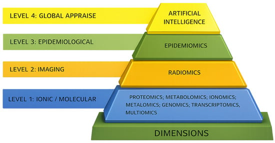
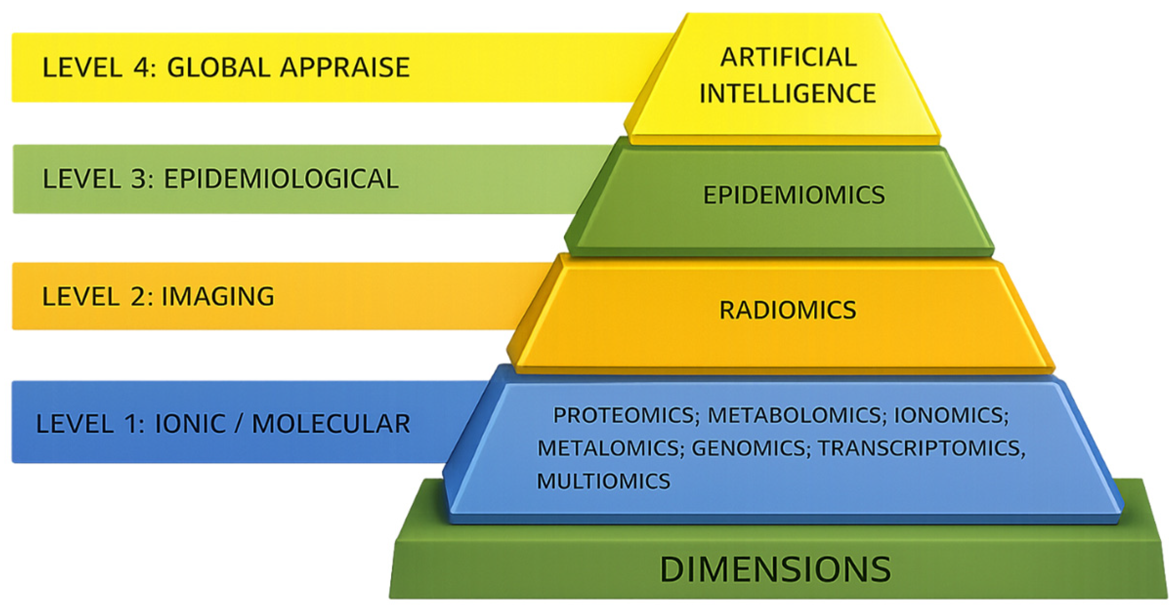
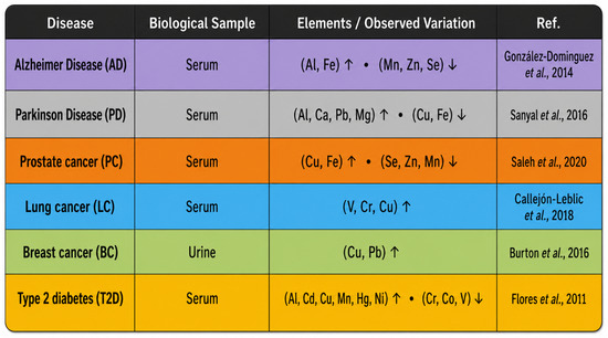
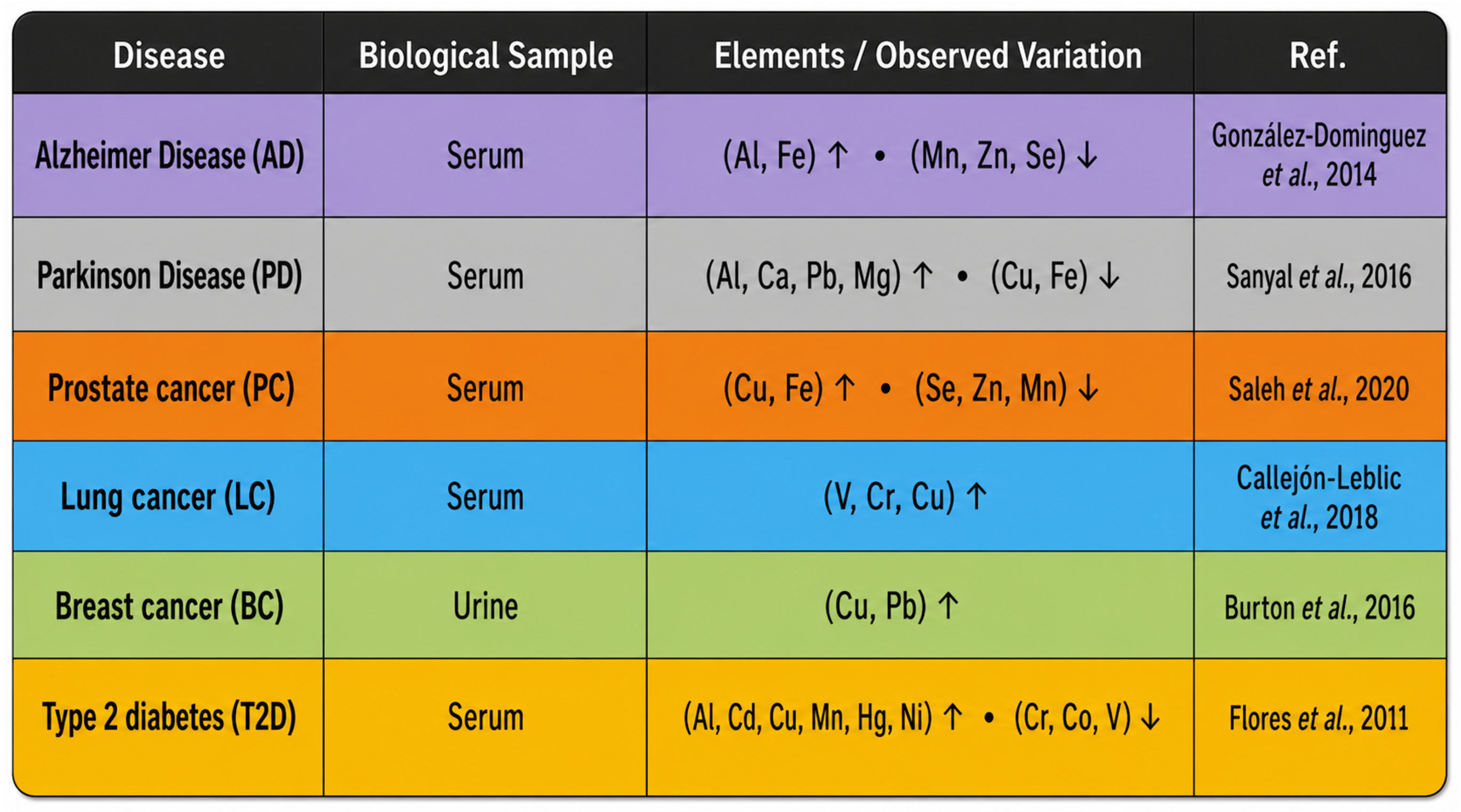
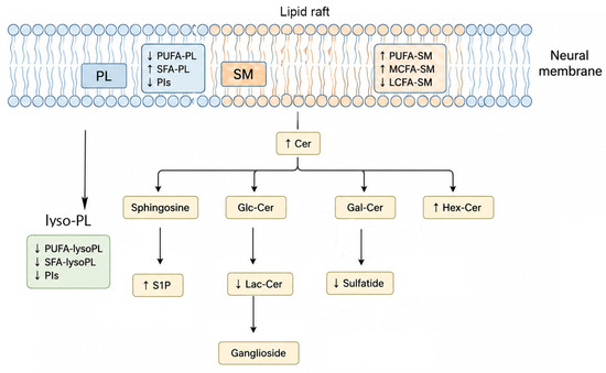
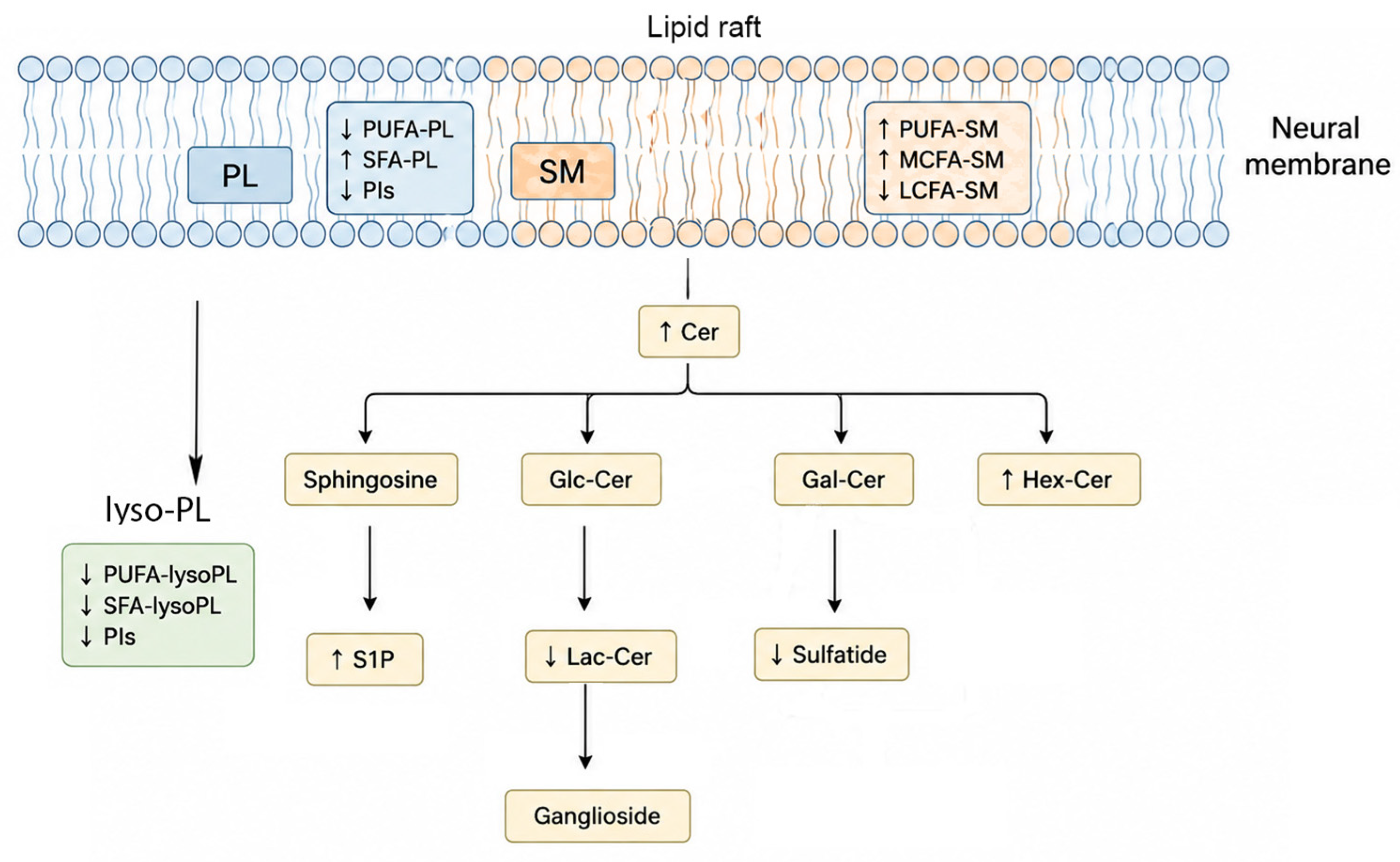
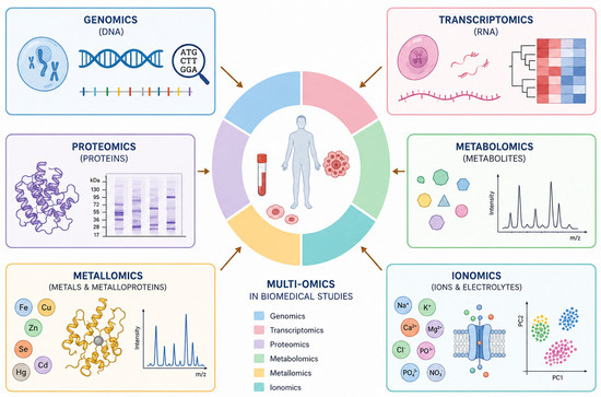
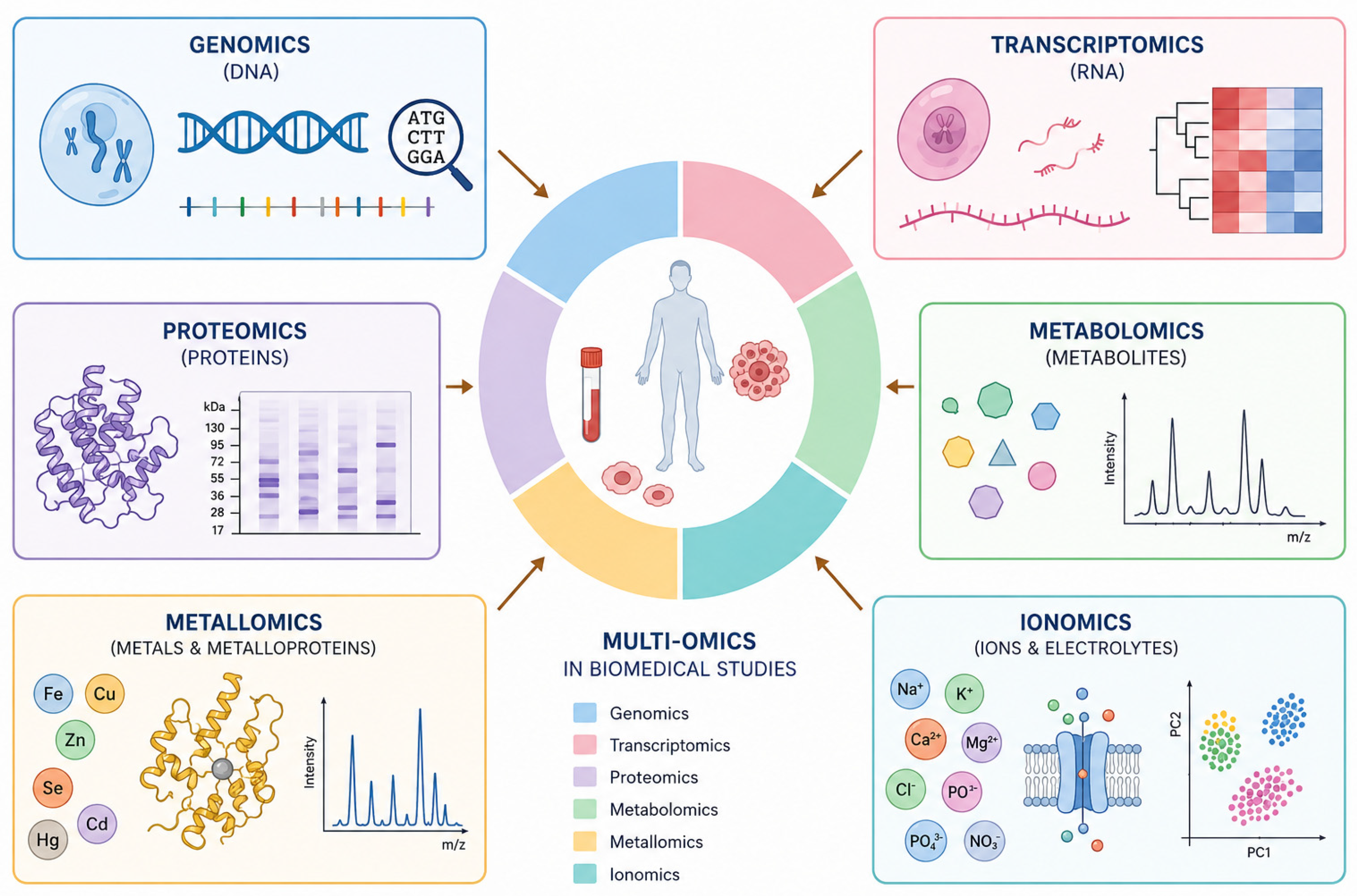

Disclaimer/Publisher’s Note: The statements, opinions and data contained in all publications are solely those of the individual author(s) and contributor(s) and not of MDPI and/or the editor(s). MDPI and/or the editor(s) disclaim responsibility for any injury to people or property resulting from any ideas, methods, instructions or products referred to in the content. |
© 2026 by the authors. Licensee MDPI, Basel, Switzerland. This article is an open access article distributed under the terms and conditions of the Creative Commons Attribution (CC BY) license.