


Abstract
Recent climate changes worldwide have negatively impacted crop yields, highlighting the urgent need to develop new cultivation strategies to counteract this phenomenon. Our study aimed to analyze the effects of saline (0 mM, 100 mM, 200 mM, and 300 mM NaCl) and heat stress (24 °C and 32 °C) on the physiological parameters of Chenopodium quinoa variety “Regalona,” evaluating the efficacy of the microalgal biostimulant Chlorella vulgaris-like extract (0%, 0.025% and 0.05%). Sprouts grown under these different conditions were assessed for antioxidant content, antioxidant activity, reactive oxygen species, and photosynthetic pigments. The analyses did not reveal significant effects of the two microalgal concentrations on germination percentage or sprout length across the various temperature and salinity conditions. However, antioxidant activity was increased in many experimental trials, especially when the microalgae were combined with salt stress, with the higher biostimulant concentration showing more pronounced effects. Conversely, a temperature of 32 °C negatively impacted the parameters measured. These findings provide a basis for further research aimed at enhancing the antioxidant and nutraceutical properties of plants valuable for human nutrition.
1. Introduction
The human impact on the planet, particularly through climate change, is now accelerating environmental shifts far beyond natural rates. Activities such as industrialization, intensive agriculture, and widespread pollution are key drivers, subjecting global ecosystems to unprecedented stress that severely compromise plant health and agricultural productivity. With the agricultural sector facing growing threats from extreme environmental conditions, innovation is essential. Among these, the application of biostimulants such as microalgae represents a promising and sustainable tool for crop growth, promoting plant tolerance to various environmental stresses [1].
Soil salinization affects approximately 800 million hectares worldwide [2,3]. Saline soil is defined by a concentration of about 40 mM NaCl in which the exchangeable sodium content is greater than 15% [4]. Salt stress inflicts damage through two primary mechanisms: osmotic stress, in which high soil NaCl lowers water potential, creating physiological drought and limiting water uptake [4]; and ionic toxicity consisting of excessive accumulation of Na+ and Cl− ions in plant tissues, which disrupt metabolic functions, enzyme activity, and membrane integrity [5,6].
The consequences of these stresses are systemic, resulting in altered root architecture, inhibition of lateral root formation, and reductions in biomass accumulation, with roots often more affected than shoots [5]. A major driver of cellular damage is the rapid generation of reactive oxygen species (ROS), which cause oxidative damage to lipids, proteins, and DNA [4].
Plants employ different strategies for salt tolerance, including detoxification, which acts by activating antioxidant systems to scavenge ROS, and compartmentalization of Na+ into vacuoles to mitigate toxicity and achieve osmotic balance. This is a key strategy in halophytes, the salt-tolerant plants that grow in high-salinity environments, like quinoa. Another strategy involves reducing photosynthetic activity via stomatal closure to conserve resources, albeit at the cost of slowed growth [7].
Plant biostimulants are defined by the European Biostimulants Industry Council (EBIC) as “a product which stimulates plant nutrition processes independently of the product’s nutrient content, with the sole aim of improving one or more of the following characteristics of the plant or the plant rhizosphere: nutrient use efficiency, tolerance to abiotic stress, quality traits, or the availability of confined nutrients in soil or rhizosphere” [8].
The principal groups of biostimulants include microorganisms, plant and algae extracts, amino acids, humic substances, mineral salts, and compounds with biostimulant properties. Unlike fertilizers, which directly supply nutrients to plants, biostimulants stimulate the plant’s own processes to better utilize nutrients and water [9].
Microalgae offer direct and indirect support to counteract salt stress. Applications of microalgal extracts enhance the plant’s endogenous antioxidant capacity, reducing ROS levels and protecting chloroplast function [9,10]. Microalgae help maintain hormonal balance (e.g., auxin levels) and improve ion selectivity, favoring K+ uptake over Na+ to preserve cellular osmotic potential [11,12].
Certain microalgae can adsorb and absorb salts, contributing to soil and water desalination [6]. Bioactive compounds in algae (e.g., phenols, flavonoids, exopolysaccharides) protect chlorophyll synthesis, improve membrane stability, and induce stress-mitigating metabolites like proline [10]. The presence of phytohormones (e.g., gibberellins, IAA) and key ions like Ca2+ (involved in the salt over-sensitivity signaling pathway) further supports adaptive responses [13].
Global temperatures are rising at ~0.2 °C per decade, causing significant heat stress that impacts plant morphology, anatomy, and phenology [14]. Key effects include inhibited root growth, altered stomatal density, and shifted harvest times [15]. Photosynthesis is highly vulnerable; heat damages Photosystem II and reduces ATP synthesis, reducing carbon fixation [7,16].
Microalgae serve as both a protective agent and a model for understanding heat tolerance. A Chlorella vulgaris-based biostimulant helped lettuce plants to maintain yield and antioxidant capacity under summer heat [17]. Studying Chlorella’s response to heat reveals adaptive strategies such as increased carotenoid production to protect the photosynthetic apparatus as in higher plants [18].
Quinoa (Chenopodium quinoa Willd.) is a facultative halophyte with a high tolerance to salinity. Native to the Andean region, this herbaceous plant has seen expanded cultivation in Asia and Europe due to its valuable agronomic and nutritional traits [19]. It is classified as a pseudocereal, along with amaranth, chia, and buckwheat. While quinoa seeds resemble true cereals in texture, starch content, and culinary use, a key botanical distinction exists: cereals are monocotyledons, whereas pseudocereals are dicotyledons [19]. Cultivated primarily for its edible seeds, quinoa shares a significant nutritional advantage with other pseudocereals. For example, the absence of gluten, which instead is present in most cereals except corn, millet, and rice [20]. This attribute makes them suitable for individuals with celiac disease. Recent interest in pseudocereals stems from their high nutritional value. They are rich in proteins and essential amino acids like lysine, which is crucial for growth and brain development [19]. Furthermore, they contain beneficial bioactive compounds including rutin, quercetin, and bioactive peptides, which help to reduce oxidative stress and may lower the risk of non-communicable diseases [20].
Research studies have confirmed that pseudocereals represent a robust nutritional alternative to traditional cereals, with higher antioxidant capacity and phenolic content than rice and rice flour. Their incorporation into the human diet can also address nutritional gaps. Many gluten-free products are deficient in minerals like iron, magnesium, and calcium. The inclusion of pseudocereals can help rebalance these profiles [19].
The use of quinoa seeds in this study was driven by the remarkable characteristics of this emerging crop. Quinoa is regarded as a highly promising species for cultivation on marginal lands due to its genetic resilience and adaptability, particularly in Mediterranean regions, which have been increasingly affected by climate change [21]. Additionally, quinoa has garnered great interest for its nutritional and nutraceutical properties, making it a valuable resource for improving food security and health [22].
Although quinoa is notably resilient for its resistance to extreme heat, such conditions alter its development, water transport, and yield [23]. Research shows variability in its response. Optimal germination ranges from 18–23 °C to 30–35 °C, but temperatures near 35 °C cause floral abortion and reduce pollen viability by up to 63% [24,25]. Plants activate heat-shock proteins (HSPs) and antioxidant defenses as innate protective responses [26].
Following our previous investigation on the positive effects of Chlorella extracts on quinoa seedlings in conditions of high salinity [22], this work examines two major abiotic stresses—salinity and high temperature—and explores the mechanistic basis for using microalgae-based biostimulants to mitigate these stresses. This study aimed to evaluate the potential of a Chlorella vulgaris-like microalgae extract (0.025% and 0.05%) in enhancing the tolerance of quinoa (Chenopodium quinoa variety ‘Regalona’) to combined salinity (0, 100, 200, and 300 mM NaCl) and temperatures (24 °C and 32 °C). We assessed physiological responses (germination and growth) and the accumulation of bioactive antioxidant compounds (total polyphenols, flavonoids, chlorophylls, and carotenoids) correlating these parameters with total antioxidant capacity (DPPH (2,2-Diphenyl-1-picrylhydrazyl), FRAP (Ferric Reducing Antioxidant Power), and ORAC (Oxygen Radical Absorbance Capacity) assays and ROS (Reactive Oxygen Species) production.
2. Materials and Methods
2.1. Plant Material
The quinoa Chenopodium quinoa Willd. variety ‘Regalona’, provided by Tuttoquinoa (Manciano, GR, Italy), was used in this study. Seeds were subjected to a fully factorial experimental design combining different salinity levels (0, 100, 200, and 300 mM NaCl), two temperature regimes (24 °C and 32 °C) and biostimulant concentrations (0, 0.025, 0.05%), resulting in 24 experimental treatments.
The biostimulant treatments utilized a lyophilized Chlorella vulgaris-like powder [27]. Two stock solutions (0.1% and a 0.05% w/v) were prepared by dispersing the powder in distilled water. Each mixture was homogenized using an Ultra-Turrax homogenizer (Kinematica Polytron PT MR2100, Lucerne, Switzerland) and then agitated continuously in the dark for three hours to ensure proper extraction. A final concentration of 0.05 and 0.025% in saline solutions was also obtained.
Germination was conducted in 9-cm Petri dishes lined with two sheets of filter paper, each wetted with 3 mL of the corresponding treatment solution. Each dish contained 25 seeds, and 3 biological replicates were prepared per treatment. All dishes were sealed with parafilm and placed in a controlled germination chamber (Termaks incubators B-series, Nordic Labtech AB, Fjärås, Sweden).
The germination protocol differed by temperature regime: for treatments at 24 °C, seeds underwent a three-day dark period followed by two days under light, with seedlings collected on the fifth day; for treatments at 32 °C, seeds were kept in the dark at this temperature for three days, then transferred to light conditions at 24 °C for two days, with collection also on the fifth day. These conditions for the 32 °C treatment were adopted because preliminary experiments revealed that if seeds were kept at 32 °C for 5 days the development of the seedlings was dramatically impaired.
2.2. Biometric Analysis of Quinoa Seedlings
Five days after germination (T5), seedlings from the different treatments were collected and the germination percentage, the length, and the fresh weight of seedlings were determined. Germination percentage was expressed as number of germinated seeds over 100 seeds and seeds were considered germinated when the radicle was as long as the seed diameter.
2.3. Preparation of Quinoa Extracts
The seedlings from each treatment were weighed and extracted by adding 80% ethanol at a 1:10 ratio (1 g plant material in 10 mL of 80% ethanol). The samples were homogenized immediately using an Ultra-Turrax homogenizer POLYTRON PT-MR 2100-CPO Kinematica, Switzerland and then agitated continuously in the dark at 4 °C for 24 h [28]. Then, the homogenates were centrifuged at 3500× g for 30 min at 4 °C with a Juan CR 31 centrifuge, Newport Pagnell, UK. The resulting supernatants were carefully collected and then analyzed using a UV/visible spectrophotometer (FLUOstar Omega, BMG LABTECH, Ortenberg, Germany), to determine total polyphenol content (TPC), total flavonoid content (TFC), and total antioxidant capacity via the FRAP, ORAC, DPPH, and ROS assays.
2.4. Determination of Total Polyphenol Content
Total polyphenol content (TPC) was determined using the Folin–Ciocalteu colorimetric assay [29]. For each sample, 50 µL of the ethanol extract was mixed with 1000 µL of Folin–Ciocalteu reagent (diluted 1:5 with distilled water) in a 2 mL microcentrifuge tube. After 6 min of incubation in the dark, 660 µL of a sodium carbonate solution (Na2CO3, 7.5% w/v) was added and the mixture was incubated in the dark for 60 min. A blank was prepared, replacing the extract with 50 µL of 80% (v/v) ethanol. Absorbance was measured at 760 nm. TPC was quantified using a gallic acid calibration curve. Results were expressed as milligrams of gallic acid equivalents per gram of fresh weight (mg GaE g−1 FW).
2.5. Determination of Total Flavonoid Content
Total flavonoid content (TFC) was determined using an aluminum chloride (AlCl3) colorimetric assay based on the method of Heimler et al. [30]. For each sample, 150 µL of the ethanol extract was transferred to a 2-mL microcentrifuge tube. Then, 600 µL of distilled water and 45 µL of the 5% NaNO2 solution were added. The mixture was incubated in the dark for 5 min. Subsequently, 90 µL of the 10% AlCl3 solution was added, followed by a 6 min incubation in the dark. To complete the reaction, 300 µL of the 1 M NaOH solution and 360 µL of distilled water were added. The final mixture was incubated in the dark for 30 min. Absorbance was measured at 430 nm against a reagent blank prepared by replacing the plant extract with 150 µL of 80% (v/v) ethanol. TFC was quantified using a calibration curve of quercetin. Results were expressed as milligrams of quercetin equivalents per gram of fresh weight (mg QE g−1 FW).
2.6. FRAP Assay
The Ferric Reducing Antioxidant Power (FRAP) of the extracts was determined using a modified method of Benzie and Strain [31]. The FRAP working reagent was prepared fresh by mixing 10 volumes of 300 mM acetate buffer (pH 3.6), 1 volume of 10 mM TPTZ (2,4,6-Tripyridyl-s-triazine) in 40 mM HCl, and 1 volume of 20 mM FeCl3·6H2O. For each sample, 51 µL of the ethanol extract was mixed with 1.5 mL of the freshly prepared FRAP working reagent in a 2-mL microcentrifuge tube. The reaction mixture was incubated in the dark at room temperature for 60 min. Absorbance was then measured at 593 nm against a blank prepared by replacing the extract with 51 µL of 80% (v/v) ethanol. Antioxidant capacity was quantified using a calibration curve of ferrous sulfate heptahydrate (FeSO4·7H2O) within a concentration range of 31.25 to 500 µM. Results were expressed as micromoles of ferrous sulfate equivalents per gram of fresh weight (µM FeSO4 eq g−1 FW).
2.7. ORAC Assay
The antioxidant capacity of the extracts against peroxyl radicals was determined using an Oxygen Radical Absorbance Capacity (ORAC) assay adapted from Ninfali et al. [32]. The assay measures the protective effect of antioxidants on fluorescein, which loses fluorescence upon oxidation by peroxyl radicals generated from the thermal decomposition of 2,2′-azobis(2-amidinopropane) dihydrochloride (AAPH) at 37 °C. Each plant extract was subjected to two serial dilutions (1:10 and 1:20) using a phosphate buffer (0.075 M, pH 7.4). For each reaction, 100 μL of the diluted extract or Trolox standard was mixed with 800 μL of fluorescein solution in a microcentrifuge tube. The reaction was initiated by adding 100 μL of the AAPH solution. Immediately after mixing, 200 μL of the reaction mixture was transferred in duplicate to a black 96-well microplate. Fluorescence was monitored kinetically at 37 °C using a fluorometric plate reader (FLUOstar Omega, BMG LABTECH, Germany) with excitation at 485 nm and emission at 514 nm. Measurements were taken every minute for 90 min to record the fluorescence decay curve. The area under the fluorescence decay curve (AUC) was calculated for each sample and standard. Antioxidant capacity was quantified by comparing the net AUC of the sample to the Trolox calibration curve. Results were expressed as micromoles of Trolox equivalents per gram of fresh weight (μmol TE g−1 FW).
2.8. DPPH Assay
The DPPH (2,2-Diphenyl-1-picrylhydrazyl) assay was based on a modified method developed by Boudjou et al. [33]. To determine the antioxidant capacity of the samples, 80 μM DPPH solution in pure methanol was used and the mixture was stirred for one hour. For the determinations, 50 μL of plant extract were added to 1450 μL of DPPH solution in 2 mL Eppendorf tubes and incubated in the dark for one hour. Absorbance was measured at 517 nm against 80% EtOH as a blank. Antiradical activity (ARA) was expressed as the percentage inhibition of DPPH and calculated using the formula:
ARA = 100 × [1 − (Abs of sample/Abs of control)]
2.9. Quantification of Intracellular Reactive Oxygen Species (ROS)
Intracellular ROS levels were quantified using the oxidation-sensitive fluorescent probe 2′,7′-dichlorofluorescein diacetate (DCFDA), following a method adapted from Bellani et al. [34].
Approximately 50 mg of quinoa sprouts from each treatment were incubated in 1 mL of a working solution containing 100 µM of DCFDA prepared in 40 mM Tris-HCl buffer (pH 7.4). The samples were shaken gently in the dark at 37 °C for 60 min to allow DCFDA uptake and oxidation by intracellular ROS. Then the samples were centrifuged at 10,000× g for 10 min at 4 °C. The supernatant was collected, and 100 µL was transferred in duplicate to a black 96-well microplate. Fluorescence was measured using a fluorometric plate reader with excitation at 488 nm and emission at 525 nm. A blank consisting of buffer alone was used for background subtraction. ROS levels were expressed as arbitrary fluorescence units per milligram of fresh weight (AU g−1 FW).
2.10. Determination of Chlorophyll and Carotenoid Content
Chlorophylls and total carotenoids were determined based on the method of Lichtenthaler [35] with modifications [36]. To 100 mg of fresh material, 3 mL of 80% acetone were added. The mixture was homogenized and then centrifuged for 5 min at 2200× g at 4 °C. The pellet was resuspended in 3 mL of 80% acetone, kept in the dark at 4 °C for 15 min, and then centrifuged again for 5 min. The two supernatants were then combined and centrifuged for an additional 5 min. The resulting final extract was subjected to spectrophotometric analysis at three wavelengths: 663 nm, 645 nm, and 470 nm. Finally, the following formulas were applied to determine the chlorophyll a (Chl a), chlorophyll b (Chl b), total chlorophyll (Chl total) and carotenoid (Car) concentrations of the samples in which VFSN is the final supernatant volume and fresh weight (FW) is in grams. Chlorophylls and carotenoid contents were expressed as mg g−1 FW.
Chl a = [(12.7 × OD 663 − 2.69 OD 645) × VFSN]/(1000 × g FW)
Chl b = [(22.9 × OD 645 − 4.68 OD 663) × VFSN]/(1000 × g FW)
Chl total = [(20.2 × OD 645 + 8.02 OD 663) × VFSN]/(1000 × g FW)
Car = [(1000 × OD 470 − 1.82 × Chl a − 85.2 × Chl b) /198] × VFSN/(1000 × g FW)
2.11. Statistical Analysis
All experimental data were subjected to a three-way analysis of variance (ANOVA) to evaluate the main effects of salinity (0, 100, 200, and 300 mM NaCl) and biostimulant treatment (Control, 0.025% and 0.05% Chlorella vulgaris-like extract) and of temperature (24 °C and 32 °C) and their interactions. Data expressed in percentage were subjected to Arcsin Square Root Transformation to meet the assumptions of normality of variances. When a significant interaction was found (p < 0.05), a post-hoc Tukey’s multiple comparisons test was performed on the means. The results were presented as the mean of three biological replicates ± standard deviation (SD).
The overall dataset was further explored using Canonical Discriminant Analysis (CDA). The CDA model was constructed with all experimental variables for samples grown at 24 °C and 32 °C: seedling length, germination percentage, total phenolic and flavonoid contents, antioxidant capacity (DPPH, FRAP, and ORAC), chlorophyll (a, b, total) and total carotenoid concentrations, and ROS content. Additionally, a Pearson’s correlation analysis was conducted between the variables and the canonical scores of the first two canonical discriminant functions to interpret the contribution of each parameter to the group separation observed in the CDA. All statistical analyses were performed using JMP software (Version 17, SAS Institute Inc., Cary, NC, USA).
3. Results
3.1. Analysis of Physiological Parameters
3.1.1. Physiology of Germination
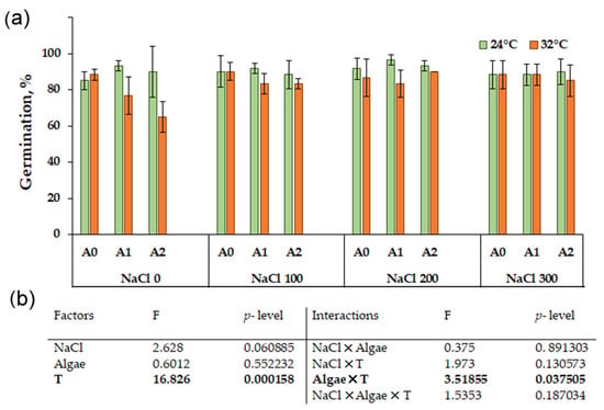
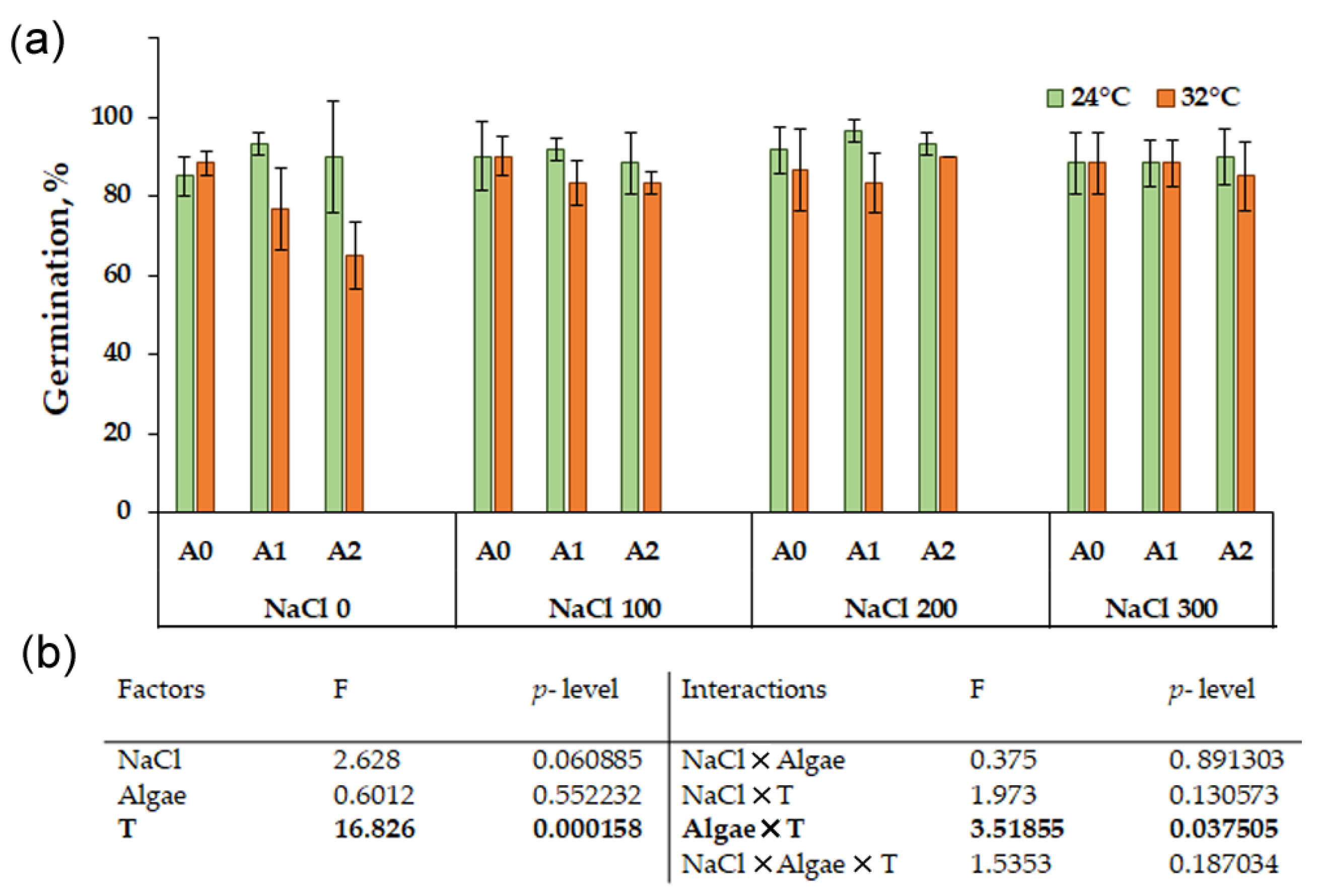
In seeds of Chenopodium quinoa, the germination percentage after 5 days at 24 °C ranged between 85% and 97% in both the control samples and those treated with NaCl at the three tested concentrations as well as in the presence of Chlorella vulgaris at both concentrations (Figure 1a). The treatment with Chlorella and NaCl slightly increased the germination percentage (Table S1).
Figure 1.
Germination percentage of Chenopodium quinoa var ‘Regalona’ after 5 days of imbibition in the presence of different NaCl concentrations (0, 100, 200, 300 mM) and Chlorella vulgaris-like at the concentration of 0 (A0), 0.025% (A1), 0.05% (A2) at temperatures of 24 °C and 32 °C for 3 days followed by 2 days recovery at 24 °C (a). The values represent the mean of three replicates ± SD (n = 3). Three-way ANOVA (b) revealed significant differences for the main factors NaCl, algae, temperature (T) and their interactions, in bold when significant (p < 0.05).
3.1.2. Seedling Length
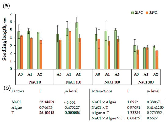
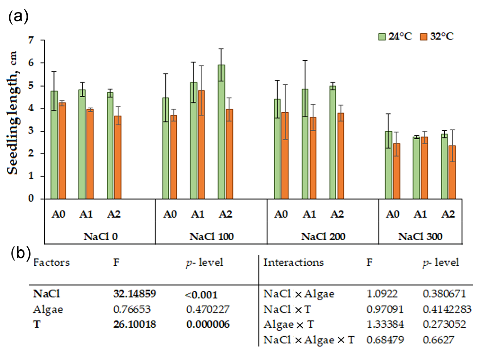
Quinoa seedling length at 24 °C (Figure 2a) ranged between 5.9 and 2.7 cm. The treatment with NaCl negatively impacted the shoot length, particularly at 300 mM (Tables S1 and S3).
Figure 2.
Seedling length of Chenopodium quinoa var ‘Regalona’ after 5 days of imbibition in the presence of different NaCl concentrations (0, 100, 200, 300 mM) and Chlorella vulgaris-like at the concentration of 0 (A0), 0.025% (A1), 0.05% (A2) at temperatures of 24 °C and 32 °C for 3 days followed by 2 days recovery at 24 °C (a). The values represent the mean of three replicates ± SD (n = 3). Three-way ANOVA (b) revealed significant differences for the main factors NaCl, algae, temperature (T) and their interactions, in bold when significant (p < 0.05).
The heat shock at 32 °C (Figure 2a) induced a significant length reduction; the values ranged between 4.8 and 2.4 cm. The interaction between the three factors was never significant (Figure 2b). The heat shock induced a decrease in respect to the control in the presence of NaCl and algae (Tables S2 and S3).
3.2. Analysis of Antioxidant Molecules
3.2.1. Total Polyphenols
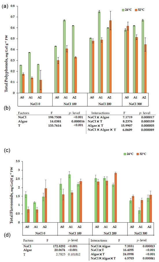
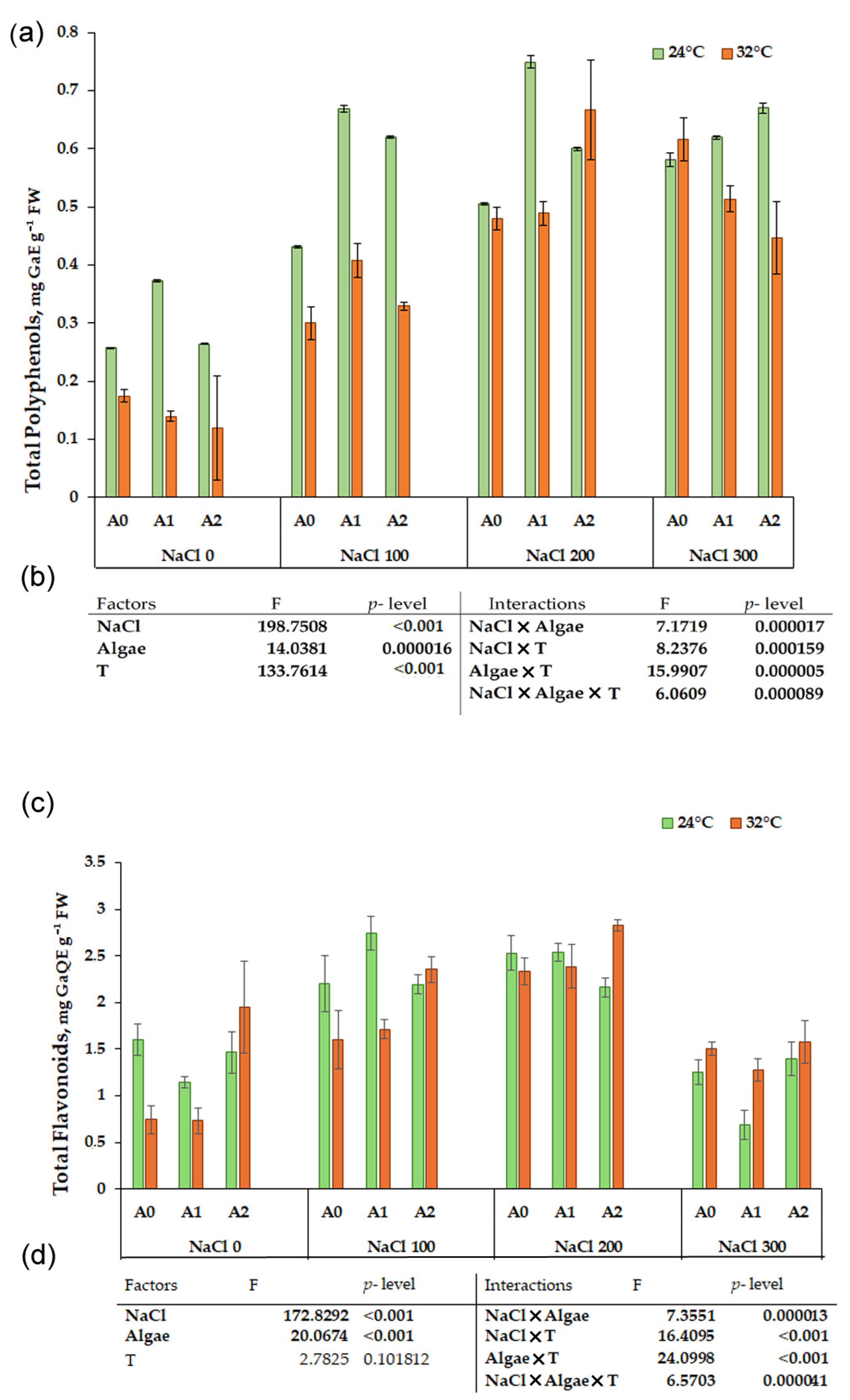
A consistent increase in polyphenols was observed at 24 °C with rising salt concentrations in samples without Chlorella. The presence of microalga significantly enhanced polyphenol levels, with the maximum increase at 0.025% Chlorella (Figure 3a), particularly in combination with 100 and 200 mM NaCl (0.75 mg GaE g−1 FW), nearly doubling the amount in samples germinated without salt or microalgae. The 0.05% Chlorella treatment in the presence of different NaCl concentrations resulted in comparable values, almost always about twice those of the corresponding control without NaCl (Table S1).
Figure 3.
Content in total polyphenols (mg GaE g−1 FW) (a) and in flavonoids (mg QE g−1 FW) (c) in Chenopodium quinoa var ‘Regalona’ after 5 days of imbibition in the presence of different NaCl concentrations (0, 100, 200, 300 mM) and Chlorella vulgaris-like extract at concentrations of 0 (A0), 0.025% (A1), 0.05% (A2) at temperatures of 24 °C and 32° C for 3 days followed by 2 days recovery at 24 °C. The values represent the mean of three replicates ± SD (n = 3). Three-way ANOVA (b,d) revealed significant differences for the main factors NaCl, algae, temperature (T), and their interactions, in bold when significant (p < 0.05).
Three-way ANOVA (Figure 3b) revealed significant differences for the factors NaCl, alga, temperature (T), and their interactions. In the samples of quinoa grown at 32 °C for three days followed by two days of recovery at 24 °C (Figure 3a), treatments with 200 and 300 mM NaCl induced a significant increase in polyphenols (0.48 and 0.62 mg GaE g−1 FW, respectively) compared to the control, which consistently showed the lowest value (0.17 mg GaE g−1 FW). The maximum amount was observed at 200 mM NaCl combined with 0.05% Chlorella (Table S2). The simple effect of the three factors and their interactions produced significant results (Figure 3b, Table S3).
3.2.2. Total Flavonoids Content
Flavonoid content (Figure 3c) significantly increased at 100 and 200 mM NaCl, regardless of the presence of Chlorella. At 24° C and 300 mM NaCl, flavonoid content showed a sharp reduction of up to 57% induced by the highest salt concentration (Figure 3c, Table S2).
It is interesting to observe that after the heat shock at 32 °C 0.05%, Chlorella caused a significant increase in flavonoids with respect to the control with no NaCl and with alga 0.025% (Table S2). The NaCl treatments at 100 and 200 mM led to dose-dependent increases in flavonoids (1.6 and 2.34 mg QE g−1 FW, respectively), whereas at 300 mM NaCl the flavonoid level decreased to 1.5 mg QE g−1 FW (Figure 3c).
3.3. Antioxidant Activity
The antioxidant activity was measured by three different tests, specifically the FRAP, ORAC (Figure 4), and DPPH assays (Figure 5).
Figure 4.
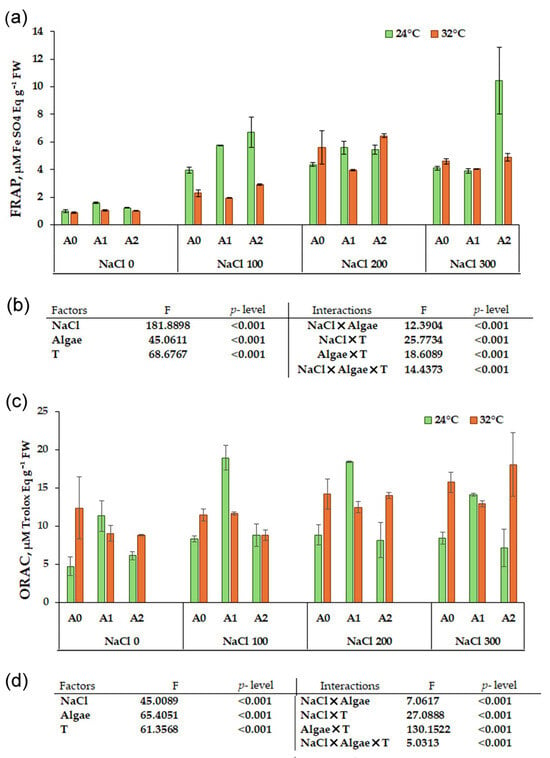
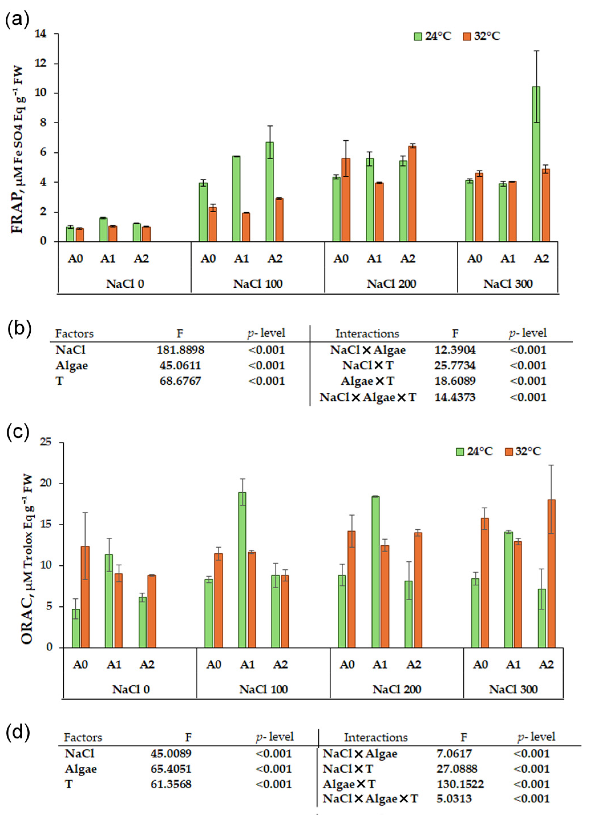
Antioxidant activity measured by FRAP (a) and ORAC (c) tests in sprouts of Chenopodium quinoa var ‘Regalona’ after 5 days of imbibition in the presence of different NaCl concentrations (0, 100, 200, 300 mM) and Chlorella vulgaris-like at the concentration of 0 (A0), 0.025% (A1), 0.05% (A2) at temperatures of 24 °C and 32 °C for 3 days followed by 2 days recovery at 24 °C. The values represent the mean of three replicates ± SD (n = 3). Three-way ANOVA (b,d) revealed significant differences for the main factors NaCl, algae, temperature (T), and their interactions, in bold when significant (p < 0.05).
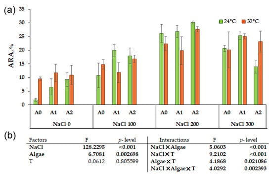
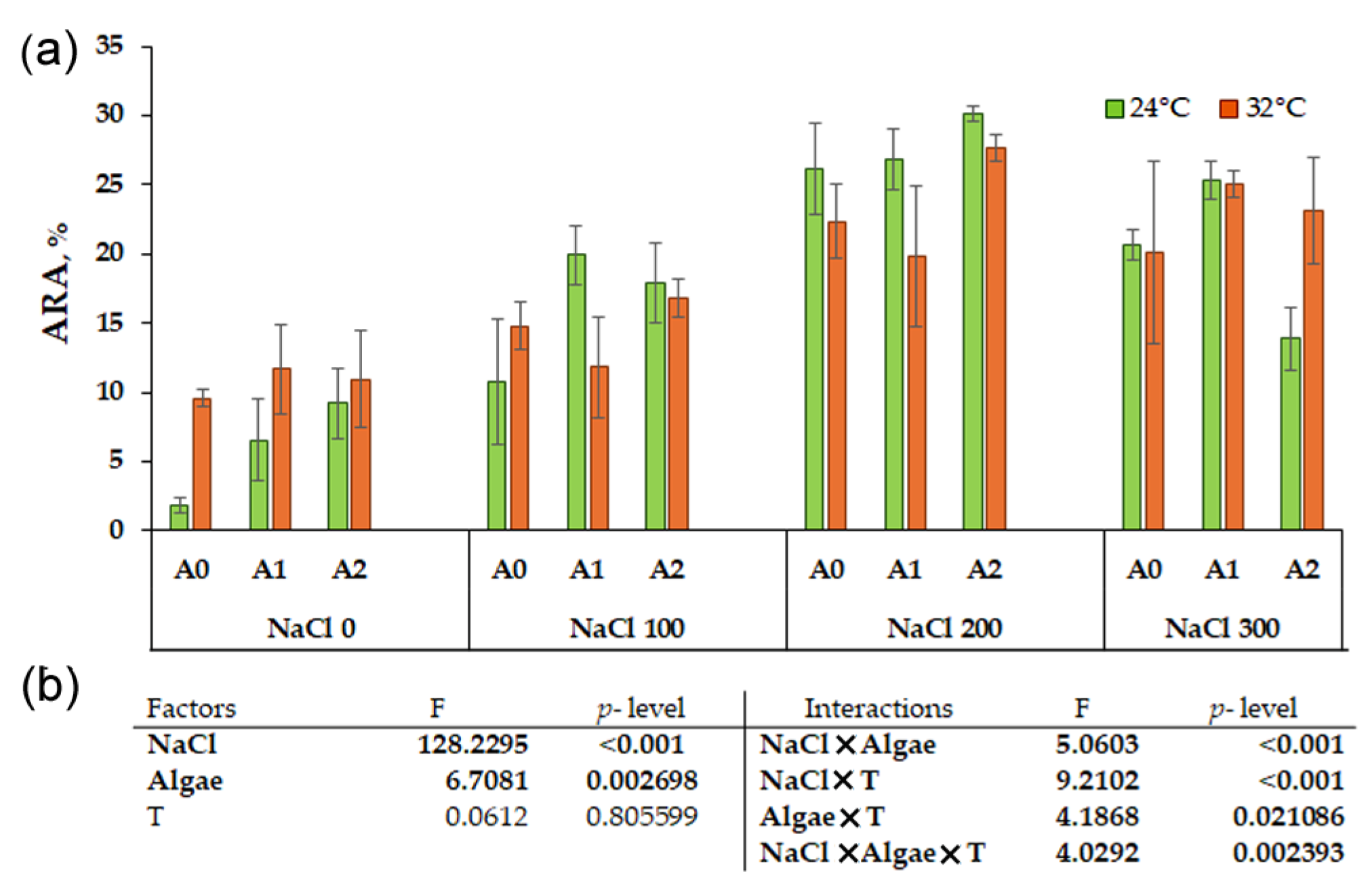
Figure 5.
Antioxidant activity measured by DPPH test in sprouts of Chenopodium quinoa var ‘Regalona’ after 5 days of imbibition in the presence of different NaCl concentrations (0, 100, 200, 300 mM) and Chlorella vulgaris-like at the concentration of 0 (A0), 0.025% (A1), 0.05% (A2) at temperatures of 24 °C and 32 °C for 3 days followed by 2 days recovery at 24 °C (a). The values represent the mean of three replicates ± SD (n = 3). Three-way ANOVA (b) revealed significant differences for the main factors NaCl, algae, temperature (T) and their interactions, in bold when significant (p < 0.05).
3.3.1. FRAP Analysis
At 24 °C, the FRAP assay (Figure 4a), showed increased reducing activity in NaCl treated samples compared to the control (Table S1). The addition of Chlorella further enhanced antioxidant activity, reaching a maximum at 0.05% (10.44 µM FeSO4 eq g−1 FW) in combination with 300 mM NaCl, representing approximately a tenfold increase over the corresponding control without Chlorella (Table S1).
After the heat shock at 32 °C (Figure 4a), the reducing activity increased, with NaCl treatments reaching a maximum of 5.6 µM FeSO4 eq g−1 FW at 200 mM NaCl, with a lower activity at 300 mM NaCl (4.6 µM FeSO4 eq g−1 FW). The treatment with Chlorella caused a smaller increase in reducing activity at 0.025% but a higher increase at 0.05% compared to the respective controls (Table S2). Three-way ANOVA revealed that the simple effect of the three factors and their interactions (Figure 4b, Table S3) produced significant results.
3.3.2. ORAC Analysis
In the ORAC assay (Figure 4c), which indicated the samples’ ability to neutralize free radicals, at 24 °C the control samples without NaCl and Chlorella showed the lowest value (4.7 μmol TE g−1). NaCl treatments induced a significant increase in antioxidant activity, reaching a maximum at 100 and 200 mM NaCl. The addition of Chlorella further enhanced antioxidant activity, particularly evident at the lower concentration (0.025%), especially in combination with 100 and 200 mM NaCl, where the highest activity was recorded (Figure 4c, Tables S1 and S3).
After the heat shock at 32 °C, the ORAC activity (Figure 4c) was also significantly influenced by NaCl, Chlorella, and their interactions. At 200 mM NaCl, an increase in antioxidant activity was observed in samples treated with Chlorella; at 300 mM NaCl the highest activity was recorded with 0.05% Chlorella (Table S2). Three-way ANOVA revealed that the simple effect of the three factors and their interactions (Figure 4d, Table S3) produced significant results.
3.3.3. DPPH Analysis
At 24 °C, the antiradical activity measured by the DPPH test (Figure 5a) showed the control sample with the lowest value compared to NaCl treatments, with a progressive increase in activity up to 200 mM NaCl, where the highest value was recorded (26.16 ARA%). The interaction between NaCl 200 mM and Chlorella at both concentrations and both temperatures induced the highest antiradical activity (Tables S1–S3).
3.4. Reactive Oxygen Species (ROS)
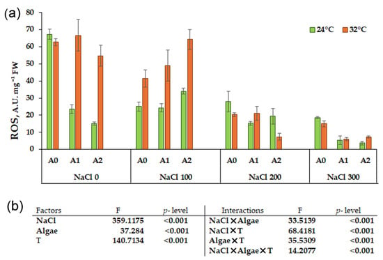
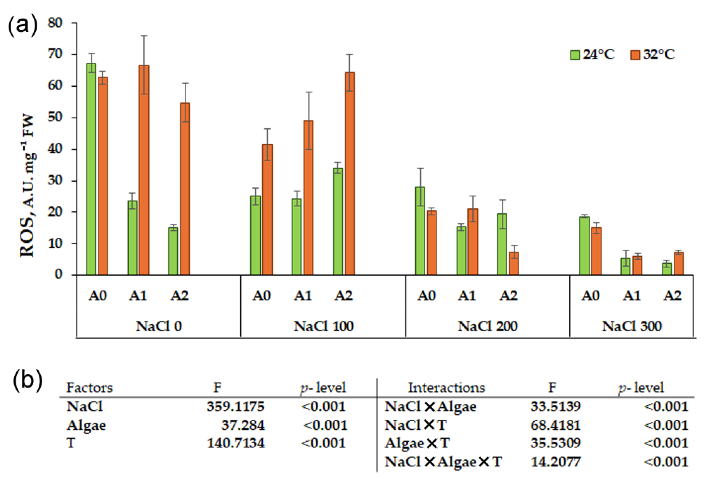
ROS analysis in treatments at 24 °C (Figure 6a) indicated that the presence of Chlorella at both concentrations significantly reduced ROS content in the samples without NaCl, particularly evident at 0.05%. The presence of 100 mM NaCl induced a significant reduction in ROS content, which was even more pronounced at 300 mM NaCl (18.66 ROS A.U. mg−1 FW), particularly in interaction with Chlorella 0.05% (3.66 ROS A.U. mg−1 FW). The highest ROS levels were observed in the sample with no salt and no algae, and the lowest appeared in samples at 300 mM NaCl and Chlorella at both concentrations (Table S1).
Figure 6.
Reactive oxygen species (ROS) content in sprouts of Chenopodium quinoa var ‘Regalona’ after 5 days of imbibition in the presence of different NaCl concentrations (0, 100, 200, 300 mM) and Chlorella vulgaris-like at the concentration of 0 (A0), 0.025% (A1), 0.05% (A2) at temperatures of 24 °C and 32 °C for 3 days followed by 2 days recovery at 24 °C (a). The values represent the mean of three replicates ± SD (n = 3). Three-way ANOVA (b) revealed significant differences for the main factors NaCl, algae, temperature (T) and their interactions, in bold when significant (p < 0.05).
After the heat shock at 32 °C (Figure 6a), ROS levels decreased at 100 mM NaCl and even more at 200 mM NaCl in samples without Chlorella. The algae interacted with the salt at 100 mM NaCl. ROS levels were high with Chlorella 0.025% and even higher with the 0.05% algae (Figure 6a). This increase was not observed at 200 and 300 mM NaCl, where the combination with algae significantly reduced the ROS content (Table S2).
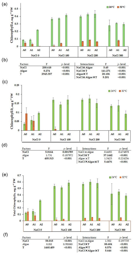
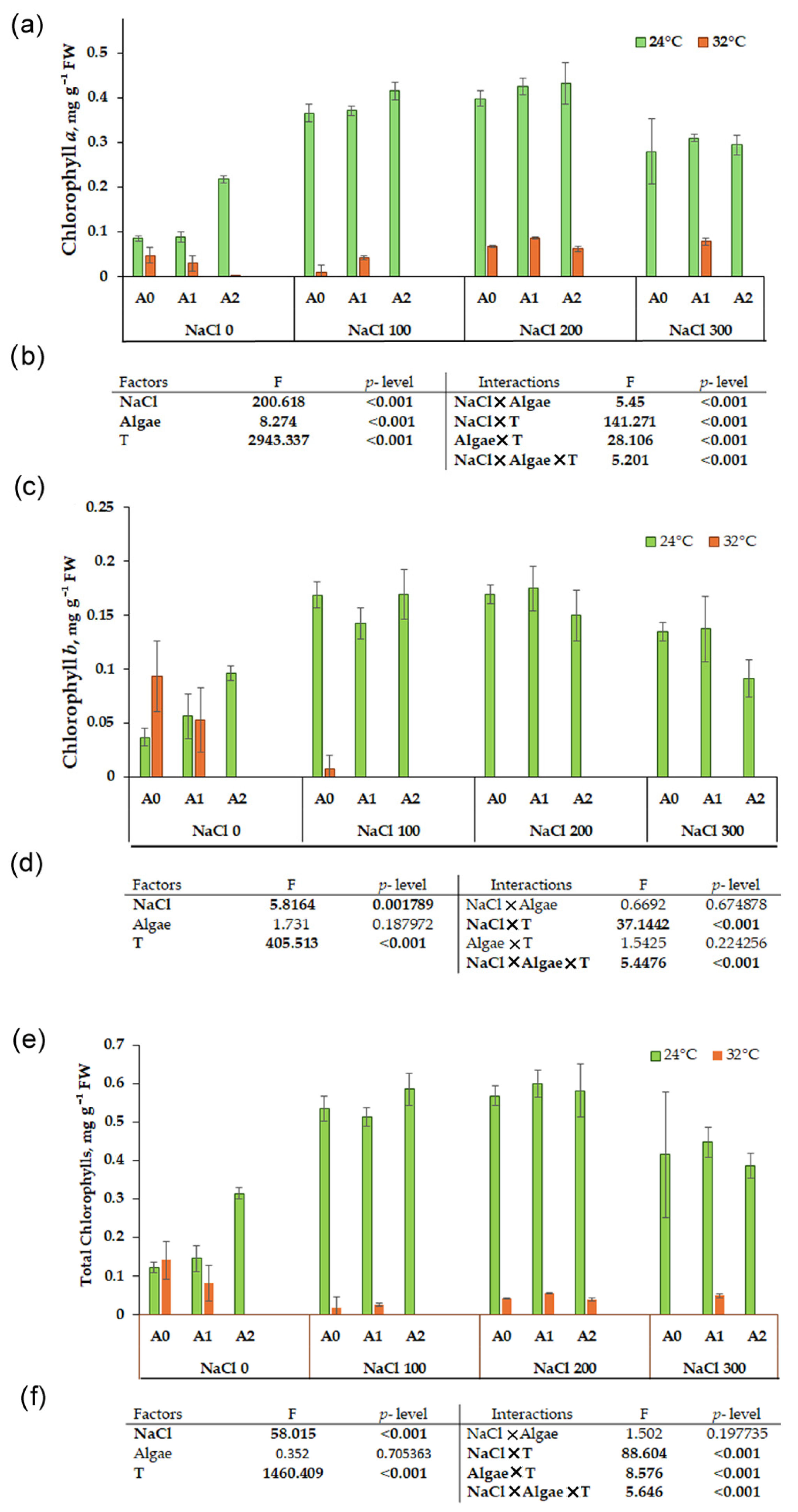
3.5. Photosynthetic Pigments
Chlorophyll a, b, total (Figure 7) and carotenoids (Figure 8) were evaluated in sprouts grown at 24 °C and after 3 days at 32 °C followed by 2 days of recovery at 24 °C. In particular, at 24 °C, the treatment with Chlorella without NaCl induced a significant increase of chlorophyll a, particularly evident at 0.05% algae. The NaCl treatment alone induced a significant increase in chlorophyll a content compared to the control (Figure 7a), with the maximum at 200 mM NaCl (0.40 mg g−1 FW) (Table S1).
Figure 7.
Chlorophyll a, b, total (a,c,e) content in sprout of Chenopodium quinoa var ‘Regalona’ after 5 days of imbibition in the presence of different NaCl concentrations (0, 100, 200, 300 mM) and Chlorella vulgaris-like at the concentration of 0 (A0), 0.025% (A1), 0.05% (A2) at temperatures of 24 °C. The values represent the mean of three replicates ± SD (n = 3). Three-way ANOVA (b,d,f) revealed significant differences for the main factors NaCl, algae, temperature (T), and their interactions, in bold when significant (p < 0.05).
Figure 8.
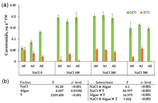
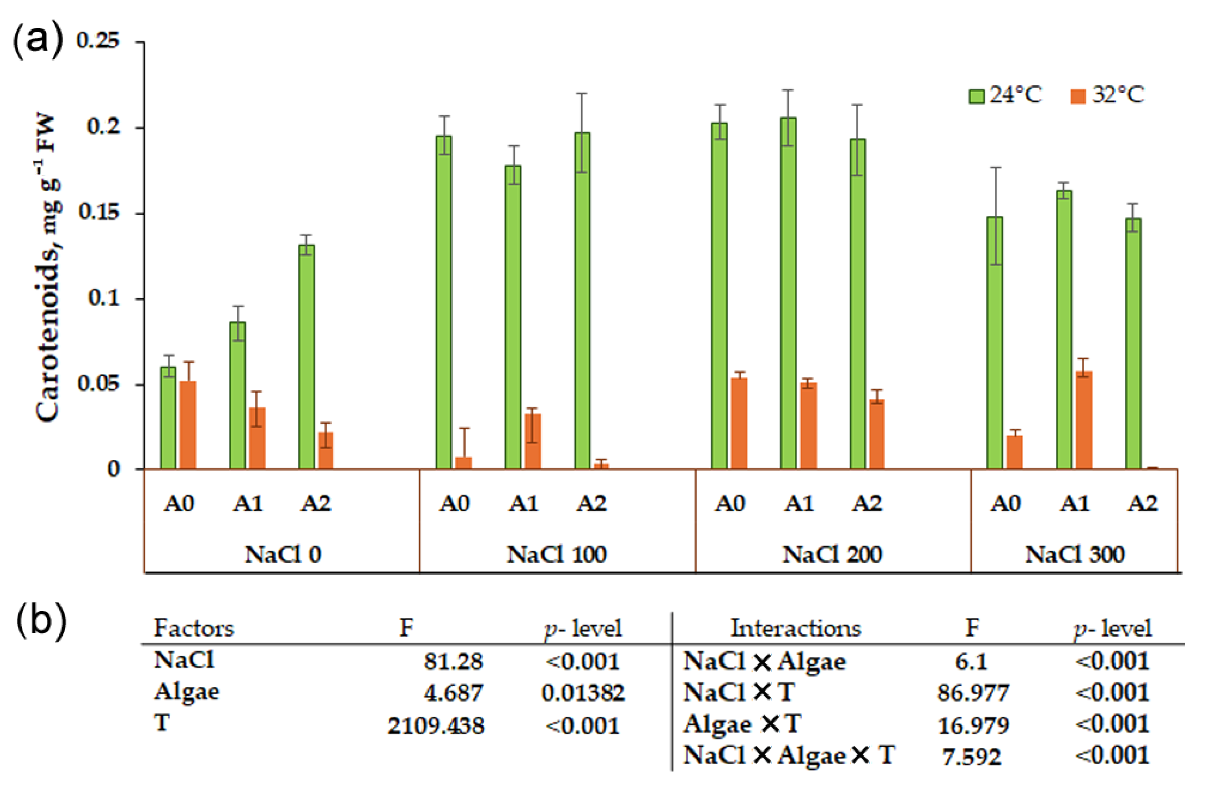
Carotenoids content (a) in sprouts of Chenopodium quinoa var ‘Regalona’ after 5 days of imbibition in the presence of different NaCl concentrations (0, 100, 200, 300 mM) and Chlorella vulgaris-like at the concentration of 0 (A0), 0.025% (A1), 0.05% (A2) at the temperature of 32 °C for 3 days followed by 2 days recovery at 24 °C. The values represent the mean of three replicates ± SD (n = 3). Three-way ANOVA (b) revealed significant differences for the main factors NaCl, algae, temperature (T) and their interactions, in bold when significant (p < 0.05).
The interactions between NaCl and algae were always significant (Figure 7b) and resulted in the highest levels of chlorophyll a at both 100 and 200 mM NaCl, with lower amounts at 300 mM NaCl. The trend of the response in chlorophyll b (Figure 7c) was similar to chlorophyll a for NaCl treatments, with the most notable increase observed at 100 and 200 mM NaCl (Table S1). The effects of Chlorella and its interaction with NaCl were not significant (Figure 7d). Total chlorophylls content in sprouts grown at 24 °C (Figure 7e) mirrored the chlorophyll a trends, with a significant influence of NaCl and its interaction with Chlorella (Figure 7f). The highest amounts of total chlorophyll were observed at 100 and 200 mM NaCl (0.53 mg g−1 FW and 0.57 mg g−1 FW, respectively.
After the heat shock at 32 °C, chlorophyll a, b, and total levels were strongly influenced, sometimes reducing them below the detection limits (Figure 7a,c,e, Table S2), mainly for the incomplete development of photosynthetic cotyledons.
Three-way ANOVA indicated that all the factors and their interactions were significant for chlorophyll a, while for chlorophyll b and total, the algae factor was not significant (Figure 7d,f, Table S3).
A similar trend to chlorophyll was observed at 24 °C for carotenoids (Figure 8a). The treatment with Chlorella but without NaCl induced a significant increase of carotenoids, particularly evident at 0.05% algae. At 100 and 200 mM NaCl in combination with algae, a significant increase compared to the control was observed. At 300 mM NaCl carotenoid levels were lower, but still higher than in the control samples (Table S1).
The carotenoid content with heat stress was significantly lower than at 24 °C in the control samples (Figure 8a). A temperature of 32 °C negatively affected the carotenoid content in combination with salt treatments. In the presence of 0.05% Chlorella, an increase of carotenoid content was observed (Table S2).
The effects of the factors NaCl, algae, and temperature were highly significant as were the interactions between the three factors by three-way ANOVA (Figure 8b).
3.6. Canonical Discriminant Analysis
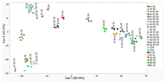
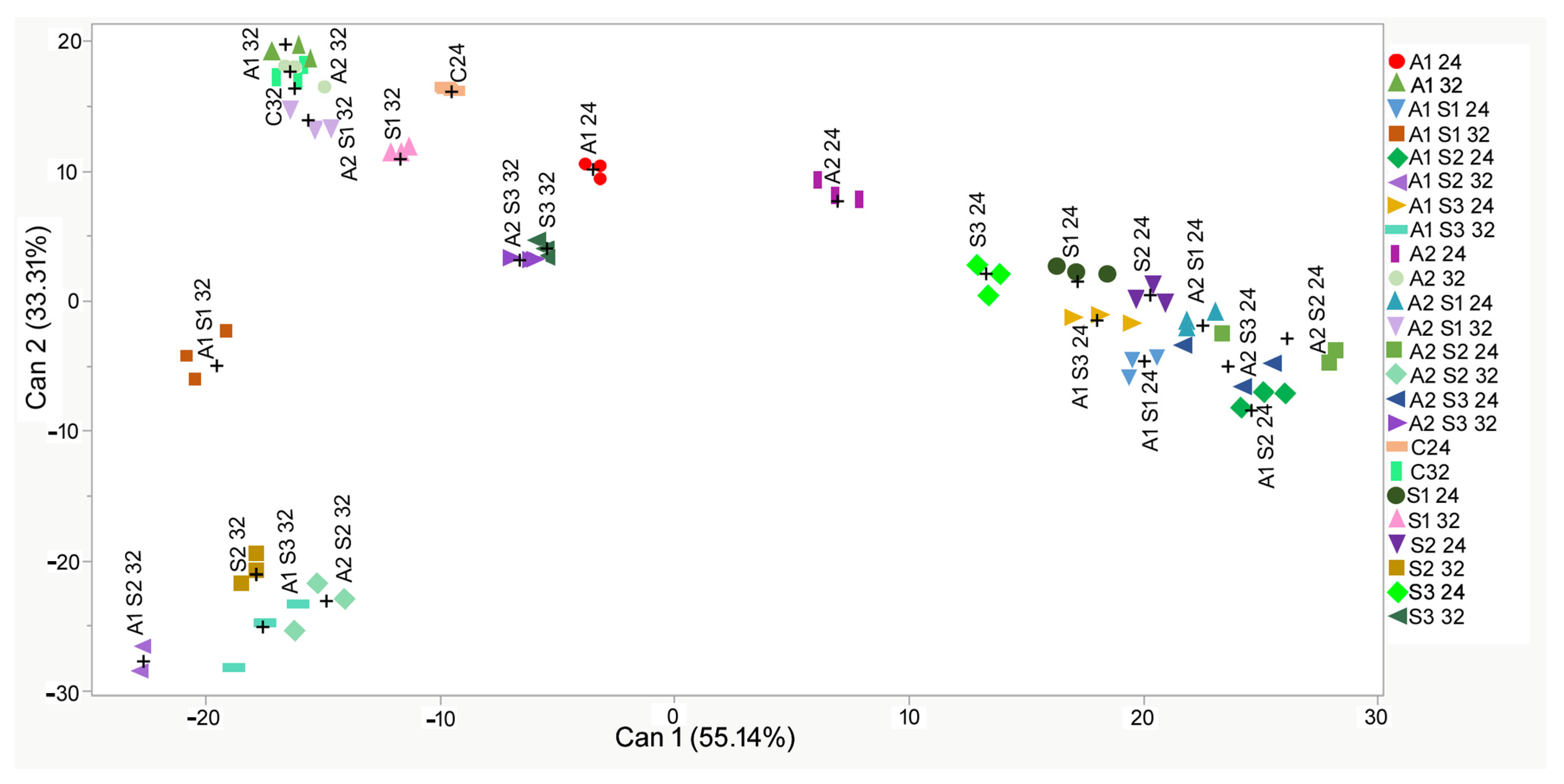
Canonical 1, which can be related to temperature, explained most of the separation (55.14%), while Canonical 2, which can be related to salt treatments, accounted for 33.31% of the separation of our samples (Figure 9).
Figure 9.
2D scatterplot of canonical discriminant analysis. Can 1 and 2 refer to canonical function 1 and 2, which consider all variables in order to maximize the separation between the groups at 24 °C and 32 °C temperatures for 3 days followed by 2 days recovery at 24 °C. C = control, S1, S2, S3 = 100, 200, 300 mM NaCl: A1, A2 = Chlorella 0.025% and 0.05%. The symbol + represents the mean value of the three determinations.
This statistical analysis allowed the identification of a dominant effect of temperature, stronger than that of salt and of biostimulant Chlorella. Pearson’s correlation analysis (Table 1) evidenced that variables mostly correlated to Can 1 are polyphenols, FRAP, chlorophyll a, b, total, and carotenoids, as explained by the high values correlated by these variables, all positives. Can 2 values are instead mostly positively correlated ROS and negatively polyphenols and FRAP.
Table 1.
Pearson’s correlation coefficients.
Shoot length, germination percentage, total phenolics, total flavonoids, total antioxidant activity (DPPH, ORAC, FRAP), ROS content, photosynthetic pigments and the canonical scores from the canonical discriminant analysis (CDA) are reported in Figure 9. In the CDA, the canonical scores from Canonical functions 1 and 2 were considered, explaining 55.14% and 33.31%, respectively. (* 0.5 > |r| > 1 highly correlated).
4. Discussion
The results of this study, which investigated a quinoa variety exposed to salt stress with co-treatment using the microalga Chlorella as a biostimulant, demonstrated that the germination percentage at 24 °C remained unaffected by both salt and Chlorella treatments, consistent with previous results for the same quinoa variety [22]. The temperature of 32 °C in combination with Chlorella induced a reduction in germination percentage to 60%, while in co-treatments with NaCl, germination remained around 80%, indicating moderate heat stress tolerance, as previously reported [25,37]. Under combined heat and salt stress, salt may induce a protective mechanism that mitigates potential adverse effects of the biostimulant at high temperatures. This temperature-dependent response demonstrates that the biostimulant’s effect is not universally beneficial but varies according to stress combination.
Considering shoot length at 24 °C at moderate salinity, the presence of the biostimulant resulted in moderate improvement, while 300 mM NaCl had a significant negative effect at both temperatures. At 32 °C, shoots were generally shorter than at 24 °C, confirming the increased sensitivity of quinoa to temperatures of 32 °C [38]. The three-way interaction revealed that the heat-induced growth reduction [39] was not mitigated by the application of Chlorella, indicating that the biostimulant’s efficacy in growth improvement is limited under combined high temperature and salinity stress.
The accumulation of bioactive antioxidant molecules was observed at both 24 °C and 32 °C under the co-treatment with NaCl and both concentrations of Chlorella, showing treatment interactions. This indicated that plants produce antioxidant protective molecules to counteract saline and heat stress. In particular, a dose-dependent increase was observed in total polyphenols level, with the 0.025% microalgae exerting its maximum positive efficacy. The biostimulant effect on these compounds, however, was mainly evident when combined with salt stress at 100 and 200 mM NaCl, in agreement with previous research reporting an increase in polyphenols in quinoa sprouts in 100 mM NaCl saline stress at [22] and a dose-dependent rise from 50 mM to 200 mM NaCl in quinoa leaves [40]. This increase supports quinoa’s classification as a halophyte [41] which can benefit from salt’s presence by synthesizing antioxidant molecules. The content of flavonoids increased at 100 and 200 mM NaCl but decreased at 300 mM, indicating that these compounds are susceptible to high salinity levels. However, this response was temperature-dependent: at 32 °C, polyphenol and flavonoid levels were consistently lower across all treatments, indicating the heat sensitivity of these biosynthetic pathways.
The increase in antioxidant activity detected by the FRAP, ORAC, and DPPH assays in samples treated with NaCl was consistent with previous reports [22,40]. The presence of the biostimulant Chlorella further enhanced reducing activity under interaction with NaCl [22]. This suggest that specific bioactive molecules in the Chlorella extract, such as polysaccharides, amino acids (e.g., proline, glutamate), and phytohormones (e.g., brassinosteroids) [42,43,44] may act as elicitors, upregulating genes encoding key enzymes in the phenylpropanoid pathway (e.g., phenylalanine ammonia-lyase, PAL) mainly when stress pathways are already activated. The antioxidant response induced by heat shock evidenced by increased ORAC and ARA was consistent with earlier studies [6,26,45].
As previously reported, the application of algal biostimulants can strengthen the plant’s natural response to salt stress, increasing antioxidant activity through elevated levels of tocopherols, polyphenols, and stress-related enzymes such as superoxide dismutase, catalase, ascorbate peroxidase, and proline [6,46]. Elevated osmolyte accumulation helps in ROS scavenging and protects vital cellular pathways such as photosynthesis, thus alleviating stress effects [10]. The role of proline could be a focal point to be analyzed in further work on quinoa seedlings. During heat shock, the biostimulant’s effect was less intense, probably for possible thermal degradation of active molecules and modifications in the algal extract composition that impaired its ability to enhance heat stress tolerance [18,43,47].
ROS accumulation revealed particularly complex interactions. At 24 °C, ROS content was highest in samples germinated without NaCl or without the biostimulant, indicating that the application of Chlorella was ineffective under non-stress conditions. Conversely, salt stress significantly reduced ROS levels, in a dose-dependent manner, while the biostimulant stimulated ROS production at moderate salt stress, corresponding to the progressive increase in polyphenols and, to a lesser extent, flavonoids. This unexpected increase may reflect controlled ROS generation as a signaling mechanism rather than oxidative damage. The interaction between high temperature and ROS production was positive in samples without NaCl and with 100 mM NaCl, but at higher salt concentrations a significant decrease in ROS levels was observed and further lowered by Chlorella application. Heat stress may induce the expression of genes coding for heat shock proteins, potentially explaining ROS reduction [44,48]. This effect appears synergistic with high salinity and biostimulant treatment. These findings align with previous reports on quinoa resistance to both low and high temperature (−4 to 30 °C) as reported in earlier studies [23,49].
Photosynthetic pigment analysis provided some of the most striking evidence for treatment interactions. At 24 °C, the content of photosynthetic pigments was increased by the presence of the Chlorella biostimulant at 0.05% in the absence of NaCl, likely due to bioactive compounds enhancing the function of enzymes responsible for chlorophyll synthesis [10,22]. Under salt stress (100–200 mM NaCl), pigment levels increased overall, an effect attributable to the tolerance of the quinoa variety “Regalona”, a facultative halophyte, through the vacuolar compartmentalization of salt, which protects chloroplasts from this toxicity. However, the stimulating effect of Chlorella was not observed. Similar results reporting an increase in photosynthetic pigments but a negligible effect of the biostimulant in two quinoa varieties have already been reported [22]. This increase in chlorophylls was considered as part of an adaptive conditioning mode by which the plant can be protected against severe environmental stress [36,50] through phytohormone signaling [51].
The increase in carotenoids under salt stress could be explained by their dual role in light harvesting and photoprotection by dissipating excess energy, stabilizing chloroplast membranes, and scavenging ROS [52].
Several studies have reported a strong impact of saline stress on the photosynthetic apparatus [7], mainly due to impaired CO2 diffusion caused by stomatal closure or modifications in the biochemical apparatus affecting CO2 fixation [53]. This was observed in the quinoa variety “Titicaca”, where 200 mM NaCl caused a decrease in chlorophyll a and carotenoids content [53] versus maintained or increased pigments in the variety “Regalona”. The contrasting responses among different quinoa varieties may be explained by their varying salt tolerance levels, depending on species and genotype [53,54].
When quinoa seeds were germinated at 32 °C, the chlorophyll levels measured spectrophotometrically were very low, sometimes below the detection limits. This dramatic heat stress effect dominated over both salinity and biostimulant treatments. High temperature either impaired sprout development resulting in shorter plants and incomplete photosynthetic cotyledon development, or induced chlorophyll oxidation, particularly in Photosystem II, leading to reduced synthesis of ATP, decreased CO2 fixation, and lower plant yield [6,18]. Therefore, Chlorella improved biochemical stress markers of salt tolerance, particular polyphenols, and antioxidant activity under salt stress but not under heat stress. These effects were absent under heat stress and did not consistently enhance growth parameters. The significant three-way interactions revealed that the biostimulant’s modest benefits required specific combinations of moderate salinity and optimal temperature.
The present research therefore revealed a complex interaction between stress intensity and microalgae efficacy, adding valuable insights for sustainable crop management based on biostimulant employment [55,56]. Biostimulant efficacy is critically dependent on environmental context and impaired by severe heat, which may directly degrade the photosynthetic machinery [57] and alter the bioactive compounds in the algal extracts. The preservation and enhancement of the photosynthetic pigment pool, particularly carotenoids, and the controlled modulation of ROS for signaling emerge as pivotal mechanisms in the Chlorella-quinoa stress resilience synergy [58].
5. Conclusions
This study provides a multifaceted assessment of how the microalgal biostimulant Chlorella influences the resilience of quinoa (Chenopodium quinoa Willd. var. Regalona) under combined salt and heat stress conditions.
For seed germination and sprout development, the present results delineate a clear response to stress gradient dependent on the interaction between salt, temperature, and biostimulant. Chlorella application slightly mitigated the salt’s effect on germination percentage and shoot length at moderate salinity at 24 °C, supporting its role as a growth promoter. However, this benefit was overcome, particularly for shoot length, by severe salt stress and by elevated temperature (32 °C), highlighting the limitation of biostimulant’ effects under extreme abiotic conditions.
The most compelling evidence for Chlorella’s role as a stress mitigator comes from the biochemical analyses under salt treatments, which consistently enhanced the plant’s antioxidant machinery, mainly increasing polyphenols and flavonoids, and antioxidant capacity. Interestingly, the ROS level was often lower in salt-stressed plants, and this reduction was further enhanced by Chlorella at higher salt concentrations. This suggests that the quinoa var. Regalona presents an efficient antioxidant response which effectively scavenges ROS. The presence of the biostimulant Chlorella reinforces this homeostasis, preventing oxidative damage, although growth or yield-related parameters were not improved.
The response of the photosynthetic apparatus was stress-dose-dependent. In quinoa var. Regalona, pigment content increased at 100–200 mM NaCl at 24 °C, reflecting its halophytic character, though Chlorella’s stimulatory effect was masked by this strong salt-induced response. The severe impairment of all the chlorophylls at 32 °C regardless of the salt treatment indicates that heat stress strongly affects photosynthetic apparatus’s development and efficacy.
In summary, this research demonstrates that Chlorella induces an effective biochemical response in quinoa under moderate stress conditions, primarily by enhancing the plant’s endogenous antioxidant machinery at optimal temperatures and against specific stress intensities. The study confirms that the quinoa var. Regalona is notably salt tolerant while identifying combined heat and salt stress as a significant challenge that limits biostimulants’ efficacy. Algal biostimulants are a promising, sustainable tool to reinforce crop performance on marginal lands, contributing to environmental stress adaptation. However, their application requires careful assessment of the environmental context and realistic expectations regarding their agronomic impact under combined or extreme stress conditions.
Supplementary Materials
The following supporting information can be downloaded at: https://www.mdpi.com/article/10.3390/environments13040182/s1, Table S1: Normalized values (% of control) of germination, growth and biochemical parameters in sprouts of Chenopodium quinoa var ‘Regalona’ after 5 days of imbibition in the presence of different NaCl concentrations (0, 100, 200, 300 mM) and Chlorella vulgaris-like at the concentration of 0 (A0), 0.025% (A1), 0.05% (A2) at the temperature of 24 °C. (+) indicates an increase compared to the control, while (−) indicates a decrease. Table S2: Normalized values (% of control) of germination, growth and biochemical parameters in sprouts of Chenopodium quinoa var ‘Regalona’ at the temperature of 32 °C for 3 days followed by 2 days recovery at 24 °C in the presence of different NaCl concentrations (0, 100, 200, 300 mM) and Chlorella vulgaris-like at the concentration of 0 (A0), 0.025% (A1), 0.05% (A2). (+) indicates an increase compared to the control, while (−) indicates a decrease. Table S3: Simple effect analyses (factors NaCl, Algae and Temperature) of significant interactions evaluated by three-way ANOVA, followed by post-hoc Tukey test within relevant simple effects. Within columns different letters indicated significant differences at p ˂ 0.05 for each factor.
Author Contributions
Conceptualization, L.G. and L.B.; methodology, A.M., L.B., L.P.F., M.G. and L.G.; software, L.G., A.M. and L.B.; validation, L.G., A.M., L.P.F., M.G. and L.B.; formal analysis, L.G., A.M. and L.B.; investigation, L.G., A.M. and L.B.; resources, L.G.; data curation, L.G., A.M. and L.B.; writing—original draft preparation, L.B., A.M. and L.G.; writing—review and editing, L.G., A.M., L.P.F., M.G. and L.B.; visualization, L.G., A.M. and L.B.; supervision, L.B. and L.G.; project administration, L.G.; funding acquisition, L.G. All authors have read and agreed to the published version of the manuscript.
Funding
This research received financial support from the CNR project FOE-2021 DBA.AD005.225 (NUTRAGE—Nutrizione, Alimentazione e Invecchiamento attivo) and from the Agritech National Research Center from the European Union Next-Generation EU [Piano Nazionale Di Ripresa e Resilienza (PNRR), Missione 4 Componente 2, Investimento 1.4—Project CN00000022] to the National Research Council.
Data Availability Statement
The raw data supporting the conclusions of this article will be made available by the authors on request.
Conflicts of Interest
The authors declare no conflicts of interest. The funders had no role in the design of the study; in the collection, analyses, or interpretation of data; in the writing of the manuscript; or in the decision to publish the results.
References
- Bhupenchandra, I.; Chongtham, S.K.; Devi, E.L.; Ramesh, R.; Choudhary, A.K.; Salam, M.D.; Sahoo, M.R.; Bhutia, T.L.; Devi, S.H.; Thounaojam, A.S.; et al. Role of biostimulants in mitigating the effects of climate change on crop performance. Front. Plant Sci. 2022, 13, 967665. [Google Scholar] [CrossRef] [Scilit]
- Badavath, H.; Saski, C.A. Plant Adaptation to Salinity: Physiological Pathways and Prospects for Crop Improvement—A Review. Genes 2026, 17, 253. [Google Scholar] [CrossRef] [Scilit]
- Ronga, D.; Biazzi, E.; Parati, K.; Carminati, D.; Carminati, E.; Tava, A. Microalgal Biostimulants and Biofertilisers in Crop Productions. Agronomy 2019, 9, 192. [Google Scholar] [CrossRef] [Scilit]
- Zhou, H.; Shi, H.; Yang, Y.; Feng, X.; Chen, X.; Xiao, F.; Lin, H.; Guo, Y. Insights into plant salt stress signaling and tolerance. J. Genet. Genom. 2024, 51, 16–34. [Google Scholar] [CrossRef] [Scilit]
- Fu, H.; Yang, Y. How plants tolerate salt stress. Curr. Issues Mol. Biol. 2023, 45, 5914–5934. [Google Scholar] [CrossRef] [Scilit] [PubMed]
- Vangenechten, B.; De Coninck, B.; Ceusters, J. How to improve the potential of microalgal biostimulants for abiotic stress mitigation in plants? Front. Plant Sci. 2025, 16, 1568423. [Google Scholar]
- Nawaz, M.; Sun, J.; Shabbir, S.; Khattak, W.A.; Ren, G.; Nie, X.; Bo, Y.; Javed, Q.; Du, D.; Sonne, C. A review of plants strategies to resist biotic and abiotic environmental stressors. Sci. Total Environ. 2023, 900, 165832. [Google Scholar]
- EBIC. European Biostimulants Industry Council [WWW Document]. 2016. Available online: http://www.biostimulants.eu/ (accessed on 20 January 2026).
- Kapoore, R.V.; Wood, E.; Llewellyn, C.A. Algae biostimulants: A critical look at microalgal biostimulants for sustainable agricultural practices. Biotechnol. Adv. 2021, 49, 107754. [Google Scholar] [CrossRef] [Scilit]
- Senousy, H.H.; Hamoud, Y.A.; Abu-Elsaoud, A.M.; Mahmoud Al Zoubi, O.; Abdelbaky, N.F.; Zia-ur-Rehman, M.; Usman, M.; Soliman, M.H. Algal Bio-Stimulants Enhance Salt Tolerance in Common Bean: Dissecting Morphological, Physiological, and Genetic Mechanisms for Stress Adaptation. Plants 2023, 12, 3714. [Google Scholar] [CrossRef] [Scilit]
- Elansary, H.O.; Yessoufou, K.; Abdel-Hamid, A.M.E.; El-Esawi, M.A.; Ali, H.M.; Elshikh, M.S. Seaweed Extracts Enhance Salam Turfgrass Performance during Prolonged Irrigation Intervals and Saline Shock. Front. Plant Sci. 2017, 8, 830. [Google Scholar] [CrossRef] [Scilit] [PubMed]
- Carillo, P.; Ciarmiello, L.F.; Woodrow, P.; Corrado, G.; Chiaiese, P.; Rouphael, Y. Enhancing Sustainability by Improving Plant Salt Tolerance through Macro- and Micro-Algal Biostimulants. Biology 2020, 9, 253. [Google Scholar] [CrossRef] [Scilit] [PubMed]
- Bachani, J.; Mahanty, A.; Aftab, T.; Kumar, K. Insight into calcium signalling in salt stress response. S. Afr. J. Bot. 2022, 151, 1–8. [Google Scholar]
- Adedeji, O.; Reuben, O.; Olatoye, O. Global Climate Change. J. Geosci. Environ. Prot. 2014, 2, 114–122. [Google Scholar]
- Hinojosa, L.; González, J.; Barrios-Masias, F.; Fuentes, F.; Murphy, K. Quinoa Abiotic Stress Responses: A Review. Plants 2018, 7, 106. [Google Scholar] [CrossRef] [Scilit]
- Allakhverdiev, S.I.; Kreslavski, V.D.; Klimov, V.V.; Los, D.A.; Carpentier, R.; Mohanty, P. Heat stress: An overview of molecular responses in photosynthesis. Photosynth. Res. 2008, 98, 541–550. [Google Scholar] [CrossRef] [Scilit]
- Kopta, T.; Pavlıkova, M.; Sekara, A.; Pokluda, R.; Marsalek, B. Effect of bacterial-algal biostimulant on the yield and internal quality of Lettuce (Lactuca sativa L.) produced for spring and summer crop. Not. Bot. Horti Agrobot. 2018, 46, 615–621. [Google Scholar]
- Brück, W.M.; Alfonso, E.; Rienth, M.; Andlauer, W. Heat Stress Resistance in Chlorella vulgaris Enhanced by Hydrolyzed Whey Proteins. Agronomy 2024, 14, 2854. [Google Scholar] [CrossRef] [Scilit]
- Morales, D.; Garcés-Rimòn, M.; Miguel, M. Pseudocereals: A novel source of biologically active peptides. Crit. Rev. Food Sci. Nutr. 2022, 62, 3729–3748. [Google Scholar]
- Shahbaz, M.; Raza, N.; Islam, M.; Imran, M.; Ahmad, I.; Meyyazhagan, A.; Pushparaj, K.; Balasubramanian, B.; Park, S.; Rengasamy, K.R.R.; et al. The nutraceutical properties and health benefits of pseudocereals: A comprehensive treatise. Crit. Rev. Food Sci. Nutr. 2024, 64, 539–560. [Google Scholar]
- Lavini, A.; Pulvento, C.; D’Andria, R.; Riccardi, M.; Choukr-Allah, R.; Belhabib, O.; Yazar, A.; Incekaya, Ç.; Metin Sezen, S.; Qadir, M.; et al. Quinoa’s Potential in the Mediterranean Region. J. Agron. Crop Sci. 2014, 200, 344–360. [Google Scholar]
- Fiorentino, S.; Bellani, L.; Santin, M.; Castagna, A.; Echeverria, M.C.; Giorgetti, L. Effects of Microalgae as Biostimulants on Plant Growth, Content of Antioxidant Molecules and Total Antioxidant Capacity in Chenopodium quinoa Exposed to Salt Stress. Plants 2025, 14, 781. [Google Scholar] [CrossRef] [Scilit] [PubMed]
- García-Parra, M.A.; Roa-Acosta, D.F.; Stechauner-Rohringer, R.; García-Molano, J.F.; Bazile, D.; Plazas-Leguizamón, N. Effect of temperature on the growth and development of quinoa plants (Chenopodium quinoa Willd.): A review on a global scale. Sylwan 2020, 164, 411–431. [Google Scholar]
- Hinojosa, L.; Matanguihan, J.B.; Murphy, K.M. Effect of high temperature on pollen morphology, plant growth and seed yield in quinoa (Chenopodium quinoa Willd.). J. Agron. Crop Sci. 2019, 205, 33–45. [Google Scholar] [CrossRef] [Scilit]
- Alvar-Beltrán, J.; Verdi, L.; Dalla Marta, A.; Dao, A.; Vivoli, R.; Sanou, J.; Orlandini, S. The effect of heat stress on quinoa (cv. Titicaca) under controlled climatic conditions. J. Agric. Sci. 2020, 158, 255–261. [Google Scholar] [CrossRef] [Scilit]
- Fahad, S.; Bajwa, A.A.; Nazir, U.; Anjum, S.A.; Farooq, A.; Zohaib, A. Crop production under drought and heat stress: Plant responses and management options. Front. Plant Sci. 2017, 8, 1147. [Google Scholar] [CrossRef] [Scilit]
- Chiellini, C.; Serra, V.; Gammuto, L.; Adriana, C.; Longo, V.; Gabriele, M. Evaluation of Nutraceutical Properties of Eleven Microalgal Strains Isolated from Different Freshwater Aquatic Environments: Perspectives for Their Application as Nutraceuticals. Foods 2022, 11, 654. [Google Scholar] [CrossRef] [Scilit]
- Vento, M.; Della Croce, C.M.; Bellani, L.; Tassi, E.L.; Echeverria, M.C.; Giorgetti, L. Effect of sprouting, fermentation and cooking on antioxidant content and total antioxidant activity in quinoa and amaranth. Int. J. Mol. Sci. 2024, 25, 10972. [Google Scholar] [CrossRef] [Scilit] [PubMed]
- Singleton, V.L.; Rossi, J.A., Jr. Colorimetry of total phenolics with phosphomolybdic–phosphotungstic acid reagents. Am. J. Enol. Vitic. 1965, 16, 144–158. [Google Scholar] [CrossRef] [Scilit]
- Heimler, D.; Vignolini, P.; Dini, M.G.; Romani, A. Rapid tests to assess the antioxidant activity of Phaseolus vulgaris L. dry beans. J. Agric. Food Chem. 2006, 53, 3053–3056. [Google Scholar] [CrossRef] [Scilit]
- Benzie, I.F.F.; Strain, J.J. Ferric reducing/antioxidant power assay: Direct measure of total antioxidant activity of biological fluids and modified version for simultaneous measurement of total antioxidant power and ascorbic acid concentration. Methods Enzymol. 1999, 299, 15–27. [Google Scholar]
- Ninfali, P.; Biagiotti, E.; Bacchiocca, M.; Avanzi, L.; Vertuani, S.; Manfredini, S. Antioxidant capacity of agrofoods and natural or synthesized antioxidant compounds. Ind. Aliment. 2002, 41, 257–261. [Google Scholar]
- Boudjou, S.; Oomah, B.D.; Zaidi, F.; Hosseinian, F. Phenolics content and antioxidant and anti-inflammatory activities of legume fractions. Food Chem. 2013, 138, 1543–1550. [Google Scholar] [CrossRef] [Scilit] [PubMed]
- Bellani, L.M.; Salvini, L.; Dell’Aquila, A.; Scialabba, A. Reactive oxygen species release, vitamin E, Fatty acid and phytosterol contents of artificially aged radish (Raphanus sativus L.) seeds during germination. Acta Physiol. Plant. 2012, 34, 1789–1799. [Google Scholar] [CrossRef] [Scilit]
- Lichtenthaler, H.K.; Buschmann, C. Chlorophylls and carotenoids: Measurement and characterization by UV-VIS spectroscopy. In Current Protocols Food Analytical Chemistry; Wiley: Hoboken, NJ, USA, 2001; pp. F4.3.1–F4.3.8. [Google Scholar]
- Muccifora, S.; Giorgetti, L.; Corsini, M.; Di Florio, G.; Bellani, L. Nano and submicron fluorescent polystyrene particles internalization and translocation in seedlings of Cichorium endivia L. Environ. Sci. Nano 2022, 9, 4585–4598. [Google Scholar] [CrossRef] [Scilit]
- Jacobsen, S.E.; Bach, A.P. The influence of temperature on seed germination rate in quinoa (Chenopodium quinoa Willd.). Seed Sci. Technol. 1998, 26, 515–523. [Google Scholar]
- Tovar, J.C.; Quillatupa, C.; Callen, S.T.; Castillo, E.; Pearson, P.; Shamin, A.; Schuhl, H.; Fahlgren, N.; Gehan, M.A. Heating quinoa shoots results in yield loss by inhibiting fruit production and delaying maturity. Plant J. 2020, 102, 1058–1073. [Google Scholar] [CrossRef] [Scilit]
- Souid, A.; Bellani, L.; Tassi, E.L.; Hamed, K.B.; Longo, V.; Giorgetti, L. Early Physiological, Cytological and Antioxidative Responses of the Edible Halophyte Chenopodium quinoa exposed to Salt Stress. Antioxidants 2023, 12, 1060. [Google Scholar] [CrossRef] [Scilit]
- Slimani, N.; Arraouadi, S.; Hajlaoui, H.; Cid-Samamed, A.; Borgi, M.A.; Snoussi, M. Salt Stress Enhanced Bioactivity of Quinoa Leaf Extracts: An in vitro and in silico Study of Acetylcholinesterase and Tyrosinase Inhibition for Sustainable Drug Development. Pharmaceuticals 2025, 18, 77. [Google Scholar] [CrossRef] [Scilit] [PubMed]
- Hinojosa, L.; Leguizamo, A.; Carpio, C.; Muñoz, D.; Mestanza, C.; Ochoa, J.; Castillo, C.; Murillo, A.; Villacréz, E.; Monar, C.; et al. Quinoa in Ecuador: Recent Advances under Global Expansion. Plants 2021, 10, 298. [Google Scholar] [CrossRef] [Scilit]
- Yadava, P.; Kaushal, J.; Gautam, A.; Parmar, H.; Singh, I. Physiological and biochemical effects of 24-epibrassinolide on heat-stress adaptation in maize (Zea mays L.). Nat. Sci. 2016, 8, 171–179. [Google Scholar]
- Hyder, Q.U.A.; Aziz, A.; Azhar, M.F.; Haider, M.H.; Hussain, N. Plant-derived biostimulants: Emerging trends, breakthroughs and challenges in 2024. Org. Agric. 2026, 16, 12. [Google Scholar] [CrossRef] [Scilit]
- Quintero-Calderón, E.H.; Sánchez-Reinoso, A.D.; Chávez-Arias, C.C.; Garcés-Varón, G.; Restrepo-Díaz, H. Rice seedlings showed a higher heat tolerance through the foliar application of biostimulants. Not. Bot. Horti Agrobot. 2021, 49, 12120. [Google Scholar] [CrossRef] [Scilit]
- Wang, W.; Vinocur, B.; Shoseyov, O.; Altman, A. Role of plant heatshock proteins and molecular chaperones in the abiotic stress response. Trends Plant Sci. 2004, 9, 244–252. [Google Scholar] [CrossRef] [Scilit]
- Kusvuran, A.; Can, A.G. Effects of microalga (Chlorella vulgaris beijerinck) on seconder metabolites and antioxidative defense system improve plant growth and salt tolerance in guar (Cyamopsis tetragonoloba). Legume Res. 2020, 43, 56–60. [Google Scholar] [CrossRef] [Scilit]
- Calvo, P.; Nelson, L.; Kloepper, J.W. Agricultural uses of plant biostimulants. Plant Soil 2014, 383, 3–41. [Google Scholar] [CrossRef] [Scilit]
- Rajewska, I.; Talarek, M.; Bajguz, A. Brassinosteroids and response of plants to heavy metals action. Front. Plant Sci. 2016, 7, 629. [Google Scholar] [CrossRef] [Scilit]
- Bazile, D.; Pulvento, C.; Verniau, A.; Al-Nusairi, M.S.; Ba, D.; Breidy, J.; Padulosi, S. Worldwide Evaluations of Quinoa: Preliminary Results from Post International Year of Quinoa FAO Projects in Nine Countries. Front. Plant Sci. 2016, 7, 850. [Google Scholar] [CrossRef] [Scilit]
- Agathokleous, E.; Feng, Z.; Peñuelas, J. Chlorophyll hormesis: Are chlorophylls major components of stress biology in higher plants? Sci. Tot. Environ. 2020, 726, 138637. [Google Scholar] [CrossRef] [Scilit]
- Gao, F.; Guo, J.; Shen, Y. Advances from chlorophyll biosynthesis to photosynthetic adaptation, evolution and signaling. Plant Stress 2024, 12, 100470. [Google Scholar] [CrossRef] [Scilit]
- Megdiche, W.; Hessini, K.M.; Gharbi, F.; Jaleel, C.A.; Ksouri, R.; Abdelly, C. Photosynthesis and photosystem 2 efficiency of two salt-adapted halophytic seashore Cakile maritima ecotypes. Photosynthetica 2008, 46, 410–419. [Google Scholar] [CrossRef] [Scilit]
- Rodríguez-Hernández, M.C.; Morcillo, L.; Garmendia, I. Sensitivity of quinoa cv. ‘Titicaca’ to low salinity conditions. Folia Hortic. 2021, 33, 135–145. [Google Scholar] [CrossRef] [Scilit]
- Brock, J.; Aboling, S.; Stelzer, R.; Esch, E.; Papenbrock, J. Genetic variation among different populations of Aster tripolium grown on naturally and anthropogenic salt-contaminated habitats: Implications for conservation strategies. J. Plant Res. 2007, 120, 99–112. [Google Scholar] [CrossRef] [Scilit] [PubMed]
- Feng, D.; Zhang, W.; Zheng, C.; Tang, G.; Adili, Y.; Ge, C.; Xu, W. Mechanisms of plant extracts in alleviating drought and saline-alkali stress in plants. Ind. Crops Prod. 2025, 233, 121346. [Google Scholar] [CrossRef] [Scilit]
- Han, M.; Kasim, S.; Yang, Z.; Deng, X.; Saidi, N.B.; Uddin, M.K.; Shuib, E.M. Plant extracts as biostimulant agents: A promising strategy for managing environmental stress in sustainable agriculture. Phyton 2024, 93, 9457. [Google Scholar] [CrossRef] [Scilit]
- Khoulati, A.; Ouahhoud, S.; Taibi, M.; Ezrari, S.; Mamri, S.; Merah, O.; Hakkou, A.; Addi, M.; Adil, M.; Saalaoui, E. Harnessing biostimulants for sustainable agriculture: Innovations, challenges, and future prospects. Discov. Agric. 2025, 3, 56. [Google Scholar] [CrossRef] [Scilit]
- Brito-Lopez, C.; Van Der Wielen, N.; Barbosa, M.; Karlova, R. Plant growth–promoting microbes and microalgae-based biostimulants: Sustainable strategy for agriculture and abiotic stress resilience. Philos. Trans. R. Soc. B Biol. Sci. 2025, 380, 1927. [Google Scholar] [CrossRef] [Scilit] [PubMed]

Disclaimer/Publisher’s Note: The statements, opinions and data contained in all publications are solely those of the individual author(s) and contributor(s) and not of MDPI and/or the editor(s). MDPI and/or the editor(s) disclaim responsibility for any injury to people or property resulting from any ideas, methods, instructions or products referred to in the content. |
© 2026 by the authors. Licensee MDPI, Basel, Switzerland. This article is an open access article distributed under the terms and conditions of the Creative Commons Attribution (CC BY) license.