


Simple Summary
While microplastics are proliferating in freshwater environments, their impact on fish nutrition and welfare is not yet fully understood. This study examined how a 14-day exposure to microplastics affects feeding, energy expenditure, stress and anxiety-like behavior in goldfish. Our results show that fish exposed to microplastics suppressed appetite, diminished feed anticipation, and increased energy expenditure, leading to stunted growth and the depletion of hepatic energy reserves. Furthermore, exposed fish also displayed signs of stress and anxiety, compromising overall well-being. Together, these findings show that microplastics disrupt energy balance and welfare in fish, suggesting that their impact extends beyond mere survival. These results highlight the importance of considering integrated behavioral and physiological indicators when assessing their effects in fish.
Abstract
The accumulation of microplastics in aquatic ecosystems poses a significant threat to fish physiology and welfare. This study investigated the impact of exposure to virgin polystyrene microplastics (15 µm) on energy balance and welfare in goldfish (Carassius auratus). Fish were exposed for 14 days, and the effects were assessed through an integrated analysis of behavioral, metabolic, neuroendocrine, and physiological parameters. Microplastic exposure significantly reduces feed intake and feed anticipatory activity, indicating a potent anorexigenic effect. This effect was driven by neuroendocrine disruption, characterized by the downregulation of orexigenic neuropeptides (npy, agrp, hcrt) and the upregulation of anorexigenic signaling (pomca, cartpt, lepa). Simultaneously, exposed fish exhibited increased oxygen consumption, suggesting elevated metabolic demands. These factors converged to impaired growth and reduced hepatosomatic index, suggesting altered energy allocation. Furthermore, microplastic exposure induced anxiety-like responses and increased plasma cortisol levels, confirming the activation of the physiological stress response. Overall, these findings demonstrate that microplastics disrupt energy homeostasis and trigger behavioral shifts that ultimately compromise fish welfare and the biological resilience of aquatic species.
1. Introduction
Global reliance on plastics has escalated at an unprecedented rate, with annual production already surpassing hundreds of millions of tons and projections indicating further dramatic increases [1,2]. The widespread use of disposable plastic items combined with inadequate waste management has resulted in the continuous leakage of plastic debris into aquatic ecosystems [3,4]. Over time, larger plastic materials fragment into smaller particles known as microplastics (1 μm to 5 mm), which are now recognized as one of the most pervasive pollutants in aquatic environments [2,5,6]. Research has predominantly focused on marine environments, leaving a significant knowledge gap regarding freshwater ecosystems [7,8], despite growing evidence of considerable microplastic contamination in freshwater aquaculture systems and freshwater fish, including cyprinid species, reported in recent studies [9,10,11].
Microplastics are highly persistent and mobile, interacting with a wide range of aquatic organisms and transferring across trophic levels. They have been detected in diverse aquatic species, including zooplankton, mollusks, crustaceans, and fish [1,12,13]. Moreover, the detection of microplastics in commercially important species underscores their relevance not only for aquatic ecosystems but also for food safety and potential human exposure [14,15,16]. In fish, once ingested, microplastics initially accumulate in the gastrointestinal tract, where they can interfere with digestion and nutrient assimilation, while additional exposure routes such as the gills may allow particle adhesion or penetration into respiratory tissues [17,18]. From these entry routes, microplastics can translocate to internal organs such as the liver, muscle, and brain, raising concerns about systemic toxicity [8]. Microplastic exposure to fish has been associated with a broad spectrum of adverse effects affecting multiple levels of biological organization. Experimental studies have shown that microplastics induce cellular and tissue damage, oxidative stress, and inflammatory responses, as well as physiological alterations affecting several systems, including digestive, neuroendocrine, and reproductive systems [8,19,20,21]. Beyond these physiological effects, microplastics can also alter behavior in fish, including feeding, locomotion, predator avoidance, and social interactions [21,22,23,24,25,26]. Such behavioral changes are progressively recognized as sensitive indicators of organismal fitness, often revealing sublethal effects that may precede overt toxicity, and complementing conventional toxicological analyses. Despite the increasing evidence of microplastic toxicity, significant gaps remain regarding their sublethal effects on energy homeostasis and welfare. This is particularly true for freshwater species such as the goldfish (Carassius auratus), a species belonging to the Cyprinidae, a family that dominates global freshwater aquaculture production [27,28], making it an ecologically and economically relevant model for assessing plastic pollution. Previous research in goldfish has addressed microplastic ingestion and egestion dynamics [29], ionoregulatory disturbances at the gills [30], effects on oxidative stress, liver damage [31,32] and impaired feeding by disrupting feeding regulators [33]. However, these studies generally focus on isolated endpoints rather than adopting an integrative perspective. The present study addresses this gap by combining these endpoints to provide a more comprehensive understanding of how microplastics affect energy homeostasis and welfare.
Energy homeostasis, defined by the balance between intake (feeding behavior) and expenditure (metabolism and activity), is fundamental for growth, reproduction, and survival, and its disruption may trigger cascading consequences that extend beyond individual physiology to population-level processes. Feed intake in fish is regulated by an integrated hypothalamic neuroendocrine system, in which orexigenic neurons expressing neuropeptide Y (NPY) and agouti-related peptide (AgRP) stimulate feeding, whereas anorexigenic neurons expressing proopiomelanocortin (POMC) and cocaine- and amphetamine-regulated transcript (CART) inhibit it [34,35,36]. This hypothalamic circuit integrates peripheral hormonal signals, primarily leptin and ghrelin, which exert opposing actions: leptin (mainly hepatic in teleosts) promotes satiety by activating POMC/CART neurons and inhibiting NPY/AgRP neurons [35,36,37], whereas ghrelin (produced in the gastrointestinal tract) stimulates feed intake through the opposite effects [34]. This gut–liver–brain axis ensures fine-tuned regulation of energy balance but may be particularly sensitive to environmental stressors such as microplastics. Although several studies have evaluated growth performance after microplastic exposure, few studies have examined direct effects on feed intake, and only a limited number have addressed the underlying neuroendocrine regulatory mechanisms. Reported effects on appetite-related signals remain inconsistent across species and particle types [33,38], highlighting the need for integrative approaches combining behavioral and molecular endpoints to better understand how microplastics disrupt energy homeostasis.
In fish, physiological stress responses are closely linked to the regulation of energy balance and welfare and are primarily mediated by activation of the hypothalamus–pituitary–interrenal (HPI) axis, leading to the release of cortisol, which promotes energy mobilization and adaptive responses to environmental challenges [39,40,41]. Under stress conditions, sustained elevation of cortisol levels is commonly observed, making this hormone a widely accepted biomarker of fish welfare [41,42,43]. Environmental stressors can also induce anxiety-like behavioral responses in fish, including altered risk assessment and reduced exploratory behavior, which are indicative of compromised welfare [44,45,46]. These behavioral alterations can be reliably assessed using non-invasive approaches, such as open field and black–white preference tests, which are widely applied in fish welfare research [47,48,49]. Together, endocrine and behavioral responses provide complementary indicators of fish welfare and allow the assessment of how environmental stressors such as microplastics may disrupt both physiological stability and adaptive behavior.
This study aims to determine the impact of microplastic exposure on energy homeostasis and welfare in fish through an integrated analysis of behavioral, physiological, neuroendocrine, and metabolic responses. The model species selected was the goldfish (Carassius auratus) for its well-established use in neuroendocrine and toxicological research [50] and its ecological relevance as a representative freshwater cyprinid. Using this teleost freshwater model, we focused on two complementary axes. First, feed intake, feed anticipatory activity, metabolic rate, and locomotor activity were evaluated as key indicators of the balance between energy intake and expenditure, and their disruption was examined in relation to growth performance. Second, anxiety-like behavior and circulating cortisol levels were assessed as relevant biomarkers of organism welfare. Furthermore, we investigated the neuroendocrine mechanisms underlying these responses to elucidate how microplastics disrupt physiology and behavior linked to molecular mechanisms.
2. Materials and Methods
2.1. Fish and Housing
Goldfish were distributed into four tanks (n = 6 fish per tank), with 60 L of aerated and filtered fresh water. Initial body weight (BW) was 16.54 ± 2.27 g in the control group and 16.32 ± 2.48 g in the microplastic-exposed group (mean ± Standard Deviation). Fish were kept in controlled temperature conditions (21 ± 1 °C), with a photoperiod of 12 h of light (8:00–20:00) and 12 h of darkness (20:00–8:00). Daily feed intake took place at 10:00 h with commercial dry pellets (Sera Pond Granulat; Heinsberg, Germany) with a ration equivalent to 1.5% BW. Fish were acclimated to these conditions at least three weeks prior to experimentation.
2.2. Experimental Design
This work was carried out in accordance with current regulations (RD 53/2013, 2010/63/EU), and all experimental procedures were approved by the Animal Experimentation Committee of the Complutense University of Madrid and the Community of Madrid (PROEX 317.7/23).
The fish were divided into two experimental groups (n = 12 fish per group), with each group conducted in duplicate tanks (6 fish per tank). One group was the control group (C), with no exposure to microplastics, while the other group (MP) was exposed for 14 days to additive-free polystyrene microspheres (15 µm diameter, #74964, Sigma Aldrich, Madrid, Spain) at a concentration of 0.5 mg/L. The 14-day exposure period was selected as a short but sufficient timeframe to detect changes in energy homeostasis and growth-related responses. This concentration was selected based on previous experimental studies in fish [32,33,38,51], and lies within the range of microplastic concentrations considered environmentally relevant in aquatic systems [52,53,54]. Continuous aeration was applied to ensure homogeneous dispersion of microplastic particles in the water and to maintain particles in suspension throughout the exposure period. The stability of the suspension was periodically checked by light microscopic observation, which did not indicate marked aggregation under the experimental conditions used. To maintain appropriate hygienic conditions while preventing microplastic loss, the filtration system was deliberately omitted, and 50% of the water volume was renewed daily. Although daily water renewal may introduce minor handling effects, these were applied equally across treatments and are therefore unlikely to have influenced the differences observed between experimental groups. Following each water exchange, microplastics were added every day at 12:00 to replenish the concentration and maintain consistent exposure levels throughout the experiment.
Before the experiment, fish were anesthetized using tricaine methanesulfonate (MS-222, 0.16 g/L, #E10521, Sigma Aldrich, Madrid, Spain), buffered with 288 mg of sodium bicarbonate (#BCCB0204, Sigma Aldrich, Madrid, Spain) to achieve a pH of 7.2, and were marked for identification via subcutaneous injection of tattoo ink (Eternal Ink, Brighton, MI, USA) using 1 mL syringes with 0.3 mm diameter needles. Throughout the 14-day experimental period, daily data were obtained on body weight, standard length, feed intake, and locomotor activity. After 7 days of treatment (on day 8), the anxiety-like behavior of the fish was assessed using the black–white preference and open field tests. On day 14, individual feeding trials were conducted, and on day 15, respirometry assays were performed to determine metabolic rate. Individual feeding and behavioral and respirometry tests were conducted under 24 h fasting conditions to eliminate potential acute effects of recent feeding and to standardize metabolic measurements, avoiding confounding effects associated with specific dynamic action. On day 16, fish fasted for 48 h were anesthetized, and blood was collected from the caudal vein, centrifuged (5 min, 12,000 g, 4 °C), and plasma was stored at −20 °C for cortisol analysis. All groups were subjected to identical handling and sampling procedures to minimize potential stress-induced variation associated with blood collection. Fish were then euthanized with an overdose of MS-222 (0.4 g/L), and the hypothalamus, anterior intestine, perivisceral fat, and liver were extracted. The latter two were weighed to calculate organosomatic indices. Hypothalamus, anterior intestine and liver samples were stored at −80 °C for subsequent gene expression analysis of different feeding regulators and transcription factors.
2.3. Physiological, Behavioral and Molecular Analysis
2.3.1. Feed Intake
Daily feed intake was monitored between 10:00 and 12:00 in each tank (six fish per tank), except on day 1 (before the first microplastic exposure), day 8 (behavioral testing day), and day 14 (individual feeding trial). On the final day of treatment (day 14), intake was measured individually in 5 L tanks, following the same time schedule, as previously described [55].
Wi, initial feed weight; Wf, final feed weight; F, dilution factor (1.15), accounting for pellet dissolution in water, calculated experimentally using the same pellet type after 2 h immersion.
2.3.2. Gene Expression Analysis
Relative transcript levels of target genes (i.e., feeding and metabolic regulators, Table 1), including agrp, npy, cartpt1, cartpt2, hcrt, and pomca (hypothalamus), ghrelin (anterior intestine), and lepa1, ppara, and pparg (liver), were quantified using real-time quantitative polymerase chain reaction (RT-qPCR). Total RNA (tRNA) was extracted from intestine, liver and hypothalamus samples using TRI Reagent (#T9424, Sigma Aldrich, Madrid, Spain) following mechanical homogenization (Ultra-Turrax T8, IKA, Staufen, Germany). RNA concentration and purity were assessed via spectrophotometry (NanoDrop 2000, Thermo Scientific, Waltham, MA, USA). Residual genomic DNA was removed using RQ1 RNase-Free DNase (#M6101, Promega, Madison, WI, USA), and tRNA was resuspended in DEPC water. cDNA synthesis was performed with 0.5 μg of tRNA using an RNase inhibitor (RNasin; #N215B, Promega, Madison, WI, USA), the SuperScript IV Reverse Transcriptase (#18090010, Invitrogen, Carlsbad, CA, USA) and random primers (#48190–011, Invitrogen, Carlsbad, CA, USA), following the manufacturer’s instructions. The RT-qPCR was conducted on 96-well plates. Each well contained 0.5 μL of reverse primer (10 μM), 0.5 μL of forward primer (10 μM), 1 μL of cDNA, and 5 μL of the SYBR Green Supermix (#1725124, Bio-Rad Laboratories, Hercules, CA, USA), with ultrapure water added to a final volume of 10 μL. Each sample was measured in duplicates; negative controls and a series of 1/3 serial dilutions of pooled cDNA were included to generate the standard amplification curve and verify reaction efficiency. All curves showed efficiencies between 95 and 110%, with r2 > 0.98. The PCR protocol consisted of initial denaturation (30 s at 95 °C) and 40 cycles (5 s at 95 °C, 30 s at 60 °C). A melting curve (65–95 °C, 0.5 °C/5 s increments) was made at the end to confirm the specificity of each specific couple of primers (Sigma Aldrich, Madrid, Spain).
Table 1.
Primers access number, sequences and product size for each gene analyzed.
Primer sequences for the target and reference gene (actb) are listed in Table 1. Relative expression was calculated using the 2−ΔΔCt method [56], by normalizing first to the reference gene and then to the control group.
2.3.3. Locomotor Activity
Locomotor activity was monitored during the last 6 days of the acclimation phase and the 14-day experimental period using 6 infrared sensors (E3S-AD12, Omron Corporation, Kyoto, Japan) affixed to the tank walls [57]. Two sensors were positioned beneath the feeders to detect feeding-related movements and record feed anticipatory activity (FAA) during the 2 h prior to feed administration, while the remaining four sensors were distributed across the mid and lower zones of the tank to assess general activity. Each sensor emitted a continuous infrared beam that was disrupted by fish movement, generating pulses recorded every 10 min via an actimeter linked to Adq16 software (Micronec, Madrid, Spain). Data were processed using El Temps® (Prof. Antoni Díez Noguera, University of Barcelona, Barcelona, Spain) to generate actograms and daily activity profiles. Locomotor data were excluded on days 1, 8, and 14 due to handling, behavioral testing, and feed intake trials, totaling 11 recording days.
2.3.4. Metabolic Rate
Oxygen consumption (MO2) was measured as an indicator of metabolic rate using an intermittent-flow respirometry system (Loligo Systems, Viborg, Denmark), following established protocols [58]. The system included four methacrylate chambers immersed in a temperature-controlled (21 ± 1 °C), aerated freshwater container, equipped with wash and recirculation pumps. Fish were individually placed in the chambers for a 24 h recording period. Oxygen levels and temperature were monitored using optode sensors connected to a Witrox-4 module, with data acquisition managed using AutoResp™ software (v2.3.0, Loligo Systems, Viborg, Denmark).
2.3.5. Growth Performance
To determine growth performance during the experimental period, several biometric indices were calculated:
- Body Weight Gain
- Body Length Gain
- Specific Growth Rate
- Fulton’s condition factor
- Organosomatic Indices
BWi, initial body weight; BWf, final body weight; Li, initial standard length; Lf, final standard length; OW, organ (liver or perivisceral fat) weight.
2.3.6. Behavioral Tests
Anxiety-like responses were evaluated through consecutive open field and black–white preference tests after 7 days of microplastic exposure as outlined in prior work [47]. Individual fish were transferred to a 5 L tank and allowed a 5 min habituation period prior to each assay. For the open field test, fish were released near the wall of a circular tank (50 cm diameter, 10 cm water depth), with the central 75% defined as the open zone and the outer 25% as the wall. For the black–white preference test, fish were released in the black side of a rectangular tank (47 cm length × 10 cm width, 10 cm water depth) divided into black and white halves (Maze Engineers, Skokie, IL, USA). Behavior was recorded for 10 min in each test and analyzed using EthoVision XT 17.5 software (Noldus, Wageningen, The Netherlands). The quantified behavioral parameters included the percentage of time spent in each predefined zone. Specifically, the proportion of time spent in the perimeter zone was used as the thigmotaxis index, while the percentage of time spent in the dark compartment of the black–white test was used as the scototaxis index. In addition to these zone-based indices, we also quantified the number of entries into each zone, the latency to the first entry, and the total distance traveled. Heat maps illustrating the mean of fish trajectories and zone occupancy for each experimental group were generated using the same software, based on a color scale. All analyses were performed automatically by the software without manual scoring, ensuring that data extraction was fully blind to treatment group.
2.3.7. Plasma Cortisol Levels
Cortisol levels were quantified using an ELISA kit (Demeditec Diagnostics GmbH, Kiel, Germany), following the manufacturer’s protocol, previously validated for goldfish plasma [59,60]. Samples and standards (10 μL) were added in duplicate, with a six-point calibration curve ranging from 0 to 800 ng/mL. After incubation with 200 μL of enzyme conjugate (1 h, room temperature), wells were washed and incubated with tetramethylbenzidine substrate (30 min, in dark conditions). The reaction was stopped with 2 N HCl, and absorbance was measured at 450 nm using a SPECTROstar nano spectrophotometer (BMG Labtech, Ortenberg, Germany) operated with MARS V5. 02 R3 data analysis software for Microsoft Windows. Cortisol concentrations were interpolated from the standard curve.
2.4. Statistical Analysis
All statistical procedures and graphical representations were performed using Sigmaplot software 12.0 (Systat Software Inc., Illinois, CA, USA). Results are expressed as mean + standard error of mean (SEM). Prior to analysis, datasets were tested for normal distribution and homogeneity of variances using the Shapiro–Wilk and Levene tests, respectively. When assumptions were not met, data were transformed using logarithmic or square root functions. Comparisons between control and microplastic groups were performed using Student’s t-test. For locomotor activity and oxygen consumption plotted in bar charts + SEM, a two-way ANOVA was applied to evaluate the effects of treatment group and time of day. No significant interactions between factors were detected; therefore, only main effects were considered, and no post hoc comparisons were required. A one-way ANOVA was used to compare feed intake among fish tanks; when significant differences were found (p < 0.05), an SNK post hoc test was performed. Circadian rhythm parameters for locomotor activity were analyzed using the El Temps® software v.313. For locomotor activity, rhythm acrophases were determined using Cosinor analysis, and their significance was assessed with the Rayleigh test. Rhythm periods were estimated using Sokolove–Bushell periodograms. All significance thresholds were set at p < 0.05. Oxygen consumption rhythms were analyzed using Cosinor Online, fitting data to a cosine function defined by the equation: , where M is the mesor (mean rhythm level), A the amplitude (peak deviation from mesor), t the time, and ɸ the acrophase (time of the peak value). Significance of rhythmicity was assessed via zero-amplitude testing [61].
3. Results
3.1. Feed Intake
The exposure to microplastics over a 14-day period led to a pronounced decline in daily feed intake in goldfish. This pattern was consistent across tank-level measurements (six fish per tank; Figure 1a) and was further confirmed in individual assessments on the final day (Figure 1b), where intake showed an 88.3% decrease. Importantly, this divergence emerged early in the exposure period, as feed intake was already significantly reduced after 7 days (C = 24.21 ± 0.81 vs. MP = 20.30 ± 0.69; p = 0.0013). To rule out handling effects, feed intake was normalized to pre-exposure baseline values (the previous day of the treatment = 100%). At day 7, control tanks showed a positive increase to baseline (+18.7%), whereas microplastic-exposed tanks exhibited a marked reduction (−17%).
Figure 1.
Feed intake after microplastic exposure in Carassius auratus. (a) Mean feed intake across 11 days for each experimental tank (n = 2 tanks per group). One-way ANOVA, post hoc Student–Newman–Keuls test, different letters indicate significant differences. (b) Individual feed intake on the last day of treatment (n = 12 fish/group). Student’s t-test, *** p < 0.001 compared to the control group. Data are presented as mean + SEM. C: control (blue); MP: microplastics (orange).
3.2. Feed Intake Regulators
In the hypothalamus, microplastic exposure led to a significant downregulation of orexigenic genes, including agrp, npy, and hcrt, with hcrt showing the most pronounced decrease (Figure 2a–c). Conversely, among anorexigenic genes, cartpt2 expression was significantly elevated (Figure 2f), while pomca exhibited a trend to increase (p = 0.058; Figure 2d). No changes were observed in cartpt1 expression (Figure 2e).
Figure 2.
mRNA abundance of hypothalamic feeding regulators after microplastic exposure in Carassius auratus. Relative expression of orexigenic neuropeptides (a–c) and anorexigenic neuropeptides: (d–f) Relative expression values are normalized to the reference gene actb. Data are presented as mean + SEM (n = 10–12/group). Student’s t-test, * p < 0.05, *** p < 0.001 compared to the control group. C: control (blue); MP: microplastics (orange).
Hepatic expression of lepa1 and ppara was significantly augmented after the microplastic exposure (Figure 3a,c), whereas pparg levels remained unchanged compared to controls (Figure 3d). Microplastics also induced upregulation of intestinal ghrelin (Figure 3b).
Figure 3.
mRNA abundance of feeding regulators (a,b) and transcription factors (c,d) after microplastic exposure in Carassius auratus. Relative expression values are normalized to the reference gene actb. Data are presented as mean + SEM (n = 10–12/group). Student’s t-test, * p < 0.05 compared to the control group. C: control (blue); MP: microplastics (orange).
3.3. Locomotor Activity
Actogram analyses revealed that both experimental groups exhibited elevated locomotor activity during the light phase (photophase, 08:00–20:00) compared to the dark phase (scotophase, 20:00–08:00), with an increase in movement observed in the hours preceding scheduled feeding (10:00) (Figure 4a,b). These activity patterns closely reflected those recorded during the six days prior to treatment initiation (Figure S1) and were further supported by the presence of statistically significant 24 h locomotor rhythms (p < 0.05) in both groups, observed before (Figure S1) and throughout the treatment period (Figure 4c,d). The control group showed a mesor of 46.04 pulses/10 min, whereas the microplastic group reached 31.53 pulses/10 min. Rhythm amplitude was 14.68 pulses/10 min in the control fish and 15.93 pulses/10 min in those exposed to microplastics. The acrophase, corresponding to the peak of locomotor activity, occurred at 11:06 in the control group and at 10:16 in the microplastics-exposed group. Notably, microplastic exposure did not alter overall activity levels during either the photophase or scotophase (Figure 5a), although FAA was markedly reduced in the microplastics-exposed group (p < 0.001; Figure 5b).
Figure 4.
Locomotor activity of Carassius auratus exposed to microplastics for 14 days. (a,b) Representative actograms are illustrated in a double plot format (48 h time scale) for better visualization. (c,d) Average waveform of locomotor activity (mean ± SEM, n = 11 days), with periodic sinusoidal function wave represented by bold black line. Light and dark phases are indicated by white and gray areas, respectively. The red line shows the feeding time (10:00). Significance of the Rayleigh test for a 24 h rhythm is shown ($ p < 0.001).
Figure 5.
Locomotor activity of Carassius auratus after microplastic exposure. (a) General locomotor activity during day (08:00–20:00) and night (20:00–08:00). Two-way ANOVA showed a significant effect of time of day, & p < 0.001 day vs. night within each group. (b) Feed anticipatory activity (FAA, 08:00–10:00). Student’s t-test, *** p < 0.001 compared to the control group. Data are presented as mean + SEM (n = 11 days). C: control (blue); MP: microplastics (orange).
3.4. Metabolic Rate
A robust circadian rhythm in oxygen consumption was detected in both control and microplastics-exposed goldfish (Figure 6a). Cosinor analysis revealed a mesor of 158.27 mg O2·kg−1·h−1 in the control group, whereas the microplastics-exposed fish showed a higher mesor (200.14 mg O2·kg−1·h−1). Rhythm amplitude was also increased in the microplastic group (67.91 mg O2·kg−1·h−1) compared to the control (40.43 mg O2·kg−1·h−1). Acrophase remained consistent between groups (15:47 h for control vs. 15:02 h for microplastics). Consistent with these results, exposure to microplastics over 14 days significantly elevated oxygen consumption during both diurnal and nocturnal periods (p < 0.001; Figure 6b).
Figure 6.
Metabolic rate of Carassius auratus after microplastic exposure. (a) Cosinor analysis of data of oxygen consumption (MO2) during a 24 h period, with data grouped by hour and presented as mean ± SEM. Blue and orange points represent control and microplastic groups, respectively. The fitted sinusoidal periodic functions are shown as dashed lines (blue, control; orange, microplastics). Zero amplitude test: $ p < 0.001. The shaded area indicates the dark phase (20:00–08:00). (b) Oxygen consumption during day and night. Two-way ANOVA test revealed significant effects of treatment (***) and time of day (&): *** p < 0.001, differences between control and microplastics; & p < 0.001, differences between day and night. Data are presented as mean + SEM (n = 8/group). Control (blue); microplastics (orange).
3.5. Growth and Biometric Indices
Fish exposed to microplastics exhibited a significant reduction in body weight and length gain and the standard growth rate (Table 2). In the case of organosomatic indices, a significant decrease in the hepatosomatic index (HSI) was observed in the microplastic group (Table 2). No statistically significant differences were detected in the perivisceral fat index (PFI), although a trend toward lower values was observed in the microplastic group. No mortality was recorded in either the control or the microplastic treatment group during the experimental period.
Table 2.
Effect of microplastics on growth and organosomatic indices in Carassius auratus.
3.6. Anxiety-like Behavior
Figure 7 illustrates the heat maps and movement trajectories obtained during the open field (Figure 7a,b) and black–white preference (Figure 7c,d) tests after 7 days of microplastic exposure. Fish exposed to microplastics exhibited clear avoidance of aversive zones in both paradigms (open zone and white compartment, respectively).
Figure 7.
Heatmaps and swimming trajectories of Carassius auratus from control and microplastics groups in the open field ((a,b): mean of 12 fish/group) and the black–white preference tests ((c,d): black side at right; mean of 6 fish/group) generated using EthoVision XT 17.5 software (Noldus). The time spent in the different areas of the arena is represented by a color gradient ranging from cool colors (blue-green, shorter time spent) to warm colors (yellow-red, longer time spent).
In the open field test, behavioral response observed in heat maps was supported by quantitative analyses, as fish exposed to microplastics spent significantly more time near the walls, which indicates a higher thigmotaxis index (Figure 8a), showed a longer latency to first entry (Figure 8c) and entered the open zone less frequently (Figure 8d). No significant differences were detected between groups in the total distance traveled (Figure 8b).
Figure 8.
Behavioral parameters of the open field test after microplastic exposure in Carassius auratus. (a) Thigmotaxis index (% time in wall and open zones), (b) total distance travelled, (c) latency to enter the open zone, and (d) number of entries into the open zone. Data are presented as mean + SEM (n = 12/group). Student’s t-test, * p < 0.05 compared to the control group. C: control (blue); MP: microplastics (orange).
In the black–white preference test, fish exposed to microplastics showed a significant reduction in the number of entries into the white compartment (Figure 9d). The percentage of time spent in the dark compartment, corresponding to the scototaxis index, tended to increase in the microplastic group (Figure 9a). Moreover, no statistically significant differences were found between groups in total traveled distance (Figure 9b) or latency to first entry (Figure 9c).
Figure 9.
Behavioral parameters of the black–white preference test after microplastic exposure in Carassius auratus. (a) Scototaxis index (% time in black and white zones), (b) total distance travelled, (c) latency to enter the white zone, and (d) number of entries into the white zone. Data are presented as mean + SEM (n = 12/group). Student’s t-test, * p < 0.05 compared to the control group. C: control (blue); MP: microplastics (orange).
3.7. Plasma Cortisol Levels
Fish exposed to microplastics for 14 days showed significantly higher circulating cortisol levels compared to the control group, with an increase of approximately 22% (Figure 10).
Figure 10.
Plasma cortisol levels after microplastic exposure in Carassius auratus. Data are presented as mean + SEM (n = 11/group). Student’s t-test, * p < 0.05 compared to the control group. C: control (blue); MP: microplastics (orange).
4. Discussion
This study provides an integrated perspective of the effects of microplastic exposure on energy balance and welfare in fish. By combining behavioral, physiological, metabolic, and neuroendocrine parameters, our results show that microplastics disrupt different components of energy homeostasis, including feed intake, anticipatory feeding behavior, metabolic demand, growth, and stress- and anxiety-related responses. These findings suggest that microplastic exposure induces alterations across central and peripheral regulatory systems, ultimately compromising energy allocation and fish welfare.
4.1. Feed Intake and Appetite Regulation
Microplastic exposure significantly reduced feed intake in goldfish, demonstrating a clear anorexigenic effect. This finding agrees with previous observations in the same species [33], with a feeding reduction after a 28-day exposure to microplastics of 50 µm and 50 nm in diameter and is also consistent with reductions observed with larger particles (250–500 µm) in pond loach (Misgurnus anguillicaudatus) for 2 weeks of exposure [38]. Our study extends this evidence by showing that the decrease in feeding also occurs with smaller particles (15 µm) and after a considerably shorter exposure (14 days). Notably, average intake was already reduced within the first week, pointing to the fact that brief contact with microplastics is sufficient to elicit a reduction in feeding. This anorexigenic effect was consistently detected using both tank- and individual-level assessments, with an approximate reduction of 15% at the tank level and 45% in individual tests. The larger effect observed in individual measurements likely reflects their higher temporal resolution and more precise endpoints, which enhance the sensitivity for detecting shifts in feed intake compared to tank-level estimates. In addition to reduced consumption, fish exposed to microplastics showed diminished feed anticipatory activity, indicating that these particles interfere not only with consummatory behavior but also with pre-feeding motivation. This pattern is consistent with the effects reported for other anorexigenic pollutants in goldfish, such as the plasticizer DEHP (di-2-ethylhexyl phthalate; [55]).
In fish, reduced feed intake following microplastic exposure has commonly been attributed to the physical consequences of ingesting these particles [6,62,63]. Accumulation in the digestive tract can cause gastrointestinal blockage and stimulate mucus secretion at the intestinal barrier, impairing digestion and nutrient absorption and ultimately evoking a sensation of satiety [23]. Beyond these mechanical effects, however, considerably less attention has been paid to the potential impact of microplastics on neuroendocrine pathways regulating appetite. Our results show marked changes in the expression of key hypothalamic neuropeptides involved in feeding control, with a downregulation of orexigenic peptides transcripts (npy, agrp, hcrt) and an upregulation of anorexigenic ones (pomca, cartpt). These findings are partially consistent with previous studies. Zhang et al. [33] reported reduced npy and increased crh (corticotropin-releasing hormone) expression in the goldfish hypothalamus, whereas Loganathan et al. [38] observed reduced hcrt but no changes in npy, cartpt or crh in pond loach brain. Importantly, despite these species- and particle-size-specific differences, all studies point toward a net anorexigenic balance. In teleosts, the hypothalamus integrates peripheral nutritional signals; among these, leptin plays a central role by inhibiting NPY/AgRP-expressing neurons and stimulating POMC/CART neurons [34,37]. The increase in hepatic leptin expression observed in the present study supports the hypothesis that microplastics activate this signaling pathway, leading to a neuroendocrine profile consistent with an anorexigenic state. While the overall transcriptomic balance clearly favors reduced appetite, additional regulatory mechanisms such as post-transcriptional regulation, receptor sensitivity, or potential desensitization processes may contribute to the final feeding response. Notably, this leptin-mediated response may be linked to the concomitant upregulation of pparα expression. Activation of PPARα has been associated with anorexigenic effects in goldfish, including increased leptin expression and suppression of hypothalamic npy, ultimately leading to reduced feed intake [64]. Overall, our results are compatible with the activation of lipid-sensing nuclear receptors by microplastic exposure, leading to changes in peripheral leptin signaling and central appetite-regulating neuropeptides that ultimately modulate feed intake.
In addition to homeostatic control, the reduction in orexin expression (hcrt) suggests that microplastics may also affect the hedonic component of feeding, since this neuropeptide is involved not only in energy balance but also in reward-related and pleasure aspects of feed intake [65]. Thus, suppression of orexin signaling, also reported by Loganathan et al. [38] in pond loach exposed to microplastics, may contribute to the diminished feed anticipatory activity in exposed goldfish, indicating an impairment of both consummatory and motivational aspects of feeding.
At a peripheral level, an upregulation of ghrelin expression was detected in the anterior intestine, the primary site of ghrelin synthesis in goldfish (an agastric species). This finding differs from previous studies in goldfish exposed to microplastics, where reduced circulating ghrelin levels were reported after longer exposure periods [33], whereas no changes in intestinal ghrelin expression were observed in pond loach [38], suggesting that ghrelin responses may depend on the tissue analyzed (intestinal expression vs. plasma levels), exposure duration (14 vs. 28 days), and species-specific differences. Although ghrelin is classically considered an orexigenic signal, its increase in the present study does not translate into elevated feed intake, suggesting a functional dissociation between peripheral ghrelin expression and central feeding regulation under microplastic exposure conditions. Rather than reflecting enhanced orexigenic drive, this increase may represent a compensatory response to reduced feed intake, a typical response to fasting periods observed in this species [66]. Moreover, the role of ghrelin in fish can be context-dependent, since under stress conditions or via interactions with central CRH neurons, it may contribute to anorexigenic outcomes [67,68]. In addition, since microplastics induce intestinal inflammation and mucosal stress [23,69,70], the observed upregulation of intestinal ghrelin may also reflect anti-inflammatory and cytoprotective mechanisms aimed at maintaining epithelial integrity [71].
It is also possible that additional peripheral and central signals contribute to microplastic-induced anorexia. In this context, increased intestinal expression of satiety-related peptides such as cck and pyy [38], activation of stress-related pathways, including increased hypothalamic crh expression and elevated plasma levels of ACTH (adrenocorticotropic hormone) and cortisol, as well as higher hypothalamic serotonin levels [33], may further contribute to the anorexigenic effects of microplastic exposure. Taken together, these observations underscore that feeding regulation is a highly integrated and multifactorial process, and that microplastics likely disrupt this balance through the combined disruption of multiple signaling pathways across the gut–brain axis.
4.2. Energy Expenditure
Our results show that an increased basal metabolic rate in microplastic-exposed fish is consistent with previous observations in rockfish (Sebastes schlegelii; [72]) and grass carp (Ctenopharyngodon idella; [73]) after comparable exposure periods (14 days), and even after shorter exposures (4 days) in black sea bass (Centropristis striata; [74]). Notably, locomotor activity remained unchanged in our study, indicating that the observed increase in oxygen consumption reflects an elevated metabolic rate and, consequently, higher energetic expenditure, suggesting that microplastic exposure imposes additional metabolic costs that are independent of physical activity. This heightened energy demand likely reflects physiological stress responses, including tissue repair and detoxification processes triggered by microplastics [73,75], as well as oxidative stress and mitochondrial dysfunction. The gills are particularly vulnerable to microplastic exposure due to their direct contact with the surrounding environment, which can lead to particle accumulation and functional impairment [69,76,77,78], potentially contributing to increased oxygen consumption. At the cellular level, microplastics have been shown to impair mitochondrial respiration, disrupt the electron transport chain, and modulate antioxidant defenses [52,78,79,80], effects that may contribute to increased oxygen consumption. This is further supported by evidence in goldfish showing increased oxidative status and antioxidant capacity in gill tissue following nanoplastic exposure [81], highlighting the gills as a main target of nanoparticle-induced stress. Future studies integrating respirometry with enzyme and molecular endpoints —such as antioxidant enzymes like superoxide dismutase, catalase, and glutathione peroxidase, and markers of mitochondrial function— would help to clarify these mechanisms.
Regarding the locomotor activity, no significant changes were detected by microplastic exposure, either in group assessments conducted in the home tanks or in individual open field and black–white preference tests. Previous studies in fish have reported variable locomotor responses, including reduced locomotion [22,25,51,62,72,82] or hyperactivity [83,84,85], highlighting the absence of a uniform locomotor response to microplastics. Differences in species, developmental stage, exposure duration, polymer type, particle size, and concentration have all been suggested as contributing factors to these divergent outcomes. In the present study, the lack of changes in locomotor activity, together with the preservation of daily rhythms in both locomotion and oxygen consumption, suggests that, under our experimental conditions, microplastic exposure does not disrupt general locomotor performance or daily rhythmic patterns associated with energy expenditure. Nevertheless, this does not exclude the possibility that microplastics may affect the circadian system at other levels. Previous studies in zebrafish (Danio rerio) have reported that exposure to nanoplastics alters brain physiology, showing phase-dependent differences between light and dark periods in behavioral and molecular responses [86,87]. More recent work has directly shown that microplastic exposure can modify the expression of core circadian clock genes, including per and cry family members, in the zebrafish brain [25]. Therefore, while daily rhythmic patterns in activity and metabolic rate appear preserved in our experimental setting, targeted chronobiological approaches would be required to determine whether microplastics interfere with the circadian system.
4.3. Growth Performance
Several studies support that microplastics generally impair growth across various fish taxa and experimental conditions [8,19,88]. Consistent with this consensus, microplastic exposure in the present study significantly reduced weight gain, length gain, and SGR in goldfish. However, Fulton’s condition factor remained unaffected. This stability in condition factor likely suggests that reductions in weight and length occurred roughly proportionally, likely reflecting the combined effects of reduced feed intake and increased energy expenditure, as described above. These findings suggest that microplastic exposure alters energy allocation and indicate that condition factor may not be sufficiently sensitive to detect sublethal growth impairments over relatively short periods, such as the 14 days of the present study.
In this context, the observed growth impairment appears to be driven by a sustained negative energy balance, where energy intake is insufficient to meet metabolic requirements, leading to the mobilization of endogenous reserves, as reflected by changes in organosomatic indices. Given that the liver is the main energy-storage organ in fish, the decrease observed in the HSI in goldfish suggests a depletion of hepatic energy reserves. Similar reductions in growth and HSI have been reported in Channa punctata and are associated with biochemical and histopathological alterations indicative of liver damage [89]. This is in line with evidence that the liver is particularly vulnerable to oxidative stress in fish exposed to microplastics [26,90,91]. A concomitant and slight decrease in perivisceral fat further supports the mobilization of energy reserves under metabolically constrained conditions. In contrast, in marine jacopever (Sebastes schlegelii), HSI increased and perivisceral fat remained unchanged despite significant reductions in growth, gross energy, and whole-body protein and lipid content [22,72], indicating that energy allocation strategies under microplastic stress may vary between species.
At the molecular level, these responses are consistent with potential changes in lipid metabolism. The upregulation of ppara suggests fatty acid β-oxidation and a greater reliance on endogenous lipid stores to sustain energy demands [92,93]. This metabolic shift is consistent with the observed reduction in hepatosomatic index, indicating depletion of hepatic energy reserves to meet demands under microplastic exposure. PPARα activation is known to promote hepatic lipid catabolism and limit excessive lipid accumulation [94,95], as described for PPARα agonists in fish models. In contrast, the absence of changes in pparg expression indicates that lipid storage pathways are not activated, reinforcing a metabolic profile oriented toward energy utilization rather than accumulation [96]. Altogether, these results suggest a stress-induced reprogramming of hepatic energy metabolism characterized by enhanced lipid catabolism and reduced energy storage capacity as part of a compensatory response to microplastic exposure. Future studies should assess key metabolic enzymes and tissue-specific lipid turnover to clarify how these transcriptional responses translate into functional metabolic adjustments.
4.4. Anxiety and Stress Responses
Goldfish exposed to microplastics avoided the unprotected areas of the open field and the light side of the black–white preference tank. Importantly, these behavioral changes occurred in the absence of alterations in total distance traveled during the tests or in basal locomotor activity measured over 14 days, indicating that the observed effects are unlikely to be driven by motor impairment, as discussed above. This behavioral profile aligns with an anxiety-like phenotype, as previously described in this species [47]. Comparable anxiogenic effects have been reported in zebrafish exposed to micro- or nanoplastics using different paradigms, including the novel tank, black–white and social preference tests [25,79,97,98]. However, while those studies involved considerably longer exposure periods (21–40 days), the present findings suggest that microplastic-induced anxiogenic effects may emerge after relatively short exposure times (7 days). The reduced exploratory behavior observed in this study is consistent with previous reports in zebrafish [88], crucian carp (Carassius carassius; [99]), and marine jacopever [100]. Moreover, microplastic exposure reduced inter-individual distances in zebrafish in shoaling tests, reflecting increased shoal cohesion as a compensatory response to heightened stress or anxiety [25]. Together, these findings suggest that microplastic exposure disrupts multiple behavioral repertoires, including exploration, risk assessment, and social interaction, potentially impairing the ability of fish to appropriately adapt to changing or novel environmental conditions.
Although further studies are needed to determine the molecular processes underlying such behavioral alterations, neurotoxic mechanisms, including increased acetylcholinesterase activity and neuroinflammation, have been suggested [25,101,102]. Our findings suggest that the hypothalamic signaling pathways involved in feeding regulation may also serve as a mechanistic link to anxiety modulation, as neuropeptides involved in energy homeostasis play key roles in anxiety regulation [103,104]. In medaka (Oryzias latipes), increased npy and agrp expression, together with reduced pomc, has been associated with anxiolytic effects [105]. In this sense, the hypothalamic neuroendocrine profile observed in our study, characterized by decreased npy/agrp and increased pomc/cartpt, aligns with a potential anxiogenic response induced by microplastics. Additionally, peripheral signals may also be involved in this anxiogenic state. Thus, the upregulation of intestinal ghrelin observed here could contribute to this response, as this hormone has recently been implicated in anxiety-related behaviors in goldfish [46]. Together, these data support a complex, multi-level neuroendocrine dysregulation driving the behavioral impacts of microplastic exposure.
In addition, elevated plasma cortisol levels in microplastic-exposed goldfish indicate a clear activation of the stress response, consistent with findings reported in other teleost species [106,107,108,109,110]. This rise in cortisol is most likely mediated by activation of the HPI axis, as supported by previous evidence showing upregulation of hypothalamic crh and pituitary acth [108], as well as elevated circulating CRH and ACTH levels in microplastic-exposed fish [33]. Such CRH activation may, at least in part, be driven by microplastic-induced neuroinflammatory processes within the hypothalamus, although this remains to be elucidated. Moreover, increased expression of cortisol receptors in metabolically active tissues such as the liver and gills has also been reported [32], suggesting an enhanced tissue sensitivity to cortisol that may further potentiate the physiological effects of HPI axis activation. Stress is known to alter energy allocation and metabolic processes, increasing energetic demands for maintenance and defense, and may limit resources available to somatic growth [111]. In this context, the elevated cortisol levels observed in the present study, together with the increased oxygen consumption, support the existence of a stress-induced rise in metabolic demands in microplastic-exposed goldfish. In parallel, microplastics have been shown to disrupt energy metabolism in fish [112]. Although fasting itself is a physiological stressor that may influence cortisol levels and neuroendocrine responses, both control and exposed fish were subjected to identical fasting conditions, and therefore, this factor is unlikely to explain the differences observed between treatments. Together, these findings suggest that microplastic-induced activation of the stress axis, coupled with increased metabolic expenditure, may contribute to the reduced growth observed in the present study. Nevertheless, reduced feed intake is likely the primary driver of the observed growth reduction, with stress-related metabolic changes playing a secondary role. Further research is needed to clarify the specific effects of microplastics on energy metabolism.
It should be noted that the effects described in this study were induced by exposure to virgin polystyrene microplastics, suggesting that the observed alterations in energy balance as well as anxiety- and stress-related responses could be associated with the particles themselves rather than to plastic additives. This is relevant given that plasticizers such as DEHP have been reported to elicit comparable anorexigenic and behavioral alterations in Carassius auratus [55]. In environmentally realistic conditions where microplastics may coexist with plastic additives and other contaminants, combined exposures could potentially contribute to a greater disruption of energy homeostasis and welfare than observed under the present experimental conditions.
5. Conclusions
This study demonstrates that microplastic exposure induces coordinated disruptions in energy balance and emotional state, ultimately compromising overall fish welfare (Figure 11). Exposed fish displayed reduced feed intake and anticipatory feeding activity, together with increased metabolic demands, leading to negative energy balance, constrained somatic growth and depleted hepatic energy reserves. Moreover, the induction of anxiety-like behaviors and activation of the stress axis indicate a clear compromise in welfare. Alterations in appetite-related neuropeptide signaling further suggest that microplastics interfere with shared neuroendocrine pathways regulating anorexigenic and anxiogenic responses. While these findings provide clear evidence under the present experimental conditions, further research is needed to determine their broader applicability across species and environmental contexts. Overall, these findings highlight the high physiological cost of microplastic pollution and emphasize the need for integrated welfare assessments to fully understand its impact on fish welfare and the biological resilience of aquatic organisms.
Figure 11.
Integrated model of multi-level disruption affecting energy homeostasis and welfare in goldfish exposed to microplastics, highlighting underlying mechanisms. For abbreviations, see the List of Abbreviations.
Supplementary Materials
The following supporting information can be downloaded at: https://www.mdpi.com/article/10.3390/ani16091381/s1, Figure S1: Locomotor activity of Carassius auratus in the previous week of the experiment.
Author Contributions
Conceptualization, N.d.P. and E.I.; methodology, L.H.-C., E.I. and N.d.P.; validation, L.H.-C., A.B., M.G.-B. and N.d.P.; formal analysis, L.H.-C. and A.B.; investigation, L.H.-C., N.N.-J., A.B., M.G.-B., E.I. and N.d.P.; data curation, L.H.-C. and N.d.P.; writing—original draft preparation, L.H.-C. and N.d.P.; writing—review and editing, L.H.-C., A.B., M.G.-B., E.I. and N.d.P.; visualization, L.H.-C. and N.N.-J.; supervision, N.d.P.; project administration and funding acquisition, N.d.P. and E.I. All authors have read and agreed to the published version of the manuscript.
Funding
This work was supported by the Spanish Ministry of Science and Innovation [PID2022-136288OB-C32 (MCIN/AEI//10.13039/501100011033)] awarded to N.d.P. and E.I. L.H.-C. is a pre-doctoral fellow from the Universidad Complutense de Madrid (CT57/21-CT58/21).
Institutional Review Board Statement
The procedures were approved by the Animal Experimentation Committee of the Complutense University of Madrid and the Community of Madrid (PROEX 317.7/23).
Informed Consent Statement
Not applicable.
Data Availability Statement
Data are available in the DOCTA database of UCM (https://hdl.handle.net/20.500.14352/134426 (accessed on 26 April 2026)).
Acknowledgments
The authors thank H. Azaoum Salhi and C. Domínguez González for their collaboration during the experiment. We also thank M.J. Delgado, A. Alonso-Gómez, and A.I. Valenciano, members of the ‘Fish Neuroendocrinology’ research group, for their valuable assistance with the sampling.
Conflicts of Interest
The authors declare no conflicts of interest.
Abbreviations
The following abbreviations are used in this manuscript:
| A | Amplitude |
| agrp | Agouti-related peptide |
| actb | Actin beta |
| ACTH | Adrenocorticotropic hormone |
| ANOVA | Analysis of variance |
| BW | Body weight |
| BWi | Initial body weight |
| BWf | Final body weight |
| C | Control group |
| cartpt1 | Cocaine and amphetamine regulated transcript 1 |
| cartpt2 | Cocaine and amphetamine regulated transcript 2 |
| CRH | Corticotropin-releasing hormone |
| F | Dilution factor |
| FAA | Feed anticipatory activity |
| FI | Feed intake |
| hcrt | Hypocretin |
| HPI | Hypothalamic–pituitary–interrenal |
| HSI | Hepatosomatic index |
| lepa1 | Leptin a1 |
| Li | Initial body length |
| Lf | Final body length |
| M | Mesor |
| MO2 | Oxygen consumption |
| MP | Microplastics group |
| MS-222 | Tricaine methanesulfonate |
| n | Sample size |
| No | Number |
| npy | Neuropeptide Y |
| OW | Organ weight |
| PCR | Polymerase chain reaction |
| PFI | Perivisceral fat index |
| pomca | Proopiomelatonocortin a |
| ppara | Peroxisome proliferator-activated receptor alpha |
| pparg | Peroxisome proliferator-activated receptor gamma |
| RNA | Ribonucleic acid |
| RNasin | RNase inhibitor |
| RT-qPCR | Real-time quantitative polymerase chain reaction |
| SEM | Standard error of mean |
| SNK | Student–Newman–Keuls |
| SGR | Specific growth rate |
| Wi | Initial feed weight |
| Wf | Final feed weight |
References
- Pal, D.; Prabhakar, R.; Barua, V.B.; Zekker, I.; Burlakovs, J.; Krauklis, A.; Hogland, W.; Vincevica-Gaile, Z. Microplastics in Aquatic Systems: A Comprehensive Review of Its Distribution, Environmental Interactions, and Health Risks. Environ. Sci. Pollut. Res. 2024, 32, 56–88. [Google Scholar] [CrossRef] [Scilit]
- Bexeitova, K.; Baimenov, A.; Varol, E.A.; Kudaibergenov, K.; Zhantikeyev, U.; Sailaukhanuly, Y.; Toshtay, K.; Tauanov, Z.; Azat, S.; Berndtsson, R. Microplastics in Freshwater Systems: A Review of Classification, Sources, and Environmental Impacts. Chem. Eng. J. Adv. 2024, 20, 100649. [Google Scholar] [CrossRef] [Scilit]
- MacLeod, M.; Arp, H.P.H.; Tekman, M.B.; Jahnke, A. The Global Threat from Plastic Pollution. Science 2021, 373, 61–65. [Google Scholar] [CrossRef] [Scilit]
- Walker, T.R.; Fequet, L. Current Trends of Unsustainable Plastic Production and Micro(Nano)Plastic Pollution. TrAC Trends Anal. Chem. 2023, 160, 116984. [Google Scholar] [CrossRef] [Scilit]
- Aransiola, S.A.; Victor-Ekwebelem, M.O.; Daza, B.X.; Oladoye, P.O.; Alli, Y.A.; Bamisaye, A.; Aransiola, A.B.; Oni, S.O.; Maddela, N.R. Micro- and Nano-Plastics Pollution in the Marine Environment: Progresses, Drawbacks and Future Guidelines. Chemosphere 2025, 374, 144211. [Google Scholar] [CrossRef] [Scilit]
- Sequeira, I.F.; Prata, J.C.; da Costa, J.P.; Duarte, A.C.; Rocha-Santos, T. Worldwide Contamination of Fish with Microplastics: A Brief Global Overview. Mar. Pollut. Bull. 2020, 160, 111681. [Google Scholar] [CrossRef] [Scilit]
- Blettler, M.C.M.; Abrial, E.; Khan, F.R.; Sivri, N.; Espinola, L.A. Freshwater Plastic Pollution: Recognizing Research Biases and Identifying Knowledge Gaps. Water Res. 2018, 143, 416–424. [Google Scholar] [CrossRef] [Scilit]
- Ghosh, T. Microplastics Bioaccumulation in Fish: Its Potential Toxic Effects on Hematology, Immune Response, Neurotoxicity, Oxidative Stress, Growth, and Reproductive Dysfunction. Toxicol. Rep. 2025, 14, 101854. [Google Scholar] [CrossRef] [Scilit]
- Xiong, X.; Xie, S.; Feng, K.; Wang, Q. Occurrence of Microplastics in a Pond-River-Lake Connection Water System: How Does the Aquaculture Process Affect Microplastics in Natural Water Bodies. J. Clean. Prod. 2022, 352, 131632. [Google Scholar] [CrossRef] [Scilit]
- Ceylan, L.; Arı, H.; Erdoğan, Ş. The Role of Habitat Preference and Feeding Strategy on Exposure to Microplastic Pollution in Freshwater Fish Species. Chemosphere 2025, 370, 143921. [Google Scholar] [CrossRef] [Scilit]
- Shamim, M.A.H.; Wang, J.; Hossain, K.B.; Rayhan, A.B.M.S.; Islam, M.M.; Chen, K.; Ke, H.; Zheng, X.; Wang, C.; Chen, D.; et al. Integrated Analysis of Microplastics Origins and Impact on Prominent Aquaculture Ecosystems in Bangladesh. Sci. Total Environ. 2025, 977, 179334. [Google Scholar] [CrossRef] [Scilit]
- Wang, W.; Ge, J.; Yu, X. Bioavailability and Toxicity of Microplastics to Fish Species: A Review. Ecotoxicol. Environ. Saf. 2020, 189, 109913. [Google Scholar] [CrossRef] [Scilit]
- Law, K.L. Plastics in the Marine Environment. Ann. Rev. Mar. Sci. 2017, 9, 205–229. [Google Scholar] [CrossRef] [Scilit] [PubMed]
- Habumugisha, T.; Zhang, Z.; Uwizewe, C.; Yan, C.; Ndayishimiye, J.C.; Rehman, A.; Zhang, X. Toxicological Review of Micro- and Nano-Plastics in Aquatic Environments: Risks to Ecosystems, Food Web Dynamics and Human Health. Ecotoxicol. Environ. Saf. 2024, 278, 116426. [Google Scholar] [CrossRef] [Scilit]
- Alberghini, L.; Truant, A.; Santonicola, S.; Colavita, G.; Giaccone, V. Microplastics in Fish and Fishery Products and Risks for Human Health: A Review. Int. J. Environ. Res. Public Health 2023, 20, 789. [Google Scholar] [CrossRef] [Scilit] [PubMed]
- Pokhriyal, R.; Tanvi; Rawat, S.; Bhattacharjee, A.; Jha, S.; Badoni, H. Impact of Micro Plastics on Fish and Human Health: A Review. Environ. Conserv. J. 2025, 26, 1022–1030. [Google Scholar] [CrossRef]
- Roch, S.; Friedrich, C.; Brinker, A. Uptake Routes of Microplastics in Fishes: Practical and Theoretical Approaches to Test Existing Theories. Sci. Rep. 2020, 10, 3896. [Google Scholar] [CrossRef] [Scilit]
- Guerrera, M.C.; Aragona, M.; Porcino, C.; Fazio, F.; Laurà, R.; Levanti, M.; Montalbano, G.; Germanà, G.; Abbate, F.; Germanà, A. Micro and Nano Plastics Distribution in Fish as Model Organisms: Histopathology, Blood Response and Bioaccumulation in Different Organs. Appl. Sci. 2021, 11, 5768. [Google Scholar] [CrossRef] [Scilit]
- Banaee, M.; Multisanti, C.R.; Impellitteri, F.; Piccione, G.; Faggio, C. Environmental Toxicology of Microplastic Particles on Fish: A Review. Comp. Biochem. Physiol. C Toxicol. Pharmacol. 2025, 287, 110042. [Google Scholar] [CrossRef] [Scilit]
- Wootton, N.; Reis-Santos, P.; Przeslawski, R.; Adyel, T.M.; Blewitt, M.; Clarke, B.; Crutchett, T.; Ghose, A.; Hajbane, S.; Hamann, M.; et al. A Field and Laboratory Manual for Sampling, Processing and Reporting Microplastics in Coastal and Marine Environments. Front. Mar. Sci. 2025, 12, 1674412. [Google Scholar] [CrossRef] [Scilit]
- Ghosh, S.; Dey, S.; Mandal, A.H.; Sadhu, A.; Saha, N.C.; Barceló, D.; Pastorino, P.; Saha, S. Exploring the Ecotoxicological Impacts of Microplastics on Freshwater Fish: A Critical Review. J. Contam. Hydrol. 2025, 269, 104514. [Google Scholar] [CrossRef] [Scilit]
- Yin, L.; Chen, B.; Xia, B.; Shi, X.; Qu, K. Polystyrene Microplastics Alter the Behavior, Energy Reserve and Nutritional Composition of Marine Jacopever (Sebastes schlegelii). J. Hazard. Mater. 2018, 360, 97–105. [Google Scholar] [CrossRef] [Scilit]
- Liang, W.; Li, B.; Jong, M.-C.; Ma, C.; Zuo, C.; Chen, Q.; Shi, H. Process-Oriented Impacts of Microplastic Fibers on Behavior and Histology of Fish. J. Hazard. Mater. 2023, 448, 130856. [Google Scholar] [CrossRef] [Scilit]
- Rojoni, S.A.; Ahmed, M.T.; Rahman, M.; Hossain, M.M.M.; Ali, M.S.; Haq, M. Advances of Microplastics Ingestion on the Morphological and Behavioral Conditions of Model Zebrafish: A Review. Aquat. Toxicol. 2024, 272, 106977. [Google Scholar] [CrossRef] [Scilit] [PubMed]
- Yang, B.; Han, Y.; Hu, S.; Xie, X.; Zhu, X.; Yuan, L. Polystyrene Microplastics Induce Depression-like Behavior in Zebrafish via Neuroinflammation and Circadian Rhythm Disruption. Sci. Total Environ. 2025, 959, 178085. [Google Scholar] [CrossRef] [Scilit]
- Hasan, A.K.M.M.; Hamed, M.; Hasan, J.; Martyniuk, C.J.; Niyogi, S.; Chivers, D.P. A Review of the Neurobehavioural, Physiological, and Reproductive Toxicity of Microplastics in Fishes. Ecotoxicol. Environ. Saf. 2024, 282, 116712. [Google Scholar] [CrossRef] [Scilit] [PubMed]
- FAO. Carassius auratus. In Fisheries and Aquaculture; FAO: Rome, Italy, 2026; Available online: https://www.fao.org/fishery/en/introsp/170/en (accessed on 22 March 2026).
- FAO. The State of World Fisheries and Aquaculture 2024—Blue Transformation in Action; FAO: Rome, Italy, 2024. [Google Scholar] [CrossRef] [Scilit]
- Xiong, X.; Tu, Y.; Chen, X.; Jiang, X.; Shi, H.; Wu, C.; Elser, J.J. Ingestion and Egestion of Polyethylene Microplastics by Goldfish (Carassius auratus): Influence of Color and Morphological Features. Heliyon 2019, 5, e03063. [Google Scholar] [CrossRef] [Scilit] [PubMed]
- Zink, L.; Morris, C.; Wood, C.M. Pulse Exposure to Microplastics Depolarizes the Goldfish Gill: Interactive Effects of DOC and Differential Degradation. Environ. Poll. 2025, 366, 125434. [Google Scholar] [CrossRef] [Scilit]
- Kim, J.A.; Kim, M.J.; Park, Y.S.; Kang, C.K.; Kim, J.H.; Choi, C.Y. Effects of Microfiber and Bead Microplastic Exposure in the Goldfish Carassius auratus: Bioaccumulation, Antioxidant Responses, and Cell Damage. Aquat. Toxicol. 2023, 263, 106684. [Google Scholar] [CrossRef] [Scilit]
- Romano, N.; Renukdas, N.; Fischer, H.; Shrivastava, J.; Baruah, K.; Egnew, N.; Sinha, A.K. Differential Modulation of Oxidative Stress, Antioxidant Defense, Histomorphology, Ion-Regulation and Growth Marker Gene Expression in Goldfish (Carassius auratus) Following Exposure to Different Dose of Virgin Microplastics. Comp. Biochem. Physiol. 2020, 238, 108862. [Google Scholar] [CrossRef] [Scilit]
- Zhang, W.; Tian, D.; Yu, Y.; Tong, D.; Zhou, W.; Yu, Y.; Lu, L.; Li, W.; Liu, G.; Shi, W. Micro/Nanoplastics Impair the Feeding of Goldfish by Disrupting the Complicated Peripheral and Central Regulation of Appetite. Sci. Total Environ. 2024, 946, 174112. [Google Scholar] [CrossRef] [Scilit]
- Delgado, M.J.; Cerdá-Reverter, J.M.; Soengas, J.L. Hypothalamic Integration of Metabolic, Endocrine, and Circadian Signals in Fish: Involvement in the Control of Food Intake. Front. Neurosci. 2017, 11, 354. [Google Scholar] [CrossRef] [Scilit]
- Volkoff, H. The Effects of Environmental Changes on the Endocrine Regulation of Feeding in Fishes. Philos. Trans. R. Soc. Lond. B Biol. Sci. 2024, 379, 20220503. [Google Scholar] [CrossRef] [Scilit]
- Soengas, J.L.; Cerdá-Reverter, J.M.; Delgado, M.J. Central Regulation of Food Intake in Fish: An Evolutionary Perspective. J. Mol. Endocrinol. 2018, 60, R171–R199. [Google Scholar] [CrossRef] [Scilit]
- Blanco, A.M.; Soengas, J.L. Leptin Signalling in Teleost Fish with Emphasis in Food Intake Regulation. Mol. Cell. Endocrinol. 2021, 526, 111209. [Google Scholar] [CrossRef] [Scilit] [PubMed]
- Loganathan, T.; Dyke, J.; Volkoff, H. Microplastics Affect Food Intake and the Expression of Appetite Regulators and Nutrient Transporters in Pond Loach (Misgurnus anguillicaudatus). Comp. Biochem. Physiol. A Mol. Integr. Physiol. 2025, 310, 111935. [Google Scholar] [CrossRef] [Scilit]
- Lemos, L.; Angarica, L.; Hauser-Davis, R.; Quinete, N. Cortisol as a Stress Indicator in Fish: Sampling Methods, Analytical Techniques, and Organic Pollutant Exposure Assessments. Int. J. Environ. Res. Public Health 2023, 20, 6237. [Google Scholar] [CrossRef] [Scilit] [PubMed]
- Mommsen, T.P.; Vijayan, M.M.; Moon, T.W. Cortisol in Teleosts: Dynamics, Mechanisms of Action, and Metabolic Regulation. Rev. Fish Biol. Fish. 1999, 9, 211–268. [Google Scholar] [CrossRef] [Scilit]
- Ellis, T.; Yildiz, H.Y.; López-Olmeda, J.; Spedicato, M.T.; Tort, L.; Øverli, Ø.; Martins, C.I.M. Cortisol and Finfish Welfare. Fish Physiol. Biochem. 2012, 38, 163–188. [Google Scholar] [CrossRef] [Scilit]
- Sadoul, B.; Geffroy, B. Measuring Cortisol, the Major Stress Hormone in Fishes. J. Fish Biol. 2019, 94, 540–555. [Google Scholar] [CrossRef] [Scilit]
- Jerez-Cepa, I.; Ruiz-Jarabo, I. Physiology: An Important Tool to Assess the Welfare of Aquatic Animals. Biology 2021, 10, 61. [Google Scholar] [CrossRef] [Scilit] [PubMed]
- Champagne, D.L.; Hoefnagels, C.C.M.; de Kloet, R.E.; Richardson, M.K. Translating Rodent Behavioral Repertoire to Zebrafish (Danio rerio): Relevance for Stress Research. Behav. Brain Res. 2010, 214, 332–342. [Google Scholar] [CrossRef] [Scilit]
- Chahardehi, A.M.; Hosseini, Y.; Mahdavi, S.M.; Naseh, I. Zebrafish, a Biological Model for Pharmaceutical Research for the Management of Anxiety. Mol. Biol. Rep. 2023, 50, 3863–3872. [Google Scholar] [CrossRef] [Scilit] [PubMed]
- Herrera-Castillo, L.; Saiz, N.; de Pedro, N.; Isorna, E. Food Reward Entrainment Increases Mealtime Anxiety in Goldfish via a Ghrelin-Dependent Mechanism. Sci. Rep. 2025, 15, 27768. [Google Scholar] [CrossRef] [Scilit]
- Saiz, N.; Herrera-Castillo, L.; de Pedro, N.; Delgado, M.J.; Arvidsson, S.D.; Marugal-López, M.Á.; Isorna, E. Assessing Chronodisruption Distress in Goldfish: The Importance of Multimodal Approaches. Animals 2023, 13, 2481. [Google Scholar] [CrossRef] [Scilit]
- Maximino, C.; Marques De Brito, T.; De Mattos Dias, C.A.G.; Gouveia, A.; Morato, S. Scototaxis as Anxiety-like Behavior in Fish. Nat. Protoc. 2010, 5, 221–228. [Google Scholar] [CrossRef] [Scilit]
- Barany, A.; Gómez-Boronat, M.; Herrera-Castillo, L.; Delgado, M.J.; de Pedro, N.; Larrán, A.M.; Isorna, E. Three Non-Invasive Tests Reveal Anxiety-like Responses During Food Anticipation in Rainbow Trout. Fishes 2025, 10, 564. [Google Scholar] [CrossRef] [Scilit]
- Blanco, A.M.; Unniappan, S. Goldfish (Carassius auratus): Biology, Husbandry, and Research Applications. In Laboratory Fish in Biomedical Research; Elsevier: Amsterdam, The Netherlands, 2022; pp. 373–408. [Google Scholar]
- Chen, Q.; Gundlach, M.; Yang, S.; Jiang, J.; Velki, M.; Yin, D.; Hollert, H. Quantitative Investigation of the Mechanisms of Microplastics and Nanoplastics toward Zebrafish Larvae Locomotor Activity. Sci. Total Environ. 2017, 584–585, 1022–1031. [Google Scholar] [CrossRef] [Scilit]
- Das, A. The Emerging Role of Microplastics in Systemic Toxicity: Involvement of Reactive Oxygen Species (ROS). Sci. Total Environ. 2023, 895, 165076. [Google Scholar] [CrossRef] [Scilit]
- Lasee, S.; Mauricio, J.; Thompson, W.A.; Karnjanapiboonwong, A.; Kasumba, J.; Subbiah, S.; Morse, A.N.; Anderson, T.A. Microplastics in a Freshwater Environment Receiving Treated Wastewater Effluent. Integr. Environ. Assess. Manag. 2017, 13, 528–532. [Google Scholar] [CrossRef] [Scilit]
- Sun, T.; Zhan, J.; Li, F.; Ji, C.; Wu, H. Evidence-Based Meta-Analysis of the Genotoxicity Induced by Microplastics in Aquatic Organisms at Environmentally Relevant Concentrations. Sci. Total Environ. 2021, 783, 147076. [Google Scholar] [CrossRef] [Scilit] [PubMed]
- Herrera-Castillo, L.; Hernández-Villasevil, C.; Barany, A.; Gómez-Boronat, M.; Isorna, E.; de Pedro, N. Anorexigenic and Anxiogenic Effects of the Plasticiser DEHP (Di-2-Ethylhexyl Phthalate) in Goldfish: Involvement of PPAR Signalling and Feeding-Related Neuropeptides. Comp. Biochem. Physiol. A Mol. Integr. Physiol. 2025, 306, 111878. [Google Scholar] [CrossRef] [Scilit] [PubMed]
- Livak, K.J.; Schmittgen, T.D. Analysis of Relative Gene Expression Data Using Real-Time Quantitative PCR and the 2−ΔΔCT Method. Methods 2001, 25, 402–408. [Google Scholar] [CrossRef] [Scilit]
- Saiz, N.; Herrera-Castillo, L.; Isorna, E.; Delgado, M.J.; Conde-Sieira, M.; Soengas, J.L.; de Pedro, N. REV-ERBα Agonist SR9009 Promotes a Negative Energy Balance in Goldfish. Int. J. Mol. Sci. 2022, 23, 2921. [Google Scholar] [CrossRef] [Scilit] [PubMed]
- Herrera-Castillo, L.; Vallejo-Palma, G.; Saiz, N.; Sánchez-Jiménez, A.; Isorna, E.; Ruiz-Jarabo, I.; de Pedro, N. Metabolic Rate of Goldfish (Carassius auratus) in the Face of Common Aquaculture Challenges. Biology 2024, 13, 804. [Google Scholar] [CrossRef] [Scilit]
- Azpeleta, C.; Delgado, M.J.; Metz, J.R.; Flik, G.; de Pedro, N. Melatonin as an Anti-Stress Signal: Effects on an Acute Stress Model and Direct Actions on Interrenal Tissue in Goldfish. Front. Endocrinol. 2024, 14, 1291153. [Google Scholar] [CrossRef] [Scilit]
- Azpeleta, C.; Martínez-Álvarez, R.M.; Delgado, M.J.; Isorna, E.; De Pedro, N. Melatonin Reduces Locomotor Activity and Circulating Cortisol in Goldfish. Horm. Behav. 2010, 57, 323–329. [Google Scholar] [CrossRef] [Scilit]
- Molcan, L. Time Distributed Data Analysis by Cosinor. Online Application. bioRxiv 2019, 805960. [Google Scholar] [CrossRef] [Scilit]
- Cong, Y.; Jin, F.; Tian, M.; Wang, J.; Shi, H.; Wang, Y.; Mu, J. Ingestion, Egestion and Post-Exposure Effects of Polystyrene Microspheres on Marine Medaka (Oryzias melastigma). Chemosphere 2019, 228, 93–100. [Google Scholar] [CrossRef] [Scilit]
- Li, B.; Liang, W.; Liu, Q.-X.; Fu, S.; Ma, C.; Chen, Q.; Su, L.; Craig, N.J.; Shi, H. Fish Ingest Microplastics Unintentionally. Environ. Sci. Technol. 2021, 55, 10471–10479. [Google Scholar] [CrossRef] [Scilit]
- Gómez-Boronat, M.; Isorna, E.; Conde-Sieira, M.; Delgado, M.J.; Soengas, J.L.; de Pedro, N. First Evidence on the Role of Palmitoylethanolamide in Energy Homeostasis in Fish. Horm. Behav. 2020, 117, 104609. [Google Scholar] [CrossRef] [Scilit]
- Muthmainah, M.; Gogos, A.; Sumithran, P.; Brown, R.M. Orexins (Hypocretins): The Intersection between Homeostatic and Hedonic Feeding. J. Neurochem. 2021, 157, 1473–1494. [Google Scholar] [CrossRef] [Scilit] [PubMed]
- Blanco, A.M.; Gómez-Boronat, M.; Redondo, I.; Valenciano, A.I.; Delgado, M.J. Periprandial Changes and Effects of Short- and Long-Term Fasting on Ghrelin, GOAT, and Ghrelin Receptors in Goldfish (Carassius auratus). J. Comp. Physiol. B 2016, 186, 727–738. [Google Scholar] [CrossRef] [Scilit] [PubMed]
- Conde-Sieira, M.; Chivite, M.; Míguez, J.M.; Soengas, J.L. Stress Effects on the Mechanisms Regulating Appetite in Teleost Fish. Front. Endocrinol. 2018, 9, 416277. [Google Scholar] [CrossRef] [Scilit]
- Cortés, R.; Teles, M.; Oliveira, M.; Fierro-Castro, C.; Tort, L.; Cerdá-Reverter, J.M. Effects of Acute Handling Stress on Short-Term Central Expression of Orexigenic/Anorexigenic Genes in Zebrafish. Fish Physiol. Biochem. 2017, 44, 257–272. [Google Scholar] [CrossRef] [Scilit]
- Abarghouei, S.; Hedayati, A.; Raeisi, M.; Hadavand, B.S.; Rezaei, H.; Abed-Elmdoust, A. Size-Dependent Effects of Microplastic on Uptake, Immune System, Related Gene Expression and Histopathology of Goldfish (Carassius auratus). Chemosphere 2021, 276, 129977. [Google Scholar] [CrossRef] [Scilit]
- Hao, Y.; Sun, Y.; Li, M.; Fang, X.; Wang, Z.; Zuo, J.; Zhang, C. Adverse Effects of Polystyrene Microplastics in the Freshwater Commercial Fish, Grass Carp (Ctenopharyngodon idella): Emphasis on Physiological Response and Intestinal Microbiome. Sci. Total Environ. 2023, 856, 159270. [Google Scholar] [CrossRef] [Scilit] [PubMed]
- Reyes, D.; Estrada, M.P.; Martínez, R. Ghrelin as a Promising Immunostimulant in Aquaculture: Mechanisms and Therapeutic Potential. Bionatura 2025, 2, 6. [Google Scholar] [CrossRef] [Scilit]
- Yin, L.; Liu, H.; Cui, H.; Chen, B.; Li, L.; Wu, F. Impacts of Polystyrene Microplastics on the Behavior and Metabolism in a Marine Demersal Teleost, Black Rockfish (Sebastes schlegelii). J. Hazard. Mater. 2019, 380, 120861. [Google Scholar] [CrossRef] [Scilit]
- Ali, M.H.; Huang, Y.P.; Johnson, D.; Tu, Z.Y.; Yuan, X. Effects of Polystyrene Microspheres on the Swimming Behavior and Metabolism of Grass Carp (Ctenopharyngodon idella). Aquat. Toxicol. 2024, 273, 107009. [Google Scholar] [CrossRef] [Scilit]
- Stienbarger, C.D.; Joseph, J.; Athey, S.N.; Monteleone, B.; Andrady, A.L.; Watanabe, W.O.; Seaton, P.; Taylor, A.R.; Brander, S.M. Direct Ingestion, Trophic Transfer, and Physiological Effects of Microplastics in the Early Life Stages of Centropristis striata, a Commercially and Recreationally Valuable Fishery Species. Environ. Pollut. 2021, 285, 117653. [Google Scholar] [CrossRef] [Scilit] [PubMed]
- Groh, K.J.; Carvalho, R.N.; Chipman, J.K.; Denslow, N.D.; Halder, M.; Murphy, C.A.; Roelofs, D.; Rolaki, A.; Schirmer, K.; Watanabe, K.H. Development and Application of the Adverse Outcome Pathway Framework for Understanding and Predicting Chronic Toxicity: I. Challenges and Research Needs in Ecotoxicology. Chemosphere 2015, 120, 764–777. [Google Scholar] [CrossRef] [Scilit]
- Kim, J.E.; Sonar, N.S.; Thakuri, L.S.; Park, J.W.; Kim, K.T.; Rhyu, D.Y. Mixtures of Polystyrene Micro and Nanoplastics Affects Fat and Glucose Metabolism in 3T3-L1 Adipocytes and Zebrafish Larvae. NanoImpact 2025, 37, 100549. [Google Scholar] [CrossRef] [Scilit]
- Santos, D.; Luzio, A.; Félix, L.; Bellas, J.; Monteiro, S.M. Oxidative Stress, Apoptosis and Serotonergic System Changes in Zebrafish (Danio rerio) Gills after Long-Term Exposure to Microplastics and Copper. Comp. Biochem. Physiol. C Toxicol. Pharmacol. 2022, 258, 109363. [Google Scholar] [CrossRef] [Scilit]
- Zheng, S.; Tang, B.Z.; Wang, W.-X. Microplastics and Nanoplastics Induced Differential Respiratory Damages in Tilapia Fish Oreochromis niloticus. J. Hazard. Mater. 2024, 465, 133181. [Google Scholar] [CrossRef] [Scilit]
- Félix, L.; Carreira, P.; Peixoto, F. Effects of Chronic Exposure of Naturally Weathered Microplastics on Oxidative Stress Level, Behaviour, and Mitochondrial Function of Adult Zebrafish (Danio rerio). Chemosphere 2023, 310, 136895. [Google Scholar] [CrossRef] [Scilit]
- Subaramaniyam, U.; Allimuthu, R.S.; Vappu, S.; Ramalingam, D.; Balan, R.; Paital, B.; Panda, N.; Rath, P.K.; Ramalingam, N.; Sahoo, D.K. Effects of Microplastics, Pesticides and Nano-Materials on Fish Health, Oxidative Stress and Antioxidant Defense Mechanism. Front. Physiol. 2023, 14, 1217666. [Google Scholar] [CrossRef] [Scilit] [PubMed]
- Blonç, M.; Brandts, I.; Cánovas, M.; Franco-Martínez, L.; Rubio, C.P.; Tort, L.; Tvarijonaviciute, A.; Gravato, C.; Teles, M. Evaluation of a Chronic Exposure to Nanoplastics in Goldfish (Carassius auratus): Analytical Validation of Automated Assays for the Measurement of Biochemical Markers. Ecol. Indic. 2023, 147, 109966. [Google Scholar] [CrossRef] [Scilit]
- Teng, M.; Zhao, X.; Wu, F.; Wang, C.; Wang, C.; White, J.C.; Zhao, W.; Zhou, L.; Yan, S.; Tian, S. Charge-Specific Adverse Effects of Polystyrene Nanoplastics on Zebrafish (Danio rerio) Development and Behavior. Environ. Int. 2022, 163, 107154. [Google Scholar] [CrossRef] [Scilit] [PubMed]
- Im, J.; Eom, H.-J.; Choi, J. Effect of Early-Life Exposure of Polystyrene Microplastics on Behavior and DNA Methylation in Later Life Stage of Zebrafish. Arch. Environ. Contam. Toxicol. 2022, 82, 558–568. [Google Scholar] [CrossRef] [Scilit]
- Yu, H.; Chen, Q.; Qiu, W.; Ma, C.; Gao, Z.; Chu, W.; Shi, H. Concurrent Water- and Foodborne Exposure to Microplastics Leads to Differential Microplastic Ingestion and Neurotoxic Effects in Zebrafish. Water Res. 2022, 219, 118582. [Google Scholar] [CrossRef] [Scilit] [PubMed]
- Chen, Q.; Lackmann, C.; Wang, W.; Seiler, T.B.; Hollert, H.; Shi, H. Microplastics Lead to Hyperactive Swimming Behaviour in Adult Zebrafish. Aquat. Toxicol. 2020, 224, 105521. [Google Scholar] [CrossRef] [Scilit]
- Sarasamma, S.; Audira, G.; Siregar, P.; Malhotra, N.; Lai, Y.-H.; Liang, S.-T.; Chen, J.-R.; Chen, K.H.-C.; Hsiao, C.-D. Nanoplastics Cause Neurobehavioral Impairments, Reproductive and Oxidative Damages, and Biomarker Responses in Zebrafish: Throwing up Alarms of Wide Spread Health Risk of Exposure. Int. J. Mol. Sci. 2020, 21, 1410. [Google Scholar] [CrossRef] [Scilit]
- Sulukan, E.; Baran, A.; Şenol, O.; Yildirim, S.; Mavi, A.; Ceyhun, H.A.; Toraman, E.; Ceyhun, S.B. The Synergic Toxicity of Temperature Increases and Nanopolystrene on Zebrafish Brain Implies That Global Warming May Worsen the Current Risk Based on Plastic Debris. Sci. Total Environ. 2022, 808, 152092. [Google Scholar] [CrossRef] [Scilit] [PubMed]
- Wang, J.; Zheng, M.; Lu, L.; Li, X.; Zhang, Z.; Ru, S. Adaptation of Life-History Traits and Trade-Offs in Marine Medaka (Oryzias melastigma) after Whole Life-Cycle Exposure to Polystyrene Microplastics. J. Hazard. Mater. 2021, 414, 125537. [Google Scholar] [CrossRef] [Scilit]
- Kumari, A.; Paul, D.K. Analysis of the Biochemical and Histopathological Impact of Polystyrene Microplastic on Channa punctata (Bloch, 1793) Fish. J. Surv. Fish. Sci. 2023, 10, 17129–17138. [Google Scholar] [CrossRef] [Scilit]
- Azarm-Karnagh, S.; Sattari, M.; Banaee, M.; Shirkavand Hadavand, B.; Falco, F. Effects of Polystyrene Nanoplastics on Oxidative Stress, Blood Biochemistry, and Digestive Enzyme Activity in Goldfish (Carassius auratus). Toxics 2025, 13, 336. [Google Scholar] [CrossRef] [Scilit]
- Ye, G.; Zhang, X.; Liu, X.; Liao, X.; Zhang, H.; Yan, C.; Lin, Y.; Huang, Q. Polystyrene Microplastics Induce Metabolic Disturbances in Marine Medaka (Oryzias melastigmas) Liver. Sci. Total Environ. 2021, 782, 146885. [Google Scholar] [CrossRef] [Scilit]
- Li, Y.; Wang, J.; Yang, G.; Lu, L.; Zheng, Y.; Zhang, Q.; Zhang, X.; Tian, H.; Wang, W.; Ru, S. Low Level of Polystyrene Microplastics Decreases Early Developmental Toxicity of Phenanthrene on Marine Medaka (Oryzias melastigma). J. Hazard. Mater. 2020, 385, 121586. [Google Scholar] [CrossRef] [Scilit]
- Montaigne, D.; Butruille, L.; Staels, B. PPAR Control of Metabolism and Cardiovascular Functions. Nat. Rev. Cardiol. 2021, 18, 809–823. [Google Scholar] [CrossRef] [Scilit] [PubMed]
- Jin, M.; Zhu, T.; Tocher, D.R.; Luo, J.; Shen, Y.; Li, X.; Pan, T.; Yuan, Y.; Betancor, M.B.; Jiao, L.; et al. Dietary Fenofibrate Attenuated High-Fat-Diet-Induced Lipid Accumulation and Inflammation Response Partly through Regulation of Pparα and Sirt1 in Juvenile Black Seabream (Acanthopagrus schlegelii). Dev. Comp. Immunol. 2020, 109, 103691. [Google Scholar] [CrossRef] [Scilit]
- Su, N.; Xu, M.; Zhou, Y.; Ye, H.; Mai, K.; Zou, C.; Sun, Z.; Chen, L.; Liu, Q.; Ye, C. The Regulation of Fenofibrate on Lipid Metabolism in Obscure Puffer (Takifugu obscurus) Is Dependent on Dietary Lipid Content. Aquac. Nutr. 2021, 27, 782–794. [Google Scholar] [CrossRef] [Scilit]
- Wafer, R.; Tandon, P.; Minchin, J.E.N. The Role of Peroxisome Proliferator-Activated Receptor Gamma (PPARG) in Adipogenesis: Applying Knowledge from the Fish Aquaculture Industry to Biomedical Research. Front. Endocrinol. 2017, 8, 265864. [Google Scholar] [CrossRef] [Scilit]
- Jabri, N.A.; Abed, R.M.M.; Al Habsi, A.; Ansari, A.; Barry, M.J. The Impacts of Microplastics on Zebrafish Behavior Depend on Initial Personality State. Environ. Toxicol. Pharmacol. 2024, 111, 104561. [Google Scholar] [CrossRef] [Scilit] [PubMed]
- Sun, D.; Wu, B.; Yue, J.; Zeng, G.; Chen, R.; Chen, J. From Particle Size to Brain Function: A Zebrafish-Based Review of Micro/Nanoplastic-Induced Neurobehavioral Toxicity and Mechanistic Pathways. Environ. Sci. Nano 2025, 12, 4197–4210. [Google Scholar] [CrossRef] [Scilit]
- Sun, Z.; Wu, B.; Yi, J.; Yu, H.; He, J.; Teng, F.; Xi, T.; Zhao, J.; Ruan, J.; Xu, P.; et al. Impacts of Environmental Concentrations of Nanoplastics on Zebrafish Neurobehavior and Reproductive Toxicity. Toxics 2024, 12, 617. [Google Scholar] [CrossRef] [Scilit]
- Mattsson, K.; Ekvall, M.T.; Hansson, L.A.; Linse, S.; Malmendal, A.; Cedervall, T. Altered Behavior, Physiology, and Metabolism in Fish Exposed to Polystyrene Nanoparticles. Environ. Sci. Technol. 2014, 49, 553–561. [Google Scholar] [CrossRef] [Scilit]
- Prüst, M.; Meijer, J.; Westerink, R.H.S. The Plastic Brain: Neurotoxicity of Micro- and Nanoplastics. Part. Fibre Toxicol. 2020, 17, 24. [Google Scholar] [CrossRef] [Scilit] [PubMed]
- Formicki, G.; Goc, Z.; Bojarski, B.; Witeska, M. Oxidative Stress and Neurotoxicity Biomarkers in Fish Toxicology. Antioxidants 2025, 14, 939. [Google Scholar] [CrossRef] [Scilit]
- Nakamachi, T.; Shibata, H.; Sakashita, A.; Iinuma, N.; Wada, K.; Konno, N.; Matsuda, K. Orexin A Enhances Locomotor Activity and Induces Anxiogenic-like Action in the Goldfish, Carassius auratus. Horm. Behav. 2014, 66, 317–323. [Google Scholar] [CrossRef] [Scilit]
- Matsuda, K.; Wada, K.; Azuma, M.; Leprince, J.; Tonon, M.C.; Sakashita, A.; Maruyama, K.; Uchiyama, M.; Vaudry, H. The Octadecaneuropeptide Exerts an Anxiogenic-like Action in Goldfish. Neuroscience 2011, 181, 100–108. [Google Scholar] [CrossRef] [Scilit]
- Lu, K.; Jia, X.; Wu, J.; Wang, Q.; Liang, X.-F. Neuropeptide Y Receptor Y2 (Npy2r) Deficiency Reduces Anxiety and Increases Food Intake in Japanese Medaka (Oryzias latipes). Front. Cell Dev. Biol. 2023, 11, 1273006. [Google Scholar] [CrossRef] [Scilit] [PubMed]
- Brun, N.R.; van Hage, P.; Hunting, E.R.; Haramis, A.P.G.; Vink, S.C.; Vijver, M.G.; Schaaf, M.J.M.; Tudorache, C. Polystyrene Nanoplastics Disrupt Glucose Metabolism and Cortisol Levels with a Possible Link to Behavioural Changes in Larval Zebrafish. Commun. Biol. 2019, 2, 382. [Google Scholar] [CrossRef] [Scilit] [PubMed]
- Choi, J.H.; Lee, J.H.; Jo, A.H.; Choi, Y.J.; Choi, C.Y.; Kang, J.C.; Kim, J.H. Microplastic Polyamide Toxicity: Neurotoxicity, Stress Indicators and Immune Responses in Crucian Carp, Carassius carassius. Ecotoxicol. Environ. Saf. 2023, 265, 115469. [Google Scholar] [CrossRef] [Scilit]
- Das, B.C.; Ramanan, P.A.; Gorakh, S.S.; Pillai, D.; Vattiringal Jayadradhan, R.K. Sub-Chronic Exposure of Oreochromis niloticus to Environmentally Relevant Concentrations of Smaller Microplastics: Accumulation and Toxico-Physiological Responses. J. Hazard. Mater. 2023, 458, 131916. [Google Scholar] [CrossRef] [Scilit]
- Kim, J.A.; Kim, J.H.; Park, Y.S.; Kang, C.K.; Choi, C.Y. Micro-Polystyrene Plastic and Benzo[α]Pyrene Exposure Affects the Endocrine System and Causes Physiological Stress in Carassius auratus. Comp. Biochem. Physiol. C Toxicol. Pharmacol. 2023, 271, 109695. [Google Scholar] [CrossRef] [Scilit]
- Lee, J.H.; Kim, J.H. Toxic Effects of Microplastic (Polyethylene) on Fish: Accumulation, Hematological Parameters and Antioxidant Responses in Korean Bullhead, Pseudobagrus fulvidraco. Sci. Total Environ. 2023, 877, 162874. [Google Scholar] [CrossRef] [Scilit] [PubMed]
- Petitjean, Q.; Jean, S.; Gandar, A.; Côte, J.; Laffaille, P.; Jacquin, L. Stress Responses in Fish: From Molecular to Evolutionary Processes. Sci. Total Environ. 2019, 684, 371–380. [Google Scholar] [CrossRef] [Scilit]
- Hodkovicova, N.; Hollerova, A.; Svobodova, Z.; Faldyna, M.; Faggio, C. Effects of Plastic Particles on Aquatic Invertebrates and Fish—A Review. Environ. Toxicol. Pharmacol. 2022, 96, 104013. [Google Scholar] [CrossRef] [Scilit]
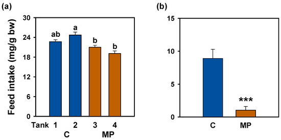
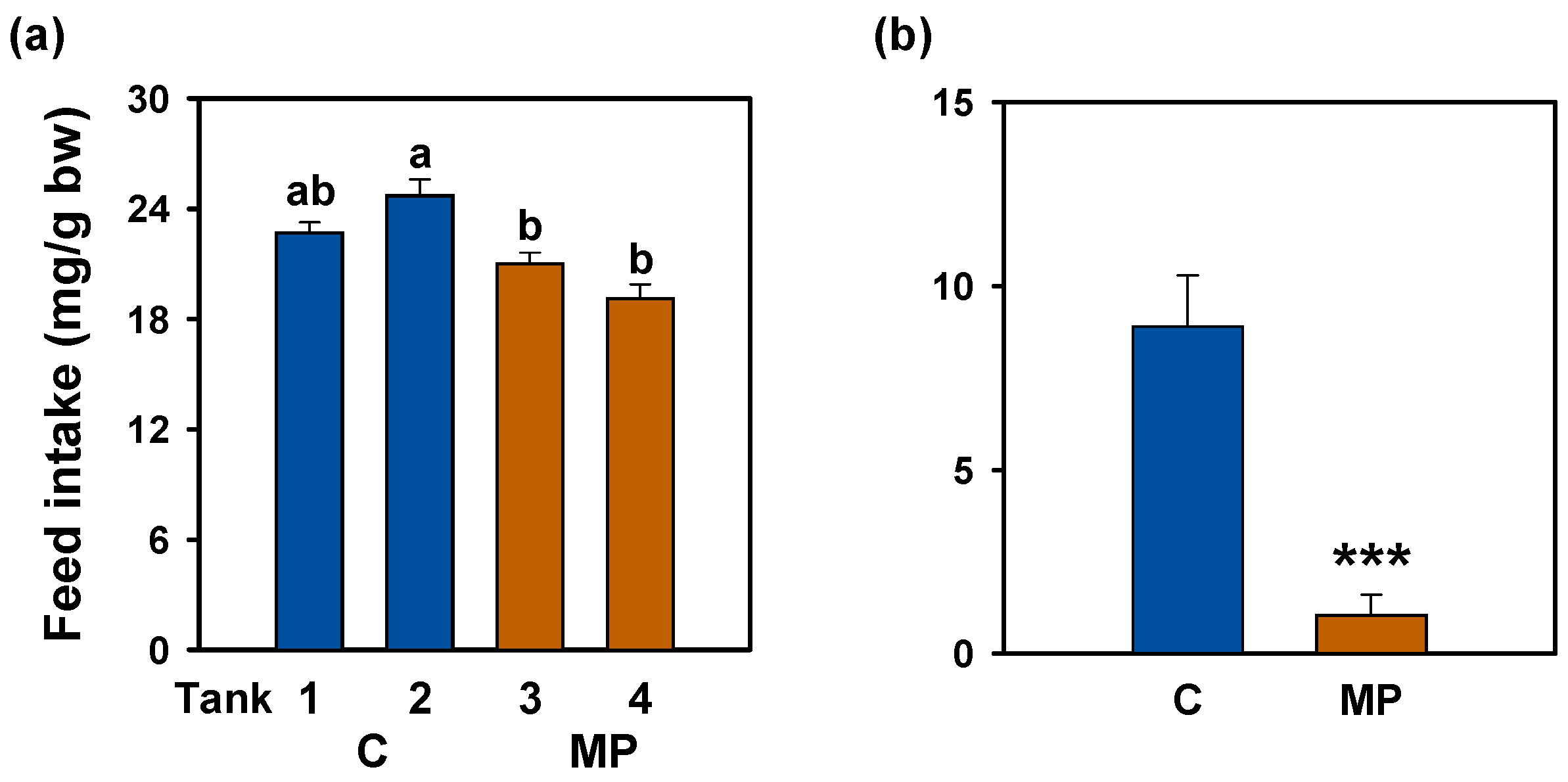
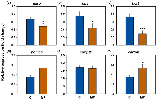
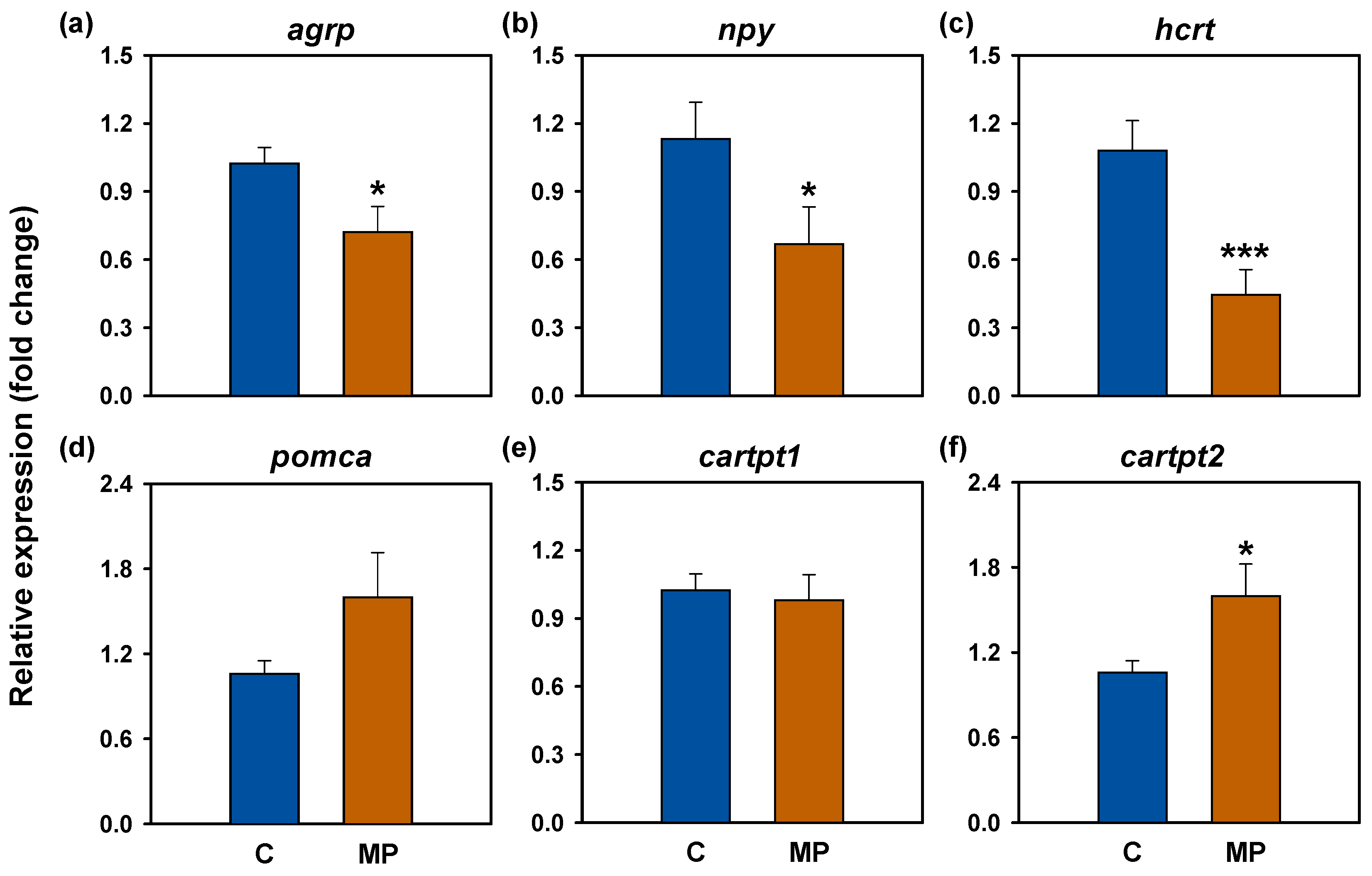
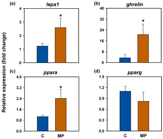
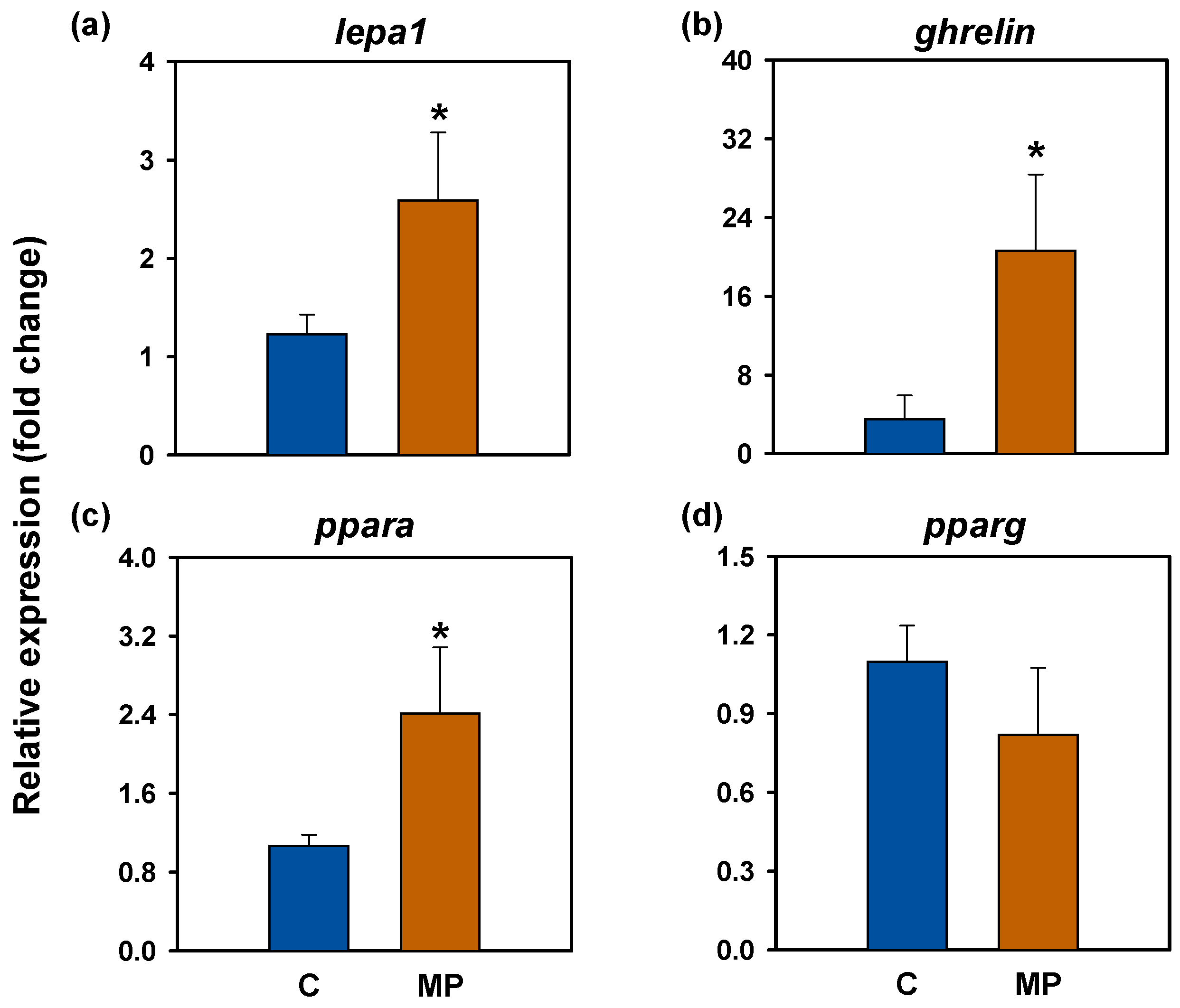
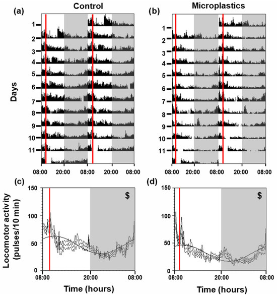
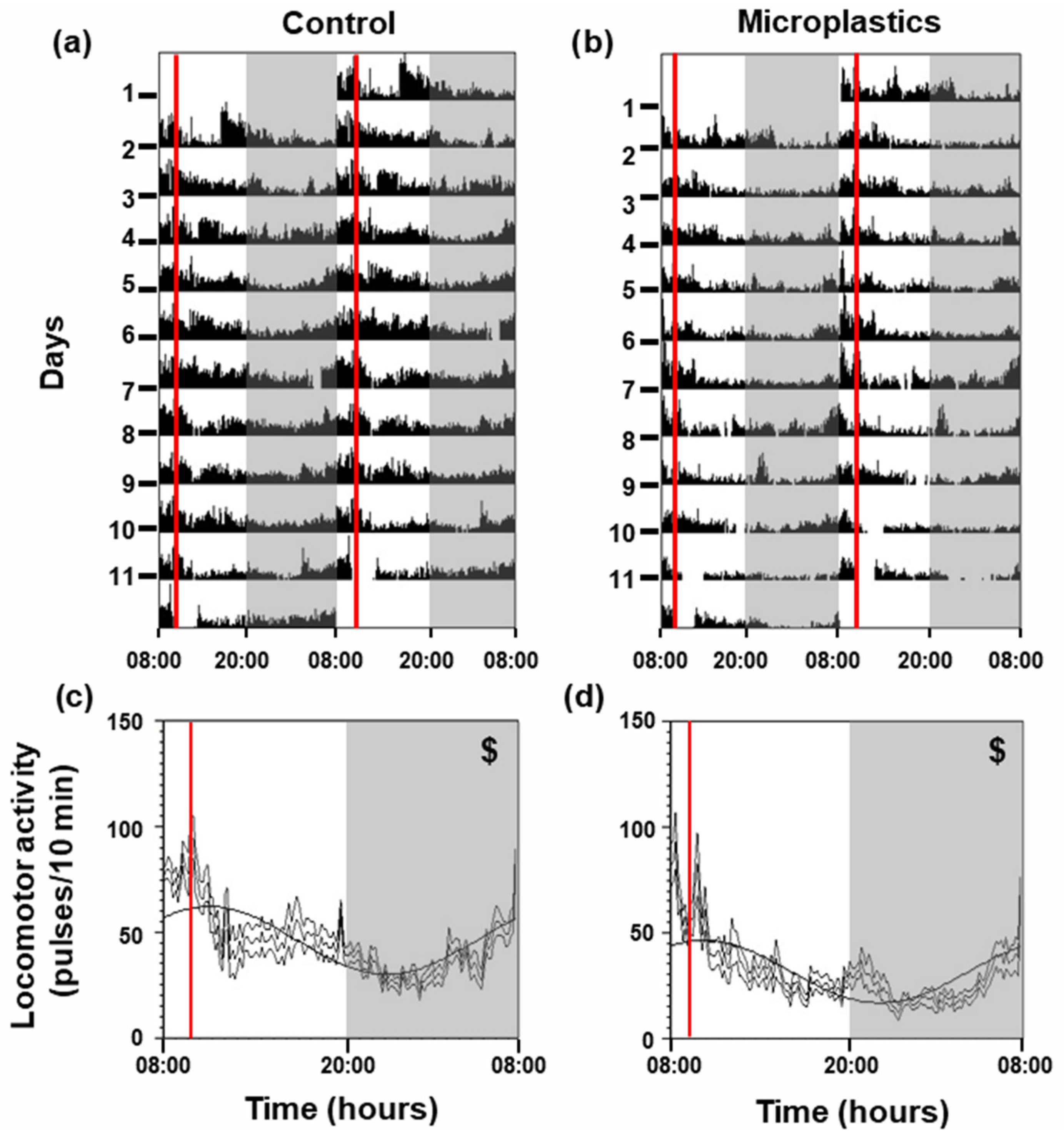
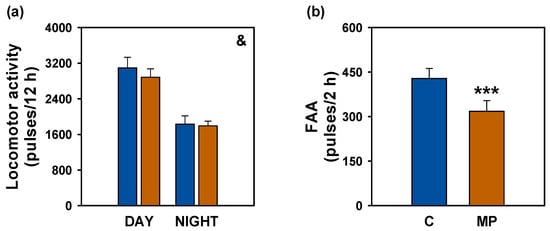
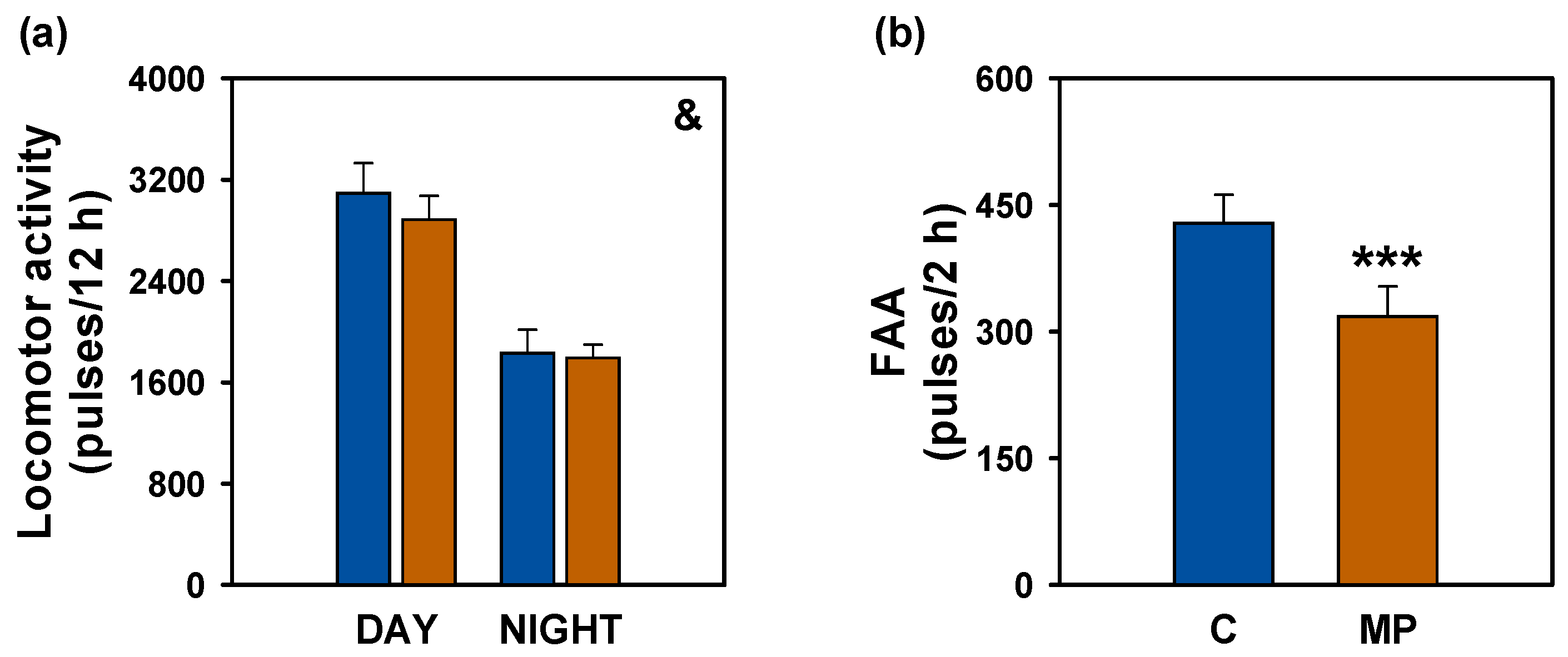
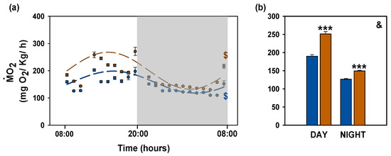
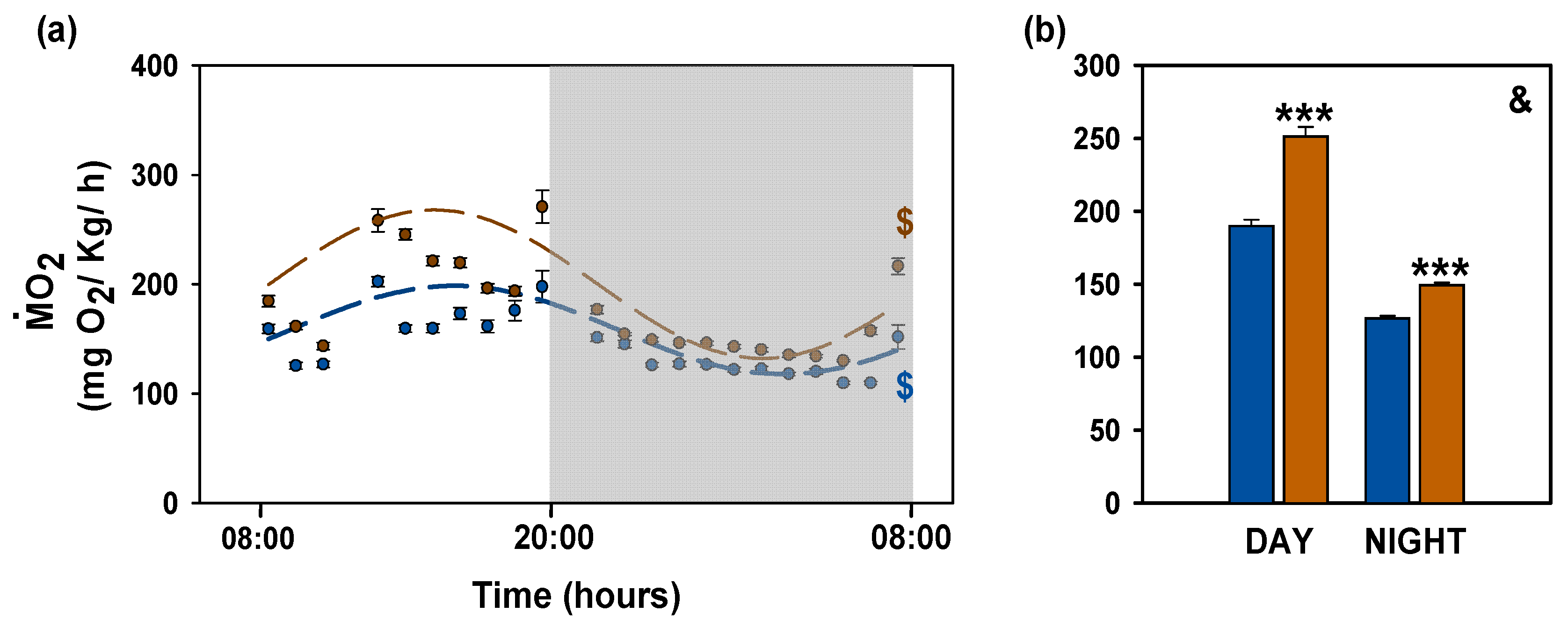
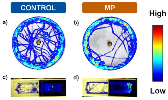
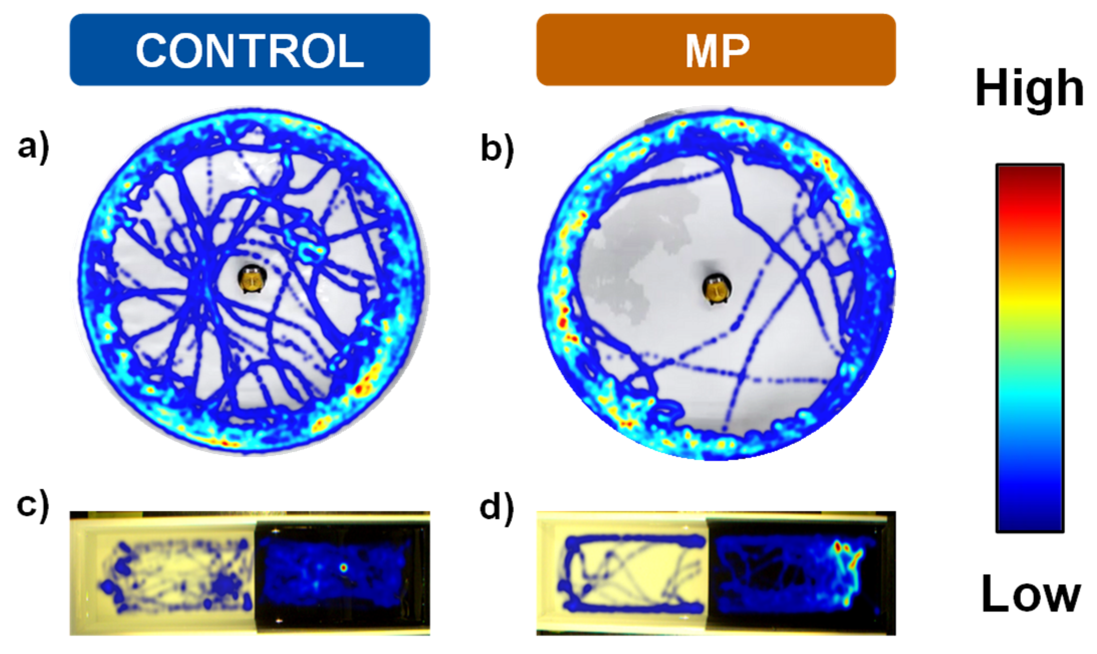
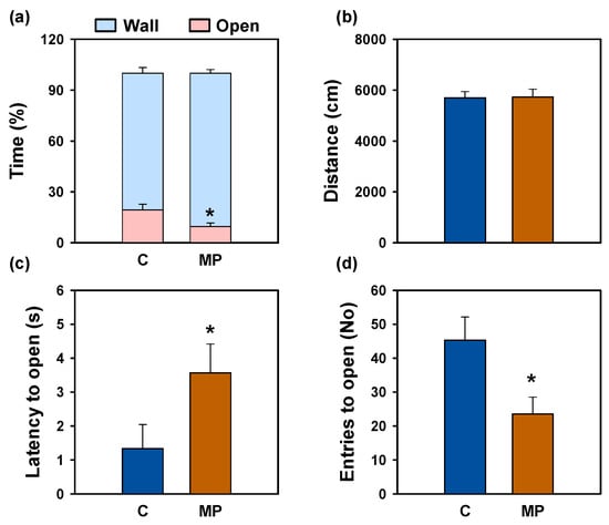
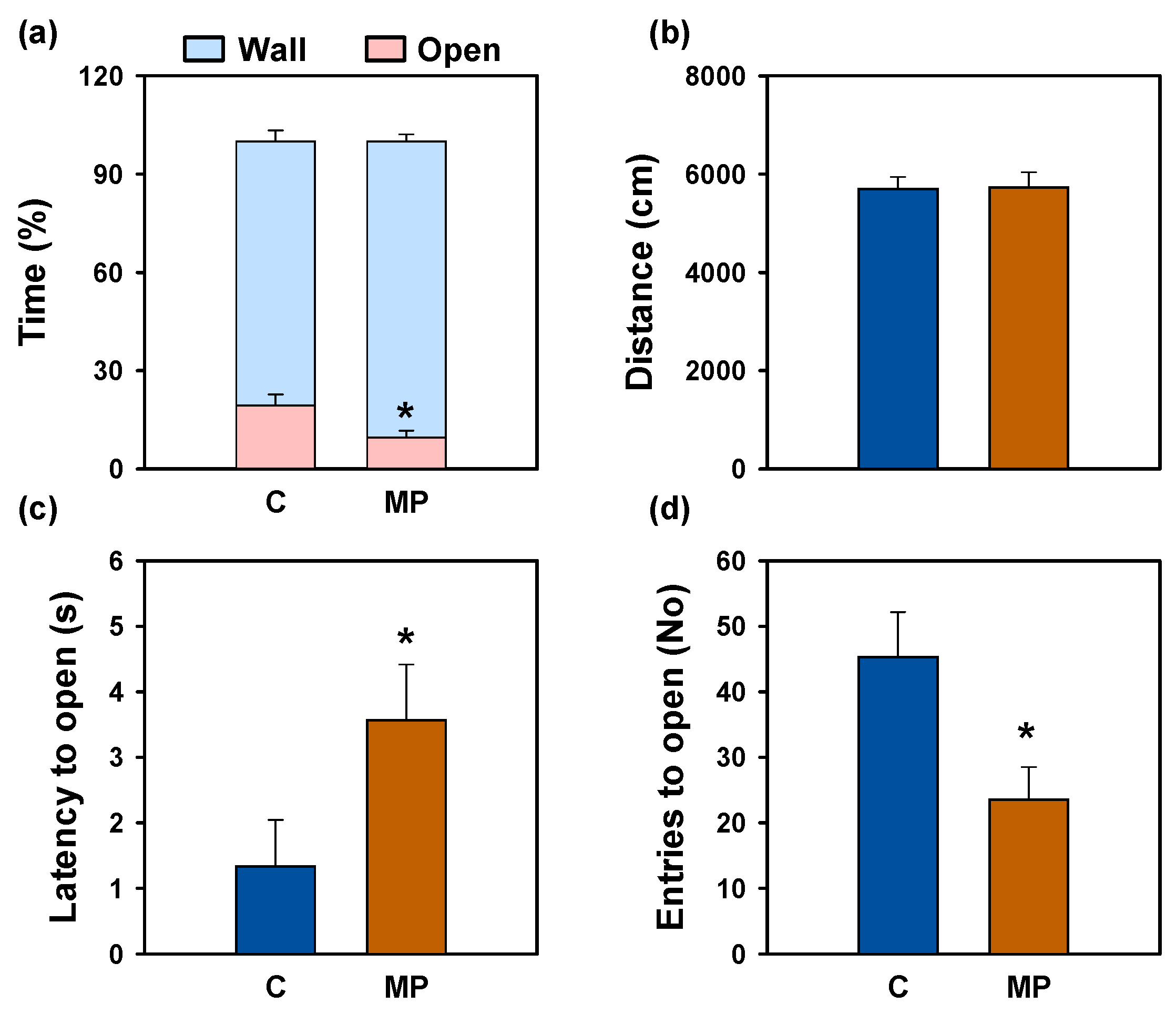
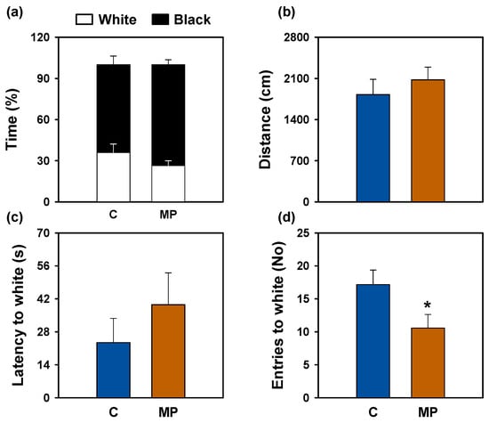
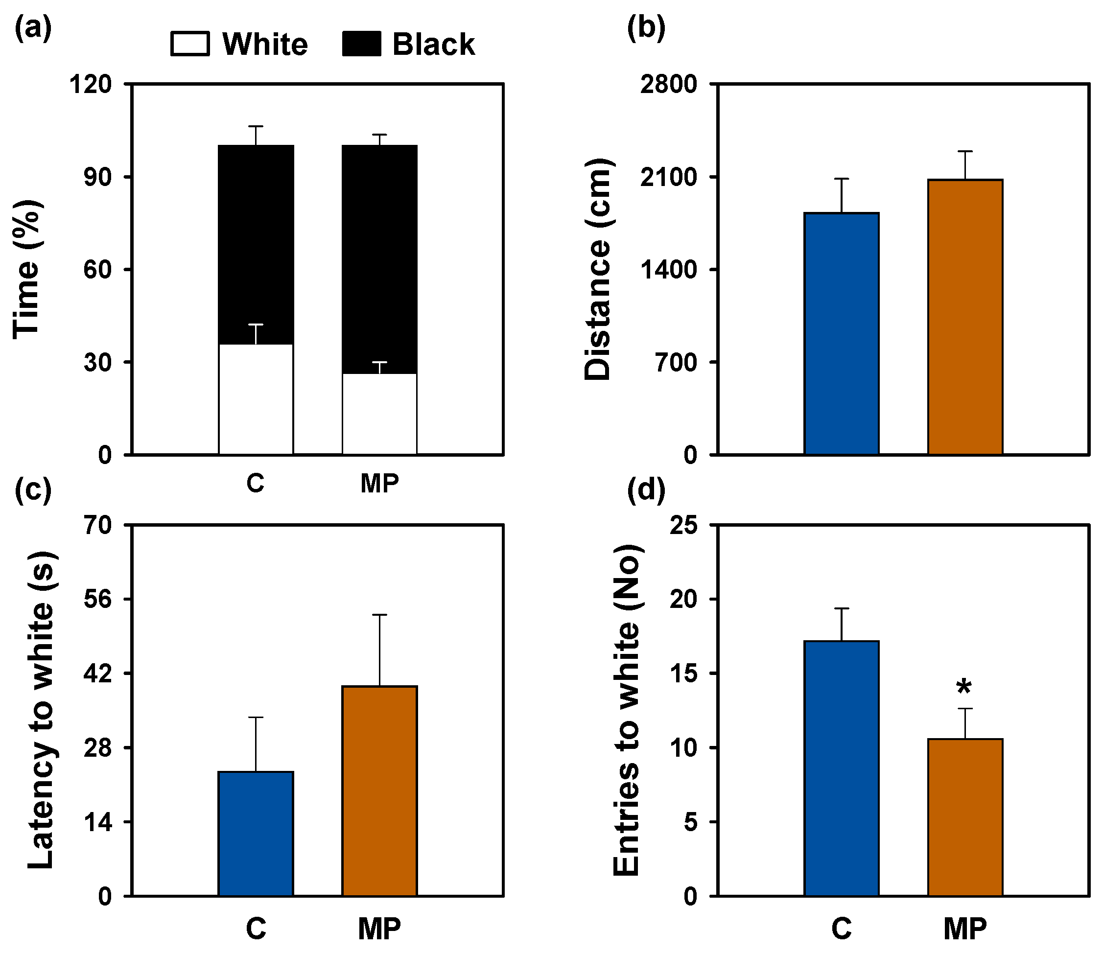
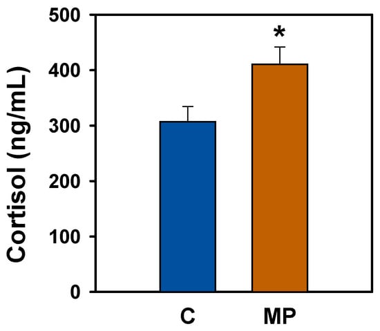
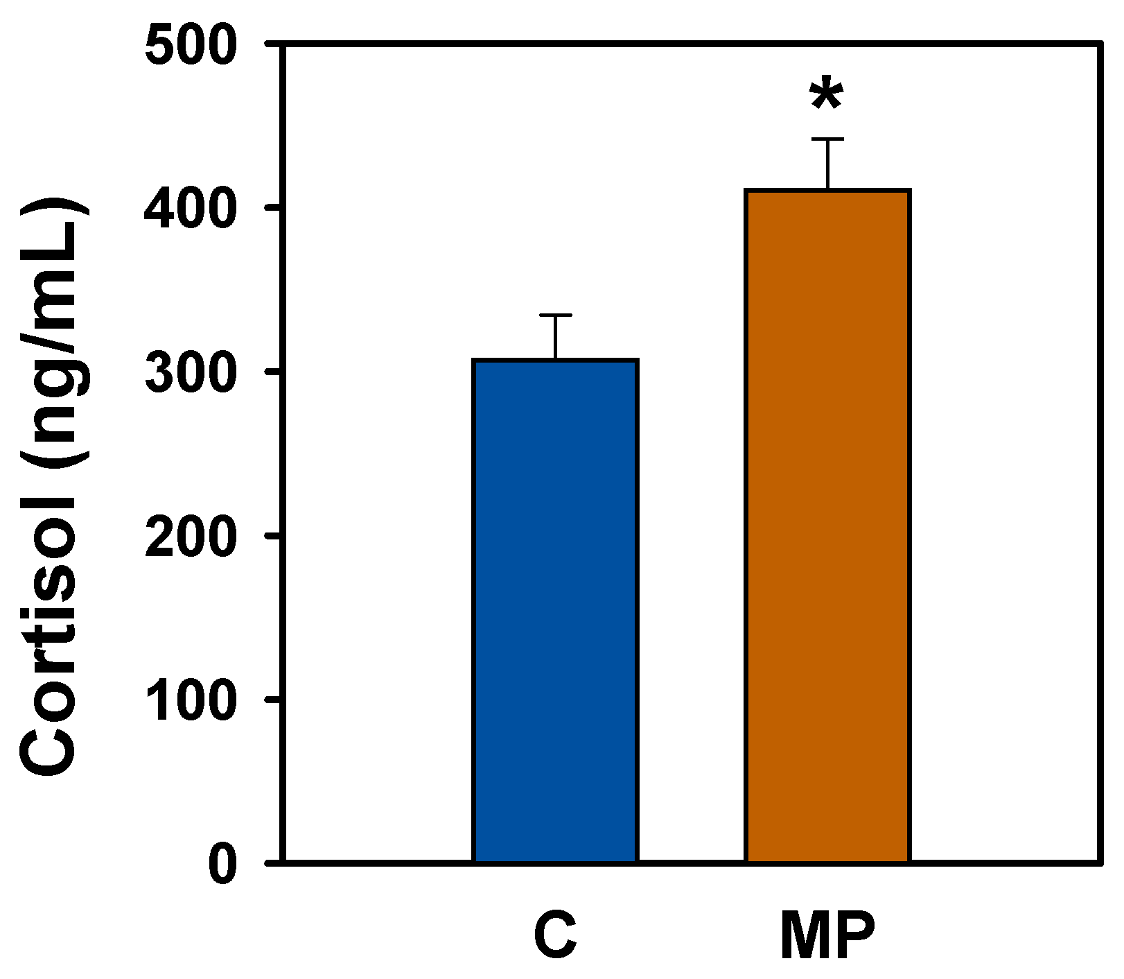
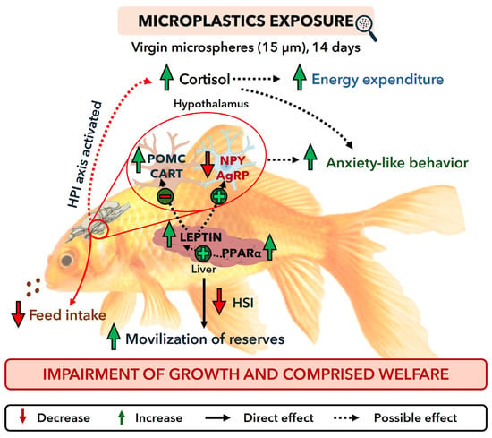
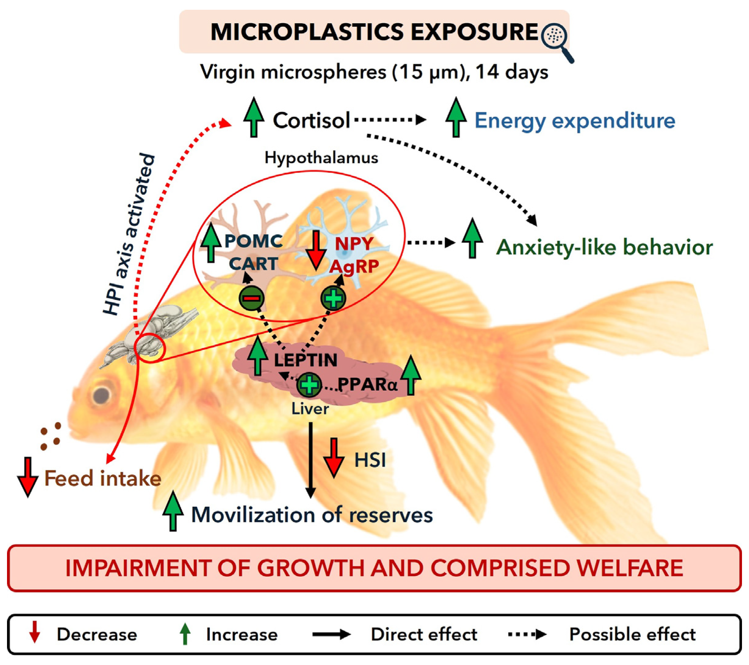

Disclaimer/Publisher’s Note: The statements, opinions and data contained in all publications are solely those of the individual author(s) and contributor(s) and not of MDPI and/or the editor(s). MDPI and/or the editor(s) disclaim responsibility for any injury to people or property resulting from any ideas, methods, instructions or products referred to in the content. |
© 2026 by the authors. Licensee MDPI, Basel, Switzerland. This article is an open access article distributed under the terms and conditions of the Creative Commons Attribution (CC BY) license.