


Simple Summary
As dogs live longer, canine cognitive dysfunction is becoming an increasingly important challenge in veterinary medicine. This progressive brain disorder causes memory loss, disorientation, altered social interactions, disturbed sleep, and behavioral changes, reducing affected dogs’ quality of life and placing an increasing emotional and practical burden on their owners. Despite advances in understanding the disease, treatment options remain limited, highlighting the need for more effective therapeutic approaches. This review provides a comprehensive overview of the role of the cholinergic nervous system in canine cognitive dysfunction and summarizes current evidence on the therapeutic potential of cholinesterase inhibitors, a group of drugs that increases the availability of acetylcholine in the brain and is widely used to treat Alzheimer’s disease in humans. By integrating evidence from both veterinary and human medicine, this review critically synthesizes the available data, identifies important knowledge gaps, and highlights priorities for future research on the use of cholinesterase inhibitors in canine cognitive dysfunction.
Abstract
Canine cognitive dysfunction is a progressive, age-related neurodegenerative disorder that shares several clinical and neuropathological features with human Alzheimer’s disease, although important differences exist between the two conditions. The central mechanisms underlying the pathophysiology of canine cognitive dysfunction include the accumulation of amyloid-β plaques, cytoplasmic deposits of phosphorylated tau protein with rarely observed neurofibrillary tangles, cortical brain atrophy, and disturbances in neurotransmitters, particularly within cholinergic pathways. Compared with those in age-matched, cognitively unimpaired and young cohorts, the number of cholinergic neurons in the basal forebrain in old, cognitively impaired dogs is significantly lower. However, cholinergic deficit does not correlate directly with cortical amyloid-β load. These neurochemical and structural changes contribute to the characteristic cognitive and behavioral deficits of canine cognitive dysfunction. Cholinesterase inhibitors, which increase synaptic acetylcholine levels by inhibiting acetylcholinesterase and/or butyrylcholinesterase, have emerged as a promising pharmacological strategy. This review critically evaluates current evidence from experimental studies investigating the effects of cholinesterase inhibitors on age-associated cognitive performance in laboratory beagle dogs, as well as clinical studies involving client-owned dogs diagnosed with canine cognitive dysfunction; describes their mechanistic basis; and outlines currently available symptomatic pharmacological interventions, such as the monoamine oxidase B inhibitor selegiline (L-deprenyl), and non-pharmacological interventions, including environmental enrichment, regular exercise, diet, and nutritional supplements. Furthermore, it identifies knowledge gaps and proposes future research directions that could support the translation of cholinesterase inhibitors into effective therapeutic interventions for improving cognitive function and quality of life in aging dog populations.
1. Introduction
Canine cognitive dysfunction (CCD), also known as canine cognitive dysfunction syndrome, is a progressive neurodegenerative disorder that predominantly affects geriatric dogs. It shares several clinical and neuropathological features with Alzheimer’s disease (AD) in humans and may therefore serve as a valuable natural animal model for investigating selected aspects of AD-related cognitive decline and neurodegeneration, although it does not fully reproduce the neuropathological characteristics of the disease [1,2]. Recent advances in veterinary diagnostics, therapeutic interventions, nutritional strategies, and improved standards of pet care, driven by the anthropomorphic perception of pets as integral family members, have collectively contributed to a significant increase in the life expectancy of dogs [3,4,5]. Human life expectancy has also increased over the past century. According to the World Health Organization’s “World Health Statistics 2025” report, the average life expectancy worldwide rose from 66.8 years in 2000 to 71.4 years in 2021.
While extending the lifespan of dogs is desirable, it has also led to an increased incidence of age-related diseases, particularly CCD [6]. Consequently, CCD is increasingly recognized in clinical practice as a significant neuropathological and welfare problem in the aging canine population, although it remains significantly underdiagnosed [7,8]. Epidemiological studies have reported CCD prevalence estimates ranging from approximately 14% to 35% in older dogs [8,9]. In a cross-sectional study of 497 community-based dogs aged 8–19.75 years, Salvin et al. (2010) estimated a CCD prevalence of 14.2% using an owner-completed behavioral questionnaire [6]. Furthermore, Neilson et al. (2001) reported age-related behavioral impairments in at least one category in 28% of dogs aged 11–12 years and 68% of dogs aged 15–16 years, based on structured telephone interviews with owners [10]. However, these estimates reflect different assessment criteria and should not be interpreted as directly comparable measures of clinically diagnosed CCD.
Although extensive research has sought to elucidate the underlying pathophysiological mechanisms of CCD, these remain incompletely characterized. Nevertheless, CCD shares several neuropathological features with AD, including the accumulation of amyloid-β (Aβ), cytoplasmic deposits of phosphorylated tau protein, with neurofibrillary tangles (NFTs) rarely observed, cortical brain atrophy, decreased cholinergic activity in the brain, cerebrovascular disease, oxidative stress, and chronic inflammatory processes resulting from abnormal microglial and astrocyte function [2,8,10,11,12,13,14,15,16]. However, important neuropathological differences exist, particularly the rarity of neurofibrillary tangles (NFTs) in dogs and the inconsistent association between Aβ accumulation and the severity of cognitive impairment [2,14,17,18,19]. Several additional pathological processes are also implicated in cognitive impairment, including neuronal mitochondrial dysfunction, glutamate-mediated excitotoxicity, impaired neuronal glucose metabolism, premature neuronal death (apoptosis), and decreased or impaired neurogenesis [8,12].
In addition to pathophysiology, CCD also reflects many of the clinical signs of AD in humans. Specifically, these signs include disorientation (e.g., becoming lost in familiar places, becoming stuck in corners, and staring into space), changes in social interaction (sudden clinginess or avoidance, inability to recognize familiar people or other pets), altered sleep patterns (wandering at night, increased daytime sleep), house-soiling despite previous training, changes in activity levels (reduced interest in play, restlessness, wandering), increased anxiety (new phobias, irritability, aggression), and reduced learning ability (loss of response to familiar commands or difficulty learning new commands) [9]. These clinical signs are summarized under the acronym DISHA (A): Disorientation, altered social Interactions, Sleep–wake cycle alterations, increasing House soiling, and alterations in Activity levels. For the latter A, some indicate increasing Anxiety and, in other cases, increasing Aggressivity [14,20].
The increase in canine life expectancy has contributed to the increasing incidence of CCD, making it a growing concern in veterinary neurology. Owing to its spontaneous age-related onset and similarities to certain clinical, cognitive, and pathological features of AD, CCD has become a valuable natural model for investigating processes associated with age-related cognitive decline and neurodegeneration [1,2,6,9]. This review examines the role of cholinergic dysfunction as an important contributor to cognitive decline in CCD. It focuses on alterations in acetylcholine (ACh) signaling and the roles of acetylcholinesterase (AChE) and butyrylcholinesterase (BChE), key regulators of cholinergic neurotransmission that have emerged as promising therapeutic targets. These mechanisms are discussed within the broader neuropathological framework of CCD, together with current diagnostic challenges and treatment approaches. Special attention should be given to cholinesterase (ChE) inhibition as a potential therapeutic strategy, along with current evidence from experimental and clinical studies of cholinesterase inhibitors (ChEIs) in dogs.
2. Review Methodology
Literature Search and Selection
This narrative review provides a comprehensive synthesis of the current knowledge on CCD, cholinergic neurotransmission, and the therapeutic potential of ChEIs for the treatment of this disease. Relevant publications were identified through searches of PubMed, Web of Science, and Scopus using combinations of the following keywords: canine cognitive dysfunction, dog, beagle, Alzheimer’s disease, clinical study, cholinergic system, cognition, neurotransmission, acetylcholine, cholinesterases, acetylcholinesterase, butyrylcholinesterase, cholinesterase inhibitor, acetylcholinesterase inhibitor, butyrylcholinesterase inhibitor, donepezil, rivastigmine, galantamine, phenserine, physostigmine, edrophonium, neostigmine, pyridostigmine, selegiline, nicergoline and propentofylline. These terms were combined according to the specific topics addressed during manuscript preparation and revision. The review included only publications written in English.
The literature search was conducted in several stages. Initial searches in late 2024 focused on experimental and clinical investigations of ChEIs in dogs as potential treatments for CCD and identified two publications reporting three relevant studies. In early 2025, the search was expanded to include literature on CCD and its underlying pathophysiology, cholinergic neurotransmission, and other relevant topics to provide broader scientific context. Database searches were also supplemented by examination of the reference lists of relevant original research articles and review papers to identify additional publications. Relevant original research articles and review papers were selected based on their relevance to the topics discussed, as assessed through examination of titles, abstracts, and, where appropriate, full texts. Further targeted searches were undertaken throughout manuscript preparation to identify publications relevant to specific aspects of the review. A follow-up search in early 2026 identified an additional publication, “Evaluation of the safety and efficacy of a donepezil depot injection in dogs with canine cognitive dysfunction” Kang et al. [21], which was subsequently included into the review. The main literature search covered publications available up to March 2026.
During manuscript revision, an additional targeted PubMed search was conducted on 7 November 2026 to reassess the available evidence from experimental and clinical investigations of ChEIs in dogs as potential treatments for CCD.
The search was restricted to the Title/Abstract fields and employed the following search string: (“dog”[Title/Abstract] OR “beagle”[Title/Abstract] OR “canine cognitive dysfunction”[Title/Abstract]) AND (“acetylcholinesterase inhibitor”[Title/Abstract] OR “butyrylcholinesterase inhibitor”[Title/Abstract] OR “cholinesterase inhibitor”[Title/Abstract] OR “cholinesterase inhibitors”[Title/Abstract] OR donepezil[Title/Abstract] OR rivastigmine[Title/Abstract] OR galantamine[Title/Abstract] OR phenserine[Title/Abstract]).
This supplementary search retrieved 51 records, including all three original research publications forming the primary evidence base of the review. It was undertaken to provide a documented and reproducible reassessment of the available primary evidence rather than to retrospectively reconstruct the original database searches.
Literature selection was guided by the objectives of this narrative review, with particular emphasis on original experimental and clinical studies investigating the effects of ChEIs on cognitive function in dogs with CCD or age-related cognitive impairment. Studies that did not directly assess these effects were not included in the primary evidence base. Accordingly, three original research publications reporting four studies, comprising two experimental and two clinical investigations, formed the core evidence of the review, with two studies reported in a single publication.
As this review followed a narrative rather than a systematic approach, no predefined systematic search or screening protocol was implemented. Consequently, the exact dates, complete search strings, and numbers of retrieved, duplicate, and excluded records from the original database searches were not systematically documented and cannot be reliably reconstructed retrospectively. Although the supplementary PubMed search provided a documented reassessment of the primary evidence, it could not reproduce the original searches across all three databases. These limitations should be considered when interpreting the findings, particularly regarding the completeness of the literature search and the potential for selection bias.
3. The Cholinergic Nervous System and Acetylcholine Signaling
The cholinergic system is a neurotransmitter system of the central and peripheral nervous systems and is defined by the synthesis, release, and receptor-mediated signaling of ACh. It regulates a wide range of physiological and cognitive functions, including learning and memory, gastrointestinal activity, cardiovascular homeostasis, and motor control. In addition to the nervous system, components of the cholinergic system are present in various non-neuronal cell types, including epithelial cells of the skin, kidney and eye, hepatocytes, placental cells, T lymphocytes, and glial cells of the central nervous system, particularly astrocytes. In these tissues, ACh acts as a local signaling molecule that regulates inflammation, cell proliferation, differentiation, tissue remodeling, and the maintenance of physiological homeostasis [22,23].
ACh is synthesized in cholinergic neurons by the acetylation of choline and catalyzed by the enzyme choline acetyltransferase, which uses acetyl coenzyme A as a substrate [24,25,26]. As previously mentioned, ACh synthesis is not restricted to neurons; various non-neuronal cell types also express choline acetyltransferase and produce ACh [26,27].
In cholinergic neurons, ACh is stored in synaptic vesicles. Two distinct ACh pools have been identified: an easily releasable pool, consisting of membrane-bound synaptic vesicles located near the presynaptic membrane, and a reserve pool, comprising ACh molecules either freely distributed in the cytoplasm or enclosed in vesicles located further from the active zone [28]. Each synaptic vesicle, often referred to as a “quantum”, contains approximately 5000–10,000 molecules of ACh [28,29,30].
Neuronal depolarization opens voltage-gated calcium channels at the presynaptic terminal, allowing calcium influx and increasing intracellular Ca2+ concentrations, which trigger ACh exocytosis by vesicle fusion with the presynaptic membrane at designated active zones. This process is mediated by accessory proteins, including synaptotagmin, synaptobrevin, syntaxin, and SNAP-25 [31,32].
When ACh is released into the synaptic cleft, it diffuses rapidly and binds to postsynaptic receptors. Two major classes of receptors mediate cholinergic signaling: ionotropic nicotinic acetylcholine receptors (nAChRs) and metabotropic muscarinic acetylcholine receptors (mAChRs) [33]. The effect of ACh on these receptors is transient, usually lasting only a few milliseconds, as it is rapidly degraded by AChE. AChE catalyzes the hydrolysis of ACh into acetate and choline. Notably, a single AChE molecule can hydrolyze up to 25,000 ACh molecules per second (Figure 1) [34,35,36].
Figure 1.
Overview of cholinergic neurotransmission and selected cholinesterase inhibitors used in human medicine for the symptomatic treatment of Alzheimer’s disease. Acetylcholine is synthesized from choline and acetyl coenzyme A by choline acetyltransferase in presynaptic neurons and released into the synaptic cleft, where it activates muscarinic (mAChRs) and nicotinic acetylcholine receptors (nAChRs). Acetylcholine is hydrolyzed by cholinesterase enzymes, including acetylcholinesterase (AChE) and butyrylcholinesterase (BChE), producing choline that is recycled into the presynaptic neuron. Cholinesterase inhibitors prevent acetylcholine degradation, thus increasing cholinergic neurotransmission and representing a therapeutic approach aimed at counteracting reduced cholinergic signaling. Created in BioRender. Trobec, T. (2026) https://BioRender.com/k8qw0lj.
4. Role of Acetylcholine in Cognition
In the mammalian brain, there are two primary sources of ACh: projection neurons that innervate distal areas and local interneurons dispersed among their cellular targets [37]. Major populations of cholinergic neurons in the mammalian brain are located in the pedunculo-pontine and laterodorsal tegmental nuclei of the brainstem, a subset of thalamic nuclei, the striatum, and the basal forebrain nuclei. In the striatum, cholinergic neurons primarily function as local interneurons, whereas the basal forebrain nuclei constitute the major source of cholinergic projections to the neocortex, hippocampus, and amygdala [37,38,39,40]. Smaller populations of cholinergic interneurons have been identified in the cortex and hippocampus, although their numbers vary among mammalian species, and the cholinergic identity of hippocampal interneurons remains debated [38,41,42,43].
The survival of animals relies on cognitive abilities such as learning and memory to adapt to environmental changes [44]. ACh is a key neurotransmitter in the central cholinergic system of the mammalian brain and plays a critical role in the modulation of cognitive functions such as attention, learning and memory consolidation [38,45]. In addition to its classical role as a neurotransmitter, ACh acts as a neuromodulator, regulating neuronal excitability and synaptic transmission, inducing synaptic plasticity and coordinating the firing of groups of neurons across widely distributed brain networks [37,46]. The role of ACh and its modulatory ability is particularly important for memory processes. The basal forebrain provides the predominant cholinergic projections that target key memory-related structures directly involved in cognitive processing [38]. These processes are well studied in humans; thus, they will be briefly described in this context. In the basal forebrain, the cell bodies of cholinergic neurons are concentrated in the medial septum and in the diagonal band of Broca, from which the main cholinergic projections extend to the hippocampus. Cholinergic cell bodies are also located in the nucleus basalis of the substantia innominata, which provides cholinergic innervation to the neocortex [46]. Studies have shown that cholinergic signaling in the hippocampus, particularly via α7 nAChRs and M1 mAChRs, is critical for long-term potentiation (LTP) and synaptic plasticity in the hippocampus, suggesting a cellular mechanism through which ACh influences memory processes [38,47,48,49]. These findings indicate that cholinergic signaling most likely modulates CA3 → CA1 synaptic plasticity in the hippocampus via α7 nAChR-triggered calcium signaling and the activation of M1 mAChRs, which inactivate SK channels [38]. The amygdala receives a dense cholinergic projection. In this region, cholinergic signaling supports the encoding and consolidation of emotionally salient memories through mechanisms that most likely involve both mAChRs and nAChRs [38,50,51].
5. Cholinesterase Enzymes
The serine hydrolase superfamily is among the largest and most functionally diverse enzyme families and plays key roles in numerous physiological processes in both prokaryotes and eukaryotes [52]. Among its most important members are ChEs, which include two enzymes: AChE and BChE [36,53]. These enzymes are widely distributed in animals but are not present in plants.
5.1. Acetylcholinesterase
AChE is a specific serine esterase with an ellipsoidal structure (~45 × 60 × 65 Å) that typically forms an amphiphilic tetramer (G4) anchored to the postsynaptic membrane via a glycosylphosphatidylinositol linker, although soluble forms also exist [36,54]. It is abundantly expressed in cholinergic synapses of the central and peripheral nervous systems, including motor endplates, sensory fibers, preganglionic sympathetic and parasympathetic fibers, postganglionic parasympathetic fibers, and erythrocyte membranes [55].
The AChE active site is located at the base of a ~20 Å deep, narrow gorge and comprises three major domains: the peripheral anion site (PAS), the aromatic gorge, and the catalytic anion site (CAS) [56,57,58].
The PAS, located at the entrance of the gorge, binds to ACh via cation–π interactions, increasing the likelihood that it will move to the catalytic site [59,60]. The aromatic gorge is formed by 14 conserved aromatic residues that help direct ACh to the active site through sequential cation–π interactions [61,62].
The CAS consists of multiple subsites:
- The choline binding site, which is responsible for the orientation of the quaternary ammonium group;
- The acyl pocket is much smaller in volume than the choline binding site, which limits the size of substrates entering the active site and is responsible for substrate specificity and for tightly binding the acetyl group;
- The esteratic site comprising the catalytic triad (Ser/His/Glu) responsible for hydrolysis; and
- The oxyanion hole, which stabilizes the tetrahedral intermediate during hydrolysis [63,64,65,66].
Together, these properties enable AChE to catalyze the rapid hydrolysis of ACh into acetate and choline with extremely high catalytic efficiency [22,36,67].
5.2. Butyrylcholinesterase
BChE is a non-specific ChE that degrades both choline and non-choline esters. It is present in blood plasma, liver, pancreas, and neuromuscular junctions and hydrolyses butyrylcholine more rapidly than ACh [36,68]. Its endogenous substrate has not yet been identified, and its physiological role has long remained unclear [69]. Given its higher plasma concentration than AChE, its presence in all vertebrates and its genetic variability, BChE is thought to have evolved from AChE as a general detoxifier while maintaining a backup role in neurotransmission [68,70]. In the brain, BChE is found mainly in glial cells but also in endothelial cells and neurons, usually outside the synaptic cleft [71].
BChE is more highly expressed than AChE in many tissues but is less concentrated in the brain, skeletal muscle, and peripheral nerves. Structurally, the gorge of BChE is larger and less aromatic, with approximately 40% of the 14 aromatic residues replaced by aliphatic or polar residues [72]. Its active pocket comprises the acylation site, choline pocket, acyl pocket, and PAS [69,73,74]. The catalytic triad is conserved in both ChEs. In contrast to Trp84 and Phe330 in AChE, Trp82 in the choline pocket mediates cation–π interactions [60,75,76]. In the acyl pocket, BChE has Leu286 and Val288 instead of the two phenylalanines in AChE, allowing it to accommodate bulkier substrates [69,77,78].
Although structurally different and often debated, the PAS of BChE consists of Asp70 and Tyr332 and binds positively charged substrates before they enter the active site. Compared with AChE, it has a weaker affinity for classical PAS ligands such as propidium and fasciculin [69].
6. Neuropathology and Cholinergic Dysfunction in Canine Cognitive Dysfunction
Clinical manifestations of CCD include disorientation, altered social behavior, disturbed sleep patterns, house-soiling, altered activity, increased anxiety and learning difficulties [79]. The mechanistic links between these clinical features and the underlying neuropathology are not yet fully understood, as is also the case with AD in humans.
Aβ plaques are detectable in the brains of dogs with CCD both before and after the onset of clinical signs. Plaque burden is typically greater in small and medium-sized dogs, likely because of their longer lifespan and the extended period available for Aβ deposition [80]. These deposits appear to originate in the prefrontal cortex and subsequently extend to the parietal, entorhinal, and occipital cortex, correlating with marked behavioral deficits. In different regions of the frontal and temporal cortex, these Aβ plaques are mainly diffuse. In addition to Aβ deposits, dogs with CCD exhibit marked astrocytosis and microgliosis [81,82]. Although Aβ accumulation in the canine prefrontal cortex increases with age, its association with the severity of cognitive impairment is not consistently observed [19]. Furthermore, no significant correlation between the extent of Aβ accumulation and neuronal number in the entorhinal cortex has been reported [18]. Studies have also revealed that soluble Aβ oligomer content in cerebrospinal fluid inversely correlates with cerebral Aβ load and that precipitates from cerebrospinal fluid are highly neurotoxic in vitro, suggesting that oligomers and protofibrils may have the greatest neurotoxic effect on the canine brain [11,82].
In contrast to Aβ plaques, NFTs have been rarely observed in CCD. In two separate studies conducted a decade ago, such inclusions in the brain were documented in only four dogs [80,81]. Cytoplasmic deposits of phosphorylated tau protein have been more frequently detected in neurons of the prefrontal cortex, albeit without NFTs [83]. Increased concentrations of phosphorylated tau protein were also detected in the synaptosomal fractions of dogs with cognitive impairment [81]. Quantification of S396 tau protein by immunohistochemistry, supported by Western blotting, revealed that compared with non-CCD controls, all the regions examined in the six dog brains with diagnosed CCD (except the cerebellum) had significantly greater S396 immunoreactivity. The increased S396 immunoreactivity was distributed in structures of the Papez circuit. Its localization in dendrites and white matter axons of the CCD brain is similar to that seen in tau dysfunction at early stages of AD. However, in CCD, S396 immunoreactivity was also observed in oligodendrocytes, a pattern not commonly observed in AD, suggesting that CCD dogs naturally exhibit a combination of tau dysfunction observed in AD along with other distinct tau pathological mechanisms [17]. Under pathological conditions, tau phosphorylation promotes its aggregation. The modified form can induce synaptic failure, the formation of intracellular deposits, and cognitive impairment [84]. Together, these results suggest that cognitive impairment may also result from synaptic impairment due to phosphorylated tau protein rather than the neurotoxic effects of NFTs, which are rarely observed. This interpretation is supported by observations of age-related tau protein hyperphosphorylation in individual cortical neurons [17,80,81,83,84]. These findings highlight important neuropathological differences between CCD and human AD, particularly the rarity of neurofibrillary tangles in dogs and the inconsistent association between Aβ deposition and cognitive impairment. Therefore, although CCD shares several neuropathological features with AD, it should not be regarded as a direct equivalent of the human disease [17,18,19,80].
In cognitively impaired dogs, neuronal loss and cortical brain atrophy have been documented in several brain regions, including the hippocampus, parts of the limbic system, and the cortex [85,86]. Compared with those in aged, cognitively unimpaired and young individuals, neuronal populations affected by decline in aged, cognitively impaired dogs include cholinergic neurons [87]. Notably, this loss of cholinergic neurons does not correlate with the extent of cortical Aβ deposits [88]. In addition, aged dogs show a decrease in mAChR density [89]. Degeneration and loss of cholinergic neurons are associated with impaired neurotransmission, leading to reduced synaptic function and chronic inflammatory processes caused by abnormal microglial and astrocyte function (Figure 2) [12,13].
Figure 2.
Neuropathological features of canine cognitive dysfunction (CCD). Schematic of the principal neuropathological alterations observed in the aging canine brain affected by CCD. These changes include (A) the accumulation of amyloid-β peptides, oligomers, aggregates, and plaques; (B) neurodegeneration and progressive neuronal loss; (C) cytoplasmic deposits of phosphorylated tau protein and rarely observed neurofibrillary tangles; and (D) chronic neuroinflammation characterized by reactive astrocytes and activated microglia. Created in BioRender. Trobec, T. (2026) https://BioRender.com/6plnr3v.
Experimental evidence from canine models has demonstrated the crucial role of the cholinergic system in regulating cognitive function. Administration of the mAChR antagonist scopolamine causes dose-dependent impairments in short-term working memory, as measured by delayed non-matching to position (DNMP) tasks, with aged dogs showing increased susceptibility to these deficits [89]. In contrast, the enhancement of cholinergic neurotransmission by ChEIs such as phenserine and donepezil improves both memory performance and complex learning in aged beagles and reverses age-related impairments in DNMP tasks and discrimination learning paradigms [87,90]. These findings highlight the pivotal role of ACh in maintaining hippocampal- and cortex-dependent cognitive processes.
The literature also highlights the importance of understanding the pathophysiological processes in aging dogs and dogs with CCD, not only for developing therapeutic approaches for the treatment of CCD but also for recognizing the translational significance of aging dogs. The pathophysiological processes in aging dogs share certain features with those in human aging and AD, as there are neuropathological and behavioral similarities. In this context, the canine model may provide a valuable translational approach for investigating selected aspects of age-related cognitive decline and evaluating potential therapeutic interventions, while recognizing that important neuropathological differences limit its ability to fully reproduce human AD [87].
Despite decades of scientific endeavor and numerous studies, many questions about the etiology, pathogenesis, and mechanistic links between neuropathological changes and the clinical progression of CCD remain unresolved.
7. Diagnosis of Canine Cognitive Dysfunction
As with AD in humans, there is no definitive ante mortem test for CCD, and a reliable protocol for ante mortem diagnosis is still lacking [20]. This common disease in aging dogs is severely underdiagnosed. In most cases, it is overlooked, and its signs are simply attributed to the dog’s age [14]. Diagnosis largely depends on owners, who first observe clinical signs or changes in the dog’s behavior, and veterinarians, who usually make the diagnosis on the basis of signs, history, and clinical features consistent with CCD [8]. The screening and diagnosis of CCD are primarily based on the observation of clinical signs [14]. To detect CCD, veterinarians can use a screening questionnaire that lists possible signs or several useful and accurate behavioral tests (e.g., the DNMP task, the attention task, a food search test (FST), and a problem-solving test (PST)) [8,12,91]. Several questionnaires can be used to determine the stage of cognitive decline in dogs [12,91]. The only imaging method used in practice is magnetic resonance imaging, but this is rarely performed, as owners often decline because of concerns about general anesthesia in older dogs and the associated costs [8,12,91]. In dogs with CCD, magnetic resonance imaging may reveal structural changes associated with cerebral atrophy, including ventricular enlargement and widening of the cerebral sulci. However, these changes are not specific to CCD, as they may also be observed in older dogs without evidence of CCD [8]. Reduced thickness of the interthalamic adhesion has also been reported in dogs with CCD [92,93]. Furthermore, Dewey et al. (2020) demonstrated that dogs with CCD had significantly smaller hippocampal volumes than similarly aged cognitively normal dogs, although substantial overlap between groups limits the diagnostic utility of this finding in individual dogs [94].
8. Treatment and Management of Canine Cognitive Dysfunction
There is currently no curative or preventive therapy for CCD. Management focuses on alleviating clinical signs and improving quality of life (QoL). Therapeutic options remain limited, as most approaches provide only supportive care, with few approved agents, limited robust evidence, and no widely accepted veterinary drug specifically developed for CCD [8,12]. Consequently, clinicians often rely on the off-label use of medications originally developed for AD in humans [21].
Pharmacological agents such as selegiline, propentofylline, and nicergoline may help slow disease progression and transiently support cognitive function. These are typically combined with non-pharmacological interventions, including environmental enrichment, regular exercise, and dietary optimization, as part of a multimodal management strategy.
8.1. Nutritional Management, Physical Activity and Environmental Enrichment
A diet containing antioxidants (e.g., vitamins E and C) and mitochondrial cofactors (lipoic acid and carnitine) can reduce oxidative damage and Aβ pathology in older dogs and improve cognitive function, but it is not sufficient to prevent irreversible neuronal loss in the hippocampus [95,96,97]. Other potentially beneficial nutritional approaches include flavonoid cyanidin-3-O-glucoside, docosahexaenoic acid (DHA), and sphingolipids, as well as supplements containing S-adenosylmethionine tosylate, medium-chain triglycerides, and Brain Protection Blend (a combination of antioxidants, DHA, L-arginine, and B vitamins) [96]. Regular physical activity has been associated with improved memory consolidation and reduced severity and progression of cognitive dysfunction in aging dogs [96,98]. Environmental enrichment, including novel toys and increased social interactions, may also improve cognitive function, either independently or in combination with nutritional interventions [97,98,99,100,101].
8.2. Current Pharmacological Treatments for Canine Cognitive Dysfunction
8.2.1. Selegiline
The use of oral selegiline (L-deprenyl), a Food and Drug Administration (FDA)-approved (1998) irreversible inhibitor of monoamine oxidase B, is indicated for the control of clinical signs associated with canine cognitive dysfunction syndrome [102]. In Europe, selegiline-containing veterinary medicinal products are authorised in certain countries (France, Italy, Ireland, Belgium, the Netherlands, Germany, Hungary, and Greece), although their approved indications may differ from the FDA-approved indication for CCD [103]. Selegiline improves dopaminergic function; decreases the degradation of various neurotransmitters in the brain; may have neuroprotective effects on dopaminergic, noradrenergic, and cholinergic neurons; increases catecholamine levels; and decreases the levels of harmful free radicals in the brain. These actions may improve cognitive function and slow disease progression in most dogs with CCD [8,12,14,104]. Most dogs with CCD show a positive response within the first month of therapy, although there is considerable variation in the degree of response between individuals. Clinical efficacy studies supporting the use of selegiline in CCD are primarily based on owner responses to questionnaires rather than standardized comparative cognitive testing of treated and control dogs. This may raise doubts, as selegiline can cause non-specific low-level hyperactivity by increasing catecholamine levels in the brain (apparent improvement observed by owners) [8]. In a non-comparative open-label study evaluating the effect of selegiline hydrochloride in a clinical setting, in which 641 dogs were treated orally with selegiline hydrochloride at dosages of 0.5 to 1.0 mg/kg once daily for 60 days, the response to treatment was similar at days 30 and 60. On day 60, 77.2% of the dogs showed overall improvement [105].
8.2.2. Nicergoline
Nicergoline is a semisynthetic ergot alkaloid derivative and an α-1A-adrenergic antagonist with vasodilatory properties that has been recommended for dogs showing signs consistent with aging-related behavioral disorders (CCD) and cerebral insufficiency of vascular origin [106,107]. Its therapeutic effects are primarily associated with increased cerebral blood flow [14]. Clinical observations have also shown that nicergoline may alleviate sleep–wake cycle alterations and the loss of learned behaviors in approximately 70% of cases [107].
8.2.3. Propentofylline
Propentofylline is a xanthine derivative that blocks adenosine uptake and inhibits the enzyme phosphodiesterase. Its use in geriatric therapy in dogs is well established, primarily because of its ability to improve hemodynamics in cerebral and peripheral tissues, enhance microcirculation, and increase the oxygen supply to these areas [107,108]. Propentofylline is used to improve dullness, lethargy, alertness, and responsiveness in older dogs. In addition, propentofylline has been reported to improve exercise tolerance and activity levels and reduce sleep disturbances in geriatric dogs [107].
9. Cholinesterase Inhibitors: Mechanism of Action and Veterinary Applications
ChEIs are a group of drugs that inhibit the activity of ChEs and are used for therapeutic or diagnostic purposes in veterinary medicine [36]. ChEIs are commonly classified according to the duration and mechanism of enzyme inhibition as reversible, pseudo-irreversible (slowly reversible), and irreversible inhibitors [109]. Reversible inhibitors are further subdivided into competitive and non-competitive inhibitors [110]. The inhibition of ChEs decreases the rate of ACh degradation and increases the amount of synaptic ACh available for nAChR and mAChR stimulation in the central and peripheral nervous systems [111].
Irreversible ChEIs are now rarely used therapeutically in either veterinary or human medicine. In veterinary medicine, most have been previously used as parasiticides and insecticides for the control of ectoparasites and endoparasites [36,112]. For example, irreversible ChEI coumaphos is currently approved in some European countries as a parasiticide to treat Varroa destructor infestations in honeybees [113]. In contrast, reversible and pseudo-reversible ChEIs are more commonly used for therapeutic and diagnostic purposes in both veterinary medicine and, especially, human medicine. In human medicine, approved, centrally acting reversible ChEIs such as donepezil, rivastigmine, and galantamine are authorised for treatment of AD in the United States and European countries, although their marketing authorisations and specific approved indications may vary by jurisdiction. In the European Union, rivastigmine is authorised through the centralised procedure, whereas donepezil and galantamine are authorised through mutual recognition procedures. Rivastigmine is also authorised for treatment of mild-to-moderate dementia associated with Parkinson’s disease in the United States and mild-to-moderately severe dementia associated with Parkinson’s disease in the European Union [65,114,115,116]. In veterinary clinical practice, the therapeutic use of ChEIs is represented mainly by carbamate-derived inhibitors such as neostigmine, pyridostigmine and physostigmine [112]. Neostigmine is used in veterinary medicine to reverse non-depolarizing neuromuscular blockade after anesthesia and as a prokinetic agent to stimulate gastrointestinal motility in several species under conditions such as ruminal atony or cecal dislocation in cattle and sheep. Pyridostigmine is used for the long-term treatment of myasthenia gravis in dogs and cats, whereas short-acting reversible ChEI edrophonium is mainly used to diagnose myasthenia gravis-like disorders in dogs and cats because it temporarily improves muscle strength [117,118]. Physostigmine can be used in veterinary medicine as an antidote in cases of intoxication with atropine, scopolamine, or belladonna [119]. Neither donepezil nor phenserine is currently authorised as a veterinary medicinal product for the treatment of CCD in the United States or the European Union. Their investigation in dogs has so far been limited primarily to experimental and clinical research studies.
10. Rationale for the Use of Cholinesterase Inhibitors in Treating Cognitive Dysfunction in Dogs
In cognitively impaired dogs, the degeneration of cholinergic neurons and reduced muscarinic receptor density contribute to impaired neurotransmission and synaptic function [12,87,88,89,90]. These changes provide the rationale for using ChEIs to attenuate cognitive decline. Several drugs have received FDA approval for the treatment of AD in the United States, although not all remain commercially available. Currently marketed therapies include the ChEIs donepezil, rivastigmine, galantamine, and benzgalantamine, the N-methyl-D-aspartate receptor antagonist memantine, and the anti-amyloid monoclonal antibodies lecanemab and donanemab. In contrast, tacrine, the first ChEI approved for AD in 1993, and anti-amyloid monoclonal antibody aducanumab, approved in 2021, have subsequently been discontinued and are no longer marketed [120,121,122,123]. However, among these FDA-approved drugs, only donepezil has been clinically tested in dogs. Further studies and clinical trials will be needed to evaluate and possibly authorize donepezil. Studies over the past two decades have provided promising experimental and clinical evidence for the potential cognitive benefits of ChEIs in dogs. Experimental studies investigating phenserine and orally administered donepezil in aged laboratory Beagles have demonstrated effects on cognitive performance, assessed using standardised behavioral tasks [90]. In contrast, studies evaluating a donepezil depot formulation and a novel BChEI in client-owned dogs with naturally occurring CCD have provided preliminary clinical evidence of potential therapeutic benefit [2,21]. Further details of these studies are provided in the next chapter (Experimental and clinical evidence for cholinesterase inhibition in canine cognitive dysfunction). Beyond donepezil, rivastigmine, a dual inhibitor of AChE and BChE, may warrant investigation as a potential treatment for CCD [124]. However, the relative contributions of these enzymes to CCD remain unclear. Pugliese et al. (2007) reported that reduced cerebellar AChE staining optical density correlated with aging but not with cognitive deficits [125]. Galantamine, a reversible AChE inhibitor, has also been proposed to enhance cholinergic neurotransmission through positive allosteric modulation of nAChRs [126]. However, this mechanism remains controversial, as Kowal et al. (2018) did not observe such modulation of human α4β2 or α7 nAChRs [127]. Given the limited veterinary evidence and potential cholinergic adverse effects, further studies are needed to evaluate the efficacy and safety of both drugs in dogs with CCD. Investigating AChE and BChE activities across different stages of CCD could also help clarify the therapeutic relevance of selective versus dual cholinesterase inhibition. Beyond ChEIs, memantine (an N-methyl-D-aspartate receptor antagonist), approved for the symptomatic treatment of AD in humans, was studied for the treatment of canine compulsive disorder and was well tolerated by the dogs [128]. However, memantine has not yet been tested for CCD. A toxicological study on the safety of memantine and its analog memantine nitrate MN-08 has already been conducted in beagle dogs. Memantine at a dose of 9 mg/kg had no adverse effects, whereas the dose of memantine nitrate MN-08 was 24 mg/kg. This novel memantine analog is therefore probably safer and less toxic than memantine [129].
11. Experimental and Clinical Evidence for Cholinesterase Inhibitors in Canine Cognitive Dysfunction
11.1. Phenserine
The effects of orally administered phenserine on age-associated cognitive performance were investigated by Araujo et al. [90] in aged laboratory beagle dogs using a DNMP task to assess working memory and an oddity discrimination task to evaluate complex learning. In the DNMP study, seven geriatric beagle dogs were randomly assigned to receive phenserine (0.5 mg/kg per os (PO); n = 4) or placebo (n = 3). Following a five-day treatment wash-in period, the dogs completed a DNMP relearning task at a 5 s delay until they reached the two-stage performance criterion. Subsequently, working memory was evaluated using a variable-delay DNMP paradigm with 20 and 80 s delays. During the treatment phase, the dogs underwent two practice sessions followed by three stabilization sessions and saline and scopolamine challenge tests in a balanced crossover design. Following discontinuation of treatment and a five-day wash-out period, the same testing protocol was repeated.
In a separate oddity discrimination study, 11 aged Beagle dogs received phenserine or placebo in a crossover design while completing two oddity discrimination tasks of different levels of difficulty. This study was conducted as an exploratory, non-blinded trial.
Phenserine-treated dogs demonstrated higher overall DNMP performance than placebo-treated dogs did. During DNMP relearning at the 5 s delay, compared with placebo-treated dogs, dogs receiving phenserine committed fewer mean errors; however, the difference was not statistically significant. The authors attributed the absence of a significant improvement in DNMP to the limited sample size. In the subsequent variable-delay DNMP paradigm, compared with those given placebo, dogs receiving phenserine showed higher overall performance, although performance decreased with increasing delay in both groups. At the 20 s delay, scopolamine significantly impaired performance in placebo-treated dogs compared with the stabilization and saline conditions, whereas no significant differences were observed in the phenserine group, indicating partial attenuation of the scopolamine-induced deficit. With respect to the 80 s delay, compared with placebo-treated dogs, phenserine-treated dogs consistently performed better under stable and saline conditions, although the differences were not statistically significant. Following the wash-out period, performance declined to placebo levels, suggesting that the memory-enhancing effect was treatment dependent.
In the oddity discrimination study, phenserine had no significant effect on the first, easier discrimination task. However, during the second more challenging task, compared with placebo, phenserine significantly reduced the number of errors required to reach the criterion, indicating that treatment effects were more readily detected under conditions of greater cognitive demand. Overall, the findings suggest that phenserine improved working memory, partially attenuated scopolamine-induced cognitive deficits, and enhanced complex learning in aged dogs (Table 1) [90].
11.2. Donepezil
The effects of orally administered donepezil on age-associated cognitive performance were evaluated by Araujo et al. [90] in a randomized, placebo-controlled experimental study involving 46 aged laboratory beagle dogs. Following five baseline testing sessions, the dogs were assigned to receive placebo (0 mg/kg) or donepezil at doses of 0.75, 1.5, or 6 mg/kg orally once daily. After a five-day wash-in period, cognitive performance was assessed over nine consecutive days using the DNMP task. The dogs were tested at 1, 3, or 5 h after dosing using a quasi-Latin square design. Memory performance was evaluated at 20 and 90 s delay intervals, and cognitive testing was repeated following a five-day wash-out period. Safety and tolerability were assessed through daily clinical observations conducted by personnel who were blinded to treatment allocation, with particular attention given to signs of cholinergic stimulation and treatment-related adverse effects. Performance varied by memory delay (20 and 90 s), donepezil dose (placebo, 0.75, 1.5, or 6 mg/kg), and post-dose assessment time (1, 3, and 5 h). No significant improvements were observed at the 20 s delay in any treatment group. At the 90 s delay, dogs receiving 1.5 mg/kg showed significantly improved working memory performance at both 3 and 5 h after administration compared with baseline, whereas no significant changes were detected in the placebo or 0.75 mg/kg groups. In contrast, the highest dose (6 mg/kg) significantly impaired performance at 3 h post-dose compared with baseline. Following the wash-out period, the cognitive performance in all groups returned to baseline levels, indicating that the observed effects were transient. Treatment was generally well tolerated at the lower doses; however, dogs receiving 6 mg/kg exhibited frequent adverse cholinergic effects, including tremors, vomiting, hypersalivation, and a greater number of non-responses during cognitive testing (Table 1) [90].
Importantly, both phenserine and donepezil were administered orally in studies conducted in aged laboratory beagles using experimental cognitive tasks. Although these findings provide evidence that ChE inhibition can influence age-associated cognitive performance, they do not establish clinical efficacy in dogs with naturally occurring CCD. Therefore, these results should be interpreted as experimental evidence supporting the therapeutic potential of ChEIs rather than direct evidence of their clinical efficacy in CCD.
11.3. Donepezil Depot
The efficacy and safety of long-acting donepezil depot injection at approximate doses of 2 mg/kg (1.973 mg/kg) and 4 mg/kg (3.946 mg/kg) were evaluated in thirty-two client-owned dogs with CCD in a multicenter, randomized, parallel-group clinical trial by Kang et al. [21]. Dogs were assigned to a low-dose group, a high-dose group, or an untreated control group and received a single intramuscular (IM) injection. Efficacy was assessed at baseline (D0), 14 days (D14), and 28 days (D28). Cognitive performance was measured using the canine cognitive dysfunction rating scale (CCDR), canine dementia scale (CADES), and DISHA (A) scale (D0, D14, D28), alongside serum neurofilament light chain (NfL) as a biomarker of axonal injury (D0, D14, D28), and caregiver-reported QoL using questionnaires (D0, D28). Safety was evaluated by serial physical examinations (D0, D14, D28), including body weight, body condition score, rectal temperature, heart rate, respiratory rate, and systolic blood pressure. The animals were monitored for signs of cholinergic overstimulation. Hematological and serum biochemical analyses were performed (D0, D28) to assess hepatic, renal, and metabolic safety. The performance varied by dose, time point, and assessment scale. At baseline, the scores were comparable across groups. The high-dose group showed significant improvements in the CCDR, CADES, and DISHA (A) at both 14 and 28 days, whereas the low-dose group demonstrated more delayed effects, with significant improvements primarily at 28 days and earlier changes limited to CADES. No significant changes were observed in the control group. Serum NfL levels remained stable or slightly decreased in both treatment groups but increased significantly in the control group at 28 days, with both treatment groups showing lower values than the control group did. Quality-of-life measures improved in activity, sociability, and overall condition in treated dogs. The treatment was well tolerated, with no clinically relevant adverse effects identified and only mild, transient events reported (Table 1) [21].
11.4. Novel Butyrylcholinesterase Inhibitor
The efficacy and safety of a novel BChEI for the treatment of canine CCD were evaluated by Zakošek Pipan et al. [2] in a randomized study with an untreated control group. Thirty-one client-owned dogs aged ≥10 years were initially screened, and seventeen small to medium-sized dogs of various breeds with moderate or severe CCD that met the inclusion criteria were included. Ten dogs were assigned to the treatment group, including seven with moderate and three with severe cognitive impairment, and they received BChEI (10 mg/kg PO twice daily) for at least six months. Seven dogs served as untreated controls. Cognitive function was evaluated at baseline and after 3 and 6 months using the CADES, the FST, and the PST. Safety was assessed through serial physical and neurological examinations, behavioral evaluations, complete blood counts, serum biochemistry, and urinalysis, with particular attention given to renal and hepatic function throughout the study.
Treatment efficacy differed according to the severity of cognitive impairment. Treatment was discontinued in all three dogs with severe CCD because of gastrointestinal adverse effects, precluding evaluation of efficacy in this subgroup. In contrast, dogs with moderate CCD showed significant improvement in cognitive function following BChEI administration. CADES scores decreased significantly after 3 months (p < 0.05) and further improved after 6 months (p < 0.001), whereas untreated dogs showed progressive worsening of cognitive impairment, reflected by significantly higher CADES scores after 3 months (p < 0.001). During the study, five of the seven untreated dogs were euthanized because of severe cognitive and physical deterioration between the third and fifth months, whereas the remaining two dogs continued to experience further cognitive decline until the end of the study. At baseline, the performance of the FST and PST did not differ significantly between the groups. Following treatment, compared with the control dogs, the BChEI-treated dogs showed significant improvement in both behavioral tests after 3 months, with additional improvement after 6 months, and the BChEI-treated dogs performed significantly better than the untreated dogs did at both follow-up evaluations (p < 0.001). Conversely, untreated dogs demonstrated progressive deterioration in behavioral performance, with most dogs showing little or no attempt to locate the hidden food or investigate the test apparatus during behavioral testing by the 3-month evaluation. Only two untreated dogs remained in the study at the 6-month evaluation; one maintained the same problem-solving performance observed at 3 months, whereas the other showed further deterioration. Owners of treated dogs also reported marked improvements in memory, learning ability, social interaction, sleep–wake behavior, and overall QoL.
Treatment was well tolerated in dogs with moderate CCD, and no clinically relevant adverse effects were observed during the six-month treatment period. Serial hematological, biochemical, and urinalysis evaluations revealed no evidence of renal or hepatic toxicity in treated dogs with moderate cognitive impairment. The authors concluded that the novel BChEI may represent a promising treatment for dogs with moderate CCD; however, they acknowledged that the absence of a placebo group and the limited sample size warrant confirmation of these findings in larger placebo-controlled clinical trials (Table 1) [2].
From a veterinary perspective, the potential use of ChEIs in geriatric dogs with CCD warrants careful consideration of concurrent medical conditions and concomitant medications, together with monitoring for possible cholinergic adverse effects, including vomiting, diarrhea, hypersalivation, tremors, and bradycardia [2,21,90,112]. Monitoring of body weight, cardiovascular parameters, and renal and hepatic function may also be appropriate, as reflected in published canine clinical studies [2,21]. Cautious dose initiation and gradual dose titration, as recommended in human medicine, may be considered for orally administered ChEIs, while treatment discontinuation may be necessary in cases of clinically significant adverse effects [2,90,130]. For long-acting depot formulations, ongoing drug exposure cannot be readily terminated following administration, emphasizing the importance of careful patient selection and post-administration safety monitoring [21,131]. Although the co-administration of peripherally acting antimuscarinic agents has been proposed as a potential strategy to mitigate gastrointestinal adverse effects associated with ChEI treatment in human medicine, particular attention should be given to agents with limited penetration of the blood–brain barrier to avoid potentially counteracting the central therapeutic effects of ChEIs [132]. However, the safety and effectiveness of this approach in dogs with CCD have not yet been established.
Table 1.
Summary of experimental and clinical evidence evaluating cholinesterase inhibitors for age-related cognitive impairment and canine cognitive dysfunction in dogs. The studies investigating orally administered phenserine and donepezil were conducted in aged laboratory beagles using experimental cognitive tasks, whereas the donepezil depot and novel BChEI studies involved client-owned dogs diagnosed with naturally occurring CCD. BChE, butyrylcholinesterase; BChEI, butyrylcholinesterase inhibitor; CADES, canine dementia scale; CCD, canine cognitive dysfunction; CCDR, canine cognitive dysfunction rating scale; DISHA (A), disorientation, altered social interactions, sleep–wake cycle alterations, house-soiling, altered activity, and anxiety/aggressivity; DNMP, delayed non-matching to position; FST, food search test; IM, intramuscular; NfL, neurofilament light chain; PO, per os; PST, problem-solving test; QoL, quality of life.
12. Discussion and Future Directions
The extended lifespan of dogs has increased the clinical relevance of age-related diseases, including CCD [6]. Reported prevalence estimates of CCD vary across studies depending on the age and characteristics of the populations investigated, as well as the assessment methods and diagnostic criteria used. Estimates generally range from approximately 14% to 35% in older dogs, with the prevalence of CCD increasing exponentially with advancing age [6,10,133]. In this context, earlier estimates suggested that more than 30 million dogs older than seven years lived in the USA and more than 15 million in Europe, highlighting the substantial population potentially at risk of developing CCD [134]. However, updated population estimates and epidemiological studies are needed to better assess the current burden of CCD. Despite its clinical and welfare importance, CCD remains substantially underdiagnosed [7,8]. Its impact extends beyond affected dogs, as caregiver burden increases with disease severity and may be particularly pronounced among owners living alone [9,135]. Given that the available evidence on caregiver burden remains limited, including one study conducted during the COVID-19 pandemic, further research is warranted [135].
ChEIs increase the availability of ACh at cholinergic synapses through inhibition of AChE and/or BChE activity, potentially influencing cognitive processes such as attention, learning, and memory. This provides a mechanistic rationale for the symptomatic treatment of CCD, as cholinergic hypofunction is a well-documented feature of aging canine brains [12,87,88,89]. Experimental studies in aged laboratory beagles showed that orally administered phenserine significantly improved DNMP performance at the longest delay compared with post-treatment wash-out, but not compared with placebo, and significantly improved performance on the more difficult oddity discrimination learning task compared with placebo, whereas orally administered donepezil (1.5 mg/kg) significantly improved DNMP performance at the longest delay compared with baseline [90]. In contrast, the highest investigated dose (6 mg/kg) impaired cognitive performance and was associated with cholinergic adverse effects, highlighting the importance of appropriate dose selection and further dose-finding studies [90]. Although these findings support the potential of ChEIs to enhance age-associated cognitive performance, they do not establish clinical efficacy in dogs with CCD. Clinical studies evaluating a donepezil depot formulation and a novel BChEI in client-owned dogs diagnosed with CCD have reported preliminary findings suggesting potential therapeutic benefits [2,21]. The donepezil depot study reported improvements in behavioral assessment scores (CCDR, CADES, and DISHA (A)) over a four-week period, whereas Zakošek Pipan et al. (2021) reported significant improvements in CADES scores and performance on cognitive tests (FST and PST) in dogs with moderate cognitive impairment during a six-month study [2,21]. Owners of dogs with moderate cognitive impairment treated with the novel BChEI also reported marked improvements in QoL and dog–owner interactions [2].
Despite these encouraging findings, the available clinical evidence remains preliminary and is subject to several important methodological limitations. Both studies involved relatively small sample sizes and included untreated rather than placebo-treated control groups, limiting the ability to distinguish treatment effects from potential expectation-related influences. In the four-week donepezil depot study, owners were aware of whether their dog’s received treatment, although they were blinded to the dose level, while attending veterinarians were aware of treatment allocation [21]. The reliance on owner-reported behavioral assessments may have introduced reporting bias, while the short follow-up period limits conclusions regarding sustained efficacy and long-term safety. In the study investigating the novel BChEI, treatment was discontinued in all three dogs with severe cognitive impairment because of gastrointestinal adverse effects, restricting longer-term treatment observations to dogs with moderate impairment [2]. Although these findings are insufficient to establish disease severity as a predictor of treatment tolerability, they highlight the importance of considering CCD severity when selecting candidates for future clinical trials. Furthermore, five of the seven untreated dogs were euthanized between the third and fifth months because of severe cognitive and physical deterioration, leaving only two dogs in the control group at the six-month assessment [2]. This substantial attrition complicates between-group comparisons and may introduce survival-related bias when interpreting longer-term outcomes [2]. Additionally, the evaluation of multiple cognitive, behavioral, and quality-of-life outcomes may increase the risk of false-positive findings. Although statistical adjustments were applied for post hoc comparisons in the donepezil depot study, these do not necessarily account for multiple testing across all assessed outcomes [21]. Collectively, these methodological limitations indicate that the reported improvements should be interpreted as preliminary signals of potential efficacy rather than definitive evidence of clinical benefit.
Future clinical trials investigating the efficacy and safety of ChEIs in dogs with CCD should employ rigorous methodological approaches to ensure reliable and clinically meaningful results. Although experimental studies in aged laboratory beagles may provide valuable preliminary information regarding cognitive effects, dose selection, and safety, direct multicenter clinical trials involving well-characterized client-owned dogs with CCD may also be scientifically justified. Such trials should include dogs of different breeds, body sizes, and stages of cognitive impairment to assess potential differences in treatment response. Eligibility criteria should account for disease severity, concurrent medical conditions, concomitant medications, and other potentially reversible causes of cognitive or behavioral changes to improve diagnostic accuracy and facilitate interpretation of treatment outcomes. Prospective sample-size calculations, trial preregistration, appropriate randomization procedures with allocation concealment, double blinding, and placebo-controlled designs should be incorporated whenever feasible and ethically appropriate. Standardized diagnostic criteria and cognitive and behavioral assessment protocols should be applied, with clearly prespecified primary outcomes and appropriate adjustments for multiple comparisons. Dose selection should be informed by dose-ranging studies and available pharmacokinetic, pharmacodynamic, and safety data in dogs. Safety monitoring should include systematic adverse-event assessment, particularly of gastrointestinal, cardiovascular, and other potential cholinergic adverse effects. Potential drug–drug interactions should also be carefully evaluated, particularly in geriatric dogs receiving multiple medications for concurrent age-related conditions. Clearly defined criteria for dose adjustment, treatment discontinuation, and withdrawal from clinical trials should also be established to ensure animal welfare and facilitate consistent safety evaluation. Clinically meaningful follow-up periods are essential to evaluate the durability of treatment effects and long-term tolerability. However, conducting such trials in geriatric dogs with progressive neurodegenerative conditions presents substantial challenges, including concurrent age-related diseases, potential loss to follow-up, difficulties in recruiting sufficiently large study populations, and considerable financial and logistical demands. Addressing these challenges through appropriate study design would substantially strengthen the evidence base and support more informed clinical decision-making regarding the potential use of ChEIs in dogs with CCD.
Furthermore, the repurposing of ChEIs approved for human use may represent a potential approach for future veterinary drug development. Human ChEIs, including donepezil, rivastigmine, and galantamine, are authorised for the symptomatic treatment of AD in humans in the United States and European countries and have established pharmacokinetic and safety profiles in human populations. However, their authorisation for human use does not imply veterinary authorisation for the treatment of CCD. This existing knowledge may facilitate veterinary translation by informing early-stage development and enabling more targeted dose-finding studies in dogs. However, species-specific differences in pharmacokinetics, pharmacodynamics, metabolism, and tolerability must be carefully evaluated before findings from human medicine can be translated into veterinary clinical practice.
13. Conclusions
Overall, ChEIs represent a mechanistically plausible potential therapeutic approach for CCD, supported by experimental findings in aged laboratory beagles and preliminary clinical observations in client-owned dogs. However, the currently available evidence remains insufficient to establish their clinical efficacy, optimal dosing regimens, long-term safety, or favorable risk–benefit profiles. Further well-designed clinical trials are therefore essential before their routine clinical use in dogs with naturally occurring CCD can be recommended.
Funding
This research was funded by the Slovenian Research and Innovation Agency, grant number P4-0053.
Institutional Review Board Statement
Not applicable.
Informed Consent Statement
Not applicable.
Data Availability Statement
The original contributions presented in this study are included in the article. Further inquiries can be directed to the corresponding author.
Acknowledgments
I would like to express my deepest gratitude to Robert Frangež for his invaluable guidance, continuous support, and encouragement throughout my academic and research career, as well as during the preparation of this review. I am also sincerely grateful to Monika C. Žužek for her valuable support and constructive discussions. I would like to thank all my colleagues at the Institute of Preclinical Sciences, Veterinary Faculty, University of Ljubljana, for their collegial support and stimulating discussions.
Conflicts of Interest
The author declares no conflicts of interest.
Abbreviations
The following abbreviations are used in this manuscript:
| Aβ | Amyloid-β |
| ACh | Acetylcholine |
| AChE | Acetylcholinesterase |
| AD | Alzheimer’s disease |
| BChE | Butyrylcholinesterase |
| BChEI | Butyrylcholinesterase inhibitor |
| CADES | Canine dementia scale |
| CAS | Catalytic anionic site |
| CCD | Canine cognitive dysfunction |
| CCDR | Canine cognitive dysfunction rating scale |
| ChE | Cholinesterase |
| ChEI | Cholinesterase inhibitor |
| DHA | Docosahexaenoic acid |
| DISHA (A) | Disorientation, altered social interactions, sleep–wake cycle alterations, house soiling, altered activity, and anxiety/aggressivity |
| DNMP | Delayed non-matching-to-position |
| FDA | Food and Drug Administration |
| FST | Food search test |
| IM | Intramuscular |
| LTP | Long-term potentiation |
| MCT | Medium-chain triglycerides |
| mAChR | Muscarinic acetylcholine receptor |
| nAChR | Nicotinic acetylcholine receptor |
| NFT | Neurofibrillary tangle |
| NfL | Neurofilament light chain |
| PAS | Peripheral anionic site |
| PO | per os |
| PST | Problem-solving test |
| QoL | Quality of life |
References
- Bray, E.E.; Raichlen, D.A.; Forsyth, K.K.; Promislow, D.E.L.; Alexander, G.E.; MacLean, E.L.; Dog Aging Project, C. Associations between physical activity and cognitive dysfunction in older companion dogs: Results from the Dog Aging Project. Geroscience 2023, 45, 645–661. [Google Scholar] [CrossRef] [Scilit] [PubMed]
- Zakosek Pipan, M.; Prpar Mihevc, S.; Strbenc, M.; Kosak, U.; German Ilic, I.; Trontelj, J.; Zakelj, S.; Gobec, S.; Pavlin, D.; Majdic, G. Treatment of canine cognitive dysfunction with novel butyrylcholinesterase inhibitor. Sci. Rep. 2021, 11, 18098. [Google Scholar] [CrossRef] [Scilit] [PubMed]
- Day, M.J. One Health: The small animal dimension. Vet. Rec. 2010, 167, 847–849. [Google Scholar] [CrossRef] [Scilit] [PubMed]
- Irvine, L.; Cilia, L. More-than-human families: Pets, people, and practices in multispecies households. Sociol. Compass 2017, 11, e12455. [Google Scholar] [CrossRef] [Scilit]
- Montoya, M.; Morrison, J.A.; Arrignon, F.; Spofford, N.; Charles, H.; Hours, M.A.; Biourge, V. Life expectancy tables for dogs and cats derived from clinical data. Front. Vet. Sci. 2023, 10, 1082102. [Google Scholar] [CrossRef] [Scilit] [PubMed]
- Salvin, H.E.; McGreevy, P.D.; Sachdev, P.S.; Valenzuela, M.J. Under diagnosis of canine cognitive dysfunction: A cross-sectional survey of older companion dogs. Vet. J. 2010, 184, 277–281. [Google Scholar] [CrossRef] [Scilit] [PubMed]
- Brisset, A.; Taylor, T.L.; Blanchard, T.; Fernandez, E.J.; Hazel, S.J. Attitudes of Australian Veterinary Professionals to Diagnosing and Managing Canine Cognitive Dysfunction. Vet. Sci. 2025, 12, 272. [Google Scholar] [CrossRef] [Scilit] [PubMed]
- Dewey, C.W.; Davies, E.S.; Xie, H.; Wakshlag, J.J. Canine Cognitive Dysfunction: Pathophysiology, Diagnosis, and Treatment. Vet. Clin. N. Am. Small Anim. Pract. 2019, 49, 477–499. [Google Scholar] [CrossRef] [Scilit] [PubMed]
- Taylor, T.L.; Smith, B.P.; Hazel, S.J. Guardians’ perceptions of caring for a dog with canine cognitive dysfunction. Vet. Rec. 2024, 194, e3266. [Google Scholar] [CrossRef] [Scilit] [PubMed]
- Neilson, J.C.; Hart, B.L.; Cliff, K.D.; Ruehl, W.W. Prevalence of behavioral changes associated with age-related cognitive impairment in dogs. J. Am. Vet. Med. Assoc. 2001, 218, 1787–1791. [Google Scholar] [CrossRef] [Scilit] [PubMed]
- Head, E.; Pop, V.; Sarsoza, F.; Kayed, R.; Beckett, T.L.; Studzinski, C.M.; Tomic, J.L.; Glabe, C.G.; Murphy, M.P. Amyloid-beta peptide and oligomers in the brain and cerebrospinal fluid of aged canines. J. Alzheimer’s Dis. 2010, 20, 637–646. [Google Scholar] [CrossRef] [Scilit] [PubMed]
- Landsberg, G.M.; Nichol, J.; Araujo, J.A. Cognitive dysfunction syndrome: A disease of canine and feline brain aging. Vet. Clin. N. Am. Small Anim. Pract. 2012, 42, 749–768, vii. [Google Scholar] [CrossRef] [Scilit] [PubMed]
- Ozawa, M.; Chambers, J.K.; Uchida, K.; Nakayama, H. The Relation between canine cognitive dysfunction and age-related brain lesions. J. Vet. Med. Sci. 2016, 78, 997–1006. [Google Scholar] [CrossRef] [Scilit] [PubMed]
- Prpar Mihevc, S.; Majdic, G. Canine Cognitive Dysfunction and Alzheimer’s Disease—Two Facets of the Same Disease? Front. Neurosci. 2019, 13, 604. [Google Scholar] [CrossRef] [Scilit] [PubMed]
- Rofina, J.E.; van Ederen, A.M.; Toussaint, M.J.; Secreve, M.; van der Spek, A.; van der Meer, I.; Van Eerdenburg, F.J.; Gruys, E. Cognitive disturbances in old dogs suffering from the canine counterpart of Alzheimer’s disease. Brain Res. 2006, 1069, 216–226. [Google Scholar] [CrossRef] [Scilit] [PubMed]
- Urfer, S.R.; Darvas, M.; Czeibert, K.; Sandor, S.; Promislow, D.E.L.; Creevy, K.E.; Kubinyi, E.; Kaeberlein, M. Canine Cognitive Dysfunction (CCD) scores correlate with amyloid beta 42 levels in dog brain tissue. Geroscience 2021, 43, 2379–2386. [Google Scholar] [CrossRef] [Scilit] [PubMed]
- Abey, A.; Davies, D.; Goldsbury, C.; Buckland, M.; Valenzuela, M.; Duncan, T. Distribution of tau hyperphosphorylation in canine dementia resembles early Alzheimer’s disease and other tauopathies. Brain Pathol. 2021, 31, 144–162. [Google Scholar] [CrossRef] [Scilit] [PubMed]
- Cummings, B.J.; Su, J.H.; Cotman, C.W.; White, R.; Russell, M.J. Beta-amyloid accumulation in aged canine brain: A model of early plaque formation in Alzheimer’s disease. Neurobiol. Aging 1993, 14, 547–560. [Google Scholar] [CrossRef] [Scilit] [PubMed]
- Schutt, T.; Helboe, L.; Pedersen, L.O.; Waldemar, G.; Berendt, M.; Pedersen, J.T. Dogs with Cognitive Dysfunction as a Spontaneous Model for Early Alzheimer’s Disease: A Translational Study of Neuropathological and Inflammatory Markers. J. Alzheimer’s Dis. 2016, 52, 433–449. [Google Scholar] [CrossRef] [Scilit] [PubMed]
- Vitturini, C.; Cerquetella, M.; Spaterna, A.; Bazzano, M.; Marchegiani, A. Diagnosis of Canine Cognitive Dysfunction Syndrome: A Narrative Review. Vet. Sci. 2025, 12, 781. [Google Scholar] [CrossRef] [Scilit] [PubMed]
- Kang, M.H.; Kang, M.A.; Jeon, H.J.; Shin, H.C.; Moon, H.; Lee, D.G.; Park, H.M. Evaluation of the safety and efficacy of a donepezil depot injection in dogs with canine cognitive dysfunction. Front. Vet. Sci. 2025, 12, 1724060. [Google Scholar] [CrossRef] [Scilit] [PubMed]
- Moodie, L.W.K.; Sepcic, K.; Turk, T.; Frange, Z.R.; Svenson, J. Natural cholinesterase inhibitors from marine organisms. Nat. Prod. Rep. 2019, 36, 1053–1092. [Google Scholar] [CrossRef] [Scilit] [PubMed]
- Oda, Y.; Nakanishi, I. The distribution of cholinergic neurons in the human central nervous system. Histol. Histopathol. 2000, 15, 825–834. [Google Scholar] [CrossRef] [Scilit] [PubMed]
- Ferreira-Vieira, T.H.; Guimaraes, I.M.; Silva, F.R.; Ribeiro, F.M. Alzheimer’s disease: Targeting the Cholinergic System. Curr. Neuropharmacol. 2016, 14, 101–115. [Google Scholar] [CrossRef] [Scilit] [PubMed]
- Fujii, T.; Masai, M.; Misawa, H.; Okuda, T.; Takada-Takatori, Y.; Moriwaki, Y.; Haga, T.; Kawashima, K. Acetylcholine synthesis and release in NIH3T3 cells coexpressing the high-affinity choline transporter and choline acetyltransferase. J. Neurosci. Res. 2009, 87, 3024–3032. [Google Scholar] [CrossRef] [Scilit] [PubMed]
- Maurer, S.V.; Williams, C.L. The Cholinergic System Modulates Memory and Hippocampal Plasticity via Its Interactions with Non-Neuronal Cells. Front. Immunol. 2017, 8, 1489. [Google Scholar] [CrossRef] [Scilit] [PubMed]
- Wessler, I.; Kirkpatrick, C.J. Acetylcholine beyond neurons: The non-neuronal cholinergic system in humans. Br. J. Pharmacol. 2008, 154, 1558–1571. [Google Scholar] [CrossRef] [Scilit] [PubMed]
- Ali, H.H.; Savarese, J.J. Monitoring of neuromuscular function. Anesthesiology 1976, 45, 216–249. [Google Scholar] [CrossRef] [Scilit] [PubMed]
- Koneczny, I.; Herbst, R. Myasthenia Gravis: Pathogenic Effects of Autoantibodies on Neuromuscular Architecture. Cells 2019, 8, 671. [Google Scholar] [CrossRef] [Scilit] [PubMed]
- Ghai, B.; Makkar, J.K.; Wig, J. Neuromuscular monitoring: A review. J. Anaesth. Clin. Pharmacol. 2006, 22, 347–356. [Google Scholar] [CrossRef] [Scilit]
- Slater, C.R. The Structure of Human Neuromuscular Junctions: Some Unanswered Molecular Questions. Int. J. Mol. Sci. 2017, 18, 2183. [Google Scholar] [CrossRef] [Scilit] [PubMed]
- Sudhof, T.C.; Rizo, J. Synaptic vesicle exocytosis. Cold Spring Harb. Perspect. Biol. 2011, 3, a005637. [Google Scholar] [CrossRef] [Scilit] [PubMed]
- Wess, J.; Eglen, R.M.; Gautam, D. Muscarinic acetylcholine receptors: Mutant mice provide new insights for drug development. Nat. Rev. Drug Discov. 2007, 6, 721–733. [Google Scholar] [CrossRef] [Scilit] [PubMed]
- Mor, I.; Sklan, E.H.; Podoly, E.; Pick, M.; Kirschner, M.; Yogev, L.; Bar-Sheshet Itach, S.; Schreiber, L.; Geyer, B.; Mor, T.; et al. Acetylcholinesterase-R increases germ cell apoptosis but enhances sperm motility. J. Cell Mol. Med. 2008, 12, 479–495. [Google Scholar] [CrossRef] [Scilit] [PubMed]
- Silman, I.; Sussman, J.L. Acetylcholinesterase: ‘classical’ and ‘non-classical’ functions and pharmacology. Curr. Opin. Pharmacol. 2005, 5, 293–302. [Google Scholar] [CrossRef] [Scilit] [PubMed]
- Colovic, M.B.; Krstic, D.Z.; Lazarevic-Pasti, T.D.; Bondzic, A.M.; Vasic, V.M. Acetylcholinesterase inhibitors: Pharmacology and toxicology. Curr. Neuropharmacol. 2013, 11, 315–335. [Google Scholar] [CrossRef] [Scilit] [PubMed]
- Picciotto, M.R.; Higley, M.J.; Mineur, Y.S. Acetylcholine as a neuromodulator: Cholinergic signaling shapes nervous system function and behavior. Neuron 2012, 76, 116–129. [Google Scholar] [CrossRef] [Scilit] [PubMed]
- Ballinger, E.C.; Ananth, M.; Talmage, D.A.; Role, L.W. Basal Forebrain Cholinergic Circuits and Signaling in Cognition and Cognitive Decline. Neuron 2016, 91, 1199–1218. [Google Scholar] [CrossRef] [Scilit] [PubMed]
- Mesulam, M.M.; Mufson, E.J.; Levey, A.I.; Wainer, B.H. Cholinergic innervation of cortex by the basal forebrain: Cytochemistry and cortical connections of the septal area, diagonal band nuclei, nucleus basalis (substantia innominata), and hypothalamus in the rhesus monkey. J. Comp. Neurol. 1983, 214, 170–197. [Google Scholar] [CrossRef] [Scilit] [PubMed]
- Woolf, N.J. Cholinergic systems in mammalian brain and spinal cord. Prog. Neurobiol. 1991, 37, 475–524. [Google Scholar] [CrossRef] [Scilit] [PubMed]
- Blusztajn, J.K.; Rinnofner, J. Intrinsic Cholinergic Neurons in the Hippocampus: Fact or Artifact? Front. Synaptic Neurosci. 2016, 8, 6. [Google Scholar] [CrossRef] [Scilit] [PubMed]
- Frotscher, M.; Vida, I.; Bender, R. Evidence for the existence of non-GABAergic, cholinergic interneurons in the rodent hippocampus. Neuroscience 2000, 96, 27–31. [Google Scholar] [CrossRef] [Scilit] [PubMed]
- Yi, F.; Catudio-Garrett, E.; Gabriel, R.; Wilhelm, M.; Erdelyi, F.; Szabo, G.; Deisseroth, K.; Lawrence, J. Hippocampal “cholinergic interneurons” visualized with the choline acetyltransferase promoter: Anatomical distribution, intrinsic membrane properties, neurochemical characteristics, and capacity for cholinergic modulation. Front. Synaptic Neurosci. 2015, 7, 4. [Google Scholar] [CrossRef] [Scilit] [PubMed]
- Echeverria, V.; Mendoza, C.; Iarkov, A. Nicotinic acetylcholine receptors and learning and memory deficits in Neuroinflammatory diseases. Front. Neurosci. 2023, 17, 1179611. [Google Scholar] [CrossRef] [Scilit] [PubMed]
- Hasselmo, M.E. The role of acetylcholine in learning and memory. Curr. Opin. Neurobiol. 2006, 16, 710–715. [Google Scholar] [CrossRef] [Scilit] [PubMed]
- Haam, J.; Yakel, J.L. Cholinergic modulation of the hippocampal region and memory function. J. Neurochem. 2017, 142, 111–121. [Google Scholar] [CrossRef] [Scilit] [PubMed]
- Cheng, Q.; Yakel, J.L. The effect of alpha7 nicotinic receptor activation on glutamatergic transmission in the hippocampus. Biochem. Pharmacol. 2015, 97, 439–444. [Google Scholar] [CrossRef] [Scilit] [PubMed]
- Gu, Z.; Lamb, P.W.; Yakel, J.L. Cholinergic coordination of presynaptic and postsynaptic activity induces timing-dependent hippocampal synaptic plasticity. J. Neurosci. 2012, 32, 12337–12348. [Google Scholar] [CrossRef] [Scilit] [PubMed]
- Gu, Z.; Yakel, J.L. Timing-dependent septal cholinergic induction of dynamic hippocampal synaptic plasticity. Neuron 2011, 71, 155–165. [Google Scholar] [CrossRef] [Scilit] [PubMed]
- Bratsch-Prince, J.X.; Warren, J.W., 3rd; Jones, G.C.; McDonald, A.J.; Mott, D.D. Acetylcholine Engages Distinct Amygdala Microcircuits to Gate Internal Theta Rhythm. J. Neurosci. 2024, 44, e1568232024. [Google Scholar] [CrossRef] [Scilit] [PubMed]
- Torres-Garcia, V.M.; Rodriguez-Nava, E.; Roldan-Roldan, G.; Katz, D.B.; Morin, J.P. The Role of Basolateral Amygdalar Cholinergic Neuromodulation in Emotional Learning. J. Integr. Neurosci. 2025, 24, 26868. [Google Scholar] [CrossRef] [Scilit] [PubMed]
- Bachovchin, D.A.; Ji, T.; Li, W.; Simon, G.M.; Blankman, J.L.; Adibekian, A.; Hoover, H.; Niessen, S.; Cravatt, B.F. Superfamily-wide portrait of serine hydrolase inhibition achieved by library-versus-library screening. Proc. Natl. Acad. Sci. USA 2010, 107, 20941–20946. [Google Scholar] [CrossRef] [Scilit] [PubMed]
- Long, J.Z.; Cravatt, B.F. The metabolic serine hydrolases and their functions in mammalian physiology and disease. Chem. Rev. 2011, 111, 6022–6063. [Google Scholar] [CrossRef] [Scilit] [PubMed]
- Dvir, H.; Silman, I.; Harel, M.; Rosenberry, T.L.; Sussman, J.L. Acetylcholinesterase: From 3D structure to function. Chem. Biol. Interact. 2010, 187, 10–22. [Google Scholar] [CrossRef] [Scilit] [PubMed]
- Tsim, K.; Soreq, H. Acetylcholinesterase: Old questions and new developments. Front. Mol. Neurosci. 2012, 5, 101. [Google Scholar] [CrossRef] [Scilit] [PubMed]
- Bourne, Y.; Taylor, P.; Radic, Z.; Marchot, P. Structural insights into ligand interactions at the acetylcholinesterase peripheral anionic site. EMBO J. 2003, 22, 1–12. [Google Scholar] [CrossRef] [Scilit] [PubMed]
- Rosenberry, T.L. Acetylcholinesterase. Adv. Enzymol. Relat. Areas Mol. Biol. 1975, 43, 103–218. [Google Scholar] [CrossRef] [Scilit] [PubMed]
- Gao, W.; Ma, X.; Yang, H.; Luan, Y.; Ai, H. Molecular engineering and activity improvement of acetylcholinesterase inhibitors: Insights from 3D-QSAR, docking, and molecular dynamics simulation studies. J. Mol. Graph. Model. 2022, 116, 108239. [Google Scholar] [CrossRef] [Scilit] [PubMed]
- Szegletes, T.; Mallender, W.D.; Thomas, P.J.; Rosenberry, T.L. Substrate binding to the peripheral site of acetylcholinesterase initiates enzymatic catalysis. Substrate inhibition arises as a secondary effect. Biochemistry 1999, 38, 122–133. [Google Scholar] [CrossRef] [Scilit] [PubMed]
- Zimmerman, G.; Soreq, H. Termination and beyond: Acetylcholinesterase as a modulator of synaptic transmission. Cell Tissue Res. 2006, 326, 655–669. [Google Scholar] [CrossRef] [Scilit] [PubMed]
- Barak, D.; Ordentlich, A.; Stein, D.; Yu, Q.S.; Greig, N.H.; Shafferman, A. Accommodation of physostigmine and its analogues by acetylcholinesterase is dominated by hydrophobic interactions. Biochem. J. 2009, 417, 213–222. [Google Scholar] [CrossRef] [Scilit] [PubMed]
- Sussman, J.L.; Harel, M.; Frolow, F.; Oefner, C.; Goldman, A.; Toker, L.; Silman, I. Atomic structure of acetylcholinesterase from Torpedo californica: A prototypic acetylcholine-binding protein. Science 1991, 253, 872–879. [Google Scholar] [CrossRef] [Scilit] [PubMed]
- Greenblatt, H.M.; Kryger, G.; Lewis, T.; Silman, I.; Sussman, J.L. Structure of acetylcholinesterase complexed with (-)-galanthamine at 2.3 A resolution. FEBS Lett. 1999, 463, 321–326. [Google Scholar] [CrossRef] [Scilit] [PubMed]
- Ordentlich, A.; Barak, D.; Kronman, C.; Flashner, Y.; Leitner, M.; Segall, Y.; Ariel, N.; Cohen, S.; Velan, B.; Shafferman, A. Dissection of the human acetylcholinesterase active center determinants of substrate specificity. Identification of residues constituting the anionic site, the hydrophobic site, and the acyl pocket. J. Biol. Chem. 1993, 268, 17083–17095. [Google Scholar] [CrossRef] [Scilit]
- Sharma, K. Cholinesterase inhibitors as Alzheimer’s therapeutics (Review). Mol. Med. Rep. 2019, 20, 1479–1487. [Google Scholar] [CrossRef] [Scilit] [PubMed]
- Bajda, M.; Wieckowska, A.; Hebda, M.; Guzior, N.; Sotriffer, C.A.; Malawska, B. Structure-based search for new inhibitors of cholinesterases. Int. J. Mol. Sci. 2013, 14, 5608–5632. [Google Scholar] [CrossRef] [Scilit] [PubMed]
- Pohanka, M. Cholinesterases, a target of pharmacology and toxicology. Biomed. Pap. Med. Fac. Univ. Palacky. Olomouc Czech Repub. 2011, 155, 219–229. [Google Scholar] [CrossRef] [Scilit] [PubMed]
- Minic, J.; Chatonnet, A.; Krejci, E.; Molgo, J. Butyrylcholinesterase and acetylcholinesterase activity and quantal transmitter release at normal and acetylcholinesterase knockout mouse neuromuscular junctions. Br. J. Pharmacol. 2003, 138, 177–187. [Google Scholar] [CrossRef] [Scilit] [PubMed]
- Masson, P.; Carletti, E.; Nachon, F. Structure, activities and biomedical applications of human butyrylcholinesterase. Protein Pept. Lett. 2009, 16, 1215–1224. [Google Scholar] [CrossRef] [Scilit] [PubMed]
- Pezzementi, L.; Nachon, F.; Chatonnet, A. Evolution of acetylcholinesterase and butyrylcholinesterase in the vertebrates: An atypical butyrylcholinesterase from the Medaka Oryzias latipes. PLoS ONE 2011, 6, e17396. [Google Scholar] [CrossRef] [Scilit] [PubMed]
- Darvesh, S.; Darvesh, K.V.; McDonald, R.S.; Mataija, D.; Walsh, R.; Mothana, S.; Lockridge, O.; Martin, E. Carbamates with differential mechanism of inhibition toward acetylcholinesterase and butyrylcholinesterase. J. Med. Chem. 2008, 51, 4200–4212. [Google Scholar] [CrossRef] [Scilit] [PubMed]
- Saxena, A.; Redman, A.M.; Jiang, X.; Lockridge, O.; Doctor, B.P. Differences in active site gorge dimensions of cholinesterases revealed by binding of inhibitors to human butyrylcholinesterase. Biochemistry 1997, 36, 14642–14651. [Google Scholar] [CrossRef] [Scilit] [PubMed]
- Ha, Z.Y.; Mathew, S.; Yeong, K.Y. Butyrylcholinesterase: A Multifaceted Pharmacological Target and Tool. Curr. Protein Pept. Sci. 2020, 21, 99–109. [Google Scholar] [CrossRef] [Scilit] [PubMed]
- Rosenberry, T.L.; Brazzolotto, X.; Macdonald, I.R.; Wandhammer, M.; Trovaslet-Leroy, M.; Darvesh, S.; Nachon, F. Comparison of the Binding of Reversible Inhibitors to Human Butyrylcholinesterase and Acetylcholinesterase: A Crystallographic, Kinetic and Calorimetric Study. Molecules 2017, 22, 2098. [Google Scholar] [CrossRef] [Scilit] [PubMed]
- Harel, M.; Schalk, I.; Ehret-Sabatier, L.; Bouet, F.; Goeldner, M.; Hirth, C.; Axelsen, P.H.; Silman, I.; Sussman, J.L. Quaternary ligand binding to aromatic residues in the active-site gorge of acetylcholinesterase. Proc. Natl. Acad. Sci. USA 1993, 90, 9031–9035. [Google Scholar] [CrossRef] [Scilit] [PubMed]
- Masson, P.; Legrand, P.; Bartels, C.F.; Froment, M.T.; Schopfer, L.M.; Lockridge, O. Role of aspartate 70 and tryptophan 82 in binding of succinyldithiocholine to human butyrylcholinesterase. Biochemistry 1997, 36, 2266–2277. [Google Scholar] [CrossRef] [Scilit] [PubMed]
- Harel, M.; Sussman, J.L.; Krejci, E.; Bon, S.; Chanal, P.; Massoulie, J.; Silman, I. Conversion of acetylcholinesterase to butyrylcholinesterase: Modeling and mutagenesis. Proc. Natl. Acad. Sci. USA 1992, 89, 10827–10831. [Google Scholar] [CrossRef] [Scilit] [PubMed]
- Radic, Z.; Pickering, N.A.; Vellom, D.C.; Camp, S.; Taylor, P. Three distinct domains in the cholinesterase molecule confer selectivity for acetyl- and butyrylcholinesterase inhibitors. Biochemistry 1993, 32, 12074–12084. [Google Scholar] [CrossRef] [Scilit] [PubMed]
- Bain, M.J.; Hart, B.L.; Cliff, K.D.; Ruehl, W.W. Predicting behavioral changes associated with age-related cognitive impairment in dogs. J. Am. Vet. Med. Assoc. 2001, 218, 1792–1795. [Google Scholar] [CrossRef] [Scilit] [PubMed]
- Schmidt, F.; Boltze, J.; Jager, C.; Hofmann, S.; Willems, N.; Seeger, J.; Hartig, W.; Stolzing, A. Detection and Quantification of beta-Amyloid, Pyroglutamyl Abeta, and Tau in Aged Canines. J. Neuropathol. Exp. Neurol. 2015, 74, 912–923. [Google Scholar] [CrossRef] [Scilit] [PubMed]
- Smolek, T.; Madari, A.; Farbakova, J.; Kandrac, O.; Jadhav, S.; Cente, M.; Brezovakova, V.; Novak, M.; Zilka, N. Tau hyperphosphorylation in synaptosomes and neuroinflammation are associated with canine cognitive impairment. J. Comp. Neurol. 2016, 524, 874–895. [Google Scholar] [CrossRef] [Scilit] [PubMed]
- Rusbridge, C.; Salguero, F.J.; David, M.A.; Faller, K.M.E.; Bras, J.T.; Guerreiro, R.J.; Richard-Londt, A.C.; Grainger, D.; Head, E.; Brandner, S.G.P.; et al. An Aged Canid with Behavioral Deficits Exhibits Blood and Cerebrospinal Fluid Amyloid Beta Oligomers. Front. Aging Neurosci. 2018, 10, 7. [Google Scholar] [CrossRef] [Scilit] [PubMed]
- Pugliese, M.; Geloso, M.C.; Carrasco, J.L.; Mascort, J.; Michetti, F.; Mahy, N. Canine cognitive deficit correlates with diffuse plaque maturation and S100beta (-) astrocytosis but not with insulin cerebrospinal fluid level. Acta Neuropathol. 2006, 111, 519–528. [Google Scholar] [CrossRef] [Scilit] [PubMed]
- Torres, A.K.; Jara, C.; Olesen, M.A.; Tapia-Rojas, C. Pathologically phosphorylated tau at S396/404 (PHF-1) is accumulated inside of hippocampal synaptic mitochondria of aged Wild-type mice. Sci. Rep. 2021, 11, 4448. [Google Scholar] [CrossRef] [Scilit] [PubMed]
- Tapp, P.D.; Siwak, C.T.; Gao, F.Q.; Chiou, J.Y.; Black, S.E.; Head, E.; Muggenburg, B.A.; Cotman, C.W.; Milgram, N.W.; Su, M.Y. Frontal lobe volume, function, and beta-amyloid pathology in a canine model of aging. J. Neurosci. 2004, 24, 8205–8213. [Google Scholar] [CrossRef] [Scilit] [PubMed]
- Siwak-Tapp, C.T.; Head, E.; Muggenburg, B.A.; Milgram, N.W.; Cotman, C.W. Region specific neuron loss in the aged canine hippocampus is reduced by enrichment. Neurobiol. Aging 2008, 29, 39–50. [Google Scholar] [CrossRef] [Scilit] [PubMed]
- Araujo, J.A.; Studzinski, C.M.; Milgram, N.W. Further evidence for the cholinergic hypothesis of aging and dementia from the canine model of aging. Prog. Neuro-Psychopharmacol. Biol. Psychiatry 2005, 29, 411–422. [Google Scholar] [CrossRef] [Scilit] [PubMed]
- Insua, D.; Corredoira, A.; Gonzalez-Martinez, A.; Suarez, M.L.; Santamarina, G.; Sarasa, M.; Pesini, P. Expression of p75(NTR), a marker for basal forebrain cholinergic neurons, in young and aged dogs with or without cognitive dysfunction syndrome. J. Alzheimer’s Dis. 2012, 28, 291–296. [Google Scholar] [CrossRef] [Scilit] [PubMed]
- Araujo, J.A.; Nobrega, J.N.; Raymond, R.; Milgram, N.W. Aged dogs demonstrate both increased sensitivity to scopolamine impairment and decreased muscarinic receptor density. Pharmacol. Biochem. Behav. 2011, 98, 203–209. [Google Scholar] [CrossRef] [Scilit] [PubMed]
- Araujo, J.A.; Greig, N.H.; Ingram, D.K.; Sandin, J.; de Rivera, C.; Milgram, N.W. Cholinesterase inhibitors improve both memory and complex learning in aged beagle dogs. J. Alzheimer’s Dis. 2011, 26, 143–155. [Google Scholar] [CrossRef] [Scilit] [PubMed]
- Madari, A.; Farbakova, J.; Katina, S.; Smolek, T.; Novak, P.; Weissova, T.; Novak, M.; Zilka, N. Assessment of severity and progression of canine cognitive dysfunction syndrome using the CAnine DEmentia Scale (CADES). Appl. Anim. Behav. Sci. 2015, 171, 138–145. [Google Scholar] [CrossRef] [Scilit]
- Hasegawa, D.; Yayoshi, N.; Fujita, Y.; Fujita, M.; Orima, H. Measurement of interthalamic adhesion thickness as a criteria for brain atrophy in dogs with and without cognitive dysfunction (dementia). Vet. Radiol. Ultrasound 2005, 46, 452–457. [Google Scholar] [CrossRef] [Scilit] [PubMed]
- Noh, D.; Choi, S.; Choi, H.; Lee, Y.; Lee, K. Evaluation of interthalamic adhesion size as an indicator of brain atrophy in dogs with and without cognitive dysfunction. Vet. Radiol. Ultrasound 2017, 58, 581–587. [Google Scholar] [CrossRef] [Scilit] [PubMed]
- Dewey, C.W.; Rishniw, M.; Johnson, P.J.; Platt, S.; Robinson, K.; Sackman, J.; O’Donnell, M. Canine cognitive dysfunction patients have reduced total hippocampal volume compared with aging control dogs: A comparative magnetic resonance imaging study. Open Vet. J. 2021, 10, 438–442. [Google Scholar] [CrossRef] [Scilit] [PubMed]
- Dowling, A.L.; Head, E. Antioxidants in the canine model of human aging. Biochim. Biophys. Acta 2012, 1822, 685–689. [Google Scholar] [CrossRef] [Scilit] [PubMed]
- Kim, S.H.; Hao, J.J. Recent advances in diagnostic and therapeutic strategies for canine cognitive dysfunction. Am. J. Vet. Res. 2025, 86, 1–8. [Google Scholar] [CrossRef] [Scilit] [PubMed]
- Milgram, N.W.; Head, E.; Zicker, S.C.; Ikeda-Douglas, C.; Murphey, H.; Muggenberg, B.A.; Siwak, C.T.; Tapp, P.D.; Lowry, S.R.; Cotman, C.W. Long-term treatment with antioxidants and a program of behavioral enrichment reduces age-dependent impairment in discrimination and reversal learning in beagle dogs. Exp. Gerontol. 2004, 39, 753–765. [Google Scholar] [CrossRef] [Scilit] [PubMed]
- Snigdha, S.; de Rivera, C.; Milgram, N.W.; Cotman, C.W. Exercise enhances memory consolidation in the aging brain. Front. Aging Neurosci. 2014, 6, 3. [Google Scholar] [CrossRef] [Scilit] [PubMed]
- Davis, P.R.; Giannini, G.; Rudolph, K.; Calloway, N.; Royer, C.M.; Beckett, T.L.; Murphy, M.P.; Bresch, F.; Pagani, D.; Platt, T.; et al. Aβ vaccination in combination with behavioral enrichment in aged beagles: Effects on cognition, Aβ, and microhemorrhages. Neurobiol. Aging 2017, 49, 86–99. [Google Scholar] [CrossRef] [Scilit] [PubMed]
- Nippak, P.M.D.; Mendelson, J.; Muggenburg, B.; Milgram, N.W. Enhanced spatial ability in aged dogs following dietary and behavioural enrichment. Neurobiol. Learn. Mem. 2007, 87, 610–623. [Google Scholar] [CrossRef] [Scilit] [PubMed]
- Pop, V.; Head, E.; Hill, M.A.; Gillen, D.; Berchtold, N.C.; Muggenburg, B.A.; Milgram, N.W.; Murphy, M.P.; Cotman, C.W. Synergistic Effects of Long-Term Antioxidant Diet and Behavioral Enrichment on β-Amyloid Load and Non-Amyloidogenic Processing in Aged Canines. J. Neurosci. 2010, 30, 9831–9839. [Google Scholar] [CrossRef] [Scilit] [PubMed]
- U.S. Food and Drug Administration. Freedom of Information Summary: ANIPRYL (Selegiline Hydrochloride) Tablets for Use in Dogs; NADA 141-080. Available online: https://animaldrugsatfda.fda.gov/adafda/app/search/public/document/downloadFoi/614 (accessed on 11 November 2026).
- European Medicines Agency. Union Product Database: Veterinary Medicines Information. Available online: https://medicines.health.europa.eu/veterinary/en (accessed on 11 November 2026).
- Magyar, K. The pharmacology of selegiline. Int. Rev. Neurobiol. 2011, 100, 65–84. [Google Scholar] [CrossRef] [Scilit] [PubMed]
- Campbell, S.; Trettien, A.; Kozan, B. A noncomparative open-label study evaluating the effect of selegiline hydrochloride in a clinical setting. Vet. Ther. 2001, 2, 24–39. [Google Scholar] [PubMed]
- Saletu, B.; Garg, A.; Shoeb, A. Safety of nicergoline as an agent for management of cognitive function disorders. BioMed Res. Int. 2014, 2014, 610103. [Google Scholar] [CrossRef] [Scilit] [PubMed]
- Seksel, K. Small Animal Clinical Pharmacology; Elsevier Ltd.: Cambridge, UK, 2008; Volume 2. [Google Scholar]
- Kapl, D.; Rudolphi, K.A. New pharmacologic aspects in the neurologic profile of propentofylline (Karsivan ad us. vet.). Tierarztl. Prax. Ausg. K. Kleintiere Heimtiere 1998, 26, 317–321. [Google Scholar] [PubMed]
- Singh, R.; Patel, P. Cholinesterase Inhibitors. In StatPearls; StatPearls Publishing: Treasure Island, FL, USA, 2026. [Google Scholar]
- Vecchio, I.; Sorrentino, L.; Paoletti, A.; Marra, R.; Arbitrio, M. The State of The Art on Acetylcholinesterase Inhibitors in the Treatment of Alzheimer’s Disease. J. Cent. Nerv. Syst. Dis. 2021, 13, 11795735211029113. [Google Scholar] [CrossRef] [Scilit] [PubMed]
- Lam, B.; Hollingdrake, E.; Kennedy, J.L.; Black, S.E.; Masellis, M. Cholinesterase inhibitors in Alzheimer’s disease and Lewy body spectrum disorders: The emerging pharmacogenetic story. Hum. Genom. 2009, 4, 91–106. [Google Scholar] [CrossRef] [Scilit] [PubMed]
- Znidarsic, N.; Strbenc, M.; Grgurevic, N.; Snoj, T. Potential revival of cholinesterase inhibitors as drugs in veterinary medicine. Front. Vet. Sci. 2023, 10, 1125618. [Google Scholar] [CrossRef] [Scilit] [PubMed]
- Kast, C.; Kilchenmann, V.; Droz, B. Distribution of coumaphos in beeswax after treatment of honeybee colonies with CheckMite® against the parasitical mite Varroa destructor. Apidologie 2020, 51, 112–122. [Google Scholar] [CrossRef] [Scilit]
- European Medicines Agency. Exelon: European Public Assessment Report (EPAR). Available online: https://www.ema.europa.eu/en/medicines/human/EPAR/exelon (accessed on 11 November 2026).
- U.S. Food and Drug Administration. Exelon (Rivastigmine Tartrate): Prescribing Information. Available online: https://www.accessdata.fda.gov/drugsatfda_docs/label/2018/020823s036,021025s024lbl.pdf (accessed on 11 November 2026).
- European Medicines Agency. Alzheimer’s Disease: EU-IN Horizon Scanning Report. Available online: https://www.ema.europa.eu/en/documents/report/alzheimers-disease-eu-horizon-scanning-report_en.pdf (accessed on 14 November 2026).
- McMurphy, M.R.; Davis, G.E.; Ranking, J.A.; Marjory, A.A.; Lutjemeier, J.B.; Kenney, J.M. Cholinergic pharmacology: Autonomic drugs. In Veterinary Pharmacology and Therapeutics; Riviere, E.J., Papich, G.M., Eds.; John Wiley and Sons, Inc.: Hoboken, NJ, USA, 2018. [Google Scholar]
- Mignan, T.; Targett, M.; Lowrie, M. Classification of myasthenia gravis and congenital myasthenic syndromes in dogs and cats. J. Vet. Intern. Med. 2020, 34, 1707–1717. [Google Scholar] [CrossRef] [Scilit] [PubMed]
- Adams, R.H. Cholinergic pharmacology: Autonomic drugs. In Veterinary Pharmacology and Therapeutics, 9th ed.; Riviere, J.E., Papich, M.G., Eds.; Wiley-Blackwell: Malden, MA, USA, 2009. [Google Scholar]
- U.S. Food and Drug Administration. NDA 218549: Approval of Zunveyl (Benzgalantamine) Delayed-Release Tablets. Available online: https://www.accessdata.fda.gov/drugsatfda_docs/appletter/2024/218549Orig1s000ltr.pdf (accessed on 11 November 2026).
- Kang, C. Donanemab: First Approval. Drugs 2024, 84, 1313–1318, Correction in Drugs 2024, 84, 1335. [Google Scholar] [CrossRef] [Scilit] [PubMed]
- Kong, L.T.; Yang, X.H.; Xu, J. Comparison of safety of lecanemab and aducanumab: A real-world disproportionality analysis using the FDA adverse event reporting system. Front. Pharmacol. 2025, 16, 1593989. [Google Scholar] [CrossRef] [Scilit] [PubMed]
- Maia, M.A.; Sousa, E. BACE-1 and γ-Secretase as Therapeutic Targets for Alzheimer’s Disease. Pharmaceuticals 2019, 12, 41. [Google Scholar] [CrossRef] [Scilit] [PubMed]
- Kandiah, N.; Pai, M.C.; Senanarong, V.; Looi, I.; Ampil, E.; Park, K.W.; Karanam, A.K.; Christopher, S. Rivastigmine: The advantages of dual inhibition of acetylcholinesterase and butyrylcholinesterase and its role in subcortical vascular dementia and Parkinson’s disease dementia. Clin. Interv. Aging 2017, 12, 697–707. [Google Scholar] [CrossRef] [Scilit] [PubMed]
- Pugliese, M.; Gangitano, C.; Ceccariglia, S.; Carrasco, J.L.; Del Fa, A.; Rodriguez, M.J.; Michetti, F.; Mascort, J.; Mahy, N. Canine cognitive dysfunction and the cerebellum: Acetylcholinesterase reduction, neuronal and glial changes. Brain Res. 2007, 1139, 85–94. [Google Scholar] [CrossRef] [Scilit] [PubMed]
- Samochocki, M.; Hoffle, A.; Fehrenbacher, A.; Jostock, R.; Ludwig, J.; Christner, C.; Radina, M.; Zerlin, M.; Ullmer, C.; Pereira, E.F.; et al. Galantamine is an allosterically potentiating ligand of neuronal nicotinic but not of muscarinic acetylcholine receptors. J. Pharmacol. Exp. Ther. 2003, 305, 1024–1036. [Google Scholar] [CrossRef] [Scilit] [PubMed]
- Kowal, N.M.; Ahring, P.K.; Liao, V.W.Y.; Indurti, D.C.; Harvey, B.S.; O’Connor, S.M.; Chebib, M.; Olafsdottir, E.S.; Balle, T. Galantamine is not a positive allosteric modulator of human alpha4beta2 or alpha7 nicotinic acetylcholine receptors. Br. J. Pharmacol. 2018, 175, 2911–2925. [Google Scholar] [CrossRef] [Scilit] [PubMed]
- Schneider, B.M.; Dodman, N.H.; Maranda, L. Use of memantine in treatment of canine compulsive disorders. J. Vet. Behav. 2009, 4, 118–126. [Google Scholar] [CrossRef] [Scilit]
- Wu, L.; Zhou, X.; Cao, Y.; Mak, S.H.; Zha, L.; Li, N.; Su, Z.; Han, Y.; Wang, Y.; Man Hoi, M.P.; et al. Therapeutic efficacy of novel memantine nitrate MN-08 in animal models of Alzheimer’s disease. Aging Cell 2021, 20, e13371. [Google Scholar] [CrossRef] [Scilit] [PubMed]
- Grossberg, G.T. Cholinesterase inhibitors for the treatment of Alzheimer’s disease:: Getting on and staying on. Curr. Ther. Res. Clin. Exp. 2003, 64, 216–235. [Google Scholar] [CrossRef] [Scilit] [PubMed]
- Flexner, C.; Thomas, D.L.; Clayden, P.; Swindells, S. What Clinicians Need to Know About the Development of Long-Acting Formulations. Clin. Infect. Dis. 2022, 75, S487–S489. [Google Scholar] [CrossRef] [Scilit] [PubMed]
- Moss, D.E. Is Combining an Anticholinergic with a Cholinesterase Inhibitor a Good Strategy for High-Level CNS Cholinesterase Inhibition? J. Alzheimer’s Dis. 2019, 71, 1099–1103. [Google Scholar] [CrossRef] [Scilit] [PubMed]
- Schutt, T.; Toft, N.; Berendt, M. Cognitive Function, Progression of Age-related Behavioral Changes, Biomarkers, and Survival in Dogs More Than 8 Years Old. J. Vet. Intern. Med. 2015, 29, 1569–1577. [Google Scholar] [CrossRef] [Scilit] [PubMed]
- Bosch, M.N.; Pugliese, M.; Gimeno-Bayon, J.; Rodriguez, M.J.; Mahy, N. Dogs with cognitive dysfunction syndrome: A natural model of Alzheimer’s disease. Curr. Alzheimer Res. 2012, 9, 298–314. [Google Scholar] [CrossRef] [Scilit] [PubMed]
- Belshaw, Z. Owners of dogs with canine cognitive dysfunction report higher levels of caregiver burden. Vet. Rec. 2024, 194, 73–75. [Google Scholar] [CrossRef] [Scilit] [PubMed]
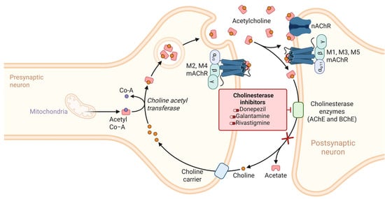
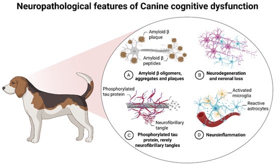
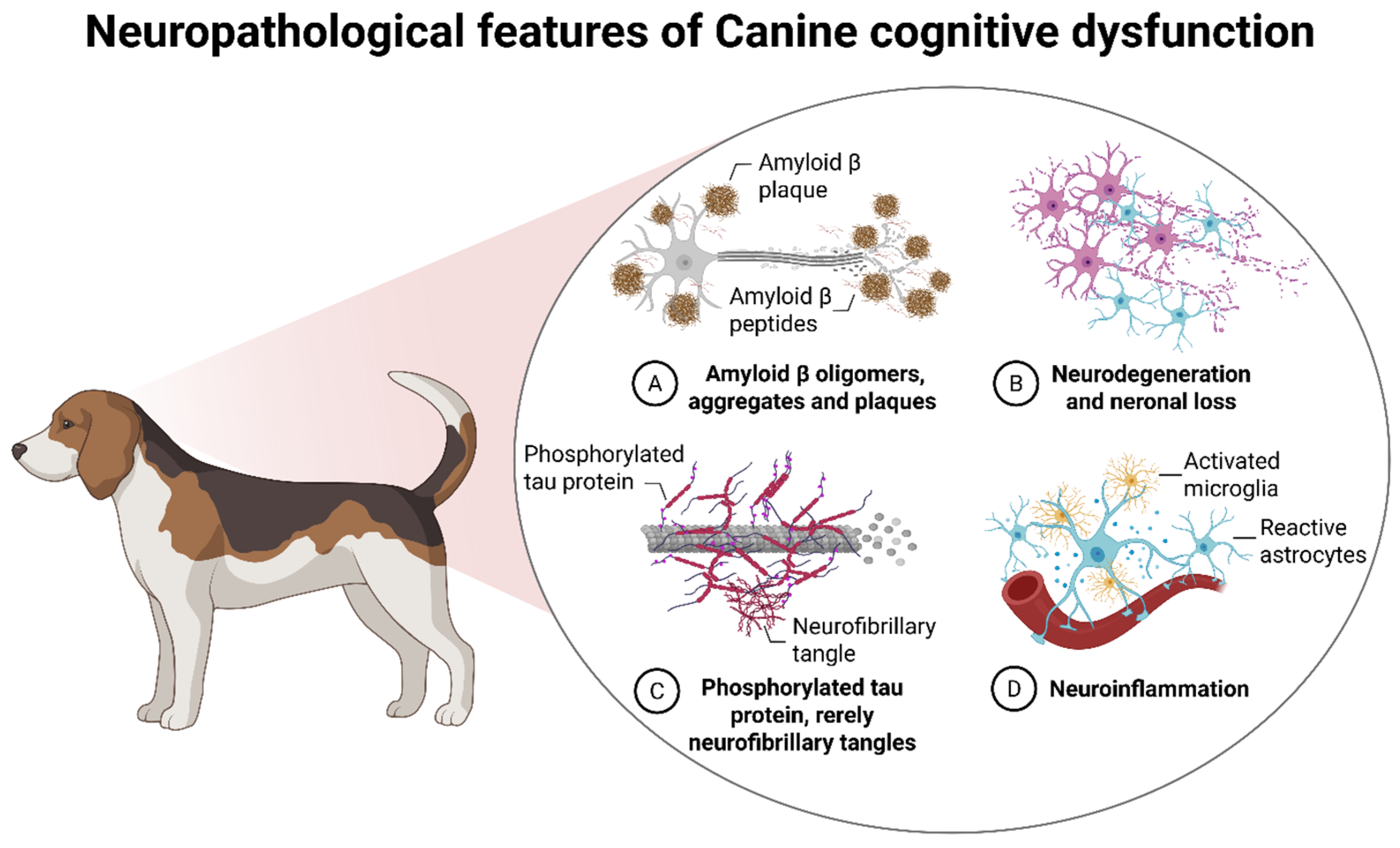
Disclaimer/Publisher’s Note: The statements, opinions and data contained in all publications are solely those of the individual author(s) and contributor(s) and not of MDPI and/or the editor(s). MDPI and/or the editor(s) disclaim responsibility for any injury to people or property resulting from any ideas, methods, instructions or products referred to in the content. |
© 2026 by the author. Licensee MDPI, Basel, Switzerland. This article is an open access article distributed under the terms and conditions of the Creative Commons Attribution (CC BY) license.