


Simple Summary
Manila clams are important shellfish farmed in China. The single-celled parasite Perkinsus olseni is widespread in these clams, but infection does not always result in disease. In August 2025, about half of the clams died at two farms in Jiangsu Province during a period of unusually warm water temperatures. We compared live clams from these farms with those from a nearby farm where no abnormal mortality was reported. Parasites were detected in all 45 clams. DNA sequencing confirmed Perkinsus olseni in clams from the affected farms. Nearly all clams from the affected farms had moderate-to-heavy infections, compared with one in five at the nearby farm. Higher infection intensity was associated with mortality events at the farms, although the cause of death could not be established. Since all sampled clams were infected, health checks should measure infection intensity as well as the presence of the parasite.
Abstract
Perkinsus olseni is widespread in Manila clam (Ruditapes philippinarum) populations in China, but the relationship between infection intensity and mortality requires further field evidence. In August 2025, about 50% of cultured Manila clams died at two neighboring sites in Nantong, Jiangsu, during a period when recorded water temperatures were 29–38 °C. Clams from these sites (A1 and A4; n = 15 each) were compared with those from a reference site with similar temperatures but no mortality (MJG; n = 15). All 45 clams were positive for Perkinsus by Ray’s fluid thioglycollate medium assay, but infection intensity on Mackin’s scale was higher at A1 and A4 (3.31 ± 1.01 and 3.38 ± 0.92) than at MJG (1.71 ± 1.36; p ≤ 0.002). Intensities above 2 occurred in 28 of 30 clams from the affected sites, compared with 3 of 15 MJG clams. Affected clams contained numerous trophozoites, often in foci enclosed by hemocytes, and internal transcribed spacer sequencing confirmed P. olseni. Ostreid herpesvirus 1 was not detected, and bacterial cycle threshold (Ct) values were higher in clams from the affected sites. Molecular identification and the site-level association between higher infection intensity and mortality support surveillance that records infection intensity alongside prevalence. The field comparison does not establish causation.
1. Introduction
Perkinsus olseni is a protozoan parasite of marine mollusks. Infection with P. olseni is a disease listed by the World Organisation for Animal Health (WOAH) [1]. It has been associated with clam mortality in Europe, Korea and Japan [2,3]. In the Manila clam, Ruditapes philippinarum, the outcome of infection depends largely on parasite burden and water temperature. In Korea, peaks in infection intensity coincided with low condition index and clam mortality [4], and heavily infected clams surfaced and died during a hot summer [5]. Experimental challenges have also demonstrated dose-dependent mortality in juvenile Manila clams infected with P. olseni [6]. Temperature also affects parasite development, which accelerates in vitro as temperature rises [7,8], and ocean warming has been associated with the northward spread of epizootics caused by the related Perkinsus marinus in eastern oysters [9].
Clams are an important mariculture product in China. According to the Chinese Fishery Statistical Yearbook 2026, national production of maricultured clams reached 4.96 million t in 2025, second only to oysters among bivalves. Of this total, 0.39 million t came from Jiangsu Province, where this study was conducted. Earlier Chinese studies reported Perkinsus sp.-associated mass mortality of Manila clams in the northern Yellow Sea and described the associated tissue lesions [10,11]. A subsequent study based on internal transcribed spacer (ITS) sequences identified Perkinsus-like parasites from Chinese Manila clams as P. atlanticus [12], now regarded as a synonym of P. olseni [3]. Later surveys documented widespread infection along the Chinese coast [13,14]. In our year-round survey around Qingdao, 96.5% of Manila clams were infected and 89.8% of infections scored 2 or less on Mackin’s scale, although heavier infections also occurred without observed mortality [15]. Thus, neither infection nor a high parasite burden invariably results in mortality. Field comparisons during mortality events may help clarify this relationship, particularly as marine heatwaves become more frequent and intense in Chinese waters [16].
In August 2025, mass mortality of cultured Manila clams occurred in Nantong, Jiangsu Province, during a period of unusually high water temperature. Several infectious and environmental factors warranted consideration. Ostreid herpesvirus 1 (OsHV-1) was associated with oyster mortality in China during the same summer [17]. Vibrio spp. and high temperature can also cause clam mortality [18,19], while heavy P. olseni infections have been associated with mortality in Korea [4,5]. We therefore compared Perkinsus prevalence, infection intensity and histopathological findings in clams from two affected sites and a nearby site without reported mortality, and used ITS sequencing to identify the parasite. The clams were also tested for OsHV-1 and for total bacterial and Vibrio loads.
2. Materials and Methods
2.1. Mortality Event and Sampling
During the mortality event in August 2025, Manila clams were collected from three culture sites in Nantong, Jiangsu Province, on the southern coast of the Yellow Sea. Clams at the three sites came from the same seed source and were stocked at the same time and density. Two sites experiencing mass mortality, A1 (32.12166° N, 121.56389° E) and A4 (32.12067° N, 121.56417° E), are approximately 250 m apart. Cumulative mortality at both sites was estimated at approximately 50% from on-site counts of dead shells and live clams in the culture plots. The third site, Maojiagang (MJG; 32.11361° N, 121.68278° E), lies approximately 12 km east of the affected sites; no abnormal mortality had been reported there, and it was sampled as a reference. Water temperatures were measured repeatedly in the culture areas with a portable water thermometer. These spot measurements were similar at the three sites (29–38 °C; mean 32 °C). According to routine on-site records, this mean was 1–2 °C higher than that for the same period in previous years. Salinity was approximately 25‰. At each site, 15 live clams were collected at random from the culture plots. Dead clams were not examined because their soft tissues had decomposed, leaving mostly empty shells. Obvious clinical signs were observed in some, but not all, clams collected at A1 and A4, whereas none were observed in clams from MJG. The clams were transported to the laboratory on ice.
2.2. Clam Morphometrics and Condition Index
Shell length, shell height, total weight and soft tissue wet weight were measured for each clam before tissues were taken for further analyses. Condition index (CI) was calculated as soft tissue wet weight (g)/shell length (mm) × 100 [4,15].
2.3. Diagnosis and Intensity of Perkinsus Infection
Perkinsus infection was diagnosed using Ray’s fluid thioglycollate medium (RFTM) assays [20]. Gill and mantle tissues from each clam were pooled and incubated in RFTM supplemented with antibiotics for 7 days at room temperature in the dark. The tissues were then stained with Lugol’s iodine and examined under a light microscope for enlarged, blue-black hypnospores. Infection intensity was scored in three preparations per clam on Mackin’s scale from 0 (absent) to 5 (very heavy) [21], as in our previous survey [15], and the mean of the three scores was used to represent infection intensity in that individual. Prevalence was calculated as the percentage of infected clams at each site.
2.4. Histological Examination
Five of the 15 clams sampled at each site were selected at random for histology. Gill, mantle and digestive gland tissues were fixed in Davidson’s fixative, embedded in paraffin, sectioned at 5 µm and stained with hematoxylin and eosin (H&E). These individuals were also examined by RFTM and are identified in Table S1. Sections were examined and photographed under a light microscope (BX53, Olympus, Tokyo, Japan). For the figures, color balance and contrast were adjusted linearly across whole images.
2.5. Molecular Identification of Perkinsus and Detection of OsHV-1 and Bacteria
Total DNA was extracted from gill tissue of each clam using the TIANamp Marine Animals DNA Kit (TIANGEN Biotech, Beijing, China) in accordance with the manufacturer’s instructions. The internal transcribed spacer (ITS) region of Perkinsus spp. was amplified from ten clams (five from A1 and five from A4) with the primers PerkITS85 and PerkITS750 [8]. Sanger sequencing was performed as described by Zhang et al. [15]. The sequences were compared with GenBank entries using the NCBI BLASTn web interface (https://blast.ncbi.nlm.nih.gov/Blast.cgi), including the P. olseni haplotypes PHap1–PHap6 (OR533517–OR533522) previously reported by our group [15].
Samples were tested for OsHV-1 by quantitative PCR targeting the B region of the viral genome, as described by Bai et al. [22] and Zhang et al. [23]. Total bacterial and Vibrio loads were measured using the corresponding real-time PCR detection kits (Qingdao Lijian Bio-Tech Co., Ltd., Qingdao, China), with 50 ng of template DNA per reaction. For each assay, all samples were analyzed on the same plate in three technical replicate wells, alongside a positive control. No amplification was detected in the no-template controls or extraction blanks. With equal template input and all samples analyzed in the same run for each assay, cycle threshold (Ct) values were used to compare relative target abundance in the extracted DNA. Higher Ct values indicate lower target abundance. Samples without amplification were assigned a Ct of 40.
2.6. Statistical Analysis
All analyses were performed in R 4.5.2 [24]. Because intensity scores are ordinal and several variables were not normally distributed, sites were compared with the Kruskal–Wallis test followed by Dunn’s test with Holm correction. Following the threshold used by Zhang et al. [15] to summarize low-intensity infections, intensities of 2 or less were regarded as light and those above 2 as moderate to heavy. The proportions of clams with intensities above 2, and above the stricter threshold of 3, were compared between the pooled affected sites and the reference site with Fisher’s exact test. The effect size for intensity was expressed as Cliff’s delta. To account for the effect of clam size on condition, log-transformed soft tissue weight was compared among sites by analysis of covariance (ANCOVA), with log-transformed shell length as the covariate. Associations of intensity with shell length and condition were tested with Spearman’s rank correlation. Significance was set at p < 0.05 for all tests. Individual data for all 45 clams are provided in Table S1.
3. Results
3.1. Gross Signs, Clam Size and Condition Index
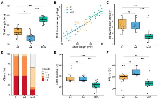
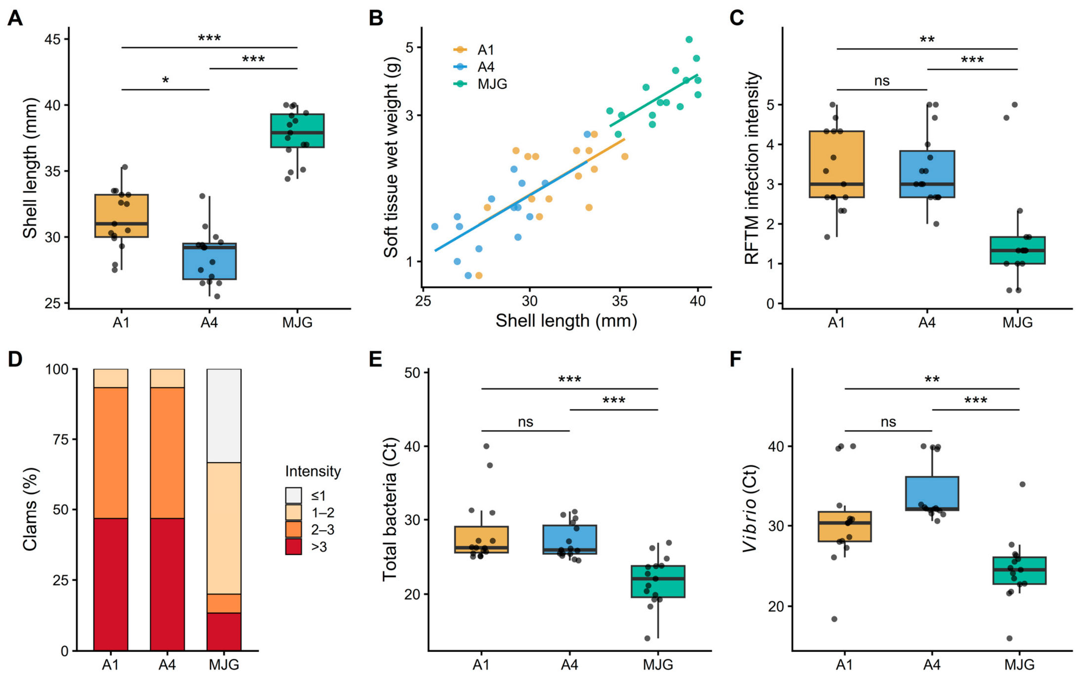
Clinically affected clams at A1 and A4 gaped and closed their valves slowly after mechanical stimulation, and whitish pustules were visible on the foot and mantle of some clams from each site. Overall, clams from these two sites were smaller than those from MJG (shell length 31.4 ± 2.3 and 28.6 ± 2.0 mm vs. 37.7 ± 1.9 mm; p < 0.001; Figure 1A) and had a lower CI (6.03 ± 1.34 and 5.18 ± 1.19 vs. 9.39 ± 1.61; p < 0.001; Table 1). The size-adjusted analysis suggested that this CI difference largely reflected differences in clam size. Soft tissue weight increased with shell length at a similar rate across sites (common slope 2.58, 95% confidence interval 1.67–3.49; test for different slopes, p = 0.79). After adjustment for shell length, no significant difference in soft tissue weight was detected among sites (ANCOVA, p = 0.34; Figure 1B).
Figure 1.
Size, condition, infection intensity and bacterial loads of Manila clams during the Perkinsus olseni-associated mortality event in Nantong, China, in August 2025. Samples came from two sites with mass mortality (A1 and A4) and a reference site without mortality (MJG); n = 15 per site. (A) Shell length. (B) Soft tissue wet weight plotted against shell length (log scales); lines are ANCOVA fits with a common slope (2.58). (C) Infection intensity on Mackin’s scale (RFTM assay). (D) Proportion of clams in each intensity class (Mackin score ≤ 1, > 1 to 2, > 2 to 3, and > 3). (E) Total bacterial load and (F) Vibrio load, expressed as Ct values (higher Ct indicates lower load; Ct = 40, not detected). Boxes show the median and interquartile range; points are individual clams. Pairwise comparisons used Dunn’s test with Holm correction: * p < 0.05, ** p < 0.01, and *** p < 0.001; ns, not significant.
Table 1.
Size, condition, Perkinsus infection and bacterial loads of Manila clams from two sites with mass mortality (A1 and A4) and a reference site without mortality (MJG) in Nantong, China, in August 2025 (n = 15 per site).
3.2. Prevalence and Intensity of Perkinsus Infection
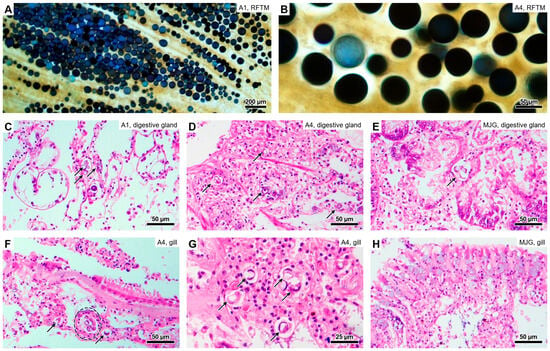
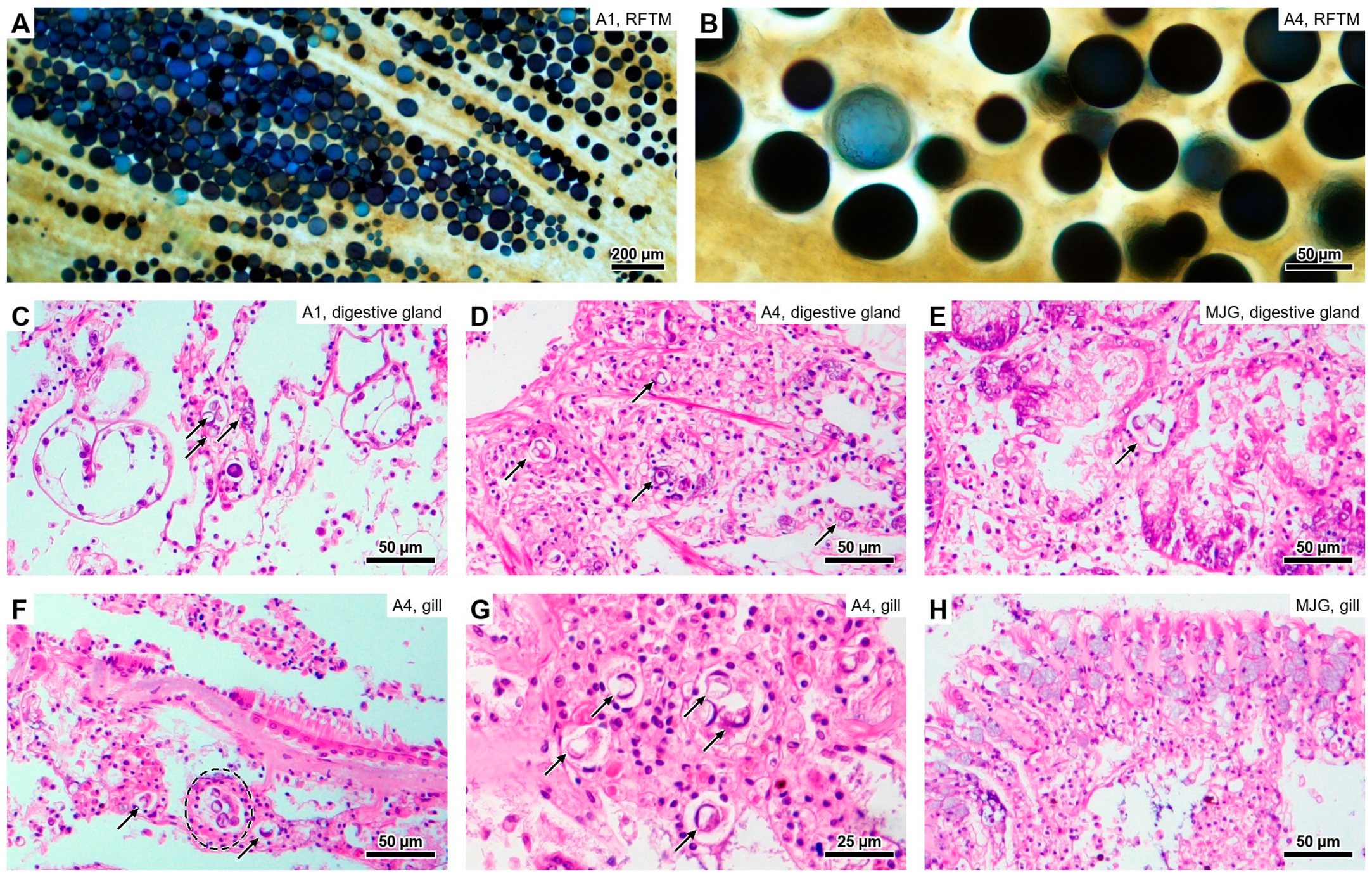
RFTM assays detected Perkinsus in all 45 clams, corresponding to 100% prevalence at each site. Preparations from the affected sites contained numerous enlarged hypnospores stained blue-black by Lugol’s iodine (Figure 2A,B). Infection intensity, however, differed markedly among sites (Kruskal–Wallis test, p < 0.001; Table 1; Figure 1C). Mean intensity was 3.31 ± 1.01 at A1 and 3.38 ± 0.92 at A4 (median 3.00 at both sites) and did not differ between these two sites (p = 0.74). Both affected sites had higher intensities than MJG (1.71 ± 1.36, median 1.33; p = 0.002 and p < 0.001, respectively; Cliff’s delta for affected vs. MJG clams = 0.74). Intensities above 2 were recorded in 28 of the 30 clams (93%) from the affected sites but in only 3 of the 15 clams (20%) from MJG (odds ratio 47, 95% confidence interval 6.6–645; p < 0.001). The corresponding proportions for intensities above 3 were 14 of 30 (47%) and 2 of 15 (13%) (p = 0.046; Figure 1D). Intensity was not correlated with shell length within any site (Spearman’s ρ = −0.30 to 0.19, all p > 0.28), nor with size-adjusted condition within the affected sites (ρ = −0.03, p = 0.87).
Figure 2.
Parasitological and histological findings in Manila clams during the Perkinsus olseni-associated mortality event in Nantong, China. (A,B) Enlarged hypnospores stained blue-black by Lugol’s iodine after RFTM incubation of gill and mantle tissues of clams from sites A1 (A) and A4 (B). (C–E) Connective tissue of the digestive gland. (C) A1: small groups of trophozoites (arrows) in hemolymph sinuses. (D) A4: numerous trophozoites (arrows) in the connective tissue. (E) MJG: an isolated trophozoite (arrow) with little hemocyte response. (F–H) Gills. (F) A4: a focus of trophozoites enclosed by hemocytes (dashed outline) and trophozoites in adjacent sinuses (arrows). (G) A4: trophozoites with a large vacuole and an eccentric nucleus (signet-ring form; arrows) among infiltrating hemocytes. (H) MJG: intact gill without visible parasites. H&E (C–H). Scale bars: (A) 200 µm; (B–F,H) 50 µm; (G) 25 µm. Color balance and contrast of the H&E images were adjusted linearly across whole images.
3.3. Histopathology
Histological findings were consistent with the RFTM results. In H&E sections, trophozoites appeared as spherical cells with a large vacuole and an eccentric nucleus (signet-ring form). In A1 clams, small groups of trophozoites occurred in the hemolymph sinuses of the connective tissue around the digestive gland (Figure 2C). In A4 clams, trophozoites were numerous in this connective tissue (Figure 2D) and formed foci in the hemolymph sinuses of the gills, where they were enclosed by infiltrating hemocytes (Figure 2F,G). Trophozoites were also found in the mantle connective tissue at both affected sites, scattered in an A1 clam and forming large foci with hemocyte infiltration in an A4 clam (Figure S1). In contrast, the gills of MJG clams were intact (Figure 2H), and only isolated trophozoites with little hemocyte response were seen in the connective tissue of the digestive gland (Figure 2E). No lesions suggestive of other infectious agents were observed.
3.4. Molecular Identification, OsHV-1 and Bacterial Loads
ITS sequences obtained from all ten clams shared 99.5–100% identity with P. olseni sequences in GenBank and matched the haplotypes that we previously reported from Manila clams in Qingdao [15]. OsHV-1 was not detected in any clam. Total bacterial and Vibrio Ct values were higher in clams from the affected sites than in those from MJG (p < 0.01 for all comparisons with MJG; Table 1; Figure 1E,F). For total bacteria, mean Ct values were 28.3 ± 4.7 at A1 and 27.1 ± 2.3 at A4, compared with 21.7 ± 3.3 at MJG; the corresponding Vibrio values were 30.8 ± 5.7, 34.0 ± 3.7 and 24.6 ± 4.1. Three clams from the affected sites showed no Vibrio amplification and were assigned Ct = 40; four others showed late amplification (Ct 39.6–39.9). All MJG clams had Ct values of 35.2 or lower.
4. Discussion
To our knowledge, this is the first molecularly confirmed report of P. olseni-associated mass mortality of cultured Manila clams in China. Earlier Chinese studies linked Perkinsus sp. to clam mortality using field observations, histopathology and infection trials [10,11], but did not molecularly identify the parasite in those mortality investigations. Zhang et al. subsequently identified P. atlanticus, now a synonym of P. olseni, in Chinese Manila clams by ITS sequencing [3,12]. The present investigation combines species identification in samples collected during the mortality event with a formal statistical comparison of infection intensity between affected and reference sites. All 45 clams were positive by RFTM, yet infection intensity was significantly higher at A1 and A4 than at MJG. Infection intensity therefore distinguished the affected populations more clearly than prevalence, with consistent results at both affected sites.
High summer temperatures may have contributed to the mortality event. Burdens at the affected sites were moderate (median 3.0), and two MJG clams with intensities near 5 appeared healthy. In Gomso Bay, Korea, mass mortality coincided with the highest monthly mean intensities recorded (up to 2.2 × 10⁶ cells g−1 wet tissue) [4]; although these counts cannot be converted directly to Mackin scores, they indicate that field mortality has been associated with heavy infections. An experimental study reported 33% mortality after two days of exposure to 36 °C at low tide [18]. At MJG, however, no abnormal mortality was reported despite similarly high spot temperature measurements. High temperature accelerates the development of P. olseni [7,8] and may also weaken host defenses. In Korea, clams that surfaced and died during a hot summer carried heavier burdens than clams that remained buried, and had a lower condition index and weaker hemocyte functions [5]. Together, these studies suggest that temperature could have modified the effects of infection during the event.
The histopathological findings are consistent with a pathogenic role of the parasite at the affected sites. In affected clams, trophozoites were numerous in the gills, digestive gland and mantle and often formed foci enclosed by hemocytes. Some of these clams also had whitish pustules on the foot and mantle. Similar tissue changes were described in earlier Chinese investigations [11] and in our previous study in Qingdao [15]. Extensive infection of the gills and digestive gland is likely to impair respiration and feeding. In experimental infections, P. olseni reduced the feeding and burrowing activity of juvenile Manila clams [25]. In MJG clams, by contrast, the few trophozoites present provoked little host response, as expected for subclinical infections [15].
The additional pathogen screening provided context for interpreting the mortality event. OsHV-1 was not detected, although the virus was associated with oyster mortality in China during the same summer [17]. Total bacterial and Vibrio Ct values were higher in clams from the affected sites than at MJG. These findings do not support an increase in the measured bacterial targets, but they do not exclude a contribution from particular pathogenic bacteria. Reduced filtration could account for lower bacterial abundance in infected clams [25], although filtration was not measured here. The lower CI at A1 and A4 was largely explained by clam size. Although the stocks shared the same seed source, stocking time and density, local growth conditions or management could have influenced both clam size and susceptibility to infection or thermal stress.
This field investigation examined 15 randomly collected live clams per site during the mortality event. Dead clams were mostly empty shells, so their infection status could not be determined; the findings therefore establish an association at the site level rather than the cause of death. The sample size, single sampling period and separation of MJG from the two neighboring affected sites limit inference about population variability, changes before mortality and unmeasured environmental or farming differences. Repeated spot temperature measurements did not establish the duration of exposure to peak temperatures, and lesions were not graded for quantitative comparison with individual RFTM scores. Longitudinal monitoring and controlled challenges would help to resolve these questions.
The event has practical implications for clam health surveillance in China. P. olseni infection is widely reported in Manila clams along the Chinese coast [14,15], and marine heatwaves in Chinese waters are becoming longer and more intense [16], so similar losses may recur. Because prevalence was 100% at all sites, surveillance based only on the presence of the parasite would not have distinguished the sampled populations with and without mortality. Baseline surveys of WOAH-listed protozoans, such as a recent PCR survey of pooled mussel and oyster samples in Korea, are designed to establish presence or absence [26]. Monitoring programs should therefore record infection intensity, examine stocks before and during hot periods, and avoid transferring seed from heavily infected areas [14].
5. Conclusions
ITS sequencing confirmed P. olseni infection in Manila clams from both affected sites in Nantong. Infection intensity was significantly higher at these sites than at MJG, despite 100% prevalence at all three sites. These findings support monitoring infection intensity alongside prevalence in farmed Manila clams, particularly during hot summers.
Supplementary Materials
The following supporting information can be downloaded at: https://www.mdpi.com/article/10.3390/ani16223686/s1, Figure S1: Histology of the mantle of Manila clams from the affected sites (H&E); Table S1: Individual data of the 45 Manila clams examined.
Author Contributions
Conceptualization, B.-W.H. and C.-M.B.; methodology, M.-L.W., X.-C.L. and X.Z.; validation, X.Z.; formal analysis, B.-W.H.; investigation, B.-W.H., M.-L.W., M.C. and X.-C.L.; resources, M.C.; writing—original draft preparation, B.-W.H.; writing—review and editing, X.Z. and C.-M.B.; visualization, B.-W.H.; supervision, C.-M.B.; funding acquisition, C.-M.B. All authors have read and agreed to the published version of the manuscript.
Funding
This research was funded by the Central Public-interest Scientific Institution Basal Research Fund, Chinese Academy of Fishery Sciences, grant number 2023TD30, and the China Agriculture Research System of MOF and MARA, grant number CARS-48.
Institutional Review Board Statement
Ethical review and approval were waived for this study because it involved only invertebrates (bivalve mollusks) collected from commercial farms during a disease investigation, and no institutional or national regulations in China require ethical approval for such samples.
Informed Consent Statement
Not applicable.
Data Availability Statement
The original contributions presented in this study are included in the article and Supplementary Materials (Table S1). The ITS sequences obtained in this study matched P. olseni haplotypes previously deposited in GenBank (accession numbers OR533517–OR533522). Further inquiries can be directed to the corresponding author.
Acknowledgments
Claude Opus 5.5 (Anthropic, San Francisco, CA, USA) was used solely to assist with the correction of scientific English language and grammar during manuscript preparation. The authors remain fully responsible for the content and interpretation of the work.
Conflicts of Interest
The authors declare no conflicts of interest.
References
- World Organisation for Animal Health (WOAH). Perkinsosis (P. olseni). Listed Disease. Available online: https://www.woah.org/en/disease/perkinsosis-p-olseni/ (accessed on 12 November 2026).
- Choi, K.-S.; Park, K.-I. Review on the protozoan parasite Perkinsus olseni (Lester and Davis 1981) infection in Asian waters. In Coastal Environmental and Ecosystem Issues of the East China Sea; Ishimatsu, A., Lie, H.-J., Eds.; TERRAPUB and Nagasaki University: Tokyo, Japan, 2010; pp. 269–281. [Google Scholar]
- Villalba, A.; Reece, K.S.; Ordás, M.C.; Casas, S.M.; Figueras, A. Perkinsosis in molluscs: A review. Aquat. Living Resour. 2004, 17, 411–432. [Google Scholar] [CrossRef] [Scilit]
- Yang, H.-S.; Park, K.-I.; Donaghy, L.; Adhya, M.; Choi, K.-S. Temporal variation of Perkinsus olseni infection intensity in the Manila clam Ruditapes philippinarum in Gomso Bay, off the west coast of Korea. J. Shellfish Res. 2012, 31, 685–690. [Google Scholar] [CrossRef] [Scilit]
- Nam, K.-W.; Jeung, H.-D.; Song, J.-H.; Park, K.-H.; Choi, K.-S.; Park, K.-I. High parasite burden increases the surfacing and mortality of the Manila clam (Ruditapes philippinarum) in intertidal sandy mudflats on the west coast of Korea during hot summer. Parasites Vectors 2018, 11, 42. [Google Scholar] [CrossRef] [Scilit] [PubMed]
- Shimokawa, J.; Yoshinaga, T.; Ogawa, K. Experimental evaluation of the pathogenicity of Perkinsus olseni in juvenile Manila clams Ruditapes philippinarum. J. Invertebr. Pathol. 2010, 105, 347–351. [Google Scholar] [CrossRef] [Scilit] [PubMed]
- Ahn, K.-J.; Kim, K.-H. Effect of temperature and salinity on in vitro zoosporulation of Perkinsus sp. in Manila clams Ruditapes philippinarum. Dis. Aquat. Org. 2001, 48, 43–46. [Google Scholar] [CrossRef] [Scilit] [PubMed]
- Casas, S.M.; Villalba, A.; Reece, K.S. Study of perkinsosis in the carpet shell clam Tapes decussatus in Galicia (NW Spain). I. Identification of the aetiological agent and in vitro modulation of zoosporulation by temperature and salinity. Dis. Aquat. Org. 2002, 50, 51–65. [Google Scholar] [CrossRef] [Scilit] [PubMed]
- Cook, T.; Folli, M.; Klinck, J.; Ford, S.; Miller, J. The relationship between increasing sea-surface temperature and the northward spread of Perkinsus marinus (Dermo) disease epizootics in oysters. Estuar. Coast. Shelf Sci. 1998, 46, 587–597. [Google Scholar] [CrossRef] [Scilit]
- Liang, Y.-B.; Zhang, X.-C.; Wang, L.-J.; Yang, B.; Zhang, Y.; Cai, C.-L. Prevalence of Perkinsus sp. in the Manila clam Ruditapes philippinarum along the northern coast of the Yellow Sea in China. Oceanol. Limnol. Sin. 2001, 32, 502–511. (In Chinese) [Google Scholar] [CrossRef]
- Li, X.; Wang, J.-B.; Wang, B.; Liang, Y.-B. Histopathological study on Perkinsus sp. disease of Ruditapes philippinarum. J. Dalian Fish. Univ. 2002, 17, 215–219. (In Chinese) [Google Scholar] [CrossRef]
- Zhang, X.-C.; Liang, Y.-B.; Fan, J.-F.; Zhang, W.; Pu, H.-Y.; Liang, B.; Chen, H.-X.; Song, L.-C. Identification of Perkinsus-like parasite in Manila clam, Ruditapes philippinarum using DNA molecular marker at ITS region. Acta Oceanol. Sin. 2005, 24, 139–144. [Google Scholar]
- Wu, S.-Q.; Wang, C.-X.; Lin, X.-M.; Wang, Z.-X.; Li, X.-F.; Liu, J.; Deng, J.-H.; Qiu, S.-Y. Infection prevalence and phylogenetic analysis of Perkinsus olseni in Ruditapes philippinarum from East China. Dis. Aquat. Org. 2011, 96, 55–60. [Google Scholar] [CrossRef] [Scilit] [PubMed]
- Ye, L.; Wu, L.; Lu, J.; Yao, T.; Wang, J.; Shi, S.; Yu, G.; Zhao, W. Diversity and distribution of Perkinsus spp. along the coast of China: Implications for widespread transmission of Perkinsus spp. in mollusks. Front. Mar. Sci. 2022, 9, 989261. [Google Scholar] [CrossRef] [Scilit]
- Zhang, X.; Zheng, Y.-D.; Yuan, T.; Liu, C.-F.; Huang, B.-W.; Xin, L.-S.; Wang, C.-M.; Bai, C.-M. Occurrence and seasonal variation of Perkinsus sp. infection in wild mollusk populations from coastal waters of Qingdao, northern China. J. Invertebr. Pathol. 2024, 202, 108044. [Google Scholar] [CrossRef] [Scilit] [PubMed]
- Yao, Y.; Wang, J.; Yin, J.; Zou, X. Marine heatwaves in China’s marginal seas and adjacent offshore waters: Past, present, and future. J. Geophys. Res. Ocean. 2020, 125, e2019JC015801. [Google Scholar] [CrossRef] [Scilit]
- Huang, B.-W.; Zhang, X.; Xu, T.; Zhang, Y.-T.; Li, X.-A.; Gao, L.; Kong, N.; Liu, M.-K.; Yu, T.; Kang, H.-G.; et al. First report of mass mortality in post-metamorphosis Magallana gigas associated with OsHV-1 in China: Epidemiological and genomic characterization. J. Invertebr. Pathol. 2026, 218, 108679. [Google Scholar] [CrossRef] [Scilit] [PubMed]
- Macho, G.; Woodin, S.A.; Wethey, D.S.; Vázquez, E. Impacts of sublethal and lethal high temperatures on clams exploited in European fisheries. J. Shellfish Res. 2016, 35, 405–419. [Google Scholar] [CrossRef] [Scilit]
- Paillard, C. A short-review of brown ring disease, a vibriosis affecting clams, Ruditapes philippinarum and Ruditapes decussatus. Aquat. Living Resour. 2004, 17, 467–475. [Google Scholar] [CrossRef] [Scilit]
- Ray, S.M. A review of the culture method for detecting Dermocystidium marinum, with suggested modifications and precautions. Proc. Natl. Shellfish Assoc. 1966, 54, 55–69. [Google Scholar]
- Mackin, J.G. Oyster disease caused by Dermocystidium marinum and other microorganisms in Louisiana. Publ. Inst. Mar. Sci. Univ. Tex. 1962, 7, 132–229. [Google Scholar]
- Bai, C.-M.; Gao, W.-H.; Wang, C.-M.; Yu, T.; Zhang, T.; Qiu, Z.; Wang, Q.; Huang, J. Identification and characterization of ostreid herpesvirus 1 associated with massive mortalities of Scapharca broughtonii broodstocks in China. Dis. Aquat. Org. 2016, 118, 65–75. [Google Scholar] [CrossRef] [Scilit] [PubMed]
- Zhang, X.; Huang, B.-W.; Yu, T.; Wang, J.-L.; Wei, M.-L.; Kang, H.-G.; Wang, C.-M.; Bai, C.-M. Epidemiological survey and genomic insights into ostreid herpesvirus 1 transmission in the blood clam Anadara broughtonii in China (2019–2025). Anim. Res. One Health 2026, early view. [Google Scholar] [CrossRef] [Scilit]
- R Core Team. R: A Language and Environment for Statistical Computing; R Foundation for Statistical Computing: Vienna, Austria, 2025. [Google Scholar]
- Waki, T.; Yoshinaga, T. Experimental evaluation of the impact of Perkinsus olseni on the physiological activities of juvenile Manila clams. J. Shellfish Res. 2018, 37, 29–39. [Google Scholar] [CrossRef] [Scilit]
- Kim, J.-H.; Kajino, N.; Shin, J.-S.; Choi, H.J.; Kwon, M.-G.; Park, C.-I.; Choi, K.-S.; Hong, H.-K. Baseline assessment of WOAH-listed protozoan parasites in wild Mediterranean mussels Mytilus galloprovincialis and Pacific oysters Crassostrea gigas from port-adjacent coastal waters of Korea in 2023. Animals 2026, 16, 1502. [Google Scholar] [CrossRef] [Scilit] [PubMed]

Disclaimer/Publisher’s Note: The statements, opinions and data contained in all publications are solely those of the individual author(s) and contributor(s) and not of MDPI and/or the editor(s). MDPI and/or the editor(s) disclaim responsibility for any injury to people or property resulting from any ideas, methods, instructions or products referred to in the content. |
© 2026 by the authors. Licensee MDPI, Basel, Switzerland. This article is an open access article distributed under the terms and conditions of the Creative Commons Attribution (CC BY) license.