


Simple Summary
Wild sika deer in Hokkaido, Japan, are known to harbor two genetically distinct Babesia taxa. One is closely related to Babesia divergens, a cattle parasite found in Europe, and was designated Babesia sp. Sika 1. The other is an unclassified Babesia taxon, designated Babesia sp. Sika 2. In Hokkaido, sika deer and grazing cattle increasingly share the same environments, where ticks can feed on both hosts. We therefore investigated whether these Babesia taxa can also infect cattle. Both taxa were found in sika deer, and Babesia sp. Sika 1 was also found in Ixodes persulcatus ticks, but neither taxon was detected in cattle. Genetic analyses further showed that Babesia sp. Sika 1 forms a lineage separate from B. divergens, supporting its interpretation as a distinct Babesia lineage rather than its direct assignment to B. divergens. Further studies are essential to clarify the findings of the present study regarding the taxonomic status and host associations of Babesia sp. Sika 1 and Babesia sp. Sika 2.
Abstract
Cervids harbor several Babesia taxa, but their potential to infect cattle remains poorly understood. The present study detected Babesia taxa in wild sika deer in Hokkaido, Japan, and subsequently examined questing ticks collected from cattle pastures and sympatric grazing cattle for their presence. The initial screening of 285 sika deer blood DNA samples using a PCR assay targeting the 18S rRNA of Babesia spp., followed by sequencing, identified two previously reported Babesia taxa: one closely related to Babesia divergens and designated Babesia sp. Sika 1, and another unclassified Babesia taxon designated Babesia sp. Sika 2. A duplex PCR assay based on the obtained 18S rRNA sequences was then used to detect these two taxa in 285 sika deer, 671 questing ticks, and 767 cattle. Babesia sp. Sika 1 was detected in 86/285 (30.2%) deer and 7/367 (1.9%) Ixodes persulcatus ticks, whereas Babesia sp. Sika 2 was detected in 4/285 (1.4%) deer but not in the examined ticks. Neither taxon was detected in any cattle samples. Phylogenetic analyses placed Babesia sp. Sika 1 in a distinct lineage from B. divergens, supporting its recognition as a distinct Babesia taxon. Similarly, Babesia sp. Sika 2 formed a distinct phylogenetic clade in the 18S rRNA analysis. Despite opportunities for exposure through shared pasture environments and I. persulcatus feeding on both hosts, neither taxon was detected in the examined cattle. Further studies are now essential to confirm and extend our findings regarding the taxonomic status and host associations of Babesia sp. Sika 1 and Babesia sp. Sika 2.
1. Introduction
Babesiosis is a tick-borne infectious disease caused by protozoan parasites of the genus Babesia, which infect a wide range of vertebrate hosts, including domestic and wild animals and humans [1]. These parasites are of veterinary and public health importance worldwide, as the infections can cause clinical manifestations such as fever, anemia, hemoglobinuria, and, in severe cases, death [2,3]. In particular, bovine babesiosis is of major concern because of its clinical impact and economic significance associated with production losses, treatment costs, and implementation of control measures [3].
Of the Babesia species infecting cattle, Babesia bovis, Babesia bigemina, Babesia divergens, and Babesia naoakii are major causative agents of clinical bovine babesiosis [3,4,5]. Among them, B. divergens is distributed in parts of Europe and Africa, corresponding to the geographical distribution of its vector, Ixodes ricinus [4]. Babesia divergens is also a zoonotic Babesia species capable of causing human babesiosis [6]. Interestingly, several B. divergens-related parasites have also been reported in cervid hosts [7].
In Japan, previous epidemiological studies identified a B. divergens-like parasite in sika deer (Cervus nippon) and Ixodes persulcatus ticks [8,9,10,11]. Based on 18S rRNA sequences, this parasite forms a lineage closely related to B. divergens [9,10]. In addition to the B. divergens-like parasite, another unclassified Babesia sp. has also been detected in sika deer in Japan [12,13]. Therefore, at least two genetically distinct Babesia taxa have been reported in sika deer in Japan. For clarity, the B. divergens-like taxon and the previously unclassified Babesia taxon identified in sika deer in Japan are provisionally referred to as Babesia sp. Sika 1 and Babesia sp. Sika 2, respectively, throughout this manuscript.
The taxonomy of B. divergens and closely related Babesia taxa has historically been difficult to resolve, particularly when based on 18S rRNA sequences alone [14]. Earlier phylogenetic studies placed B. divergens, Babesia capreoli, Babesia sp. MO1, and Babesia sp. Sika 1 within a closely related group, making species-level assignment difficult [8]. More recently, Babesia sp. MO1 was formally described as Babesia hegotelforum, establishing it as a species distinct from B. divergens based on genome-wide sequence divergence, phylogenetic placement, host associations, and other biological characteristics [15]. This revised taxonomic framework provides an opportunity to reassess the phylogenetic position of Babesia sp. Sika 1.
The expansion of sika deer populations in Japan has increased ecological overlap between wild deer and grazing cattle [16]. In Hokkaido, deer frequently inhabit or move through cattle pastures, increasing opportunities for exposure to the same tick species [17]. Moreover, I. persulcatus, a potential vector of Babesia taxa infecting sika deer in Hokkaido, is known to feed on both deer and cattle [18,19,20]. Therefore, cattle may encounter ticks carrying these Babesia parasites within shared pasture environments. However, such ecological contact does not necessarily result in cattle infection. For example, a previous study in Hokkaido detected a Theileria species infecting sika deer in both deer and questing ticks collected from cattle pastures, but not in sympatric grazing cattle [21]. Whether the same pattern applies to Babesia taxa detected in sika deer remains unknown, as no epidemiological study has examined their potential to infect sympatric grazing cattle in Japan. Based on this epidemiological context, the present study first identified Babesia taxa detected in sika deer in Hokkaido and subsequently surveyed questing ticks collected from cattle pastures and sympatric grazing cattle for these parasites to assess their potential to infect cattle, while reassessing the taxonomic position of Babesia sp. Sika 1 using phylogenetic analyses.
2. Materials and Methods
2.1. DNA Samples from Wild Sika Deer, Grazing Cattle, and Questing Ticks
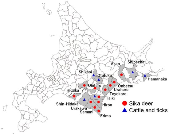
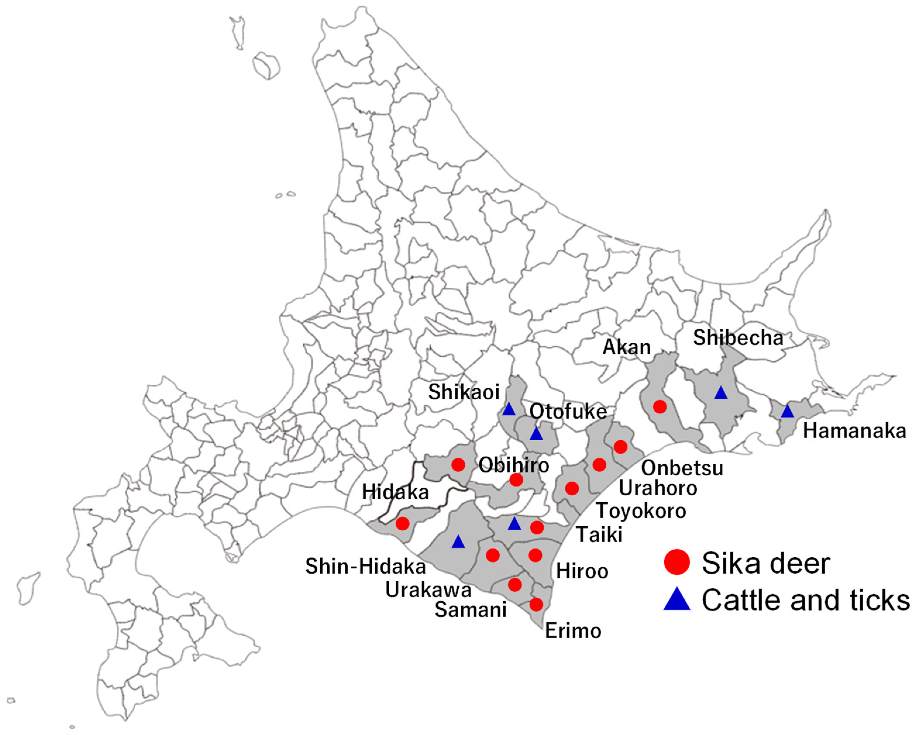
Archived DNA samples from the blood of 285 wild sika deer [21,22] and blood of 767 grazing cattle [21] raised in the same or adjacent regions as the deer sampling sites, and 671 questing ticks collected from cattle pastures [21] in Hokkaido, Japan, were used in the present study. The deer samples were originally obtained from deer during hunting or slaughter at deer farms. Detailed sampling locations and sample numbers for the deer samples are provided in Table 1, and the sampling locations of the deer, cattle, and ticks are shown in Figure 1. Some of these samples were used in previous studies [21,22]. Based on morphological characteristics, the ticks were previously identified as I. persulcatus, Ixodes ovatus, Haemaphysalis japonica, and Haemaphysalis megaspinosa (Table 2) [21,23].
Table 1.
Detection of Babesia sp. Sika 1 and Babesia sp. Sika 2 in blood DNA samples from sika deer by universal PCR and taxon-specific duplex PCR assays targeting the 18S rRNA.
Figure 1.
Map showing the sampling locations of sika deer, cattle, and questing ticks in Hokkaido, Japan.
Table 2.
Detection of Babesia sp. Sika 1 in questing ticks by duplex PCR assay.
The deer and cattle DNA samples were extracted from whole blood using the QIAamp DNA Blood Mini Kit (Qiagen, Hilden, Germany), while tick DNA was extracted using phenol:chloroform:isoamyl alcohol after digestion with the lysis buffer provided in the ISOHAIR kit (Nippon Gene, Tokyo, Japan). All DNA samples were extracted within a short period after sample collection and stored at −30 °C in aliquots to minimize repeated freeze–thaw cycles.
2.2. Molecular Identification of Babesia Taxa Infecting Sika Deer
To identify Babesia taxa infecting sika deer, all 285 deer DNA samples were screened using a previously described universal PCR assay targeting the 18S rRNA sequences of Babesia spp. [24] (forward primer: 5′-CATTACAACAGTTATAGTTTCTTTGG-3′; reverse primer: 5′-CTAGGCATTCCTCGTTCATGATTTAG-3′). Briefly, a 10-μL PCR reaction mixture was prepared containing 2 μL of DNA template, 1.0 μL of 10× PCR buffer (Applied Biosystems, Foster City, CA, USA), 1.0 μL of dNTP mixture (Applied Biosystems), 0.5 μL of forward primer (10 μM), 0.5 μL of reverse primer (10 μM), 0.1 μL of AmpliTaq Gold DNA polymerase (Applied Biosystems), and 4.9 μL of nuclease-free water. The mixture was subjected to the following cycling conditions: initial denaturation at 95 °C for 5 min; 40 cycles of denaturation at 95 °C for 30 s, annealing at 62 °C for 1 min, and extension at 72 °C for 2 min; and a final extension at 72 °C for 10 min. PCR products were analyzed by electrophoresis on 1.5% agarose gels, stained with ethidium bromide, and visualized under UV light. The presence of an approximately 1400-bp amplicon indicated a positive result for Babesia spp. Amplicons of the expected size (~1400 bp) were purified, cloned into the pCR2.1-TOPO vector (Invitrogen, Carlsbad, CA, USA), and sequenced. All amplicons obtained from the 28 Babesia spp. PCR-positive deer samples were subjected to sequencing.
Sequence analysis identified the two previously described Babesia taxa in sika deer in Japan, namely Babesia sp. Sika 1 and Babesia sp. Sika 2. To further characterize Babesia sp. Sika 1, a 1271-bp fragment of the beta-tubulin gene was amplified from selected Babesia sp. Sika 1-positive deer samples using Babesia-specific forward (5′-AGATCGGTGCCAARTTCTGGGAG-3′) and reverse (5′-TCCGATACCCTCTTGAACATGTC-3′) primers [8]. The PCR reaction mixture and cycling conditions were the same as those described above, except that the annealing temperature was adjusted to 57 °C. Following agarose gel electrophoresis and ethidium bromide staining, the presence of a 1271-bp product under UV light indicated a positive result. The amplicons were cloned and sequenced.
2.3. Duplex PCR Assay for Simultaneous Detection of Babesia sp. Sika 1 and Babesia sp. Sika 2
A duplex PCR assay was used for simultaneous detection of Babesia sp. Sika 1 and Babesia sp. Sika 2. Briefly, the 18S rRNA sequences obtained from positive sika deer samples were aligned with available relevant sequences of hemopathogens infecting animals in Hokkaido, as well as sequences from sika deer, ticks, and cattle. The alignment was used to identify regions specific to Babesia sp. Sika 1 and Babesia sp. Sika 2. Taxon-specific forward primers for Babesia sp. Sika 1 (5′-GGGGAAACTCACCAGGTCCAGACAAT-3′) and Babesia sp. Sika 2 (5′-CAGCCTGTGGCATTCTCGCGCGTGC-3′) were then manually designed, with particular attention to sequence differences at or near the 3′ ends of the primers. A common reverse primer (5′-CTAGGCATTCCTCGTTCATGATTTAG-3′) was selected from a region conserved between Babesia sp. Sika 1 and Babesia sp. Sika 2 but divergent from non-target sequences.
PCR amplification was performed in a final reaction volume of 10 μL containing 2 μL of DNA template, 1.0 μL of 10× PCR buffer (Applied Biosystems, Foster City, CA, USA), 1.0 μL of dNTP mixture (Applied Biosystems), 0.5 μL of each forward primer (10 μM), 0.5 μL of reverse primer (10 μM), 0.1 μL of AmpliTaq Gold DNA polymerase (Applied Biosystems), and 4.4 μL of nuclease-free water. Thermal cycling conditions consisted of an initial denaturation at 95 °C for 5 min, followed by 40 cycles of denaturation at 95 °C for 30 s, annealing at 62 °C for 1 min, and extension at 72 °C for 2 min, with a final extension at 72 °C for 5 min. PCR products were analyzed by electrophoresis on 1.5% agarose gels, stained with ethidium bromide, and observed under UV light. The presence of 394-bp and 889-bp amplicons indicated positive results for Babesia sp. Sika 1 and Babesia sp. Sika 2, respectively.
The specificity of the duplex PCR assay was evaluated using DNA samples from related hemopathogens, including B. bovis, B. bigemina, Babesia ovata, B. naoakii, Babesia sp. Hue-1, Theileria orientalis, Theileria annulata, Theileria parva, Theileria sp. Sika-1, Trypanosoma evansi, Trypanosoma brucei, Trypanosoma theileri, Anaplasma marginale, and Anaplasma centrale, as well as uninfected bovine blood DNA [21,24].
2.4. Detection of Babesia sp. Sika 1 and Babesia sp. Sika 2 in Deer, Ticks, and Cattle Using Duplex PCR Assay
The duplex PCR assay was used to detect Babesia sp. Sika 1 and Babesia sp. Sika 2 in DNA samples obtained from wild sika deer, questing ticks, and grazing cattle. Two I. persulcatus DNA samples that tested positive using the duplex PCR assay were then subjected to the same universal 18S rRNA PCR assay used for the initial detection of Babesia taxa in deer. The resulting amplicons were cloned and sequenced to confirm the duplex PCR results.
2.5. Phylogenetic Analysis
Phylogenetic trees were constructed separately for three sequence datasets: 18S rRNA sequences of Babesia sp. Sika 1 from sika deer and ticks, beta-tubulin gene sequences of Babesia sp. Sika 1 from positive sika deer, and 18S rRNA sequences of Babesia sp. Sika 2 from sika deer. Separate 18S rRNA phylogenetic trees were constructed for Babesia sp. Sika 1 and Babesia sp. Sika 2 as they addressed different phylogenetic questions. For Babesia sp. Sika 1, closely related taxa were selected to improve resolution of its relationship with B. divergens and other closely related taxa, whereas a broader set of Babesia species was included to assess the phylogenetic position of Babesia sp. Sika 2.
The sequences obtained in the present study, together with reference sequences retrieved from GenBank, were aligned using MAFFT online software (version 7) [25]. The resulting alignments were analyzed using MEGA version 11 to determine the best-fitting nucleotide substitution models [26]. Maximum likelihood phylogenetic trees were constructed using MEGA11 based on the Tamura–Nei substitution model [27], with the complete deletion option selected to exclude alignment positions containing gaps and missing data. The final numbers of nucleotide positions included in the phylogenetic analyses were 1413 bp for the 18S rRNA sequences of Babesia sp. Sika 1; 902 bp for the beta-tubulin sequences of Babesia sp. Sika 1; and 1226 bp for the 18S rRNA sequences of Babesia sp. Sika 2.
3. Results
3.1. Babesia Taxa Detected in Wild Sika Deer in Hokkaido
Among 285 blood DNA samples from sika deer, 28 (9.8%) tested positive using the initial universal PCR targeting the 18S rRNA sequences of Babesia spp., yielding amplicons of the expected size (~1400 bp). Sequence analysis of the 28 PCR amplicons identified two genetically distinct Babesia lineages, which were designated Babesia sp. Sika 1 and Babesia sp. Sika 2. Of these, 25 samples were positive only for Babesia sp. Sika 1, two were positive only for Babesia sp. Sika 2, and one was positive for both taxa (Table 1). The obtained sequences (Accession Nos. LC951429-LC951454) were 1420–1423 bp in length and shared 99.5–99.9% sequence identities with previously reported 18S rRNA sequences of Babesia sp. Sika 1 (Accession Nos. KC493555, AB975389, LC279018, KC465977, and KC465976).
On the other hand, Babesia sp. Sika 2 was detected only in three deer samples, including one sample that was co-infected with Babesia sp. Sika 1. The obtained sequences (Accession Nos. LC951455-LC951457) were 1392 bp in length and shared 99.8% sequence identity with previously reported 18S rRNA sequences of Babesia sp. Sika 2 from sika deer in Japan (Accession No. LC536169).
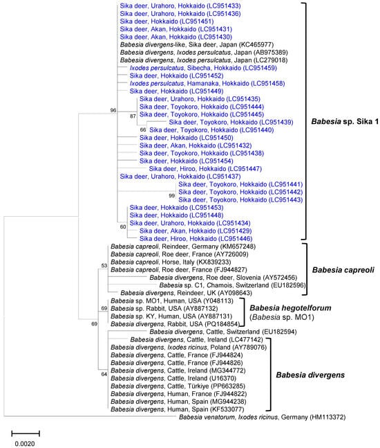
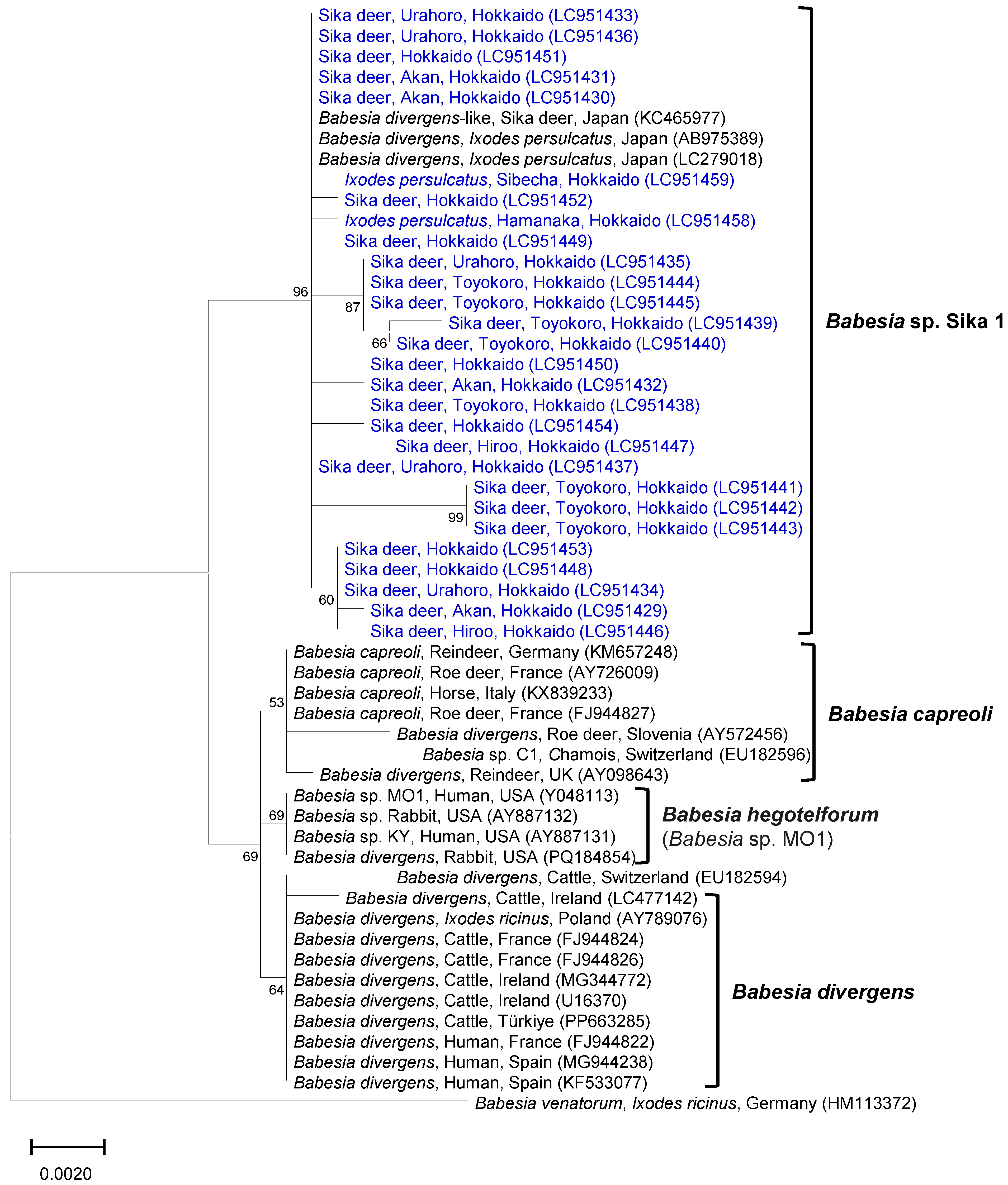
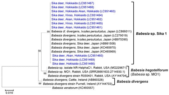
In the 18S rRNA phylogeny, the newly obtained Babesia sp. Sika 1 sequences clustered with previously reported Babesia sp. Sika 1 sequences from sika deer and I. persulcatus ticks in Japan (Figure 2). The Babesia sp. Sika 1 clade was sister to the common ancestor of B. divergens, B. capreoli, and B. hegotelforum (formerly Babesia sp. MO1). The analysis of beta-tubulin gene sequences (Accession Nos. LC951460-LC951469) from Babesia sp. Sika 1-positive deer further supported the distinct phylogenetic position of Babesia sp. Sika 1 relative to B. divergens (Figure 3). The obtained sequences formed a sister clade to B. hegotelforum, and the common ancestor of these two clades was sister to the B. divergens clade (Figure 3).
Figure 2.
Phylogenetic analysis of 18S rRNA sequences of Babesia sp. Sika 1 obtained from sika deer and questing ticks. The sequences obtained in this study (highlighted in blue) clustered with previously reported Babesia sp. Sika 1 sequences and were sister to the common ancestor of Babesia divergens, Babesia capreoli, and Babesia hegotelforum.
Figure 3.
Phylogenetic analysis of beta-tubulin sequences of Babesia sp. Sika 1 obtained from sika deer. The sequences obtained in this study (highlighted in blue) clustered with previously reported Babesia sp. Sika 1 sequences and were sister to the Babesia hegotelforum clade. The common ancestor of these two clades was sister to the Babesia divergens clade.
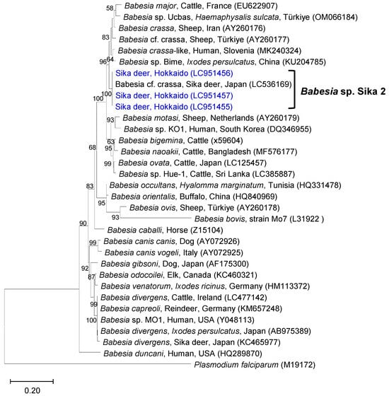
In the 18S rRNA phylogeny, Babesia sp. Sika 2 sequences clustered with a previously reported Babesia sequence from sika deer in Japan, and formed a sister clade to the common ancestor of Babesia major, Babesia crassa, and B. crassa-like species (Figure 4).
Figure 4.
Phylogenetic analysis of 18S rRNA sequences of Babesia sp. Sika 2 obtained from sika deer. The sequences obtained in this study (highlighted in blue) clustered with a previously reported Babesia sp. Sika 2 sequence and were sister to the common ancestor of Babesia crassa, Babesia crassa-like species, and Babesia major.
3.2. Distribution of Babesia sp. Sika 1 and Babesia sp. Sika 2 in Deer, Ticks, and Cattle as Determined by Duplex PCR Assay
Based on the sequence polymorphisms in 18S rRNA, taxon-specific primers were designed for simultaneous detection of Babesia sp. Sika 1 and Babesia sp. Sika 2 by duplex PCR assay. The assay produced the expected taxon-specific amplicons, but no amplification was observed from the other tested hemoparasites and uninfected bovine blood DNA, indicating specificity for the tested DNA samples.
The duplex PCR assay was then used to screen all the deer, tick, and cattle DNA samples. Babesia sp. Sika 1 was detected in 86/285 deer samples (30.2%), with positive deer distributed across multiple sampling sites in Hokkaido (Table 1). By contrast, Babesia sp. Sika 2 was detected in 4/285 deer samples (1.4%) at only a limited number of sampling sites. All samples identified as positive for Babesia sp. Sika 1 or Babesia sp. Sika 2 by the initial universal PCR and sequencing were also detected as positive for the corresponding taxon by duplex PCR. The sample identified as co-infected with both taxa by the initial PCR and sequencing was also positive for both taxa using the duplex PCR. Among the samples identified as negative by the initial PCR, the duplex PCR additionally detected Babesia sp. Sika 1 in 60 samples and Babesia sp. Sika 2 in one sample; no additional co-infections were detected.
Among the 671 tick DNA samples, Babesia sp. Sika 1 was detected in 7/367 (1.9%) I. persulcatus ticks collected from Hamanaka, Shibecha, and Taiki, while no other tick species tested positive for this taxon (Table 2). Positive ticks included both adults and nymphs, whereas no other tick species tested positive. By contrast, Babesia sp. Sika 2 was not detected in any tick samples. The Babesia sp. Sika 1-positive ticks included both adults and nymphs. Two selected I. persulcatus DNA samples that tested positive for Babesia sp. Sika 1 were additionally subjected to the universal 18S rRNA PCR assay. The sequences (Accession Nos. LC951458-LC951459) obtained from the resulting amplicons shared 99.2–99.9% sequence identity with the deer-derived Babesia sp. Sika 1 sequences and clustered with these sequences in the phylogenetic analysis, supporting the duplex PCR results (Figure 2).
Blood DNA samples from a total of 767 cattle from six districts in Hokkaido, including 50, 100, 300, 77, 146, and 94 cattle from Hamanaka, Shibecha, Shikaoi, Otofuke, Taiki, and Shin-Hidaka districts, respectively, that had been collected between 2008 and 2016 [21], were screened using the duplex PCR assay. We found that none of the 767 DNA samples tested positive for either Babesia sp. Sika 1 or Babesia sp. Sika 2.
4. Discussion
The present study identified two genetically distinct Babesia lineages in sika deer in Hokkaido prefecture and designated them Babesia sp. Sika 1 and Babesia sp. Sika 2. Of these two taxa, Babesia sp. Sika 1 was also detected in I. persulcatus ticks collected from cattle pastures, whereas neither taxon was detected in the examined cattle. These findings provided an opportunity to examine whether Babesia taxa infecting sika deer, particularly Babesia sp. Sika 1, which is closely related to the bovine parasite B. divergens, can infect cattle when opportunities for transmission are present within the deer–tick–cattle interface.
4.1. Taxonomic Clarification and Host and Tick Associations of Babesia sp. Sika 1
A relatively high proportion of the sika deer examined in the present study were identified as positive for Babesia sp. Sika 1 by the taxon-specific duplex PCR assay. The detection rate obtained with the duplex PCR was substantially higher than that obtained with the initial Babesia spp. PCR. This difference may reflect differences in amplification efficiency between the two assays. The initial PCR used broad-range primers targeting a relatively long 18S rRNA fragment, whereas the duplex PCR used a Babesia sp. Sika 1-specific forward primer targeting a shorter region. In addition, the primers used for the initial PCR may not completely match the 18S rRNA sequence of Babesia sp. Sika 1, which may have reduced amplification efficiency. These differences may have contributed to the higher detection rate of Babesia sp. Sika 1 obtained with the duplex PCR assay.
In the present study, the 18S rRNA phylogeny separated Babesia sp. Sika 1 from B. divergens, B. capreoli, and B. hegotelforum, placing it in a distinct lineage. Analysis of beta-tubulin sequences also placed Babesia sp. Sika 1 separately from B. divergens, although the topology differed from that obtained with 18S rRNA sequences. The concordant separation across two independent molecular markers strengthens the evidence that Babesia sp. Sika 1 represents a distinct Babesia lineage, although these phylogenetic findings do not by themselves establish formal species status. Some sequences derived from hosts other than cattle and humans that were previously identified as B. divergens were also positioned outside the B. divergens clade. Their phylogenetic positions and reported host origins suggest that some of these sequences may represent other Babesia taxa rather than B. divergens. Together, these phylogenetic findings support the recognition of Babesia sp. Sika 1 as a distinct Babesia taxon rather than direct assignment to B. divergens.
The phylogenetically distinct Babesia species closely related to B. divergens also differs in their vertebrate hosts and Ixodes tick species. Babesia divergens primarily infects cattle and is transmitted by I. ricinus in Europe [4], whereas B. capreoli mainly infects cervids in Europe and is transmitted by I. ricinus [28], and B. hegotelforum has been reported from cottontail rabbits and is transmitted by Ixodes dentatus in North America [15]. In addition, B. divergens and B. hegotelforum are zoonotic species capable of infecting humans [4,29]. In contrast, Babesia sp. Sika 1 has been frequently detected in sika deer and was also detected in I. persulcatus in Hokkaido, consistent with previous reports of this parasite in the same deer–tick system [8,9,11]. Therefore, the distinct phylogenetic position, together with differences in vertebrate hosts and tick vectors compared with B. divergens, further supports the recognition of Babesia sp. Sika 1 as a distinct Babesia lineage.
Accurate taxonomic recognition of Babesia sp. Sika 1 is important because referring to this parasite as B. divergens-like or as the B. divergens Asian lineage may lead to assumptions about its host range and epidemiological significance based on the established biology of B. divergens. The present study provided phylogenetic evidence that Babesia sp. Sika 1 is distinct from B. divergens, but the available data are not sufficient for formal species description. Formal taxonomic recognition would require additional biological and genomic evidence, including morphological characterization and comparative genomic analysis with B. divergens and other closely related taxa [15]. Such information would provide the additional evidence needed to formally describe Babesia sp. Sika 1 as a separate Babesia species.
4.2. Babesia sp. Sika 2 in Sika Deer, Ticks, and Cattle
Unlike Babesia sp. Sika 1, Babesia sp. Sika 2 has been poorly characterized, with only limited information available on its distribution and host associations [13]. In the 18S rRNA phylogeny, Babesia sp. Sika 2 formed a distinct phylogenetic clade separate from the other Babesia species included in the analysis. However, its biological and epidemiological characteristics remain largely unknown. In contrast to Babesia sp. Sika 1, the prevalence of Babesia sp. Sika 2 was very low in the wild sika deer population, and this taxon was not detected in any of the questing ticks. These contrasting patterns of the two taxa suggest that genetically distinct Babesia taxa may differ substantially in their prevalence within the same wildlife host population and their distribution among questing tick populations.
The low prevalence of Babesia sp. Sika 2 in deer may be related to low parasite burdens, which could reduce the probability of detection by PCR [30]. Alternatively, it is possible that other vertebrate hosts may serve as the primary hosts for Babesia sp. Sika 2, while sika deer may represent a spillover host rather than the principal host [31]. The low prevalence of Babesia sp. Sika 2 in the deer examined in the present study may have limited the opportunities for acquisition by feeding ticks and consequently contributed to its absence from the tick samples. Alternatively, Babesia sp. Sika 2 may be transmitted by tick species other than those surveyed in the present study. Further studies involving additional tick species, other sympatric wildlife species, and broader geographic sampling would help clarify the host range and transmission ecology of Babesia sp. Sika 2.
4.3. Epidemiological Implications at the Deer–Tick–Cattle Interface
The present study provided field evidence on Babesia taxa at the deer–tick–cattle interface in Hokkaido. Wild sika deer and grazing cattle share pasture environments, and I. persulcatus feeds on both hosts [18,19,20]. Despite this ecological overlap, neither of the two Babesia taxa examined in the present study was detected in cattle. A similar pattern has been reported for a Theileria species in Hokkaido, which was detected in sika deer and questing ticks collected from cattle pastures, but not in sympatric grazing cattle [21]. Together, these observations suggest that the presence of tick-borne protozoan parasites in wildlife and questing ticks within cattle environments may not necessarily result in detectable infection of cattle.
The absence of Babesia sp. Sika 1 in cattle is notable given its close phylogenetic relationship with B. divergens, which primarily infects cattle [4]. The present field observations suggest that close phylogenetic relatedness does not necessarily correspond to a similar host range. Host specificity may instead depend on biological compatibility between the parasite and its vertebrate host.
Biological compatibility may depend on the ability of the parasites to establish and multiply within host cells. In Babesia, infection involves a successful invasion of erythrocytes followed by intracellular development and multiplication, and differences in these processes may contribute to host range [32,33]. For example, an in vitro study of B. bovis showed that host specificity can involve differences in erythrocyte invasion, multiplication, and reinvasion among host species [34]. Similar differences in erythrocyte invasion or intracellular development could contribute to the absence of Babesia sp. Sika 1 from cattle. Therefore, additional studies are needed to experimentally examine the interaction of Babesia sp. Sika 1 with bovine erythrocytes and determine whether its absence from cattle reflects limited compatibility with bovine erythrocytes.
Successful isolation and in vitro cultivation of Babesia sp. Sika 1 would enable comparative experiments using sika deer and bovine erythrocytes to directly examine erythrocyte invasion, intracellular development, and multiplication. In parallel, larger-scale epidemiological studies using deer, questing ticks, and sympatric cattle collected from multiple geographic regions would help determine whether the absence of Babesia sp. Sika 1 and Babesia sp. Sika 2 from cattle is consistent across different ecological settings. These approaches could clarify the host range and transmission ecology of Babesia taxa detected in sika deer in Hokkaido, Japan.
4.4. Limitations of the Study
Several limitations should be considered when interpreting the present findings. First, the analytical sensitivity of the duplex PCR assay was not formally evaluated, and its ability to detect low levels of Babesia sp. Sika 1 and Babesia sp. Sika 2 is therefore unknown. This limitation is particularly relevant to the low detection rate of Babesia sp. Sika 2 and to the interpretation of negative results. In addition, preferential amplification of Babesia sp. Sika 1 over Babesia sp. Sika 2 in samples containing both taxa cannot be excluded.
Second, the molecular evidence available for the two taxa was not equivalent. Babesia sp. Sika 1 was characterized using both 18S rRNA and beta-tubulin sequences, whereas Babesia sp. Sika 2 was characterized only by 18S rRNA sequences. Therefore, the phylogenetic evidence supporting the distinct position of Babesia sp. Sika 2 is more limited, and additional genetic markers will be needed to clarify its taxonomic position.
Third, the deer, tick, and cattle samples were collected during different sampling periods. Although these hosts share environments and I. persulcatus feeds on both deer and cattle, the present study therefore does not demonstrate contemporaneous circulation or direct transmission of the same Babesia taxa among these hosts.
Finally, the absence of detectable Babesia sp. Sika 1 and Babesia sp. Sika 2 in the examined cattle should be interpreted cautiously. Negative PCR results may reflect low parasite burdens, differences in sample timing, or limitations in assay sensitivity, and the present findings do not establish whether either taxon is biologically incapable of infecting cattle. Larger studies using samples collected contemporaneously from deer, ticks, and cattle, together with more sensitive and analytically validated molecular assays, will be needed to further clarify the transmission potential and host range of these Babesia taxa.
5. Conclusions
The present study identified two genetically distinct Babesia lineages in sika deer in Hokkaido, provisionally designated Babesia sp. Sika 1 and Babesia sp. Sika 2. While Babesia sp. Sika 1 was also detected in questing I. persulcatus ticks, neither lineage was detected in the examined grazing cattle. Phylogenetic analyses using 18S rRNA and beta-tubulin sequences supported the distinct position of Babesia sp. Sika 1 relative to B. divergens, while Babesia sp. Sika 2, which was characterized only by 18S rRNA, formed a distinct phylogenetic clade. These findings support the recognition of Babesia sp. Sika 1 and Babesia sp. Sika 2 as distinct Babesia lineages, but do not establish formal species status. The absence of detectable infection in cattle should be interpreted cautiously in view of the limited analytical assessment of the duplex PCR and the different sampling periods among the examined host animals.
Author Contributions
Conceptualization, I.Y., T.S. and N.Y.; methodology, I.Y., Y.M., R.U.-S., S.F., T.S. and N.Y.; formal analysis, I.Y., Y.M., R.U.-S., S.F., T.S. and N.Y.; investigation, I.Y., Y.M. and R.U.-S.; writing—original draft preparation, I.Y.; writing—review and editing, Y.M., R.U.-S., S.F., T.S. and N.Y.; supervision, T.S. and N.Y.; funding acquisition, N.Y. All authors have read and agreed to the published version of the manuscript.
Funding
This study was supported by a grant from the Japan Agency for Medical Research and Development (Grant No. 25wm0225031h0003).
Institutional Review Board Statement
The animal study protocol was approved by The Animal Care and Use Committee of OBIHIRO UNIVERSITY OF AGRICULTURE AND VETERINARY MEDICINE, HOKKAIDO, JAPAN (Approval number: 20-04; date of approval: 16 April 2020).
Informed Consent Statement
Not applicable.
Data Availability Statement
All data generated in the present study are reported in this article.
Acknowledgments
We thank all the staff from the cattle farms, field veterinarians, and hunters who participated in this study for their kind cooperation. In addition, we thank Hiroko Yomamoto from the National Research Center for Protozoan Diseases, Obihiro University of Agriculture and Veterinary Medicine, for her outstanding technical assistance.
Conflicts of Interest
The authors declare no conflicts of interest.
References
- Homer, M.J.; Aguilar-Delfin, I.; Telford, S.R., III; Krause, P.J.; Persing, D.H. Babesiosis. Clin. Microbiol. Rev. 2000, 13, 451–469. [Google Scholar] [CrossRef]
- Vannier, E.; Gewurz, B.E.; Krause, P.J. Human babesiosis. Infect. Dis. Clin. N. Am. 2008, 22, 469–488. [Google Scholar] [CrossRef] [Scilit]
- Bock, R.; Jackson, L.; de Vos, A.; Jorgensen, W. Babesiosis of cattle. Parasitology 2004, 129, S247–S269. [Google Scholar] [CrossRef] [Scilit]
- Zintl, A.; Mulcahy, G.; Skerrett, H.E.; Taylor, S.M.; Gray, J.S. Babesia divergens, a bovine blood parasite of veterinary and zoonotic importance. Clin. Microbiol. Rev. 2003, 16, 622–636. [Google Scholar] [CrossRef] [Scilit]
- Sivakumar, T.; Tuvshintulga, B.; Otgonsuren, D.; Batmagnai, E.; Ahedor, B.; Kothalawala, H.; Vimalakumar, S.C.; Silva, S.S.P.; Yamagishi, J.; Yokoyama, N. Phylogenetic analyses of the mitochondrial, plastid, and nuclear genes of Babesia sp. Mymensingh and its naming as Babesia naoakii n. sp. Parasites Vectors 2022, 15, 299. [Google Scholar] [CrossRef] [Scilit]
- Ord, R.L.; Lobo, C.A. Human babesiosis: Pathogens, prevalence, diagnosis and treatment. Curr. Clin. Microbiol. Rep. 2015, 2, 173–181. [Google Scholar] [CrossRef] [Scilit]
- Fanelli, A. A historical review of Babesia spp. associated with deer in Europe: Babesia divergens/Babesia divergens-like, Babesia capreoli, Babesia venatorum, Babesia cf. odocoilei. Vet. Parasitol. 2021, 294, 109433. [Google Scholar] [CrossRef] [Scilit]
- Ma, Y.; Sivakumar, T.; Mumbi, N.N.M.; Umemiya-Shirafuji, R.; Yokoyama, N. An epidemiological survey of zoonotic Babesia species in questing ticks in Hokkaido, Japan, using a newly developed PCR-sequencing approach. Parasitol. Int. 2026, 110, 103136. [Google Scholar] [CrossRef] [Scilit]
- Zamoto-Niikura, A.; Tsuji, M.; Imaoka, K.; Kimura, M.; Morikawa, S.; Holman, P.J.; Hirata, H.; Ishihara, C. Sika deer carrying Babesia parasites closely related to B. divergens, Japan. Emerg. Infect. Dis. 2014, 20, 1398–1400. [Google Scholar] [CrossRef] [Scilit]
- Zamoto-Niikura, A.; Tsuji, M.; Qiang, W.; Morikawa, S.; Hanaki, K.I.; Holman, P.J.; Ishihara, C. The Babesia divergens Asia lineage is maintained through enzootic cycles between Ixodes persulcatus and sika deer in Hokkaido, Japan. Appl. Environ. Microbiol. 2018, 84, e02491-17. [Google Scholar] [CrossRef] [Scilit]
- Zamoto-Niikura, A.; Hagiwara, K.; Imaoka, K.; Morikawa, S.; Ishihara, C.; Hanaki, K.I. Epidemiological survey of Babesia divergens Asia lineage in wild sika deer (Cervus nippon) by using direct PCR in Japan. Jpn. J. Infect. Dis. 2020, 73, 68–71. [Google Scholar] [CrossRef] [Scilit]
- Elbaz, E.; Moustafa, M.A.M.; Lee, K.; Ching, A.L.C.; Shimozuru, M.; Sashika, M.; Nakao, R.; El-Khodery, S.A.; Tsubota, T. Utilizing attached hard ticks as pointers to the risk of infection by Babesia and Theileria species in sika deer (Cervus nippon yesoensis), in Japan. Exp. Appl. Acarol. 2020, 82, 411–429. [Google Scholar] [CrossRef] [Scilit]
- Masatani, T.; Hayashi, K.; Morikawa, M.; Ozawa, M.; Kojima, I.; Okajima, M.; Takano, A.; Shimoda, H.; Maeda, K.; Matsuu, A.; et al. Molecular detection of tick-borne protozoan parasites in sika deer (Cervus nippon) from western regions of Japan. Parasitol. Int. 2020, 79, 102161. [Google Scholar] [CrossRef] [Scilit]
- Hrazdilová, K.; Rybářová, M.; Široký, P.; Votýpka, J.; Zintl, A.; Burgess, H.; Steinbauer, V.; Žákovčík, V.; Modrý, D. Diversity of Babesia spp. in cervid ungulates based on the 18S rDNA and cytochrome c oxidase subunit I phylogenies. Infect. Genet. Evol. 2020, 77, 104060. [Google Scholar] [CrossRef] [Scilit]
- Singh, P.; Estrada, K.; Gonzalez, L.M.; Grande, R.; Sánchez-Prieto, S.; Cornillot, E.; Harb, O.; Sanchez-Flores, A.; Montero, E.; Le Roch, K.G.; et al. Babesia hegotelforum sp. nov., a zoonotic Babesia species previously referred to as Babesia sp. MO1. Emerg. Microbes Infect. 2026, 15, 2637280. [Google Scholar] [CrossRef] [Scilit]
- Hata, A.; Tsukada, H.; Washida, A.; Mitsunaga, T.; Takada, M.B.; Suyama, T.; Takeuchi, M. Temporal and spatial variation in the risk of grazing damage to sown grasslands by sika deer (Cervus nippon) in a mountainous area, central Japan. Crop Prot. 2019, 119, 185–190. [Google Scholar] [CrossRef] [Scilit]
- Takafumi, H.; Kamii, T.; Murai, T.; Yoshida, R.; Sato, A.; Tachiki, Y.; Akamatsu, R.; Yoshida, T. Seasonal and year-round use of the Kushiro Wetland, Hokkaido, Japan by sika deer (Cervus nippon yesoensis). PeerJ 2017, 5, e3869. [Google Scholar] [CrossRef] [Scilit]
- Shimizu, K.; Shimozuru, M.; Yamanaka, M.; Ito, G.; Nakao, R.; Tsubota, T. Seasonal infestation patterns of ticks on Hokkaido sika deer (Cervus nippon yesoensis). Parasitology 2024, 151, 1317–1325. [Google Scholar] [CrossRef] [Scilit]
- Shimizu, K.; Shimozuru, M.; Tsuruga, H.; Shirane, Y.; Yamanaka, M.; Ito, G.; Shinjo, K.; Murakami, T.; Ijuin, A.; Kovba, A.; et al. Patterns of tick infestation and tick-borne pathogen prevalence in sika deer and brown bears in Hokkaido, Japan. Exp. Appl. Acarol. 2025, 96, 10. [Google Scholar] [CrossRef] [Scilit]
- Yamane, I.; Nishiguchi, A.; Kobayashi, S.; Zeniya, Y. Cross-sectional survey of ixodid tick species on grazing cattle in Japan. Exp. Appl. Acarol. 2006, 38, 67–74. [Google Scholar] [CrossRef] [Scilit]
- Shibata, S.; Sivakumar, T.; Igarashi, I.; Umemiya-Shirafuji, R.; Inokuma, H.; Fukumoto, S.; Yokoyama, N. Epidemiological survey of a cervine Theileria in wild deer, questing ticks, and cattle in Hokkaido, Japan. Ticks Tick-Borne Dis. 2018, 9, 1235–1240. [Google Scholar] [CrossRef] [Scilit]
- Shirozu, T.; Badolo, A.; Soga, A.; Yoshimura, A.; Morishita, Y.-K.; Koketsu, M.; Umemiya-Shirafuji, R.; Inokuma, H.; Yokoyama, N.; Fukumoto, S. Development and evaluation of a novel loop-mediated isothermal amplification (LAMP) method targeting Theileria parasites infecting Yezo sika deer. Parasitol. Int. 2020, 77, 102130. [Google Scholar] [CrossRef] [Scilit]
- Yamaguti, N.; Tipton, V.J.; Keegan, H.L.; Toshioka, S. Ticks of Japan, Korea, and the Ryukyu Islands. Brigham Young Univ. Sci. Bull. Biol. Ser. 1971, 15, 1–226. [Google Scholar]
- Sivakumar, T.; Tuvshintulga, B.; Zhyldyz, A.; Kothalawala, H.; Yapa, P.R.; Kanagaratnam, R.; Vimalakumar, S.C.; Abeysekera, T.S.; Weerasingha, A.S.; Yamagishi, J.; et al. Genetic analysis of Babesia isolates from cattle with clinical babesiosis in Sri Lanka. J. Clin. Microbiol. 2018, 56, e00895-18. [Google Scholar] [CrossRef] [Scilit]
- Katoh, K.; Rozewicki, J.; Yamada, K.D. MAFFT online service: Multiple sequence alignment, interactive sequence choice and visualization. Brief. Bioinform. 2019, 20, 1160–1166. [Google Scholar] [CrossRef] [Scilit]
- Tamura, K.; Stecher, G.; Kumar, S. MEGA11: Molecular Evolutionary Genetics Analysis version 11. Mol. Biol. Evol. 2021, 38, 3022–3027. [Google Scholar] [CrossRef] [Scilit]
- Tamura, K.; Nei, M. Estimation of the number of nucleotide substitutions in the control region of mitochondrial DNA in humans and chimpanzees. Mol. Biol. Evol. 1993, 10, 512–526. [Google Scholar] [CrossRef] [Scilit]
- Malandrin, L.; Jouglin, M.; Sun, Y.; Brisseau, N.; Chauvin, A. Redescription of Babesia capreoli (Enigk and Friedhoff, 1962) from roe deer (Capreolus capreolus): Isolation, cultivation, host specificity, molecular characterisation and differentiation from Babesia divergens. Int. J. Parasitol. 2010, 40, 277–284. [Google Scholar] [CrossRef] [Scilit]
- Montero, E.; Gray, J.; Lobo, C.A.; González, L.M. Babesia and human babesiosis. Pathogens 2022, 11, 399. [Google Scholar] [CrossRef] [Scilit]
- Bloch, E.M.; Lee, T.H.; Krause, P.J.; Telford, S.R., III; Montalvo, L.; Chafets, D.; Usmani-Brown, S.; Lepore, T.J.; Busch, M.P. Development of a real-time polymerase chain reaction assay for sensitive detection and quantitation of Babesia microti infection. Transfusion 2013, 53, 2299–2306. [Google Scholar] [CrossRef] [Scilit]
- Lempereur, L.; Wirtgen, M.; Nahayo, A.; Caron, Y.; Shiels, B.; Saegerman, C.; Losson, B.; Linden, A. Wild cervids are host for tick vectors of Babesia species with zoonotic capability in Belgium. Vector-Borne Zoonotic Dis. 2012, 12, 275–280. [Google Scholar] [CrossRef] [Scilit]
- Elsworth, B.; Duraisingh, M.T. A framework for signaling throughout the life cycle of Babesia species. Mol. Microbiol. 2021, 115, 882–890. [Google Scholar] [CrossRef] [Scilit]
- Yokoyama, N.; Okamura, M.; Igarashi, I. Erythrocyte invasion by Babesia parasites: Current advances in the elucidation of the molecular interactions between the protozoan ligands and host receptors in the invasion stage. Vet. Parasitol. 2006, 138, 22–32. [Google Scholar] [CrossRef] [Scilit]
- Gaffar, F.R.; Franssen, F.F.; de Vries, E. Babesia bovis merozoites invade human, ovine, equine, porcine and caprine erythrocytes by a sialic acid-dependent mechanism followed by developmental arrest after a single round of cell fission. Int. J. Parasitol. 2003, 33, 1595–1603. [Google Scholar] [CrossRef] [Scilit]

Disclaimer/Publisher’s Note: The statements, opinions and data contained in all publications are solely those of the individual author(s) and contributor(s) and not of MDPI and/or the editor(s). MDPI and/or the editor(s) disclaim responsibility for any injury to people or property resulting from any ideas, methods, instructions or products referred to in the content. |
© 2026 by the authors. Licensee MDPI, Basel, Switzerland. This article is an open access article distributed under the terms and conditions of the Creative Commons Attribution (CC BY) license.