


Simple Summary
Bats play an important role as a natural reservoir of pathogens in nature and can carry viruses that affect humans and other animals without becoming seriously ill themselves. Understanding how bats resist infections and protect their brains from inflammation may help scientists develop new strategies for studying diseases in both animals and people. However, there are still very few laboratory models available to study bat brain cells. In this study, we created the first primary and immortalized brain cell cultures from the bat species Hypsugo savii, a common European bat that often lives close to human environments. We tested different methods to produce long-lasting cell cultures and found that a method based on Simian virus 40 successfully generated stable immortalized cells. We also compared the genetic activity of the original and immortalized cells and found important differences related to cell growth, immune responses, and cell survival. These new bat brain cell models provide a valuable tool for studying how bats interact with viruses, how they tolerate infections without severe disease, and how their brains respond to inflammation. The results may support future research on emerging zoonotic diseases and improve our understanding of animal and human health within a One Health approach.
Abstract
Bats are important reservoirs of zoonotic pathogens and valuable models for studying antiviral tolerance and neuroinflammation within a One Health framework. However, chiropteran neural 2D-in vitro models remain limited. Here, we established and characterized the first chiropteran primary (CpBCs) and immortalized (CiBCs) cell lines from Hypsugo savii species. To overcome the limited lifespan of CpBCs, immortalization strategies based on human telomerase reverse transcriptase (hTERT) and Simian virus 40 large T antigen (SV40) were evaluated. Electroporation-mediated transfection with SV40 successfully generated CiBCs, whereas liposome-mediated and hTERT-based approaches were unsuccessful. RNA sequencing revealed marked transcriptional changes comparing CiBCs with CpCBs, such as the upregulation of pathways related to cell cycle progression, DNA replication, and proliferation in CiBCs, together with the downregulation of apoptosis, inflammatory signaling, and immune-related pathways. Immortalized cells also exhibited enrichment of neural stem cell-like and cancer-associated signatures, suggesting partial dedifferentiation induced by SV40-mediated immortalization. Overall, this study provides a novel chiropteran brain-derived 2D-in vitro platform for investigating bat neurobiology, host–pathogen interactions, viral tolerance, and neurotropic infectious diseases relevant to emerging zoonoses.
1. Introduction
The One Health framework emphasizes the interconnectedness of human, animal, and environmental health, highlighting the need for integrated approaches to understand and mitigate emerging biological threats [1,2]. Recent global events, particularly the COVID-19 pandemic, have intensified scientific attention toward wildlife species as reservoirs of zoonotic pathogens [1,2,3]. Among these, bats (order Chiroptera) have emerged as particularly significant due to their remarkable ecological diversity and unique host–pathogen interactions [1,2]. Several studies suggest that bats possess a “tuned-down” immune system, characterized by constitutive antiviral defenses and a controlled inflammatory profile [1,2,4,5]. Additionally, the high metabolic demands associated with powered flight result in elevated production of reactive oxygen species [1]. Consequently, bats have evolved enhanced DNA repair pathways and antioxidant systems, which mitigate cellular damage and reduce inflammation-associated tissue injury during viral replication [1,2,4,5,6]. Their tendency to form dense roosting colonies, relatively long lifespans for their body size, and capacity for torpor or hibernation promote sustained low-dose pathogen exposure and may favor the evolution of immune tolerance while limiting viral replication dynamics [1,3,4]. Importantly, these adaptations have broader biomedical implications, particularly towards the study of diseases affecting the nervous system (NS) [2,4].
Notably, bats are recognized as natural reservoirs for a wide range of NS zoonotic infections. While some of these agents—particularly Lyssaviruses—can occasionally induce mild pathological manifestations in bats, most do not cause any pathology, especially within the NS [2]. Additionally, regarding NS non-infectious diseases, the dampened inflammatory responses observed in bats are of particular interest for understanding human neuroinflammatory and neurodegenerative disorders such as Multiple Sclerosis, Alzheimer’s disease, and Parkinson’s disease [7,8,9,10,11]. Furthermore, bats exhibit exceptional longevity and maintain cognitive function with age, suggesting enhanced resistance to neurodegeneration and cancer [6,11,12,13]. Intriguingly, bats’ specialized sensory systems, particularly echolocation, rely on highly adapted hippocampal and cortical circuits involved in spatial navigation and sensorimotor integration [1]. These systems might also inform research into neurological conditions such as epilepsy and schizophrenia [11]. Moreover, the metabolic demands of flight expose bats to intermittent hypoxia-like conditions, suggesting potential adaptations relevant to ischemic tolerance in conditions such as stroke and traumatic brain injury [11,14].
Despite their scientific value, bats remain underrepresented as experimental models. Limitations include challenges in breeding and maintenance, limited genetic manipulation tools compared to traditional laboratory organisms, and high interspecies variability [1,2,3]. In this context, standardized 2D-in vitro systems may provide a valuable alternative model for improving our understanding of bat biology and physiology.
Primary somatic cells (pSCs), either isolated in-house or commercially available, are widely used in human biomedical research [15]. However, their limited lifespan and susceptibility to replicative senescence restrict long-term applications [15,16]. Therefore, to overcome these limitations, immortalization of pSCs is required to stop cell-aging mechanisms [17,18,19,20,21,22]. In mammals, common genes used to generate immortalized cells employ viral oncogenes—such as Simian virus 40 large T antigen (SV40), human papillomavirus E6/E7, or Epstein–Barr virus latent membrane protein 1—or non-viral oncogenes (e.g., KRAS and MYC), often in combination with telomerase reverse transcriptase (hTERT) expression and/or short-hairpin RNA constructs [15,16,17,18,20,21,22,23,24,25,26,27,28,29,30,31]. Expression by pSCs of one of these genes can be achieved using different methodologies, including biological (e.g., transduction), chemical, or physical approaches, with liposome- and electroporation-mediated transfection being the most commonly used ones [15,16,32,33,34].
In human biomedical research, a broad array of primary NS non-cancerous cell types—including neurons, astrocytes, oligodendrocytes, Schwann cells and microglia—are commercially available together with their immortalized counterparts (except for neurons). Numerous laboratories have also successfully isolated and immortalized human astrocytes, Schwann cells, microglia, and neural stem cells (NSCs), whereas oligodendrocytes remain restricted to pSCs [29,30,35,36,37,38,39,40,41,42,43,44,45]. In contrast, non-tumorigenic NS-derived cells from veterinary species—excluding conventional laboratory animals such as rodents and rabbits—remain limited. Commercially available NS pSCs include canine neurons, astrocytes, and microglia, as well as bovine, feline, and non-human primate astrocytes. Available immortalized NS cells include feline astrocytes, alongside non-characterized mustelid and ovine NS-derived cells. In-house isolations have included porcine neurons, astrocytes, and oligodendrocytes, as well as canine, bovine, caprine, and porcine NSCs [46,47,48,49,50,51,52,53,54,55,56,57]. In addition, immortalization efforts have yielded porcine NSCs and ovine astrocytes and microglia [55,58,59,60]. With specific regard to chiropteran NS 2D-in vitro models, several cell lines have been reported, both commercially available and in-house isolated.
In light of the previous considerations, the present study aimed to isolate and establish post-natal chiropteran primary brain-derived cells (CpBCs), herein called brain cells, and their immortalized counterparts (CiBCs) to generate a robust 2D-in vitro model that can support investigations into bat-specific antiviral mechanisms, neuroprotection, and NS-related host–pathogen interactions within a One Health context. Additionally, we examined for the first time the transcriptomic alterations associated with the immortalization of CpBCs to better characterize the changes underlying this process at the RNA level [2].
2. Materials and Methods
2.1. Tissue Sampling and DNA Extraction for Species Identification
A young male bat was humanely euthanized due to an irreparable humerus fracture at a specialized veterinary clinic following a thorough clinical evaluation. Necropsy was performed by a trained pathologist immediately after death, and samples were transported to the Department of Comparative Biomedicine and Food Safety of the University of Padua (Legnaro, Italy) for processing.
For species identification, skin, muscle, and liver tissue fragments (approximately 0.5 × 0.5 cm) were immediately collected and stored at −20 °C until DNA extraction using the DNeasy Blood and Tissue Kit (Qiagen, Venlo, The Netherlands, #69506), following the manufacturer’s protocol. Extracted DNA was then quantified using Nanodrop ND-1000 technology (Wilmington, DE, USA). Only high-quality DNA samples obtained from muscle and liver tissues were selected and submitted for species identification through DNA barcoding analysis targeting the mitochondrial gene Cytochrome c oxidase subunit I (COI) [61]. PCR amplifications were performed using the universal primers for COI-barcode [62], and PCR products were sent for sequencing to BMR Genomics Company (Padua, Italy).
2.2. Isolation and Establishment of Chiropteran Primary Brain Cells
For the isolation of CpBCs, brain tissue fragments (approximately 0.5 × 0.5 cm) were immediately suspended in sterile phosphate-buffered saline (PBS) supplemented with 10% penicillin–streptomycin (P/S, Thermo Fisher Scientific, Waltham, MA, USA, #15140122). After transport at 4 °C, under sterile conditions, brain biopsies were washed with PBS, dissected to remove visible blood vessels, and minced with a sterile razor blade. Tissue dissociation was then performed using the Papain Dissociation System (Worthington Biochemical Corporation, Lakewood, NJ, USA, #LK003150) following the manufacturer’s protocol. Briefly, samples were digested at 37 °C for 1.5 h, filtered through a 70 µm cell strainer, and centrifuged at 300× g for 5 min. The resulting pellet was subjected to density gradient centrifugation with an albumin-ovomucoid inhibitor solution at 100× g for 5 min, plated in two wells of a 24-well standard plate (sP1), and cultured in Dulbecco’s Modified Eagle Medium/Nutrient Mixture F-12 (DMEM/F-12, Thermo Fisher Scientific, #11320033) with 1% GlutaMAX™ supplement (Glutamax, Thermo Fisher Scientific, #35050061), 1% MEM Non-Essential Amino Acids solution (NEAA, Thermo Fisher Scientific, #11140050), 1% N-2 supplement (N-2, Thermo Fisher Scientific, #17502048), 2% B-27™ supplement (B-27, Thermo Fisher Scientific, #17504044), 1% P/S, 20 ng/mL of human basic fibroblast growth factor (hFGF, Thermo Fisher Scientific, #100-18B-50UG), and 20 ng/mL of human epidermal growth factor (hEGF, Merck, Darmstadt, Germany, #E9644) (herein as cBCsm1) in an atmosphere of humidified air and 5% CO2 at 37 °C. Cytokines were always added fresh to the cell culture media. Additionally, 72 h after isolation, the supernatant from the wells of sP1 was replated in one well each of a 24-well standard plate (sP2) in cBCsm1 supplemented with 2% fetal bovine serum (FBS, Thermo Fisher Scientific, #A5256701) (herein as cBCsm2), and fresh cBCm1 was replaced in all wells of sP1. Furthermore, 48 h after the first replating, fresh cBCsm1 or cBCsm2 was added to all wells. Four days after the first replating, media changes with cBCsm1 or cBCsm1 supplemented with 10% FBS (herein as cBCsm3) were performed for all wells of sP1 and sP2, respectively. The day after, fresh cBCsm1 and cBCsm3 were added to all wells. Eight days after the first replating, media changes with cBCsm1 were performed for sP1 and one well of sP2, while for the other well of sP2, a medium change was performed using cBCsm3. When the day after CpBCs reached nearly 100% confluence in all wells (except for the sP2 well cultured in cBCsm1), cells were single-washed with PBS and enzymatically detached using Accutase (Merck, #A6964) diluted (1:3) in PBS at 37 °C for 5 min. Then, CpBCs were centrifuged at 400× g for 5 min and seeded (1:2) into either one well each of a 12-well standard (sP3) or two wells of a 24-well standard plate (sP4) for the wells of sP1 and sP2, respectively. Either cBCsm1 or cBCsm3 was replaced with the leftover cells in sP1 and sP2 wells and changed every other day to sP3 and sP4, respectively. When the cells reached nearly 100% confluence in sP3, CpBCs were enzymatically split using Accutase and seeded into two wells of a 12-well standard (sP5) and in one well of a 24-well standard plate (sP6) in cBCsm1. From this moment on, all wells were cultured in cBCsm3, and subsequently changed the day after splitting and every other day. CpBCs were further expanded in the 6-well standard plate format with a seeding rate ranging from 1:2 to 1:4, according to downstream experiments. cBCsm3 was always replaced the day after splitting and three times per week, following a single wash with PBS when observing large amounts of cellular death and debris. CpBCs were regularly cryopreserved in a freezing medium composed of 90% FBS and 10% dimethyl sulfoxide and screened every three months for mycoplasma contamination using the MycoAlert® Mycoplasma Detection kit (Lonza, Basel, Switzerland, #LT07-703) and the Venor®GeM Classic (Minerva Biolabs, Berlin, Germany, #11-1025) kit.
2.3. Immortalization of Chiropteran Primary Brain Cells
DH5-alpha Escherichia coli bacteria, transduced with either plasmid DNA encoding SV40 (pSV3-neo, ATCC, Manassas, VA, USA, #37150) or hTERT (pCl neo-hEST2, Addgene, Watertown, MA, USA, #1781), were grown overnight at 37 °C in Luria–Bertani medium (LB, Microbiol, Catania, Italy, #70402) agar plates supplemented with 100 μg/mL of ampicillin (Merck, #A5354-10). The day after, single antibiotic-resistant bacterial colonies were picked and allowed to grow on a shaking platform overnight at 37 °C in 3 mL of liquid LB with 100 μg/mL of ampicillin. Then, plasmid DNA was extracted using the QIAprep Spin Miniprep Kit (Qiagen, Hilden, Germany, #27104) following the manufacturer’s protocol. After assessment of plasmid DNA integrity using Hind III (Promega, Madison, WI, USA, #R6041) and EcoRI (Promega, #R6011) restriction enzymes, antibiotic-resistant bacterial colonies were allowed to grow on a shaking platform overnight at 37 °C in 500 mL of liquid LB with 100 μg/mL of ampicillin. The day after, plasmid DNA was extracted using the QIAGEN Plasmid Maxi Kit (Qiagen, #12162) using the manufacturer’s protocol. Then, extracted plasmid DNA was quantified using Nanodrop ND-1000 technology.
Two different immortalization methods were tested: one based on liposomes and one mediated by electroporation. To reduce culture heterogeneity, CpBCs after seven passages in culture were employed for immortalization experiments.
For liposome-mediated transfection, one well of a 6-well standard plate of CpBCs at nearly 100% confluence was enzymatically detached using Accutase and seeded (1:2–1:4) in new 6-well standard plates. Upon reaching 80% confluence, CpBCs were transfected using Lipofectamine™ 3000 transfection reagent with either plasmid DNA encoding SV40 or hTERT or a combination of these latter two, following the manufacturer’s protocol. Lipofectamine™ 3000 was removed after 24 h. One 6-well plate each of CpBCs transfected with either a GFP-expressing plasmid (Monster Green® Fluorescent Protein Vector, Promega, #E6421) or incubated with transfection reagent only was included to evaluate transfection efficiency and cytotoxicity through estimation of GFP-positive CpBCs using a fluorescence microscope and evaluation of cell recovery (e.g., attachment at the bottom of the plate) the day following electroporation, respectively.
For electroporation-mediated transfection, one well of a 6-well standard plate of CpBCs and CiBCs at nearly 100% confluence was enzymatically detached using Accutase and seeded (1:2–1:4) in new 6-well standard plates. Upon reaching 100% confluence, cells were enzymatically detached using Accutase and centrifuged at 400× g for 5 min. Cells were washed two times with PBS and resuspended in a 4 mm cuvette in 80–145 µL of Opti-MEM™ (Thermo Fisher Scientific, #31985062) at a cell density of 5–10 × 106 cells/mL. Cells were then transfected with 3–8 μg each of plasmid DNA encoding either SV40 or hTERT, alongside a GFP-expressing plasmid using the ECM®630 Electroporation System (BTX, Harvard Apparatus, Holliston, MA, USA) and applying the following electroporation parameters: 280–500 V, 50 uF, 129–1575 Ω, and 1–3 pulses. After electroporation, cells were incubated for 5 min on ice, and media were changed the day after. Negative controls were included to assess cell viability.
One week post-transfection, CpBCs were subjected to selection with 350 µg/mL of G418 antibiotic (Merck, #G8168-10ML) in cBCsm3 for four weeks to establish CiBCs.
2.4. Protein Extraction and Western Blot of Chiropteran Primary and Immortalized Brain Cells
To characterize CpBCs and CiBCs, antibodies targeting phenotypic markers were selected based on the orthology of the targeted antigens to chiropteran proteins. The orthologous sequences were identified as the reciprocal best hits in GenBank through BLASTp v2.17.0 searches [63]. Sequences with E-value = 0.0 and 100% coverage were aligned with CLUSTALW to assess the conservation level [64]. Proteins showing ≥65% sequence identity were therefore selected for Western blot (WB) (Table 1). To reduce culture heterogeneity, CpBCs after seven passages in culture were employed for WB experiments.
Table 1.
Primary antibodies used in Western blot and immunofluorescence to characterize chiropteran primary brain cells and chiropteran immortalized brain cells.
For protein extraction, one well each of a 6-well standard plate of CpBCs and CiBCs at nearly 100% confluence was enzymatically detached using Accutase and seeded (1:2–1:4) in new 6-well standard plates. Upon reaching nearly 100% confluence, cells were lysed for 5 min using 120 µL of Pierce™ Ripa buffer (Thermo Fisher Scientific, #89901) supplemented with cOmplete Mini EDTA-free protease inhibitor cocktail tablets (Roche, #11836170001) following the manufacturer’s protocol. Cell lysates were then centrifuged at 4 °C at 14,000× g for 15 min. Protein concentrations were calculated using a Pierce™ BCA protein Assay kit (Thermo Fisher Scientific, #23225) following the manufacturer’s protocol.
For WB, 15 μg of proteins were first denatured at 70 °C for 10 min or at 95 °C for 5 min and then resolved using Bolt™ Bis-Tris Plus Mini protein gels, 4–12% (Thermo Fisher Scientific, # NW04125BOX), or NuPAGE™ Tris-Acetate Mini protein gels, 3–8% (Thermo Fisher Scientific, # EA03755BOX), and transferred to a nitrocellulose membrane using the iBlot™ Transfer Stack kit (Thermo Fisher Scientific, # IB301002). To verify equal protein loading and transfer efficiency, membranes were stained with AdvanStain Ponceau (Aurogene, Rome, Italy, #R-03021-D50) for 5 min at room temperature. To block non-specific binding sites, blots were incubated for 90 min in 5–10% non-fat dry milk in Tris-buffered saline (TBS) containing 0.05–2% Tween-20 (TBS-T) at room temperature. Then, blots were incubated at 4 °C overnight with selected primary antibodies diluted in TBS-T containing 1–5% non-fat dry milk (Table 1). To improve signal detection, several protocols were tested.Then, membranes were incubated with goat anti-rabbit IgG (H + L) poly-HRP secondary antibody (1:3000, Thermo Fisher Scientific, #32260) or goat anti-mouse IgG (H + L) poly-HRP secondary antibody (1:3000, Thermo Fisher Scientific, #32230) diluted in TBS-T for 1 h at room temperature. Reactive bands were visualized using the SuperSignal™ West Pico PLUS Chemiluminescent Substrate kit (Thermo Fisher Scientific, #34577). Protein extracted from canine tissues (brain, spleen, and smooth muscle), canine mammary tumor cells (Cellosaurus, Geneva, Switzerland, CIPp, #CVCL_L149), and dolphin immortalized fibroblast cells (patent n°102020000003248; https://www.unipd.it/en/brevetti/scheda/sea-sentinel-system-studio-ambiente) were included as positive controls.
2.5. Immunofluorescence of Chiropteran Primary and Immortalized Brain Cells
For immunofluorescence (IF), only the antibodies that showed binding to the expected-size proteins upon WB were investigated. To reduce culture heterogeneity, CpBCs after seven passages in culture were employed for IF experiments. One well of a 6-well standard plate of CpBCs and CiBCs at nearly 100% confluence was enzymatically detached using Accutase and seeded (1:2–1:4) in new 24-well standard plates. Upon reaching 60% confluence, cells were fixed with 4% paraformaldehyde solution for 20 min at room temperature. Cells were then washed with PBS containing 0.05% Tween-20 (PBS-T) and permeabilized with PBS containing 0.1% Triton X-100 for 15 min at room temperature. To block non-specific binding sites, cells were incubated with PBS containing 4% bovine serum albumin for 1 h at room temperature and then incubated at 4 °C overnight with selected primary antibodies (Table 1). Several protocols were tested.Cells were then incubated with goat anti-mouse IgG (H + L) Alexa Fluor® 647 (1:200–1:1000, Aurogene, #AB150115-500UG) for 1 h at room temperature in the dark. Nuclei were counterstained with Hoechst33342 for 10 min (Thermo Fisher Scientific, #62249) and washed with PBS before imaging using the THUNDER Imager 3D Assay (Leica, Wetzlar, Germany).
2.6. RNA Extraction and Quantification of Chiropteran Primary and Immortalized Brain Cells
To reduce culture heterogeneity, CpBCs after seven passages in culture were employed for transcriptomic experiments. For RNA extraction, one well of a 6-well standard plate of CpBCs and CiBCs at nearly 100% confluence was enzymatically detached using Accutase and seeded (1:2–1:4) in new 12-well standard plates (three biological replicates). Upon reaching nearly 100% confluence, cells were enzymatically detached using Accutase and centrifuged at 400× g for 5 min. The obtained cell pellet was snap frozen at −80 °C until acid nucleic extraction was performed using the miRNAeasy Micro kit (Qiagen, #217084) following the manufacturer’s protocol. Extracted RNA was then quantified using both Nanodrop ND-1000 technology and the Qbit™ RNA BR Assay kit (Thermo Fisher Scientific, #Q10211). Samples were sent for RNA-sequencing (RNA-seq) to Biomarker Technologies (BMK) GmbH (Münster, Germany).
2.7. Bioinformatic Analyses
A total of six RNA-seq libraries derived from chiropteran brain cells were processed using the nf-core/rnaseq pipeline v3.22.0 (https://nf-co.re/rnaseq/3.23.0/). Sequencing reads were aligned to the Eptesicus fuscus reference genome (NCBI assembly GCA_027574615.1) using STAR. Gene-level quantification was performed with RSEM, generating raw read counts for each gene. The resulting gene-level count matrix was analyzed using iDEP v2.4.4. Lowly expressed genes were filtered out by retaining those with counts ≥0.5 counts per million in at least one sample. Data normalization and transformation were performed using the EdgeR pipeline with a pseudocount of 4 to stabilize variance. Missing values were imputed using the gene median approach. Gene identifiers were mapped to Ensembl/STRING databases, and differential expression analysis (DEG) was conducted using DESeq2 with the standard Wald test and independent filtering enabled. Genes were considered differentially expressed if they met a false discovery rate (FDR) threshold of 0.05 and a minimum fold-change (FC) of 2. Functional enrichment analysis was performed using Gene Set Enrichment Analysis (GSEA, pre-ranked mode) across Gene Ontology Biological Process (GOBP), Molecular Signatures Database Hallmark (MSigDB Hallmark), and Kyoto Encyclopedia of Genes and Genomes (KEGG) set collections, with pathway sizes restricted to 5–2000 genes and an FDR cutoff of 0.1. Directionality of FC was preserved. Similarly, to identify significantly enriched cellular signatures, a custom-made Signature database was realized using gene expression data obtained from the DropViz resource v2018.04.01 (http://dropviz.org/), based on the single-cell transcriptomic atlas of the adult mouse brain [65] (Table S1).
3. Results
3.1. DNA Barcoding Analysis Revealed Species Identification of Hypsugo savii
DNA barcoding analysis of the mitochondrial COI gene sequences revealed a high degree of similarity (>99%) with reference sequences of Hypsugo savii (Savi’s pipistrelle, Bonaparte, 1837), allowing the assignment of the sampled specimens to this species.
3.2. cBCsm3 Cell Culture Media Enables the Growth of Chiropteran Primary Brain Cells
The day following plating, undigested tissue fragments were observed in both wells, sP1. After 72 h and five days following dissociation, CpBCs displaying a heterogeneous but mainly elongated fibroblast-like morphology began to adhere to sP1 and sP2 wells, respectively (Figure 1A). Eleven days after tissue dissociation, when CpBCs in both sP1 and sP2 cultures had reached approximately 100% confluence, only one well of sP2 was switched to cBCsm1. Within this well, after 24 h of cBCsm1 exposure, spheroidal aggregates resembling neurospheres emerged. However, these formations exhibited signs of cellular suffering, leading to a medium change back to cBCsm3 after six days. Four days after the introduction of cBCsm3, CpBCs in sP1 cultures again reached approximately 100% confluence and were further expanded, as previously described.
Figure 1.
Brightfield images of chiropteran primary brain cells and chiropteran immortalized brain cells. (A) 50% confluent chiropteran primary brain cells, displaying a heterogeneous but mainly elongated fibroblast-like morphology; (B) nearly 50% confluent chiropteran immortalized brain cells, displaying a homogeneous but mainly elongated fibroblast-like and compact morphology (10×). Scale bar: 400 μm.
3.3. Simian Virus 40 Large T Antigen Mediates the Immortalization of Chiropteran Primary Brain Cells
After transfection with either pSV3-neo or pCl neo-hEST2, CpBCs showed different morphology, having a more ovaloid shape mixed with cellular debris when transfected with only the latter one, in comparison with the negative control. Similarly, when transfecting both plasmids at the same time, increased signs of cell suffering were observed; thus, the immortalization protocol was carried out only for pSV3-neo. At first, Lipofectamine™ 3000 was used following the manufacturer’s protocol, replacing the exhausted medium after 24 h. Due to no detection of GFP-positive cells in the controls, only the optimization of electroporation conditions was carried out.
Several protocol parameters were systematically optimized. Cell density and DNA concentration were adjusted to 7.5 × 10⁶ cells/mL and 8 µg of each plasmid DNA per reaction, respectively. CpBCs were resuspended in the minimum 80 µL volume recommended by the manufacturer for the 4 mm cuvette in order to maximize conductivity. Resistance (1575 Ω) and capacitance (50 µF) were set to achieve the longest possible pulse duration. Standard voltage conditions (280 V), reported in the literature, for mouse primary brain cell electroporation did not yield detectable GFP-positive CpBCs. Therefore, the voltage was increased to 500 V, resulting in optimal transfection efficiency. Having defined all the previous parameters, the number of electroporation pulses was set to two, as this condition provided the best balance between transfection efficiency and cell viability. Additionally, electroporated CpBCs were incubated on ice for 5 min to facilitate membrane pore stabilization and increase plasmid DNA uptake.
One week post-transfection, CpBCs were subjected to selection with 350 µg/mL of G418 cBCsm3 for four weeks to establish CiBCs. After six days, CiBCs-resistant colonies started appearing, showing a more homogeneous and elongated fibroblast-like morphology and growing in more compact cell colonies compared to CpBCs (Figure 1B). After four weeks, with a cell viability rate of approximately 50%, CiBCs were expanded and cryopreserved as previously described, with a splitting ratio between 1:10 and 1:20 according to planned experiments.
3.4. Chiropteran and Immortalized Brain Cells Express a Mesenchymal Marker by Western Blot
Of the two mesenchymal cell markers, only vimentin (VIM), a 54–57 kDa protein, was detected in protein extracts from both CpBCs (Figure 2A) and CiBCs (Figure 2B), while alpha-smooth muscle actin (SMA), a 42 kDa protein (Figure 2C,D), was not detected. Glial fibrillary acidic protein (GFAP) and pan-keratin (panCK), 52 and 40–56.5 kDa proteins marking glial and epithelial cells, respectively, were both not detected in the CpBCs (Figure 2E) and CiBCs (Figure 2F). Similarly, synaptophysin (Figure 2G,H), neurofilament 200 (Figure 2I,J), and Von Willebrand factor (Figure 2K,L), 40, 200, and 500–10,000 kDa proteins, respectively, were detected neither in the CpBCs nor in the CiBCs. All protocols were validated by the presence of positive controls. Full gel images are available in Supplemental Information (Figure S1).
Figure 2.
Western blot analysis of protein lysates of chiropteran primary brain cells and chiropteran immortalized brain cells. (A) Western blot expression of vimentin in chiropteran primary brain cells; (B) Western blot expression of vimentin in chiropteran immortalized brain cells; (C) Western blot expression of alpha-smooth muscle actin in chiropteran primary brain cells; (D) Western blot expression of alpha-smooth muscle actin in chiropteran immortalized brain cells; (E) Western blot expression of glial fibrillary acidic protein in chiropteran primary brain cells; (F) Western blot expression of pan-keratin in chiropteran immortalized brain cells; (G) Western blot expression of synaptophysin in chiropteran primary brain cells; (H) Western blot expression of synaptophysin in chiropteran immortalized brain cells; (I) Western blot expression of neurofilament 200 in chiropteran primary brain cells; (J) Western blot expression of neurofilament 200 in chiropteran immortalized brain cells; (K) Western blot expression of Von Willebrand factor in chiropteran primary brain cells; (L) Western blot expression of Von Willebrand factor in chiropteran immortalized brain cells, related to Figure S1. L: ladder; CpBCs: chiropteran primary brain cells; Ctrl + 1: dolphin immortalized fibroblast cells; CiBCs: chiropteran immortalized brain cells; Ctrl + 2: canine smooth muscle; Ctrl + 3: canine brain; Ctrl + 4: canine mammary tumor cells; Ctrl + 5: canine spleen.
3.5. Chiropteran and Immortalized Brain Cells Express a Mesenchymal Marker at Immunofluorescence
Mild and strong cytoplasmic positivity for VIM (Figure 3A,B) was detected in both CpBCs and CiBCs, respectively.
Figure 3.
Immunofluorescence of chiropteran primary brain cells and chiropteran immortalized brain cells. (A) Immunofluorescence expression of vimentin in chiropteran primary brain cells; (B) immunofluorescence expression of vimentin in chiropteran immortalized brain cells (20×). Vimentin-positive staining is observed in red within the cytoplasm, while cell nuclei are counterstained in blue.
3.6. Chiropteran Immortalized Brain Cells Show Activation of Cell Cycle Mitotic Pathways and a Less Heterogeneous Cell Composition Compared to Chiropteran Primary Brain Cells
Following preprocessing, 25,780 unique genes were detected across the samples, of which 12,933 passed the filtering criteria and were retained for downstream DEGs. An initial unsupervised analysis was conducted by means of principal component analysis (PCA) which demonstrated that the primary source of the total variance (PC1, 90.48%) segregated CiBCs from CpBCs, whereas the secondary remaining of the total variance (PC2, 3.02%) was attributable to intra-group differences (Figure 4A), driven by genes such as ADGRF1, PTGS2, CADPS, EDIL3, LYPD1, KCNMA1, NDUFA3, TMEM50B, and PDGFB, respectively (Figure 4B,C). DEGs revealed marked transcriptional differences between CpBCs and CiBCs with 3073 differentially expressed genes (1527 upregulated and 1546 downregulated). The full list of up- and downregulated genes is available in Supplemental Information (Table S2). Among the differentially expressed genes with the highest FC (i.e., upregulated), genes such as ENPP3, MYRF, RASGRF1, CGN, ENO2, PLEKHG6, CELSR1, ENTREP1, P2RY2, ISG15, FGFR4, MAG, DUSP26, EFNB3, RASAL1, and LGI1, were found, while genes such as IL13RA2, ADGRF1, BRINP1, PCDH19, MMP1, NTM, NEFM, MMP13, KCNMA1, PCDH20, CDH6, GREM1, FRMPD4, CNTNAP2, OLFM4, THBS4, SEMA6A, RUBCNL, NPY, PAPPA, PTGS2, LYPD1, ZIC1, CDH13, CADPS, SRPX2, ADGRL4, EVI2A, SERPINE1, GRIK2, ALK, DIO2, NRP2, GRIA3, and VIM, were found among the top downregulated genes in CiBCs compared to CpBCs.
Figure 4.
Differential expression analysis summary plots. (A) Principal component analysis showing the primary and secondary components of the total variance between chiropteran primary brain cells (blue circles) and chiropteran immortalized brain cells (red squares); (B) histogram showing the genes contributing the most to the primary component of the total variance; (C) histogram showing the genes contributing the most to the secondary component of the total variance. X-axis: gene names; y-axis: contribution (%).
To gain insight into the different pathways between the two populations, GSEA, run on the GOBP database, was performed. GSEA revealed strong upregulation of the pathways associated with the regulation of antimicrobial humoral responses and negative regulation of humoral immune responses, alongside the downregulation of processes related to mesenchymal–epithelial cell signaling, negative regulation of the insulin-like growth factor receptor signaling pathway, adenylate cyclase activity, and microglial cell proliferation in CiBCs (Figure 5A). In a similar fashion, GSEA run on the MSigDB Hallmark database showed significant enrichment of proliferative and metabolic programs in CiBCs, including E2F targets, G2/M checkpoints, KRAS-downregulated pathways, and interferon alpha response, while TNFA signaling, epithelial–mesenchymal transition, KRAS-upregulated pathways, inflammatory response, p53, apoptosis and TGF-beta signaling were downregulated (Figure 5B). Similar results were also obtained when running GSEA against the KEGG database, which highlighted the upregulation of core pathways such as DNA replication and homologous recombination, alongside the downregulation of metabolic and signaling pathways, including IL-17-driven processes, viral protein interactions with cytokines and cytokine receptors, and cell adhesion molecules (Figure 5C). Complementary, GSEA against the custom-made Signature database revealed that brain cell-type-associated signatures were predominantly enriched in CpBCs compared to CiBCs, in which an NSC-like phenotype seemed to be predominant (Figure 5D). The full list of pathways resulting from the GSEA against GOBP, MSigDB Hallmark, KEGG, and the custom-made Signature database is available in Supplemental Information (Table S3).
Figure 5.
Gene Set Enrichment Analysis summary plots. (A) Histogram showing the Gene Set Enrichment Analysis resulting pathways run on the Gene Ontology Biological Process; (B) histogram showing the Gene Set Enrichment Analysis resulting pathways run on the Molecular Signatures Database Hallmark; (C) histogram showing the Gene Set Enrichment Analysis resulting pathways run on Kyoto Encyclopedia of Genes and Genomes; (D) histogram showing the Gene Set Enrichment Analysis resulting pathways run on the custom-made Signature database. X-axis: Normalized Enrichment Score (NES); y-axis: name of the pathway.
4. Discussion
Recent pandemics and the continuing impacts of climate change have further highlighted the significance of the One Health approach, which recognizes the strong interdependence between human, animal, and environmental health [1,2,3]. Within this framework, bats (order Chiroptera) have emerged as key reservoir hosts for a wide range of zoonotic pathogens, while also attracting attention for their unique immunological traits, including modulated inflammatory responses and tolerance to viral infections [1,4,5]. These characteristics make them valuable models for studying both infectious and non-infectious diseases, particularly those affecting the brain [2]. Therefore, in our study, we challenged the isolation and establishment of CpBCs and CiBCs, with a specific focus on the analysis of transcriptomic changes underlying the immortalization process to gain a better insight into the reliability of these 2D-in vitro brain models.
Given the high species diversity within bats, accurate identification is essential not only for assessing species-specific zoonotic risk but also for understanding their distinct ecological roles [1,2,3]. In this study, DNA barcoding analysis identified the sampled individual as Hypsugo savii (Savi’s pipistrelle, Bonaparte, 1837). This species’ frequent roosting in anthropogenic structures facilitates closer proximity to humans and domestic animals [2,3,7]. Therefore, environmental pressures, including urbanization, habitat fragmentation, and climate change, may further influence its distribution, immune dynamics, and pathogen shedding patterns [1,2,3]. With particular regard to infectious diseases associated with this species, viruses such as Coronavirus, Issyk-Kul virus, and Lyssavirus are considered zoonotic, while other species-related pathogens, Hexametra angusticaecoides, Hypsugopoxvirus, and Ixodes simplex, have so far not been linked to human infection [7,8,9,10,66,67,68,69,70,71,72,73,74]. Additionally, Hypsugo savii parasites such as Diptera and Siphonaptera may act as mechanical or biological vectors of zoonotic agents under specific ecological conditions [68]. Hence, integrated surveillance approaches combining ecological, molecular, and epidemiological data with 2D-in vitro models of Hypsugo savii are essential to better understand host–pathogen interactions and spillover risks [2].
Regarding bat 2D-in vitro models, only one lung epithelial cell line (e.g., Tb 1 Lu) from the Tadarida brasiliensis species is commercially available, but several in-house isolation protocols have optimized the establishment of chiropteran primary and immortalized cells from several tissues, either from embryos or post-natal individuals [5,6,12,14,71]. To our knowledge, however, chiropteran brain cell lines still remain scarce, being the current ones reported only for some bat species, excluding Hypsugo spp. [75,76,77,78,79,80,81,82,83,84,85,86,87,88,89,90,91,92,93,94,95,96,97,98].
In this study, cerebral tissue was rapidly processed for cell isolation. Rapid brain tissue processing is important because of its abundance in lipids and intrinsic cell susceptibility to post-mortem lysis [99,100]. Several brain dissociation protocols have been reported both in human and other animal species, involving commercial kits, such as mechanical or enzymatic digestion employing the use of Accutase, Collagenase, Dispase, or Trypsin [92,99,100,101]. In the current literature in veterinary medicine, Papain digestion is the most effective strategy used for isolating viable cells from the brain, despite other studies’ enriched CpBCs using mechanical dissociation and Trypsin digestion [75,76,77,78,79,92,102]. Additionally, tissue filtration, delayed media change, and repeated transfer of the supernatants to fresh plates allowed the maximization of CpBCs recovery [53,54,99,101,103,104,105,106]. To enrich NSCs present in the adult mammal brain, because of their self-replicative tendency and extended lifespan, we tried culturing CpBCs with cBCsm1, given their reported tendency to form neurospheres when grown in media without serum [38,107]. However, increased cell suffering was noticed, leading to a return to cBCsm3 medium [38,51,99,100,101,107,108,109,110]. CpBCs showed heterogeneous but mainly elongated fibroblast-like morphology, as previously reported both in human and veterinary brain studies. Moreover, these findings seem to be in accordance with other isolated bat brain cell lines [75,76,77,89,92]. Accutase was used for cell detachment, and cBCsm3 was subsequently adopted as the maintenance medium based on established protocols for human and veterinary brain cells, despite the composition of the media for the culture of other CpBCs being less rich, and, in most cases, requiring only basal media and FBS [75,76,77,78,79,99].
In the literature, for continuous cell establishment, CpBCs were transfected with constructs encoding either SV40 or hTERT, targeting p53 suppressor pathways and the maintenance of telomere length, respectively [15,16,17,20,22,23,24,27,31,48,110,111,112]. In our study, CpBCs hTERT-mediated immortalization (in combination or not with SV40) resulted in increased cell suffering, whereas SV40-only transfected cells did not, as confirmed also by the current literature [55,76,77,89,102]. Interestingly, although the co-expression of SV40 and hTERT is often reported to enhance immortalization efficiency, hTERT-induced toxicity has been described in sensitive human cell types such as epithelial cells [20,24]. Different from our manuscript, other studies involving the immortalization of human, veterinary, and bat brain cells also reported the establishment of hTERT- and spontaneously immortalized CpBCs, highlighting a great variability across several in vitro systems [58,59,75,76,92]. Moreover, immortalization via liposome-mediated transfection was first approached in our study due to its relative simplicity, cost-effectiveness, and minimal technical requirements, although its efficiency strongly depends on the endocytic capacity of the target cells [15,16,32,33,34]. On the other hand, electroporation requires special equipment and more optimization, despite it being much more effective and useful to engineer hard-to-transfect cells, which could be a translatable characteristic to CpBCs, given their high resistance to pathogen entry [21,22,32,33,34]. Different from the immortalization of CiBCs carried out by He et al., in our study, no liposome-mediated transfection was achieved [77]. This might be due to either low endocytic activity, confirmed by resilience to a viral infection of chiropters, or a peculiar composition in the membrane lipids [15,16,32,33,34]. However, no further studies have described bat cell membrane characteristics so far. Despite Crameri et al. and Gonzalez et al. achieving the immortalization of CpBCs with retroviral and lentiviral transduction, in our study, we employed electroporation for the first time to avoid permanent genome integration and viral host-species specificity [76,102]. No morphological changes were previously reported in immortalized bat brain cells compared to primary cultures, despite the fact that, in our case and in other veterinary species, a less heterogeneous phenotype was observed [30,35,38,42,44,58,59,105].
Despite the high sequence homology between bat proteins and the epitopes targeted by the selected antibodies, both WB and IF analyses of CpBCs and CiBCs revealed expression exclusively of VIM, suggesting a mesenchymal phenotype [113,114]. However, the RNA-seq showed downregulated VIM expression in CiBCs compared to CpBCs, supporting the loss of cell-specific phenotypes due to oncogene-driven immortalization, also reported only in human studies [115,116]. Indeed, PCA revealed that immortalization was the dominant source of transcriptional variance, exceeding intra-group heterogeneity. Therefore, the immortalization process appeared to generate a more homogeneous and clonal population, likely due to selective pressure favoring a specific cell type capable of successfully transcribing the plasmid and surviving the oncogenic stress induced by antibiotic-based treatment despite the original heterogeneity of CpBCs. Supporting this hypothesis, CiBCs exhibited marked upregulation of genes, including ENPP3, CGN, P2RY2, ISG15, FGFR4, RASAL1, and LGI1, which are involved in pathways regulating cell proliferation and epithelial morphogenesis and have been reported to be enriched in brain cancers (e.g., glioma) [117]. Moreover, DUSP26, a gene implicated in glioblastoma progression through its ability to dephosphorylate and inactivate p53, was also upregulated, consistent with pathways expected to be specifically enriched in SV40-driven immortalization [117]. These findings, alongside the downregulation of genes that drive differentiation, apoptosis, and inflammatory and oncosuppressor mechanisms (e.g., IL13RA2, BRINP1, GREM1, FRMPD4, OLFM4, SEMA6A, RUBCN, CDH13, and DIRAS3), as well as the enrichment of the NSC-like signature in GSEA against our custom-made database, seem to support the partial dedifferentiation process in CiBCs compared to CpBCs, also reported in human studies [115,116,117,118,119,120,121,122]. Interestingly, GSEA that is run on the GOBP database showed negative regulation of the humoral immune response, suggesting a shift in suppressing inflammatory processes to avoid apoptosis and promote survival, while downregulated mesenchymal–epithelial signaling, negative feedback IGF, and microglial differentiation processes may be due to a loss in heterogeneity of CiBCs compared to CpBCs and uncontrolled proliferation mechanisms common in cancer cells [4,117,118,119,120,121,122]. Indeed, GSEA run on the MSigDB Hallmark and KEGG database confirmed the upregulation of cell cycle progression pathways, highlighting all the processes involving immune response, apoptosis, and p53 signaling as downregulated [4,118,119,120,121,122].
Limitations of the Study
Tissue freshness and transport conditions likely affected pSCs yield and viability, highlighting the need for further improvement of sample handling strategies, such as immediate processing limiting time for transport. In addition, the neurogenic niche within the chiropteran brain remains poorly characterized, and more refined area-specific isolation approaches may be required to enrich NSCs. Further, although CiBCs clones survived antibiotic selection, G418 sensitivity assays could not be performed due to the limited replicative potential of CpBCs. Additionally, the genome of Hypsugo savii species was not yet available, so reads were therefore aligned to the genome of the most closely related species. Finally, to further validate the RNA-seq findings and determine whether the observed transcriptional changes associated with cell immortalization are representative at the species level, future studies should include DEGs of samples derived from multiple individuals.
Moreover, chiropteran positive controls and housekeeping target antibodies were not available, making the WB analysis specifically qualitative and not quantitative. Therefore, future studies should include comprehensive molecular characterization of CpBCs and CiBCs using approaches such as single-cell RNA sequencing and karyotype analysis to better define cellular identity and genomic stability.
Finally, as CpBCs and CiBCs represent 2D-in vitro culture systems, they cannot fully recapitulate the structural, cellular, or functional complexity of brain tissue in vivo. While these models provide valuable tools for mechanistic investigations, their limitations should be acknowledged. Future studies should therefore complement these systems with more physiologically relevant 3D-in vitro models and in vivo approaches to obtain a more comprehensive understanding of the biological processes involving these cells and to better assess their relevance within the native tissue context.
5. Conclusions
In conclusion, we successfully established the first brain-derived 2D-in vitro models from Hypsugo savii, including CpBCs and CiBCs, and characterized the molecular and phenotypic changes associated with their derivation and immortalization. Our findings indicate that SV40-driven immortalization promotes the selection of a more homogeneous, proliferative cell population, accompanied by transcriptional reprogramming toward cell cycle progression, partial dedifferentiation, and suppression of apoptosis and immune-related pathways. These features, together with the enrichment of cancer-associated and NSC-like signatures, highlight both the utility and the limitations of CiBCs as experimental models, particularly when investigating physiological versus transformation-associated processes. Importantly, the establishment of brain-derived cells from Hypsugo savii, a widespread synanthropic bat species in Europe, can provide a novel and regionally relevant platform for studying processes such as neurotropism, host–pathogen interactions, and species-specific immune dynamics, reinforcing the importance of integrating bat-derived 2D-in vitro systems within a One Health framework [1,2,3].
Supplementary Materials
The following supporting information can be downloaded at: https://www.mdpi.com/article/10.3390/ani16132037/s1, Figure S1: Full Western blot gel pictures for the characterization of chiropteran primary brain cells and chiropteran immortalized brain cells, related to Figure 2. * = other primary antibody stained on the same membrane (to be ignored); # = aspecific signal; Table S1: Excel table of gene expression data obtained from the DropViz resource, based on the single-cell transcriptomic atlas of the adult mouse brain; Table S2: Excel table of the up- and downregulated genes in CiBCs compared to CpBCs, their FC and FDR; Table S3: Excel table of the GSEA enriched pathways across GOBP, MSigDB Hallmark, KEGG and custom-made Signature database.
Author Contributions
Conceptualization, A.M., C.C., S.M. and V.E.G.Z.; methodology, A.M. and V.E.G.Z.; formal analysis, A.M., V.M., M.B., L.P. and E.N.; investigation, A.M., C.C., S.M. and V.E.G.Z.; writing—original draft preparation, A.M.; writing—review and editing, A.M., V.M., M.B., L.P., E.N., C.C., S.M. and V.E.G.Z.; visualization, A.M., M.B. and L.P.; supervision, C.C., S.M. and V.E.G.Z.; project administration, A.M. and V.E.G.Z.; funding acquisition, C.C., S.M. and V.E.G.Z. All authors have read and agreed to the published version of the manuscript.
Funding
This research was funded by PNRR-Next Generation Europe—“INF-ACT One Health Basic and translational Research Actions addressing Unmet Needs on Emerging Infectious Diseases”, MUR PE_00000007—Spoke 1 “Emerging and Re-emerging Viral Threats”, grant number C93C22005170007.
Institutional Review Board Statement
Ethical review and approval were waived for this study because no animals were euthanized or handled specifically for research purposes. The bat used for cell isolation had been euthanized for reasons unrelated to this study, in accordance with applicable animal welfare and wildlife regulations, by an experienced wildlife veterinarian. Tissue samples were collected post-mortem, and all research activities were conducted exclusively on tissues obtained after the animal’s death.
Informed Consent Statement
Not applicable.
Data Availability Statement
Cell lines generated in this study will be made available on request, but we may require a payment and/or a completed materials transfer agreement if there is potential for commercial application. RNA-sequencing data have been deposited in the NCBI SRA database as PRJNA1466837 and are publicly available as of the date of publication. This paper does not report original code. Any additional information required to reanalyze the data reported in this paper is available from the lead contact upon request.
Acknowledgments
The authors would like to thank Diego Cattarossi for the availability of the clinical structure, Rafaella Franch for her assistance with PCR amplification for species identification, and Luca Sella for providing access to the electroporation system used in this study.
Conflicts of Interest
The authors declare no conflicts of interest.
Abbreviations
The following abbreviations are used in this manuscript:
| NS | Nervous system |
| pSCs | Primary somatic cells |
| SV40 | Simian virus 40 large T antigen |
| TERT | Human telomerase reverse transcriptase |
| NSCs | Neural stem cells |
| CpBCs | Chiropteran primary brain cells |
| CiBCs | Chiropteran immortalized brain cells |
| COI | Cytochrome c oxidase subunit I |
| PBS | Phosphate-buffered saline |
| P/S | Penicillin–streptomycin |
| sP | Standard plate |
| DMEM/F-12 | Dulbecco’s Modified Eagle Medium/Nutrient Mixture F-12 |
| Glutamax | GlutaMAX™ supplement |
| NEEA | MEM Non-Essential Amino Acids solution |
| N-2 | N-2 supplement |
| B-27 | B-27™ supplement |
| hFGF | Human basic fibroblast growth factor |
| hEGF | Human epidermal growth factor |
| FBS | Fetal bovine serum |
| WB | Western blot |
| TBS | Tris-buffered saline |
| TBS-T | Tris-buffered saline containing 0.05–2% Tween-20 |
| IF | Immunofluorescence |
| FDR | False discovery rate |
| FC | Fold-change |
| GOBP | Gene Ontology Biological Process |
| MSigDB Hallmark | Molecular Signatures Database Hallmark |
| KEGG | Kyoto Encyclopedia of Genes and Genomes |
| VIM | Vimentin |
| SMA | Alpha-smooth muscle actin |
| GFAP | Glial fibrillary acidic protein |
| panCK | Pan-keratin |
| SYN | Synaptophysin |
| NF200 | Neurofilament 200 |
| VWF | Von Willebrand factor |
| PCA | Principal component analysis |
| NES | Normalized Enrichment Score |
References
- Gonzalez, V.; Banerjee, A. Molecular, Ecological, and Behavioral Drivers of the Bat-Virus Relationship. iScience 2022, 25, 104779. [Google Scholar] [CrossRef] [Scilit] [PubMed]
- Allocati, N.; Petrucci, A.G.; Di Giovanni, P.; Masulli, M.; Di Ilio, C.; De Laurenzi, V. Bat–Man Disease Transmission: Zoonotic Pathogens from Wildlife Reservoirs to Human Populations. Cell Death Discov. 2016, 2, 16048. [Google Scholar] [CrossRef] [Scilit] [PubMed]
- Ancillotto, L.; Budinski, I.; Nardone, V.; Di Salvo, I.; Della Corte, M.; Bosso, L.; Conti, P.; Russo, D. What Is Driving Range Expansion in a Common Bat? Hints from Thermoregulation and Habitat Selection. Behav. Process. 2018, 157, 540–546. [Google Scholar] [CrossRef] [Scilit] [PubMed]
- Das, S.; Jain, D.; Chaudhary, P.; Quintela-Tizon, R.M.; Banerjee, A.; Kesavardhana, S. Bat Adaptations in Inflammation and Cell Death Regulation Contribute to Viral Tolerance. mBio 2025, 16, e0320423. [Google Scholar] [CrossRef] [Scilit] [PubMed]
- Prescott, J.; Guito, J.C.; Spengler, J.R.; Arnold, C.E.; Schuh, A.J.; Amman, B.R.; Sealy, T.K.; Guerrero, L.W.; Palacios, G.F.; Sanchez-Lockhart, M.; et al. Rousette Bat Dendritic Cells Overcome Marburg Virus-Mediated Antiviral Responses by Upregulation of Interferon-Related Genes While Downregulating Proinflammatory Disease Mediators. mSphere 2019, 4, 10-1128. [Google Scholar] [CrossRef] [Scilit] [PubMed]
- Athar, F.; Zheng, Z.; Riquier, S.; Zacher, M.; Lu, J.Y.; Zhao, Y.; Volobaev, V.; Alcock, D.; Galazyuk, A.; Cooper, L.N.; et al. Limited Cell-Autonomous Anticancer Mechanisms in Long-Lived Bats. Nat. Commun. 2025, 16, 4125. [Google Scholar] [CrossRef] [Scilit] [PubMed]
- Festa, F.; Priori, P.; Chiarello, G.; Palumbo, E.; Zamperin, G.; Cosentino, F.; Maiorano, L.; Menandro, M.L.; Scaravelli, D.; De Benedictis, P.; et al. A Multi-Disciplinary Approach to Identify Spillover Interfaces of Bat Coronaviruses to Pig Farms in Italy. PLoS ONE 2025, 20, e0332117. [Google Scholar] [CrossRef] [Scilit] [PubMed]
- López-Roig, M.; Bourhy, H.; Lavenir, R.; Serra-Cobo, J. Seroprevalence Dynamics of European Bat Lyssavirus Type 1 in a Multispecies Bat Colony. Viruses 2014, 6, 3386–3399. [Google Scholar] [CrossRef] [Scilit] [PubMed]
- Lelli, D.; Papetti, A.; Sabelli, C.; Rosti, E.; Moreno, A.; Boniotti, M.B. Detection of Coronaviruses in Bats of Various Species in Italy. Viruses 2013, 5, 2679–2689. [Google Scholar] [CrossRef] [Scilit] [PubMed]
- Mendenhall, I.H.; Kerimbayev, A.A.; Strochkov, V.M.; Sultankulova, K.T.; Kopeyev, S.K.; Su, Y.C.F.; Smith, G.J.D.; Orynbayev, M.B. Discovery and Characterization of Novel Bat Coronavirus Lineages from Kazakhstan. Viruses 2019, 11, 356. [Google Scholar] [CrossRef] [Scilit] [PubMed]
- Li, F.F.; Zhang, Q.; Wang, G.Y.; Liu, S.L. Comparative Analysis of SARS-CoV-2 and Its Receptor ACE2 with Evolutionarily Related Coronaviruses. Aging 2020, 12, 20938–20945. [Google Scholar] [PubMed]
- Hua, R.; Ma, Y.S.; Yang, L.; Hao, J.J.; Hua, Q.Y.; Shi, L.Y.; Yao, X.Q.; Zhi, H.Y.; Liu, Z. Experimental Evidence for Cancer Resistance in a Bat Species. Nat. Commun. 2024, 15, 1401. [Google Scholar] [CrossRef] [Scilit] [PubMed]
- Kacprzyk, J.; Locatelli, A.G.; Hughes, G.M.; Huang, Z.; Clarke, M.; Gorbunova, V.; Sacchi, C.; Stewart, G.S.; Teeling, E.C. Evolution of Mammalian Longevity: Age-Related Increase in Autophagy in Bats Compared to Other Mammals. Aging 2021, 13, 7998–8025. [Google Scholar] [CrossRef] [Scilit] [PubMed]
- Deng, F.; Peña, V.; Morales-Sosa, P.; Bernal-Rivera, A.; Yang, B.; Huang, S.; Ghosh, S.; Katt, M.; Castellano, L.A.; Maddera, L.; et al. From Development to Regeneration: Insights into Flight Muscle Adaptations from Bat Muscle Cell Lines. Cells 2025, 14, 1190. [Google Scholar] [CrossRef] [Scilit] [PubMed]
- Stacey, G. Primary Cell Cultures and Immortal Cell Lines. In Encyclopedia of Life Sciences; Wiley: Hoboken, NJ, USA, 2006. [Google Scholar]
- Stacey, G.; Macdonald, C. Chapter 3 Immortalisation of Primary Cells; Kluwer Academic Publishers: Dordrecht, The Netherlands, 2001. [Google Scholar]
- de Bardet, J.C.; Cardentey, C.R.; González, B.L.; Patrone, D.; Mulet, I.L.; Siniscalco, D.; Robinson-Agramonte, M.d.l.A. Cell Immortalization: In Vivo Molecular Bases and In Vitro Techniques for Obtention. BioTech 2023, 12, 14. [Google Scholar] [CrossRef] [Scilit] [PubMed]
- De Semir, D.; Maurisse, R.; Vock, E.H.; Gruenert, D.C. Immortalization Strategies for Epithelial Cells in Primary Culture. In Drug Absorption Studies; Springer: Berlin/Heidelberg, Germany, 2008. [Google Scholar]
- Wang, Y.; Chen, S.; Yan, Z.; Pei, M. A Prospect of Cell Immortalization Combined with Matrix Microenvironmental Optimization Strategy for Tissue Engineering and Regeneration Biological Sciences Biochemistry and Cell Biology. Cell Biosci. 2019, 9, 7. [Google Scholar] [PubMed]
- Chalak, M.; Hesaraki, M.; Mirbahari, S.N.; Yeganeh, M.; Abdi, S.; Rajabi, S.; Hemmatzadeh, F. Cell Immortality: In Vitro Effective Techniques to Achieve and Investigate Its Applications and Challenges. Life 2024, 14, 417. [Google Scholar] [CrossRef] [Scilit] [PubMed]
- Voloshin, N.; Tyurin-Kuzmin, P.; Karagyaur, M.; Akopyan, Z.; Kulebyakin, K. Practical Use of Immortalized Cells in Medicine: Current Advances and Future Perspectives. Int. J. Mol. Sci. 2023, 24, 12716. [Google Scholar] [CrossRef] [Scilit] [PubMed]
- Maqsood, M.I.; Matin, M.M.; Bahrami, A.R.; Ghasroldasht, M.M. Immortality of Cell Lines: Challenges and Advantages of Establishment. Cell Biol. Int. 2013, 37, 1038–1045. [Google Scholar] [CrossRef] [Scilit] [PubMed]
- Wang, A.Y.L. Application of Modified Mrna in Somatic Reprogramming to Pluripotency and Directed Conversion of Cell Fate. Int. J. Mol. Sci. 2021, 22, 8148. [Google Scholar] [CrossRef] [Scilit] [PubMed]
- Vidale, P.; Magnani, E.; Nergadze, S.G.; Santagostino, M.; Cristofari, G.; Smirnova, A.; Mondello, C.; Giulotto, E. The Catalytic and the RNA Subunits of Human Telomerase Are Required to Immortalize Equid Primary Fibroblasts. Chromosoma 2012, 121, 475–488. [Google Scholar] [CrossRef] [Scilit] [PubMed]
- Lowe, D.B.; Shearer, M.H.; Tarbox, J.A.; Kang, H.S.; Jumper, C.A.; Bright, R.K.; Kennedy, R.C. In Vitro Simian Virus 40 Large Tumor Antigen Expression Correlates with Differential Immune Responses Following DNA Immunization. Virology 2005, 332, 28–37. [Google Scholar] [CrossRef] [Scilit] [PubMed]
- Gao, K.; Lu, Y.R.; Wei, L.L.; Lu, X.F.; Li, S.F.; Wan, L.; Li, Y.P.; Cheng, J.Q. Immortalization of Mesenchymal Stem Cells From Bone Marrow of Rhesus Monkey by Transfection With Human Telomerase Reverse Transcriptase Gene. Transplant. Proc. 2008, 40, 634–637. [Google Scholar] [CrossRef] [Scilit] [PubMed]
- Guo, D.; Zhang, L.; Wang, X.; Zheng, J.; Lin, S. Establishment Methods and Research Progress of Livestock and Poultry Immortalized Cell Lines: A Review. Front. Vet. Sci. 2022, 9, 956357. [Google Scholar] [CrossRef] [Scilit] [PubMed]
- Kim, H.-S.; Shin, J.-Y.; Yun, J.-Y.; Ahn, D.-K.; Lee, J.-Y. Immortalization of Human Embryonic Fibroblasts by Overexpression of C-Myc and Simian Virus 40 Large T Antigen. Exp. Mol. Med. 2001, 33, 293–298. [Google Scholar] [CrossRef] [Scilit]
- Li, Z.; Oganesyan, D.; Mooney, R.; Rong, X.; Christensen, M.J.; Shahmanyan, D.; Perrigue, P.M.; Benetatos, J.; Tsaturyan, L.; Aramburo, S.; et al. L-MYC Expression Maintains Self-Renewal and Prolongs Multipotency of Primary Human Neural Stem Cells. Stem Cell Rep. 2016, 7, 483–495. [Google Scholar] [CrossRef] [Scilit] [PubMed]
- Li, H.; Chang, L.J.; Neubauer, D.R.; Muir, D.F.; Wallace, M.R. Immortalization of Human Normal and NF1 Neurofibroma Schwann Cells. Lab. Investig. 2016, 96, 1105–1115. [Google Scholar] [CrossRef] [Scilit] [PubMed]
- Yu, Y.; Zhong, J.; Chen, C.; Gou, Y.; Zhao, G.; Zhao, P.; Wang, Y.; Zeng, W.; Wang, A.; Wagstaff, W.D.; et al. SV40 Large T Antigen-Induced Immortalization Reprograms Mouse Cardiomyocyte Progenitors with Mesenchymal Stem Cell Characteristics and Osteogenic Potential. Genes Dis. 2023, 10, 1161–1164. [Google Scholar] [CrossRef] [Scilit] [PubMed]
- Kim, T.K.; Eberwine, J.H. Mammalian Cell Transfection: The Present and the Future. Anal. Bioanal. Chem. 2010, 397, 3173–3178. [Google Scholar] [CrossRef] [Scilit] [PubMed]
- Chong, Z.X.; Yeap, S.K.; Ho, W.Y. Transfection Types, Methods and Strategies: A Technical Review. PeerJ 2021, 9, e11165. [Google Scholar] [CrossRef] [Scilit] [PubMed]
- Fus-Kujawa, A.; Prus, P.; Bajdak-Rusinek, K.; Teper, P.; Gawron, K.; Kowalczuk, A.; Sieron, A.L. An Overview of Methods and Tools for Transfection of Eukaryotic Cells in Vitro. Front. Bioeng. Biotechnol. 2021, 9, 701031. [Google Scholar] [CrossRef] [Scilit] [PubMed]
- Furihata, T.; Ito, R.; Kamiichi, A.; Saito, K.; Chiba, K. Establishment and Characterization of a New Conditionally Immortalized Human Astrocyte Cell Line. J. Neurochem. 2016, 136, 95–105. [Google Scholar] [CrossRef] [Scilit] [PubMed]
- Speth, C.; Stöckl, G.; Mohsenipour, I.; Würzner, R.; Stoiber, H.; Lass-Flörl, C.; Dierich, M.P. Human Immunodeficiency Virus Type 1 Induces Expression of Complement Factors in Human Astrocytes. J. Virol. 2001, 75, 2604–2615. [Google Scholar] [CrossRef] [Scilit] [PubMed]
- Peng, B.H.; Borisevich, V.; Popov, V.L.; Zacks, M.A.; Estes, D.M.; Campbell, G.A.; Paessler, S. Production of IL-8, IL-17, IFN-Gamma and IP-10 in Human Astrocytes Correlates with Alphavirus Attenuation. Vet. Microbiol. 2013, 163, 223–234. [Google Scholar] [CrossRef] [Scilit] [PubMed]
- Klimczak, A.; Kozłowska, U.; Sanford, J.; Walczak, P.; Małysz-Cymborska, I.; Kurpisz, M. Immunological Characteristics and Properties of Glial Restricted Progenitors of Mice, Canine Primary Culture Suspensions, and Human QSV40 Immortalized Cell Lines for Prospective Therapies of Neurodegenerative Disorders. Cell Transplant. 2019, 28, 1140–1154. [Google Scholar] [CrossRef] [Scilit] [PubMed]
- Liu, D.D.; Gao, D.; Womack-Gambrel, N.L.; Eastman, A.E.; Ohene-Gambill, B.F.; Weissman, I.L. Modeling Glioma Intratumoral Heterogeneity with Primary Human Neural Stem and Progenitor Cells. Stem Cell Rep. 2025, 20, 102597. [Google Scholar] [CrossRef] [Scilit] [PubMed]
- Donato, R.; Miljan, E.A.; Hines, S.J.; Aouabdi, S.; Pollock, K.; Patel, S.; Edwards, F.A.; Sinden, J.D. Differential Development of Neuronal Physiological Responsiveness in Two Human Neural Stem Cell Lines. BMC Neurosci. 2007, 8, 36. [Google Scholar] [CrossRef] [Scilit] [PubMed]
- Kandel, E.R.; Schwartz, J.H.; Jessell, T.M.; Siegelbaum, S.A.; Hudspeth, A.J. Principles of Neural Science, 5th ed.; McGraw-Hill Professional: New York, NY, USA, 2013. [Google Scholar]
- Alwin Prem Anand, A.; Gowri Sankar, S.; Kokila Vani, V. Immortalization of Neuronal Progenitors Using SV40 Large T Antigen and Differentiation towards Dopaminergic Neurons. J. Cell. Mol. Med. 2012, 16, 2592–2610. [Google Scholar] [CrossRef] [Scilit] [PubMed]
- Rawat, P.; Spector, S.A. Development and Characterization of a Human Microglia Cell Model of HIV-1 Infection. J. Neurovirol. 2017, 23, 33–46. [Google Scholar] [CrossRef] [Scilit] [PubMed]
- Garcia-Mesa, Y.; Jay, T.R.; Checkley, M.A.; Luttge, B.; Dobrowolski, C.; Valadkhan, S.; Landreth, G.E.; Karn, J.; Alvarez-Carbonell, D. Immortalization of Primary Microglia: A New Platform to Study HIV Regulation in the Central Nervous System. J. Neurovirol. 2017, 23, 47–66. [Google Scholar] [CrossRef] [Scilit] [PubMed]
- Buntinx, M.; Vanderlocht, J.; Hellings, N.; Vandenabeele, F.; Lambrichts, I.; Raus, J.; Ameloot, M.; Stinissen, P.; Steels, P. Characterization of Three Human Oligodendroglial Cell Lines as a Model to Study Oligodendrocyte Injury: Morphology and Oligodendrocyte-Specific Gene Expression. J. Neurocytol. 2003, 32, 25–38. [Google Scholar] [CrossRef] [Scilit] [PubMed]
- Santos, S.I.P.; de Oliveira, V.C.; Pieri, N.C.G.; Bressan, F.F.; Ambrósio, C.E.; Feitosa, M.L.T. Isolation and Characterization of Neural Stem Cells from Fetal Canine Spinal Cord. Neurosci. Lett. 2021, 765, 136293. [Google Scholar] [CrossRef] [Scilit] [PubMed]
- Jensen, P.; Jensen, P.S.; Lyck, L.; Zimmer, J.; Meyer, M. Characterization of Porcine Ventral Mesencephalic Precursor Cells Following Long-Term Propagation in 3d Culture. Stem Cells Int. 2012, 2012, 761843. [Google Scholar] [CrossRef] [Scilit] [PubMed]
- Gao, Y.; Li, X.; Zheng, D.; Guan, W.; Ma, Y. Isolation of a Pluripotent Neural Stem Cell from the Embryonic Bovine Brain. Int. J. Mol. Sci. 2015, 16, 5990–5999. [Google Scholar] [CrossRef] [Scilit] [PubMed]
- Duncan, T.; Lowe, A.; Dalton, M.A.; Valenzuela, M. Isolation and Expansion of Adult Canine Hippocampal Neural Precursors. J. Vis. Exp. 2016, 117, 54953. [Google Scholar] [CrossRef] [Scilit] [PubMed]
- Milward, E.A.; Lundberg, C.G.; Ge, B.; Lipsitz, D.; Zhao, M.; Duncan, I.D. Isolation and Transplantation of Multipotential Populations of Epidermal Growth Factor-Responsive, Neural Progenitor Cells from the Canine Brain. J. Neurosci. Res. 1997, 50, 862–871. [Google Scholar] [CrossRef]
- Walton, R.M.; Parmentier, T.; Wolfe, J.H. Postnatal Neural Precursor Cell Regions in the Rostral Subventricular Zone, Hippocampal Subgranular Zone and Cerebellum of the Dog (Canis Lupus Familiaris). Histochem. Cell Biol. 2013, 139, 415–429. [Google Scholar] [CrossRef] [Scilit] [PubMed]
- Liard, O.; Segura, S.; Pascual, A.; Gaudreau, P.; Fusai, T.; Moyse, E. In Vitro Isolation of Neural Precursor Cells from the Adult Pig Subventricular Zone. J. Neurosci. Methods 2009, 182, 172–179. [Google Scholar] [CrossRef] [Scilit] [PubMed]
- Moloney, R.A.; Pavy, C.L.; Kahl, R.G.S.; Palliser, H.K.; Hirst, J.J.; Shaw, J.C. Dual Isolation of Primary Neurons and Oligodendrocytes from Guinea Pig Frontal Cortex. Front. Cell. Neurosci. 2023, 17, 1298685. [Google Scholar] [CrossRef] [Scilit] [PubMed]
- Agarwal, P.; Kumar, M.; Kumar, K.; Singh, R.; Mahapatra, P.S.; Kumar, A.; Bhure, S.K.; Malakar, D.; Sarkar, M.; Bag, S. Isolation and Propagation of Neural Stem Cells in Caprine (Capra Hircus). Cell Biol. Int. 2014, 38, 953–961. [Google Scholar] [CrossRef] [Scilit] [PubMed]
- Vilette, D.; Madelaine, M.F. Establishment of Astrocyte Cell Lines From Sheep Genetically Susceptible to Scrapie; Society for In Vitro Biology: Millersville, MD, USA, 2000; Volume 36. [Google Scholar]
- Bobilya, D.J. Isolation and Cultivation of Porcine Astrocytes. Methods Mol. Biol. 2012, 814, 127–135. [Google Scholar] [CrossRef] [Scilit] [PubMed]
- Lim, J.H.; Koh, S.; Olby, N.J.; Piedrahita, J.; Mariani, C.L. Isolation and Characterization of Neural Progenitor Cells from Adult Canine Brains. AJVR 2012, 73, 1963–1968. [Google Scholar] [CrossRef] [Scilit] [PubMed]
- Stanton, J.B.; Swanson, B.; Orozco, E.; Muñoz-Gutiérrez, J.F.; Evermann, J.F.; Ridpath, J.F. Immortalized Sheep Microglial Cells Are Permissive to a Diverse Range of Ruminant Viruses. Vet. Q. 2017, 37, 52–56. [Google Scholar] [CrossRef] [Scilit] [PubMed]
- Uebing-Czipura, A.U.; Dawson, H.D.; Scherba, G. Immortalization and Characterization of Lineage-Restricted Neuronal Progenitor Cells Derived from the Porcine Olfactory Bulb. J. Neurosci. Methods 2008, 170, 262–276. [Google Scholar] [CrossRef] [Scilit] [PubMed]
- Ludwig, P.E.; Patil, A.A. Adult Neural Stem Cell Isolation from Whole Rat Brain and Neurosphere Culture and Differentiation. Bratisl. Med. J. 2019, 120, 19–25. [Google Scholar] [CrossRef] [Scilit] [PubMed]
- Hebert, P.D.N.; Ratnasingham, S.; DeWaard, J.R. Barcoding Animal Life: Cytochrome c Oxidase Subunit 1 Divergences among Closely Related Species. Proc. R. Soc. B Biol. Sci. 2003, 270, S96–S99. [Google Scholar] [CrossRef] [Scilit] [PubMed]
- Folmer, O. DNA Primers for Amplification of Mitochondrial Cytochrome c Oxidase Subunit I from Diverse Metazoan Invertebrates. Mol. Mar. Biol. Biotechnol. 1994, 3, 294–299. [Google Scholar] [PubMed]
- Altschul, S.F.; Gish, W.; Miller, W.; Myers, E.W.; Lipman, D.J. Basic Local Alignment Search Tool. J. Mol. Biol. 1990, 215, 403–410. [Google Scholar] [CrossRef] [PubMed]
- Thompson, J.D.; Higgins, D.G.; Gibson, T.J. CLUSTAL W: Improving the Sensitivity of Progressive Multiple Sequence Alignment through Sequence Weighting, Position-Specific Gap Penalties and Weight Matrix Choice. Nucleic Acids Res. 1994, 22, 4673–4680. [Google Scholar] [CrossRef] [Scilit]
- Saunders, A.; Macosko, E.Z.; Wysoker, A.; Goldman, M.; Krienen, F.M.; de Rivera, H.; Bien, E.; Baum, M.; Bortolin, L.; Wang, S.; et al. Molecular Diversity and Specializations among the Cells of the Adult Mouse Brain. Cell 2018, 174, 1015–1030.e16. [Google Scholar] [CrossRef] [Scilit] [PubMed]
- Lelli, D.; Moreno, A.; Canziani, S.; Soliani, L.; Carrera, M.; Castelli, A.; Faccin, F.; Trogu, T.; Sozzi, E.; Cavallari, G.L.; et al. Isolation of Batborne Neglected Zoonotic Agent Issyk-Kul Virus, Italy. Emerg. Infect. Dis. 2024, 30, 786–790. [Google Scholar] [CrossRef] [Scilit] [PubMed]
- Lelli, D.; Lavazza, A.; Prosperi, A.; Sozzi, E.; Faccin, F.; Baioni, L.; Trogu, T.; Cavallari, G.L.; Mauri, M.; Gibellini, A.M.; et al. Hypsugopoxvirus: A Novel Poxvirus Isolated from Hypsugo Savii in Italy. Viruses 2019, 11, 568. [Google Scholar] [CrossRef] [Scilit] [PubMed]
- Léger, C. Bat Parasites (Acari, Anoplura, Cestoda, Diptera, Hemiptera, Nematoda, Siphonaptera, Trematoda) in France (1762-2018): A Literature Review and Contribution to a Checklist. Parasite 2020, 27, 61. [Google Scholar] [PubMed]
- Moraga-Fernández, A.; Sánchez-Sánchez, M.; Muñoz-Hernández, C.; Pardavila, X.; Sereno-Cadierno, J.; Queirós, J.; Vicente, J.; Fernández de Mera, I.G. Beware with the Backpack! New Hosts and Pathogens Identified for Ixodes Simplex Ticks Collected from Bats in the Iberian Peninsula. Res. Vet. Sci. 2024, 176, 105316. [Google Scholar] [CrossRef] [Scilit] [PubMed]
- Fagundes-Moreira, R.; Mendoza-Roldan, J.A.; Mori, E.; Ancillotto, L.; Vergari, S.; Dondini, G.; Chini, C.; Lia, R.P.; Otranto, D. Detection of Hexametra Angusticaecoides (Nematoda: Ascarididae) in Italian Bats: A Squamate-Associated Nematode in Mammals. Int. J. Parasitol. Parasites Wildl. 2026, 29, 101184. [Google Scholar] [CrossRef] [Scilit] [PubMed]
- Carvalho, V.S.; Rissino, J.D.; Nagamachi, C.Y.; Pieczarka, J.C.; Noronha, R.C.R. Isolation and Establishment of Skin-Derived and Mesenchymal Cells from South American Bat Artibeus Planirostris (Chiroptera–Phyllostomidae). Tissue Cell 2021, 71, 101507. [Google Scholar] [CrossRef] [Scilit] [PubMed]
- Lau, S.K.P.; Fan, R.Y.Y.; Luk, H.K.H.; Zhu, L.; Fung, J.; Li, K.S.M.; Wong, E.Y.M.; Ahmed, S.S.; Chan, J.F.W.; Kok, R.K.H.; et al. Replication of MERS and SARS Coronaviruses in Bat Cells Offers Insights to Their Ancestral Origins. Emerg. Microbes Infect. 2018, 7, 1–11. [Google Scholar] [CrossRef] [Scilit] [PubMed]
- Aurine, N.; Baquerre, C.; Gaudino, M.; Jean, C.; Dumont, C.; Rival-Gervier, S.; Kress, C.; Horvat, B.; Pain, B. Reprogrammed Pteropus Bat Stem Cells as a Model to Study Host-Pathogen Interaction during Henipavirus Infection. Microorganisms 2021, 9, 2567. [Google Scholar] [CrossRef] [Scilit] [PubMed]
- Bokelmann, M.; Vogel, U.; Debeljak, F.; Düx, A.; Riesle-sbarbaro, S.; Lander, A.; Wahlbrink, A.; Kromarek, N.; Neil, S.; Couacy-hymann, E.; et al. Tolerance and Persistence of Ebola Virus in Primary Cells from Mops Condylurus, a Potential Ebola Virus Reservoir. Viruses 2021, 13, 2186. [Google Scholar] [CrossRef] [Scilit] [PubMed]
- Bokelmann, M.; Edenborough, K.; Hetzelt, N.; Kreher, P.; Lander, A.; Nitsche, A.; Vogel, U.; Feldmann, H.; Couacyhymann, E.; Kurth, A. Utility of Primary Cells to Examine Npc1 Receptor Expression in Mops Condylurus, a Potential Ebola Virus Reservoir. PLoS Negl. Trop. Dis. 2020, 14, e0007952. [Google Scholar] [CrossRef] [Scilit] [PubMed]
- Crameri, G.; Todd, S.; Grimley, S.; McEachern, J.A.; Marsh, G.A.; Smith, C.; Tachedjian, M.; de Jong, C.; Virtue, E.R.; Yu, M.; et al. Establishment, Immortalisation and Characterisation of Pteropid Bat Cell Lines. PLoS ONE 2009, 4, e8266. [Google Scholar] [CrossRef] [Scilit] [PubMed]
- He, X.; Korytář, T.; Zhu, Y.; Pikula, J.; Bandouchova, H.; Zukal, J.; Köllner, B. Establishment of Myotis Myotis Cell Lines—Model for Investigation of Host-Pathogen Interaction in a Natural Host for Emerging Viruses. PLoS ONE 2014, 9, e109795. [Google Scholar] [CrossRef] [Scilit] [PubMed]
- Violet-Lozano, L.; Paredes-Galarza, B.; Gasparetto, R.; Mangini, A.T.; Timm, F.B.; Melgarejo, A.S.; Prandi, B.A.; Witt, A.; Oliveira, M.T.; Batista, H.B.d.C.R.; et al. Establishment of a Cell Line from the Hematophagous Bat Desmodus Rotundus Susceptible to Lyssavirus Rabies. Braz. J. Microbiol. 2025, 56, 1311–1320. [Google Scholar] [CrossRef] [Scilit] [PubMed]
- Banerjee, A.; Rapin, N.; Miller, M.; Griebel, P.; Zhou, Y.; Munster, V.; Misra, V. Generation and Characterization of Eptesicus Fuscus (Big Brown Bat) Kidney Cell Lines Immortalized Using the Myotis Polyomavirus Large T-Antigen. J. Virol. Methods 2016, 237, 166–173. [Google Scholar] [CrossRef] [Scilit] [PubMed]
- Moreira-Soto, A.; Soto-Garita, C.; Corrales-Aguilar, E. Neotropical Primary Bat Cell Lines Show Restricted Dengue Virus Replication. Comp. Immunol. Microbiol. Infect. Dis. 2017, 50, 101–105. [Google Scholar] [CrossRef] [Scilit] [PubMed]
- Benfield, C.T.O.; Smith, S.E.; Wright, E.; Wash, R.S.; Ferrara, F.; Temperton, N.J.; Kellam, P. Bat and Pig IFN-Induced Transmembrane Protein 3 Restrict Cell Entry by Influenza Virus and Lyssaviruses. J. General. Virol. 2015, 96, 991–1005. [Google Scholar] [CrossRef] [Scilit] [PubMed]
- Biesold, S.E.; Ritz, D.; Gloza-Rausch, F.; Wollny, R.; Drexler, J.F.; Corman, V.M.; Kalko, E.K.V.; Oppong, S.; Drosten, C.; Müller, M.A. Type I Interferon Reaction to Viral Infection in Interferon-Competent, Immortalized Cell Lines from the African Fruit Bat Eidolon Helvum. PLoS ONE 2011, 6, e28131. [Google Scholar] [CrossRef] [Scilit] [PubMed]
- Chan, L.L.Y.; Gamage, A.M.; Tan, C.W.; Tan, K.S.; Liu, J.; Tay, D.J.W.; Foo, R.J.H.; Rénia, L.; Wang, D.Y.; Wang, L.F. Generation of Self-Replicating Airway Organoids from the Cave Nectar Bat Eonycteris Spelaea as a Model System for Studying Host–Pathogen Interactions in the Bat Airway Epithelium. Emerg. Microbes Infect. 2023, 12, e2148561. [Google Scholar] [CrossRef] [Scilit] [PubMed]
- Nakagawa, K.; Miyawaki, S.; Moribe, J.; Masatani, T.; Saito, T.; Takashima, Y. Infection of Animal Coronaviruses into a Bat Cell Line from the Kidney of Pipistrellus Abramus. J. Vet. Med. Sci. 2025, 87, 1107–1111. [Google Scholar] [CrossRef] [Scilit] [PubMed]
- Eckerle, I.; Ehlen, L.; Kallies, R.; Wollny, R.; Corman, V.M.; Cottontail, V.M.; Tschapka, M.; Oppong, S.; Drosten, C.; Müller, M.A. Bat Airway Epithelial Cells: A Novel Tool for the Study of Zoonotic Viruses. PLoS ONE 2014, 9, e84679. [Google Scholar] [CrossRef] [Scilit] [PubMed]
- Mourya, D.T.; Lakra, R.J.; Yadav, P.D.; Tyagi, P.; Raut, C.G.; Shete, A.M.; Singh, D.K. Establishment of Cell Line from Embryonic Tissue of Pipistrellus Ceylonicus Bat Species from India & Its Susceptibility to Different Viruses. Indian J. Med. Res. 2013, 138, 224–231. [Google Scholar] [PubMed]
- Bai, L.; Tani, T.; Kobayashi, T.; Nouda, R.; Kanai, Y.; Sano, Y.; Takami, K.; Tomita, H.; Sugano, E.; Ozaki, T.; et al. Establishment of Immortalized Egyptian Rousettus Bat Cell Lines. FEBS Open Bio 2024, 14, 598–612. [Google Scholar] [CrossRef] [Scilit] [PubMed]
- Jagannathan, N.S.; Koh, J.Y.P.; Lee, Y.; Sobota, R.M.; Irving, A.T.; Wang, L.; Itahana, Y.; Itahana, K.; Tucker-Kellogg, L. Multi-Omic Analysis of Bat versus Human Fibroblasts Reveals Altered Central Metabolism. Elife 2024, 13, e94007. [Google Scholar] [CrossRef] [Scilit] [PubMed]
- Aicher, S.-M.; Streicher, F.; Chazal, M.; Planas, D.; Luo, D.; Buchrieser, J.; Nemcova, M.; Seidlova, V.; Zukal, J.; Serra-Cobo, J.; et al. Species-Specific Molecular Barriers to SARS-CoV-2 Replication in Bat Cells. J. Virol. 2022, 96, e0060822. [Google Scholar] [CrossRef] [Scilit] [PubMed]
- Jordan, I.; Horn, D.; Oehmke, S.; Leendertz, F.H.; Sandig, V. Cell Lines from the Egyptian Fruit Bat Are Permissive for Modified Vaccinia Ankara. Virus Res. 2009, 145, 54–62. [Google Scholar] [CrossRef] [Scilit] [PubMed]
- Dlugolenski, D.; Jones, L.; Tompkins, S.M.; Crameri, G.; Wang, L.F.; Tripp, R.A. Bat Cells from Pteropus Alecto Are Susceptible to Influenza A Virus Infection and Reassortment. Influenza Other Respir. Viruses 2013, 7, 900–903. [Google Scholar] [CrossRef] [Scilit] [PubMed]
- Auerswald, H.; Low, D.H.W.; Siegers, J.Y.; Ou, T.; Kol, S.; In, S.; Linster, M.; Su, Y.C.F.; Mendenhall, I.H.; Duong, V.; et al. A Look inside the Replication Dynamics of SARS-CoV-2 in Blyth’s Horseshoe Bat (Rhinolophus Lepidus) Kidney Cells. Microbiol. Spectr. 2022, 10, e0044922. [Google Scholar] [CrossRef] [Scilit] [PubMed]
- Krähling, V.; Dolnik, O.; Kolesnikova, L.; Schmidt-Chanasit, J.; Jordan, I.; Sandig, V.; Günther, S.; Becker, S. Establishment of Fruit Bat Cells (Rousettus Aegyptiacus) as a Model System for the Investigation of Filoviral Infection. PLoS Negl. Trop. Dis. 2010, 4, e802. [Google Scholar] [CrossRef] [Scilit] [PubMed]
- Irving, A.T.; Rozario, P.; Kong, P.S.; Luko, K.; Gorman, J.J.; Hastie, M.L.; Chia, W.N.; Mani, S.; Lee, B.P.H.; Smith, G.J.D.; et al. Robust Dengue Virus Infection in Bat Cells and Limited Innate Immune Responses Coupled with Positive Serology from Bats in IndoMalaya and Australasia. Cell. Mol. Life Sci. 2020, 77, 1607–1622. [Google Scholar] [CrossRef] [Scilit] [PubMed]
- Jordan, I.; Munster, V.J.; Sandig, V. Authentication of the R06E Fruit Bat Cell Line. Viruses 2012, 4, 889–900. [Google Scholar] [CrossRef] [Scilit] [PubMed]
- Hoffmann, M.; Müller, M.A.; Drexler, J.F.; Glende, J.; Erdt, M.; Gützkow, T.; Losemann, C.; Binger, T.; Deng, H.; Schwegmann-Weßels, C.; et al. Differential Sensitivity of Bat Cells to Infection by Enveloped RNA Viruses: Coronaviruses, Paramyxoviruses, Filoviruses, and Influenza Viruses. PLoS ONE 2013, 8, e72942. [Google Scholar] [CrossRef] [Scilit] [PubMed]
- Bisht, P.; Gallagher, M.D.; Barrasa, M.I.; Boucau, J.; Harding, A.; Déjosez, M.; Godoy-Parejo, C.; Bisher, M.E.; de Nola, G.; Lytton-Jean, A.K.R.; et al. Abortive Infection of Bat Fibroblasts with SARS-CoV-2. Proc. Natl. Acad. Sci. USA 2024, 121, e2406773121. [Google Scholar] [CrossRef] [Scilit] [PubMed]
- Slater, T.; Eckerle, I.; Chang, K.C. Bat Lung Epithelial Cells Show Greater Host Species-Specific Innate Resistance than MDCK Cells to Human and Avian Influenza Viruses. Virol. J. 2018, 15, 68. [Google Scholar] [CrossRef] [Scilit] [PubMed]
- Babu, H.; Claasen, J.H.; Kannan, S.; Rünker, A.E.; Palmer, T.; Kempermann, G. A Protocol for Isolation and Enriched Monolayer Cultivation of Neural Precursor Cells from Mouse Dentate Gyrus. Front. Neurosci. 2011, 5, 89. [Google Scholar] [CrossRef] [Scilit] [PubMed]
- Afhami, M.; Behnam-Rassouli, M.; Gorji, A.; Karima, S.; Shahpasand, K. Isolation and Culture of Neural Stem/Progenitor Cells from the Hippocampal Dentate Gyrus of Young Adult and Aged Rats. Bio. Protoc. 2023, 13, e4843. [Google Scholar] [CrossRef] [Scilit] [PubMed]
- Ahmed, A.K.M.A.; Isaksen, T.J.; Yamashita, T. Protocol for Mouse Adult Neural Stem Cell Isolation and Culture. STAR Protoc. 2021, 2, 100522. [Google Scholar] [CrossRef] [Scilit] [PubMed]
- Gonzalez, V.; Word, C.; Guerra-Pilaquinga, N.; Mazinani, M.; Fawcett, S.; Portfors, C.; Falzarano, D.; Kell, A.M.; Jangra, R.K.; Banerjee, A.; et al. Expanding the Bat Toolbox: Carollia Perspicillata Bat Cell Lines and Reagents Enable the Characterization of Viral Susceptibility and Innate Immune Responses. PLoS Biol. 2025, 23, e3003098. [Google Scholar] [CrossRef] [Scilit] [PubMed]
- Eberhardt, C.; Amann, B.; Stangassinger, M.; Hauck, S.M.; Deeg, C.A. Isolation, Characterization and Establishment of an Equine Retinal Glial Cell Line: A Prerequisite to Investigate the Physiological Function of Müller Cells in the Retina. J. Anim. Physiol. Anim. Nutr. 2012, 96, 260–269. [Google Scholar] [CrossRef] [Scilit] [PubMed]
- John, H.A.; Laffling, A.J.; Marrs, J.; Baird, A.; Jat, P.S.; Holdstock, N.B.; Rossdale, P.D. Establishment and Characterization of Equine Autonomic Ganglion Cell Lines to Enable Direct Testing of Candidate Toxins Involved in Equine Dysautonomia (Grass Sickness). Cell Biol. Toxicol. 2000, 16, 63–74. [Google Scholar] [CrossRef] [Scilit] [PubMed]
- Choi, S.S.; Yoon, S.B.; Lee, S.R.; Kim, S.U.; Cha, Y.J.; Lee, D.; Kim, S.U.; Chang, K.T.; Lee, H.J. Establishment and Characterization of Immortalized Minipig Neural Stem Cell Line. Cell Transplant. 2017, 26, 271–281. [Google Scholar] [CrossRef] [Scilit] [PubMed]
- Kasem, S.; Abdel-Moneim, A.S.; Fukushi, H. Establishment of a New Equine Embryo Brain Primary Cell Culture with Long-Term Expansion. J. Virol. Methods 2024, 328, 114952. [Google Scholar] [CrossRef] [Scilit] [PubMed]
- da Silva Siqueira, L.; Majolo, F.; da Silva, A.P.B.; da Costa, J.C.; Marinowic, D.R. Neurospheres: A Potential in Vitro Model for the Study of Central Nervous System Disorders. Mol. Biol. Rep. 2021, 48, 3649–3663. [Google Scholar] [CrossRef] [Scilit] [PubMed]
- Malik, S.Z.; Lewis, M.; Isaacs, A.; Haskins, M.; van Winkle, T.; Vite, C.H.; Watson, D.J. Identification of the Rostral Migratory Stream in the Canine and Feline Brain. PLoS ONE 2012, 7, e36016. [Google Scholar] [CrossRef] [Scilit] [PubMed]
- Bekiari, C.; Grivas, I.; Tsingotjidou, A.; Papadopoulos, G.C. Adult Neurogenesis and Gliogenesis in the Dorsal and Ventral Canine Hippocampus. J. Comp. Neurol. 2020, 528, 1216–1230. [Google Scholar] [CrossRef] [Scilit] [PubMed]
- Lowe, A.; Dalton, M.; Sidhu, K.; Sachdev, P.; Reynolds, B.; Valenzuela, M. Neurogenesis and Precursor Cell Differences in the Dorsal and Ventral Adult Canine Hippocampus. Neurosci. Lett. 2015, 593, 107–113. [Google Scholar] [CrossRef] [Scilit] [PubMed]
- Ahuja, D.; Sáenz-Robles, M.T.; Pipas, J.M. SV40 Large T Antigen Targets Multiple Cellular Pathways to Elicit Cellular Transformation. Oncogene 2005, 24, 7729–7745. [Google Scholar] [CrossRef] [Scilit] [PubMed]
- Katwal, P.; Thomas, M.; Uprety, T.; Hildreth, M.B.; Kaushik, R.S. Development and Biochemical and Immunological Characterization of Early Passage and Immortalized Bovine Intestinal Epithelial Cell Lines from the Ileum of a Young Calf. Cytotechnology 2019, 71, 127–148. [Google Scholar] [CrossRef] [Scilit] [PubMed]
- Yoshida, K.; Chambers, J.K.; Uchida, K. Immunohistochemical Study of Neural Stem Cell Lineage Markers in Canine Brains, Gliomas, and a Glioma Cell Line. Vet. Pathol. 2023, 60, 35–46. [Google Scholar] [CrossRef] [Scilit] [PubMed]
- Meneses, C.S.; Müller, H.Y.; Herzberg, D.E.; Uberti, B.; Bustamante, H.A.; Werner, M.P. Immunofluorescence Characterization of Spinal Cord Dorsal Horn Microglia and Astrocytes in Horses. PeerJ 2017, 5, e3965. [Google Scholar] [CrossRef] [Scilit] [PubMed]
- Deng, L.; Pollmeier, L.; Zhou, Q.; Bergemann, S.; Bode, C.; Hein, L.; Lother, A. Gene Expression in Immortalized versus Primary Isolated Cardiac Endothelial Cells. Sci. Rep. 2020, 10, 2241. [Google Scholar] [CrossRef] [Scilit] [PubMed]
- Furuya, K.; Wu, T.; Orimoto, A.; Sugano, E.; Tomita, H.; Kiyono, T.; Kurose, T.; Takai, Y.; Fukuda, T. The Transcriptome of Wild-Type and Immortalized Corneal Epithelial Cells. Sci. Data 2021, 8, 126. [Google Scholar] [CrossRef] [Scilit] [PubMed]
- Stelzer, G.; Rosen, N.; Plaschkes, I.; Zimmerman, S.; Twik, M.; Fishilevich, S.; Stein, T.I.; Nudel, R.; Lieder, I.; Mazor, Y. The GeneCards Suite: From Gene Data Mining to Disease Genome Sequence Analyses. Curr. Protoc. Bioinform. 2016, 54, 1–30. [Google Scholar] [CrossRef] [Scilit]
- Kao, C.W.; Yeh, Y.P.; Chen, T.W.; Lin, M.Y.; Der Leu, J.; Lee, Y.J. Differential Gene Expression Analysis Reveals Signaling Pathways of Proliferation, Angiogenesis and Metastasis Is Associated with Interferon Suppression in Advanced Hypopharyngeal Cancer Cells. Oncol. Lett. 2026, 31, 56. [Google Scholar] [CrossRef] [Scilit] [PubMed]
- Pan, C.; Kumar, C.; Bohl, S.; Klingmueller, U.; Mann, M. Comparative Proteomic Phenotyping of Cell Lines and Primary Cells to Assess Preservation of Cell Type-Specific Functions. Mol. Cell. Proteom. 2009, 8, 443–450. [Google Scholar] [CrossRef] [Scilit] [PubMed]
- Hare, D.; Collins, S.; Cuddington, B.; Mossman, K. The Importance of Physiologically Relevant Cell Lines for Studying Virus-Host Interactions. Viruses 2016, 8, 297. [Google Scholar] [PubMed]
- Oshi, M.; Patel, A.; Le, L.; Tokumaru, Y.; Yan, L.; Matsuyama, R.; Endo, I.; Takabe, K. G2M Checkpoint Pathway Alone Is Associated with Drug Response and Survival among Cell Proliferation-Related Pathways in Pancreatic Cancer. Am. J. Cancer Res. 2021, 11, 3070–3084. [Google Scholar] [PubMed]
- Pipiya, V.V.; Gilazieva, Z.E.; Issa, S.S.; Rizvanov, A.A.; Solovyeva, V.V. Comparison of Primary and Passaged Tumor Cell Cultures and Their Application in Personalized Medicine. Explor. Target. Anti-Tumor Ther. 2024, 5, 581–599. [Google Scholar] [CrossRef] [Scilit]
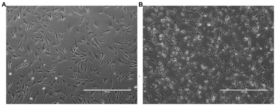
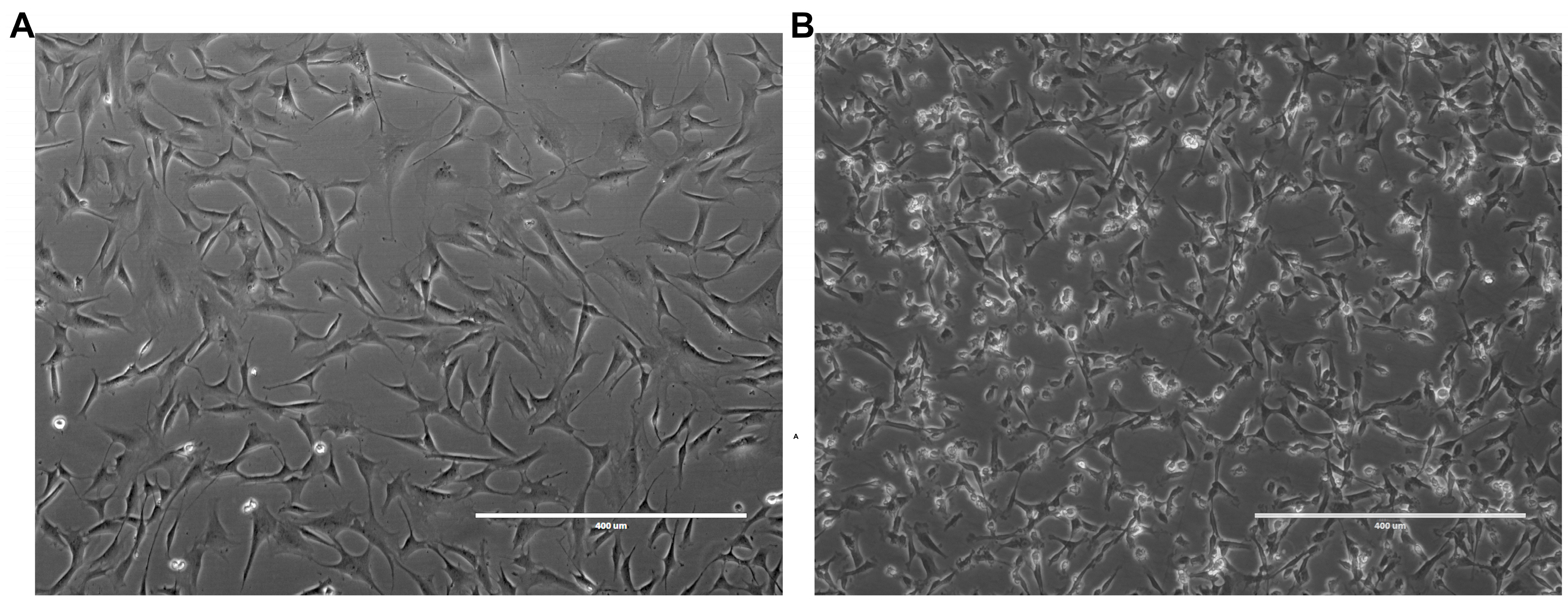
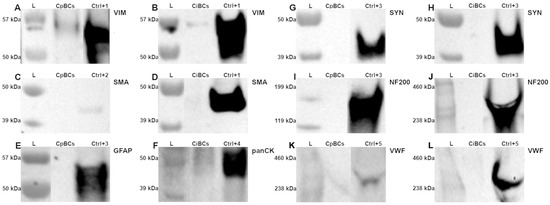
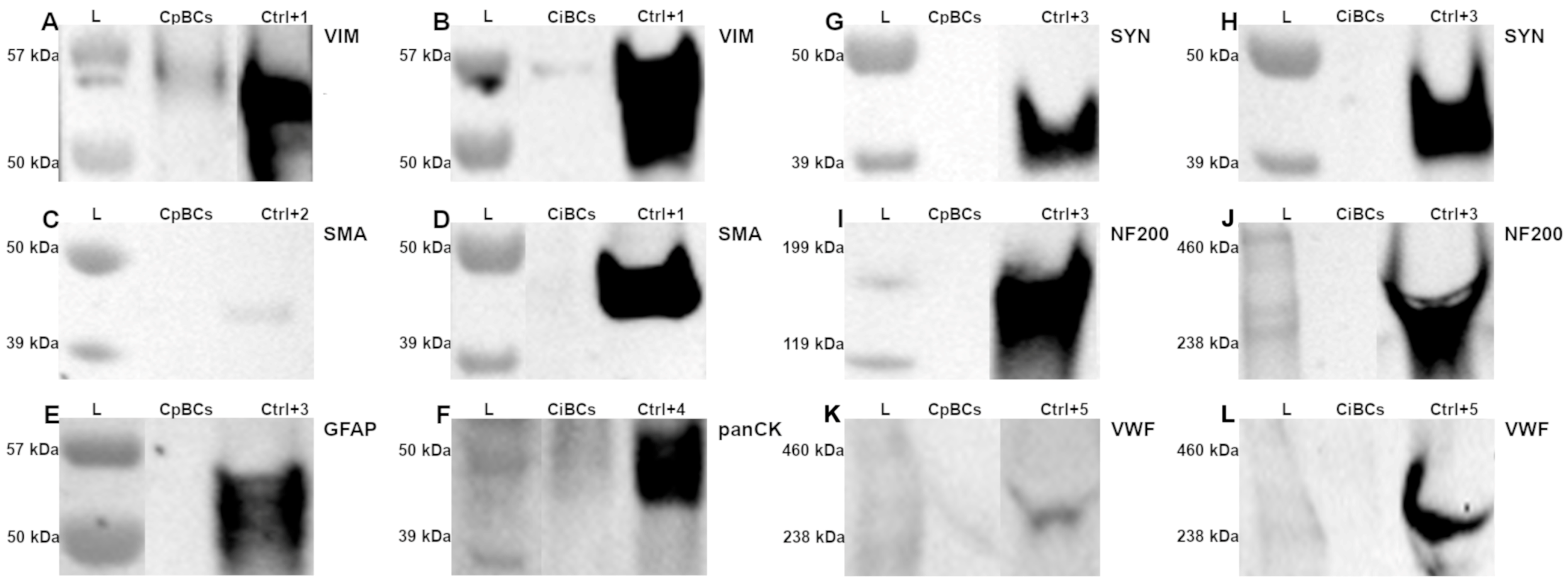
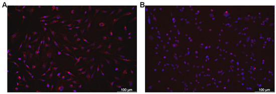
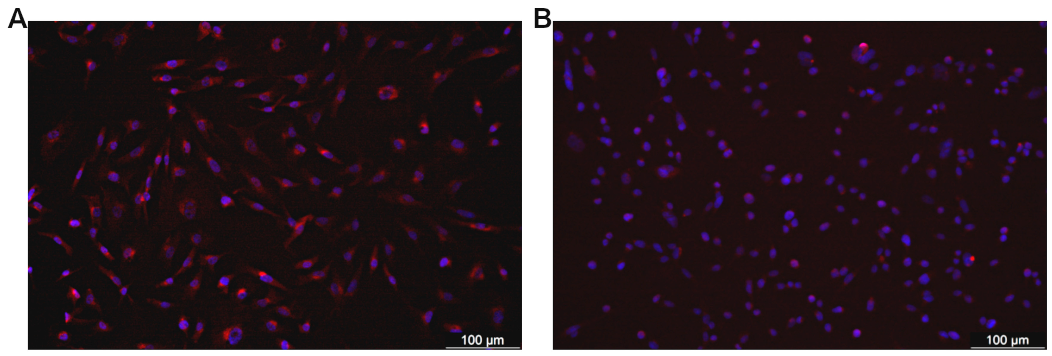
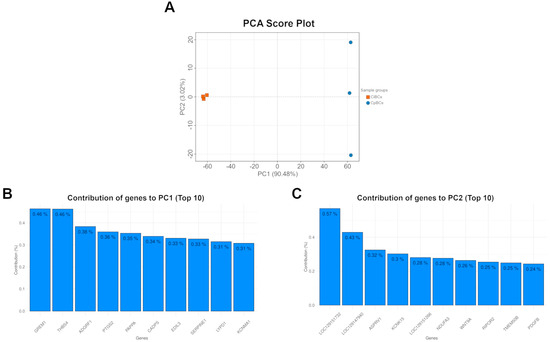
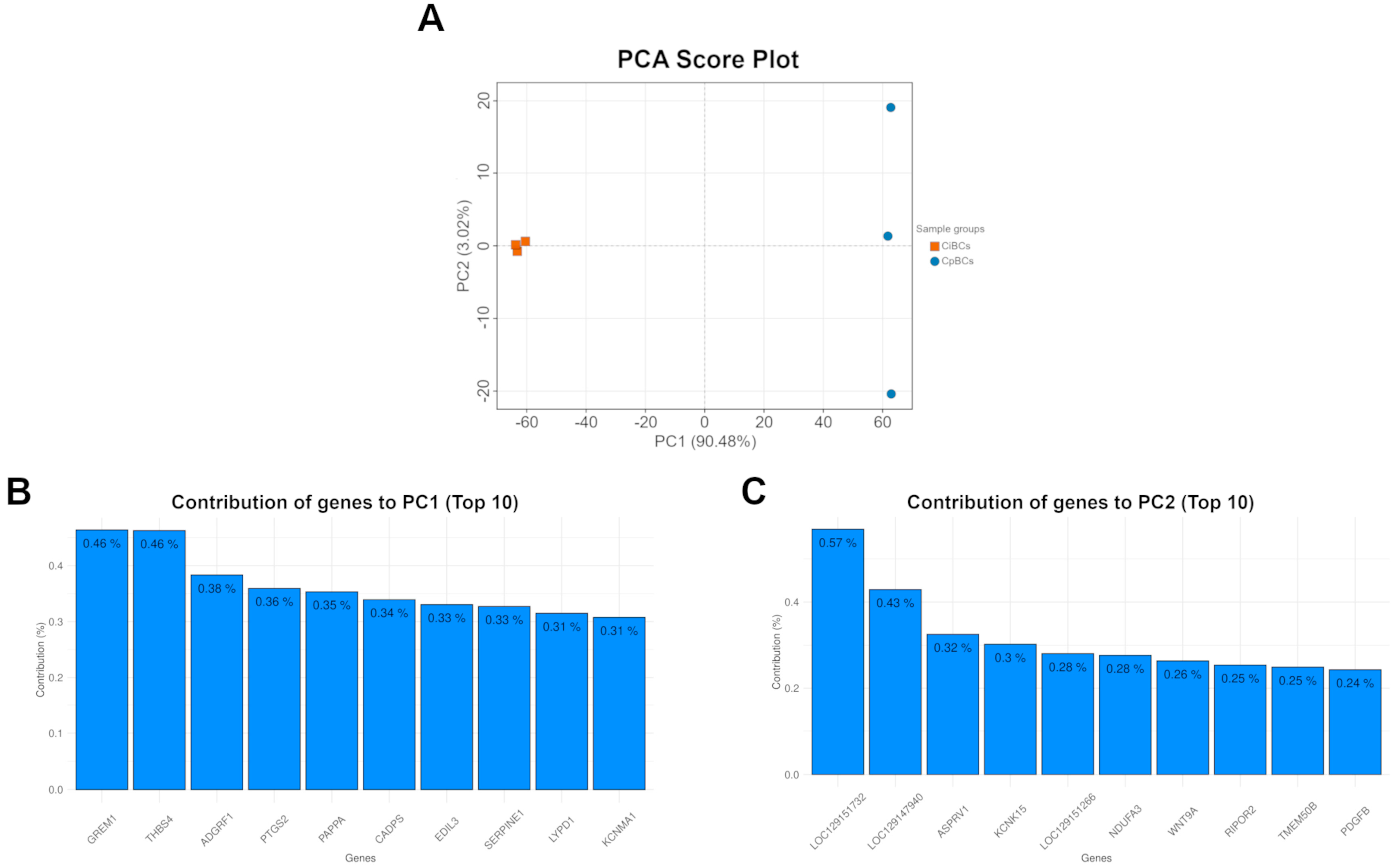
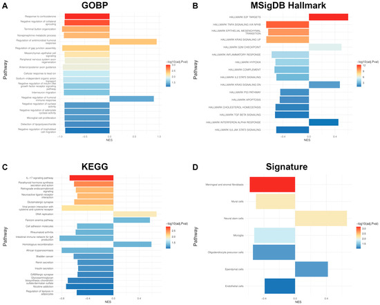
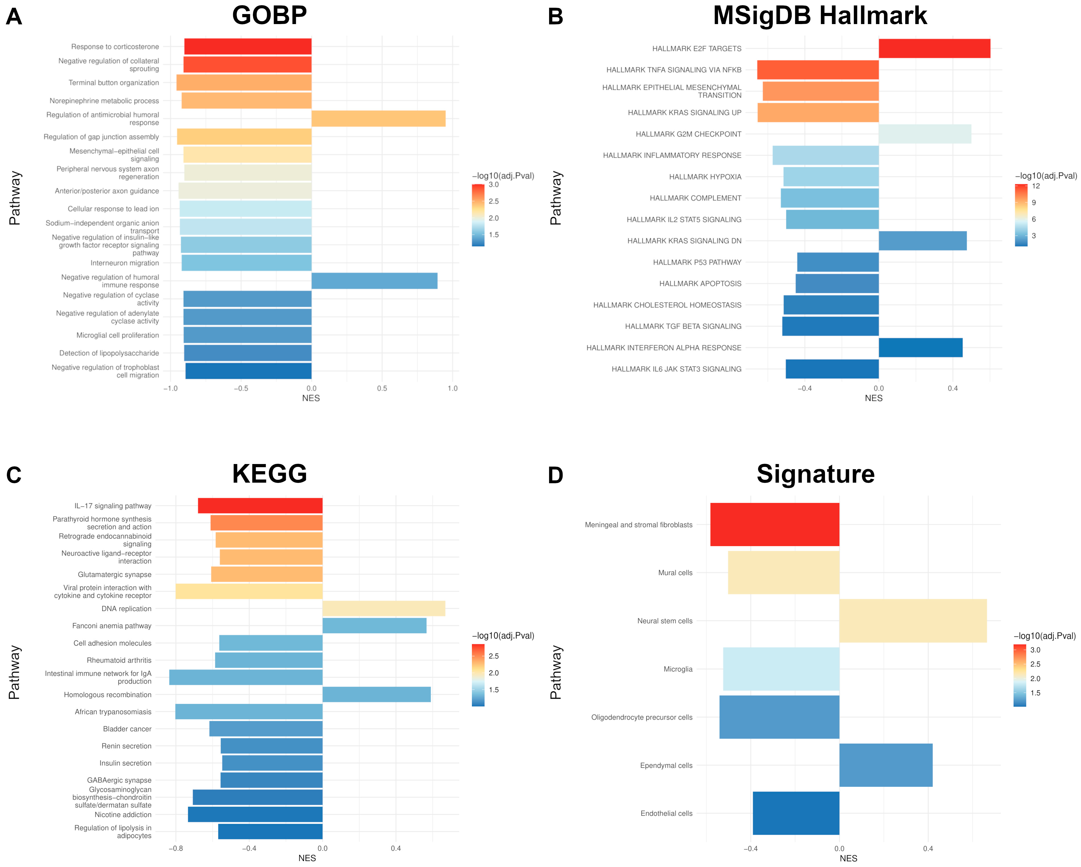

Disclaimer/Publisher’s Note: The statements, opinions and data contained in all publications are solely those of the individual author(s) and contributor(s) and not of MDPI and/or the editor(s). MDPI and/or the editor(s) disclaim responsibility for any injury to people or property resulting from any ideas, methods, instructions or products referred to in the content. |
© 2026 by the authors. Licensee MDPI, Basel, Switzerland. This article is an open access article distributed under the terms and conditions of the Creative Commons Attribution (CC BY) license.