


Simple Summary
Antilles Islands inhabit interesting freshwater snails’ fauna, shaped by the geological history of the archipelago, isolation, and migrations. This fauna, however, is still poorly studied. At Hispaniola Island, in the Dominican Republic, in the Dudu cenote, a small pond originated by partial collapse of a cave, some minute snails were found in 2001, representing the Truncatelloidea, the most diverse and species-rich, globally distributed superfamily of fresh- and brackish-water gastropods. Both morphological and molecular (DNA) characters showed that these animals represent the family Cochliopidae but are as distinct as to be classified as new to science, both as a species and a genus. The genus is named in honour of Dr Robert Hershler, a highly merited malacologist devoted to the study of the Cochliopidae. These snails still inhabit this pond.
Abstract
A new genus and species of the Cochliopidae are described from the Dudu cenote, Dominican Republic, Hispaniola Island. Shell and its measurements, soft parts morphology and pigmentation, radula, female reproductive organs, and penes are described. Mitochondrial cytochrome oxidase subunit I (COI), 16S and 12S, and nuclear 18S sequences have been applied to reconstruct phylogenetic relationships. The Hispaniolan taxon clearly belongs to a clade that also includes many Heleobia W. Stimpson, 1865, species from the Americas, the European Eupaludestrina Mabille, 1877, as well as North American Heleobops F. G. Thompson, 1968. All analyses support the assignment of the Hispaniolan taxon to a distinct genus, new to science. Its known geographic distribution is restricted to this cenote so far.
1. Introduction
In the Dominican Republic (Hispaniola Island), Greater Antilles, 14 species of freshwater gastropods have been recorded so far [1,2,3,4,5,6,7]: nine species represent Caenogastropoda, and five—Hygrophila. In the Antilles, the superfamily Truncatelloidea J. E. Gray, 1840, is represented by the family Cochliopidae Tryon, 1866. Two species of the genus Nanivitrea Thiele, 1927, have been recorded from Cuba, N. alcaldei Jaume and Abbot, 1947, and N. helicoides (Poey, 1865) [8], and another two from Jamaica, N. inconspicua (C. B. Adams, 1851) and N. pygmaea (C. B. Adams, 1849). These low-spired, valvatoid gastropods, initially assigned to the Assimineidae H. Adams et A. Adams, 1856 [9], were later transferred to the Cochliopidae [10,11,12]. Pyrgophorus Ancey, 1888, is another cochliopid genus reported from this area—from Venezuela [13,14,15] (six species), Guadalupe [16,17] (two species), and Martinique [18] (one species). In Venezuela and the latter two islands, Pyrgophorus parvulus (Guilding, 1828) has been found. Pfeiffer [19] described Paludina coronata L. Pfeiffer, 1840, from Cuba, later assigned to the genus Pyrgophorus and recorded from several localities in Cuba [20].
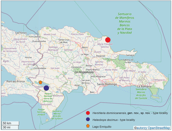
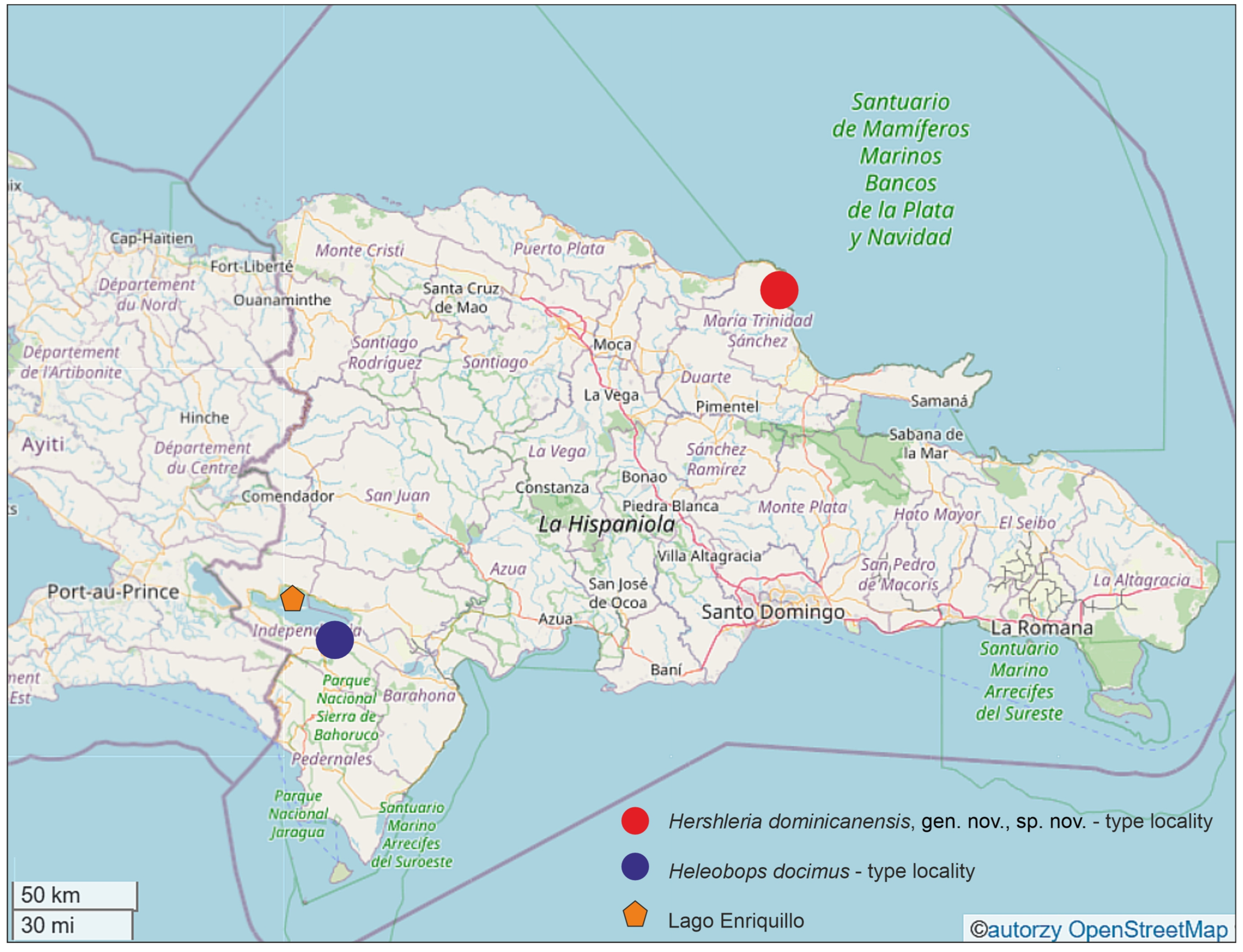
On Hispaniola Island, P. coronatus has been recorded from Lake Miraguane in the western part of Haiti [2], and on the same island, in the Dominican Republic, P. coronatus bermudezi (Aguayo, 1947) has been found in the Pleistocene sediments, near Lake Enriquillo (Figure 1) [21]. In the Dominican Republic, another representative of the Cochliopidae, genus Heleobops F. G. Thompson, 1968, has been found [6]: Heleobops clytus F. G. Thompson et Hershler, 1991 (now assigned, as a junior synonym, to H. docimus F. G. Thompson, 1968 [22]), reported from 2 km ESE of Duvergé (Figure 1), Independencia Province, Lake Enriquillo basin, and spring Boca de Cachón. Antilobia margalefi Altaba, 1993, was described from Lake Enriquillo [23], the taxon later synonymized with Pyrgophorus coronatus bermudezi [22]. Heleobops docimus inhabits also a pond in the Cayman Islands [24]. Heleobops torquatus F. G. Thompson et Hershler, 1991, has been found on Jamaica [6].
Figure 1.
Map showing localities of Hershleria dominicanensis and other representatives of Cochliopidae in the Dominican Republic. The map is based on OpenStreetMap data, and all locality names are presented in their original forms.
In the materials deposited in the Zoological Museum of the Jagiellonian University by an anonymous collector there was a jar containing a few minute truncatelloid snails collected in the Dudu Blue Lagoon cenote, Dominican Republic, in April 2001. Recently, on February 27th, 2025, we inspected this cenote (Figure 1) and confirmed the presence of these snails there. The Dudu cenote is recently a protected area; thus, we did not collect these snails then, and the entire study is based entirely on these old museum materials. The snails represent the family Cochliopidae. In the paper, we present the morphology and the molecularly inferred phylogenetic relationships of this new taxon.
2. Materials and Methods
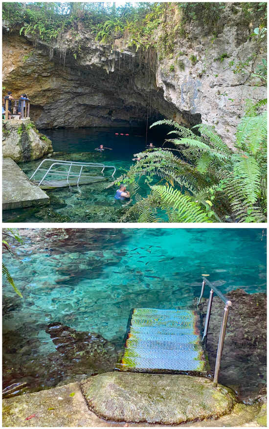
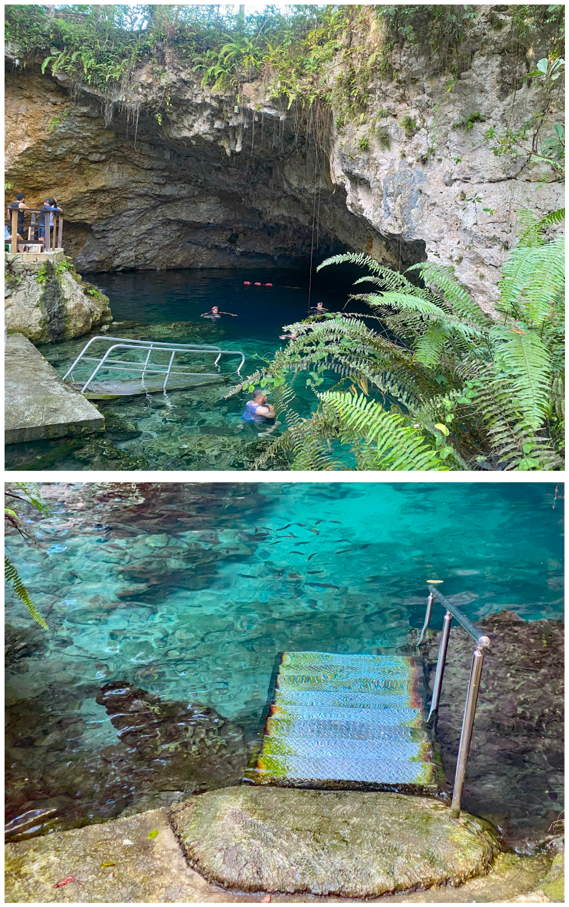
The snails still occur on a delicate layer of filamentous algae growing on stones, at a depth of approximately 0.5 m, in the Dudu Blue Lagoon cenote, 19°38′51.0″ N 70°04′38.9″ W, near Cabrera, María Trinidad Sánchez Province, Hispaniola Island, Dominican Republic, at the eastern end of the country’s northern coast (Figure 1). A cenote is a natural pit or sinkhole, resulting from the collapse of limestone bedrock that exposes groundwater underneath. The main lake, 16 m deep, is partially covered by a ceiling with stalactites. Underground branches of the cave run radially from the lake and are 90–135–350 m long. The temperature of the water ranges between 25 and 27 °C. Like in most limestone-based cenotes, the water is neutral to slightly alkaline and slightly brackish. Without permission, we were not able to analyse the water chemistry in this protected area. The snails inhabit the light zone (Figure 2) and show no trace of troglomorphism. The snails were kept in 80% analytical ethanol and stored at −20 °C.
Figure 2.
Type locality of Hershleria dominicanensis, Dudu cenote, María Trinidad Sánchez Province, Dominican Republic.
The shells were photographed with a Canon EOS 50D digital camera, under a Nikon SMZ18 microscope. The dissections were done under a Nikon SMZ18 microscope with dark-field, equipped with a Nikon DS-5 digital camera, whose captured images were used to draw female reproductive organs with a graphic tablet. Morphometric parameters of the shell were measured by one person using ImageJ v. 1.53g image analysis software [25]. Protoconchs were cleaned using an ultrasonic cleaner, mounted and examined, and the radulae were extracted with Clorox, all applying the techniques described by Falniowski [26]. Protoconchs and radulae were photographed using a Hitachi S-4700 scanning electron microscope. Penes were photographed under a Motic B3 Professional microscope with dark field. Morphological terminology after Hershler and Ponder [27].
DNA was extracted from whole specimens; tissues were hydrated in Tris-EDTA (TE) buffer (3 × 10 min); then, total genomic DNA was extracted with the Sherlock extraction kit (A&A Biotechnology, Gdańsk, Poland), and the final product was dissolved in 20 μL of TE buffer. The extracted DNA was stored at −80 °C at the Department of Malacology, Institute of Zoology and Biomedical Research, Jagiellonian University in Kraków (Poland).
Mitochondrial cytochrome oxidase subunit I (COI), 16S ribosomal RNA (16S), and 12S ribosomal RNA (12S) were sequenced. Details of PCR conditions, primers used, and sequencing were given in Szarowska et al. [28] and Collado et al. [29]. Phylogenetic analyses using COI were performed by adding sequences from GenBank (Table 1), available for the representatives of all the cochliopid genera inhabiting Central America, the Antilles, the northern part of South America, and the southern part of the USA. The representatives of the genera Aphaostracon J. G. Thompson, 1968, Cochliopa W. Stimpson, 1865, Cochliopina J. P. E. Morrison, 1946, Littoridina Souleyet, 1852 [30], Littoridinops Pilsbry, 1952, Tryonia Stimpson, 1865 [31], as well as widely distributed but geographically far Heleobia W. Stimpson, 1865, were included in the COI-based analyses. Unfortunately, from among the geographically close genera, there were no sequences of Emmericiella Pilsbry, 1909, Subcochliopa J. P. E. Morrison, 1946, and Texadina Abbott & Ladd, 1951. There were no sequences of the representatives of these genera in GenBank, and we were unable to obtain these specimens for sequencing.
Sequences were initially aligned using MUSCLE [32] software, implemented within MEGA 7 [33], and then visually checked in BioEdit 7.1.3.0 [34]. Uncorrected p-distances were calculated in MEGA 7. The estimation of the proportion of invariant sites and the saturation test for entire data sets [35,36] were performed using DAMBE [37]. The data were analysed using Bayesian inference (BI) and maximum likelihood (ML), in MrBayes v. 3.2.7a [38] and RAxML-NG v. 0.8.0 [39], respectively. The appropriate substitution model for ML analysis was selected using jModelTest2 via the CIPRES Science Gateway [40]; the model TPM2uf+I+G was used. Partitioning of the data into three codon positions for COI was performed. The ML analysis was conducted in RAxML-NG v. 0.8.0 via the web service available at https://raxml-ng.vital-it.ch/ (accessed on 20 March 2025), with 10 random and 10 parsimony-starting trees. In the BI analysis, the K81+G model of nucleotide substitution was applied in tree reconstruction. The substitution model was selected using MrModelTest 2.4 [41]. The analyses were run using MrBayes v. 3.2.7a [38] with default priors. Two simultaneous analyses were performed, each with 10,000,000 generations, with one cold chain and three heated chains, starting from random trees and sampling the trees every 1000 generations. The first 25% of the trees were discarded as a burn-in. The analyses were summarized as a 50% majority-rule consensus tree. Convergence of the runs was checked in Tracer v.1.7.1 [42], and all effective sample sizes exceeded 200, the proposed measure of good sample mixing. Fig Tree v. 1.4.4 [43] was used to visualize the trees. Two species delimitation methods were performed: Poisson Tree Processes (PTPs) [44] and Assemble Species by Automatic Partitioning (ASAP) [45]. The PTP approach was run on the web server (https://species.h-its.org/ptp/ accessed on 20 March 2025), with 100,000 MCMC generations, 100 thinning intervals, and 0.1 burn-in. We used the RAxML output phylogenetic tree. The ASAP approach was run on the web server (https://bioinfo.mnhn.fr/abi/public/asap/, accessed on 20 March 2025) using simple distance (p-distances). The Fastachar application [46] and DeSignate [47] were used to determine the Molecular Diagnostic Characters (MDCs) for COI. Two types of characters were accepted: at binary positions (where the character state in the query group is different from the uniform character state of the reference group) and at asymmetric positions (where the character state in the query group is not different from the uniform character state of the reference group). The letters used in the MDCs list denote the nucleotide position in our sequence set, and the letter in parentheses denotes the specific nucleotide characteristic for the particular group. These analyses were performed on the set of sequences shown in the Supplementary Materials (Supplementary File S1).
Table 1.
Reference sequences used in phylogenetic analyses.
3. Results
3.1. Molecular Phylogeny
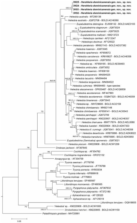
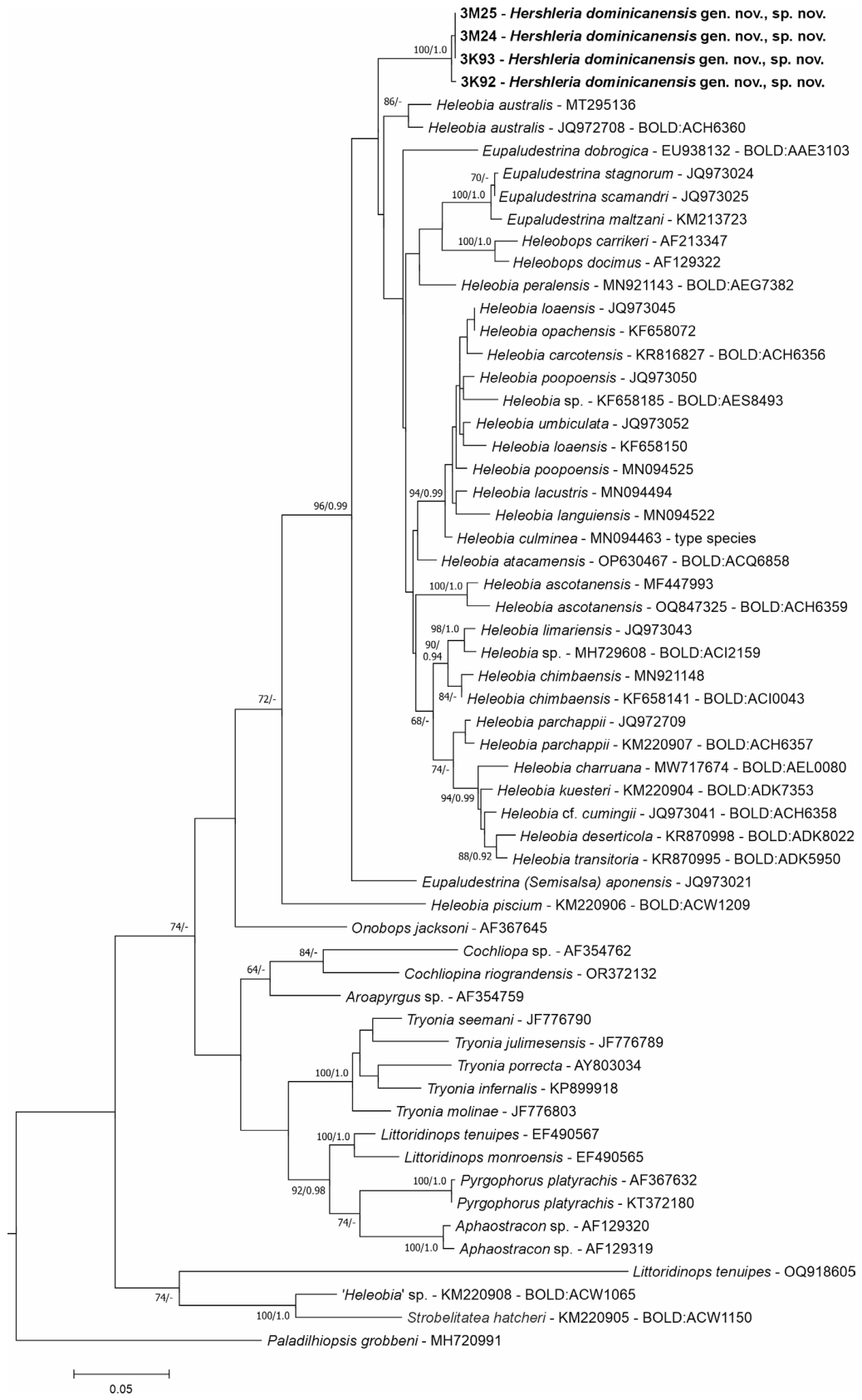
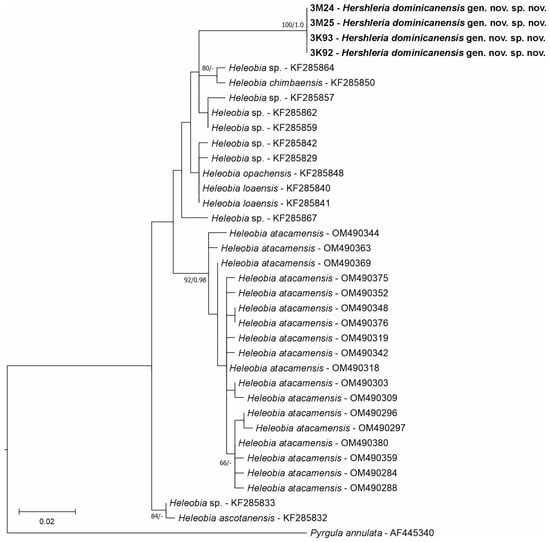
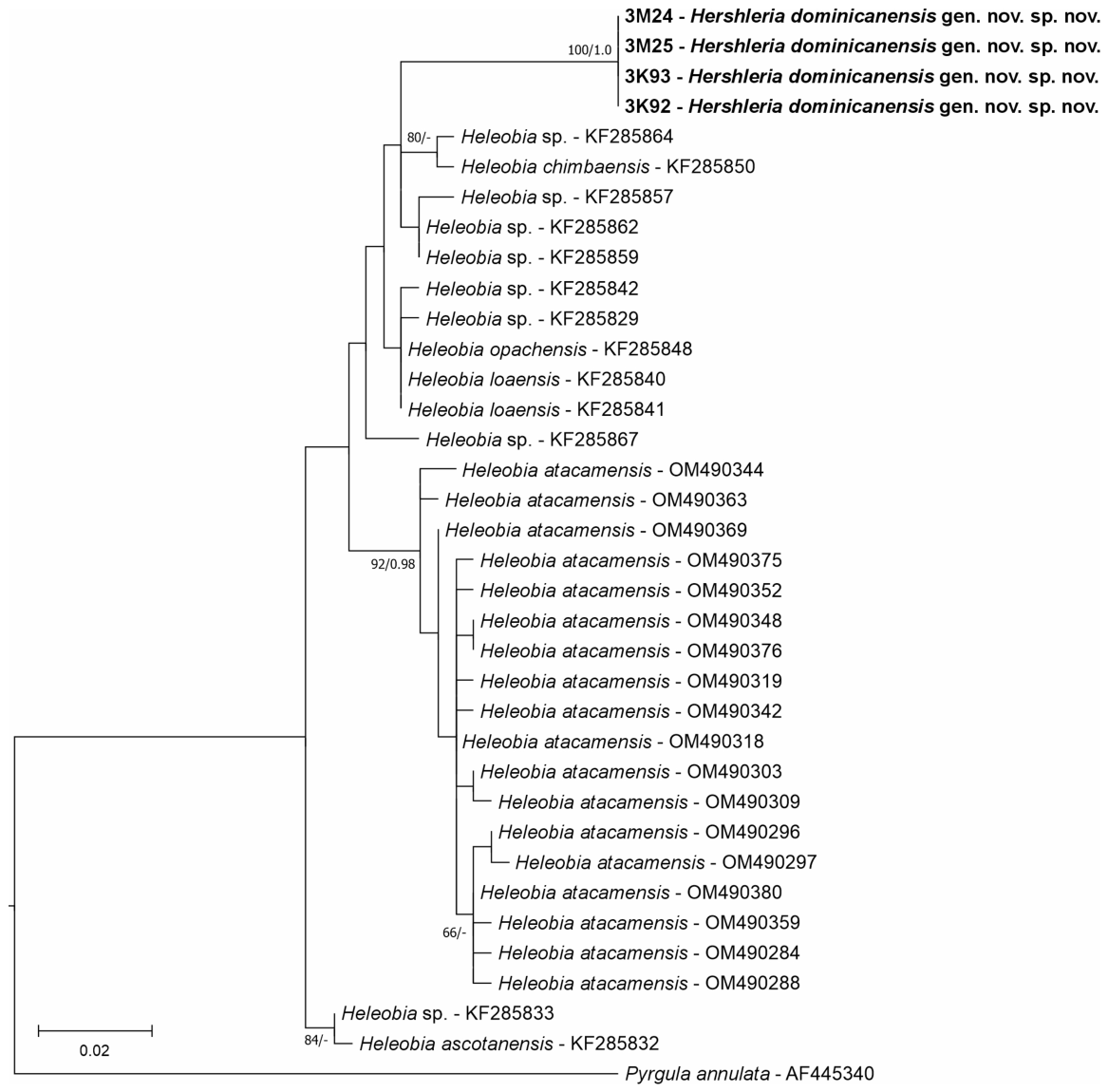
We obtained four new sequences of COI (551 bp, GenBank accession numbers PZ349653-PZ349656). The test for substitution saturation analysis showed an ISS value (0.74) significantly below the critical value (ISSC: 0.95), indicating that sequences are not saturated and are therefore suitable for phylogenetic reconstruction. The topologies of the resulting phylograms were identical in both the ML and BI phylogram analyses; thus, we present the phylogram computed with RAxML (Figure 3). All algorithms used for species delimitation supported the distinctiveness of the Dominican snails. The COI sequences unambiguously (bootstrap support 100%) placed the Dominican specimens within one Marker gene Operational Taxonomic Unit (mOTU).
Figure 3.
Maximum-likelihood tree computed for COI; bootstrap supports over 60%, and Bayesian probabilities over 0.90 are given.
This mOTU belonged to the highly supported clade (bootstrap 96%, BP 0.99) of Heleobia W. Stimpson, 1865 ([69] type species Paludina culminea A. d’Orbigny, 1838), Eupaludestrina Mabille, 1877 (type species Hydrobia macei Paladilhe, 1867), and Heleobops (type species H. docimus F. G. Thompson, 1968), and our Dominican taxon. Within this clade, one subclade grouped most of Heleobia, another one included Heleobops and European/circum-Mediterranean Eupaludestrina. In addition, the three genera constituted quite distinct clades (Figure 3). All the genera whose ranges were geographically close to Hispaniola were grouped outside this clade. The p-distances between the Dominican cochliopid snail and the other two geographically most closely related cochliopid taxa were 7.4% for Dominican Heleobops docimus, and 15.2% for Pyrgophorus platyrachis F. G. Thompson, 1968 from Florida. Considering the genetic distances, the closest to our taxon was Heleobia australis (A. d’Orbigny, 1835) from Argentina (p-distance = 5.3%), although these two taxa did not form a monophyletic group in the COI tree, and a large unresolved polytomy was observed there. As shown in the COI-based phylogram (Figure 3), these distances are similarly high as the ones between the other genera of the Cochliopidae.
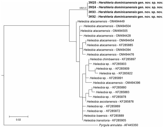
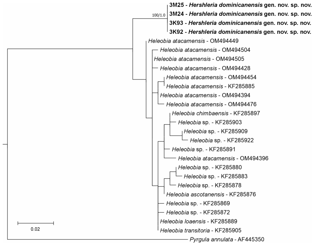
We also obtained four new sequences of 12S (311 bp, GenBank accession numbers PZ349056-PZ349059), four new sequences of 16S (323 bp, GenBank accession numbers PZ349060-PZ349063), and one 18S sequence (GenBank accession number PZ349601). The number of cochliopid taxa for which mitochondrial 16S and 12S sequences are available is very restricted, but our analyses placed the Dominican taxon far from the other Heleobia (Figure 4 and Figure 5). All algorithms used for species delimitation also supported the distinctiveness of this taxon.
Figure 4.
Maximum-likelihood tree computed for 12S; bootstrap supports over 60%, and Bayesian probabilities over 0.90 are given. The GB numbers of the sequences obtained by Valladares et al. [70] and Collado et al. [29] are also shown.
Figure 5.
Maximum-likelihood tree computed for 16S; bootstrap supports over 60%, and Bayesian probabilities over 0.90 are given. The GB numbers of the sequences obtained by Valladares et al. [70] and Collado et al. [29] are also shown.
Considering our phylogeny analyses, species delimitations, as well as geography -thousands of kilometres between Hispaniola and the northern limit of the range of Heleobia (Amazonia in South America), the most justified solution seems to describe a new genus for our Dominican taxon.
3.2. Systematic Part
Gastropoda Cuvier, 1795
Caenogastropoda Cox, 1960
Littorinimorpha Golikov and Starobogatov, 1975
Truncatelloidea Gray, 1840
Cochliopidae Tryon, 1866
Hershleria Falniowski et Jaszczyńska, gen. nov.
ZooBank number: urn:lsid:zoobank.org:act:1000A30E-8FD9-45B0-8E88-0A5786BAFB80
Type species Hershleria dominicanensis Falniowski et Jaszczyńska, sp. nov., by original designation.
Diagnosis
Minute cochliopid snail with conic, moderately broad shell with relatively low spire, whorls slightly convex, ovoid moderately broad aperture, parietal lip complete, radula with one pair of basal cusps and six broad cusps on the cutting edge of central tooth, with big bursa copulatrix closely overlapping on albumen gland, with long duct, long and worm-shaped receptaculum seminis with no distinct duct, sperm tube short, opening into posterior end of pallial cavity, and penis long, flattened, tapering distally, with 5–6 stout, short-stalked apocrine glands similar in size, small and slim non-glandular outgrowth on the right side.
Etymology
The genus is named in honour of Dr Robert Hershler, a highly merited malacologist devoted to the study of the Cochliopidae.
Differential diagnosis
As for Hershleria dominicanensis Falniowski et Jaszczyńska, sp. nov. for this monotypic genus.
Hershleria dominicanensis Falniowski et Jaszczyńska, sp. nov.
ZooBank number: urn:lsid:zoobank.org:act:E95B08FE-BFCA-491A-B3F8-4AC5B77F4743
GenBank sequence numbers for COI: PZ349653-PZ349656
Etymology: dominicanensis derived from the Dominican Republic, harbouring the type locality.
Type locality: Dudu Blue Lagoon cenote, 19°33′51.9″ N 69°54′28.8″ W, near Cabrera, María Trinidad Sánchez province, Hispaniola Island, Dominican Republic (Figure 1 and Figure 2), on a delicate layer of filamentous algae growing on the stones, at a depth of approximately 0.5 m.
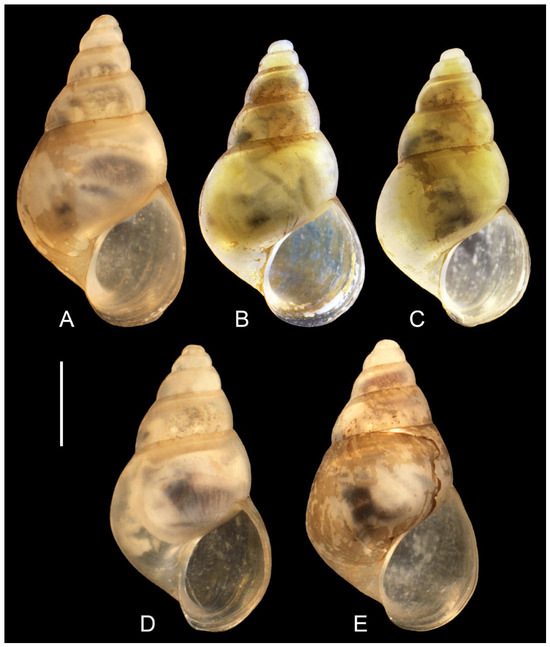
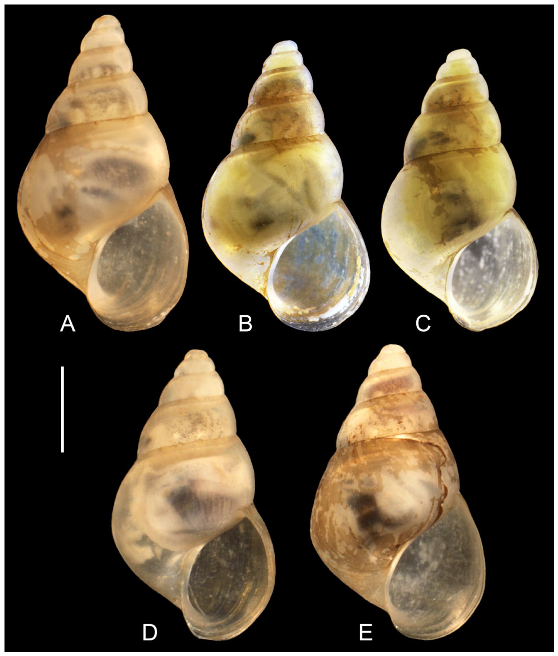
Holotype: Ethanol-fixed specimen (Figure 6A) collected by an anonymous collector, at Dudu Blue Lagoon cenote, 19°33′51.9″ N 69°54′28.8″ W, Hispaniola Island, Dominican Republic, April 2001, deposited in the Zoological Museum of the Jagiellonian University, voucher number ZMUJ/2026/07.
Figure 6.
Shells of Hershleria dominicanensis: (A)—holotype, (B–E)—paratypes; bar equals 1 mm.
Paratypes: Seven ethanol-fixed specimens collected at the type locality, voucher number ZMUJ/P/08–13, two paratypes destroyed for DNA extraction, three others for dissection.
Description
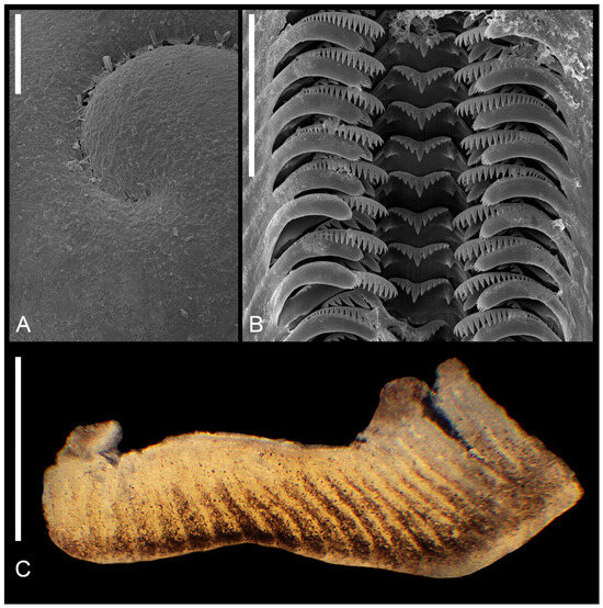
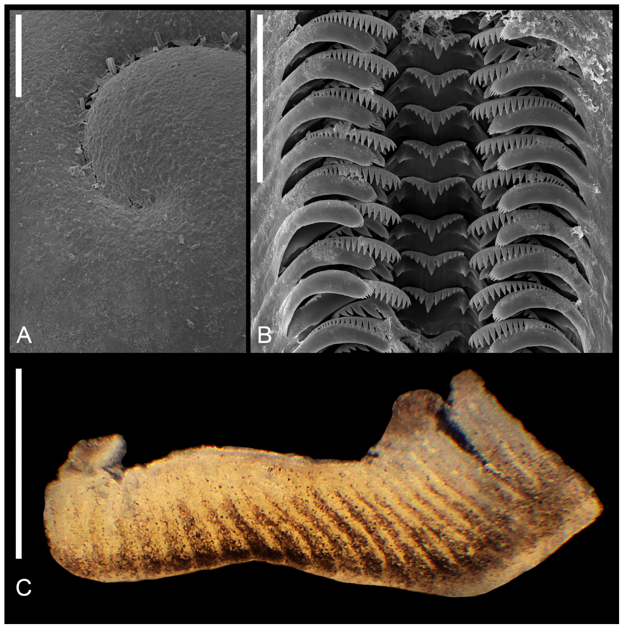
Shell (Figure 6A–E) conic, moderately broad, with relatively low spire, up to 3.79 mm high, with about six whorls, spire height approximately 33% of shell height, and 66% of body whorl width. Teleoconch whorls slightly convex, evenly rounded, growing regularly in diameter. Aperture ovoid, moderately broad, with an angle at its upper part, outer lip simple, parietal lip complete, umbilicus slit-like. Teleoconch glossy, growth lines hardly visible, periostracum white to yellowish. Protoconch (Figure 7A) smooth, no sharp border between proto- and teleoconch. Operculum paucispiral, without any outgrowth. Measurements: Table 2.
Figure 7.
Hershleria dominicanensis: (A)—protoconch, bar equals 50 µm; (B)—radula bar equals 50 µm; (C)—pigmentation of mantle along ctenidial lamellae, bar equals 500 µm.
Table 2.
Shell measurements (in mm): a—shell height; b—body whorl breadth; c—aperture height; d—spire height; e—aperture breadth; α—apex angle measured between the lines tangential to the spire; β—angle between the body whorl suture and the line perpendicular to the columella; M—mean; SD—standard deviation; Min—minimum value; Max—maximum value.
Radula (Figure 7B) with central tooth with lateral angles highly expanded, ventral process well excavated, cusps rather long and massive, following the formula:
Median cusp twice longer than adjacent ones, basal cusps broad, located relatively high on lateral angles of central tooth. Lateral tooth with 3(4)–1–6 blunt cusps, inner marginal tooth with 22–24 sharp cusps, outer marginal tooth with 8–10 delicate sharp cusps.
Morphology and anatomy of soft parts
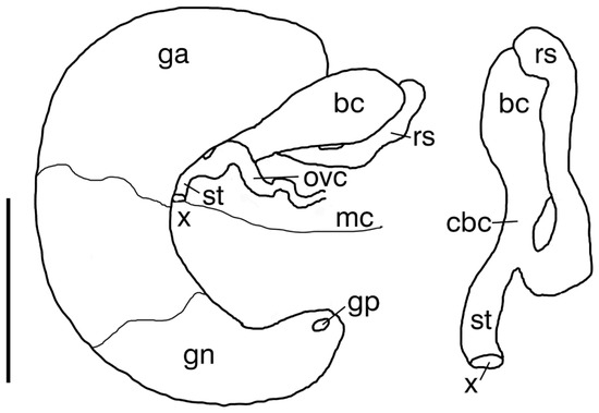
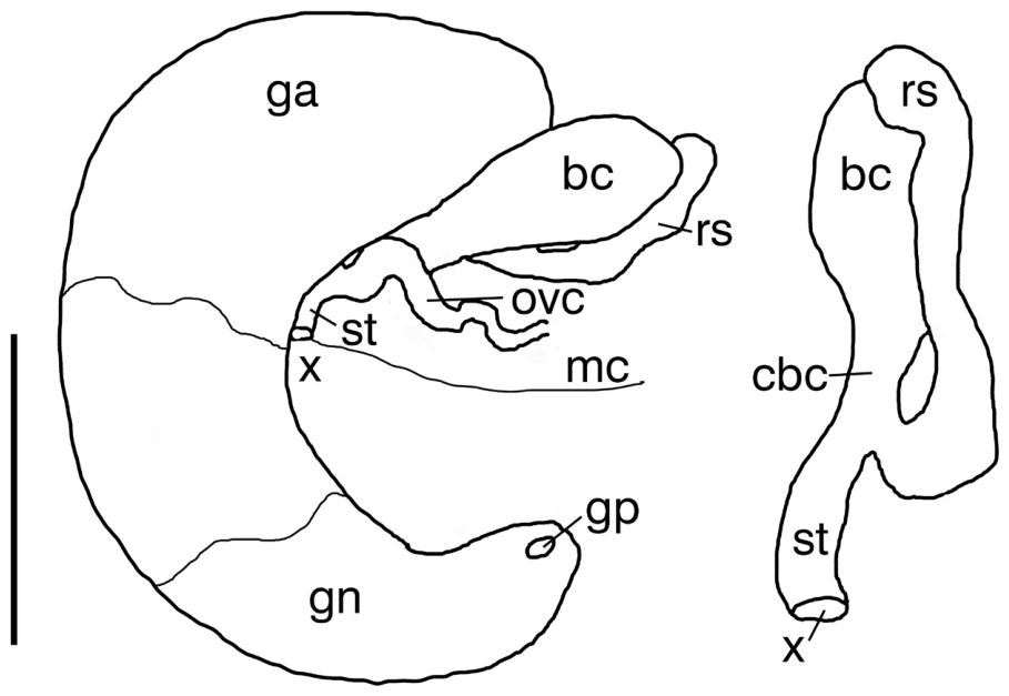
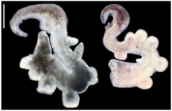
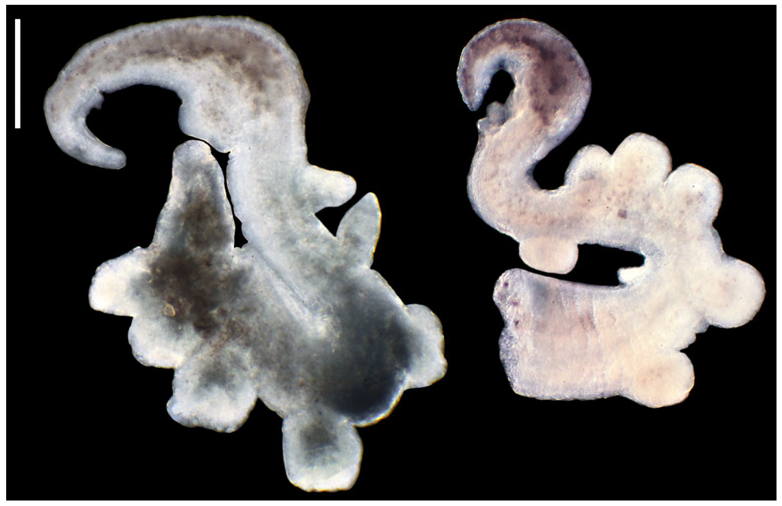
The head pigmented black, eye spots present, hyperciliation of the tentacles. On the mantle some spots of pigment, lines of pigment along the bases of the ctenidial lamellae (Figure 7C), ctenidium with about 22 lamellae, osphradium small. Female reproductive organs typical of Heleobia [11,52,71,72] with big bursa copulatrix closely overlapping on albumen gland, with long duct, long and worm-shaped seminal receptacle with no distinct duct, sperm tube short, opening into posterior end of pallial cavity (Figure 8). Penis (Figure 9) long, flattened, tapering distally, with 5–6 stout, short-stalked apocrine glands arising from outer curvature at proximal part of penis. All glands similar in size. At the half of the penis length small and slim non-glandular outgrowth on the right side. On the left side, at base od terminal tapering section, flat outgrowth, terminal section with long spot of brown pigment.
Figure 8.
Female reproductive organs of Hershleria dominicanensis (bc—bursa copulatrix; cbc—bursal duct; ga—albumen gland; gn—capsule gland; gp—gonoporus; mc—posterior end of mantle cavity; ovc—coiled (renal) oviduct; rs—receptaculum seminis; st—sperm tube; x—second (copulatory) gonoporus); bar equals 1 mm.
Figure 9.
Penes of Hershleria dominicanensis; bar equals 200 µm.
Differential diagnosis
Hershleria dominicanensis differs from Heleobia australis in having a non-turreted shell with a narrower spire, less sharp basal cusps, and more cusps on cutting edge of the central tooth and lateral tooth of the radula (H. australis crassa M. Gaillard, 1974, while in H. australis nana Er. Marcus et Ev. Marcus, 1963, the radula is similar as Hershleria dominicanensis), smaller and flatter outgrowth at base of terminal tapering section of the penis, and five–six, instead of one, apocrine glands (anatomy of H. australis: [18]). Heleobia australis differs also in its brackish-water habitat (although there is a very low admixture of the sea water in the Dudu Cenote) and free-swimming larva [73]. In the case of the Dudu cenote, with a very limited access of the sea water, only direct development would be possible. Combined shell and penis characters distinguish Hershleria dominicanensis from all Heleobia whose anatomy is known (see Discussion for references). The molecular diagnostic for COI characters in comparison with all Heleobia sequences used in molecular analysis: binary: 11(A), 183(C), 282(C), 341(T); asymmetric: 9(A), 408(T), 510(G).
Geographically, but not molecularly, the closest cochliopid taxon is Heleobops docimus [3,11], recorded from the Dominican Republic. Hershleria dominicanensis differs from Heleobops docimus in shell morphology by having less flat whorls and an unreflexed outer lip, in radula’s central tooth with broader basal cusps and more cusps on its cutting edge, in a bigger bursa copulatrix, and in penis with much bigger apocrine glands, lack of a lobe close to the terminal end of the penis (in Heleobops present or not), presence of two non-glandular outgrowths, and brown pigmentation of the terminal part of the penis. From another cochliopid genus recorded from the Dominican Republic, Pyrgophorus [3,11], Hershleria dominicanensis differs in the radula by having one instead of two basal cusps, and in the penis by lacking a distal bifurcation and having less numerous but bigger papillae (apocrine glands).
Distribution and habitat
Known only from the type locality.
4. Discussion
Despite more than one hundred described species of Heleobia [74], anatomical characters have been described for only a few of them, the same concerns molecular data [24,51,52,59,63,75]. Considering all the descriptions of the anatomy of Heleobia [11,52,58,71,72,76,77,78,79,80,81], Hershleria dominicanensis differs in its penis, and molecularly is clearly distinct. Contrary to a species, a genus is not a natural unit whose threshold distinguishing value can be estimated. Thus, in a given systematic group, the values of genetic distances should be similar between the distinguished genera.
The geographic range of Heleobia covers South America, but only its southern part and a narrow belt along the eastern coast, expanding northward into Amazonia, and, on the eastern side of the Atlantic Ocean, in several areas around the Mediterranean and on the Atlantic coasts of South Morocco and the Netherlands [11,82]. However, as concerns the European Heleobia, Radoman [83] described the genus Semisalsa Radoman, 1974 for S. dalmatica, inhabiting the spring Pirovac on the Dalmatian coast of the Adriatic Sea, and representatives of Semisalsa were later found at many localities along the European coasts. Giusti and Pezzoli [84] created a distinct subfamily, Semisalsinae Giusti et Pezzoli, 1980. Later, Semisalsa was synonymized with Eupaludestrina. It should be noted that, according to Szarowska et al. [52], E. dalmatica, as well as all the other European brackish-water Eupaludestrina, should be synonymized with E. stagnorum (Gmelin, 1791) (described as Helix stagnorum Gmelin, 1791, from the Kaaskenswaters, Zierikzee, the Netherlands [85,86]. A species of Eupaludestrina is also known from the Canary Islands [87]. Our COI tree (Figure 3) does not support the monophyly of Eupaludestrina, as well as Heleobia, and Heleobops is not clearly distinct from the Eupaludestrina/Heleobia group. It has to be noted that the anatomy, as usual in the Truncatelloidea [88], is variable, and the anatomic characters of Heleobops, especially of its penis [3,11], may rather fall within the range observed for Eupaludestrina/Heleobia. Similarly, female reproductive organs showing no differences between Hershleria and Heleobia may not deny the genus distinctness of the former.
The assignment of Heleobia australis to the genus Heleobia was already questioned [53,59,81]. The occurrence of Heleobia in Hispaniola Island, thousands of kilometres far from all the other localities of this genus is improbable. Passive transport by birds, even over such long distances, cannot be excluded: such transportation was observed several times for the truncatelloid snails [88]. The Antilles are on the route of bird migration between S and N America [50]. South American Heleobia charruana (d’Orbigny, 1841) from Uruguay has been recorded from the United Kingdom (2003), the Netherlands (2014), and Belgium (2017) [57]. Despite the possibility of long-distance passive transport, considering the Cretaceous origin of Hispaniola, there was a long time for evolution of an invader to become molecularly distinct. One should expect the occurrence of Hershleria at the neighbouring islands, at least, but the minute truncatelloid fauna of the latter remains poorly studied so far. Fortunately, the Dudu cenote is a protected area, which supports the necessary protection of this taxon, so far unique to this small habitat. However, there is some human impact in this area visited by numerous tourists and divers; thus, monitoring of this snail seems important.
5. Conclusions
The snails inhabiting the Dudu cenote in the Dominican Republic belong to the family Cochliopidae and represent a new genus and species. This is confirmed by both morphological and molecular data. This finding enriches our knowledge of the still-poorly studied, but interesting, malacofauna of the Antilles.
Supplementary Materials
The following supporting information can be downloaded at: https://www.mdpi.com/article/10.3390/ani16121810/s1. Supplementary File S1: Molecular Diagnostic Characters (MDCs) for COI.
Author Contributions
Conceptualization, A.F. and S.H.; methodology, A.J., S.H. and A.F.; formal analysis, S.H. and A.F.; investigation, A.J. and S.H.; resources, A.F.; writing—original draft, A.J., S.H. and A.F.; writing—review and editing, A.J., S.H. and A.F.; visualization, A.J.; funding acquisition, A.F. All authors have read and agreed to the published version of the manuscript.
Funding
The study was supported by the grant to Andrzej Falniowski, N18/DBS/000012 from the Institute of Zoology and Biomedical Research, Jagiellonian University.
Institutional Review Board Statement
Ethical review and approval were waived for this study because it is entirely based on museum materials collected several years ago and because neither animals have been killed nor any conflict of interest is involved.
Informed Consent Statement
Not applicable in this case.
Data Availability Statement
The sequences were deposited in GenBank (accession numbers: PZ349056-PZ349063, PZ349601, PZ349653-PZ349656) at https://www.ncbi.nlm.nih.gov (accessed on 20 April 2026).
Acknowledgments
We are grateful to Anna Łatkiewicz (Laboratory of FE Scanning Microscopy and Microanalysis, Institute of Geological Sciences, Jagiellonian University, Kraków, Poland) for her assistance with the SEM.
Conflicts of Interest
The authors declare no conflicts of interest.
References
- Pilsbry, H.A. On a collection of land Mollusca from the Island of Dominica, West Indies. Trans. Conn. Acad. Arts Sci. 1892, 8, 358–366. [Google Scholar]
- Clench, W.J.; Aguayo, C.G. Notes and descriptions of some new land and freshwater mollusks from Hispaniola. Mem. Soc. Cuba. Hist. Nat. “Felipe Poey” 1937, 11, 61–76. [Google Scholar]
- Thompson, F.G. The Aquatic Snails of the Family Hydrobiidae of Peninsular Florida; University of Florida Press: Gainesville, FL, USA, 1968; pp. xv+268. [Google Scholar]
- Prentice, M.A. Schistosomiasis and its intermediate hosts in the Lesser Antillean Islands of the Caribbean. Bull. Pan Am. Health Organ. 1980, 14, 258–268. [Google Scholar] [PubMed]
- von Starmühlner, F. Ergebnisse der Österreichisch-Französischen Hydrobiologischen Mission 1979 nach Guadeloupe, Dominica und Martinique (Kleine Antillen). Teil II: Beiträge zur Kenntnis der Süß- und Brackwassermollusken von Guadeloupe, Dominica und Martinique. Ann. Naturhist. Mus. Wien. Ser. B 1988, 90, 221–340. [Google Scholar]
- Thompson, F.G.; Hershler, R. New hydrobiid snails (Mollusca: Gastropoda: Prosobranchia: Truncatelloidea) from North America. Proc. Biol. Soc. Wash. 1991, 104, 669–683. [Google Scholar]
- Reeves, W.K.; Dillon, R.T., Jr.; Dasch, G.A. Freshwater snails (Mollusca: Gastropoda) from the Commonwealth of Dominica with a discussion of their roles in the transmission of parasites. Am. Malacol. Bull. 2008, 24, 59–63. [Google Scholar] [CrossRef] [Scilit]
- Vázquez, A.A.; Perera, S. Endemic Freshwater molluscs of Cuba and their conservation status. Trop. Conserv. Sci. 2010, 3, 190–199. [Google Scholar] [CrossRef] [Scilit]
- Thiele, J. Über die Schneckenfamilie Assimineidae. Zool. Jahrbücher. Zool. Jahrb. Abt. Syst. Geogr. Biol. Tiere. 1927, 53, 113–146. [Google Scholar]
- Taylor, D.W. A Remarkable Snail Fauna from Coahuila, Mexico. Veliger 1966, 9, 152–228. [Google Scholar]
- Hershler, R.; Thompson, F.G. A review of the aquatic gastropod subfamily Cochliopinae (Prosobranchia: Hydrobiidae). Malacol. Rev. 1992, 5, 1–140. [Google Scholar]
- Kabat, A.R.; Hershler, R. The prosobranch snail family Hydrobiidae (Gastropoda: Rissooidea): Review of classification and supraspecific taxa. Smithson. Contrib. Zool. 1993, 547, 1–94. [Google Scholar] [CrossRef] [Scilit]
- Baker, H.B. The mollusca collected by the University of Michigan—Williamson expedition in Venezuela. Occas. Pap. Mus. Zool. Univ. Mich. 1930, 210, 1–95. [Google Scholar]
- Rodríguez, G. El Sistema del Lago de Maracaibo: Sedimentación y Estudio Biológico; Instituto Venezolano de Investigaciones Científica (IVIC): Caracas, Venezuela, 1973; 398p. [Google Scholar]
- Nava, M.; Severin, H.; Machado, N. Distribución y taxonomía de Pyrgophorus platyrachis (Caenogastropoda: Hydrobiidae), en el sistema de Maracaibo, Venezuela. Rev. Biol. Trop. 2011, 59, 165–172. [Google Scholar] [CrossRef] [Scilit]
- Pointier, J.P. Faune malacologique dulçaquicole de l’ile de la Guadaloupe (Antilles françaises). Bull. Mus. Natl. Hist. Nat. 1974, 3, 905–933. [Google Scholar]
- Bouchet, P.; Pointier, J.P. Les Mollusques Terrestres et Dulçaquicoles de la Guadeloupe; MNHN, EPHE, Parc National de la Guadeloupe: Paris, France, 1998; 7p. [Google Scholar]
- Delannoye, R.; Charles, L.; Pointier, J.P.; Massemin, D. Mollusques Continentaux de La Martinique—Non-Marine Molluscs of Martinique, Lesser Antilles; Muséum National d’Histoire Naturelle: Paris, France, 2015. [Google Scholar]
- Pfeiffer, L. Uebersicht der im Januar, Februar und März, 1839 auf Cuba gesammelten Mollusken. Arch. Naturgesch. 1840, 6, 250–264. [Google Scholar]
- Pointier, J.P.; Yong, M.; Gutiérrez, A. Guide to the Freshwater Molluscs of Cuba; ConchBooks: Hackenheim, Germany, 2005. [Google Scholar]
- Aguayo, C.G. Notas y variadades (VIII). Rev. Soc. Malacol. Carlos Torre. 1947, 5, 81–83. [Google Scholar]
- Thompson, F.G. The taxonomic status of the freshwater snail Antillobia margalefi Altaba, 1993, from Hispaniola (Hydrobiidae: Cochliopinae). Veliger 2002, 45, 264–267. [Google Scholar]
- Altaba, C.R. Description and relationships of a new brackish-water snail genus (Gastropoda: Hydrobiidae: Littoridininae) from Hispaniola. Zool. J. Linn. Soc. 1993, 107, 73–90. [Google Scholar] [CrossRef]
- Hershler, R.; Liu, H.P.; Mulvey, M. Phylogenetic relationships within the aquatic snail genus Tryonia: Implications for biogeography of the North American Southwest. Mol. Phylogenet. Evol. 1999, 13, 377–391. [Google Scholar] [CrossRef] [Scilit] [PubMed]
- Rueden, D.T.; Schindelin, J.; Hiner, M.C.; Dezonia, B.E.; Walter, A.E.; Arena, E.T.; Eliceiri, K.W. ImageJ2: ImageJ for the next generation of scientific image data. BMC Bioinform. 2017, 18, e529. [Google Scholar] [CrossRef] [Scilit]
- Falniowski, A. Anatomical characters and SEM structure of radula and shell in the species-level taxonomy of freshwater prosobranchs (Mollusca: Gastropoda: Prosobranchia): A comparative usefulness study. Folia Malacol. 1990, 4, 53–142+78 tabs. [Google Scholar] [CrossRef] [Scilit]
- Hershler, R.; Ponder, W.F. A review of morphological characters of hydrobioid snails. Smithson. Contrib. Zool. 1998, 600, 1–55. [Google Scholar] [CrossRef] [Scilit]
- Szarowska, M.; Osikowski, A.; Hofman, S.; Falniowski, A. Pseudamnicola Paulucci, 1878 (Caenogastropoda: Truncatelloidea) from the Aegean Islands: A long or short story? Org. Divers. Evol. 2016, 16, 121–139. [Google Scholar] [CrossRef] [Scilit]
- Collado, G.A.; Valladares, M.A.; Méndez, M.A. Hidden diversity in spring snails from the Andean Altiplano, the second highest plateau on Earth, and the Atacama Desert, the driest place in the world. Zool. Stud. 2013, 52, 50. [Google Scholar] [CrossRef] [Scilit]
- Eydoux, J.F.T.; Souleyet, L.F.A. Voyage Autour du Monde Exécuté Pendant les Années 1836 et 1837 sur la Corvette La Bonite Commandée par M. Vaillant. Zoologie, Tome Deuxième; Zoologie: Bertrand, Paris, 1852; 664p, Available online: https://www.biodiversitylibrary.org/page/28264738 (accessed on 30 March 2005).
- Stimpson, W. Diagnoses of newly discovered genera of gasteropods, belonging to the sub-fam. Hydrobiinae of the family Rissoidae. Am. J. Conchol. 1865, 1, 52–54. [Google Scholar]
- Edgar, R.C. MUSCLE: Multiple sequence alignment with high accuracy and high throughput. Nucleic Acids Res. 2004, 32, 1792–1797. [Google Scholar] [CrossRef] [Scilit] [PubMed]
- Kumar, S.; Stecher, G.; Tamura, K. MEGA7: Molecular Evolutionary Genetics Analysis Version 7.0 for Bigger Datasets. Mol. Biol. Evol. 2016, 33, 1870–1874. [Google Scholar] [CrossRef] [Scilit]
- Hall, T.A. BioEdit: A user-friendly biological sequence alignment editor and analysis program for Windows 95/98/NT. Nucleic Acids Symp. Ser. 1999, 41, 95–98.35. [Google Scholar]
- Xia, X. Data Analysis in Molecular Biology and Evolution; Kluwer Academic Publishers: Boston, MA, USA, 2000. [Google Scholar]
- Xia, X.; Xie, Z.; Salemi, M.; Chen, L.; Wang, Y. An index of substitution saturation and its application. Mol. Phylogenet. Evol. 2003, 26, 1–7. [Google Scholar] [CrossRef] [Scilit]
- Xia, X. DAMBE: A comprehensive software package for data analysis in molecular biology and evolution. Mol. Biol. Evol. 2013, 30, 1720–1728. [Google Scholar] [CrossRef] [Scilit]
- Ronquist, F.; Teslenko, M.; van der Mark, P.; Ayres, D.L.; Darling, A.; Höhna, S.; Larget, B.; Liu, L.; Suchard, M.A.; Huelsenbeck, J.P. Efficient Bayesian phylogenetic inference and model choice across a large model space. Syst. Biol. 2012, 61, 539–542. [Google Scholar] [CrossRef] [Scilit] [PubMed]
- Kozlov, A.M.; Darriba, D.; Flouri, T.; Morel, B.; Stamatakis, A. RAxML-NG: A fast, scalable and user-friendly tool for maximum likelihood phylogenetic inference. Bioinformatics 2019, 35, 4453–4455. [Google Scholar] [CrossRef] [Scilit]
- Miller, M.; Pfeiffer, A.W.; Schwartz, T. Creating the CIPRES Science Gateway for inference of large phylogenetic trees. In Proceedings of the Gateway Computing Environments Workshop (GCE), New Orleans, LA, USA, 14 November 2010; pp. 1–8. [Google Scholar]
- Nylander, J.A.A. MrModeltest v.2. Program Distributed by the Author; Evolutionary Biology Centre, Uppsala University: Uppsala, Sweden, 2004. [Google Scholar]
- Rambaut, A.; Drummond, A.J.; Xie, D.; Baele, G.; Suchard, A.A. Posterior summarisation in Bayesian phylogenetics using Tracer1.7. Syst. Biol. 2018, 67, 901–904. [Google Scholar] [CrossRef] [Scilit]
- Rambaut, A. FigTree, v1.3.1; Github: San Francisco, CA, USA, 2010. Available online: http://tree.bio.ed.ac.uk/software/figtree (accessed on 12 February 2026).
- Zhang, J.; Kapli, P.; Pavlidis, P.; Stamatakis, A. A general species delimitation method with applications to phylogenetic placements. Bioinformatics 2013, 29, 2869–2876. [Google Scholar] [CrossRef] [Scilit]
- Puillandre, N.; Brouillet, S.; Achaz, G. ASAP: Assemble species by automatic partitioning. Mol. Ecol. Resour. 2021, 21, 609–620. [Google Scholar] [CrossRef] [Scilit]
- Merckelbach, L.; Borges, L. Make every species count: FastaChar software for rapid determination of molecular diagnostic characters to describe species. Mol. Ecol. Resour. 2020, 20, 1761–1768. [Google Scholar] [CrossRef] [Scilit]
- Hütter, T.; Ganser, M.H.; Kocher, M.; Halkic, M.; Agatha, S.; Augsten, N. DeSignate: Detecting signature characters in gene sequence alignments for taxon diagnoses. BMC Bioinform. 2020, 21, 151. [Google Scholar] [CrossRef] [Scilit]
- Liu, H.P.; Hershler, R.; Thompson, F.G. Phylogenetic Relationships of the Cochliopinae (Rissooidea: Hydrobiidae): An Enigmatic Group of Aquatic Gastropods. Mol. Phylogenet. Evol. 2001, 21, 17–25. [Google Scholar] [CrossRef] [Scilit] [PubMed]
- Perez, K.E.; Guerrero, Y.; Castañeda, R.; Diaz, P.H.; Gibson, R.; Schwartz, B.; Hutchins, B.T. Two new phreatic snails (Mollusca, Caenogastropoda, Cochliopidae) from the Edwards and Edwards-Trinity aquifers, Texas. Subterr. Biol. 2023, 47, 1–27. [Google Scholar] [CrossRef] [Scilit]
- Williams, T.C. Autumnal Bird Migration Over the Windward Caribbean Islands. Auk 1985, 102, 163–167. [Google Scholar] [CrossRef] [Scilit]
- Falniowski, A.; Szarowska, M.; Sirbu, I.; Hillebrand, A.; Baciu, M. Heleobia dobrogica (Grossu & Negrea, 1989) (Gastropoda: Rissooidea: Cochliopidae) and the estimated time of its isolation in a continental analogue of hydrothermal vents. Molluscan Res. 2008, 28, 165–170. [Google Scholar]
- Szarowska, M.; Hofman, S.; Osikowski, A.; Falniowski, A. Heleobia maltzani (Westerlund, 1886) (Caenogastropoda: Truncatelloidea: Cochliopidae) from Crete and species-level diversity of Heleobia Stimpson, 1865 in Europe. J. Nat. Hist. 2014, 8, 2487–2500. [Google Scholar] [CrossRef] [Scilit]
- Kroll, O.; Hershler, R.; Albrecht, C.; Terrazas, E.M.; Apaza, R.; Fuentealba, C.; Wolff, C.; Wilke, T. The endemic gastropod fauna of Lake Titicaca: Correlation between molecular evolution and hydrographic history. Ecol. Evol. 2012, 2, 1517–1530. [Google Scholar] [CrossRef] [Scilit] [PubMed] [PubMed Central]
- Valladares, M.A.; Méndez, M.A.; Collado, G.A. Influenced but not determined by historical events: Genetic, demographic and morphological differentiation in Heleobia ascotanensis from the Chilean Altiplano. PeerJ 2018, 6, e5802. [Google Scholar] [CrossRef] [Scilit]
- Valladares, M.A.; Fabres, A.A.; Sánchez-Rodríguez, F.; Collado, G.A.; Méndez, M.A. Population structure and microscale morphological differentiation in a freshwater snail from the Chilean Altiplano. BMC Ecol. Evol. 2024, 24, 5. [Google Scholar] [CrossRef] [Scilit]
- Collado, G.A.; Valladares, M.A.; Méndez, M.A. A new species of Heleobia (Caenogastropoda: Cochliopidae) from the Chilean Altiplano. Zootaxa 2016, 4137, 277–280. [Google Scholar] [CrossRef] [Scilit]
- van Haaren, T.; Worsfold, T.M.; Stelbrink, B.; Collado, G.A.; Gonçalves, I.C.B.; Serra, W.S.; Scarabino, F.; Gittenberger, A.; Gittenberger, E. Heleobia charruana (Gastropoda, Truncatelloidea, Cochliopidae), a South American brackish water snail in northwest European estuaries. Basteria 2021, 85, 82–91. [Google Scholar]
- Collado, G.A.; Fuentealba, C.G.; Cazzaniga, N.J.; Valladares, M.A. Assessing biodiversity within the range of Heleobia chimbaensis (Caenogastropoda: Cochliopidae) on the Atacama Desert coast. Syst. Biodivers. 2020, 18, 708–719. [Google Scholar] [CrossRef] [Scilit]
- Koch, E.; Martin, S.M.; Ciocco, N.F. A molecular contribution to the controversial taxonomical status of some freshwater snails (Caenogastropoda: Rissooidea, Cochliopidae) from the Central Andes desert to Patagonia. Iheringia Sér. Zool. 2015, 105, 69–75. [Google Scholar] [CrossRef] [Scilit]
- Collado, G.A.; Aguayo, K.P.; Cazzaniga, N.J.; Gutiérrez Gregoric, D.E.; de Lucía, M.; Haase, M. Systematic evaluation of cryptic freshwater snails from central Chile, including the enigmatic Littoridina santiagensis (Gastropoda, Truncatelloidea). Eur. J. Taxon. 2019, 524, 1–15. [Google Scholar] [CrossRef] [Scilit]
- Wilke, T.; Davis, G.M.; Gong, X.; Liu, H.X. Erhaia (Gastropoda: Rissooidea): Phylogenetic relationships and the question of Paragonimus coevolution in Asia. Am. J. Trop. Med. Hyg. 2000, 62, 453–459. [Google Scholar] [CrossRef] [Scilit]
- Hershler, R.; Davis, C.L.; Kitting, C.L.; Liu, H.P. Discovery of introduced and cryptogenic cochliopid gastropods in the San Francisco Estuary, California. J. Molluscan Stud. 2007, 73, 323–332. [Google Scholar] [CrossRef] [Scilit]
- Wilke, T.; Davis, G.M.; Falniowski, A.; Giusti, F.; Bodon, M.; Szarowska, M. Molecular systematics of Hydrobiidae (Gastropoda: Rissooidea): Testing monophyly and phylogenetic relationships. Proc. Acad. Nat. Sci. Phila. 2001, 151, 1–21. [Google Scholar] [CrossRef] [Scilit]
- Hofman, S.; Rysiewska, A.; Osikowski, A.; Grego, J.; Sket, B.; Prevorčnik, S.; Zagmajster, M.; Falniowski, A. Phylogenetic relationships of the Balkan Moitessieriidae (Caenogastropoda: Truncatelloidea). Zootaxa 2018, 4486, 311–339. [Google Scholar] [CrossRef] [Scilit]
- Ng, T.H.; Liew, J.H.; Song, J.Z.E.; Yeo, D.C.J. First record of the cryptic invader Pyrgophorus platyrachis Thompson, 1968 (Gastropoda: Truncatelloidea: Cochliopidae) outside the Americas. BioInvasions Rec. 2016, 5, 75–80. [Google Scholar] [CrossRef] [Scilit]
- Hershler, R.; Liu, H.P.; Simpson, J.S. Assembly of a micro-hotspot of caenogastropod endemism in the southern Nevada desert, with a description of a new species of Tryonia (Truncatelloidea, Cochliopidae). ZooKeys 2015, 492, 107–122. [Google Scholar] [CrossRef] [Scilit]
- Hershler, R.; Liu, H.P.; Landye, J. New species and records of springsnails (Caenogastropoda: Cochliopidae: Tryonia) from the Chihuahuan Desert (Mexico and United States), an imperiled biodiversity hotspot. Zootaxa 2011, 3001, 1–32. [Google Scholar] [CrossRef] [Scilit]
- Hershler, R.; Mulvey, M.; Liu, H.P. Genetic variation in the Desert Springsnail (Tryonia porrecta): Implications for reproductive mode and dispersal. Mol. Ecol. 2005, 14, 1755–1765. [Google Scholar] [CrossRef] [Scilit] [PubMed]
- Stimpson, W. Researches upon the Hydrobiinae and allied forms: Chiefly made upon materials in the Museum of the Smithsonian Institution. In Smithsonian Miscellaneous Collections; Smithsonian Institution: Washington, DC, USA, 1895; Volume 7, pp. 1–59. [Google Scholar] [CrossRef] [Scilit]
- Valladares, M.A.; Fabres, A.A.; Collado, G.A.; Sáez, P.A.; Méndez, M.A. Coping with Dynamism: Phylogenetics and Phylogeographic Analyses Reveal Cryptic Diversity in Heleobia Snails of Atacama Saltpan, Chile. Front. Ecol. Evol. 2022, 10, 869626. [Google Scholar] [CrossRef] [Scilit]
- da Silva, M.C.P. Hydrobiidae (Gastropoda, Neotaenioglossa, Rissooidea) da Planície Costeira do Rio Grande do Sul, Brasil. Ph.D. Thesis, Porto Alegre, Universidade Federal do Rio Grande do Sul, Porto Alegre, Brazil, 2003; p. 382. Available online: https://lume.ufrgs.br/bitstream/handle/10183/3446/000400850.pdf?sequence=1&isAllowed=y (accessed on 20 January 2026).
- Szarowska, M. Molecular phylogeny, systematics and morphological character evolution in the Balkan Rissooidea (Caenogastropoda). Folia Malacol. 2006, 14, 99–168. [Google Scholar] [CrossRef] [Scilit]
- Neves, R.A.F.; Valentin, J.L.; Figueiredo, G.M. Morphological description of the gastropod Heleobia australis (Hydrobiidae) from egg to hatching. Braz. J. Oceanogr. 2010, 58, 47–250. [Google Scholar] [CrossRef] [Scilit]
- WoRMS Editorial Board. World Register of Marine Species. 2025. Available online: https://www.marinespecies.org (accessed on 19 April 2025).
- Jaszczyńska, A.; Grego, J.; Hofman, S.; Ozimec, R.; Osikowski, A.; Falniowski, A. Croatian brackish karst spring lake Jezero Blaca in Pelješac Peninsula reveals interesting truncatelloid gastropods assemblage. Aquat. Conserv. 2024, 34, 1–27. [Google Scholar] [CrossRef] [Scilit]
- da Silva, M.C.P.; Thomé, J.W. Uma nova Heleobia (Prosobranchia: Hydrobiidae) do “rio” Guaíba, Rio Grande do Sul. Rev. Bras. Biol. 1985, 45, 515–534. [Google Scholar]
- da Silva, P.; Cristina, M. Dados morfológicos de Heleobia parchappei (Orbigny, 1835) (Prosobranchia, Hydrobiidae, Littoridininae). Iheringia Sér. Zool. 1993, 75, 81–87. [Google Scholar]
- da Silva, M.C.P.; Veitenheimer-Mendes, I.L. Nova espécie de Heleobia (Rissooidea, Hydrobiidae) da planície costeira do sul do Brasil. Iheringia Sér. Zool. 2004, 94, 89–94. [Google Scholar] [CrossRef] [Scilit]
- Simone, L.L. Accounts on the phylogeny of the Rissooidea (=Hydrobioidea) and Littorinoidea, based on some American representatives, as base for a future taxonomic revaluation (Mollusca, Caenogastropoda). Strombus 2006, 13, 18–26. [Google Scholar]
- Simone, L.R.L. Phylogeny of the Caenogastropoda (Mollusca), based on comparative morphology. Arq. Zool. 2011, 42, 161–323. [Google Scholar] [CrossRef] [Scilit]
- Simone, L.R.L.; de Oliveira, G.V. A new species of the micro snail genus Heleobia (Caenogastropoda, Cochliopidae) from Bahia, Brazil. Pap. Avulsos Zool. 2021, 61, e20216143. [Google Scholar] [CrossRef] [Scilit]
- Lydeard, C.; Cummings, K.S. Freshwater Mollusks of the World: A Distribution Atlas; Johns Hopkins University Press: Baltimore, MD, USA, 2019; 256p. [Google Scholar]
- Radoman, P. Some new gastropod representatives from the brackish waters of the Adriatic and Aegean seasides. Veliger 1974, 16, 283–288. [Google Scholar]
- Giusti, F.; Pezzoli, E. Guide per il Riconoscimento delle Specie Animali delle Acque Interne Italiane 8. Gasteropodi 2 (Gastropoda: Prosobranchia: Hydrobioidea, Pyrguloidea) [Guide for the Recognition of Animal Species in Inland Waters of Italy 8. Gastropods 2 (Gastropoda: Prosobranchia: Hydrobioidea, Pyrguloidea)]; Consiglio Nazionale delle Ricerche—CNR: Verona, Italy, 1980. [Google Scholar]
- Bank, R.A.; Butot, L.J.M.; Gittenberger, E. On the identity of Hydrobia stagnorum Gmelin, 1791 and Turbo ventrosus Montagu, 1803 (Prosobranchia, Hydrobiidae). Basteria 1979, 43, 51–60. [Google Scholar]
- Bank, R.A.; Butot, L.J.M. Some more data on Hydrobia ventrosa (Montagu, 1803) and “Hydrobia” stagnorum (Gmelin, 1791) with remarks on the genus Semisalsa. Malakol. Abh. 1984, 10, 5–15. [Google Scholar]
- Miller, J.P. Redescription of the poorly known Eupaludestrina canariensis (Mousson, 1872) (Caenogastropoda, Cochliopidae) from Fuerteventura, Canary Islands, Spain using an integrative approach. Folia Malacol. 2023, 31, 136–146. [Google Scholar] [CrossRef] [Scilit]
- Falniowski, A. Species Distinction and Speciation in Hydrobioid Gastropods (Mollusca: Caenogastropoda: Truncatelloidea). Arch. Zool. Stud. 2018, 1, 1–6. [Google Scholar] [CrossRef] [Scilit] [PubMed]

Disclaimer/Publisher’s Note: The statements, opinions and data contained in all publications are solely those of the individual author(s) and contributor(s) and not of MDPI and/or the editor(s). MDPI and/or the editor(s) disclaim responsibility for any injury to people or property resulting from any ideas, methods, instructions or products referred to in the content. |
© 2026 by the authors. Licensee MDPI, Basel, Switzerland. This article is an open access article distributed under the terms and conditions of the Creative Commons Attribution (CC BY) license.