


Abstract
Irritable bowel syndrome (IBS) is a common disorder of gut–brain interaction (DGBI) of multifactorial genesis. Studies consistently show a disrupted intestinal barrier with increased permeability in IBS patients, regardless of subtype. This allows facultative pathogenic bacteria to translocate into underlying body tissue and to initiate or exacerbate IBS symptoms. Protecting the intestinal barrier is therefore a primary therapeutic target. Bifidobacterium bifidum MIMBb75 has proven its efficacy in IBS both in its viable and heat-inactivated forms. Its efficacy is thought to be mediated by the physical adhesion of B. bifidum MIMBb75 to intestinal epithelial cells, thereby protecting the intestinal barrier. In the present study, we show—using a Caco-2 model—that this strain-specific adhesion is facilitated by the high cell surface hydrophobicity of B. bifidum MIMBb75, which is retained following heat inactivation. In line with these adhesive properties, both viable and heat-inactivated B. bifidum MIMBb75 protect the epithelial barrier, as indicated by an increased transepithelial electrical resistance in Caco-2 monolayers. Together, these findings strongly support a physical mode of action in which both viable and heat-inactivated B. bifidum MIMBb75 adhere to the epithelial surface and act, figuratively, as a protective plaster on the epithelial barrier.
1. Introduction
Irritable bowel syndrome (IBS) is a common disorder of gut–brain interaction (DGBI) affecting about 10% of the population worldwide [1,2]. Although the disease is of multifactorial genesis, biopsy examinations and lactulose-mannitol tests in IBS patients have consistently shown a damaged intestinal barrier at the epithelial cell layer with an increased intestinal permeability, independent of IBS subtype [3,4,5,6,7,8]. Moreover, it could be demonstrated that the severity of typical IBS symptoms, such as abdominal pain and diarrhea, is increased with increased intestinal permeability [6,9].
In a healthy intestine, a two-tiered mucus layer forms a physical barrier between the intestinal lumen and the epithelium, preventing contact of bacteria, antigens, toxins, and other extrinsic substances with the epithelial cells [10,11]. The inner mucus layer is entirely devoid of bacteria [12]. In IBS patients, however, this mucus layer is more permeable, exposing epithelial cells directly to bacteria, bacterial antigens, and other pathogenic substances [13].
Direct exposure of intestinal epithelial cells to pathogenic substances, such as lipopolysaccharides (LPS), initiates a pathogenic process and causes the release of pro-inflammatory mediators and reactive oxygen species (“oxidative stress”), as well as low-grade immune activation [14,15]. As a result, the structural integrity of the epithelial cells deteriorates further, and the intercellular tight junctions disintegrate, thereby providing a conduit for more facultative pathogenic bacteria to enter the host tissues [16,17,18,19]. Additionally, due to these host defense activities, the mucus layer is thinned and degraded [20], further perpetuating the disease process. One primary therapeutic target in IBS is therefore to protect the epithelial layer.
Recently, there has been increased interest in probiotic bacteria as a treatment of IBS that is generally regarded as safe—many carry the Qualified Presumption of Safety status assigned by the EFSA. However, only selected probiotic strains were proven to be effective [21,22,23,24,25,26], and even fewer are believed to protect the intestinal barrier [27,28]. In this context, Bifidobacterium bifidum MIMBb75, a strain originally isolated from human feces [29], is among the best-studied bacterial strains. B. bifidum MIMBb75 has proven its effectiveness in alleviating IBS and all its main symptoms, both in its viable [30] and heat-inactivated forms [31], and has recently been named in the German S3 IBS Clinical Practice Guideline [32]. B. bifidum MIMBb75 was suggested to have a strong adhesive property that exerts a protective effect on the intestinal barrier—yet its strain-specific mode of action in either form (viable/heat-inactivated) has not yet been comprehensively described in the literature.
In previous publications, viable B. bifidum MIMBb75 was shown to adhere to Caco-2 cells, a well-known and widely accepted model of the intestinal barrier, up to 500 times stronger compared to other known probiotic bacteria, including B. animalis subsp. lactis BB-12 and L. rhamnosus GG. Further, Andresen and co-workers noted that the adhesiveness of B. bifidum MIMBb75 is retained following heat inactivation [31]. The aim of this study is to investigate the protective effect of viable and heat-inactivated B. bifidum MIMBb75 on the epithelial barrier using a Caco-2 cell model.
2. Materials and Methods
2.1. Culturing of Caco-2 Cells
We validated the adhesion ability of heat-inactivated B. bifidum MIMBb75 on a Caco-2 cell line, a widely applied standard in vitro model of the human intestinal epithelium [33]. Caco-2 cells form fully differentiated, polarized epithelial monolayers, including apical brush border microvilli and tight junctions. Caco-2 cells (human colon adenocarcinoma cell line, CLS GmbH, Eppelheim Germany) were cultured in Minimum Essential Medium Eagle (CLS GmbH, Eppelheim Germany) or Dulbecco’s Modified Eagle Medium/Nutrient Mixture F-12 (PAN Biotech, Aidenbach Germany) supplemented with 10% (v/v) heat-inactivated fetal bovine serum (Gibco, Thermo Fisher Scientific, Waltham, MA, USA) and 1% (v/v) antibiotics solution (penicillin 10,000 U/mL and streptomycin 10 mg/mL, Gibco, Thermo Fisher Scientific, Waltham, MA, USA) at 37 °C in a humidified atmosphere (95% humidity) with 5% CO2. The culture medium was replaced every 2–3 days. At 70–80% confluence, cells were passaged (every 2–3 days) by trypsinization using a 0.05% trypsin-EDTA solution (Gibco, Thermo Fisher Scientific, Waltham, MA, USA) for 10 min and re-diluted in a medium well plate for seeding.
Clinical evidence suggests that the intestinal mucus barrier is often significantly thinned or compromised in the context of IBS [34]. To focus specifically on the direct interaction between B. bifidum MIMBb75 and the epithelial cell surface, we deliberately chose a Caco-2 monoculture rather than a mucin-producing Caco-2/HT29-MTX co-culture.
2.2. Culturing of Bacteria
B. bifidum MIMBb75 or B. animalis subsp. lactis BB-12 were grown on separate MRS agar plates (Thermo Scientific, Waltham, MA, USA) for at least 24 h at 37 ± 2 °C under an anaerobic atmosphere. For experiments, 50 mL of MRS broth (Dr. Möller & Schmelz, Göttingen, Germany) supplemented with 0.5 g/L L-cysteine hydrochloride (Sigma Aldrich, Taufkirchen, Germany) was inoculated from the plates and incubated for a max of 24 h at 37 ± 2 °C under anaerobic conditions. The bacteria were centrifuged at 7000 rpm for 10 min and washed once with sterile phosphate-buffered saline (PBS, pH 7.2) prior to resuspending in PBS. The number of bacterial cells was determined by spectroscopy using optical density at a 600 nm wavelength (OD600).
L. rhamnosus GG was grown on MRS agar plates for 24 h at 37 ± 2 °C in an anaerobic atmosphere. E. coli Nissle 1917 was grown on CASO agar plates (bioMérieux, Marcy-l’Étoile, France) for 24 h at 30–35 °C under aerobic conditions. For experiments, the bacteria were flushed off the agar plates using sterile PBS. The bacterial suspension was collected, centrifuged at 7000 rpm for 10 min, and washed once with PBS. The number of bacteria was determined by measuring OD600.
2.3. Heat Inactivation of B. bifidum MIMBb75
To obtain heat-inactivated B. bifidum MIMBb75, viable cells of B. bifidum MIMBb75 were subjected to a proprietary time- and temperature-controlled heat inactivation process that preserves the morphology of the bacteria, as confirmed by light microscopy. Successful heat inactivation was confirmed by >99.9% growth inhibition on MRS agar plates across several dilutions of the heat-treated bacterial suspension. The bacterial concentration used for the inactivated preparation was calculated based on the viable cell count prior to inactivation and is reported as cells/well rather than CFU/well.
2.4. Adhesion Assay and Light Microscopy
Caco-2 cells were seeded at a density of 3 × 10⁵ cells/well on untreated tissue culture 24-well Transwell inserts (0.47 cm2) growth area (Corning, Corning, NY, USA) and incubated at 37 °C in an atmosphere of 95% humidity and 5% CO2 for 21 days.
The cell monolayers were carefully washed twice with PBS (pH 7.4) before adding approximately 1 × 10⁷ CFU/well of viable B. bifidum MIMBb75 or 1 × 10⁷ cells/well of heat-inactivated B. bifidum MIMBb75, respectively, resuspended in antibiotic-free medium. After 1 h of incubation at 37 °C in an atmosphere of 95% humidity and 5% CO2, all monolayers were washed three times with PBS to release unbound bacterial cells. The monolayers were fixed with 2 mL of methanol for 8 min at room temperature. After methanol was removed, cells were stained with 3 mL of 1:20 Giemsa stain solution (Carlo Erba, Cornaredo, Italy) and incubated in the dark for 30 min at room temperature. Wells were then washed at least three times with PBS until no color was observed in the washing solution and dried in an incubator at 30 °C for 1 h. Microscope cover glasses were removed and examined using phase-contrast light microscopy (magnification of 40×; Di-Li 2025, Distelkamp-Electronic, Kaiserslautern, Germany). The adhesion indices of viable and heat-inactivated B. bifidum MIMBb75 were determined by counting attached bacterial cells to 100 Caco-2 cells. Counting was performed by Tentamus (Berlin, Germany) as an external CRO by a trained microbiologist in 20 randomly selected microscope fields per cover glass at 40× magnification. The applied bacterial dose corresponds to 2.13 × 10⁵ cells/mm2 of Caco-2 monolayer (1 × 10⁷ cells per 0.47 cm2 insert). Cell numbers of heat-inactivated B. bifidum MIMBb75 were calculated from OD600 measurements using an internally calibrated factor of OD600 1.0 = 1 × 10⁸ cells/mL, derived from serial plating and colony counting of the corresponding viable preparation prior to inactivation.
2.5. Determination of Cell Surface Hydrophobicity
Cell surface hydrophobicity (CSH) is a strong predictor of bacterial adhesion to Caco-2 cells [35]. We evaluated the CSH of viable and heat-inactivated B. bifidum MIMBb75 and of other probiotic strains (L. rhamnosus GG, B. animalis subsp. lactis BB-12 and E. coli Nissle 1917) using the n-hexadecane extraction method [35]. To this end, we measured the absorbance at 560 nm (A560nm) of the aqueous phase before and after extraction with n-hexadecane. In detail, the bacteria suspension was centrifuged at 7500 rpm for 7 min, washed three times with PBS (pH 7.4), and resuspended in the same buffer. The bacteria suspension was then diluted and, by measurement of A560nm using the Fluidlab R-300 device (Anvajo, Dresden, Germany), adjusted to OD»0.6. Subsequently, 2 mL of the bacterial suspension was mixed with the corresponding volume of n-hexadecane (Sigma-Aldrich, Taufkirchen, Germany) in a 5 mL Eppendorf tube (Eppendorf, Hamburg, Germany) and mixed vigorously for 2 min using a vortexer (Phoenix Instrument, Garbsen, Germany). The following ratios of n-hexadecane/bacteria suspension were measured: 0.1; 0.2; 0.5; 1.0; 2.0. Following a settling time of 2 min at room temperature, 1 mL of the lower, aqueous phase was carefully transferred to a cuvette, and A560nm was determined again.
The decrease in the absorbance value was taken as a measure for CSH and was calculated as follows:
where A0 and A were absorbance values before and after extraction of the bacteria with n-hexadecane. Cell-free PBS buffer served as the spectrometer blank to correct for background absorbance and yielded an OD of 0.00115 ± 0.00383.
CSH = [(A0 − A)/A0] × 100
2.6. Caco-2 Transwell Cultures and Measurement of Transepithelial Electrical Resistance
To assess the protective properties of viable and heat-inactivated B. bifidum MIMBb75 on an intact and damaged intestinal barrier, we measured Caco-2 cell layer permeability using transepithelial electrical resistance (TEER). TEER, a widely applied measurement to investigate intestinal barrier function in vitro, is the electrical resistance measured across a cellular monolayer in response to an applied current that depends on the resistances of the transcellular and paracellular pathways [36]. The degree of epithelial permeability can be expressed by the ratio of the two resistances. The lower the paracellular resistance compared to transcellular resistance, the more permeable the epithelial monolayer [37].
Two separate experiments were conducted with transwell cultures: one Caco-2 cell culture without disruptors and one with disruptors [E. coli LPS/hydrogen peroxide (HPO)]. The effects of E. coli LPS and HPO on Caco-2 cell membrane permeability have previously been explored in various in vitro studies [38,39,40,41]. E. coli LPS in the apical compartment imitates mechanisms of microbial dysbiosis and disrupts epithelial barrier proteins as a result of the subsequent inflammatory response [39,41], whereas HPO in the basolateral medium was used as a model of sub-mucosal inflammation in IBS and induces, amongst others, oxidative stress, ultimately disrupting the epithelial barrier proteins [38,40].
Caco-2 cells were seeded on polyester (PET) membrane Transwell inserts (24-well format, 0.47 cm2 growth area, 0.4 µm pore size; Corning, Corning, NY, USA) at a density of 7 × 10⁴ cells/well (1.5 × 10⁵ cells/cm2). The same insert format was used for both experimental arms (without disruptors and with disruptors [E. coli LPS/hydrogen peroxide]). In each plate, one well was left cell-free and served as a blank for TEER measurements. The cells were cultured at 37 °C in an atmosphere of 95% humidity and 5% CO2 for 21 days until fully differentiated. The culture medium was changed every 2–3 days. TEER was measured using an ohm voltmeter equipped with a chopstick electrode (EVOM2, World Precision Instruments, Friedberg, Germany, or Millicell-ERS-2, Merck Millipore, Burlington, MA, USA) to monitor its development during cell differentiation. Only morphologically and physiologically well-developed Caco-2 cell monolayers with TEER values around 1200 Ω were used for the experiments. On day 21, before any experiment, the medium was replaced with fresh, antibiotic-free medium in both apical and basolateral chambers.
In the experiment without disruptors, a baseline TEER was measured, and the apical medium was replaced with fresh medium containing either 1 × 10⁷ CFU/well of viable B. bifidum MIMBb75, or 1 × 10⁷ cells/well of heat-inactivated B. bifidum MIMBb75, or medium without bacteria (untreated control). This concentration was selected as an experimentally established dose commonly applied in comparable in vitro adhesion and epithelial barrier assays [42,43,44,45,46]. Subsequent TEER measurements were performed for t = 1, 2, 3, 4, 5, 6, and 24 h.
In the LPS experiment, an initial TEER was measured, and the apical medium was replaced with 500 µL of medium containing 24 ng/mL LPS (E. coli 0113, K-endotoxin, BRP batch 5). The cells were incubated for approx. 3 h at 37 °C (95% humidity, 5% CO2). The apical medium was then exchanged with medium containing either 1 × 10⁷ CFU/well of viable B. bifidum MIMBb75, or 1 × 10⁷ cells/well of heat-inactivated B. bifidum MIMBb75, or medium without bacteria (LPS-only control). The baseline (t = 0 h) TEER was measured immediately after the exchange of medium, and subsequent TEER measurements were performed for t = 1, 2, 3, 4, 5, 6, and 24 h.
In the HPO experiment, an initial TEER was measured before the basolateral medium was replaced with 1 mL of medium containing 50 mM HPO (Carl Roth, Karlsruhe, Germany). Cells were incubated for approx. 30 min at 37 °C (95% humidity, 5% CO2). After the incubation period, the basolateral medium was replaced with fresh medium. The apical medium was then exchanged with medium containing either 1 × 10⁷ CFU/well of viable B. bifidum MIMBb75, or 1 × 10⁷ cells/well of heat-inactivated B. bifidum MIMBb75, or no bacteria (HPO–only control). Again, baseline (t = 0 h) TEER was measured immediately after the exchange of the medium, and subsequent TEER measurements were performed for t = 1, 2, 3, 4, 5, 6, and 24 h.
Net TEER values (Ω × cm2) were obtained by subtracting the TEER of the blank and multiplying it by the surface area of the wells. To determine the change in TEER over time, the obtained net TEER values were normalized to baseline.
2.7. Statistical Analysis
The data are given in terms of mean ± standard deviation (SD). For the determination of the hydrophobicity, an ANOVA with all pairwise comparisons and Holm-Bonferroni Type I corrections was applied, testing the differences between the average n-hexadecane/bacteria suspension ratio of each strain. Statistical differences in the TEER experiments were evaluated using a two-tailed unpaired t-test (predefined function in Microsoft Excel, Microsoft Corporation, Redmond, WA, USA). A p-value < 0.05 was considered statistically significant.
3. Results
3.1. Both Viable and Heat-Inactivated B. bifidum MIMBb75 Show Strong Adherence to Caco-2 Cells Through Hydrophobic Interactions
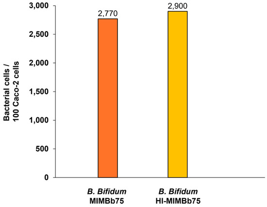
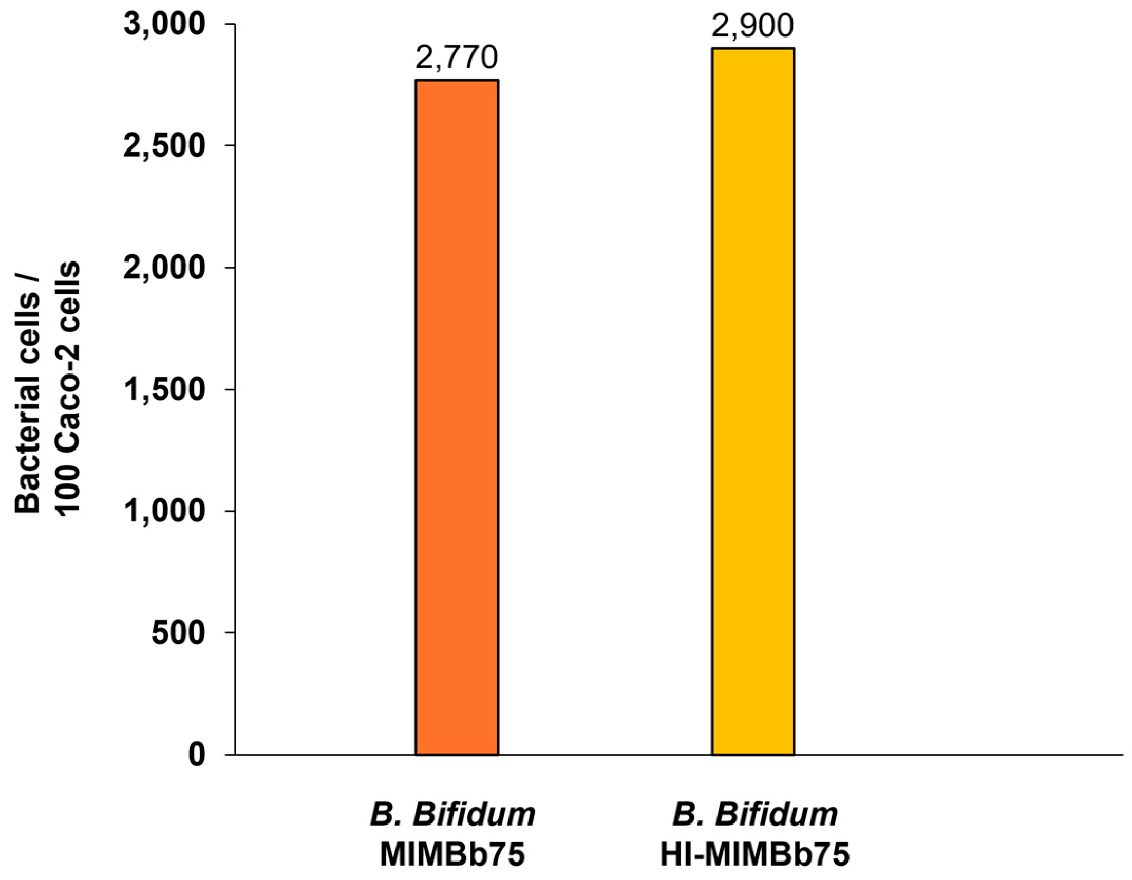
We observed that viable and heat-inactivated B. bifidum MIMBb75 possess a similarly high physical adhesion ability: The adhesion index, i.e., the number of bacterial cells firmly adhering after at least three washing steps to a set of 100 Caco-2 cells, was 2770 for viable B. bifidum MIMBb75 and 2900 for heat-inactivated B. bifidum MIMBb75 (Figure 1).
Figure 1.
Adhesion index of viable and heat-inactivated B. bifidum MIMBb75 adhering to 100 Caco-2 cells.
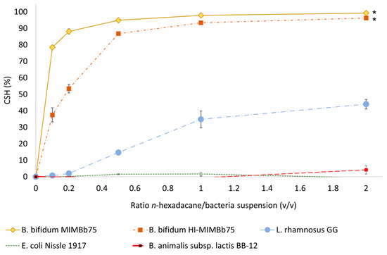
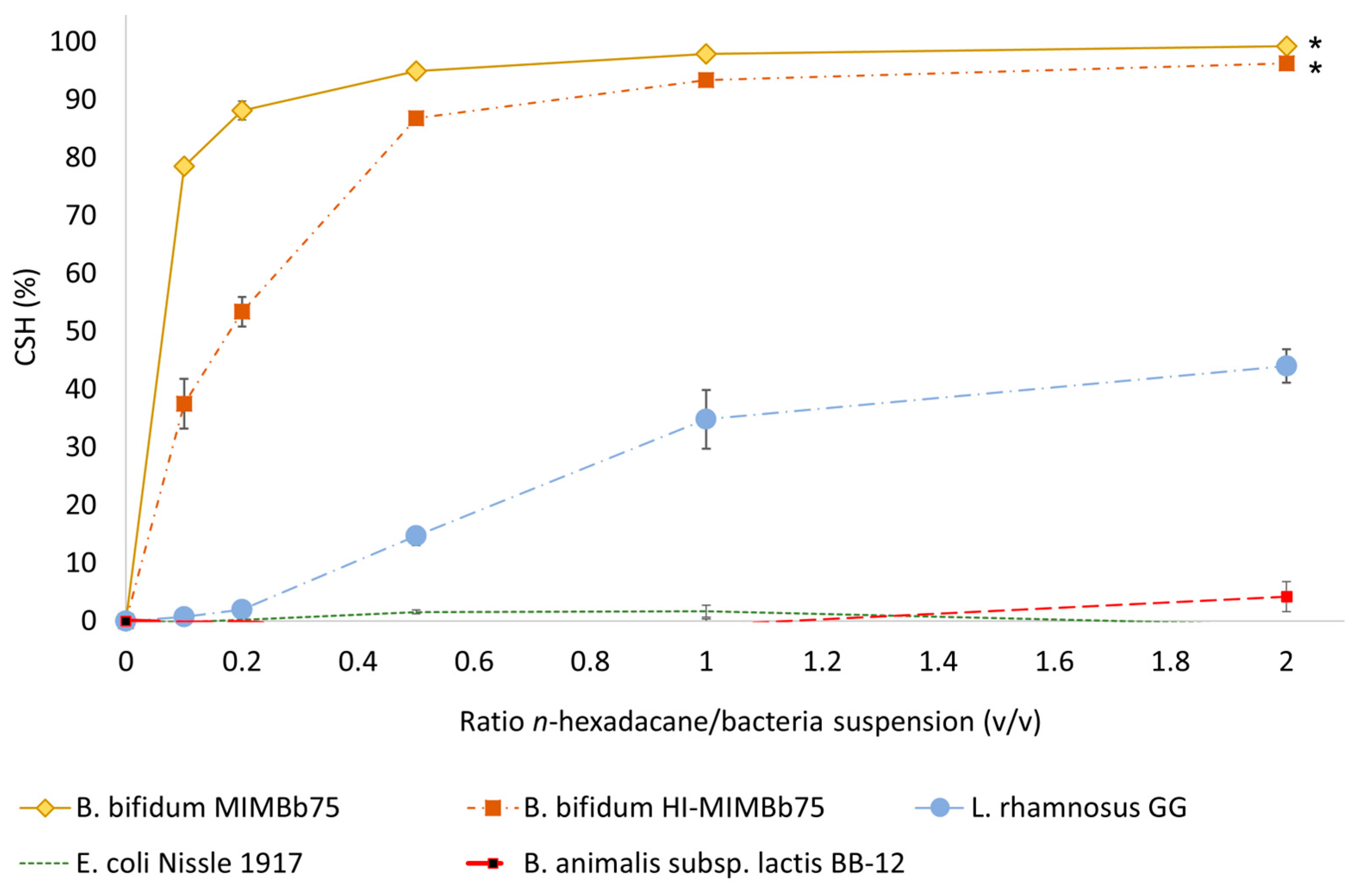
Viable and heat-inactivated B. bifidum MIMBb75 revealed by far the highest CSH when compared to L. rhamnosus GG, B. animalis subsp. lactis BB-12 and E. coli Nissle 1917, all of which have been successfully used in the treatment of IBS [47,48,49,50,51] (Figure 2).
Figure 2.
Cell Surface Hydrophobicity (CSH) of viable B. bifidum MIMBb75 and heat-inactivated B. bifidum MIMBb75 compared to L. rhamnosus GG, B. animalis subsp. lactis BB-12 and E. coli Nissle 1917, respectively, as assessed by relative absorbance rates at 560 nm of increasing n-hexadecane/bacteria suspension extraction ratios. Results are shown as mean ± SD (n = 3). * CSH values of viable and heat-inactivated B. bifidum MIMBb75 do not differ significantly from each other (p = 0.2492), but they do pairwise from all others (p < 0.0001).
L. rhamnosus GG, B. animalis subsp. lactis BB-12 and E. coli Nissle 1917 were used as well-characterized benchmark strains to contextualize the CSH of B. bifidum MIMBb75, not as definitive positive or negative controls. Because CSH is a continuous physical property, values can vary substantially even among closely related strains. Accordingly, Pan et al. reported CSH values ranging from 0.30% to 37.24% across 23 bifidobacterial strains [35]. For both viable and heat-inactivated B. bifidum MIMBb75, the aqueous-phase absorbance decreased progressively with increasing n-hexadecane/bacterial suspension ratios, approaching maximal extraction at the highest ratios.
Already at very low extraction ratios of 0.1 and 0.2, both viable and heat-inactivated B. bifidum MIMBb75 show a hydrophobic affinity to n-hexadecane that is several-fold higher than for any of the other tested bacteria (including another bifidobacterium strain, B. animalis subsp. lactis BB-12). The only other strain showing a moderate CSH is L. rhamnosus GG (fluid-fluid interface absorbance of 44% at an extraction ratio of 2.0). In contrast, E. coli Nissle 1917 and B. animalis subsp. lactis BB-12 do not appear to be hydrophobic.
3.2. Both Viable and Heat-Inactivated B. bifidum MIMBb75 Increase TEER
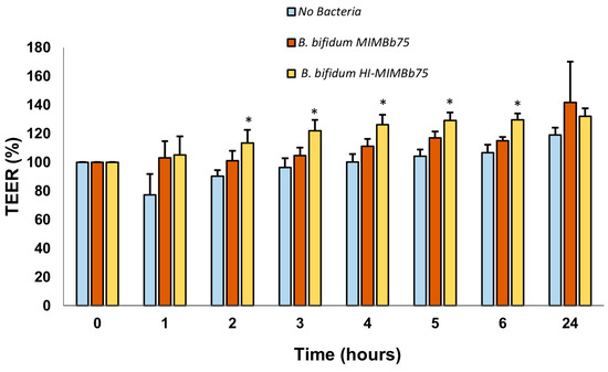
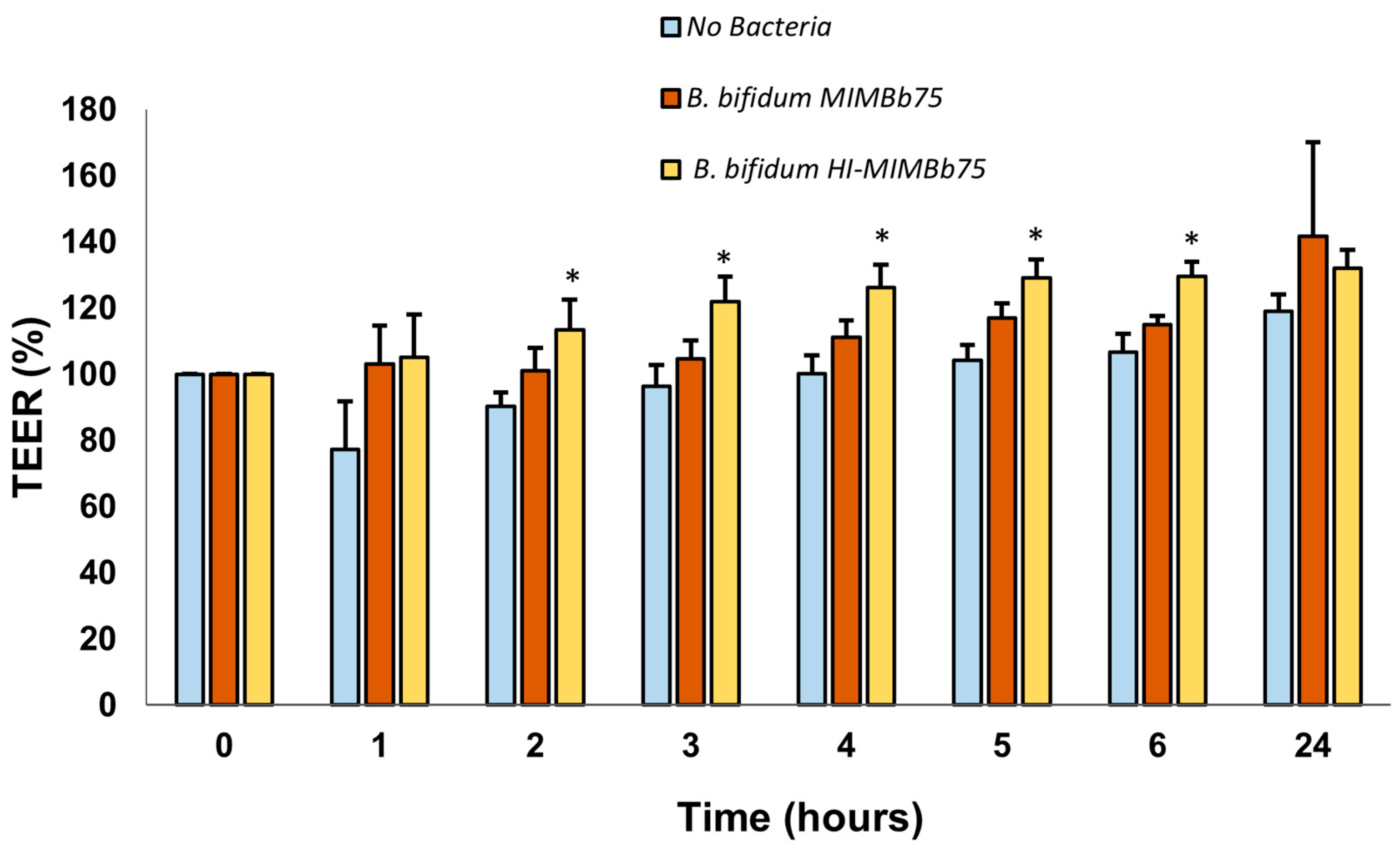
In the experiment without any disruptors, TEER (normalized to baseline) of Caco-2 cells exposed to viable or heat-inactivated B. bifidum MIMBb75 was consistently higher than the untreated control (medium without bacteria) in all measurements. At 24 h, the TEERs (normalized to baseline) of Caco-2 cells exposed to viable B. bifidum MIMBb75 increased to 141.78 ± 28.31% (p = 0.40 vs. untreated control 118.90 ± 5.11%), while the TEER of Caco-2 cells exposed to heat-inactivated B. bifidum MIMBb75 increased to 132.07 ± 5.38% (p = 0.05 vs. untreated control 118.90 ± 5.11%) (Figure 3).
Figure 3.
Change in intestinal barrier function by viable B. bifidum MIMBb75 (107 CFU/well) and heat-inactivated B. bifidum MIMBb75 (107 cells/well), in comparison to the untreated cell control (medium without bacteria), as indicated by an increase in TEER over 24 h measured in Caco-2 transwell bacterial co-cultures. Results are normalized to baseline and shown as mean ± SD (n = 2–5). * p < 0.05 versus untreated control.
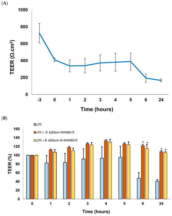
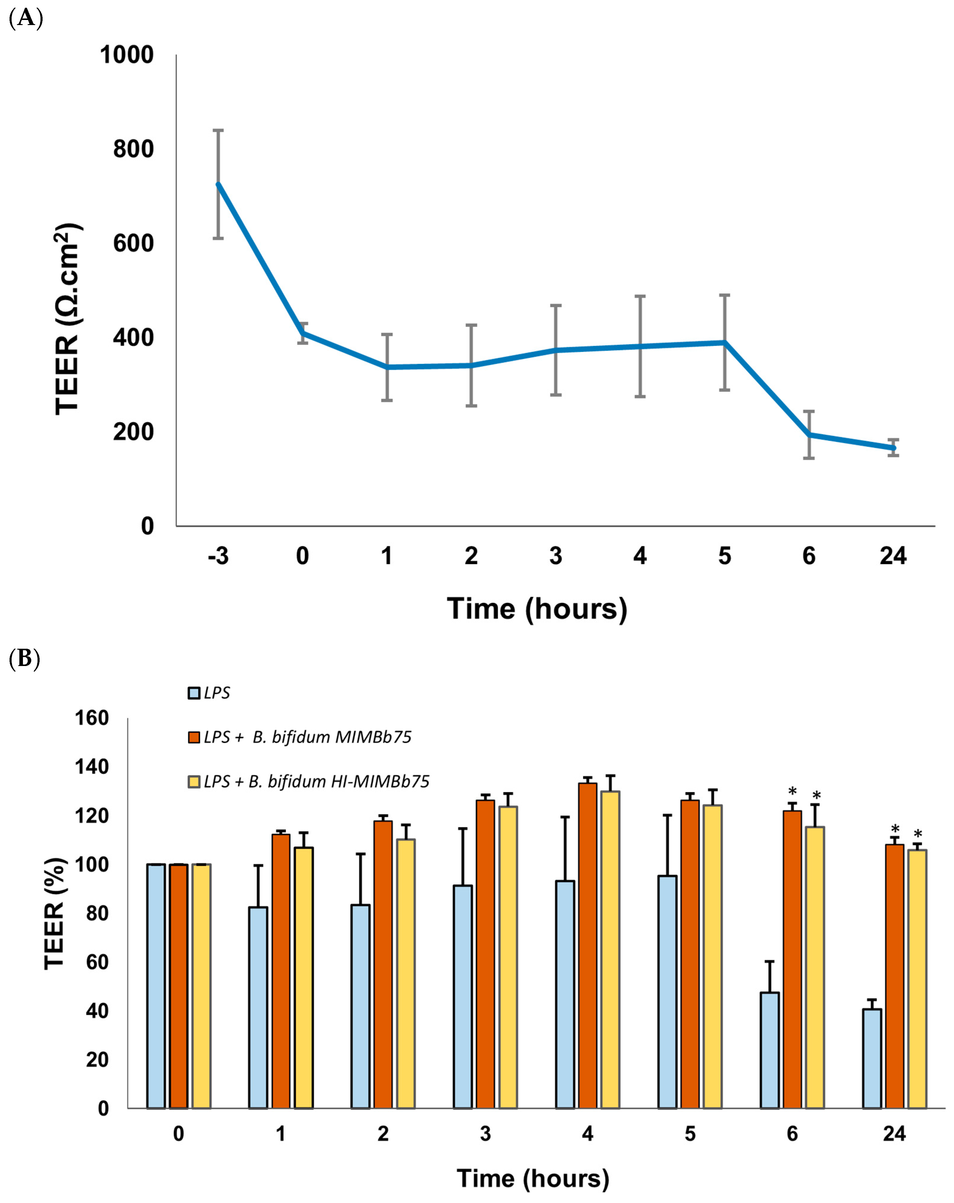
Treating Caco-2 cells with 24 ng/mL LPS induced a TEER drop from 725.18 ± 114.76 Ω × cm2 to 408.40 ± 20.77 Ω × cm2 during the incubation period of three hours. Afterwards, TEER was almost stable at a value around 370 Ω × cm2 for the first five hours, before decreasing to 166.11 ± 16.68 Ω × cm2 in 24 h (Figure 4A). E. coli LPS significantly disrupted the membrane integrity of Caco-2 cells at a 24 ng/mL concentration.
Figure 4.
(A) Caco-2 cells were pre-treated with 24 ng/mL E. coli LPS and incubated for 3 h. During the incubation period, TEER dropped from 725.18 ± 114.76 Ω × cm2 to 408.40 ± 20.77 Ω × cm2. (B) Addition of 107 CFU/well of viable B. bifidum MIMBb75 or 107 cells/well of heat-inactivated B. bifidum MIMBb75 to LPS-pre-treated cells increased intestinal barrier function as measured by TEER (normalized to baseline), whereas it dropped to 40.71 ± 3.95% for the LPS-only control (medium without bacteria). Results are shown as mean ± SD (n = 3). * p < 0.05 versus LPS-only control.
Upon addition of viable or heat-inactivated B. bifidum MIMBb75 to the LPS pretreated cells at t = 0, TEER (normalized to baseline, t = 0) at 24 h (Figure 4B) was significantly higher than the LPS-only control (medium-without bacteria) for both viable B. bifidum MIMBb75 (108.22 ± 2.90% vs. 40.71 ± 3.95%, p < 0.001) and heat-inactivated B. bifidum MIMBb75 (105.96 ± 2.64% vs. 40.71 ± 3.95%, p < 0.001).
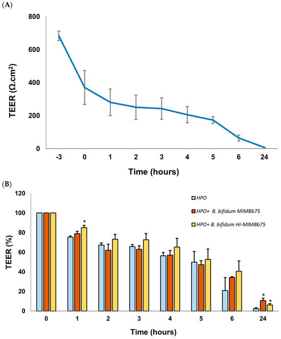
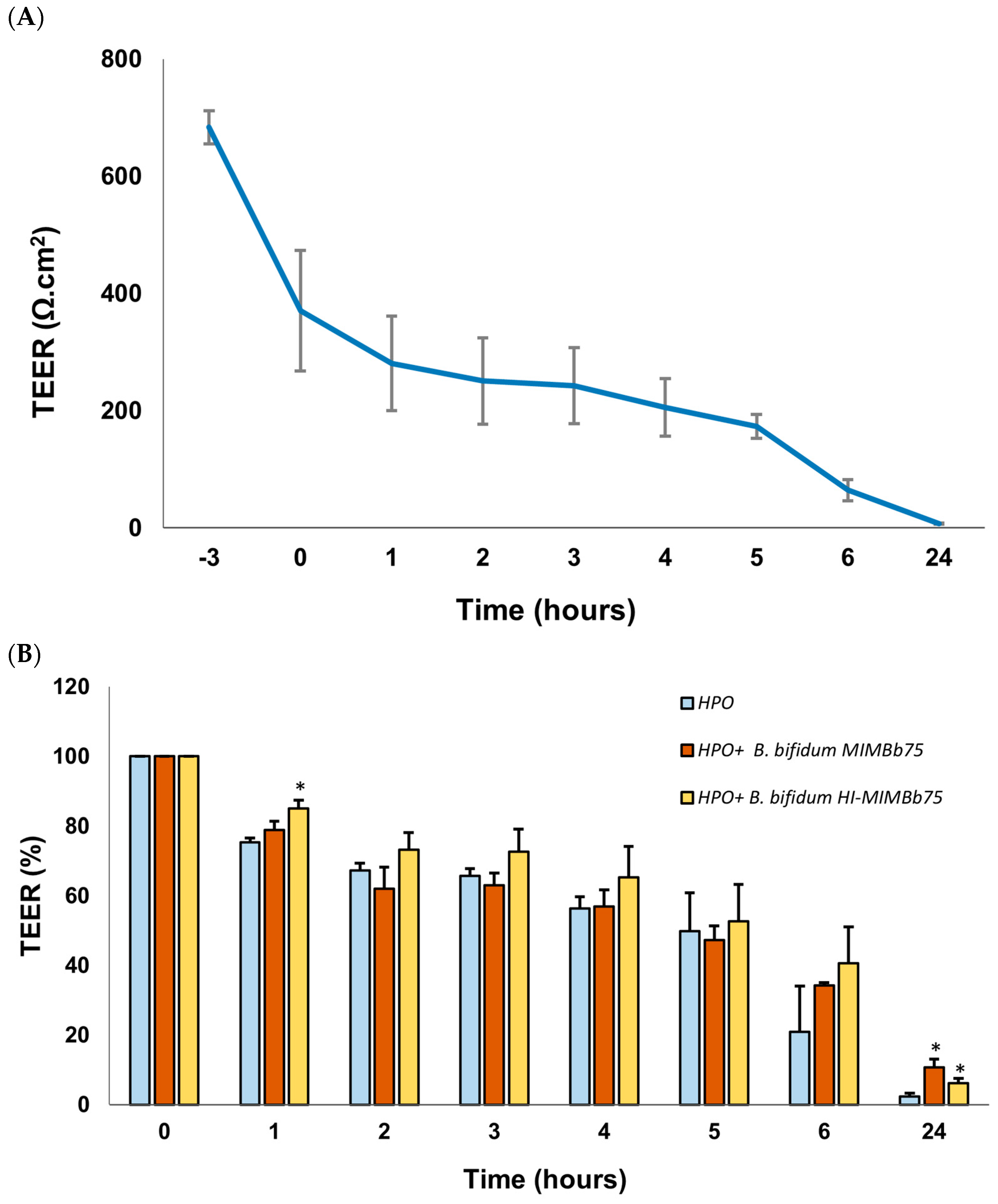
Unlike LPS, the addition of 50 mM HPO reduced TEER continuously throughout the entire measurement period from an initial 683.43 ± 28.39 Ω × cm2 to 7.57 ± 0.84 Ω × cm2 (Figure 5A). Over the course of 24 h, TEER (normalized to baseline, t = 0) in pre-treated Caco-2 cells decreased to 2.31 ± 0.97%. In comparison, TEER was roughly three to five times higher for cells treated with either viable or heat-inactivated B. bifidum MIMBb75 at the end of measurement (12.14 ± 1.45%; p = 0.006 vs. HPO-only control and 6.20 ± 1.35%; p = 0.030 vs. HPO-only control) (Figure 5B).
Figure 5.
(A) Caco-2 cells were pre-treated with 50 mM HPO and incubated for 0.5 h. During the incubation period, TEER dropped from 683.43 ± 28.39 Ω × cm2 (measured initially 2.5 h before adding HPO) to 370.72 ± 102.73 Ω × cm2. (B) TEER (%) dropped to 2.31 ± 0.97% for the HPO-only control (medium without bacteria), whereas it dropped to 12.14 ± 1.45% and 6.2 ± 1.35% with the addition of 107 CFU/well of viable B. bifidum MIMBb75 or 107 cells/well of heat-inactivated B. bifidum MIMBb75. Results are shown as mean ± SD (n = 3). * p < 0.05 versus HPO-only control.
4. Discussion
IBS is a common gastrointestinal disease of multifactorial genesis. Various studies have consistently found a disrupted intestinal barrier with increased permeability in patients with IBS, irrespective of the underlying IBS subtype. Increased intestinal permeability contributes to the translocation of luminal bacterial antigens, which in turn causes irritation of the intestinal nervous system [9,16,18,19,52,53,54,55]. In consequence, the typical symptoms of IBS, such as abdominal pain and diarrhea, exacerbate. Protecting the intestinal barrier is hence a key target within IBS treatment. In the present study, we demonstrated that both viable and heat-inactivated B. bifidum MIMBb75 physically adhere to intestinal epithelial cells in high numbers and protect both the intact and the damaged epithelial barrier. To our knowledge, this is the first systematic study that systematically investigates the barrier-protecting effects of viable and heat-inactivated B. bifidum MIMBb75 within the same experimental framework.
Gentle heating leads to the inactivation of probiotic bacteria, preserving their overall structure but eliminating their ability to grow and multiply. Heat-inactivated probiotics present notable advantages with respect to standardization, transport, and storage stability [56]. For many years, cell viability has been regarded as one of the key—if not the single most crucial—properties of probiotics thought to underlie their health benefits [57,58,59,60,61,62]. However, evidence indicating that microbial viability is not essential for the efficacy has been demonstrated for several strains and conditions [63,64,65].
Both viable and heat-inactivated B. bifidum MIMBb75 show a distinctly high adherence to Caco-2 cells. Evaluation by light microscopy showed that the adhesion index, i.e., the number of bacterial cells adhering to an average set of 100 Caco-2 cells, was 2770 for viable B. bifidum MIMBb75 and 2900 for heat-inactivated B. bifidum MIMBb75, in line with previous findings on clinical efficacy by Guglielmetti [30] and Andresen [31]. The adhesion index of B. bifidum MIMBb75 is up to 500-fold higher compared to adhesion indices reported by others under comparable experimental conditions for other known probiotic bacteria, including B. animalis subsp. lactis BB-12 and L. rhamnosus GG [29]. These data confirm that physical adhesion to the intestinal epithelial tissue is a characteristic feature of B. bifidum MIMBb75 that is fully maintained upon heat inactivation.
Various mechanisms have been proposed by others to explain the forces driving this strong adhesion ability. In particular, an involvement of the specific surface protein BopA was initially hypothesized [29,66] but was subsequently disproven [67]. In contrast, our study indicates that the adhesion to the disrupted intestinal barrier is primarily mediated by the high CSH of both viable and heat-inactivated B. bifidum MIMBb75 through a physical mode of action.
Our results regarding the high CSH of viable and heat-inactivated B. bifidum MIMBb75 and its strong adhesion abilities to Caco-2 cells are consistent with a previous report of Pan and co-workers [35], who found a strong positive correlation between the two metrics. Indeed, already at very low extraction ratios of 0.1 and 0.2, viable and heat-inactivated B. bifidum MIMBb75 both show a hydrophobic affinity to n-hexadecane that is several-fold higher compared to other strains tested, including B. animalis subsp. lactis BB-12, which pertains to the same genus of bifidobacteria. These findings suggest that hydrophobic interactions—purely physical forces—are the primary mechanism underlying the adherence of viable and heat-inactivated B. bifidum MIMBb75 to epithelial cells.
Comparable to the findings by Briske-Anderson and co-workers [68], TEER values measured in this study were stable and only increased minimally in fully differentiated, untreated Caco-2 cells at day 21. However, following the addition of either viable or heat-inactivated B. bifidum MIMBb75, a marked increase in TEER values was observed. The consistently higher TEER values were also observed when the Caco-2 monolayers were pre-treated with LPS and HPO. The adhesion of both viable and heat-inactivated B. bifidum MIMBb75 to Caco-2 cells is in accordance with previous hypotheses on the clinical effect of B. bifidum MIMBb75 on the intestinal barrier in IBS patients [30,31].
One limitation of this study is that our findings are based on in vitro data using a Caco-2 cell model. However, Caco-2 monolayers are an established and widely accepted standard model for studying direct interactions between probiotic bacteria and the intestinal epithelium [69,70,71,72,73,74], and they are recognized by regulatory authorities as supportive mechanistic evidence. Conducting an invasive clinical trial for the sole purpose of elucidating the mode of action would, moreover, not be ethically justifiable, particularly given that the clinical efficacy of both viable and heat-inactivated B. bifidum MIMBb75 has already been demonstrated in two independent double-blind randomized clinical trials [30,31].
The use of a Caco-2 monolayer model is also consistent with the pathophysiological situation in IBS patients, in whom the protective mucus layer is significantly thinner than in healthy controls [34], so that direct bacteria–epithelium interactions are of particular physiological relevance.
Another limitation of the present study is that epithelial barrier integrity was assessed using a single functional readout. TEER is a well-established and widely accepted method to evaluate epithelial monolayer integrity in Caco-2 models and was therefore appropriate for the objective of this study. Nevertheless, the inclusion of complementary barrier assays would further strengthen the evidence for the observed barrier-stabilizing effect and should be considered in future studies.
A further limitation is the limited number of biological replicates in the TEER experiments, which reflects the strict inclusion criteria applied.
While the aim of the present study was to explicitly compare and examine the hydrophobicity and the protective effect of both viable and heat-inactivated B. bifidum MIMBb75, it might also be useful in the in vitro screening of other future probiotic treatments for IBS. Our results underpin the validity of the strong correlation between the CSH of bifidobacteria and their adhesiveness to the disrupted epithelium.
5. Conclusions
In summary, our results clearly demonstrate that B. bifidum MIMBb75—in both the viable and heat-inactivated forms—adheres exceptionally well to intestinal epithelial cells. Together with a significant increase in TEER values after the addition of viable and heat-inactivated B. bifidum MIMBb75, this suggests a protection of the intestinal epithelial barrier and provides mechanistic support for the efficacy of B. bifidum MIMBb75 in previously conducted clinical studies. Its adhesion is primarily mediated through physical forces for both viable and heat-inactivated B. bifidum MIMBb75, i.e., their extraordinarily high cell surface hydrophobicity, figuratively comparable to a physical plaster. Our findings also confirm that the barrier-protective capacity is independent of viability and, hence, metabolic activity. The strong adhesion of both viable and heat-inactivated B. bifidum MIMBb75 forms a protective layer above the epithelial surface and results in a significant increase in TEER, indicating protection of the intestinal epithelial barrier.
Author Contributions
M.S. made substantial contributions to the conception of the work, interpretation of data and critical revision of the manuscript. M.G. edited and reviewed the manuscript. All authors have read and agreed to the published version of the manuscript.
Funding
We would like to thank Synformulas GmbH for funding the study.
Institutional Review Board Statement
Not applicable.
Informed Consent Statement
Not applicable.
Data Availability Statement
The original contributions presented in this study are included in the article. Further inquiries can be directed to the corresponding author.
Acknowledgments
The experiments were executed at Tentamus Group GmbH as assigned by Synformulas GmbH.
Conflicts of Interest
The authors declare that this study received funding from Synformulas GmbH. The funder contributed to the study design and provided support with medical writing.
Abbreviations
The following abbreviations are used in this manuscript:
| A | Absorbance values after extraction of bacteria with n-hexadecane |
| A0 | Absorbance values before extraction of bacteria with n-hexadecane |
| ANOVA | Analysis of variance |
| B. animalis subsp. lactis BB-12 | Bifidobacterium animalis subspecies lactis BB-12 |
| B. bifidum | Bifidobacterium bifidum |
| B. bifidum MIMBb75 | Viable Bifidobacterium bifidum MIMBb75 |
| B. bifidum HI-MIMBb75 | Heat-inactivated B. bifidum MIMBb75 |
| CFU | Colony-forming units |
| CSH | Cell surface hydrophobicity |
| DGBI | Disorder of gut–brain interaction |
| E. coli | Escherichia coli |
| HPO | Hydrogen peroxide |
| IBS | Irritable Bowel Syndrome |
| LPS | Lipopolysaccharide(s) |
| L. rhamnosus GG | Lactobacillus rhamnosus GG |
| OD | Optical density |
| SD | Standard deviation |
| TEER | Transepithelial electrical resistance |
References
- Enck, P.; Qasim, A.; Barbara, G.; Farmer, A.D.; Fukudo, S.; Mayer, E.A.; Niesler, B.; Quigley, E.M.M.; Rajilić-Stojanović, M.; Schemann, M.; et al. Irritable bowel syndrome. Nat. Rev. Dis. Primers 2016, 2, 16014. [Google Scholar] [CrossRef] [Scilit]
- Ford, A.C.; Sperber, A.D.; Corsetti, M.; Camilleri, M. Irritable bowel syndrome. Lancet 2020, 396, 1675–1688. [Google Scholar] [CrossRef] [Scilit] [PubMed]
- Dunlop, S.P.; Hebden, J.; Campbell, E.; Naesdal, J.; Olbe, L.; Perkins, A.C.; Spiller, R.C. Abnormal intestinal permeability in subgroups of diarrhea-predominant irritable bowel syndromes. Am. J. Gastroenterol. 2006, 101, 1288–1294. [Google Scholar] [CrossRef] [Scilit]
- Gecse, K.; Róka, R.; Séra, T.; Rosztóczy, A.; Annaházi, A.; Izbéki, F.; Nagy, F.; Molnár, T.; Szepes, Z.; Pávics, L.; et al. Leaky gut in patients with diarrhea-predominant irritable bowel syndrome and inactive ulcerative colitis. Digestion 2012, 85, 40–46. [Google Scholar] [CrossRef] [Scilit] [PubMed]
- Mujagic, Z.; Ludidi, S.; Keszthelyi, D.; Hesselink, M.A.M.; Kruimel, J.W.; Lenaerts, K.; Hanssen, N.M.J.; Conchillo, J.M.; Jonkers, D.M.A.E.; Masclee, A.A.M. Small intestinal permeability is increased in diarrhoea predominant IBS, while alterations in gastroduodenal permeability in all IBS subtypes are largely attributable to confounders. Aliment. Pharmacol. Ther. 2014, 40, 288–297. [Google Scholar] [CrossRef] [Scilit]
- Piche, T.; Barbara, G.; Aubert, P.; Des Varannes, S.B.; Dainese, R.; Nano, J.L.; Cremon, C.; Stanghellini, V.; De Giorgio, R.; Galmiche, J.P.; et al. Impaired Intestinal barrier integrity in the colon of patients with irritable bowel syndrome: Involvement of soluble mediators. Gut 2009, 58, 196–201. [Google Scholar] [CrossRef] [Scilit]
- Turcotte, J.F.; Kao, D.; Mah, S.J.; Claggett, B.; Saltzman, J.R.; Fedorak, R.N.; Liu, J.J. Breaks in the wall: Increased gaps in the intestinal epithelium of irritable bowel syndrome patients identified by confocal laser endomicroscopy (with videos). Gastrointest. Endosc. 2013, 77, 624–630. [Google Scholar] [CrossRef] [Scilit] [PubMed]
- Vivinus-Nébot, M.; Dainese, R.; Anty, R.; Saint-Paul, M.C.; Nano, J.L.; Gonthier, N.; Marjoux, S.; Frin-Mathy, G.; Bernard, G.; Hébuterne, X.; et al. Combination of allergic factors can worsen diarrheic irritable bowel syndrome: Role of barrier defects and mast cells. Am. J. Gastroenterol. 2012, 107, 75–81. [Google Scholar] [CrossRef] [Scilit]
- Martínez, C.; Lobo, B.; Pigrau, M.; Ramos, L.; González-Castro, A.M.; Alonso, C.; Guilarte, M.; Guilá, M.; de Torres, I.; Azpiroz, F.; et al. Diarrhoea-predominant irritable bowel syndrome: An organic disorder with structural abnormalities in the jejunal epithelial barrier. Gut 2013, 62, 1160–1168. [Google Scholar] [CrossRef] [Scilit]
- Johansson, M.E.V.; Sjövall, H.; Hansson, G.C. The gastrointestinal mucus system in health and disease. Nat. Rev. Gastroenterol. Hepatol. 2013, 10, 352–361. [Google Scholar] [CrossRef] [Scilit]
- Pelaseyed, T.; Bergström, J.H.; Gustafsson, J.K.; Ermund, A.; Birchenough, G.M.H.; Schütte, A.; van der Post, S.; Svensson, F.; Rodríguez-Piñeiro, A.M.; Nyström, E.E.L.; et al. The mucus and mucins of the goblet cells and enterocytes provide the first defense line of the gastrointestinal tract and interact with the immune system. Immunol. Rev. 2014, 260, 8–20. [Google Scholar] [CrossRef] [Scilit] [PubMed]
- Johansson, M.E.V.; Phillipson, M.; Petersson, J.; Velcich, A.; Holm, L.; Hansson, G.C. The inner of the two Muc2 mucin-dependent mucus layers in colon is devoid of bacteria. Proc. Natl. Acad. Sci. USA 2008, 105, 15064–15069. [Google Scholar] [CrossRef] [Scilit]
- Jabbar, K.S.; Dolan, B.; Eklund, L.; Wising, C.; Ermund, A.; Johansson, Å.; Törnblom, H.; Simren, M.; Hansson, G.C. Association between Brachyspira and irritable bowel syndrome with diarrhoea. Gut 2021, 70, 1117–1129. [Google Scholar] [CrossRef] [Scilit]
- Dam, B.; Misra, A.; Banerjee, S. Role of Gut Microbiota in Combating Oxidative Stress. In Oxidative Stress in Microbial Diseases; Springer: Singapore, 2019; pp. 43–82. [Google Scholar] [CrossRef] [Scilit]
- Shukla, R.; Ghoshal, U.; Ranjan, P.; Ghoshal, U.C. Expression of Toll-like Receptors, Pro-, and Anti- inflammatory Cytokines in Relation to Gut Microbiota in Irritable Bowel Syndrome: The Evidence for Its Micro-organic Basis. J. Neurogastroenterol. Motil. 2018, 24, 628–642. [Google Scholar] [CrossRef] [Scilit]
- Bercik, P.; Verdu, E.F.; Collins, S.M. Is irritable bowel syndrome a low-grade inflammatory bowel disease? Gastroenterol. Clin. North Am. 2005, 34, 235–245. [Google Scholar] [CrossRef] [Scilit]
- Holtmann, G.J.; Ford, A.C.; Talley, N.J. Pathophysiology of irritable bowel syndrome. Lancet Gastroenterol. Hepatol. 2016, 1, 133–146. [Google Scholar] [CrossRef] [Scilit] [PubMed]
- Matricon, J.; Meleine, M.; Gelot, A.; Piche, T.; Dapoigny, M.; Muller, E.; Ardid, D. Review article: Associations between immune activation, intestinal permeability and the irritable bowel syndrome. Aliment. Pharmacol. Ther. 2012, 36, 1009–1031. [Google Scholar] [CrossRef] [Scilit]
- Ng, Q.X.; Soh, A.Y.S.; Loke, W.; Lim, D.Y.; Yeo, W.S. The role of inflammation in irritable bowel syndrome (IBS). J. Inflamm. Res. 2018, 11, 345–349. [Google Scholar] [CrossRef] [Scilit]
- Baumgartner, M.; Lang, M.; Holley, H.; Crepaz, D.; Hausmann, B.; Pjevac, P.; Moser, D.; Haller, F.; Hof, F.; Beer, A.; et al. Mucosal Biofilms Are an Endoscopic Feature of Irritable Bowel Syndrome and Ulcerative Colitis. Gastroenterology 2021, 161, 1245–1256. [Google Scholar] [CrossRef] [Scilit]
- Ford, A.C.; Quigley, E.M.M.; Lacy, B.E.; Lembo, A.J.; Saito, Y.A.; Schiller, L.R.; Soffer, E.E.; Spiegel, B.M.R.; Moayyedi, P. Efficacy of prebiotics, probiotics, and synbiotics in irritable bowel syndrome and chronic idiopathic constipation: Systematic review and meta-analysis. Am. J. Gastroenterol. 2014, 109, 1547–1562. [Google Scholar] [CrossRef] [Scilit] [PubMed]
- Moayyedi, P.; Ford, A.C.; Talley, N.J.; Cremonini, F.; Foxx-Orenstein, A.E.; Brandt, L.J.; Quigley, E.M.M. The efficacy of probiotics in the treatment of irritable bowel syndrome: A systematic review. Gut 2010, 59, 325–332. [Google Scholar] [CrossRef] [Scilit]
- Whelan, K.; Quigley, E.M.M. Probiotics in the management of irritable bowel syndrome and inflammatory bowel disease. Curr. Opin. Gastroenterol. 2013, 29, 184–189. [Google Scholar] [CrossRef] [Scilit]
- Zhang, T.; Zhang, C.; Zhang, J.; Sun, F.; Duan, L. Efficacy of Probiotics for Irritable Bowel Syndrome: A Systematic Review and Network Meta-Analysis. Front. Cell. Infect. Microbiol. 2022, 12, 859967. [Google Scholar] [CrossRef] [Scilit] [PubMed]
- Zhang, Y.; Li, L.; Guo, C.; Mu, D.; Feng, B.; Zuo, X.; Li, Y. Effects of probiotic type, dose and treatment duration on irritable bowel syndrome diagnosed by Rome III criteria: A meta-analysis. BMC Gastroenterol. 2016, 16, 58. [Google Scholar] [CrossRef] [Scilit]
- Rana, A.; Smriti. Probiotics: Mechanism of action and gastrointestinal health: Gut guardians: Unlocking the power of probiotics. J. Sci. Food Agric. 2026, 106, 3938–3951. [Google Scholar] [CrossRef] [Scilit] [PubMed]
- Bron, P.A.; Kleerebezem, M.; Brummer, R.J.; Cani, P.D.; Mercenier, A.; MacDonald, T.T.; Garcia-Ródenas, C.L.; Wells, J.M. Can probiotics modulate human disease by impacting intestinal barrier function? Br. J. Nutr. 2017, 117, 93–107. [Google Scholar] [CrossRef] [Scilit]
- Camilleri, M. Human intestinal barrier: Effects of stressors, diet, prebiotics, and probiotics. Clin. Transl. Gastroenterol. 2021, 12, e00308. [Google Scholar] [CrossRef] [Scilit] [PubMed]
- Guglielmetti, S.; Tamagnini, I.; Mora, D.; Minuzzo, M.; Scarafoni, A.; Arioli, S.; Hellman, J.; Karp, M.; Parini, C. Implication of an outer surface lipoprotein in adhesion of Bifidobacterium bifidum to Caco-2 cells. Appl. Environ. Microbiol. 2008, 74, 4695–4702. [Google Scholar] [CrossRef] [Scilit]
- Guglielmetti, S.; Mora, D.; Gschwender, M.; Popp, K. Randomised clinical trial: Bifidobacterium bifidum MIMBb75 significantly alleviates irritable bowel syndrome and improves quality of life—A double-blind, placebo-controlled study. Aliment. Pharmacol. Ther. 2011, 33, 1123–1132. [Google Scholar] [CrossRef] [Scilit]
- Andresen, V.; Gschossmann, J.; Layer, P. Heat-inactivated Bifidobacterium bifidum MIMBb75 (SYN-HI-001) in the treatment of irritable bowel syndrome: A multicentre, randomised, double-blind, placebo-controlled clinical trial. Lancet Gastroenterol. Hepatol. 2020, 5, 658–666. [Google Scholar] [CrossRef] [Scilit]
- Layer, P.; Andresen, V.; Allescher, H.; Bischoff, S.C.; Claßen, M.; Elsenbruch, S.; Freitag, M.; Frieling, T.; Gebhard, M.; Goebel-Stengel, M.; et al. Update S3-Leitlinie Reizdarmsyndrom: Definition, Pathophysiologie, Diagnostik und Therapie. Gemeinsame Leitlinie der Deutschen Gesellschaft für Gastroenterologie, Verdauungs- und Stoffwechselkrankheiten (DGVS) und der Deutschen Gesellschaft für Neurogastroenterologie und Motilität (DGNM)—Juni 2021—AWMF-Registriernummer: 021/016. Z. Gastroenterol. 2021, 59, 1323–1415, Erratum in Z. Gastroenterol. 2021, 59, e1. https://doi.org/10.1055/a-1738-5962. [Google Scholar] [CrossRef] [Scilit] [PubMed]
- Press, B.; Di Grandi, D. Permeability for Intestinal Absorption: Caco-2 Assay and Related Issues. Curr. Drug Metab. 2008, 9, 893–900. [Google Scholar] [CrossRef] [Scilit] [PubMed]
- Inczefi, O.; Eutamene, H.; Placide, F.; Tondereau, V.; Pallagi, P.; Bagyánszki, M.; Bódi, N.; Gémes, N.; Szebeni, G.; Molnár, T.; et al. Translational evaluation of Gelsectan® effects on gut barrier dysfunction and visceral pain in animal models and irritable bowel syndrome with diarrhoea. United Eur. Gastroenterol. J. 2024, 12, 1102–1113. [Google Scholar] [CrossRef] [Scilit] [PubMed]
- Pan, W.H.; Li, P.L.; Liu, Z. The correlation between surface hydrophobicity and adherence of Bifidobacterium strains from centenarians’ faeces. Anaerobe 2006, 12, 148–152. [Google Scholar] [CrossRef] [Scilit]
- Srinivasan, B.; Kolli, R.A.; Esch, M.B.; Abaci, H.E.; Shuler, M.L.; Hickman, J.J. TEER Measurement Techniques for In Vitro Barrier Model Systems. J. Lab. Autom. 2015, 20, 107–126. [Google Scholar] [CrossRef] [Scilit]
- Hou, J. Paracellular Channel Recording. In The Paracellular Channel; Elsevier: Amsterdam, The Netherlands, 2019; pp. 29–49. [Google Scholar] [CrossRef] [Scilit]
- Fatmawati, N.N.D.; Goto, K.; Mayura, I.P.B.; Nocianitri, K.A.; Ramona, Y.; Sakaguchi, M.; Matsushita, O.; Sujaya, I.N. Caco-2 cells monolayer as an in-vitro model for probiotic strain translocation. Bali Med. J. 2020, 9, 137–142. [Google Scholar] [CrossRef] [Scilit]
- Guo, S.; Al-Sadi, R.; Said, H.M.; Ma, T.Y. Lipopolysaccharide Causes an Increase in Intestinal Tight Junction Permeability in Vitro and in Vivo by Inducing Enterocyte Membrane Expression and Localization of TLR-4 and CD14. Am. J. Pathol. 2013, 182, 375–387. [Google Scholar] [CrossRef] [Scilit]
- Iraha, A.; Chinen, H.; Hokama, A.; Yonashiro, T.; Kinjo, T.; Kishimoto, K.; Nakamoto, M.; Hirata, T.; Kinjo, N.; Higa, F.; et al. Fucoidan enhances intestinal barrier function by upregulating the expression of claudin-1. World J. Gastroenterol. 2013, 19, 5500–5507. [Google Scholar] [CrossRef] [Scilit]
- Nighot, M.; Al-Sadi, R.; Guo, S.; Rawat, M.; Nighot, P.; Watterson, M.D. Lipopolysaccharide-Induced Increase in Intestinal Epithelial Tight Permeability Is Mediated by Toll-Like Receptor 4/Myeloid Differentiation Primary Response 88 (MyD88) Activation of Myosin Light Chain Kinase Expression. Am. J. Pathol. 2017, 187, 2698–2710. [Google Scholar] [CrossRef] [Scilit]
- Tazuke, Y.; Drongowski, R.A.; Teitelbaum, D.H.; Coran, A.G. The effect of hypoxia on permeability and bacterial translocation in Caco-2 adult and I-407 fetal enterocyte cell culture models. Pediatr. Surg. Int. 2003, 19, 316–320. [Google Scholar] [CrossRef] [Scilit]
- Chen, H.L.; Tung, Y.T.; Chuang, C.L.; Tu, M.Y.; Tsai, T.C.; Chang, H.C.; Chen, C.M. Lactobacillus kefiranofaciens M1 isolated from milk kefir grains ameliorates experimental colitis in vitro and in vivo. J. Dairy Sci. 2012, 95, 63–74. [Google Scholar] [CrossRef] [Scilit]
- Han, H.; You, Y.; Cha, S.; Kim, T.R.; Sohn, M.; Park, J. Multi-Species Probiotic Strain Mixture Enhances Intestinal Barrier Function by Regulating Inflammation and Tight Junctions in Lipopolysaccharides Stimulated Caco-2 Cells. Microorganisms 2023, 11, 656. [Google Scholar] [CrossRef] [Scilit]
- de Gregorio, A.; Serafino, A.; Krasnowska, E.K.; Superti, F.; Di Fazio, M.R.; Fuggetta, M.P.; Hammarberg Ferri, I.; Fiorentini, C. Protective Effect of Limosilactobacillus fermentum ME-3 against the Increase in Paracellular Permeability Induced by Chemotherapy or Inflammatory Conditions in Caco-2 Cell Models. Int. J. Mol. Sci. 2023, 24, 6225. [Google Scholar] [CrossRef] [Scilit]
- Liu, X.; Wang, L.; Huang, B.; Jiao, Y.; Guan, Y.; Nuli, R. Barnesiella intestinihominis improves gut microbiota disruption and intestinal barrier integrity in mice with impaired glucose regulation. Front. Pharmacol. 2025, 16, 1635579. [Google Scholar] [CrossRef] [Scilit]
- Eskesen, D.; Jespersen, L.; Michelsen, B.; Whorwell, P.J.; Müller-Lissner, S.; Morberg, C.M. Effect of the probiotic strain Bifidobacterium animalis subsp. lactis, BB-12®, on defecation frequency in healthy subjects with low defecation frequency and abdominal discomfort: A randomised, double-blind, placebo-controlled, parallel-group trial. Br. J. Nutr. 2015, 114, 1638–1646. [Google Scholar] [CrossRef] [Scilit] [PubMed]
- Francavilla, R.; Miniello, V.; Magistà, A.M.; De Canio, A.; Bucci, N.; Gagliardi, F.; Lionetti, E.; Castellaneta, S.; Polimeno, L.; Peccarisi, L.; et al. A randomized controlled trial of lactobacillus GG in children with functional abdominal pain. Pediatrics 2010, 126, e1445–e1452. [Google Scholar] [CrossRef] [Scilit]
- Gawrońska, A.; Dziechciarz, P.; Horvath, A.; Szajewska, H. A randomized double-blind placebo-controlled trial of Lactobacillus GG for abdominal pain disorders in children. Aliment. Pharmacol. Ther. 2007, 25, 177–184. [Google Scholar] [CrossRef] [Scilit] [PubMed]
- Kajander, K.; Myllyluoma, E.; Rajilić-Stojanović, M.; Kyrönpalo, S.; Rasmussen, M.; Järvenpää, S.; Zoetendal, E.G.; DE Vos, W.M.; Vapaatalo, H.; Korpela, R. Clinical trial: Multispecies probiotic supplementation alleviates the symptoms of irritable bowel syndrome and stabilizes intestinal microbiota. Aliment. Pharmacol. Ther. 2008, 27, 48–57. [Google Scholar] [CrossRef] [Scilit]
- Kruis, W.; Chrubasik, S.; Boehm, S.; Stange, C.; Schulze, J. A double-blind placebo-controlled trial to study therapeutic effects of probiotic Escherichia coli Nissle 1917 in subgroups of patients with irritable bowel syndrome. Int. J. Color. Dis. 2012, 27, 467–474. [Google Scholar] [CrossRef] [Scilit]
- de Vos, W.M.; Tilg, H.; van Hul, M.; Cani, P.D. Gut microbiome and health: Mechanistic insights. Gut 2022, 71, 1020–1032. [Google Scholar] [CrossRef] [Scilit] [PubMed]
- Bertiaux-Vandaële, N.; Youmba, S.B.; Belmonte, L.; Lecleire, S.; Antonietti, M.; Gourcerol, G.; Leroi, A.-M.; Déchelotte, P.; Ménard, J.-F.; Ducrotté, P.; et al. The expression and the cellular distribution of the tight junction proteins are altered in irritable bowel syndrome patients with differences according to the disease subtype. Am. J. Gastroenterol. 2011, 106, 2165–2173. [Google Scholar] [CrossRef] [Scilit]
- Piche, T. Tight junctions and IBS—The link between epithelial permeability, low-grade inflammation, and symptom generation? Neurogastroenterol. Motil. 2014, 26, 296–302. [Google Scholar] [CrossRef] [Scilit] [PubMed]
- Wilcz-Villega, E.; Mcclean, S.; O’Sullivan, M. Reduced E-cadherin expression is associated with abdominal pain and symptom duration in a study of alternating and diarrhea predominant IBS. Neurogastroenterol. Motil. 2014, 26, 316–325. [Google Scholar] [CrossRef] [Scilit]
- Piqué, N.; Berlanga, M.; Miñana-Galbis, D. Health benefits of heat-killed (Tyndallized) probiotics: An overview. Int. J. Mol. Sci. 2019, 20, 2534. [Google Scholar] [CrossRef] [Scilit]
- Khalighi, A.; Behdani, R.; Kouhestani, S. Probiotics: A Comprehensive Review of Their Classification, Mode of Action and Role in Human Nutrition. In Probiotics and Prebiotics in Human Nutrition and Health; InTech: London, UK, 2016. [Google Scholar] [CrossRef] [Scilit]
- Kleerebezem, M.; Binda, S.; Bron, P.A.; Gross, G.; Hill, C.; van Hylckama Vlieg, J.E.; Lebeer, S.; Satokari, R.; Ouwehand, A.C. Understanding mode of action can drive the translational pipeline towards more reliable health benefits for probiotics. Curr. Opin. Biotechnol. 2019, 56, 55–60. [Google Scholar] [CrossRef] [Scilit]
- Oelschlaeger, T.A. Mechanisms of probiotic actions—A review. Int. J. Med. Microbiol. 2010, 300, 57–62. [Google Scholar] [CrossRef] [Scilit]
- Ohashi, Y.; Ushida, K. Health-beneficial effects of probiotics: Its mode of action. Anim. Sci. J. 2009, 80, 361–371. [Google Scholar] [CrossRef] [Scilit]
- Ouwehand, A.C.; Kirjavainen, P.V.; Shortt, C.; Salminen, S. Probiotics: Mechanisms and established effects. Int. Dairy J. 1999, 9, 43–52. [Google Scholar] [CrossRef] [Scilit]
- Chen, X.; Bai, H.; Mo, W.; Zheng, X.; Chen, H.; Yin, Y.; Liao, Y.; Chen, Z.; Shi, Q.; Zuo, Z.; et al. Lactic Acid Bacteria Bacteriocins: Safe and Effective Antimicrobial Agents. Int. J. Mol. Sci. 2025, 26, 4124. [Google Scholar] [CrossRef] [Scilit]
- Eslami, M.; Azin, P.; Farzad, P.; Atefe, K.; Hanieh-Sadat, E.; Mohammadreza, M.T.; Hasani-Ranjbar, S.; Larijani, B. The anti-obesity effects of postbiotics: A systematic review of pre-clinical and clinical studies. Clin. Nutr. ESPEN 2024, 64, 370–389. [Google Scholar] [CrossRef] [Scilit] [PubMed]
- Kerksick, C.M.; Moon, J.M. It’s Dead! Can Postbiotics Really Help Performance and Recovery? A Systematic Review. Nutrients 2024, 16, 720. [Google Scholar] [CrossRef] [Scilit] [PubMed]
- Zdybel, K.; Sliwka, A.; Polak-Berecka, M.; Polak, P.; Wasko, A. Postbiotics Formulation and Therapeutic Effect in Inflammation: A Systematic Review. Nutrients 2025, 17, 2187. [Google Scholar] [CrossRef] [Scilit]
- Gleinser, M.; Grimm, V.; Zhurina, D.; Yuan, J.; Riedel, C.U. Improved adhesive properties of recombinant bifidobacteria expressing the Bifidobacterium bifidum-specific lipoprotein BopA. Microb. Cell Fact. 2012, 11, 80. [Google Scholar] [CrossRef] [Scilit]
- Kainulainen, V.; Reunanen, J.; Hiippala, K.; Guglielmetti, S.; Vesterlund, S.; Palva, A.; Satokari, R. BopA does not have a major role in the adhesion of Bifidobacterium bifidum to intestinal epithelial cells, extracellular matrix proteins, and mucus. Appl. Environ. Microbiol. 2013, 79, 6989–6997. [Google Scholar] [CrossRef] [Scilit]
- Briske-Anderson, M.J.; Finley, J.W.; Newman, S.M. The influence of culture time and passage number on the morphological and physiological development of Caco-2 cells. Proc. Soc. Exp. Biol. Med. 1997, 214, 248–257. [Google Scholar] [CrossRef] [Scilit] [PubMed]
- González-Orozco, B.D.; Kosmerl, E.; Jiménez-Flores, R.; Alvarez, V.B. Enhanced probiotic potential of Lactobacillus kefiranofaciens OSU-BDGOA1 through co-culture with Kluyveromyces marxianus bdgo-ym6. Front Microbiol. 2023, 14, 1236634. [Google Scholar] [CrossRef] [Scilit]
- Wei, B.; Peng, Z.; Xiao, M.; Huang, T.; Zheng, W.; Xie, M.; Xiong, T. Limosilactobacillus fermentum NCU003089 and Lactiplantibacillus plantarum NCU001261, two probiotics with inhibition of Escherichia coli and Cronobacter sakazakii translocation in vitro. Microb. Pathog. 2023, 181, 106216. [Google Scholar] [CrossRef] [Scilit]
- Zhao, L.; Xie, Q.; Evivie, S.E.; Liu, D.; Dong, J.; Ping, L.; Liu, F.; Li, B.; Huo, G. Bifidobacterium dentium N8 with potential probiotic characteristics prevents LPS-induced intestinal barrier injury by alleviating the inflammatory response and regulating the tight junction in Caco-2 cell monolayers. Food Funct. 2021, 12, 7171–7184. [Google Scholar] [CrossRef] [Scilit]
- Veisseire, P.; Bonnet, M.; Saraoui, T.; Poupet, C.; Camarès, O.; Gachinat, M.; Callon, C.; Febvre, G.; Chassard, C.; Bornes, S. Investigation into in vitro and in vivo Caenorhabditis elegans models to select cheese yeasts as probiotic candidates for their preventive effects against Salmonella Typhimurium. Microorganisms 2020, 8, 922. [Google Scholar] [CrossRef] [Scilit] [PubMed]
- Valero-Cases, E.; Roy, N.C.; Frutos, M.J.; Anderson, R.C. Influence of the fruit juice carriers on the ability of Lactobacillus plantarum DSM20205 to improve in vitro intestinal barrier integrity and its probiotic properties. J. Agric. Food Chem. 2017, 65, 5632–5638. [Google Scholar] [CrossRef] [Scilit]
- Ramos, C.L.; Thorsen, L.; Schwan, R.F.; Jespersen, L. Strain-specific probiotics properties of Lactobacillus fermentum, Lactobacillus plantarum and Lactobacillus brevis isolates from Brazilian food products. Food Microbiol. 2013, 36, 22–29. [Google Scholar] [CrossRef] [Scilit] [PubMed]
Disclaimer/Publisher’s Note: The statements, opinions and data contained in all publications are solely those of the individual author(s) and contributor(s) and not of MDPI and/or the editor(s). MDPI and/or the editor(s) disclaim responsibility for any injury to people or property resulting from any ideas, methods, instructions or products referred to in the content. |
© 2026 by the authors. Licensee MDPI, Basel, Switzerland. This article is an open access article distributed under the terms and conditions of the Creative Commons Attribution (CC BY) license.