


Abstract
Listeria monocytogenes is already a well-known foodborne bacterial pathogen, ubiquitously dispersed not only in the food production environment but also in the primary animal production environment as well. The present study performed whole-genome characterization of the multidrug-resistant (MDR) L. monocytogenes strain BF11, previously isolated from raw pet food and phenotypically described for antimicrobial resistance. To this end, the genomic analysis performed on the isolate confirmed the pathogen’s designation as a serotype 1/2b strain belonging to ST5 and CC5 (Lineage I), carrying multiple MDR genes, stress-related genes, and mobile genetic elements, despite the absence of plasmids. The strain is phylogenetically closely related to Lineage I epidemic strains (e.g., F2365), as it has a full-length inlA and a functional prfA, rendering it capable of invading human cells and marking its high virulence. Overall, this strain may represent a potentially novel genomic profile when core genome multilocus sequence typing (cgMLST) is used, although further data from additional isolates would be required to confirm its classification within a new Complex Type, while displaying a hybrid unique profile. It is an evolved ST5 L. monocytogenes strain that has acquired genetic material conferring a “clinical signature” (Lineage I-like) and an extensive resistance network. Therefore, presence of L. monocytogenes strain BF11 in pet food is alarming, since such hybrid strains often evade surveillance monitoring as they do not fit strictly into classical categories, posing a serious food safety and public health threat in the concept of One Health.
1. Introduction
Listeria monocytogenes is a major Gram-positive foodborne pathogen responsible for listeriosis, a severe illness posing significant public health risks, particularly to pregnant women, neonates, the elderly, and immunocompromised individuals [1,2,3]. This bacterium is widely distributed in nature and occurs in both food production facilities and primary animal production systems [3,4]. Its capacity to survive and proliferate under adverse conditions, including refrigeration temperatures, elevated salt concentrations, and acidic environments, contributes to its persistence in diverse ecological niches [5,6].
The clinical management of listeriosis relies on early administration of antibiotics, with ampicillin or penicillin often combined with gentamicin as first-line therapy, and trimethoprim–sulfamethoxazole as an alternative for penicillin-allergic patients [7,8]. Nevertheless, antimicrobial resistance (AMR) in L. monocytogenes has become a growing concern in both clinical and food safety contexts [9]. While intrinsic resistance to certain antimicrobial classes is well established, acquired resistance determinants have also been reported and may contribute to multidrug-resistant (MDR) phenotypes [10,11]. Resistance to clinically relevant antibiotics, including tetracycline, erythromycin, clindamycin, fluoroquinolones such as ciprofloxacin, and sulfonamides, has been detected among isolates from food, environmental, and clinical origins [12,13,14]. In some cases, these phenotypes are mediated by acquired genes such as tet(M), erm(B), and lnu(B), frequently associated with mobile genetic elements [15,16,17].
The pathogenicity of L. monocytogenes is mediated by a diverse set of genes, many of which are organized within pathogenicity islands such as Listeria Pathogenicity Island 1 (LIPI-1). Key factors include the transcriptional regulator prfA, hly, inlA, inlB, plcA, and actA [18,19,20]. Expression of these factors enables host–cell invasion, intracellular survival, and dissemination within host tissues [21]. Moreover, the ability of L. monocytogenes to form biofilms on abiotic surfaces common in food-processing environments contributes to its persistence and recurrent contamination. Biofilm-associated cells may show increased tolerance to disinfectants and sanitizers, complicating eradication efforts and contributing to stress adaptation [22,23,24,25].
Most genomic and AMR studies of L. monocytogenes have focused on foods intended for human consumption, food-processing environments, clinical isolates, livestock-associated matrices, and environmental waters [26,27,28,29,30,31,32,33,34]. In comparison, pet food remains less extensively characterized, despite its relevance at the food–feed–household interface. Unlike many foods intended for human consumption, raw pet foods may be handled repeatedly in domestic settings, stored alongside household items, and dispensed through bowls or utensils that can become points of cross-contamination. This is particularly relevant for households or occupational settings involving vulnerable individuals, veterinary personnel, animal handlers, or service dogs [35,36].
In a previous study, L. monocytogenes strain BF11 was isolated from biologically appropriate raw food (BARF) intended for military service dogs and exhibited an MDR phenotype, with resistance to ciprofloxacin, meropenem, penicillin, trimethoprim–sulfamethoxazole, and tetracycline [37]. The present study aimed to perform whole-genome characterization of this MDR raw pet-food isolate and to evaluate whether its resistance phenotype co-occurs with genetic features that increase its relevance for One Health surveillance, including association with clinically relevant lineage background, virulence potential, stress adaptation, mobile genetic elements, and persistence-associated traits. Whole-genome sequencing was therefore used to determine the serogroup, multilocus sequence typing (MLST) and/or core genome MLST (cgMLST) profile, resistance-associated loci, virulence-associated genes, prophage content, putative CRISPR-Cas elements, plasmid status, and comparative genomic position of BF11, thereby enabling genomic risk characterization of this MDR L. monocytogenes strain from raw pet food.
2. Materials and Methods
2.1. Sampling Source and Microbiological Analysis for Pathogen’s Isolation and Confirmation
A commercially available frozen raw meat-based pet food sample, marketed as a complete BARF diet for dogs, was collected by the Hellenic Army Biological Research Centre on 26 May 2022. According to the product label, it was intended for dogs of all breeds and ages and consisted mainly of poultry-derived ingredients, together with vegetables and added nutritional components. The product was supplied as a sealed 1 kg frozen retail package. The sample was analyzed in accordance with current microbiological criteria for the presence of L. monocytogenes among other microorganisms, as described elsewhere [37]. The detection and isolation of L. monocytogenes were performed according to the ISO 11290-1 protocol [38]. Briefly, the sample was primary enriched in half-Fraser broth (BioKar Diagnostics, Pantin, France) at 30 °C for 24 h, followed by secondary enrichment in full-Fraser broth (BioKar Diagnostics) at 37 °C for 48 h. The enriched culture was streaked in duplicate onto COMPASS® Listeria agar (Biokar Diagnostics), an ALOA-type medium. Presumptive L. monocytogenes colony was confirmed through biochemical testing, including catalase reaction, oxidase test, motility test at 25 °C, rhamnose and xylose utilization, haemolysis reaction, and CAMP test.
2.2. Antimicrobial Susceptibility Testing
The antimicrobial susceptibility profile of the confirmed L. monocytogenes isolate was determined using the Kirby–Bauer disk diffusion method [39] on Mueller–Hinton agar plates supplemented with 5% defibrinated horse blood and 20 mg/L β-NAD (MH-F; Bioprepare, Keratea, Attica, Greece), in accordance with the guidelines of the European Committee on Antimicrobial Susceptibility Testing (EUCAST v.15.0) [40]. The minimum inhibitory concentration (MIC) for antimicrobials to which the isolate showed resistance was further determined using the E-test method (BioMérieux, Marcy-l’Étoile, France) [41]. The antimicrobial panel included ampicillin (AMP, 2 μg), benzylpenicillin (P, 1U), ciprofloxacin (CIP, 5 μg), erythromycin (E, 15 μg), meropenem (MEM, 10 μg), trimethoprim-sulfamethoxazole (SXT, 25 μg), and tetracycline (TE, 30 μg) (Oxoid, Basingstoke, UK).
2.3. Molecular Serotyping
Genomic DNA was extracted from the bacterial isolate using a commercial kit (Nucleospin® Tissue, Macheray-Nagel GmbH & Co. KG, Düren, Germany) according to the manufacturer’s instructions. The major serogroup (IIb) was determined by multiplex PCR which targets the L. monocytogenes genes prs, lmo0737, lmo1118, ORF2819, and ORF2110 to differentiate between the major serotypes 1/2a, 1/2b, 1/2c, and 4b [42].
2.4. Bioinformatic Analysis
Assembly and annotation of the genome were carried out on the Bacterial and Viral Bioinformatics Resource Center (BV-BRC) platform [43] with the Genome Assembly Service, and the proposed pipeline, i.e., Flye as long-read assembler [44] and correction (polishing) of the assembly errors with minimap2 [45] and Racon [46], and the Genome Annotation Service, respectively. Species confirmation was performed using the Type (Strain) Genome Server (TYGS) (https://tygs.dsmz.de/, accessed on 16 December 2025) [47], the Average Nucleotide Identity (ANI) as estimated by the OrthANI software (https://www.ezbiocloud.net/, accessed on 16 December 2025) [48], and the BIGSdb/rMLST tools [49,50]. Assembly quality was evaluated with QUAST [51]. Other quality checks performed involved Kraken 2 contaminants [52], total assembly length deviation, percentage of typing loci detected, percentage of complete BUSCO genes [53], CheckM [54,55] and ContEst 16S [56] for completeness and contamination of the assembled genome. Pathogenicity, virulence and antimicrobial resistance of the strain was analyzed with the following bioinformatic tools: PathogenFinder [57], AMRFinder [58], ResFinder [59,60], VirulenceFinder along with the reference database VFDB [61]. The identification of mobile genetic elements and plasmids were done with MobileElementFinder [62] and PlasmidFinder [63], respectively. MLST [64] was employed for sequence type (ST) classification. Finally, CRISPR-Cas and prophages were searched through the CRISPRCasFinder [65] and PHASTEST [66] webtools, respectively. All the aforementioned tools and software were run through the use of the Center for Genomic Epidemiology (CGE) services (http://www.genomicepidemiology.org/services/, accessed on 16 December 2025), the European public Galaxy server (https://usegalaxy.eu/, accessed on 16 December 2025) [67], and the Bionumerics software v.8.1. (bioMérieux, Marcy l’ Etoile, France).
All bioinformatic tools and platforms were used with their latest available stable versions at the time of analysis (December 2025), including Flye (v2.9), minimap2 (v2.24), Racon (v1.5.0), QUAST (v5.2.0), Kraken 2 (v2.1.3), BUSCO (v5.4.7), CheckM (v1.2.2), AMRFinder (v3.11.2), ResFinder (v4.1), VirulenceFinder (v2.0), MobileElementFinder (v1.0.3), PlasmidFinder (v2.1), CRISPRCasFinder (v4.2.20), and PHASTEST (accessed December 2025). Analyses performed through web-based platforms (e.g., BV-BRC, CGE, and Galaxy) utilized the versions available on the respective servers at the time of access. Reference genomes used for phylogenomic and comparative analyses were retrieved from the NCBI and BV-BRC databases, and their accession numbers and associated metadata are provided in Supplementary Table S1.
3. Results
3.1. Bacterial Isolation and Confirmation
Phenotypic confirmation showed that L. monocytogenes strain BF11 was catalase-positive and oxidase-negative, exhibited characteristic tumbling motility at 25 °C, and produced β-hemolysis on sheep blood agar. The isolate showed a positive CAMP reaction with Staphylococcus aureus and a negative reaction with Rhodococcus equi. Carbohydrate utilization testing demonstrated L-rhamnose fermentation and absence of D-xylose utilization.
3.2. Antimicrobial Resistance Profile
L. monocytogenes strain BF11 exhibited an MDR phenotype, showing resistance to five antimicrobials; ciprofloxacin (CIP), meropenem (MEM), penicillin (P), trimethoprim–sulfamethoxazole (SXT), and tetracycline (TE), representing more than three antimicrobial classes. MIC testing confirmed resistance to MEM, P, SXT, and TE, with MIC values of 4 µg/mL, 4 µg/mL, >1024 µg/mL, and 48 µg/mL, respectively.
3.3. Serotyping of the Isolate
Serological typing classified strain BF11 as L. monocytogenes serogroup IIb strain, corresponding to serotypes 1/2b, 3b. The molecular and phenotypic serotyping results were fully concordant and consistently identified BF11 as belonging to serogroup IIb.
3.4. Genome Assembly and Quality Assessment
The genome of L. monocytogenes strain BF11 was sequenced using long-read technology and assembled into a single contig. The assembled genome had a total length of 2,993,949 bp and a G + C content of 37.98%. Assembly quality metrics indicated a high-quality genome suitable for downstream comparative and functional analyses. Genome completeness was estimated at 99.45%, with no detectable contamination or heterogeneity, and no ambiguous bases were identified (Table 1).
Table 1.
Quality assessment of the assembled genome for L. monocytogenes BF11.
Advanced quality assessment confirmed the absence of taxonomic contamination, with 99.89% of typing loci detected and 99.20% of Benchmarking Universal Single-Copy Orthologs (BUSCO) genes identified as complete (Table 2).
Table 2.
Advanced quality assessment of the assembled genome for L. monocytogenes BF11.
The assembled genome contained 2948 predicted protein-coding sequences (CDSs), 68 tRNA genes, and 18 rRNA genes (Table 3).
Table 3.
Properties of the assembled genome for L. monocytogenes BF11.
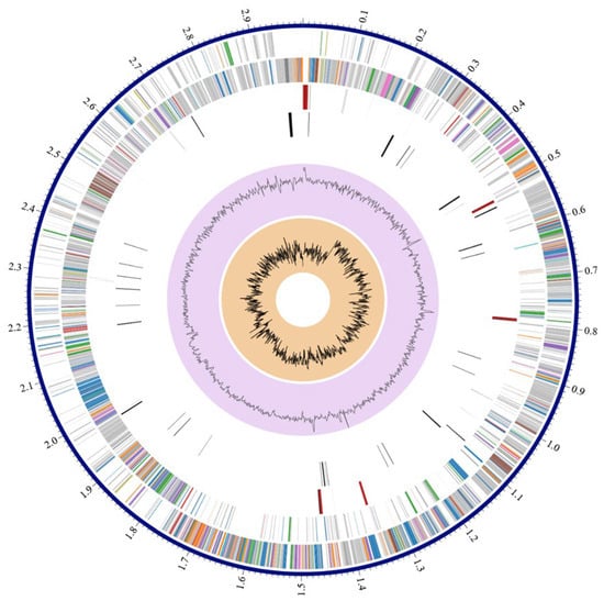
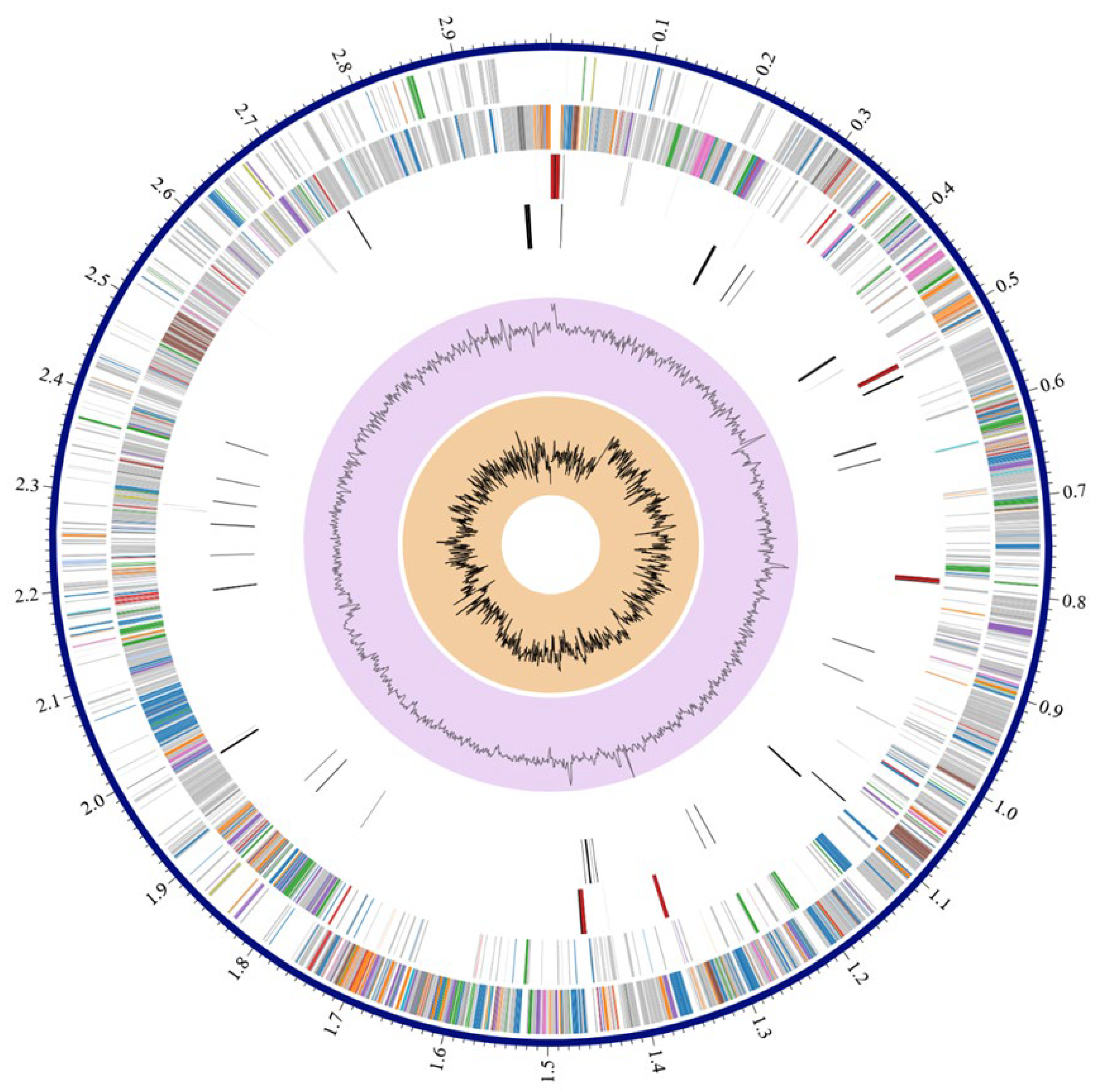
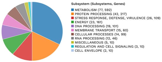
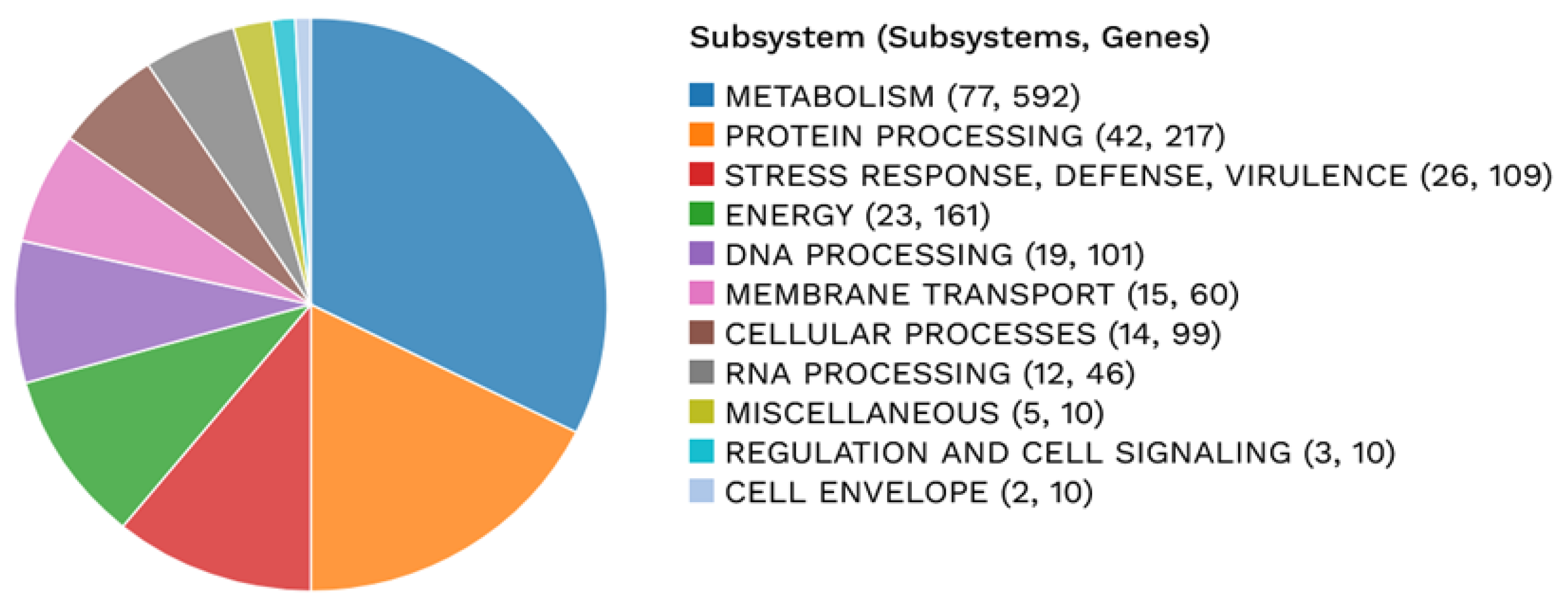
Among the annotated CDSs, 2423 were assigned a predicted function, while 525 were classified as hypothetical proteins. Functional annotation identified 784 proteins associated with Enzyme Commission (EC) numbers, 637 with Gene Ontology (GO) terms, and 551 mapped to Kyoto Encyclopedia of Genes and Genomes (KEGG) pathways. Pathosystems Resource Integration Center (PATRIC) annotation indicated that 2896 proteins belonged to genus-specific protein families (PLFams), and 2918 proteins were assigned to cross-genus protein families (PGFams). A circular graphical representation of the genome annotation, illustrating the distribution of coding sequences, RNA genes, and functional categories across the chromosome, is shown in Figure 1. In addition, an overview of functional subsystems, defined as sets of proteins collectively implementing specific biological processes or structural complexes, is provided in Figure 2.
Figure 1.
Annotation of the sequenced genome for L. monocytogenes strain BF11. From outer to inner rings, the contigs, CDS on the forward strand, CDS on the reverse strand, RNA genes, CDS with homology to known antimicrobial resistance genes, CDS with homology to known virulence factors, GC content and GC skew. The colors of the CDS on the forward and reverse strand indicate the subsystem that these genes belong to (see Figure 2).
Figure 2.
Annotation of the genome for L. monocytogenes strain BF11 including an analysis of the subsystems unique to the genome.
3.5. Taxonomic Identification, MLST, and cgMLST Analysis
Taxonomic assignment using rMLST classified BF11 as a L. monocytogenes strain. Serotyping identified the strain as belonging to serogroup IIb, serovar 1/2b. MLST analysis classified BF11 as a sequence type ST5, clonal complex CC5 strain (Table 4). The strain was predicted to be a human pathogen with a probability of 90.9%.
Table 4.
MLST and pathogenicity of the L. monocytogenes strain BF11 isolated from pet food.
cgMLST analysis was performed using a scheme of 1748 loci. No genomes within the PubMLST and BIGSdb-Pasteur databases were identified within a threshold of ≤10 allelic differences from BF11. Therefore, the strain did not cluster with any existing genomes within the applied threshold (≤10 allelic differences), suggesting that it may represent a potentially distinct cgMLST cluster within the currently available datasets. Analysis further revealed potentially novel alleles in 15 loci of the cgMLST scheme, indicating genetic divergence relative to previously deposited ST5 genomes.
3.6. Antimicrobial Resistance Phenotype and Genotypic Determinants
As previously mentioned, original phenotypic analyses demonstrated that L. monocytogenes strain BF11 exhibited resistance to CIP, MEM, P, SXT, and TE during antimicrobial screening, with MIC testing confirming resistance to MEM, P, SXT, and TE [37]. These results established the MDR phenotype of the strain. In silico analysis of antimicrobial resistance determinants identified a broad repertoire of resistance-associated genes and regulatory systems (Table 5).
Table 5.
Antimicrobial resistance genes of the L. monocytogenes BF11 isolate.
Genes encoding antimicrobial inactivation enzymes, antimicrobial target modification, target replacement proteins, and regulatory systems were detected. The identified genes include both intrinsic (housekeeping-related) targets commonly associated with antimicrobial susceptibility (e.g., alr, ddl, murA, gyrA, gyrB) and acquired or resistance-associated determinants (e.g., vga(G), norB, sul, regulatory systems such as liaFSR). The latter are more directly linked to the observed MDR phenotype. No plasmids were detected in the genome, indicating that the resistance determinants identified were chromosomally encoded.
Genes associated with environmental stress tolerance were also identified, including the bcrABC cassette and Stress Survival Islet 1 (SSI-1). These loci are known to be involved in tolerance to quaternary ammonium compounds, acidic conditions, and high salt concentrations.
3.7. Virulence-Associated Genes and Pathogenicity Determinants
Analysis of virulence-associated loci revealed the presence of a complete LIPI-1, including hly, plcA, plcB, actA, and prfA. Multiple internalin genes were also detected, including inlA, inlB, inlC, inlF, inlK, and inlP.
Sequence analysis of the inlA gene demonstrated full-length integrity without premature stop codon mutations. BLAST analysis (https://blast.ncbi.nlm.nih.gov/Blast.cgi, accessed on 16 December 2025) showed 100% query coverage against the reference strain EGD-e, with 96% sequence identity and an E-value of 1.23 × 10−298, indicating a statistically significant and complete alignment. Sequence analysis also confirmed the presence of a functional prfA gene.
Analysis of virulence-associated databases further confirmed the presence of a complete virulence gene repertoire. In addition, sequence variation in inlB was identified compared to reference genomes, indicating the presence of potentially distinct allelic variants.
3.8. Mobile Genetic Elements, Prophages, and CRISPR-Cas System
Mobilome analysis identified multiple genes associated with recombination and genetic mobility, including recT_2 and int-Tn_2. Several resistance- and virulence-associated genes showed 100% sequence identity to known reference genes.
Four integrated prophages were detected within the BF11 genome, occupying large genomic regions. Identified prophages belonged to the Siphoviridae family and included Listeria phages phi A006, phi LP-101, vB_LmoS_188, and B025. These prophage regions encompassed substantial portions of the chromosome.
A CRISPR-Cas system was also identified, containing three spacers. The presence of both multiple prophages and a CRISPR system indicates the coexistence of phage integration events and phage defense mechanisms within the BF11 genome.
3.9. Phylogenomic and Comparative Genomic Analyses
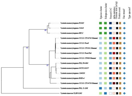
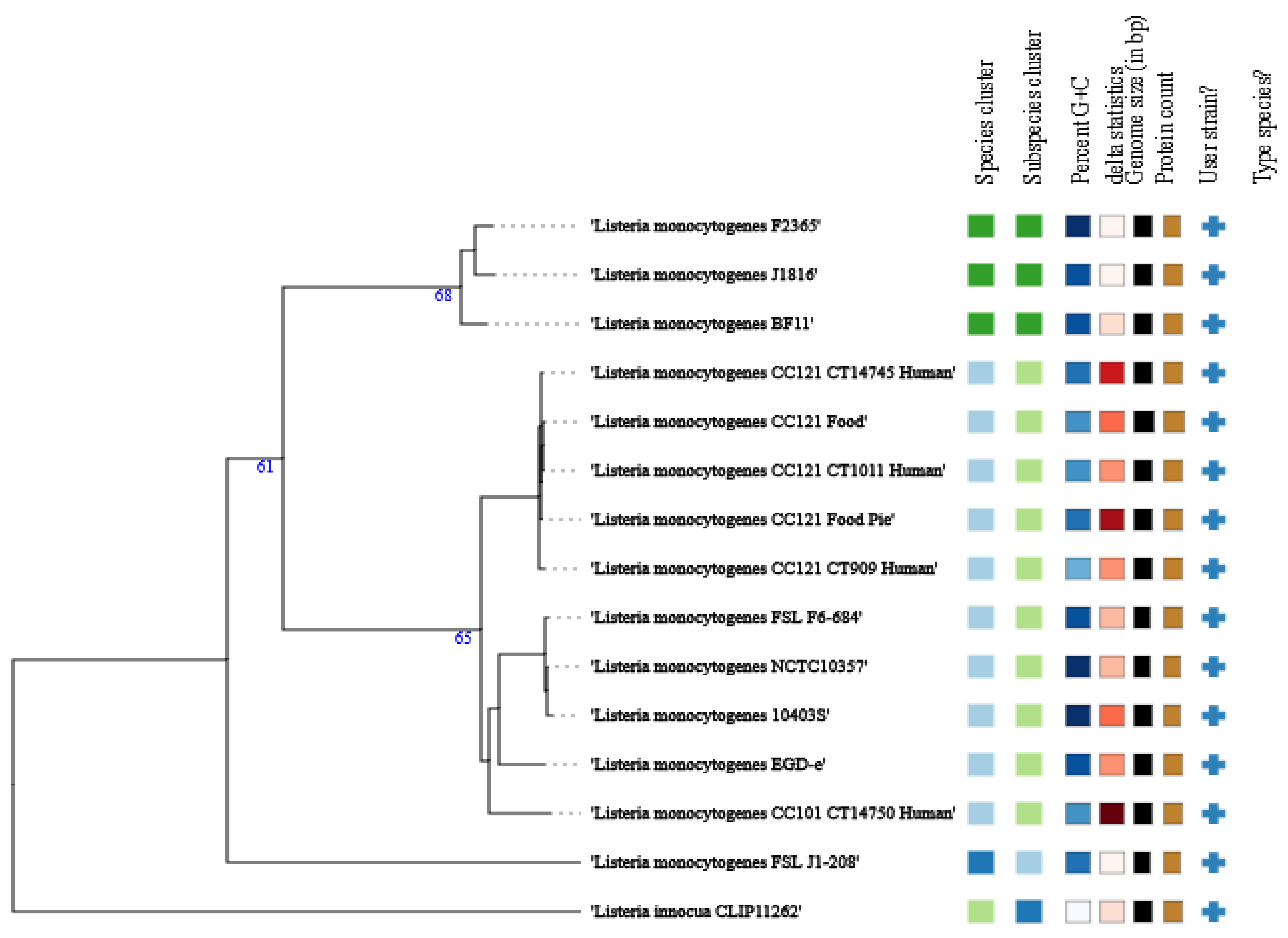
Whole-genome-based taxonomic analysis using TYGS assigned BF11 to L. monocytogenes (Figure 3).
Figure 3.
TYGS-generated WGS-based phylogram of the BF11 strain, as compared with other Listeria spp. reference strains. Strains with the same color are grouped together. All strains were added by the user (+) for comparison and were not TYGS-generated.
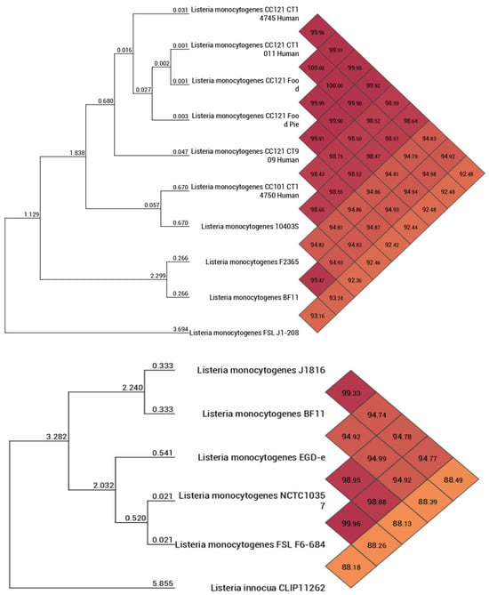
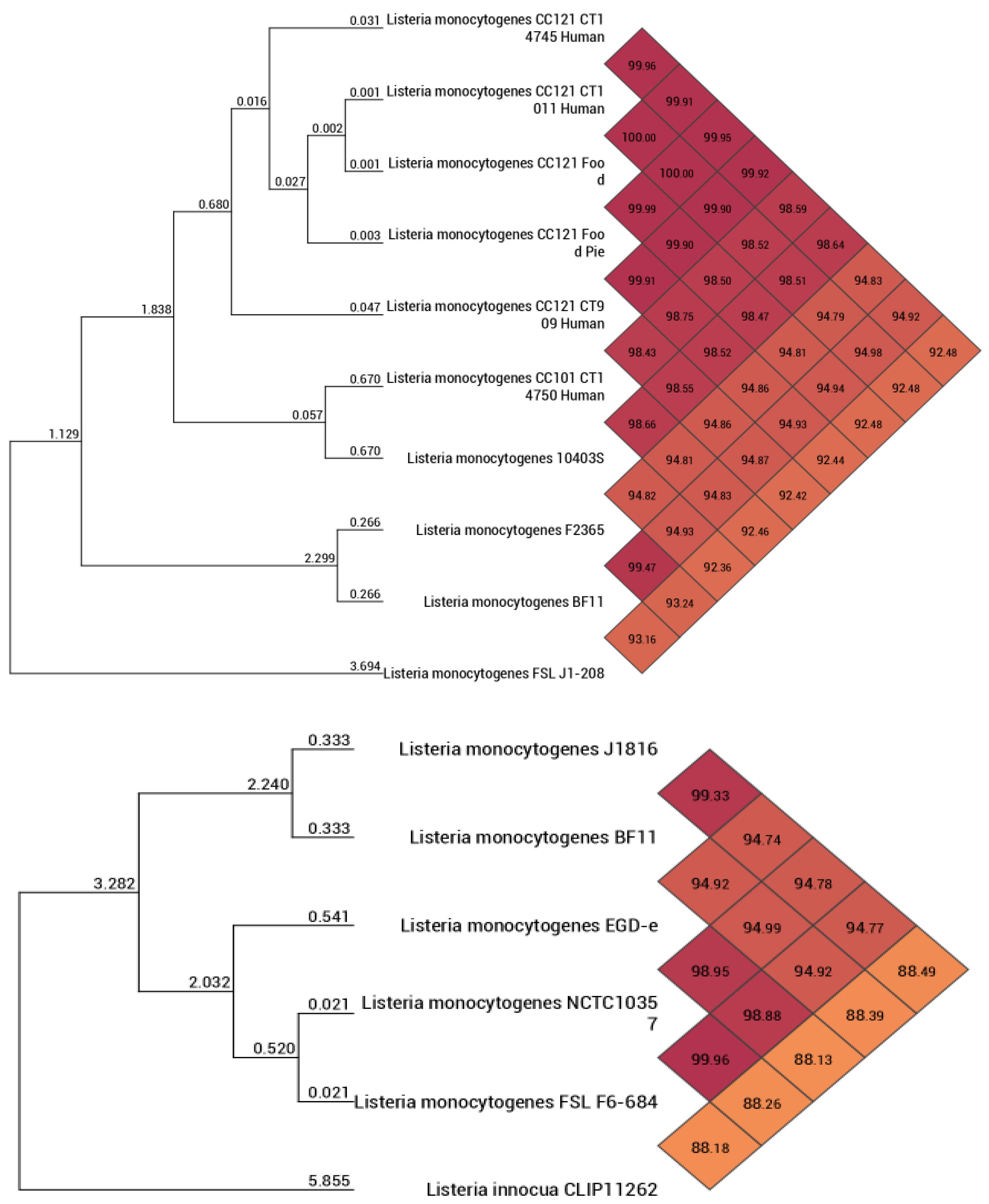
ANI analysis confirmed species-level identity, with ANI values exceeding the 95–96% threshold for species delineation when compared with reference L. monocytogenes genomes (Figure 4).
Figure 4.
Heatmap–dendrogram graph (two parts, up and down) displaying the ANI values between the BF11 strain and different Listeria spp. strains. Strains with the same color are grouped together.
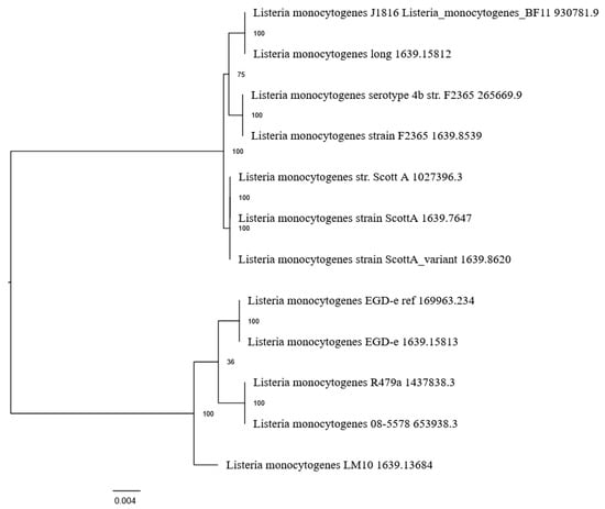
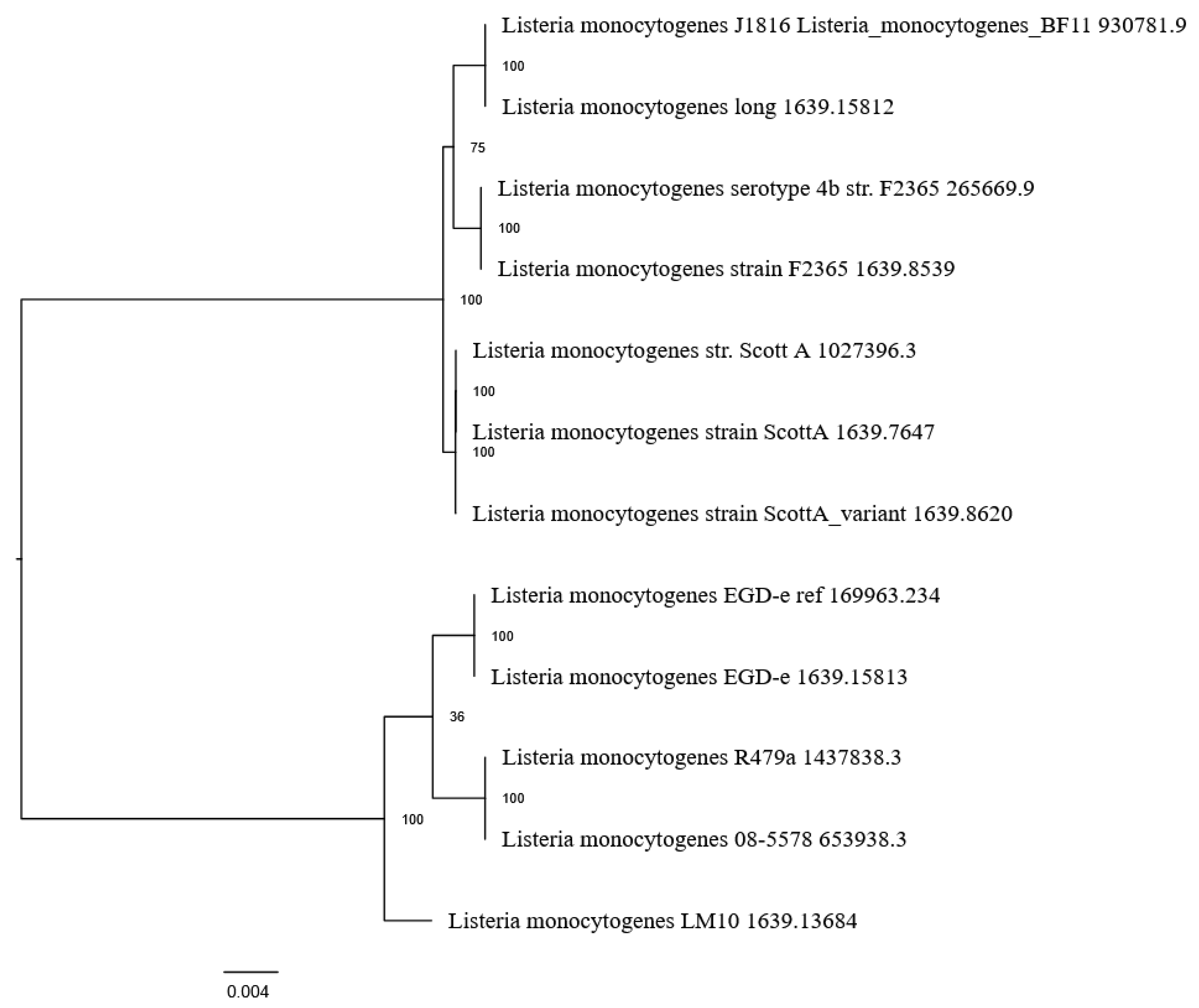
Proteome-based phylogenetic analysis revealed multiple clusters within the species, indicating proteomic divergence among strains (Figure 5). BF11 clustered in close proximity to the Lineage I reference strains F2365 and Scott A.
Figure 5.
Phylogenetic dendrogram between the BF11 strain (the first two IDs 930781.9 and 1639.15812 at the beginning of the dendrogram) and selected reference L. monocytogenes strains. Multiple inputs per strain are displayed in the graph because BV-BRC platform contained the same strain with different ID. All IDs were included in the analysis to avoid missing data.
Comparative genome alignment analysis between BF11, a reference ST5 strain (08-5578), and the Lineage I strain F2365 identified multiple contiguous genomic regions shared between BF11 and F2365 that were absent from the reference ST5 genome, supporting the mosaic genome structure of BF11. Additional regions showed alignment between BF11 and the ST5 reference, indicating the presence of an ST5 genomic backbone with integrated genomic segments shared with Lineage I strains. Resistance-associated genes such as vga(G), norB, and sul were located within regions shared with the Lineage I reference strain.
4. Discussion
This study was structured as a whole-genome risk characterization of an MDR L. monocytogenes strain isolated from raw pet food. The main objective was to determine whether its MDR phenotype co-occurs with genomic features relevant to public health and One Health surveillance. The findings can therefore be interpreted along a single logical axis: BF11 combines an ST5/CC5, serogroup IIb/1/2b background with phenotypic resistance to multiple clinically relevant antimicrobials, retained virulence-associated loci, stress/persistence-associated determinants, multiple prophage regions, and absence of detected plasmids. cgMLST divergence further indicates that BF11 was not closely matched to genomes currently represented in the queried databases within the applied allelic threshold (i.e., ≤10 allelic differences).
The detected resistance-associated genes can be linked to the observed phenotypic resistance through known antimicrobial resistance mechanisms. Resistance (intermediate resistance) based on the MIC results of Andritsos et al. [37] to fluoroquinolones (e.g., CIP) may be associated with mutations or intrinsic features of DNA gyrase subunits (gyrA, gyrB), as well as efflux-related mechanisms such as norB. Reduced susceptibility to beta-lactams (e.g., P, MEM) may be related to alterations in cell wall synthesis pathways involving genes such as murA, ddl, and regulatory systems such as liaFSR, which contribute to cell envelope stress responses. Resistance to tetracyclines may be partially explained by the presence of vga(G), which is associated with antimicrobial target protection mechanisms. SXT resistance may be linked to genes involved in folate biosynthesis pathways (e.g., folA, folP, dfr), affecting the antimicrobial target pathway. Overall, the resistance phenotype observed in strain BF11 likely reflects the combined effect of intrinsic resistance determinants, regulatory systems, and efflux-associated mechanisms rather than the presence of a single dominant acquired resistance gene.
MLST classified L. monocytogenes BF11 as an ST5 (CC5) strain (Table 4), a sequence type repeatedly associated with persistence in food processing environments. ST5 strains are frequently implicated in long-term colonization of production facilities and recurrent contamination events, reflecting their capacity to tolerate environmental stressors and routine sanitation procedures [30,68]. Consistent with this persistence-associated profile, strain BF11 harbored the bcrABC cassette and SSI-1, genetic elements known to confer tolerance to quaternary ammonium compounds, acidic conditions, and high salt concentrations [6,22,25]. The co-occurrence of disinfectant tolerance and antimicrobial resistance supports the concept of biocide–antimicrobial co-selection, whereby exposure to sanitizers may indirectly favor the maintenance of antimicrobial resistance [10,25,69].
The virulence-associated gene profile of BF11 indicates that the isolate retains the principal genomic components associated with the canonical pathogenicity repertoire of L. monocytogenes. BF11 carried a complete LIPI-1, including prfA, hly, plcA, plcB, and actA. These genes are central to the intracellular infection cycle of L. monocytogenes: prfA regulates major virulence determinants, hly encodes listeriolysin O, plcA and plcB encode phospholipases involved in vacuolar escape, and actA is associated with actin-based intracellular motility and cell-to-cell spread [2,18,19,20,21]. BF11 also carried several internalin genes, including inlA, inlB, inlC, inlF, inlK, and inlP. Among these, inlA and inlB are particularly relevant because they are major invasion-associated determinants of L. monocytogenes [2,19,20,21]. Sequence analysis showed that inlA was full-length and lacked premature stop codons. This finding is relevant because premature stop codons in inlA have been associated with truncated Internalin A and reduced invasion of human intestinal epithelial cells, and are reported more frequently among food isolates than among human listeriosis isolates [70,71]. The presence of an intact prfA sequence further supports retention of the regulatory component required for coordinated expression of major virulence genes. In addition, sequence variation was detected in inlB relative to reference genomes, suggesting the presence of a distinct allelic variant. Overall, these findings support the presence of a preserved virulence-associated genomic repertoire in BF11.
Phylogenomic analyses revealed that, although strain BF11 retains an ST5 genomic backbone, it clusters in close proximity to well-characterized Lineage I outbreak-associated strains such as F2365 and Scott A (Figure 3, Figure 4 and Figure 5). This apparent phylogenetic incongruence, supported by ANI and proteome-based analyses, indicates a mosaic genome architecture shaped by recombination and horizontal gene transfer. Recombination-driven convergence between persistence-associated ST5 backgrounds and virulence-associated Lineage I traits has been documented previously and represents a key mechanism in the adaptive evolution of L. monocytogenes [10,70,72]. In L. monocytogenes BF11, this convergence provides a mechanistic explanation for the coexistence of environmental resilience and virulence-associated characteristics.
Analysis of the mobilome further highlighted the potential contribution of phage-mediated genome remodeling to the BF11 genome. Four integrated prophage regions were detected, assigned to Listeria phages phi A006, phi LP-101, vB_LmoS_188, and B025. In L. monocytogenes, prophages are important components of the accessory genome and can contribute to strain diversification through integration, excision, genome rearrangement, and gene gain or loss. Horizontal gene transfer in this species has been associated with plasmids, conjugative transposons, recombination events, and prophages, indicating that mobile genetic elements can introduce new genetic information into otherwise highly conserved genomic backgrounds [10]. This is relevant for BF11 because the isolate combines an ST5/CC5 backbone with multiple accessory genome features, including prophages, recombination-associated genes, and a putative CRISPR-Cas locus. Previous genome-based studies have also emphasized that L. monocytogenes populations show heterogeneity in virulence and stress-resistance features despite a relatively conserved core-genome structure, supporting the importance of accessory genome variation in strain differentiation [73,74]. Prophage-associated loci have also been proposed as markers of niche-specific adaptation and persistence in food-processing environments, particularly through comK-prophage junctions [74,75]. Therefore, the prophage content of BF11 supports the interpretation that this isolate has undergone accessory-genome remodeling that may contribute to its genomic distinctiveness.
Directly comparable ST5/CC5 MDR pet-food isolates with equivalent WGS-based resistance, virulence, and mobilome characterization remain limited. Therefore, BF11 was compared with available ST5/CC5 strains from food-processing environments and with acquired MDR strains from other clonal backgrounds. ST5 has been detected among food-associated L. monocytogenes isolates, while multiresistance patterns were reported in 27.4% of isolates, mainly involving oxacillin, clindamycin, and ceftriaxone; however, all tested isolates were susceptible to P and SXT [76]. ST5/CC5 has also been associated with food-processing environments, including cheese-processing and ready-to-eat meat-processing settings, where ST5/CC5 strains have been linked with persistence-associated features such as SSI-1, bcrABC, prophages, and plasmid carriage [77,78].
Comparison with acquired MDR L. monocytogenes further emphasizes this distinction. Recent surveillance identified acquired MDR in only 34 of 8344 food isolates from China, mostly belonging to ST9/CC9 rather than ST5/CC5, with MDR frequently associated with plasmid-borne genes such as dfrG, erm(B), lnu(B), lsa(E), tet(S), and catA [14]. This is consistent with earlier WGS-based screening of 2862 food isolates from China, in which resistant isolates were uncommon, and with reports of foodborne MDR L. monocytogenes carrying plasmid-associated resistance genes such as cat, erm(B), and tet(S) [79,80]. By contrast, BF11 belonged to ST5/CC5, showed resistance to P, MEM, SXT, TE, and CIP, retained complete LIPI-1 and full-length inlA, and had no detected plasmids. Thus, BF11 should be described as a concerning ST5/CC5 MDR pet-food isolate with a notable combination of resistance, virulence-associated, stress/persistence-associated, and mobilome features.
It is worth mentioning that this study has certain limitations. BF11 showed β-hemolysis and a positive CAMP reaction during species confirmation, but no virulence-specific assays were performed. Therefore, the presence of LIPI-1, prfA, hly, plcA, plcB, actA, and internalin genes should be interpreted as evidence of retained virulence-associated genomic potential. In particular, epithelial-cell invasion, intracellular survival, plaque formation, actin-based motility, macrophage survival, and animal-model virulence were not assessed. Similarly, although BF11 carried stress- and persistence-associated loci such as SSI-1 and bcrABC, biofilm formation, sanitizer tolerance, acid or salt tolerance, and other environmental-adaptability phenotypes were not experimentally evaluated. This distinction is important because biofilm formation and environmental persistence in L. monocytogenes are condition-dependent and can vary among strains, growth temperatures, nutrient conditions, salt concentrations, and genetic backgrounds [22,23,24,25,30]. Therefore, future studies should combine WGS with phenotypic assays to determine whether the genomic profile observed in BF11 translates into increased host–cell invasion, intracellular survival, biofilm formation, sanitizer tolerance, or persistence under food/feed-processing conditions.
From a One Health perspective, the isolation of L. monocytogenes BF11 strain from pet food is particularly significant because this matrix may connect food/feed production, animals, handlers, and household environments. Raw and minimally processed pet foods are increasingly recognized as potential reservoirs of zoonotic and antimicrobial-resistant bacteria, yet they remain comparatively under-surveilled relative to foods intended for human consumption [35,36]. L. monocytogenes has been detected in commercial frozen raw meat-based diets for dogs and cats, and broader pet-food surveys have recovered Listeria, including L. monocytogenes, and Salmonella from raw pet-food products [35,81]. Potential exposure routes include handling of contaminated raw pet food, contact with feeding bowls, utensils, storage containers, kitchen surfaces, direct animal contact, and possible fecal shedding after ingestion [35,36]. These routes are especially relevant for vulnerable household members, veterinary personnel, animal handlers, and people caring for service or working dogs. The genomic profile of strain BF11 illustrates how selective pressures in food and feed production environments can drive the emergence of L. monocytogenes strains that unite multidrug resistance, environmental persistence, and virulence-associated traits, underscoring the need for integrated genomic surveillance and targeted control strategies across food, feed, and veterinary sectors.
5. Conclusions
This study identifies Listeria monocytogenes strain BF11 as an MDR strain from raw pet food with a concerning genomic and phenotypic profile. BF11 belonged to serogroup IIb/serovar 1/2b and ST5/CC5, and exhibited resistance to multiple clinically relevant antimicrobials. Whole-genome analysis identified resistance-associated loci, stress/persistence-associated determinants, multiple prophage regions, putative CRISPR-Cas elements, and no detected plasmids under the analytical conditions used.
BF11 also retained major virulence-associated genomic components, including complete LIPI-1, intact inlA, and prfA, indicating retained virulence-associated genomic potential and absence of a common inlA-mediated attenuation marker. Similarly, the detection of SSI-1 and bcrABC supports possible stress- and persistence-associated potential.
The isolation of an MDR L. monocytogenes ST5/CC5 strain from raw pet food is relevant within a One Health framework because this matrix may connect food/feed production, animals, handlers, and household environments. The combination of multidrug resistance, disinfectant tolerance, and virulence-associated traits in the BF11 strain represents a particularly concerning profile, especially given its isolation from pet food. These findings highlight L. monocytogenes strain BF11 as an example of how selective pressures in food and feed production systems can drive the emergence of L. monocytogenes strains that unite persistence and pathogenicity. Enhanced genomic surveillance, improved sanitation strategies, and prudent antimicrobial use across food, feed, and veterinary sectors are therefore essential to limit the dissemination of strains with similar high-risk characteristics.
Supplementary Materials
The following supporting information can be downloaded at: https://www.mdpi.com/article/10.3390/microorganisms14051097/s1, Table S1: Reference genomes of Listeria spp. strains used in this study for phylogenomic and comparative analyses.
Author Contributions
Conceptualization, M.M. and N.D.A.; methodology, A.M., M.M., I.G. and P.S.; software, M.M. and P.S.; validation, A.M., M.M., S.P. and N.D.A.; formal analysis, M.M.; investigation, A.M., N.T. and N.D.A.; resources, M.M., N.T., P.S.; data curation, M.M.; writing—original draft preparation, A.M.; writing—review and editing, M.M., S.P., J.I. and N.D.A.; visualization, A.M. and M.M.; supervision, M.M. and J.I.; project administration, N.D.A.; funding acquisition, N.D.A. All authors have read and agreed to the published version of the manuscript.
Funding
This research received no external funding.
Institutional Review Board Statement
Not applicable.
Informed Consent Statement
Not applicable.
Data Availability Statement
The original data regarding the microbial strain used in the study are included in the article and supplementary material of Andritsos et al. (2025) [37], while they are publicly available at https://doi.org/10.3390/vetsci12090844 and https://www.mdpi.com/article/10.3390/vetsci12090844/s1 (accessed on 16 February 2026). Genome accession number for L. monocytogenes strain BF11 is BioProject ID: PRJNA1464443 (BioSample accession: SAMN59708363).
Acknowledgments
The authors acknowledge the Hellenic Army Biological Research Centre for providing the microbial strain used in the study.
Conflicts of Interest
The authors declare no conflicts of interest.
References
- Buchanan, R.L.; Gorris, L.G.M.; Hayman, M.M.; Jackson, T.C.; Whiting, R.C. A review of Listeria monocytogenes: An update on outbreaks, virulence, dose-response, ecology, and risk assessments. Food Control 2017, 75, 1–13, Erratum in Food Control 2018, 88, 236. https://doi.org/10.1016/j.foodcont.2018.01.004. [Google Scholar] [CrossRef] [Scilit]
- Vázquez-Boland, J.A.; Kuhn, M.; Berche, P.; Chakraborty, T.; Domínguez-Bernal, G.; Goebel, W.; González-Zorn, B.; Wehland, J.; Kreft, J. Listeria pathogenesis and molecular virulence determinants. Clin. Microbiol. Rev. 2001, 14, 584–640. [Google Scholar] [CrossRef] [Scilit]
- Lyautey, E.; Lapen, D.R.; Wilkes, G.; McCleary, K.; Pagotto, F.; Tyler, K.; Hartmann, A.; Piveteau, P.; Rieu, A.; Robertson, W.J.; et al. Distribution and characteristics of Listeria monocytogenes isolates from surface waters of the South Nation River watershed, Ontario, Canada. Appl. Environ. Microbiol. 2007, 73, 5401–5410. [Google Scholar] [CrossRef] [Scilit] [PubMed]
- Srinivasan, V.; Nam, H.M.; Nguyen, L.T.; Tamilselvam, B.; Murinda, S.E.; Oliver, S.P. Prevalence of antimicrobial resistance genes in Listeria monocytogenes isolated from dairy farms. Foodborne Pathog. Dis. 2005, 2, 201–211. [Google Scholar] [CrossRef] [Scilit]
- Gandhi, M.; Chikindas, M.L. Listeria: A foodborne pathogen that knows how to survive. Int. J. Food Microbiol. 2007, 113, 1–15. [Google Scholar] [CrossRef] [Scilit]
- Bucur, F.I.; Grigore-Gurgu, L.; Crauwels, P.; Riedel, C.U.; Nicolau, A.I. Resistance of Listeria monocytogenes to stress conditions encountered in food and food processing environments. Front. Microbiol. 2018, 9, 2700. [Google Scholar] [CrossRef] [Scilit]
- Troxler, R.; von Graevenitz, A.; Funke, G.; Wiedemann, B.; Stock, I. Natural antibiotic susceptibility of Listeria species: L. grayi, L. innocua, L. ivanovii, L. monocytogenes, L. seeligeri and L. welshimeri strains. Clin. Microbiol. Infect. 2000, 6, 525–535. [Google Scholar] [CrossRef] [Scilit] [PubMed]
- Morvan, A.; Moubareck, C.; Leclercq, A.; Hervé-Bazin, M.; Bremont, S.; Lecuit, M.; Courvalin, P.; Le Monnier, A. Antimicrobial resistance of Listeria monocytogenes strains isolated from humans in France. Antimicrob. Agents Chemother. 2010, 54, 2728–2731. [Google Scholar] [CrossRef] [Scilit]
- Olaimat, A.N.; Al-Holy, M.A.; Shahbaz, H.M.; Al-Nabulsi, A.A.; Abu Ghoush, M.H.; Osaili, T.M.; Ayyash, M.M.; Holley, R.A. Emergence of antibiotic resistance in Listeria monocytogenes isolated from food products: A comprehensive review. Compr. Rev. Food Sci. Food Saf. 2018, 17, 1277–1292. [Google Scholar] [CrossRef] [Scilit] [PubMed]
- Allen, K.J.; Wałecka-Zacharska, E.; Chen, J.C.; Katarzyna, K.P.; Devlieghere, F.; van Meervenne, E.; Osek, J.; Wieczorek, K.; Bania, J. Listeria monocytogenes—An examination of food chain factors potentially contributing to antimicrobial resistance. Food Microbiol. 2016, 54, 178–189. [Google Scholar] [CrossRef] [Scilit]
- Andriyanov, P.A.; Zhurilov, P.A.; Liskova, E.A.; Karpova, T.I.; Sokolova, E.V.; Yushina, Y.K.; Zaiko, E.V.; Bataeva, D.S.; Voronina, O.L.; Psareva, E.K.; et al. Antimicrobial resistance of Listeria monocytogenes strains isolated from humans, animals, and food products in Russia in 1950–1980, 2000–2005, and 2018–2021. Antibiotics 2021, 10, 1206. [Google Scholar] [CrossRef] [Scilit]
- Anwar, T.M.; Pan, H.; Chai, W.; Ed-Dra, A.; Fang, W.; Li, Y.; Yue, M. Genetic diversity, virulence factors, and antimicrobial resistance of Listeria monocytogenes from food, livestock, and clinical samples between 2002 and 2019 in China. Int. J. Food Microbiol. 2022, 366, 109572. [Google Scholar] [CrossRef] [Scilit]
- Kayode, A.J.; Okoh, A.I. Antibiotic resistance profile of Listeria monocytogenes recovered from ready-to-eat foods surveyed in South Africa. J. Food Prot. 2022, 85, 1807–1814. [Google Scholar] [CrossRef] [Scilit]
- Li, H.; Sheng, H.; Zhao, J.; Zhang, X.; Li, M.; Zhao, L.; Li, L.; Zhang, X.; Yang, B.; Fanning, S.; et al. Emerging threats: Listeria monocytogenes with acquired multidrug resistance from food in China, 2012–2022. Int. J. Food Microbiol. 2025, 439, 111236. [Google Scholar] [CrossRef] [Scilit] [PubMed]
- Poyart-Salmeron, C.; Carlier, C.; Trieu-Cuot, P.; Courvalin, P.; Courtieu, A.L. Transferable plasmid-mediated antibiotic resistance in Listeria monocytogenes. Lancet 1990, 335, 1422–1426. [Google Scholar] [CrossRef] [Scilit] [PubMed]
- Poyart-Salmeron, C.; Trieu-Cuot, P.; Carlier, C.; MacGowan, A.; McLauchlin, J.; Courvalin, P. Genetic basis of tetracycline resistance in clinical isolates of Listeria monocytogenes. Antimicrob. Agents Chemother. 1992, 36, 463–466. [Google Scholar] [CrossRef] [Scilit]
- Bertsch, D.; Muelli, M.; Weller, M.; Uruty, A.; Lacroix, C.; Meile, L. Antimicrobial susceptibility and antibiotic resistance gene transfer analysis of foodborne, clinical, and environmental Listeria spp. isolates including Listeria monocytogenes. MicrobiologyOpen 2014, 3, 118–127. [Google Scholar] [CrossRef] [Scilit] [PubMed]
- Jacquet, C.; Gouin, E.; Jeannel, D.; Cossart, P.; Rocourt, J. Expression of ActA, Ami, InlB, and listeriolysin O in Listeria monocytogenes of human and food origin. Appl. Environ. Microbiol. 2002, 68, 616–622. [Google Scholar] [CrossRef] [Scilit]
- Dussurget, O.; Pizarro-Cerda, J.; Cossart, P. Molecular determinants of Listeria monocytogenes virulence. Annu. Rev. Microbiol. 2004, 58, 587–610. [Google Scholar] [CrossRef] [Scilit]
- Camejo, A.; Carvalho, F.; Reis, O.; Leitão, E.; Sousa, S.; Cabanes, D. The arsenal of virulence factors deployed by Listeria monocytogenes to promote its cell infection cycle. Virulence 2011, 2, 379–394. [Google Scholar] [CrossRef] [Scilit]
- Radoshevich, L.; Cossart, P. Listeria monocytogenes: Towards a complete picture of its physiology and pathogenesis. Nat. Rev. Microbiol. 2018, 16, 32–46. [Google Scholar] [CrossRef] [Scilit] [PubMed]
- Colagiorgi, A.; Bruini, I.; Di Ciccio, P.A.; Zanardi, E.; Ghidini, S.; Ianieri, A. Listeria monocytogenes biofilms in the wonderland of food industry. Pathogens 2017, 6, 41. [Google Scholar] [CrossRef] [Scilit]
- Lee, B.-H.; Cole, S.; Badel-Berchoux, S.; Guillier, L.; Felix, B.; Krezdorn, N.; Hébraud, M.; Bernardi, T.; Sultan, I.; Piveteau, P. Biofilm formation of Listeria monocytogenes strains under food processing environments and pan-genome-wide association study. Front. Microbiol. 2019, 10, 2698. [Google Scholar] [CrossRef] [Scilit]
- Pan, Y.; Breidt, F.; Kathariou, S. Resistance of Listeria monocytogenes biofilms to sanitizing agents in a simulated food processing environment. Appl. Environ. Microbiol. 2006, 72, 7711–7717. [Google Scholar] [CrossRef] [Scilit]
- Wiktorczyk-Kapischke, N.; Skowron, K.; Grudlewska-Buda, K.; Wałecka-Zacharska, E.; Korkus, J.; Gospodarek-Komkowska, E. Adaptive response of Listeria monocytogenes to the stress factors in the food processing environment. Front. Microbiol. 2021, 12, 710085. [Google Scholar] [CrossRef] [Scilit] [PubMed]
- Kayode, A.J.; Semerjian, L.; Osaili, T.; Olapade, O.; Okoh, A.I. Occurrence of multidrug-resistant Listeria monocytogenes in environmental waters: A menace of environmental and public health concern. Front. Environ. Sci. 2021, 9, 737435. [Google Scholar] [CrossRef] [Scilit]
- Keet, R.; Rip, D. Listeria monocytogenes isolates from Western Cape, South Africa exhibit resistance to multiple antibiotics and contradicts certain global resistance patterns. AIMS Microbiol. 2021, 7, 40–58. [Google Scholar] [CrossRef] [Scilit]
- Chen, M.; Cheng, J.; Wu, Q.; Zhang, J.; Chen, Y.; Zeng, H.; Ye, Q.; Wu, S.; Cai, S.; Wang, J.; et al. Prevalence, potential virulence, and genetic diversity of Listeria monocytogenes isolates from edible mushrooms in Chinese markets. Front. Microbiol. 2018, 9, 1711. [Google Scholar] [CrossRef] [Scilit]
- Soni, D.K.; Singh, R.K.; Singh, D.V.; Dubey, S.K. Characterization of Listeria monocytogenes isolated from Ganges water, human clinical and milk samples at Varanasi, India. Infect. Genet. Evol. 2013, 14, 83–91. [Google Scholar] [CrossRef] [Scilit]
- Di Ciccio, P.; Rubiola, S.; Panebianco, F.; Lomonaco, S.; Allard, M.; Bianchi, D.M.; Civera, T.; Chiesa, F. Biofilm formation and genomic features of Listeria monocytogenes strains isolated from meat and dairy industries located in Piedmont (Italy). Int. J. Food Microbiol. 2022, 378, 109784. [Google Scholar] [CrossRef] [Scilit]
- Bille, J.; Blanc, D.S.; Schmid, H.; Boubaker, K.; Baumgartner, A.; Siegrist, H.H.; Tritten, M.L.; Lienhard, R.; Berner, D.; Anderau, R.; et al. Outbreak of human listeriosis associated with tomme cheese in northwest Switzerland, 2005. Eurosurveillance 2006, 11, 11–12. [Google Scholar] [CrossRef] [Scilit]
- Parussolo, L.; Sfaciotte, R.A.P.; Dalmina, K.A.; Melo, F.D.; da Costa, U.M.; Ferraz, S.M. Detection of virulence genes and antimicrobial susceptibility profile of Listeria monocytogenes isolates recovered from artisanal cheese produced in the southern region of Brazil. An. Acad. Bras. Cienc. 2021, 93, e20190200. [Google Scholar] [CrossRef] [Scilit]
- Shourav, A.H.; Hasan, M.; Ahmed, S. Antibiotic susceptibility pattern of Listeria spp. isolated from cattle farm environment in Bangladesh. J. Agric. Food Res. 2020, 2, 100082. [Google Scholar] [CrossRef] [Scilit]
- Raschle, S.; Stephan, R.; Stevens, M.J.A.; Cernela, N.; Zurfluh, K.; Muchaamba, F.; Nüesch-Inderbinen, M. Environmental dissemination of pathogenic Listeria monocytogenes in flowing surface waters in Switzerland. Sci. Rep. 2021, 11, 9066. [Google Scholar] [CrossRef] [Scilit]
- van Bree, F.P.J.; Bokken, G.C.A.M.; Mineur, R.; Franssen, F.; Opsteegh, M.; van der Giessen, J.W.B.; Lipman, L.J.A.; Overgaauw, P.A.M. Zoonotic bacteria and parasites found in raw meat-based diets for cats and dogs. Vet. Rec. 2018, 182, 50. [Google Scholar] [CrossRef] [Scilit] [PubMed]
- Davies, R.H.; Lawes, J.R.; Wales, A.D. Raw diets for dogs and cats: A review, with particular reference to microbiological hazards. J. Small Anim. Pract. 2019, 60, 329–339. [Google Scholar] [CrossRef] [Scilit] [PubMed]
- Andritsos, N.D.; Mataragka, A.; Tzimotoudis, N.; Chatzopoulou, A.-S.; Kotsikori, M.; Ikonomopoulos, J. Serotyping and antibiotic resistance profiles of Salmonella spp. and Listeria monocytogenes strains isolated from pet food and feed samples: A One Health perspective. Vet. Sci. 2025, 12, 844. [Google Scholar] [CrossRef] [Scilit]
- ISO 11290-1:2017; Microbiology of the Food Chain—Horizontal Method for the Detection and Enumeration of Listeria monocytogenes and of Listeria spp. Part 1: Detection Method, 2nd ed. ISO: Geneva, Switzerland, 2017.
- Hudzicki, J. Kirby-Bauer Disk Diffusion Susceptibility Test Protocol; American Society for Microbiology: Washington, DC, USA, 2009; pp. 1–23. [Google Scholar]
- EUCAST (European Committee on Antimicrobial Susceptibility Testing). Breakpoint Tables for Interpretation of MICs and Zone Diameters, version 15.0; EUCAST: Växjö, Sweden, 2025; Available online: https://www.eucast.org (accessed on 6 February 2026).
- Brown, D.F.; Brown, L. Evaluation of the E test, a novel method of quantifying antimicrobial activity. J. Antimicrob. Chemother. 1991, 27, 185–190. [Google Scholar] [CrossRef] [Scilit] [PubMed]
- Doumith, M.; Buchrieser, C.; Glaser, P.; Jacquet, C.; Martin, P. Differentiation of the major Listeria monocytogenes serovars by multiplex PCR. J. Clin. Microbiol. 2004, 42, 3819–3822. [Google Scholar] [CrossRef] [Scilit]
- Olson, R.D.; Assaf, R.; Brettin, T.; Conrad, N.; Cucinell, C.; Davis, J.J.; Dempsey, D.M.; Dickerman, A.; Dietrich, E.M.; Kenyon, R.W.; et al. Introducing the Bacterial and Viral Bioinformatics Resource Center (BV-BRC): A resource combining PATRIC, IRD and ViPR. Nucleic Acids Res. 2023, 51, D678–D689. [Google Scholar] [CrossRef] [Scilit]
- Kolmogorov, M.; Yuan, J.; Lin, Y.; Pevzner, P.A. Assembly of long, error-prone reads using repeat graphs. Nat. Biotechnol. 2019, 37, 540–546. [Google Scholar] [CrossRef] [Scilit] [PubMed]
- Li, H. Minimap2: Pairwise alignment for nucleotide sequences. Bioinformatics 2018, 34, 3094–3100. [Google Scholar] [CrossRef] [Scilit] [PubMed]
- Vaser, R.; Sović, I.; Nagarajan, N.; Šikić, M. Fast and accurate de novo genome assembly from long uncorrected reads. Genome Res. 2017, 27, 737–746. [Google Scholar] [CrossRef] [Scilit]
- Meier-Kolthoff, J.P.; Göker, M. TYGS is an automated high-throughput platform for state-of-the-art genome-based taxonomy. Nat. Commun. 2019, 10, 2182. [Google Scholar] [CrossRef] [Scilit]
- Lee, I.; Ouk Kim, Y.; Park, S.C.; Chun, J. OrthoANI: An improved algorithm and software for calculating average nucleotide identity. Int. J. Syst. Evol. Microbiol. 2016, 66, 1100–1103. [Google Scholar] [CrossRef] [Scilit]
- Jolley, K.A.; Bray, J.E.; Maiden, M.C.J. Open-access bacterial population genomics: BIGSdb software, the PubMLST.org website and their applications. Wellcome Open Res. 2018, 3, 124. [Google Scholar] [CrossRef] [Scilit]
- Jolley, K.A.; Maiden, M.C. BIGSdb: Scalable analysis of bacterial genome variation at the population level. BMC Bioinform. 2010, 11, 595. [Google Scholar] [CrossRef] [Scilit]
- Gurevich, A.; Saveliev, V.; Vyahhi, N.; Tesler, G. QUAST: Quality assessment tool for genome assemblies. Bioinformatics 2013, 29, 1072–1075. [Google Scholar] [CrossRef] [Scilit]
- Wood, D.E.; Lu, J.; Langmead, B. Improved metagenomic analysis with Kraken 2. Genome Biol. 2019, 20, 257. [Google Scholar] [CrossRef] [Scilit] [PubMed]
- Manni, M.; Berkeley, M.R.; Seppey, M.; Simão, F.A.; Zdobnov, E.M. BUSCO update: Novel and streamlined workflows along with broader and deeper phylogenetic coverage for scoring of eukaryotic, prokaryotic, and viral genomes. Mol. Biol. Evol. 2021, 38, 4647–4654. [Google Scholar] [CrossRef] [Scilit]
- Parks, D.H.; Imelfort, M.; Skennerton, C.T.; Hugenholtz, P.; Tyson, G.W. CheckM: Assessing the quality of microbial genomes recovered from isolates, single cells, and metagenomes. Genome Res. 2015, 25, 1043–1055. [Google Scholar] [CrossRef] [Scilit]
- Chklovski, A.; Parks, D.H.; Woodcroft, B.J.; Tyson, G.W. CheckM2: A rapid, scalable and accurate tool for assessing microbial genome quality using machine learning. Nat. Methods 2023, 20, 1203–1212, Erratum in Nat. Methods 2024, 21, 735. https://doi.org/10.1038/s41592-024-02248-z. [Google Scholar] [CrossRef] [Scilit]
- Lee, I.; Chalita, M.; Ha, S.M.; Na, S.I.; Yoon, S.H.; Chun, J. ContEst16S: An algorithm that identifies contaminated prokaryotic genomes using 16S RNA gene sequences. Int. J. Syst. Evol. Microbiol. 2017, 67, 2053–2057. [Google Scholar] [CrossRef] [Scilit]
- Cosentino, S.; Voldby Larsen, M.; Møller Aarestrup, F.; Lund, O. PathogenFinder—Distinguishing friend from foe using bacterial whole genome sequence data. PLoS ONE 2013, 8, e77302, Erratum in PLoS ONE 2013, 8, 10.1371/annotation/b84e1af7-c127-45c3-be22-76abd977600f. https://doi.org/10.1371/annotation/b84e1af7-c127-45c3-be22-76abd977600f. [Google Scholar] [CrossRef] [Scilit]
- Feldgarden, M.; Brover, V.; Haft, D.H.; Prasad, A.B.; Slotta, D.J.; Tolstoy, I.; Tyson, G.H.; Zhao, S.; Hsu, C.H.; McDermott, P.F.; et al. Validating the AMRFinder tool and resistance gene database by using antimicrobial resistance genotype-phenotype correlations in a collection of isolates. Antimicrob. Agents Chemother. 2019, 63, e00483-19, Erratum in Antimicrob. Agents Chemother. 2020, 64, e00361-20. https://doi.org/10.1128/aac.00361-20. [Google Scholar] [CrossRef] [Scilit]
- Zankari, E.; Hasman, H.; Cosentino, S.; Vestergaard, M.; Rasmussen, S.; Lund, O.; Aarestrup, F.M.; Larsen, M.V. Identification of acquired antimicrobial resistance genes. J. Antimicrob. Chemother. 2012, 67, 2640–2644. [Google Scholar] [CrossRef] [Scilit] [PubMed]
- Bortolaia, V.; Kaas, R.S.; Ruppe, E.; Roberts, M.C.; Schwarz, S.; Cattoir, V.; Philippon, A.; Allesoe, R.L.; Rebelo, A.R.; Florensa, A.F.; et al. ResFinder 4.0 for predictions of phenotypes from genotypes. J. Antimicrob. Chemother. 2020, 75, 3491–3500. [Google Scholar] [CrossRef] [Scilit] [PubMed]
- Chen, L.; Zheng, D.; Liu, B.; Yang, J.; Jin, Q. VFDB 2016: Hierarchical and refined dataset for big data analysis—10 years on. Nucleic Acids Res. 2016, 44, D694–D697. [Google Scholar] [CrossRef] [Scilit] [PubMed]
- Johansson, M.H.K.; Bortolaia, V.; Tansirichaiya, S.; Aarestrup, F.M.; Roberts, A.P.; Petersen, T.N. Detection of mobile genetic elements associated with antibiotic resistance in Salmonella enterica using a newly developed web tool: MobileElementFinder. J. Antimicrob. Chemother. 2021, 76, 101–109. [Google Scholar] [CrossRef] [Scilit]
- Carattoli, A.; Zankari, E.; García-Fernández, A.; Voldby Larsen, M.; Lund, O.; Villa, L.; Møller Aarestrup, F.; Hasman, H. In silico detection and typing of plasmids using PlasmidFinder and plasmid multilocus sequence typing. Antimicrob. Agents Chemother. 2014, 58, 3895–3903. [Google Scholar] [CrossRef] [Scilit]
- Larsen, M.V.; Cosentino, S.; Rasmussen, S.; Friis, C.; Hasman, H.; Marvig, R.L.; Jelsbak, L.; Sicheritz-Pontén, T.; Ussery, D.W.; Aarestrup, F.M.; et al. Multilocus sequence typing of total-genome-sequenced bacteria. J. Clin. Microbiol. 2012, 50, 1355–1361. [Google Scholar] [CrossRef] [Scilit]
- Couvin, D.; Bernheim, A.; Toffano-Nioche, C.; Touchon, M.; Michalik, J.; Néron, B.; Rocha, E.P.C.; Vergnaud, G.; Gautheret, D.; Pourcel, C. CRISPRCasFinder, an update of CRISRFinder, includes a portable version, enhanced performance and integrates search for Cas proteins. Nucleic Acids Res. 2018, 46, W246–W251. [Google Scholar] [CrossRef] [Scilit]
- Wishart, D.S.; Han, S.; Saha, S.; Oler, E.; Peters, H.; Grant, J.R.; Stothard, P.; Gautam, V. PHASTEST: Faster than PHASTER, better than PHAST. Nucleic Acids Res. 2023, 51, W443–W450. [Google Scholar] [CrossRef] [Scilit]
- Afgan, E.; Baker, D.; Batut, B.; van den Beek, M.; Bouvier, D.; Cech, M.; Chilton, J.; Clements, D.; Coraor, N.; Grüning, B.A.; et al. The Galaxy platform for accessible, reproducible and collaborative biomedical analyses: 2018 update. Nucleic Acids Res. 2018, 46, W537–W544. [Google Scholar] [CrossRef] [Scilit] [PubMed]
- Acciari, V.A.; Ruolo, A.; Torresi, M.; Ricci, L.; Pompei, A.; Marfoglia, C.; Valente, F.M.; Centorotola, G.; Conte, A.; Salini, R.; et al. Genetic diversity of Listeria monocytogenes strains contaminating food and food producing environment as single based sample in Italy (retrospective study). Int. J. Food Microbiol. 2022, 366, 109562. [Google Scholar] [CrossRef] [Scilit]
- Kampf, G. Biocidal agents used for disinfection can enhance antibiotic resistance in Gram-negative species. Antibiotics 2018, 7, 110. [Google Scholar] [CrossRef] [Scilit]
- Nightingale, K.K.; Windham, K.; Martin, K.E.; Yeung, M.; Wiedmann, M. Select Listeria monocytogenes subtypes commonly found in foods carry distinct nonsense mutations in inlA, leading to expression of truncated and secreted internalin A and a reduced invasion phenotype for human intestinal epithelial cells. Appl. Environ. Microbiol. 2005, 71, 8764–8772. [Google Scholar] [CrossRef] [Scilit] [PubMed]
- Van Stelten, A.; Simpson, J.M.; Ward, T.J.; Nightingale, K.K. Revelation by single-nucleotide polymorphism genotyping that mutations leading to a premature stop codon in inlA are common among Listeria monocytogenes isolates from ready-to-eat foods but not human listeriosis cases. Appl. Environ. Microbiol. 2010, 76, 2783–2790. [Google Scholar] [CrossRef] [Scilit]
- Ragon, M.; Wirth, T.; Hollandt, F.; Lavenir, R.; Lecuit, M.; Le Monnier, A.; Brisse, S. A new perspective on Listeria monocytogenes evolution. PLoS Pathog. 2008, 4, e1000146. [Google Scholar] [CrossRef] [Scilit] [PubMed]
- Kim, S.W.; Haendiges, J.; Keller, E.N.; Myers, R.; Kim, A.; Lombard, J.E.; Karns, J.S.; van Kessel, J.A.S.; Haley, B.J. Genetic diversity and virulence profiles of Listeria monocytogenes recovered from bulk tank milk, milk filters, and milking equipment from dairies in the United States (2002 to 2014). PLoS ONE 2018, 13, e0197053. [Google Scholar] [CrossRef] [Scilit]
- Moura, A.; Criscuolo, A.; Pouseele, H.; Maury, M.M.; Leclercq, A.; Tarr, C.; Björkman, J.T.; Dallman, T.; Reimer, A.; Enouf, V.; et al. Whole-genome-based population biology and epidemiological surveillance of Listeria monocytogenes. Nat. Microbiol. 2016, 2, 16185. [Google Scholar] [CrossRef] [Scilit]
- Andritsos, N.D.; Mataragas, M. Characterization and antibiotic resistance of Listeria monocytogenes strains isolated from Greek Myzithra soft whey cheese and related food processing surfaces over a two-and-a-half years of safety monitoring in a cheese processing facility. Foods 2023, 12, 1200. [Google Scholar] [CrossRef] [Scilit]
- Sosnowski, M.; Lachtara, B.; Wieczorek, K.; Osek, J. Antimicrobial resistance and genotypic characteristics of Listeria monocytogenes isolated from food in Poland. Int. J. Food Microbiol. 2019, 289, 1–6. [Google Scholar] [CrossRef] [Scilit]
- Muhterem-Uyar, M.; Ciolacu, L.; Wagner, K.H.; Wagner, M.; Schmitz-Esser, S.; Stessl, B. New Aspects on Listeria monocytogenes ST5-ECVI Predominance in a Heavily Contaminated Cheese Processing Environment. Front. Microbiol. 2018, 9, 64. [Google Scholar] [CrossRef] [Scilit] [PubMed]
- Jashari, B.; Stessl, B.; Félix, B.; Cana, A.; Bisha, B.; Jankuloski, D.; Blagoevska, K.; Kayode, A.J. Multilocus Sequence Typing and Antimicrobial Susceptibility of Listeria monocytogenes Isolated from Foods Surveyed in Kosovo. Microorganisms 2024, 12, 2441. [Google Scholar] [CrossRef] [Scilit]
- Li, L.; Olsen, R.H.; Shi, L.; Ye, L.; He, J.; Meng, H. Characterization of a Plasmid Carrying cat, ermB and tetS Genes in a Foodborne Listeria monocytogenes Strain and Uptake of the Plasmid by Cariogenic Streptococcus mutans. Int. J. Food Microbiol. 2016, 238, 68–71. [Google Scholar] [CrossRef] [Scilit]
- Yan, S.; Li, M.; Luque-Sastre, L.; Wang, W.; Hu, Y.; Peng, Z.; Dong, Y.; Gan, X.; Nguyen, S.; Anes, J.; et al. Susceptibility (Re)-Testing of a Large Collection of Listeria monocytogenes from Foods in China from 2012 to 2015 and WGS Characterization of Resistant Isolates. J. Antimicrob. Chemother. 2019, 74, 1786–1794. [Google Scholar] [CrossRef] [Scilit] [PubMed]
- Nemser, S.M.; Doran, T.; Grabenstein, M.; McConnell, T.; McGrath, T.; Pamboukian, R.; Smith, A.C.; Achen, M.; Danzeisen, G.; Kim, S.; et al. Investigation of Listeria, Salmonella, and Toxigenic Escherichia coli in Various Pet Foods. Foodborne Pathog. Dis. 2014, 11, 706–709. [Google Scholar] [CrossRef] [Scilit] [PubMed]

Disclaimer/Publisher’s Note: The statements, opinions and data contained in all publications are solely those of the individual author(s) and contributor(s) and not of MDPI and/or the editor(s). MDPI and/or the editor(s) disclaim responsibility for any injury to people or property resulting from any ideas, methods, instructions or products referred to in the content. |
© 2026 by the authors. Licensee MDPI, Basel, Switzerland. This article is an open access article distributed under the terms and conditions of the Creative Commons Attribution (CC BY) license.