


Abstract
Toll-like receptor 7 (TLR7) agonism offers a promising avenue for antiviral intervention. This study characterizes AXC-715, a novel small-molecule agonist that selectively targets TLR7 to elicit broad-spectrum antiviral effects. Structural analysis of the AXC-715–hTLR7 complex (PDB ID: 5GMH) elucidates the molecular basis of receptor activation. AXC-715 occupies the interface of TLR7 monomers, establishing critical hydrogen bonds with D555 and T586, alongside π-π and π-alkyl interactions with F408, V381, and L557. These interactions effectively promote and stabilize the active TLR7 dimeric conformation. Functionally, AXC-715 activates NF-κB signaling in a P65-dependent manner without inducing cytotoxicity in PK-15 or THP-1 cells. In vitro assays demonstrated that AXC-715 potently inhibits the replication of both pseudorabies virus (PRV) and vesicular stomatitis virus (VSV) by specifically impairing viral replication, distinct from adsorption, entry, assembly, or release processes. The antiviral effect was abolished in TLR7-knockout PK-15 cells, confirming the strict dependence of AXC-715 on on-target TLR7 signaling. These findings highlight AXC-715 as a potent TLR7 agonist that stabilizes receptor dimerization to inhibit viral replication, providing a valuable framework for developing TLR7-based antiviral therapeutics.
1. Introduction
Innate immune defense against viral pathogens relies on pattern recognition receptors (PRRs) that detect conserved pathogen-associated molecular patterns (PAMPs) and initiate host defense programs [1,2]. Among these, Toll-like receptor 7 (TLR7) is localized within endosomal compartments, where it senses viral single-stranded RNA (ssRNA). Upon ligand recognition, TLR7 triggers MyD88-dependent signaling cascades, leading to the activation of nuclear factor-κB (NF-κB) and interferon regulatory factor 7 (IRF7). This activation results in the induction of type I interferons (IFN-I) and proinflammatory cytokines, which constitute a critical axis of antiviral innate immunity [3,4,5,6]. Dysregulated TLR7 signaling is associated with impaired viral clearance and excessive inflammatory responses [7], underscoring the necessity for precise modulation of TLR7 activity to balance antiviral protection with immunopathology.
Pharmacological activation of TLR7 has emerged as a viable strategy to enhance host antiviral immunity [8,9,10]. Early investigations demonstrated that small-molecule compounds could activate immune responses via TLR7-dependent pathways, exerting potent antiviral effects [11]. Subsequently, multiple TLR7 agonists, including imiquimod and its derivatives, have been developed for antiviral and immunotherapeutic applications [12,13,14]. Recent advances in medicinal chemistry have facilitated the discovery of novel small-molecule TLR7 agonists with improved potency and pharmacological profiles [15]. However, challenges persist, including limited receptor selectivity and an incomplete understanding of the mechanisms governing broad-spectrum efficacy. Whether pharmacological TLR7 activation can confer effective protection against both DNA and RNA viruses remains insufficiently explored [16]. Thus, the identification of structurally defined, functionally potent TLR7 agonists with broad-spectrum activity addresses a critical unmet need [17,18].
Structural biology has provided profound insights into the molecular mechanisms of TLR7 activation. Crystallographic and cryo-electron microscopy studies have revealed that TLR7 forms an active dimer upon ligand binding [19,20], with both guanosine and ssRNA contributing to activation through coordinated interactions within the ligand-binding pocket [20,21]. These findings established the structural basis for ligand-induced dimerization and signal initiation, offering a rational framework for structure-guided drug design. Nevertheless, relatively few small molecules have been structurally characterized in complex with TLR7, and the precise conformational mechanisms governing agonist-induced activation require further elucidation.
Pseudorabies virus (PRV), a neurotropic double-stranded DNA alphaherpesvirus, is the causative agent of Aujeszky’s disease and poses a significant threat to the global swine industry. PRV has evolved multiple immune evasion strategies that antagonize host innate immune signaling, particularly type I interferon responses [22], thereby facilitating efficient replication and persistence [23,24]. Conversely, vesicular stomatitis virus (VSV), a negative-strand RNA virus, is highly sensitive to interferon-mediated restriction and serves as a robust model for investigating innate antiviral immunity [25,26]. Given their distinct genomic architectures, replication strategies, and immune susceptibilities, PRV and VSV represent complementary models for evaluating antiviral mechanisms and the breadth of therapeutic efficacy.
This study reports the identification and characterization of AXC-715, a novel small-molecule TLR7 agonist. Structural analyses reveal that AXC-715 binds at the TLR7 monomer interface, stabilizing the active dimeric conformation through coordinated hydrogen-bonding and hydrophobic interactions. Functionally, AXC-715 robustly activates NF-κB signaling without detectable cytotoxicity. Using PRV and VSV infection models, we demonstrate that AXC-715 markedly suppresses viral replication in vitro, exhibiting broad-spectrum activity against both DNA and RNA viruses. Mechanistic studies confirm that the antiviral effects are strictly dependent on TLR7 signaling, defining its specific mode of action. These results establish AXC-715 as a structurally defined, functionally potent TLR7 agonist, providing mechanistic insights into TLR7-targeted antiviral immunomodulation.
2. Materials and Methods
2.1. General Information on Chemistry
All solvents and reagents were commercially available and used without further purification unless otherwise stated. 11H NMR and 1313C NMR spectra were acquired in CDCl3, D2O or DMSO-d6 using a Bruker DPX-400 Spectrometer (Rheinstetten, Germany), with data processed by MestReNova 14.0.23239 (Santiago de Compostela, Spain) software. In 1313C NMR spectra, the middle signal of CDCl3 was referenced to 77.16 ppm. Chemical shifts are reported in δ units (ppm) relative to an internal standard. Signal multiplicity is reported as singlet (s), doublet (d), triplet (t), quartet (q), and multiplet (m). Coupling constants (J) are reported in hertz. High-resolution mass spectra were obtained using a Thermo Fisher Q-Tof MicroTM instrument (Thermo Fisher Scientific, Waltham, MA, USA)
N-Boc-1,4-butanediamine (2). A solution of 1,4-diaminobutane (10.00 g, 113.44 mmol) in CH2Cl2 (200 mL) was treated with di-tert-butyl dicarbonate (Diboc, 4.95 g, 22.698 mmol) at 0 °C and stirred for 12 h. The mixture was filtered, and the filtrate was concentrated under reduced pressure. The residue was extracted with ethyl acetate (200 mL), washed with saturated NaCl solution, dried over anhydrous Na2SO4, and concentrated to afford compound 2 (3.89 g, 91%) as a pale yellow oil.
3-Nitroquinolin-4-ol (4). Nitric acid (31.51 g, 500 mmol) was added dropwise to a solution of 4-hydroxyquinoline (14.52 g, 100 mmol) in acetic acid (150 mL) at reflux. The reaction was stirred for 6 h. Upon completion, the mixture was filtered, washed with acetic acid and water, and dried at 65 °C to yield compound 4 (14.76 g, 93%) as a white solid. 1H NMR (400 MHz, DMSO-d6) δ 13.01 (s), 9.18 (s), 8.25 (dd, J = 8.2, 1.5 Hz), 7.79 (ddd, J = 8.4, 6.9, 1.5 Hz), 7.71 (dd, J = 8.4, 1.2 Hz), 7.51 (ddd, J = 8.2, 6.9, 1.2 Hz). 13C NMR (101 MHz, DMSO-d6) δ 167.70, 142.42, 138.29, 133.26, 131.01, 128.11, 126.01, 125.87, 119.52. HRMS (ESl): called for C9H6N4O3 [M + Na]+: 213.0276, found: 213.0258
4-Chloro-3-nitroquinoline (5). Thionyl chloride (SOCl2, 28.55 g, 240 mmol) and DMF (8.77 g, 120 mmol) were added to a solution of compound 4 (19.00 g, 100 mmol) in CH2Cl2 (200 mL) at reflux. The mixture was stirred for 12 h. After cooling, the reaction mixture was poured onto ice, and saturated sodium bicarbonate solution was added until gas evolution ceased. The mixture was extracted with dichloromethane. The organic layer was washed with saturated sodium bicarbonate, water, and brine, dried over anhydrous sodium sulfate, filtered, and concentrated to give the crude product (19.40 g, 93%), which was used directly in the next step.
tert-Butyl (4-((3-nitroquinolin-4-yl)amino)butyl)carbamate (6). A mixture of compound 5 (19.40 g, 93 mmol), N-Boc-1,4-butanediamine (17.51 g, 93 mmol), and K2CO3 (19.3 g, 120 mmol) in DMF (200 mL) was stirred at 80 °C for 4 h. The reaction was poured into water, and the precipitated solid was filtered and washed with ethanol to afford compound 6 (26.81 g, 80%) as a yellow solid. 1H NMR (400 MHz, CDCl3) δ 9.67 (s), 9.33 (s), 8.28 (d, J = 8.5 Hz), 7.96 (d, J = 8.3 Hz), 7.74 (t, J = 7.0 Hz), 7.47 (t, J = 7.1 Hz), 4.70 (s), 4.00–3.93 (m, 2H), 3.19 (q, J = 6.7 Hz, 2H), 1.94–1.80 (m, 2H), 1.67 (p, J = 7.1 Hz, 2H), 1.42 (s, 9H). 13C NMR (101 MHz, CDCl3) δ 156.14, 151.05, 150.63, 147.56, 132.79, 130.49, 127.08, 125.90, 125.55, 119.42, 49.16, 39.98, 28.50, 28.41, 27.53. HRMS (ESl): called for C18H24N4O4 [M + Na]+: 383.1690, found: 383.1667.
tert-Butyl (4-((3-aminoquinolin-4-yl)amino)butyl)carbamate (7). Compound 6 (5.00 g, 13.87 mmol) and palladium on activated charcoal (10% Pd, 500 mg, 1% w/w) were suspended in THF (70 mL). The reaction was stirred under a hydrogen atmosphere for 4 h. The mixture was filtered through a Celite pad, and the filtrate was concentrated under reduced pressure to yield compound 7 (4.35 g, 95%) as a thick solid, used directly in the next step.
tert-Butyl (4-(2-butyl-1H-imidazo[4,5-c]quinolin-1-yl)butyl)carbamate (8). Trimethyl orthovalerate (1.47 g, 9.08 mmol) was added to a solution of compound 7 (2.00 g, 6.05 mmol) in toluene (30 mL) at reflux. The reaction was stirred for 4 h. The solvent was removed under reduced pressure, and the crude product was purified by column chromatography (5–20% methanol/dichloromethane) to afford compound 8 (1.80 g, 75%) as a white solid. 1H NMR (400 MHz, DMSO-d6) δ 7.99 (d, J = 7.9 Hz), 7.61 (dd, J = 8.4, 1.3 Hz), 7.41 (ddd, J = 8.3, 6.9, 1.3 Hz), 7.30–7.20 (m), 6.83 (t, J = 5.8 Hz), 6.47 (s, 2H), 4.49 (t, J = 7.5 Hz, 2H), 3.00–2.85 (m, 4H), 1.94–1.68 (m, 5H), 1.58–1.50 (m, 2H), 1.45 (h, J = 7.4 Hz, 2H), 1.33 (s, 9H), 0.96 (t, J = 7.3 Hz, 3H). 13C NMR (101 MHz, DMSO-d6) δ 155.65, 152.98, 151.67, 144.65, 132.20, 126.41, 126.25, 126.21, 121.16, 119.93, 114.79, 77.40, 44.59, 29.72, 28.20, 27.28, 26.57, 26.15, 21.97, 13.82. HRMS (ESl): called for C23H33N5O2 [M + H]+: 412.2712, found: 412.2697.
tert-Butyl(4-(4-amino-2-butyl-1H-imidazo[4,5-c]quinolin-1-yl)butyl)carbamate (9). m-Chloroperbenzoic acid (m-CPBA, 1.3 g, 7.56 mmol) was added to a solution of compound 8 (1.5 g, 3.78 mmol) in CHCl3 (20 mL) at 50 °C. The mixture was stirred for 2 h. Subsequently, 20% aqueous ammonium hydroxide (19.85 g, 113.4 mmol) was added, and stirring continued at 50 °C for 1 h. After cooling to room temperature, p-toluenesulfonyl chloride was added, and the reaction was stirred for an additional 4 h. The mixture was diluted with CHCl3 (80 mL) and washed with aqueous bicarbonate and brine. The organic layer was dried with Na2SO4 and concentrated. The crude product was purified by silica gel chromatography (20% DCM/MeOH) to yield compound 9 (1.10 g, 71%). 1H NMR (400 MHz, DMSO-d6) δ 7.99 (d, J = 7.9 Hz), 7.61 (dd, J = 8.4, 1.3 Hz), 7.41 (ddd, J = 8.3, 6.9, 1.3 Hz), 7.30–7.20 (m), 6.83 (t, J = 5.8 Hz), 6.47 (s, 2H), 4.49 (t, J = 7.5 Hz, 2H), 3.00–2.85 (m, 4H), 1.94–1.68 (m, 5H), 1.58–1.50 (m, 2H), 1.45 (h, J = 7.4 Hz, 2H), 1.33 (s, 9H), 0.96 (t, J = 7.3 Hz, 3H). 13C NMR (101 MHz, DMSO-d6) δ 155.65, 152.98, 151.67, 144.65, 132.20, 126.41, 126.25, 126.21, 121.16, 119.93, 114.79, 77.40, 44.59, 29.72, 28.20, 27.28, 26.57, 26.15, 21.97, 13.82. HRMS (ESl): called for C23H33N5O2 [M + H]+: 412.2712, found: 412.2697.
1-(4-aminobutyl)-2-Butyl-1H-imidazo[4,5-c]quinolin-4-amine (10). Compound 9 (1.35 g, 3.28 mmol) was dissolved in THF (20 mL) and stirred under a hydrogen chloride atmosphere for 4 h. The mixture was filtered to afford compound 10 (1.06 g, 93%). 1H NMR (400 MHz, D2O) δ 7.73 (d, J = 7.7 Hz), 7.51 (t, J = 7.4 Hz), 7.43 (t, J = 7.1 Hz), 7.30 (d, J = 7.2 Hz), 4.25 (t, J = 6.5 Hz, 2H), 2.93 (s, 3H), 2.84 (t, J = 7.8 Hz, 2H), 1.74 (tt, J = 8.4, 4.1 Hz, 6H), 1.44 (h, J = 7.4 Hz, 2H), 0.95 (t, J = 7.4 Hz, 3H). 13C NMR (101 MHz, D2O) δ 157.33, 146.72, 134.76, 132.69, 130.06, 125.85, 121.72, 120.77, 118.04, 111.42, 45.16, 38.83, 28.29, 26.07, 26.05, 23.73, 21.77, 13.04 C18H25N5 [M + H]+: 312.2188, found: 312.2174.
2.2. Biological Evaluation
2.2.1. Reagents and Antibodies
TRIzol Reagent (D9108B) and SYBR Premix Ex Taq (RR420A) were purchased from TaKaRa (Kyoto, Japan). The anti-PRV-gB antibody was developed in-house. Anti-GFP (50430-2-AP) and anti-TLR7 (17232-1-AP) antibodies were purchased from Proteintech (Wuhan, China). HRP-conjugated goat anti-rabbit IgG (A16104) and goat anti-mouse IgG (PA1-74421) were obtained from Thermo Fisher Scientific (Waltham, MA, USA).
2.2.2. Cells and Virus
PK-15 and THP-1 cells were maintained as monolayers at 37 °C with 5% CO2. PK-15 cells were cultured in DMEM (GIBCO), while THP-1 cells were cultured in RPMI 1640 (GIBCO, Waltham, MA, USA) supplemented with 10% FBS (GIBCO, Waltham, MA, USA), 100 units/mL penicillin, and 100 μg/mL streptomycin sulfate (Sangon, Shanghai, China). PRV-GFP (Recombinant PRV containing the GFP reporter gene) was gift from Han-zhong Wang (Wuhan institute of virology, China). VSV-GFP and PRV-QXX were gifts from Yong-Tao Li (Henan Agricultural University, China).
2.2.3. Cell Viability Analysis
Cell viability was assessed using the CCK-8 assay (DingGuo, Beijing, China). PK-15 and THP-1 cells (1 × 10⁴ cells/well) were seeded into 96-well plates and treated with AXC-715 (0–50 μM) or DMSO for 24 h. CCK-8 reagent (10 μL) was added to each well, and plates were incubated at 37 °C for 3 h. Absorbance was measured at 450 nm using a Varioskan Flash microplate reader (Thermo Fisher Scientific, Waltham, MA, USA).
2.2.4. Luciferase Reporter Assay
THP-1 Lucia NF-κB cells (WT and p65 KO) were seeded in 96-well plates (1 × 10⁶ cells/well) and treated with AXC-715 (0–50 μM) for 24 h. For luminescence measurement, 20 μL of cell suspension was mixed with 180 μL QUANTI-Luc (Invivogen, San Diego, CA, USA), and luminescence was recorded immediately.
2.2.5. Molecular Docking
The crystal structure of hTLR7 (PDB ID: 5GMH) was prepared in PyMOL 3.1.0 (New York, NY, USA) by removing water molecules and non-relevant ligands (NAG, SO4). The native ligand RX8 was retained to define the binding pocket. AXC-715 was designed in ChemBioDraw 14.0.0.117 (Waltham, MA, USA) and saved in .sdf format. Protein and ligand were imported into Discovery Studio 19.1.0 (San Diego, CA, USA for preparation: hydrogen atoms were added, charges were calculated, force field parameters were assigned, and energy minimization was performed. Rotatable bonds were defined for the ligand. The binding site sphere (radius: 6.53686 Å) was centered on the RX8 coordinates (X = −15.0035, Y = −28.3855, Z = −12.0789). After removing RX8, molecular docking was performed, and results were visualized in PyMOL 3.1.0 (New York, NY, USA).
2.2.6. Flow Cytometry
PK-15 cells were seeded into 24-well plates at a density of 1 × 10⁵ cells/mL. When the cells reached approximately 50% confluence, they were pretreated with AXC-715 at concentrations ranging from 0 to 20 μM for 4 h. The cells were infected with PRV-GFP at an MOI of 0.1 or VSV-GFP at an MOI of 0.1 for 1 h. After infection, the medium was replaced with fresh DMEM containing 1% fetal bovine serum (FBS) and AXC-715 at the corresponding concentrations, and the cells were incubated for an additional 24 h. GFP fluorescence in infected cells was observed using an inverted fluorescence microscope (OLYMPUS, Tokyo, Japan), and the percentage of GFP-positive cells was quantified by flow cytometry on a CytoFLEX instrument (BECKMAN COULTER, Miami, FL, USA) with data analyzed using CytExpert 2.5.0.77 (Beckman Coulter, Miami, FL, USA).
2.2.7. RNA Extraction and Real-Time Quantitative PCR (RT-qPCR)
Total RNA was extracted from cell samples using TRIzol Reagent (9108, TaKaRa, Kyoto, Japan), and reverse transcription into cDNA was performed using the PrimeScript RT Reagent Kit (RR047A, TaKaRa, Kyoto, Japan). Quantitative real-time PCR (qRT-PCR) was conducted in triplicate using SYBR Premix Ex Taq (RR820A, TaKaRa, Kyoto, Japan). Gene expression levels were normalized to ACTB (β-actin), and relative quantification was calculated using the 2−ΔΔCt method. The primer sequences used for qRT-PCR are presented in Table 1.
Table 1.
Primers used for gene cloning and RT-qPCR analysis.
2.2.8. Western Blot Analysis
Cells were lysed in a buffer containing 50 mM Tris-HCl (pH 8.0), 150 mM NaCl, 1% Triton X-100, 1% sodium deoxycholate, 0.1% SDS, and 2 mM MgCl2, supplemented with protease and phosphatase inhibitor cocktails (HY-K0010, HY-K0022; MedChemExpress, Monmouth Junction, NJ, USA). Protein concentrations were determined using a BCA Protein Assay Kit (BCA01, DingGuo, Beijing, China). Equal amounts of protein were separated by SDS-PAGE and transferred onto PVDF membranes (ISEQ00010, Millipore, Billerica, MA, USA). Membranes were blocked with 5% nonfat milk (A600669, Sangon, Shanghai, China) at room temperature for 1 h, followed by overnight incubation at 4 °C with primary antibodies against PRV gB, GFP, TLR7, and β-actin. After washing, membranes were incubated with horseradish peroxidase-conjugated secondary antibodies (goat anti-mouse IgG and goat anti-rabbit IgG) for 1 h at room temperature. Protein bands were visualized using Luminata Crescendo Western HRP Substrate (WBLUR0500, Millipore, Billerica, MA, USA) and detected with a GE AI600 imaging system.
2.2.9. TCID50 Assay
PK-15 cells or TLR7−/− PK-15 were seeded into 96-well plates at a density of 1 × 10⁴ cells/well. After 24 h, cells were infected with serially diluted virus (10−1 to 10−12) and incubated at 37 °C for 1 h. Unbound virus was removed by washing with PBS, and 100 μL of maintenance medium (DMEM supplemented with 1% fetal bovine serum) was added to each well. Cells were cultured for 3–5 days, and cytopathic effects (CPEs) were monitored daily. The 50% tissue culture infectious dose (TCID50) was calculated using the Reed–Muench method.
2.2.10. Viral Attachment Assay
PK-15 cells were seeded in 6-well plates (1 × 10⁵ cells/well). At approximately 50% confluence, the medium was replaced with viral inoculum containing 10 μM AXC-715 and PRV-QXX (MOI = 1). Cells were incubated at 4 °C for 1 h to facilitate viral adsorption. Following adsorption, cells were washed three times with ice-cold PBS. Total RNA was extracted, reverse transcribed into cDNA, and the mRNA expression level of the PRV gB gene was quantified by qRT-PCR.
2.2.11. Viral Entry Assay
Cells were inoculated with pre-chilled PRV-QXX (MOI = 1) and incubated at 4 °C for 1 h to allow viral adsorption. After washing three times with ice-cold PBS, cells were cultured in fresh medium containing 10 μM AXC-715 at 37 °C for 1 h to permit entry. Residual viral particles on the plasma membrane were removed by trypsin treatment (1 mg/mL). Total RNA was extracted, and viral genome copy number was quantified by qRT-PCR.
2.2.12. Viral Replication Assay
Cells were infected with PRV-GFP (MOI = 0.1) at 37 °C for 1 h. The inoculum was replaced with fresh medium containing 10 μM AXC-715. Viral replication kinetics were monitored at 0, 6, 12, 24, and 36 h post-infection.
2.2.13. Viral Assembly Assay
Cells were infected with PRV-QXX (MOI = 10) at 37 °C for 1 h. The inoculum was replaced with fresh medium containing 10 μM AXC-715. After 24 h, the efficiency of viral assembly in the supernatant was evaluated by determining the infectious titer (TCID50/mL) and quantifying the PRV genomic copy number.
2.2.14. Viral Secretion Assay
Cells were infected with PRV-QXX (MOI = 10) at 37 °C for 1 h. The inoculum was replaced with fresh medium containing 10 μM AXC-715. At 24 h post-infection, samples were collected for viral titer determination. The ratio of intracellular to extracellular infectious particles relative to the total infectious titer was calculated to evaluate viral release efficiency.
2.2.15. Statistical Analysis
All data were analyzed using Prism 8 software (GraphPad Software) and are presented as mean ± SEM. Each experiment was performed in triplicate (n = 3). Differences between groups were assessed using two-tailed Student’s t-tests. Statistical significance relative to the corresponding controls was indicated as follows: * p < 0.05, ** p < 0.01, and *** p < 0.001.
3. Results
3.1. Chemistry
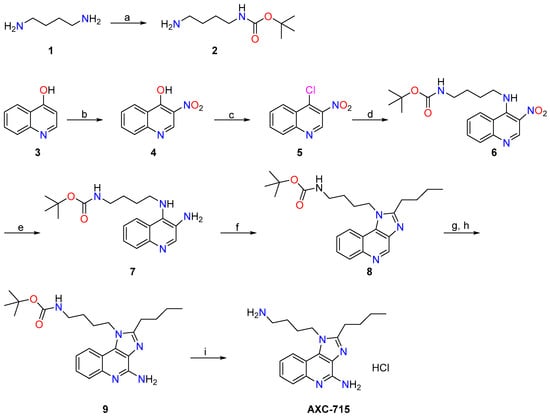
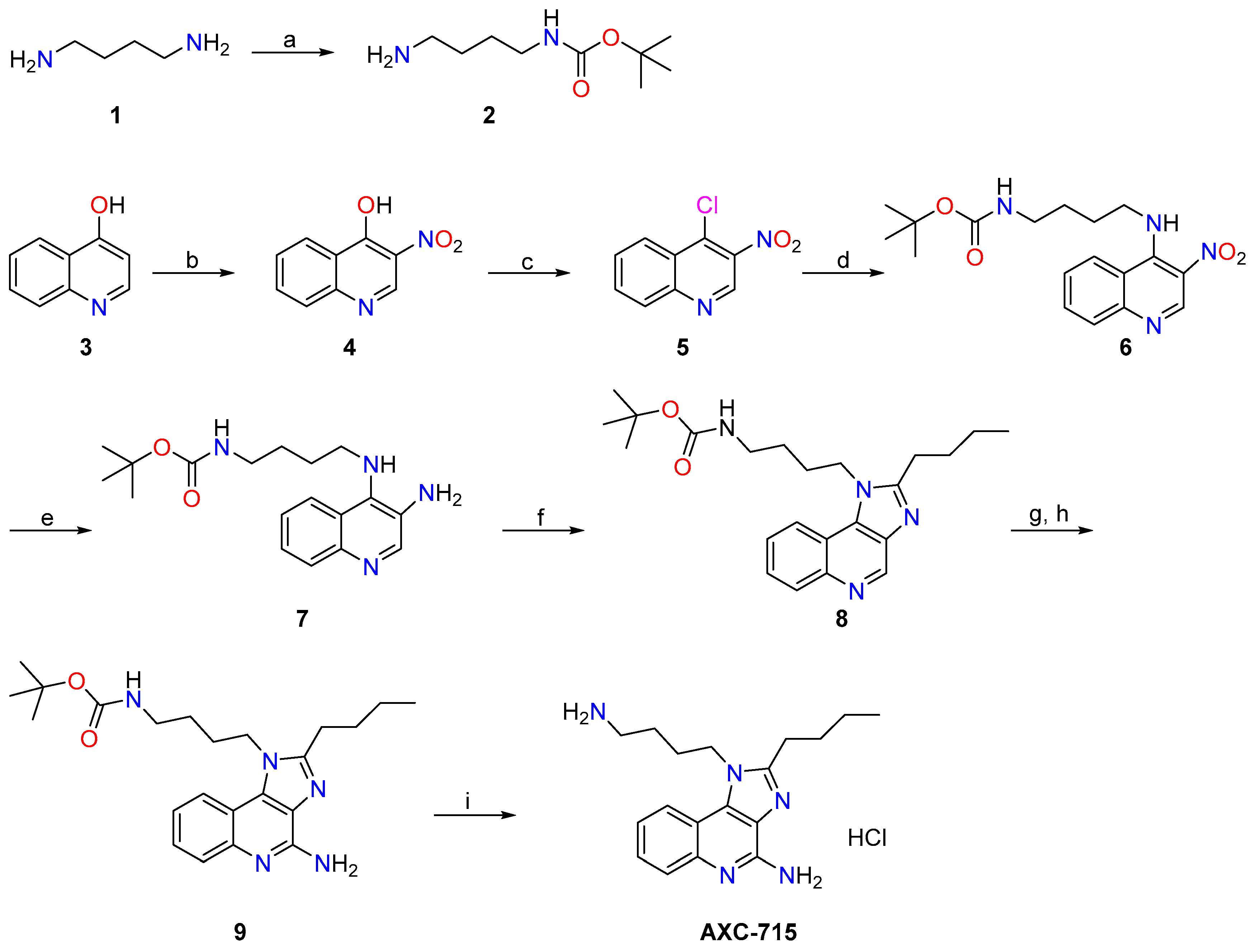
The synthetic route for AXC-715 is illustrated in Scheme 1. The synthesis commenced with commercially available 1,4-diaminobutane (1) and 4-hydroxyquinoline (3). Compound 1 was mono-protected via reaction with di-tert-butyl dicarbonate (Boc2O) to yield intermediate 2. In parallel, 4-hydroxyquinoline was nitrated using nitric acid to afford the nitro-substituted derivative. Subsequent chlorination of the 4-hydroxyl group with thionyl chloride in dichloromethane yielded intermediate 4. Nucleophilic substitution of the chlorine in 4 with amine 2 furnished compound 6. Reduction of the nitro group in 6 under a hydrogen atmosphere generated amine 7, which underwent intramolecular cyclization with trimethyl orthovalerate to form the imidazoquinoline core 8. Oxidation of 8 with m-chloroperbenzoic acid (m-CPBA), followed by treatment with aqueous ammonia, produced compound 9. Finally, deprotection of 9 with dry hydrogen chloride gas in tetrahydrofuran (THF) yielded the target compound AXC-715 as a hydrochloride salt.
Scheme 1.
Synthesis of AXC-715. Reagent and conditions: (a) Diboc, CH2Cl2, 0 °C, 12 h; (b) HNO3, CH3COOH, reflux, 6 h; (c) SOCl2, CH2Cl2, DMF, reflux,12 h; (d) N-boc-1,4-butanediamine, K2CO3, DMF, 80 °C, 4 h; (e) Pd/C, H2, THF, 12 h; (f) trimethyl orthovalerate, Toluene, reflux, 4 h; (g) m-CPBA, CHCl3, 50 °C, 2 h; (h) NH4OH, p-toluenesulfonyl chloride, CHCl3, rt,4 h; (i) dry HCl gas, THF, ice-water 2 h.
3.2. AXC-715 Promotes and Stabilizes TLR7 Dimer Formation
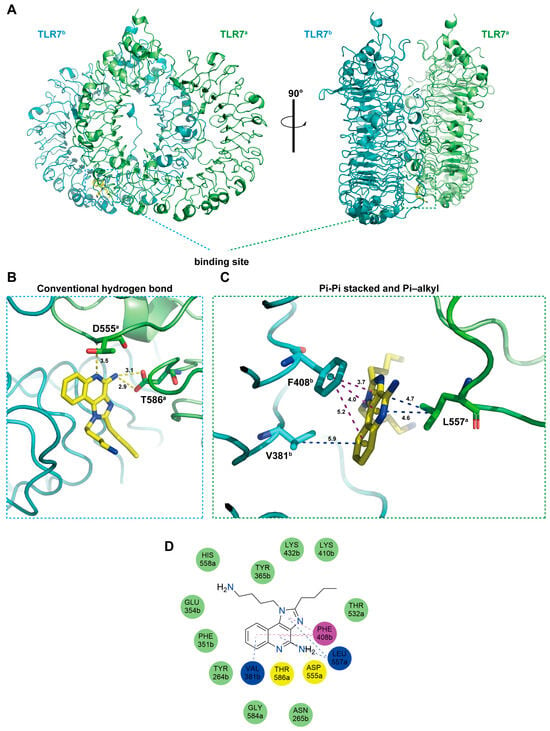
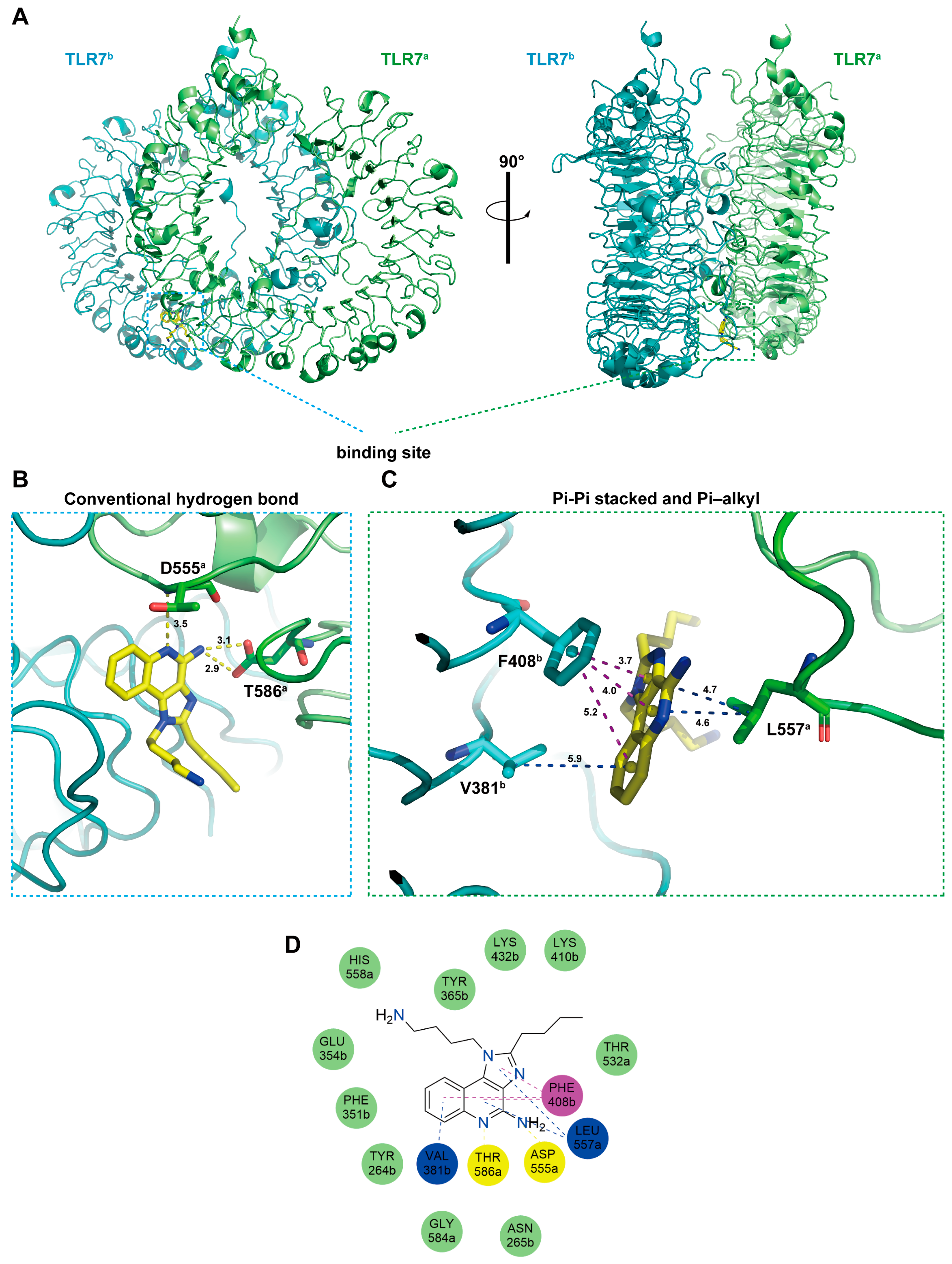
The binding mode of AXC-715 within the hTLR7 structure (PDB ID: 5GMH) was investigated to elucidate the mechanism of receptor activation. AXC-715 occupies the interface between TLR7 monomers, inserting into the binding pocket formed by the two protomers, thereby promoting and stabilizing the active dimer (Figure 1A). Molecular docking simulations indicate that AXC-715 forms specific hydrogen bonds with the side chains of D555 and T586 on monomer A. The pyridine nitrogen acts as a hydrogen bond acceptor for D555, while the amino group interacts with the hydroxyl of T586 (Figure 1B,D). Furthermore, the phenyl, pyridine, and imidazole rings of AXC-715 engage in π-π stacking with F408 of monomer B. The phenyl ring participates in a π-alkyl interaction with V381 (monomer B), while the pyridine and imidazole rings interact with L557 (monomer A) via π-alkyl forces (Figure 1C,D). By occupying this pocket, AXC-715 enhances ligand-protein affinity and locks the dimer in its active conformation. The combined hydrogen bonding and hydrophobic interactions stabilize the dimer interface, preventing dissociation and facilitating downstream signal transduction.
Figure 1.
AXC-715 exhibits direct binding affinity to TLR7 protein. (A) Predicted binding model of the 2.2 Å cocrystal structure of the AXC-715–hTLR7 complex (PDB ID: 5GMH). TLR7 and its dimerization partner are colored green and cyan respectively, and AXC-715 is shown in yellow. (B) Magnified view of the binding pocket highlighting key hydrogen bond interactions. (C) Close-up of the binding pocket illustrating π–π stacking and π–alkyl interactions. (D) Schematic representation of interactions between AXC-715 and TLR7.
3.3. AXC-715 Induces Activation of NF-κB Response Element
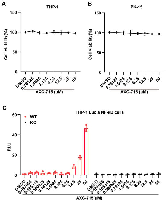
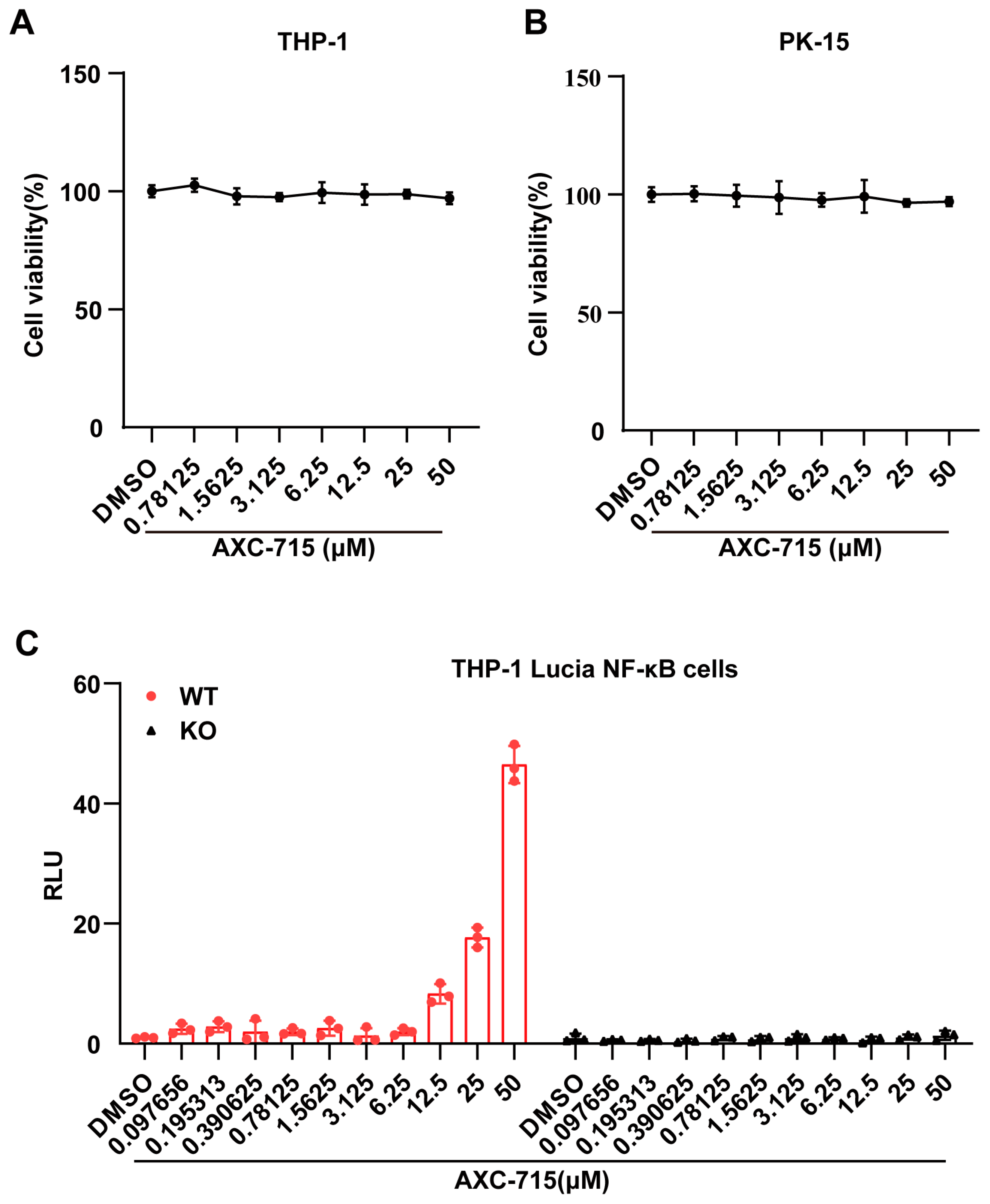
The cytotoxicity of AXC-715 was assessed in PK-15 and THP-1 cells using the CCK-8 assay. Cells were treated with AXC-715 at concentrations ranging from 0 to 50 μM. As shown in Figure 2A,B, AXC-715 did not significantly reduce cell viability in either line, indicating a non-toxic profile at the tested concentrations. Subsequent evaluation of NF-κB pathway activation using THP-1 Lucia NF-κB reporter cells demonstrated a dose-dependent increase in promoter activity in wild-type cells (Figure 2C). This induction was absent in p65-knockout cells, confirming that AXC-715 activates NF-κB signaling via a p65-dependent mechanism.
Figure 2.
AXC-715 induces NF-κB activation in vitro. (A,B) THP-1 cells (A) and PK-15 cells (B) were treated with AXC-715 (0–50 μM) for 24 h. Cell viability was assessed using the CCK-8 assay. (C) THP-1 Lucia NF-κB (WT and p65 KO) cell lines were treated with the AXC-715 for 24 h, then the cell-based activities of the selected compounds were quantified by normalizing compounds-induced reporter activity to DMSO-treated reporter activity. Data are mean ± SEM; n = 3. * p < 0.05, ** p < 0.01, *** p < 0.001.
3.4. AXC-715 Inhibits PRV Replication In Vitro
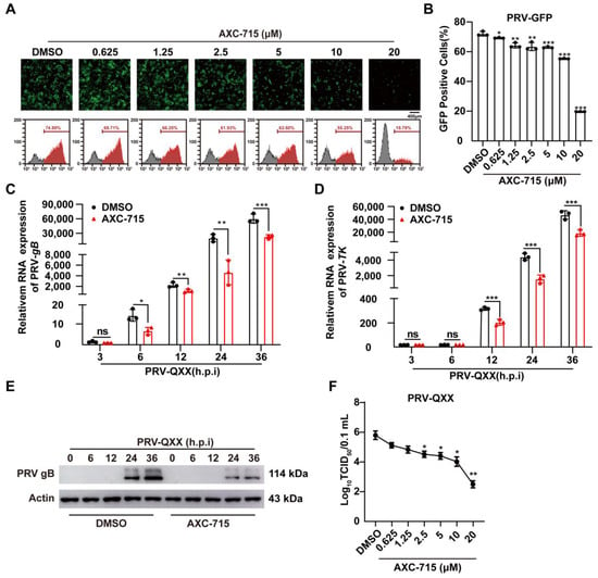
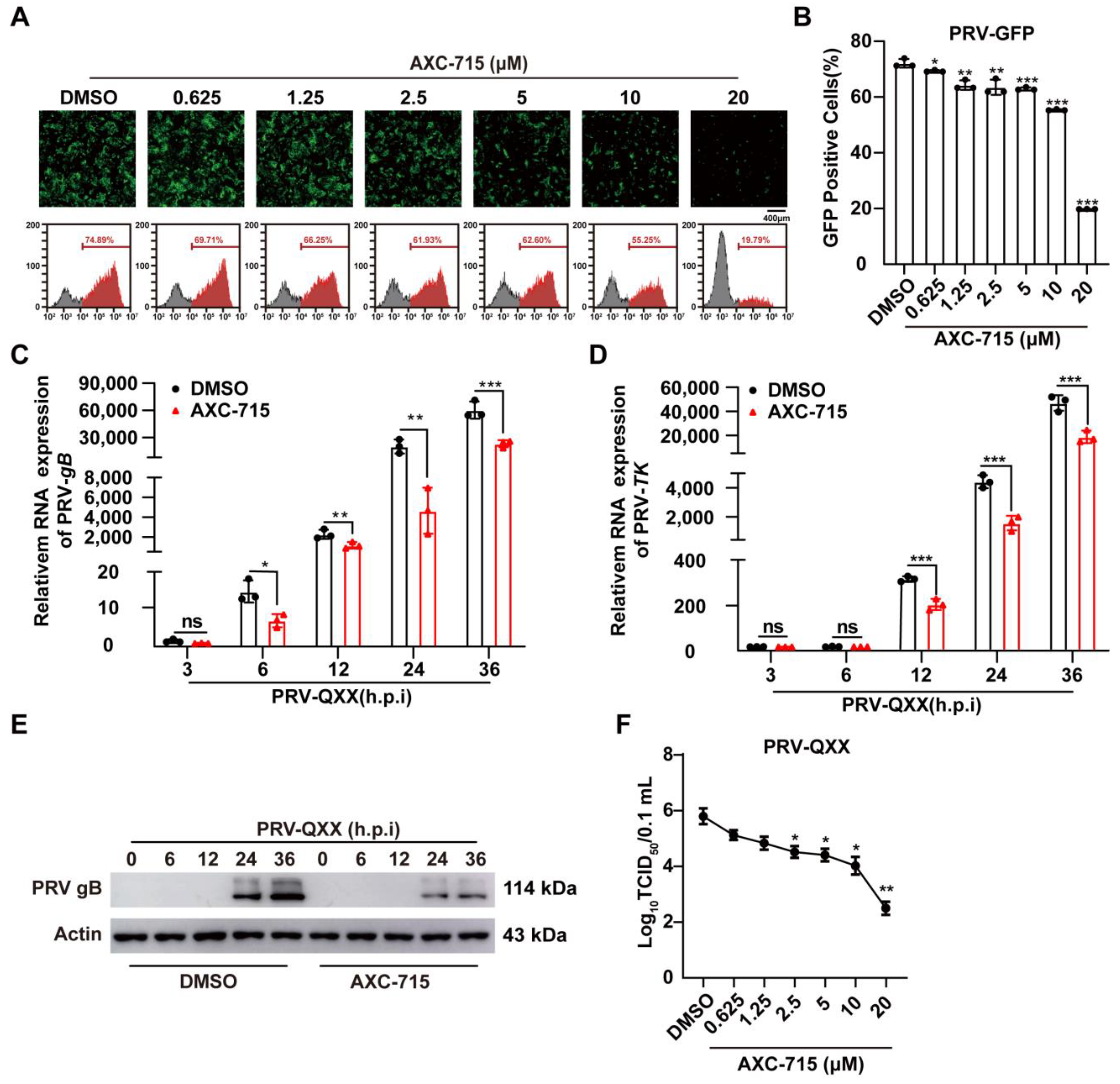
The antiviral efficacy of AXC-715 against PRV was evaluated using a recombinant PRV-GFP strain. Fluorescence microscopy and flow cytometry showed a dose-dependent reduction in the GFP signal upon AXC-715 treatment, indicating inhibition of PRV proliferation (Figure 3A,B). qRT-PCR analysis confirmed a significant downregulation of PRV gB and TK gene transcription (Figure 3C,D). Western blot analysis further revealed decreased PRV-gB protein levels (Figure 3E). Viral titer assays (TCID50) corroborated these results, showing reduced production of progeny PRV-QXX (Figure 3F). These data demonstrate the potent anti-PRV activity of AXC-715.
Figure 3.
AXC-715 inhibits PRV replication in vitro. (A,B) PK-15 cells were infected with PRV-GFP and treated with AXC-715 for 24 h. GFP expression was visualized by fluorescence microscopy and quantified by flow cytometry. (C–E) PK-15 cells were infected with PRV-QXX and then treated with AXC-715 (10 μM). The mRNA expression levels of PRV gB and TK were analyzed by real-time quantitative PCR (RT-qPCR), and the protein expression level of PRV gB was assessed by Western blot analysis. (F) The effect of AXC-715 on progeny virus production of PRV-QXX was evaluated by viral titration assay. Data are mean ± SEM; n = 3. ns p > 0.05, * p < 0.05, ** p < 0.01, *** p < 0.001.
3.5. AXC-715 Inhibits VSV Replication In Vitro
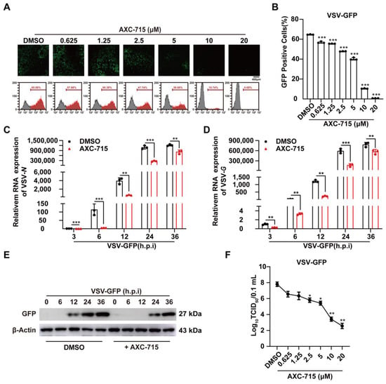
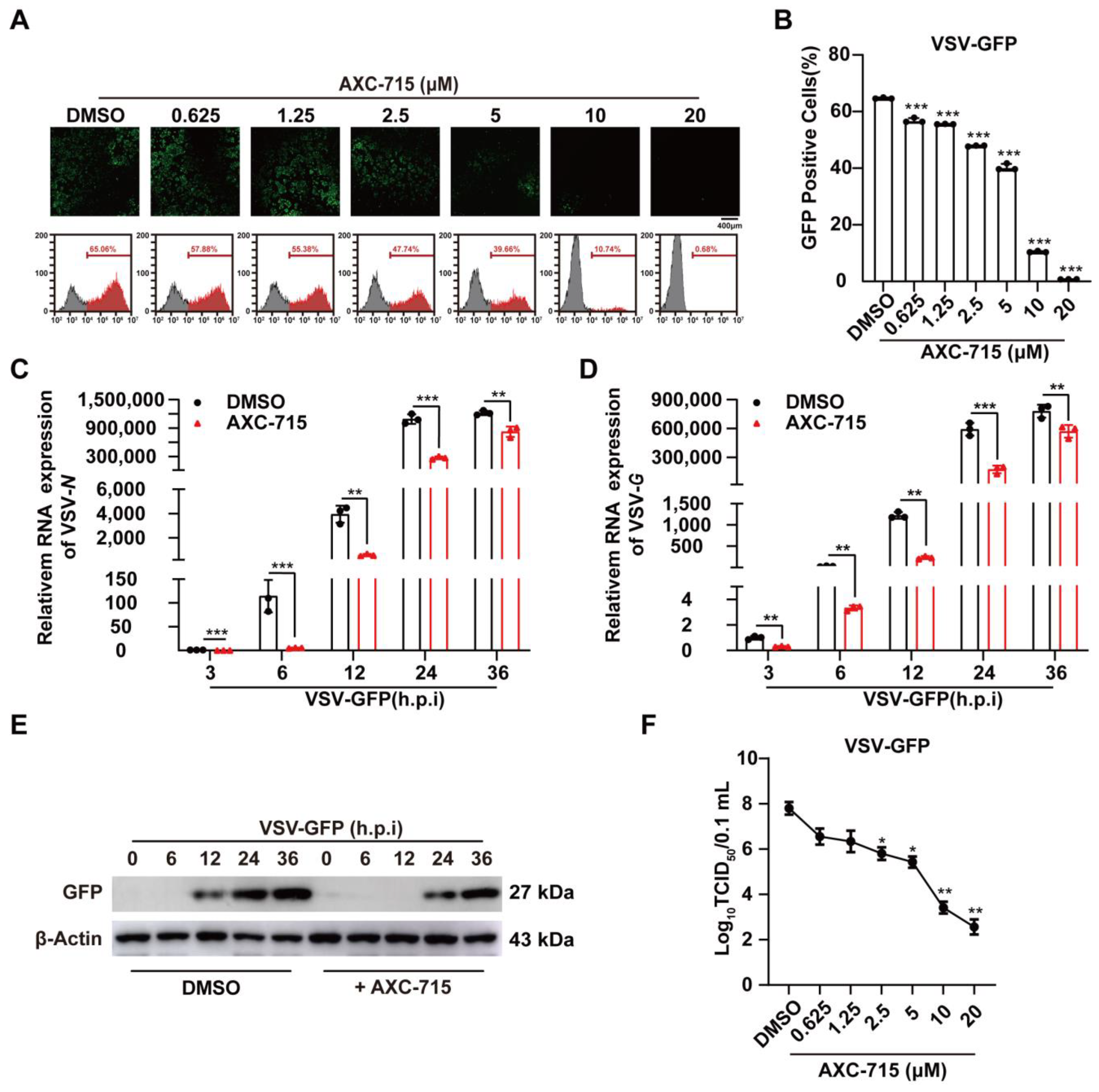
To assess broad-spectrum activity, the effect of AXC-715 on VSV was examined using VSV-GFP. Both fluorescence microscopy and flow cytometry indicated a dose-dependent decrease in GFP intensity, suggesting suppression of VSV replication (Figure 4A,B). qRT-PCR results showed significant inhibition of VSV N and G gene transcription (Figure 4C,D). Consistent with these findings, Western blotting detected reduced GFP protein expression (Figure 4E), and viral titer assays confirmed a decrease in progeny VSV-GFP production (Figure 4F). AXC-715 thus exhibits potent antiviral activity against VSV.
Figure 4.
AXC-715 inhibits VSV replication in vitro. (A,B) PK-15 cells were infected with VSV-GFP and treated with AXC-715 for 24 h. GFP expression was visualized by fluorescence microscopy and quantified by flow cytometry. (C–E) PK-15 cells were infected with VSV-GFP and then treated with AXC-715 (10 μM). The mRNA expression levels of VSV-GFP N and G were analyzed by quantitative real-time PCR (qRT-PCR), and the protein expression level of PRV gB was assessed by Western blot analysis. (F) The effect of AXC-715 on progeny virus production of VSV-GFP was evaluated by viral titration assay. Data are mean ± SEM; n = 3. * p < 0.05, ** p < 0.01, *** p < 0.001.
3.6. AXC-715 Inhibits Viral Replication
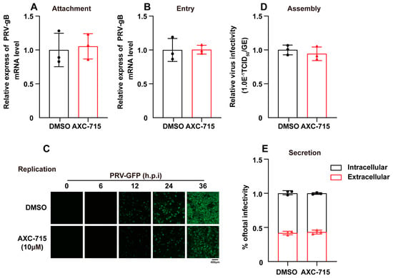
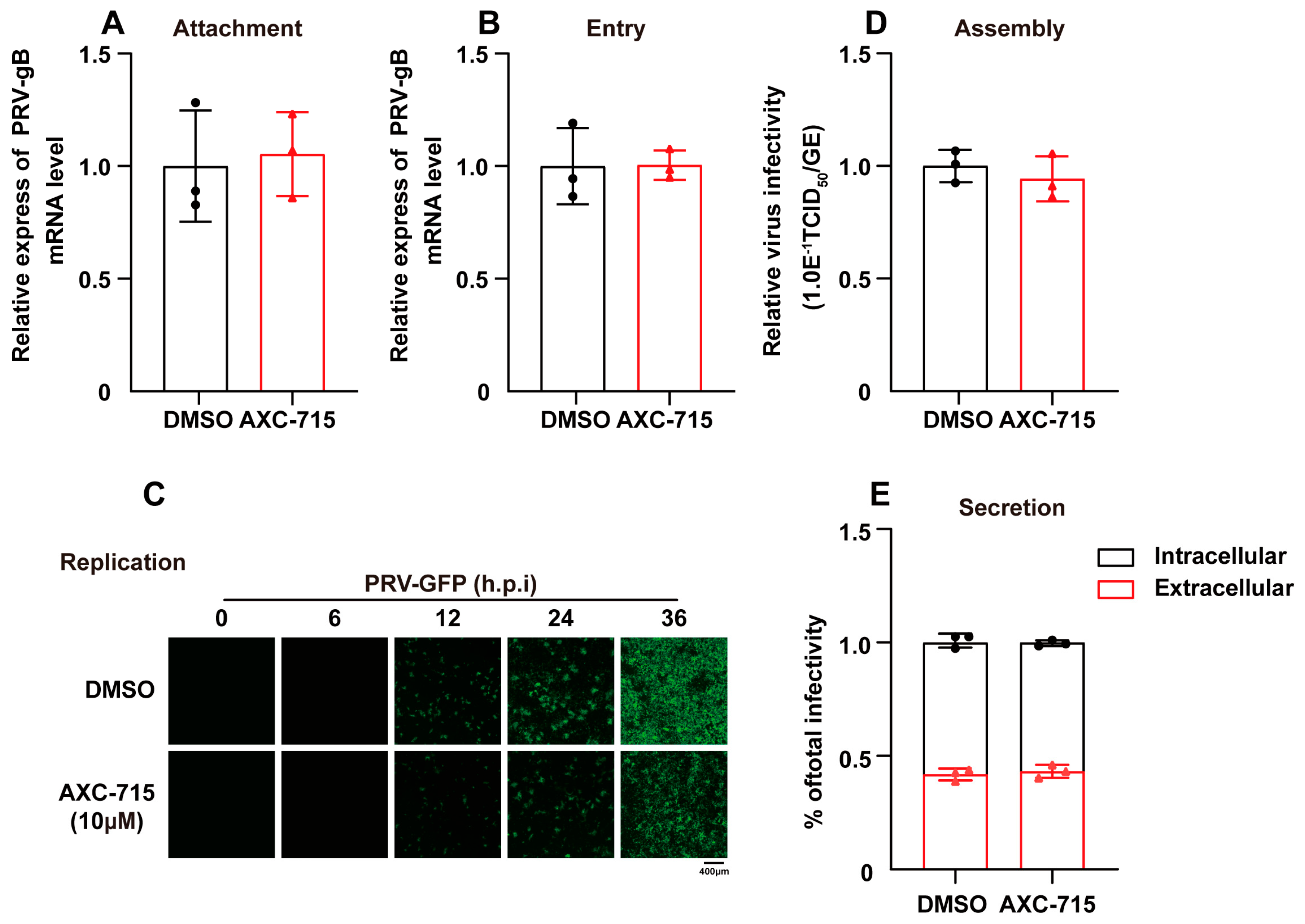
Mechanistic studies were performed to identify the stage of the viral life cycle targeted by AXC-715. First, attachment assays showed that co-incubation of PRV-QXX with AXC-715 at 4 °C did not alter gB mRNA levels compared to the control, indicating no effect on viral adsorption (Figure 5A). Next, entry assays were conducted; pre-adsorbed virus was allowed to enter in the presence of AXC-715 at 37 °C. qRT-PCR analysis revealed no significant difference in viral entry (Figure 5B). In contrast, replication assays monitoring GFP fluorescence over time showed a marked reduction in the signal upon AXC-715 treatment (Figure 5C). Additionally, AXC-715 did not interfere with viral assembly or release. These results suggest that AXC-715 specifically impairs the intracellular replication phase of PRV.
Figure 5.
Study on the effect of AXC-715 on the life cycle of PRV. (A,B) Real-time PCR analysis of the effect of AXC-715 (10 µM) on the adsorption and entry phases of PRV-QXX in PK-15 cells. (C) Immunofluorescence assay (IFA) assessing the impact of AXC-715 (10 µM) on PRV-QXX replication in PK-15 cells. (D) Evaluation of AXC-715 (10 µM) on the assembly phase of PRV-QXX in PK-15 cells by analyzing the ratio of infectious viral titers to viral genome copy numbers. (E) Analysis of the effect of AXC-715 (10 µM) on the release phase of PRV-QXX in PK-15 cells by the ratio of viral titers in the supernatant to those inside the cells.
3.7. AXC-715 Targets TLR7 to Inhibit Viral Replication
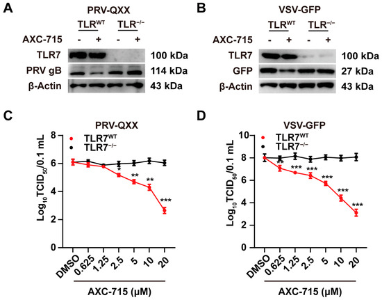
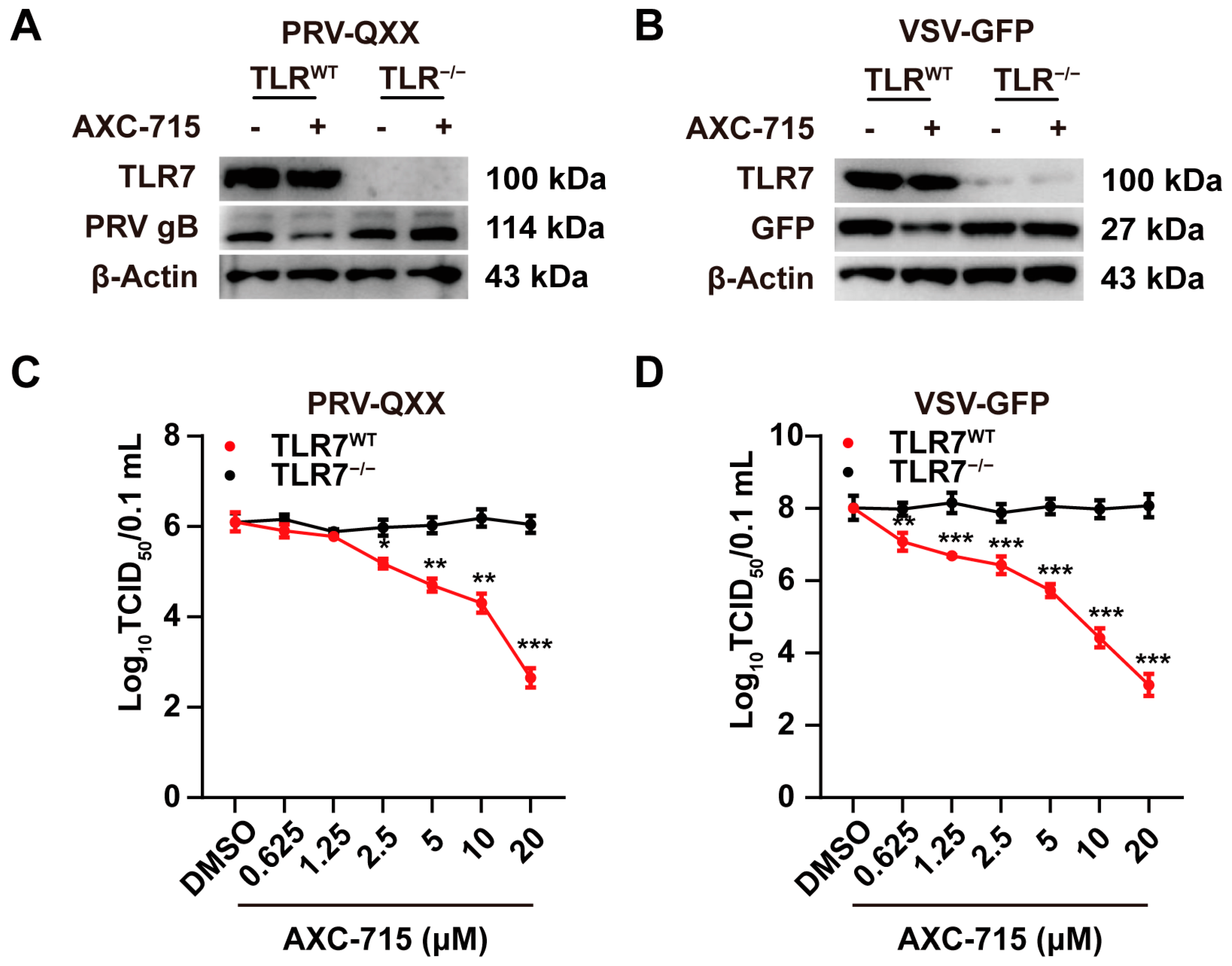
To verify TLR7 dependence, TLR7 wild-type (TLR7WT) and TLR7 knockout (TLR7−/−) PK-15 cells were treated with AXC-715 and infected with PRV-QXX or VSV-GFP. Western blot analysis of PRV-gB and GFP protein expression revealed that AXC-715 significantly suppressed viral replication in TLR7WT cells (Figure 6A,B). In contrast, no antiviral effect was observed in TLR7−/− cells. This confirms that the antiviral activity of AXC-715 is strictly mediated through the TLR7 signaling pathway.
Figure 6.
AXC-715 suppresses viral replication in a strictly TLR7-dependent manner. (A) Western blot analysis of PRV gB protein expression in TLR7WT and TLR7−/− PK-15 cells infected with PRV-QXX following AXC-715 treatment. (B) Western blot analysis of GFP protein expression in TLR7WT and TLR7−/− PK-15 cells infected with VSV-GFP following AXC-715 treatment. (C) The effect of AXC-715 on the production of PRV-QXX progeny virus in TLR7WT and TLR7−/− cells was evaluated by viral titration assay. (D) The effect of AXC-715 on the production of VSV-GFP progeny virus in TLR7WT and TLR7−/− cells was evaluated by viral titration assay. Data are mean ± SEM; n = 3. * p < 0.05, ** p < 0.01, *** p < 0.001.
4. Discussion
This study elucidates the structural and functional mechanisms underlying the antiviral activity of AXC-715, a novel small-molecule agonist of TLR7. We demonstrate that AXC-715 functions as a potent molecular stabilizer of TLR7. Structural analysis of the AXC-715–hTLR7 complex revealed that the ligand occupies a critical position at the dimer interface, engaging key residues—D555 and T586 via hydrogen bonding, and F408, V381, and L557 through π-π and π-alkyl interactions. This binding mode facilitates the formation of the active “m”-shaped dimer, bringing the C-termini of the receptor into close proximity to recruit downstream adaptor proteins [27,28]. Unlike endogenous ligands such as guanosine or ssRNA, which often require specific coordination within the binding pocket [29], AXC-715 acts as a “molecular glue,” effectively locking the receptor in an active conformation through direct intermolecular contacts. This stabilization likely lowers the energy threshold for dimerization and sustains signaling output, accounting for the robust activation of NF-κB observed in our experiments.
Functionally, the capacity of AXC-715 to inhibit the replication of both DNA (PRV) and RNA (VSV) viruses underscores the potential of TLR7 agonists as broad-spectrum, host-directed antiviral therapeutics. Time-of-addition assays indicated that AXC-715 specifically impairs viral replication without affecting viral adsorption, entry, assembly, or release. This phenotype suggests that the antiviral effect is not due to direct virucidal activity but is mediated by the induction of a cellular antiviral state. Upon TLR7 activation, the NF-κB and IRF7 pathways are triggered, leading to the upregulation of IFNs and ISGs [30]. These ISGs establish an intracellular environment restrictive to viral replication. The complete abolition of antiviral activity in TLR7-knockout cells confirms the on-target specificity of AXC-715 and highlights its role as a host-directed therapeutic agent capable of circumventing viral resistance mechanisms that often plague direct-acting antivirals [31,32].
Beyond its immediate antiviral efficacy, AXC-715 provides critical insights into the pharmacological regulation of TLR7. The ability of small-molecule ligands to promote dimer formation offers a strategy to fine-tune immune responses. By stabilizing the active dimer, AXC-715 may enhance receptor sensitivity to low-level viral PAMPs that might otherwise evade detection [33]. This sensitization effect could be particularly beneficial during early infection or in cases of immune evasion. However, the potent activation of TLR7 necessitates a careful evaluation of the therapeutic window, as chronic or excessive signaling has been associated with immunopathology, including autoimmune disorders and cytokine storms [34]. While our in vitro studies showed no detectable cytotoxicity in PK-15 or THP-1 cells, the in vivo safety profile remains to be determined. Future studies must focus on the pharmacokinetics and biodistribution of AXC-715 to ensure that sustained receptor activation does not lead to excessive inflammation or tissue damage.
In conclusion, AXC-715 exemplifies a structure-guided, receptor-targeted strategy that leverages TLR7 dimer stabilization to modulate innate immunity. Its dual capacity to enhance NF-κB signaling and inhibit viral replication across distinct viral families highlights its potential as a versatile immunomodulatory agent. While alternative antiviral strategies, including immunomodulators, interferons, and antioxidants, have been explored, our study specifically focused on the host-directed approach using the TLR7 agonist AXC-715. This strategy activates innate immunity to establish a broad-spectrum antiviral state, potentially overcoming virus-specific resistance mechanisms commonly observed with direct-acting antivirals. Future studies could explore combinatorial approaches, assessing the synergy between TLR7 agonists and other biological response modifiers to further enhance antiviral efficacy and minimize potential side effects. These findings provide a valuable framework for the rational design of next-generation TLR7 agonists, aiming to optimize the balance between potent antiviral efficacy and acceptable safety profiles for clinical application.
Author Contributions
C.Y.: Conception and Design, Methodology, Data analysis, Software, Validation, Writing—original draft and editing. M.-H.D., Y.-J.M., H.W., L.H.: Investigation; Methodology; Resources; Software; Validation; Visualization. Z.-C.L., H.-Y.Y.: Data curation; Validation; Software; G.-Y.Y.: Formal analysis, Investigation, Funding acquisition and Methodology. M.-D.W., S.-L.M.: Conceptualization, Project administration, Resources Funding acquisition. All authors have read and agreed to the published version of the manuscript.
Funding
This research was supported by grants from the National Key R&D Program of China (2021YFD1301200),the China Postdoctoral Science Foundation (2025M770267, GZC20240430).
Institutional Review Board Statement
Not applicable.
Informed Consent Statement
Not applicable.
Data Availability Statement
The data that support the findings of this study are available from the corresponding author upon request.
Conflicts of Interest
The authors declare no conflicts of interest.
References
- Thompson, M.R.; Kaminski, J.J.; Kurt-Jones, E.A.; Fitzgerald, K.A. Pattern recognition receptors and the innate immune response to viral infection. Viruses 2011, 3, 920–940. [Google Scholar] [CrossRef] [Scilit]
- Carty, M.; Guy, C.; Bowie, A.G. Detection of Viral Infections by Innate Immunity. Biochem. Pharmacol. 2021, 183, 114316. [Google Scholar] [CrossRef] [Scilit] [PubMed]
- Diebold, S.S.; Kaisho, T.; Hemmi, H.; Akira, S.; Reis e Sousa, C. Innate antiviral responses by means of TLR7-mediated recognition of single-stranded RNA. Science 2004, 303, 1529–1531. [Google Scholar] [CrossRef] [Scilit] [PubMed]
- Lund, J.M.; Alexopoulou, L.; Sato, A.; Karow, M.; Adams, N.C.; Gale, N.W.; Iwasaki, A.; Flavell, R.A. Recognition of single-stranded RNA viruses by Toll-like receptor 7. Proc. Natl. Acad. Sci. USA 2004, 101, 5598–5603. [Google Scholar] [CrossRef] [Scilit] [PubMed]
- Honda, K.; Taniguchi, T. IRFs: Master regulators of signalling by Toll-like receptors and cytosolic pattern-recognition receptors. Nat. Rev. Immunol. 2006, 6, 644–658. [Google Scholar] [CrossRef] [Scilit]
- Kawai, T.; Akira, S. The role of pattern-recognition receptors in innate immunity: Update on Toll-like receptors. Nat. Immunol. 2010, 11, 373–384. [Google Scholar] [CrossRef] [Scilit]
- Lester, S.N.; Li, K. Toll-like receptors in antiviral innate immunity. J. Mol. Biol. 2014, 426, 1246–1264. [Google Scholar] [CrossRef] [Scilit]
- Hemmi, H.; Kaisho, T.; Takeuchi, O.; Sato, S.; Sanjo, H.; Hoshino, K.; Horiuchi, T.; Tomizawa, H.; Takeda, K.; Akira, S. Small anti-viral compounds activate immune cells via the TLR7 MyD88-dependent signaling pathway. Nat. Immunol. 2002, 3, 196–200. [Google Scholar] [CrossRef] [Scilit] [PubMed]
- Gorden, K.B.; Gorski, K.S.; Gibson, S.J.; Kedl, R.M.; Kieper, W.C.; Qiu, X.; Tomai, M.A.; Alkan, S.S.; Vasilakos, J.P. Synthetic TLR agonists reveal functional differences between human TLR7 and TLR8. J. Immunol. 2005, 174, 1259–1268. [Google Scholar] [CrossRef] [Scilit]
- Jurk, M.; Heil, F.; Vollmer, J.; Schetter, C.; Krieg, A.M.; Wagner, H.; Lipford, G.; Bauer, S. Human TLR7 or TLR8 independently confer responsiveness to the antiviral compound R-848. Nat. Immunol. 2002, 3, 499. [Google Scholar] [CrossRef] [Scilit] [PubMed]
- Heil, F.; Hemmi, H.; Hochrein, H.; Ampenberger, F.; Kirschning, C.; Akira, S.; Lipford, G.; Wagner, H.; Bauer, S. Species-specific recognition of single-stranded RNA via toll-like receptor 7 and 8. Science 2004, 303, 1526–1529. [Google Scholar] [CrossRef] [Scilit] [PubMed]
- Stanley, M.A. Imiquimod and the imidazoquinolones: Mechanism of action and therapeutic potential. Clin. Exp. Dermatol. 2002, 27, 571–577. [Google Scholar] [CrossRef] [Scilit]
- Bilu, D.; Sauder, D.N. Imiquimod: Modes of action. Br. J. Dermatol. 2003, 149, 5–8. [Google Scholar] [CrossRef] [Scilit]
- Tyring, S. Imiquimod applied topically: A novel immune response modifier. Skin. Therapy Lett. 2001, 6, 1–4. [Google Scholar]
- Dowling, J.K.; Mansell, A. Toll-like receptors: The swiss army knife of immunity and vaccine development. Clin. Transl. Immunol. 2016, 5, e85. [Google Scholar] [CrossRef] [Scilit]
- Miller, R.L.; Meng, T.C.; Tomai, M.A. The antiviral activity of Toll-like receptor 7 and 7/8 agonists. Drug News Perspect. 2008, 21, 69–87. [Google Scholar] [CrossRef] [Scilit]
- Iwasaki, A.; Pillai, P.S. Innate immunity to influenza virus infection. Nat. Rev. Immunol. 2014, 14, 315–328. [Google Scholar] [CrossRef] [Scilit]
- Schoggins, J.W. Interferon-Stimulated Genes: What Do They All Do? Annu. Rev. Virol. 2019, 6, 567–584. [Google Scholar] [CrossRef] [Scilit] [PubMed]
- Zhang, Z.; Ohto, U.; Shibata, T.; Taoka, M.; Yamauchi, Y.; Sato, R.; Shukla, N.M.; David, S.A.; Isobe, T.; Miyake, K.; et al. Structural Analyses of Toll-like Receptor 7 Reveal Detailed RNA Sequence Specificity and Recognition Mechanism of Agonistic Ligands. Cell Rep. 2018, 25, 3371–3381. [Google Scholar] [CrossRef] [Scilit]
- Tanji, H.; Ohto, U.; Shibata, T.; Miyake, K.; Shimizu, T. Structural reorganization of the Toll-like receptor 8 dimer induced by agonistic ligands. Science 2013, 339, 1426–1429. [Google Scholar] [CrossRef] [Scilit] [PubMed]
- Zhang, Z.; Ohto, U.; Shibata, T.; Krayukhina, E.; Taoka, M.; Yamauchi, Y.; Tanji, H.; Isobe, T.; Uchiyama, S.; Miyake, K.; et al. Structural Analysis Reveals that Toll-like Receptor 7 Is a Dual Receptor for Guanosine and Single-Stranded RNA. Immunity 2016, 45, 737–748. [Google Scholar] [CrossRef] [Scilit] [PubMed]
- Zhang, R.; Tang, J. Evasion of I Interferon-Mediated Innate Immunity by Pseudorabies Virus. Front. Microbiol. 2021, 12, 801257. [Google Scholar] [CrossRef] [Scilit]
- Pomeranz, L.E.; Reynolds, A.E.; Hengartner, C.J. Molecular biology of pseudorabies virus: Impact on neurovirology and veterinary medicine. Microbiol. Mol. Biol. Rev. 2005, 69, 462–500. [Google Scholar] [CrossRef] [Scilit]
- Wang, C.; Li, L.; Zhai, X.; Chang, H.; Liu, H. Evasion of the Antiviral Innate Immunity by PRV. Int. J. Mol. Sci. 2024, 25, 13140. [Google Scholar] [CrossRef] [Scilit]
- Stojdl, D.F.; Lichty, B.D.; tenOever, B.R.; Paterson, J.M.; Power, A.T.; Knowles, S.; Marius, R.; Reynard, J.; Poliquin, L.; Atkins, H.; et al. VSV strains with defects in their ability to shutdown innate immunity are potent systemic anti-cancer agents. Cancer Cell 2003, 4, 263–275. [Google Scholar] [CrossRef] [Scilit]
- Moglan, A.M.; Albaradie, O.A.; Alsayegh, F.F.; Alharbi, H.M.; Samman, Y.M.; Jalal, M.M.; Saeedi, N.H.; Mahmoud, A.B.; Alkayyal, A.A. Preclinical efficacy of oncolytic VSV-IFNβ in treating cancer: A systematic review. Front. Immunol. 2023, 14, 1085940. [Google Scholar] [CrossRef] [Scilit]
- Shingo, T.; Zhikuan, Z.; Hiroyuki, M.; Masahiro, T.; Mitsunori, I.; Mami, K.; Mami, K.; Hideki, S.; Akihisa, T.; Naruhiko, A.; et al. Structural analysis reveals TLR7 dynamics underlying antagonism. Nat. Commun. 2020, 11, 5204. [Google Scholar] [CrossRef] [Scilit]
- Bérouti, M.; Wagner, M.; Greulich, W.; Piseddu, I.; Gärtig, J.; Hansbauer, L.; Müller-Hermes, C.; Heiss, M.; Pichler, A.; Tölke, A.J.; et al. Pseudouridine RNA avoids immune detection through impaired endolysosomal processing and TLR engagement. Cell 2025, 188, 4880–4895. [Google Scholar] [CrossRef] [Scilit]
- Taro, K.; Shizuo, A. Innate immune recognition of viral infection. Nat. Immunol. 2006, 7, 131–137. [Google Scholar] [CrossRef] [Scilit] [PubMed]
- Pamela, O.; Ville, V.; Jukka, S.; Kevin, M.K.; John, H.; Sampsa, M.; Ilkka, J. Gene expression and antiviral activity of alpha/beta interferons and interleukin-29 in virus-infected human myeloid dendritic cells. J. Virol. 2005, 79, 9608–9617. [Google Scholar] [CrossRef] [Scilit]
- Kaufmann, S.H.; Dorhoi, A.; Hotchkiss, R.S.; Bartenschlager, R. Host-directed therapies for bacterial and viral infections. Nat. Rev. Drug Discov. 2017, 17, 35–56. [Google Scholar] [CrossRef] [Scilit] [PubMed]
- Kenya, H.; Akinori, T.; Tadatsugu, T. Type I inteferon gene induction by the interferon regulatory factor family of transcription factors. Immunity 2006, 25, 349–360, Erratum in Immunity 2006, 25, 849. https://doi.org/10.1016/j.immuni.2006.10.004. [Google Scholar] [CrossRef] [Scilit] [PubMed]
- Yang, J.; He, Y.; Zhang, M.; Liang, C.; Li, T.; Ji, T.; Zu, M.; Ma, X.; Zhang, Z.; Liang, C.; et al. Programmed initiation and enhancement of cGAS/STING pathway for tumour immunotherapy via tailor-designed ZnFe2O4-based nanosystem. Exploration 2024, 3, 20230061. [Google Scholar] [CrossRef] [Scilit]
- Christine, W.; Ee Lyn, L.; Mohammad, M.; Barbara, K.; Alexandru, O.; Eusebia, L.-V.; Sarah, K.; Simon, M.; Katharina, M.; Kerstin, E.; et al. UNC93B1 variants underlie TLR7-dependent autoimmunity. Sci. Immunol. 2024, 9, eadi9769. [Google Scholar] [CrossRef] [Scilit]

Disclaimer/Publisher’s Note: The statements, opinions and data contained in all publications are solely those of the individual author(s) and contributor(s) and not of MDPI and/or the editor(s). MDPI and/or the editor(s) disclaim responsibility for any injury to people or property resulting from any ideas, methods, instructions or products referred to in the content. |
© 2026 by the authors. Licensee MDPI, Basel, Switzerland. This article is an open access article distributed under the terms and conditions of the Creative Commons Attribution (CC BY) license.