


Abstract
Among Candida species, several are major opportunistic fungal pathogens capable of causing a wide spectrum of infections, ranging from superficial mucosal conditions to severe systemic diseases. Their success as human pathogens is largely due to their ability to rapidly adapt to diverse host environments and develop resistance to antifungal agents. Experimental evolution provides a powerful framework for understanding these adaptive processes by observing evolutionary change in real-time. Although most studies rely on in vitro systems and a limited set of Candida species, there is strong evidence that genome plasticity, including aneuploidy, loss of heterozygosity, and copy number variation, plays a central role in driving rapid adaptation. Experimental evolution has also been applied to study the dynamics of antifungal resistance, particularly to azoles, although relatively fewer studies have explored resistance to echinocandins and polyenes. This review summarizes current knowledge on experimental evolution in pathogenic Candida species, with a focus on genome plasticity, adaptation to host-imposed stress, and particularly on the emergence of antifungal resistance. It also identifies critical research gaps, including the need for broader species coverage, investigation of underexplored antifungal classes, and evaluation of combined therapies. A deeper understanding of these dynamics is essential to improve antifungal strategies and counter the growing threat of drug-resistant Candida spp. infections.
1. Introduction
Experimental evolution (EE) involves the study of evolutionary change in populations subjected to defined environmental, demographic, genetic, social, and other constraints imposed under controlled laboratory or natural conditions [1]. Over time, EE has been adapted and refined across multiple disciplines, leading to the development of distinct methodologies suited to specific research questions and model systems [2,3,4,5,6]. Its growing importance is highlighted by the sharp increase in EE studies over the past decade, reinforcing the relevance of reviewing its applications.
Microbial EE typically uses well-controlled laboratory populations combined with molecular and genomic tools to investigate mechanisms of adaptation, validate theoretical predictions, and discover novel evolutionary outcomes [7,8,9]. In the context of human health, EE has been instrumental in studying the development of antimicrobial resistance, particularly among invasive fungal pathogens [10,11].
Fungal infections represent a major clinical concern, particularly in immunocompromised or critically ill patients [12,13,14]. The range of potential pathogens responsible for these infections is expanding, with Candida spp., Cryptococcus neoformans, Pneumocystis jirovecii, and Aspergillus spp. being the most prevalent [15]. Candida species can cause invasive fungal infections, particularly bloodstream infections, which are associated with high morbidity and mortality rates [16]. While C. albicans remains the most commonly isolated species, non-albicans Candida (NAC) species, such as C. glabrata (reclassified as Nakaseomyces glabratus [17]), C. krusei (reclassified as Pichia kudriavzevii [18]), C. parapsilosis, and C. tropicalis, are increasingly prevalent and often show resistance to azole antifungals, especially fluconazole [19]. More recently, the emergence of the multidrug-resistant yeast C. auris (reclassified as Candidozyma auris [19]) has further underscored the urgent need to understand how antifungal resistance evolves [20,21,22].
Previous reviews of EE assays have highlighted their value for investigating antifungal drug resistance in pathogenic fungi, particularly via drug-driven selection experiments that reconstruct resistance evolution under controlled conditions. However, most studies have focused exclusively on resistance phenotypes, whereas the use of EE to study other fungal traits, such as genome plasticity and the response to host-related stresses, has not yet been comprehensively synthesized.
In this review, we focus on EE studies in pathogenic Candida species and aim at capturing the multiple evolutionary dimensions shaping fungal pathogens beyond antifungal exposure. We synthesize current knowledge on three major themes: (i) evolutionary dynamics of genome plasticity, (ii) adaptation to host-imposed stressors, and (iii) the evolution of antifungal drug resistance. By integrating these areas, we aim to provide a unified view of how EE can help elucidate clinically relevant evolutionary processes and guide the development of more effective strategies to combat antifungal resistance.
2. Literature Search Strategy
A comprehensive literature search was conducted across the electronic database PubMed (PubMed.gov from the National Library of Medicine). The search covered publications from 1990 to November 2025. Keyword combinations included “experimental evolution”, “in vitro experimental evolution”, “in vivo experimental evolution”, “antifungal drug resistance”, “fungal pathogens”, “Candida species”, “Candida albicans”, “Non-albicans Candida”, “host-imposed stress”, and “genomic flexibility”. After the initial retrieval, titles and abstracts were screened to identify original research and key reviews examining the potential of experimental evolution to study fungal adaptation. Papers aligned with the aims of this review were subsequently selected for in-depth analysis and synthesis. PubMed was chosen as the primary database because of its strong coverage of biomedical, microbiological, and clinical literature relevant to pathogenic fungi. Nevertheless, we acknowledge that some experimental evolution studies may also be indexed in other databases.
3. Experimental Evolution as a Tool to Study Pathogenic Candida spp.
EE has become a powerful tool for investigating adaptive processes in fungal pathogens. While EE has been widely applied to diverse fungal genera (e.g., Cryptococcus, Aspergillus, Saccharomyces), this review deliberately focuses on pathogenic Candida species as they combine high clinical relevance, genetic tractability, and a substantial body of in vivo and in vitro EE studies directly linked to antifungal resistance and host adaptation. Candida species are especially suitable for EE due to their relatively short generation times, large population sizes, and well-developed molecular and genomic toolkits. In the study of pathogenic Candida spp., understanding adaptation requires integrating both in vitro EE and in vivo evolution approaches (Figure 1).
Figure 1.
Overview of experimental evolution approaches. (a) In vitro evolution includes two main approaches: serial transfer, where microbial populations are repeatedly propagated in fresh medium, promoting adaptation (e.g., antifungals, macrophages, H2O2) across successive growth cycles, and chemostat culture, a continuous system in which fresh medium is supplied and culture is removed at a constant rate, maintaining a steady state and allowing adaptation under stable selective pressures (e.g., antifungals). (b) In vivo evolution relies on different host models: the mouse model, often established through antimicrobial treatment of pathogens in immunocompetent or immunocompromised mice to study infection dynamics and immune adaptation; the Caenorhabditis elegans model, where nematodes are infected on agar plates or liquid culture to monitor pathogen adaptation in a simple host; the Drosophila model, typically involving infection through pricking or feeding assays to explore genetic determinants of host–pathogen interactions; and the human host, where evolutionary dynamics are observed directly during clinical infections, for instance through longitudinal sampling of isolates from patients under antimicrobial treatment.
EE techniques such as “laboratory natural selection” and “laboratory culling” are frequently used to simulate environmental stressors without explicitly selecting for specific traits. In “laboratory natural selection,” researchers alter environmental parameters, such as temperature, nutrient availability, or antifungal presence, while maintaining an unaltered control population for comparison [1]. In contrast, “laboratory culling” involves applying lethal or sublethal stress to a population and allowing only the survivors to seed the next generation, thereby mimicking strong selection pressure [1]. These experimental designs help reveal the diversity of adaptive strategies that pathogenic Candida species can evolve, including changes in drug susceptibility, stress tolerance, or virulence factors expression.
However, one major limitation of laboratory-based EE is its ecological relevance. The controlled and simplified conditions used in vitro often fail to replicate the complexity of host environments, where Candida encounters dynamic pressures such as immune responses, microbiota interactions, fluctuating nutrient availability, and tissue-specific conditions. These limitations can restrict the applicability of in vitro findings to clinical or natural scenarios. Furthermore, the relatively short timescales typical of laboratory studies might not capture long-term evolutionary dynamics that are relevant to chronic or recurrent infections [1].
To address these limitations, in vivo evolution studies have been conducted using animal models (e.g., mice, nematodes and insects) or through the longitudinal analysis of clinical isolates (clinical cohorts). These studies are particularly valuable for understanding how Candida adapts to immune responses, anatomical niches, and therapeutic interventions within living hosts [23]. In vivo evolution provides essential insights into the development of antifungal resistance, persistence mechanisms, and virulence evolution. Nevertheless, ethical and logistical constraints, especially in studies involving humans or animals, limit the frequency, reproducibility, and scope of such investigations. Additionally, the complexity of host environments makes it difficult to isolate specific selective forces, and the lack of environmental control introduces confounding variables that can complicate result interpretation and make experimental replication challenging.
The integration of EE with next-generation sequencing has further advanced the field by enabling the detailed characterization of genomic responses to selection [5]. For example, comparative studies have shown that Candida albicans populations evolved in vivo (e.g., within oral host niches) exhibit slower but more diverse genomic and phenotypic changes than those evolved under in vitro conditions [24]. Isolates from host environments can generate highly divergent isolates, with mutation and diversity rates up to two orders of magnitude greater than isolates from laboratory setting [25].
Despite their differences, both in vitro and in vivo EE approaches are essential for understanding the evolutionary dynamics of pathogenic Candida spp. and for developing strategies to manage the high burden of fungal infections. In vitro EE is particularly well-suited for dissecting specific mechanisms, such as the emergence of antifungal resistance biomarkers, while in vivo evolution is indispensable for exploring complex host–pathogen interactions in realistic biological settings.
4. Evolutionary Dynamics of Genome Plasticity in Pathogenic Candida Species
Genetic variation is a central feature of pathogenic Candida species biology, playing a critical role in their ability to adapt to diverse environments, evade host immune responses, and develop resistance to antifungal agents. C. albicans, in particular, exhibits extensive genome plasticity, driven by genetic mutations, chromosomal rearrangements, loss of heterozygosity (LOH), copy number variation (CNV), and variation in ploidy. EE studies have been instrumental in uncovering the extent and mechanisms of this plasticity under both stress-free and selective conditions [26]. Table 1 summarizes key studies on these genomic changes and their adaptive outcomes, revealing that most research has focused on C. albicans while non-albicans Candida (NAC) species remain largely unexplored.
Ploidy shifts between haploid, diploid, and/or polyploid states occur frequently in C. albicans and are mediated by both sexual and asexual processes. These transitions introduce genetic diversity within a cell population and enable rapid adaptation [26,27]. Hickman and colleagues studied the stability of C. albicans tetraploid and aneuploid isolates by performing in vitro EE over 28 days in nutrient-rich medium. They tracked ploidy changes using a combination of flow cytometry and restriction-site-associated DNA sequencing [27]. Their findings showed that isolates have a strong tendency towards euploidy, with diploids being the most stable state. Polyploid and aneuploid states were fundamentally unstable, and isolates that remained polyploid showed increased variability, providing a genetic basis for selection [27]. Gerstein et al. extended this analysis by evolving C. albicans strains with different initial ploidy states (haploid, diploid, polyploid) across four media—synthetic defined complete medium (SDC), minimal medium (MM), phosphorus-depletion medium (Pdep), and nitrogen-depletion medium (Ndep)—over approximately 140 generations [28]. They found that, in the four different media, 96% of diploid lines remained diploid, while haploid and polyploid populations were more prone to ploidy shifts. The frequency and direction of ploidy changes depended on the growth environment: while SDC favoured haploid-to-diploid transitions, MM and Pdep impaired them. Genome size was also influenced by environmental conditions, with larger genomes observed in SDC and Ndep [28]. These studies emphasize that ploidy instability generates transient genetic diversity that can be shaped by selection and environmental stress.
EE has also allowed quantification of spontaneous mutation rates and genome rearrangement events. Ene et al. conducted a long-term EE study in C. albicans strain SC5314 and clinical isolates over 80 days and estimated a de novo base substitution rate of 1.17 × 10⁻¹⁰ per base pair per generation, and an LOH rate of 1.61 × 10⁻¹⁰ per base pair per generation via gene conversion [29]. Other study found that CNV rates ranged from ~1.3 × 10⁻⁶ to 1.5 × 10⁻⁶ per gene per cell division [30]. These mutations were disproportionately located in repeat-rich and subtelomeric regions, intergenic sequences, and genes encoding GPI-linked cell wall proteins. Notably, these rates were observed under standard laboratory conditions; mutation rates are likely increased in vivo due to the activation of stress responses during host colonization and immune evasion [29,30].
Repetitive elements, such as transposons, tandem repeats, and long inverted sequences, contribute significantly to the genomic plasticity of C. albicans. Subtelomeric regions exhibit high variability, often containing lineage-specific genes with roles in stress response and pathogenesis. In the C. albicans SC5314 reference strain, subtelomeric regions harbour approximately 50 protein-coding genes, many linked to biofilm formation, hyphal growth, and metabolism [31]. Anderson and colleagues evolved this strain over 4000 generations in nutrient-rich media to investigate subtelomeric genome evolution. They found that these regions accumulated high levels of Single-Nucleotide Polymorphisms (SNPs), Insertion and Deletions (INDELs), and CNVs, driven by ectopic recombination and point mutations [32]. About 25% of the rapidly evolving subtelomeric genes belonged to the TLO family, which regulates virulence-associated traits including stress resistance and filamentation [31].
Todd and colleagues also studied genome plasticity driven by long repeat sequences in C. albicans strains evolved in vitro [33]. Their work showed that an isolate with a long-inverted repeat within the centromere of chromosome IV can generate an isochromosome 4L, which remains stable for at least 300 generations and enhances fitness in the presence of fluconazole. This structure was associated with LOH breakpoints on chromosome 4L. Another isolate featured a CNV breakpoint on chromosome 3L, associated with intergenic sequences [33]. These studies collectively emphasize the significance of repetitive elements and long repeat sequences in shaping the genomic architecture and plasticity of pathogenic Candida species.
Most EE studies to date have focused on C. albicans, leaving a gap in our understanding of genome plasticity in NAC species such as C. glabrata, C. tropicalis, and C. auris. Given their increasing clinical importance and divergent evolutionary strategies (e.g., reduced recombination in C. glabrata), further comparative evolution studies are needed to assess whether similar plasticity mechanisms operate in these species.
From a clinical and experimental perspective, these findings emphasize that genome plasticity is a central driver of adaptation, virulence, and potential antifungal resistance in Candida spp. Environmental conditions shape the pace and nature of these genomic changes, and EE is uniquely suited to dissect these dynamics. Understanding how genome instability contributes to antifungal resistance and virulence can inform therapeutic development and long-term infection management strategies.
Table 1.
Summary of relevant EE studies on genome plasticity and adaptive phenotypes in Candida albicans.
5. Evolutionary Adaptation of Candida to Host-Imposed Stressors
5.1. Candida albicans
Pathogenic Candida species exhibit remarkable adaptability to various environments, allowing them to thrive in diverse ecological niches. This adaptability is crucial for their persistence, pathogenesis, and capacity to colonize and infect both host-associated and external environments. Within the human host, pathogenic Candida spp. colonizes mucosal surfaces such as the gastrointestinal tract, oral cavity, and reproductive organs, where they encounter dynamic stressors including pH fluctuations, immune surveillance, nutrient competition, and interactions with commensal microbes [34].
To investigate host-associated adaptation, EE studies using in vivo models have been instrumental. Forche and colleagues demonstrated that C. albicans undergoes frequent LOH events during serial passage in mouse models, along with karyotypic changes and phenotypic diversification [24,35]. LOH events, arising from mitotic recombination, were distributed across various genomic loci and often co-occurred with phenotypic shifts, such as hypha formation, indicating that C. albicans adapts through diverse genomic routes in response to host-induced selection. Further, in an oral mouse model, Forche and colleagues showed that evolved isolates displayed a variety of genomic changes in all chromosomes, including aneuploidies in small chromosomes and LOH in larger chromosomes, enabling a possible rapid adaptation to the diversity of stress environments it encounters inside the host [25].
Studies using the nematode Caenorhabditis elegans as an infection model revealed similar trends. Smith et al. explored the influence of the genetic background and ploidy on C. albicans genome stability, finding that host-induced LOH events and genome size changes varied significantly depending on the strain and ploidy state. Tetraploid strains were particularly unstable, often undergoing genome reductions associated with attenuated virulence, while diploid strains showed smaller genomic shifts but increased virulence [36]. In a subsequent study, Smith and colleagues demonstrated that host immune competence further modulates genome instability. When C. albicans was passage through immunocompromised nematodes lacking key immune effectors such as reactive oxygen species (ROS) or antimicrobial peptides (AMPs), the frequency of LOH and aneuploidies decreased, and the evolved isolates exhibited lower virulence [37]. The same research group published a follow-up study, demonstrating that tetraploid C. albicans strains evolved increased virulence while undergoing extensive genome size reductions, ultimately approaching a diploid state. This rapid evolution of virulence and genome size occurred in hosts irrespective of immune status; however, in immunocompetent hosts, the process followed complex temporal dynamics, characterized by an initial rapid increase in virulence followed by its subsequent decline [38]. These findings emphasize the selective role of host immunity in shaping fungal genome dynamics and virulence evolution.
Furthermore, Ene and colleagues demonstrated that C. albicans undergoes elevated base-substitution rates during gastrointestinal colonization in mice [29]. Large-scale chromosomal changes were relatively rare, although chromosome VII’s trisomy frequently emerged during passaging in this model and was associated with increased fitness for this niche. Multiple chromosomal features impacted mutational patterns, with mutation rates elevated in repetitive regions, subtelomeric regions, and in gene families encoding cell surface proteins involved in host adhesion.
Other studies also validated the fact that genomes show biased patterns of mutations suggestive of extensive purifying selection during evolution [29]. Tso et al. exposed C. albicans to evolutionary pressure through repeated passages in antibiotic-treated mice devoid of gut bacteria. This process led to selection for low-virulence phenotypes with mutations near the FLO8 gene, which lost hyphal formation capacity but still stimulated immune responses. However, in microbiota-replete hosts, only hyphal (virulent) forms persisted, suggesting the microbiome acts as an additional evolutionary filter promoting pathogenic traits [39]. A recent study employing a Drosophila melanogaster infection model demonstrated its utility for investigating C. albicans pathogenesis and assessing antifungal interventions [40]. C. albicans 4372, a clinical isolate obtained from a pleural fluid sample, displayed enhanced virulence by eliciting a dysregulated innate immune response, resulting in rapid host mortality despite effective control of fungal burden. This phenomenon was characterized by accelerated and amplified production of AMPs and other immune effectors. These findings highlight that strain-specific virulence can arise not only from direct pathogenic mechanisms but also from the induction of immunocompromised host responses [40].
C. albicans also adapts by altering their morphology, switching between yeast and filamentous forms, modifying their cell wall composition, and expressing virulence factors to evade host defences and establish infection. To explore the relevance of the filamentation phenotype, Wartenberg et al. undertook in vitro evolution of a non-filamentous C. albicans cph1/efg1 double mutant in the presence of macrophages [41]. Remarkably, after 42 passages, the strain regained the ability to filament due to a compensatory missense mutation (R352Q) in SSN3, a gene encoding a Cdk8 module component of the Mediator complex involved in the co-regulation of transcription [42]. This mutation bypassed the need for CPH1 and EFG1, restoring filamentation and upregulating core filamentation genes [41].
Collectively, these studies describe how genomic alterations, such as LOH, aneuploidy and point mutations influence adaptability to the host immune system and affect its response to the pathogen. However, further EE studies are needed to explore other virulence traits, such as adhesion and biofilm formation.
5.2. Candida glabrata
Although C. albicans is the most used fungal pathogen in EE studies, other Candida pathogens also demonstrate adaptive flexibility. Brunke et al. serially passaged C. glabrata with macrophages over six months. This evolution resulted in a striking alteration in fungal morphology. The growth form changed from typical spherical yeasts to pseudohyphae-like structures, a phenotype which was stable over several generations without any selective pressure [43]. This morphology was accompanied by changes in cell wall architecture, faster escape from macrophages, increased macrophage damage, and enhanced virulence. This phenotype was linked to an N556K substitution caused by a genetic point mutation in CHS2, a gene encoding a chitin synthase, illustrating how single-nucleotide changes can significantly impact pathogenic traits [43].
Adaptation to oxidative stress is another hallmark of Candida survival within the host. Huang and colleagues performed short-term adaptive evolution of C. glabrata under increasing hydrogen peroxide concentrations, from 80 mM to 350 mM, over ~180 generations [44]. Through genome resequencing and transcriptomic profiling, key resistance genes including CgCTH2 (CAGL0E01243g) and CgMGA2 (CAGL0F06831g) were identified. These genes were implicated in NADPH regeneration, cell wall remodelling, membrane composition, and global transcriptional regulation. Evolved strains exhibited enhanced detoxification and growth in oxidative environments, underscoring the multi-layered genetic and functional adaptations C. glabrata employs to counter host-derived ROS.
Overall, pathogenic Candida species, particularly C. albicans, display remarkable genomic plasticity during host colonization and infection, including LOH, aneuploidy, base-substitution mutations, and compensatory changes that restore lost traits. These genomic changes are tightly linked to phenotypic adaptation, such as altered virulence, filamentation, and stress resistance, and are strongly shaped by host-imposed pressures including immune effectors, nutrient availability, and microbiota composition. Evolutionary trajectories are influenced by strain background and ploidy, with tetraploid strains often evolving more rapidly but undergoing extensive genome instability, while diploid strains show more gradual genomic shifts. Collectively, these studies highlight that Candida adaptation is context-dependent, dynamic, and capable of generating diverse strategies to persist and thrive within the host.
Clinically, these findings underscore the importance of understanding host-driven adaptation for predicting virulence and resistance emergence, particularly in immunocompromised patients or those receiving microbiota-disrupting treatments. From an experimental perspective, in vivo and in vitro EE models provide critical frameworks for dissecting evolutionary mechanisms, allowing researchers to identify generalizable principles, such as the interplay between genome instability, compensatory mutations, and phenotypic outcomes. Integrating genomic, phenotypic, and host-context data can reveal potential intervention targets and guide the design of future studies aimed at anticipating the adaptive potential of pathogenic Candida species in clinical settings.
6. Evolution of Antifungal Drug Resistance in Candida Species
There are five major classes of antifungal agents, polyenes, azoles, echinocandins, allylamines, and antimetabolites; however, only three (polyenes, azoles, and echinocandins) are routinely used for systemic treatment of invasive candidiasis. Allylamines and antimetabolites are primarily used for superficial infections or for specific niche indications and are therefore less relevant to the experimental evolution studies discussed here. The eukaryotic nature of fungal pathogens and their hosts accounts for the absence of specialized targets and therefore restricts the efficacy of these antifungal agents. Also, the extensive use of antifungals has created strong selective pressures on pathogenic Candida spp., driving the rapid emergence of resistant populations [45,46,47,48]. EE and in vivo studies have been instrumental in providing key insights into how these pathogens acquire and refine mechanisms of drug tolerance, defined as the capacity of a drug-susceptible fungal strain to grow in the presence of antifungal concentrations exceeding the minimal inhibitory concentration, and drug resistance, defined as the ability to grow at drug levels that inhibit susceptible isolates [49]. Importantly, EE approaches often mimic clinically relevant treatment scenarios, such as prolonged drug exposure, stepwise increases in antifungal concentration, or intermittent dosing, reflecting therapeutic regimens used in patients. These designs provide mechanistic insights into clinically observed phenomena, including treatment failure, persistence, and relapse during antifungal therapy [10]. In this context, several EE studies have investigated the adaptive responses of different Candida species to distinct antifungal classes, including azoles in C. albicans and C. auris, echinocandins in C. glabrata, and polyenes in multiple Candida species. Key experimental evolution studies investigating the mechanisms of antifungal resistance and the resulting genomic adaptations in Candida species are summarized in Table 2.
6.1. Adaptive Responses to Azole Exposure
Azoles, such as fluconazole, are the first-line antifungals used against fungal infections. They inhibit lanosterol 14α-demethylase (encoded by ERG11), a key enzyme in the ergosterol biosynthesis pathway [50]. This inhibition blocks the conversion of lanosterol to ergosterol, leading to the accumulation of toxic sterols such as 14-methyl-3,6-diol, which disrupt membrane integrity and inhibit fungal growth [50]. Fluconazole remains widely prescribed due to its efficacy, safety, bioavailability, and low cost. However, as it is a fungistatic drug, the emergence of resistant isolates is a prevalent problem [51,52]. Known resistance mechanisms include changes in sterol biosynthesis that cause sterols to replace ergosterol; overexpression of the target enzyme; upregulation of drug efflux pumps; and changes in the target gene sequence that cause a reduction in the target protein’s drug binding affinity [53,54,55].
6.1.1. In Vivo Evolution Studies in Candida albicans
Among in vivo studies, initial research conducted in the 1990s focused on HIV patients with oral candidiasis resulting from C. albicans infection, who received fluconazole treatment over a specific period [56,57,58,59,60,61,62,63]. These studies consistently demonstrated that resistance arises through the overexpression and mutation of key genes, including ERG11, CDR1, CDR2, MDR1, TAC1, UPC2, and MRR1, which are now established as canonical mechanisms of azole resistance.
Overexpression of ERG11 increases the cellular concentration of the azole target enzyme, thereby reducing drug efficacy [57,58,62,63]. This is often linked to mutations in UPC2, a Zn(II)2Cys6 transcription factor that regulates sterol uptake and biosynthesis [64]. For example, the G1927A mutation results in an A643T substitution in Upc2, driving ERG11 overexpression [65]. Additionally, other studies found that point mutations in ERG11 itself, resulting in substitutions R467K, D116E, G450E, Y132F, D446N, F126L, K143R, S405F, F449S, T229A, Y132H, and G464S, alter the drug-binding pocket and reduce azole affinity [60,66,67].
Azole exposure also induces overexpression of CDR1 and CDR2, which encode ATP-binding cassette (ABC) efflux pumps [56,58,62,63,66,68]. This overexpression is often linked to the upregulation of TAC1, a Zn(II)2Cys6 binuclear cluster-type transcriptional activator that regulates these and other drug-responsive genes [69]. Gain-of-function mutations in TAC1, such as A736V, N977D, and ΔL962–ΔN969, have been associated with increased expression of these efflux pumps [67,70].
A parallel mechanism involves the major facilitator superfamily transporter MDR1 [71]. Several studies have demonstrated that resistant isolates frequently display elevated MDR1 mRNA levels [57,58,62,63,68]. These increased levels are driven by the overexpression of MRR1, a putative Zn(II)2Cys6 transcription factor that regulates MDR1 transcription [72]. Studies showed that gain-of-function mutations such as P683H, P683S, G997V, T896I, Q350L and N803D are responsible for the overexpression of MRR1 leading to the overexpression of MDR1 [57,58,66,72,73].
Other studies demonstrated that, in addition to single-gene mutations, structural genomic alterations also play a major role. Coste et al. demonstrated that the isochromosome 5L [i(5L)] increases the expression of ERG11 and TAC1, although the isochromosome is unstable and lost in the absence of fluconazole [67]. Ford et al. identified a trisomy on chromosome V and LOH on chromosome 3R and 5L, which impact efflux and ergosterol biosynthesis genes [74]. Todd et al. further linked LOH breakpoints at long repeats across multiple chromosomes (II, III, V, VI, VII, and R) to genome plasticity in resistant isolates [33].
Collectively, in vivo studies revealed that fluconazole resistance in C. albicans is driven by a combination of mechanisms: (i) target alteration via ERG11 mutations, (ii) efflux activation through CDR1/CDR2 and MDR1, (iii) transcriptional rewiring by UPC2, TAC1, and MRR1, and (iv) large-scale genomic rearrangements that amplify resistance gene dosage. These findings underscore the dynamic and multifactorial nature of resistance evolution, reflecting both gene-specific mutations and genome-wide adaptations under antifungal pressure.
6.1.2. In Vitro Evolution Studies in Candida albicans
Although there are several in vivo EE studies, most experiments are carried out in vitro. Cowen and colleagues conducted one of the first experiments, evolving drug-sensitive C. albicans from an oral swab of an HIV patient for 330 generations in the presence and absence of fluconazole [75]. At the end of the experiment, populations evolved without fluconazole remained susceptible to fluconazole, itraconazole, ketoconazole, and amphotericin B, whereas those evolved with fluconazole acquired resistance to azoles but retained susceptibility to amphotericin B [75]. Fitness assays revealed that fluconazole-evolved populations had greater fitness in the presence of the drug, but reduced fitness in its absence [76]. Further expression analyses showed that resistance emerged via multiple routes: overexpression of CDR2 in one lineage, MDR1 upregulation in several lineages (both early and late microevolution), and alternative mechanisms in others [77].
Using a similar in vitro approach, Selmecki et al. found that fluconazole-evolved strains often carried trisomies of smaller chromosomes (III–VII) and the isochromosome 5L (i(5L)), which also appeared during EE in vivo [78]. Strains harbouring i(5L) displayed enhanced fitness both in the presence and absence of fluconazole, suggesting the existence of compensatory mechanisms that mitigate the fitness costs associated with resistance acquisition [78]. Todd and colleagues further investigated the role of CNV in resistance acquisition. In their evolution experiments with fluconazole, CNVs arose at high frequencies (~1.3 × 10⁻⁶ to 1.5 × 10⁻⁶ per gene per cell division), highlighting that fluconazole exposure accelerates CNV [30]. Breakpoints frequently occurred within long inverted repeats, generating dicentric chromosomes that increased genome instability and heterogeneity, both linked to drug tolerance and resistance [30]. Overall, CNVs proved to be rapid, reversible, and capable of conferring cross-resistance to multiple azoles.
Interestingly, when isolates evolved under fluconazole at different concentrations (0, 1, 8, 64 μg/mL) for 100 generations, distinct evolutionary outcomes were observed [79]. While high drug concentrations (8 and 64 μg/mL) selected for drug-tolerant phenotypes without major aneuploidies or LOH events, low drug concentrations (1 μg/mL) conditions favoured the emergence of resistance mutations, including segmental aneuploidies on chromosomes I, III, and IV, formation of i(5L), and LOH on chromosomes V and R [79].
Other evolution studies explored additional mechanisms. Weil and coworkers demonstrated that elevated mistranslation of the CUG codon from serine to leucine in C. albicans facilitated fluconazole resistance acquisition through CDR1/CDR2 overexpression driven by a gain-of-function mutation (A736V) in TAC1, extensive LOH on chromosome V, and CNV increase across multiple chromosomes [80]. Hill et al. showed that combining fluconazole with inhibitors of HSP90 (geldanamycin) or calcineurin (F506K) uncovered resistance mechanisms dependent on stress-response regulators. Resistance emerged through point mutations in HSP90 (G271T) and CNA1 (S401*), as well as chromosomal aneuploidies affecting chromosomes IV, V, VI, and VII [81].
Huang and colleagues employed Visualizing Evolution in Real-Time (VERT), a novel experimental evolution approach in which cells of the reference strain SC5314 expressed fluorescent proteins (GFP, YFP, or RFP) during chemostat culture. By tracking coloured colonies that increased in abundance under fluconazole exposure, they identified adaptive events appearing within 10–40 generations, which persisted for at least 30 generations even in the absence of the drug [82]. Similarly, Gerstein et al. evolved 20 diverse clinical isolates, each with different resistance and tolerance phenotypes, for 100 generations with fluconazole, showing that strain background strongly influenced evolutionary dynamics. While clade or mating type had no effect, parental genomic background correlated with fitness, tolerance, and genome size variation [83]. Notably, parental strains with MIC values above 1 μg/mL exhibited slow growth of a subpopulation of cells at high drug concentrations, leading to increased tolerance and greater diversity in genome sizes. Furthermore, parental strains with low fitness were associated with high levels of heterogeneity in fitness, tolerance, and genome size during evolution [83].
Other azoles, such as posaconazole, have also been investigated. Kukurudz et al. performed an EE study using multiple replicates of seven clinical C. albicans isolates and the reference strain SC5314, all exposed to high concentrations of the drug [84]. The study demonstrated that the emergence of stable drug resistance to posaconazole was rare; however, increased drug tolerance was frequently observed across strains. Moreover, whole-genome sequencing revealed that 11 of 12 evolved SC5314 replicates exhibited trisomy of at least one of chromosomes III, VI, or R, suggesting a potential genetic adaptation underlying tolerance to posaconazole [84].
6.1.3. Experimental Evolution Studies of Other Candida Species
Beyond C. albicans, EE studies have shed light on resistance mechanisms in other pathogenic Candida species. In C. glabrata, in vivo evolution was documented in two AIDS patients with oropharyngeal candidiasis treated with fluconazole, where clinical isolates acquired resistance through CDR1 overexpression [85]. This overexpression was caused by the hyperactivity of the transcription factor PDR1 through gain-of-function mutations (R376W, D1026, T588A, E1083Q, Y584C, L280F, P822L) [86]. These PDR1 mutations not only conferred resistance but also enhanced virulence, as demonstrated by fluconazole treatment failure in a mouse model [87]. This effect is likely due to decreased macrophage adherence and uptake, coupled with increased epithelial adherence via upregulation of the adhesin encoding gene EAP1 [87,88].
In vitro evolution studies of C. glabrata confirmed this pattern. Cavalheiro and colleagues conducted a transcriptomic analysis of a C. glabrata clinical isolate that transitioned from azole susceptibility to resistance against posaconazole, clotrimazole, fluconazole, and voriconazole following long-term fluconazole exposure [89]. Only the population that acquired resistance to all azoles carried a gain-of-function mutation in PDR1 (Y372C), which resulted in the overexpression of multidrug resistance transporter genes [89]. In contrast, strains exhibiting resistance to posaconazole and clotrimazole, along with elevated MIC values for fluconazole and voriconazole, were found to rely on alternative resistance mechanisms, including adhesin gene upregulation and biofilm-related mechanisms, such as EPA3-mediated reduction in intracellular drug accumulation [89]. C. glabrata fluconazole-induced resistance has also been associated with duplications of chromosomes A and E, as well as point mutations in ERG11 within the azole-binding pocket (K152, Y141) [90]. Furthermore, Galocha et al. demonstrated a novel mechanism in which loss-of-function mutations in hexose transporters (CgHXT4/6/7) reduced azole import, leading to resistance independently of PDR1 or ERG11 alterations [91].
In the emerging pathogen C. auris, in vitro evolution studies revealed rapid acquisition of azole resistance, primarily through aneuploidies of chromosome V leading to overexpression of TAC1B, NCP1, ERG9, and ERG13 [92,93,94]. Point mutations in TAC1B (R495G, F214S, FS191S, V742A) were also common and associated with increased CDR1 expression [95,96]. Additional mechanisms include UPC2 mutations (C444Y, A506V), overexpression of ergosterol biosynthesis genes (ERG1, ERG7, ERG8, ERG11, ERG26), efflux pump upregulation, and aneuploidies affecting chromosomes I, III, and VI [92,93,94,95,96].
Overall, experimental evolution studies under azole selection demonstrate that Candida species exhibit remarkable adaptability to these antifungals through a diverse and multifactorial set of mechanisms. The trajectory of resistance is highly context-dependent, influenced by genetic background, ploidy, drug concentration, and environmental conditions, with resistance and tolerance emerging as distinct but interacting outcomes. Importantly, these adaptive processes are observed both in vivo, mimicking clinical treatment conditions, and in vitro under controlled experimental evolution, highlighting the predictive value of EE for understanding antifungal adaptation.
Clinically, these findings emphasize the need to consider both genetic and environmental variables when designing antifungal strategies. The rapid and repeatable emergence of resistance underscores the limitations of monotherapy and the importance of monitoring evolutionary trajectories during treatment, particularly in immunocompromised patients or in the presence of microbiome-altering interventions.
6.2. Evolutionary Responses to Echinocandin Exposure
Echinocandins are lipopeptides derived from pneumocandins that inhibit the 1,3-β-D-glucan synthase enzyme, composed of the intracellular regulatory subunit Rho1 and the transmembrane catalytic subunits Fks [97]. By targeting the Fks1 subunit, echinocandins block the conversion of glucose uridine diphosphate into 1,3-β-D-glucan, a key cell wall polymer. The resulting impairment of cell wall synthesis causes osmotic instability and ultimately cell death [97]. Resistance arises through mutations in specific regions (“hotspots”) of the FKS1 gene, which reduce the affinity of the drug for its target enzyme [98].
EE studies with echinocandins in C. albicans are scarce. However, one study demonstrated that, under caspofungin treatment, tetraploid strains adapted more rapidly and reached higher resistance levels than diploid strains [99]. Tetraploid populations underwent rapid genome-size reductions prior to acquiring resistance, whereas diploids generally maintained stable genome sizes. Resistance was linked to distinct types of FKS1 mutations, and aneuploidies on chromosome II and V. Furthermore, tetraploid-evolved lines exhibited lower fitness costs in the absence of drug pressure. Collectively, these findings suggest that the transient tetraploid state enhances adaptability and may contribute to the emergence of antifungal drug resistance [99].
Most research in this area has been carried out with other species, particularly C. glabrata. In in vivo evolution, Singh-Babak and colleagues analyzed clinical isolates of C. glabrata from a patient with recurrent bloodstream candidemia treated with caspofungin over 10 months [100]. They identified missense mutations in multiple genes, including MOH1 (Y5H), GPH1 (H409Y), TCB1/2 (A1161V), DOT6 (K347*), MRPL11 (Y161H), SUI2 (I121M), FSK2 (S663P), CDC6 (K171E), and CDC55 (P155S). Of these, only two were functionally validated: CDC6-A511G(K171E), which conferred modest resistance, and FKS2-T1987C(S663P) which was the primary driver of echinocandin resistance. The FSK2 substitution reduced fitness, but this cost was compensated by CDC55-C463T(P155S) that resulted in CDC55 overexpression. Moreover, FKS2-T1987C(S663P)-mediated resistance was dependent on Hsp90 and calcineurin signalling [100]. Additional missense mutations in FSK2, including D66H, L664R, D666E, D66N, and ΔF659, have also been identified in resistant clinical isolates of C. glabrata [101].
In in vitro EE, resistance has been documented in C. glabrata [90,101,102,103,104], C. parapsilosis [104], and C. auris [94,105]. Across these species, resistance emerged through mutations in FKS1 or FKS2, either within or outside hotspot regions of the genes encoding 1,3-β-glucan synthase. In C. glabrata, FKS1 resistance was linked to mutations outside hotspots (R1422L, F708S, W681L) [90], while FKS2 resistance involved hotspot mutations (S663P, ΔF658, W715L) [101,102,103] and non-hotspot alterations (K265*, A651T, ΔF659) [90]. In C. parapsilosis, resistance was conferred by a heterozygous hotspot 1 mutation (FKS1 S656P) and homozygous mutations in hotspot 2 (FKS1 W1370R) and hotspot 3 (FKS1 L703F) [104]. In C. auris, resistance was associated with a codon deletion in hotspot 1 (FKS1 FL635L) [94]. Additional resistance mechanisms include a truncation in CEN1 and duplication of chromosome L in C. glabrata [90], as well as an ERG3 L207I substitution in C. auris [94].
In summary, EE studies with echinocandins remain scarce in C. albicans, but work in C. glabrata, C. parapsilosis, and C. auris has shown that resistance is largely driven by mutations in FKS1 and FKS2. These mutations, whether inside or outside hotspot regions, reduce drug binding affinity to glucan synthase. Other genomic alterations, such as gene truncations, chromosomal duplications, and mutations in sterol biosynthesis genes, further contribute to resistance. These studies highlight that echinocandin resistance, although less frequent than azole resistance, can emerge rapidly under sustained drug pressure and may involve fitness trade-offs that are subsequently compensated. The strong dependence of resistance on specific FKS mutations supports their use as molecular markers for surveillance and diagnostics.
6.3. Adaptive Mechanisms Under Polyene Evolutionary Pressure
Polyenes, such as amphotericin B, act through a unique mechanism: instead of targeting an enzyme, they directly bind to ergosterol, creating pores in the fungal membrane. Resistance is less common but can emerge through alterations in sterol composition, typically through mutations in genes of the ergosterol biosynthesis pathway [106].
Table 2.
Summary of adaptive mechanisms and genetic drivers of antifungal resistance in Candida species identified through EE.
In C. albicans, Vicent and colleagues investigated the rare mechanisms of amphotericin B resistance, focusing on LOH events in genes of the ergosterol biosynthesis pathway [107]. During in vitro evolution with amphotericin B in the laboratory strain C. albicans ATCC-10231, resistance emerged through point mutations and LOH in ERG6 [107]. These mutations were dependent on Hsp90 activity for cell survival and were associated with defective filamentation [107]. Importantly, in a mouse model, amphotericin B–resistant C. albicans strains were avirulent. The mutations impaired the ability of Candida to withstand host-associated stresses, crippled filamentation (a major virulence factor), and abolished lethality in mice [107].
In C. auris, in vitro EE revealed additional resistance mechanisms. Resistance to amphotericin B arose through nonsense mutations in ERG3 (W182*) and ERG11 (E429*), as well as a missense mutation in MEC3 (A272V), a gene involved in DNA damage checkpoint signalling [94]. The ERG3 and ERG11 mutations also conferred cross-resistance to fluconazole, further complicating treatment [94].
Overall, EE studies have shown that amphotericin B resistance in Candida species is rare and often incurs substantial fitness and virulence costs. These findings highlight a pronounced evolutionary trade-off between resistance and pathogenicity. Amphotericin B–resistant strains often show reduced fitness, attenuated virulence, and diminished ability to cope with host-associated stresses, which may limit their persistence in clinical settings. This helps explain the continued clinical durability of polyenes despite decades of use.
7. Conclusions and Future Perspectives
EE has proven to be a powerful tool for investigating how Candida species adapt to diverse selective pressures, from host-imposed stresses to antifungal drugs. By allowing real-time observation of adaptive trajectories, this approach has revealed the remarkable genomic plasticity of Candida and its ability to exploit multiple evolutionary routes towards survival. These studies have deepened our understanding of the mechanisms underpinning antifungal resistance, the role of chromosomal rearrangements and aneuploidy in adaptation, and the trade-offs between fitness, virulence, and drug resistance.
Despite these advances, important challenges remain. Most EE studies have focused on a limited set of species (C. albicans and C. glabrata), while emerging pathogens such as C. auris and C. parapsilosis are underrepresented. Furthermore, most studies have concentrated on azoles, leaving echinocandins and polyenes, the other two major antifungal classes, relatively underexplored, despite their frontline clinical use. Another notable gap is the lack of EE studies involving drug combinations or sequential therapies, even though these approaches are increasingly used in clinical settings to manage challenging infections.
A particularly critical area for future work is the expansion of in vivo EE studies. While in vitro models provide valuable insights under controlled conditions, they cannot fully replicate the complex and fluctuating selective pressures encountered within the host. In vivo studies capture interactions with the immune system, nutrient limitation, tissue-specific environments, and drug pharmacodynamics, all of which strongly influence evolutionary trajectories. Another key frontier lies in understanding the interplay between resistance and virulence. Evidence suggests that resistance mutations often impose fitness costs or attenuate pathogenic traits, yet compensatory mechanisms can restore fitness and maintain virulence. Dissecting these evolutionary trade-offs will be crucial for predicting the trajectories of resistant strains in clinical contexts. Thus, in vivo EE is essential for understanding the clinical relevance of resistance pathways and for predicting how Candida adapts during infections. Priority questions for future work include how specific immune pressures shape resistance pathways, how drug penetration in different tissues constrains evolution, and whether resistance–virulence trade-offs observed in vitro persist during infection. Addressing these questions will require standardized animal models, longitudinal sampling, and integration of genomic and phenotypic data across time.
Looking forward, the field would benefit from broadening EE to encompass a wider diversity of Candida species, antifungal classes, and therapeutic strategies, with a deliberate integration of in vivo models. Key directions include systematic EE of underrepresented species such as C. auris and C. parapsilosis; experimental testing of combination and sequential therapies; and direct comparison of resistance trajectories across antifungal classes. When coupled with advanced approaches such as long-read sequencing, single-cell omics, CRISPR-based genome editing, evolutionary modelling, and machine learning, EE can generate mechanistic and predictive insights directly applicable to the clinical practice. These integrative EE strategies can inform antifungal stewardship by identifying treatment regimens that minimize resistance emergence, and guide drug discovery by revealing evolutionarily constrained targets. This will support personalized antifungal therapy by anticipating patient-specific evolutionary trajectories.
Author Contributions
Conceptualization, G.S. and A.R.B.; writing—original draft preparation, G.S. and A.R.B.; writing—review and editing, G.S., I.C. and A.R.B.; supervision, I.C. and A.R.B.; funding acquisition, I.C. and A.R.B. All authors have read and agreed to the published version of the manuscript.
Funding
This work was funded by FEDER—Fundo Europeu de Desenvolvimento Regional funds through the COMPETE 2030 and by Portuguese funds through FCT—Fundação para a Ciência e a Tecnologia in the framework of projects COMPETE2030-FEDER-00840900, FunResist (https://doi.org/10.54499/PTDC/BIA-MIC/1141/2021), and UID/04501/2025 (https://doi.org/10.54499/UID/04501/2025). G.S. is supported directly by FCT grant 2022.11692.BD (https://doi.org/10.54499/2022.11692.BD), I.C. is supported by national funds (OE), through 2021.00329.CEECIND/CP1659/CT0009 (https://doi.org/10.54499/2021.00329.CEECIND/CP1659/CT0009), and A.R.B. is supported through 2022.07484.CEECIND/CP1720/CT0015 (https://doi.org/10.54499/2022.07484.CEECIND/CP1720/CT0015).
Institutional Review Board Statement
Not applicable.
Informed Consent Statement
Not applicable.
Data Availability Statement
No new data were created or analyzed in this study. Data sharing is not applicable to this article.
Acknowledgments
During the preparation of this manuscript, the authors used ChatGPT (GPT-5.1; OpenAI, San Francisco, CA, USA) to assist with text editing and to improve the grammar and clarity of the English language. The authors have reviewed and edited the output and take full responsibility for the content of this publication.
Conflicts of Interest
The authors declare no conflicts of interest.
Abbreviations
The following abbreviations are used in this manuscript:
| EE | Experimental evolution |
| LOH | Loss of heterozygosity |
| CNV | Copy number variation |
| NAC | Non-albicans Candida |
| SDC | Synthetic defined medium |
| MM | Minimal Medium |
| Pdep | Phosphorus-depletion medium |
| Ndep | Nitrogen-depletion medium |
| SNP | Single-Nucleotide Polymorphism |
| INDEL | Insertion and Deletions |
| ROS | Reactive oxygen species |
| AMPs | Antimicrobial peptides |
| ABC | ATP-binding cassette |
| VERT | Visualizing Evolution in Real-Time |
References
- Garland, T.; Rose, M.R. Experimental Evolution: Concepts, Methods, and Applications of Selection Experiments; University of California Press: London, UK, 2009; p. 752. [Google Scholar]
- Remigi, P.; Masson-Boivin, C.; Rocha, E.P.C. Experimental Evolution as a Tool to Investigate Natural Processes and Molecular Functions. Trends Microbiol. 2019, 27, 623–634. [Google Scholar] [CrossRef] [Scilit]
- Sandberg, T.E.; Salazar, M.J.; Weng, L.L.; Palsson, B.O.; Feist, A.M. The emergence of adaptive laboratory evolution as an efficient tool for biological discovery and industrial biotechnology. Metab. Eng. 2019, 56, 1–16. [Google Scholar] [CrossRef] [Scilit] [PubMed]
- Fischer, M.; Kang, M.; Brindle, N.P. Using experimental evolution to probe molecular mechanisms of protein function. Protein Sci. 2016, 25, 352–359. [Google Scholar] [CrossRef] [Scilit] [PubMed]
- Schlötterer, C.; Kofler, R.; Versace, E.; Tobler, R.; Franssen, S.U. Combining experimental evolution with next-generation sequencing: A powerful tool to study adaptation from standing genetic variation. Heredity 2015, 114, 431–440, Erratum in Heredity 2016, 116, 248. https://doi.org/10.1038/hdy.2015.85. [Google Scholar] [CrossRef] [Scilit]
- Lenski, R.E.; Travisano, M. Dynamics of adaptation and diversification: A 10,000-generation experiment with bacterial populations. Proc. Natl. Acad. Sci. USA 1994, 91, 6808–6814. [Google Scholar] [CrossRef] [Scilit]
- Elena, S.F.; Lenski, R.E. Evolution experiments with microorganisms: The dynamics and genetic bases of adaptation. Nat. Rev. Genet. 2003, 4, 457–469. [Google Scholar] [CrossRef] [Scilit] [PubMed]
- Barrick, J.E.; Lenski, R.E. Genome dynamics during experimental evolution. Nat. Rev. Genet. 2013, 14, 827–839. [Google Scholar] [CrossRef] [Scilit]
- McDonald, M.J. Microbial Experimental Evolution—A proving ground for evolutionary theory and a tool for discovery. EMBO Rep. 2019, 20, e46992. [Google Scholar] [CrossRef] [Scilit]
- Jacobs, S.; Boccarella, G.; van den Berg, P.; Van Dijck, P.; Carolus, H. Unlocking the potential of experimental evolution to study drug resistance in pathogenic fungi. NPJ Antimicrob. Resist. 2024, 2, 48. [Google Scholar] [CrossRef] [Scilit]
- Gerstein, A.C.; Sethi, P. Experimental evolution of drug resistance in human fungal pathogens. Curr. Opin. Genet. Dev. 2022, 76, 101965. [Google Scholar] [CrossRef] [Scilit]
- Brown, G.D.; Ballou, E.R.; Bates, S.; Bignell, E.M.; Borman, A.M.; Brand, A.C.; Brown, A.J.P.; Coelho, C.; Cook, P.C.; Farrer, R.A.; et al. The pathobiology of human fungal infections. Nat. Rev. Microbiol. 2024, 22, 687–704. [Google Scholar] [CrossRef] [Scilit]
- Perlroth, J.; Choi, B.; Spellberg, B. Nosocomial fungal infections: Epidemiology, diagnosis, and treatment. Med. Mycol. 2007, 45, 321–346. [Google Scholar] [CrossRef] [Scilit] [PubMed]
- Douglas, A.P.; Stewart, A.G.; Halliday, C.L.; Chen, S.C. Outbreaks of Fungal Infections in Hospitals: Epidemiology, Detection, and Management. J. Fungi 2023, 9, 1059. [Google Scholar] [CrossRef] [Scilit] [PubMed]
- Sun, S.; Hoy, M.J.; Heitman, J. Fungal pathogens. Curr. Biol. 2020, 30, R1163–R1169. [Google Scholar] [CrossRef] [Scilit]
- Denning, D.W. Global incidence and mortality of severe fungal disease. Lancet Infect. Dis. 2024, 24, e428–e438. [Google Scholar] [CrossRef] [Scilit]
- Takashima, M.; Sugita, T. Taxonomy of Pathogenic Yeasts Candida, Cryptococcus, Malassezia, and Trichosporon. Med. Mycol. J. 2022, 63, 119–132. [Google Scholar] [CrossRef] [Scilit] [PubMed]
- Douglass, A.P.; Offei, B.; Braun-Galleani, S.; Coughlan, A.Y.; Martos, A.A.R.; Ortiz-Merino, R.A.; Byrne, K.P.; Wolfe, K.H. Population genomics shows no distinction between pathogenic Candida krusei and environmental Pichia kudriavzevii: One species, four names. PLoS Pathog. 2018, 14, e1007138. [Google Scholar] [CrossRef] [Scilit]
- Wiederhold, N.P. Emerging Fungal Infections: New Species, New Names, and Antifungal Resistance. Clin. Chem. 2021, 68, 83–90. [Google Scholar] [CrossRef] [Scilit]
- Liu, F.; Hu, Z.D.; Zhao, X.M.; Zhao, W.N.; Feng, Z.X.; Yurkov, A.; Alwasel, S.; Boekhout, T.; Bensch, K.; Hui, F.L.; et al. Phylogenomic analysis of the Candida auris-Candida haemuli clade and related taxa in the Metschnikowiaceae, and proposal of thirteen new genera, fifty-five new combinations and nine new species. Persoonia 2024, 52, 22–43. [Google Scholar] [CrossRef] [Scilit]
- Miranda, I.M.; Gonçalves, M.F.M.; Pinheiro, D.; Hilário, S.; Paiva, J.A.; Guimarães, J.T.; de Oliveira, S.C. First Report of Candida auris Candidemia in Portugal: Genomic Characterisation and Antifungal Resistance-Associated Genes Analysis. J. Fungi 2025, 11, 716. [Google Scholar] [CrossRef] [Scilit]
- Ahmad, S.; Alfouzan, W. Candida auris: Epidemiology, Diagnosis, Pathogenesis, Antifungal Susceptibility, and Infection Control Measures to Combat the Spread of Infections in Healthcare Facilities. Microorganisms 2021, 9, 807. [Google Scholar] [CrossRef] [Scilit]
- Hahn, M.A.; Dheilly, N.M. Experimental Models to Study the Role of Microbes in Host-Parasite Interactions. Front. Microbiol. 2016, 7, 1300. [Google Scholar] [CrossRef] [Scilit]
- Forche, A.; Magee, P.T.; Selmecki, A.; Berman, J.; May, G. Evolution in Candida albicans populations during a single passage through a mouse host. Genetics 2009, 182, 799–811. [Google Scholar] [CrossRef] [Scilit]
- Forche, A.; Cromie, G.; Gerstein, A.C.; Solis, N.V.; Pisithkul, T.; Srifa, W.; Jeffery, E.; Abbey, D.; Filler, S.G.; Dudley, A.M.; et al. Rapid Phenotypic and Genotypic Diversification After Exposure to the Oral Host Niche in Candida albicans. Genetics 2018, 209, 725–741. [Google Scholar] [CrossRef] [Scilit]
- Selmecki, A.; Forche, A.; Berman, J. Genomic plasticity of the human fungal pathogen Candida albicans. Eukaryot. Cell 2010, 9, 991–1008. [Google Scholar] [CrossRef] [Scilit] [PubMed]
- Hickman, M.A.; Paulson, C.; Dudley, A.; Berman, J. Parasexual Ploidy Reduction Drives Population Heterogeneity Through Random and Transient Aneuploidy in Candida albicans. Genetics 2015, 200, 781–794. [Google Scholar] [CrossRef] [Scilit]
- Gerstein, A.C.; Lim, H.; Berman, J.; Hickman, M.A. Ploidy tug-of-war: Evolutionary and genetic environments influence the rate of ploidy drive in a human fungal pathogen. Evolution 2017, 71, 1025–1038, Erratum in Evolution 2017, 71, 1922. [Google Scholar] [CrossRef] [Scilit]
- Ene, I.V.; Farrer, R.A.; Hirakawa, M.P.; Agwamba, K.; Cuomo, C.A.; Bennett, R.J. Global analysis of mutations driving microevolution of a heterozygous diploid fungal pathogen. Proc. Natl. Acad. Sci. USA 2018, 115, E8688–E8697. [Google Scholar] [CrossRef] [Scilit] [PubMed]
- Todd, R.T.; Selmecki, A. Expandable and reversible copy number amplification drives rapid adaptation to antifungal drugs. eLife 2020, 9, e58349. [Google Scholar] [CrossRef] [Scilit] [PubMed]
- Dunn, M.J.; Anderson, M.Z. To Repeat or Not to Repeat: Repetitive Sequences Regulate Genome Stability in Candida albicans. Genes 2019, 10, 866. [Google Scholar] [CrossRef] [Scilit]
- Anderson, M.Z.; Wigen, L.J.; Burrack, L.S.; Berman, J. Real-Time Evolution of a Subtelomeric Gene Family in Candida albicans. Genetics 2015, 200, 907–919. [Google Scholar] [CrossRef] [Scilit]
- Todd, R.T.; Wikoff, T.D.; Forche, A.; Selmecki, A. Genome plasticity in Candida albicans is driven by long repeat sequences. eLife 2019, 8, e45954. [Google Scholar] [CrossRef] [Scilit]
- Robbins, N.; Cowen, L.E. . Curr. Biol. 2025, 35, R522–R526. [Google Scholar] [CrossRef] [Scilit] [PubMed]
- Forche, A.; May, G.; Magee, P.T. Demonstration of loss of heterozygosity by single-nucleotide polymorphism microarray analysis and alterations in strain morphology in Candida albicans strains during infection. Eukaryot. Cell 2005, 4, 156–165. [Google Scholar] [CrossRef] [Scilit] [PubMed]
- Smith, A.C.; Hickman, M.A. Host-Induced Genome Instability Rapidly Generates Phenotypic Variation across Candida albicans Strains and Ploidy States. mSphere 2020, 5, e00433-20. [Google Scholar] [CrossRef] [Scilit]
- Smith, A.C.; Morran, L.T.; Hickman, M.A. Host Defense Mechanisms Induce Genome Instability Leading to Rapid Evolution in an Opportunistic Fungal Pathogen. Infect. Immun. 2022, 90, e0032821. [Google Scholar] [CrossRef] [Scilit] [PubMed]
- Smith, A.C.; Rizvi, H.; Hickman, M.A.; Morran, L.T. Increased Virulence and Large-Scale Reduction in Genome Size of Tetraploid. Front. Fungal Biol. 2022, 3, 903135. [Google Scholar] [CrossRef] [Scilit]
- Tso, G.H.W.; Reales-Calderon, J.A.; Tan, A.S.M.; Sem, X.; Le, G.T.T.; Tan, T.G.; Lai, G.C.; Srinivasan, K.G.; Yurieva, M.; Liao, W.; et al. Experimental evolution of a fungal pathogen into a gut symbiont. Science 2018, 362, 589–595. [Google Scholar] [CrossRef] [Scilit]
- Cortacans, M.; Arch, M.; Fuentes, E.; Cardona, P.J. Candida albicans infection model in Drosophila melanogaster suggests a strain-specific virulent factor boosting a stormy innate immune response. Front. Immunol. 2024, 15, 1474516. [Google Scholar] [CrossRef] [Scilit]
- Wartenberg, A.; Linde, J.; Martin, R.; Schreiner, M.; Horn, F.; Jacobsen, I.D.; Jenull, S.; Wolf, T.; Kuchler, K.; Guthke, R.; et al. Microevolution of Candida albicans in macrophages restores filamentation in a nonfilamentous mutant. PLoS Genet. 2014, 10, e1004824. [Google Scholar] [CrossRef] [Scilit]
- Hollomon, J.M.; Liu, Z.; Rusin, S.F.; Jenkins, N.P.; Smith, A.K.; Koeppen, K.; Kettenbach, A.N.; Myers, L.C.; Hogan, D.A. The Candida albicans Cdk8-dependent phosphoproteome reveals repression of hyphal growth through a Flo8-dependent pathway. PLoS Genet. 2022, 18, e1009622. [Google Scholar] [CrossRef] [Scilit]
- Brunke, S.; Seider, K.; Fischer, D.; Jacobsen, I.D.; Kasper, L.; Jablonowski, N.; Wartenberg, A.; Bader, O.; Enache-Angoulvant, A.; Schaller, M.; et al. One small step for a yeast--microevolution within macrophages renders Candida glabrata hypervirulent due to a single point mutation. PLoS Pathog. 2014, 10, e1004478. [Google Scholar] [CrossRef] [Scilit]
- Huang, M.; Kao, K.C. Identifying novel genetic determinants for oxidative stress tolerance in Candida glabrata via adaptive laboratory evolution. Yeast 2018, 35, 605–618. [Google Scholar] [CrossRef] [Scilit]
- Gintjee, T.J.; Donnelley, M.A.; Thompson, G.R. Aspiring Antifungals: Review of Current Antifungal Pipeline Developments. J. Fungi 2020, 6, 28. [Google Scholar] [CrossRef] [Scilit]
- Vallières, C.; Raulo, R.; Dickinson, M.; Avery, S.V. Novel Combinations of Agents Targeting Translation That Synergistically Inhibit Fungal Pathogens. Front. Microbiol. 2018, 9, 2355. [Google Scholar] [CrossRef] [Scilit]
- Tscherner, M.; Kuchler, K. A Histone Acetyltransferase Inhibitor with Antifungal Activity against CTG clade. Microorganisms 2019, 7, 201. [Google Scholar] [CrossRef] [Scilit] [PubMed]
- Hetta, H.F.; Melhem, T.; Aljohani, H.M.; Salama, A.; Ahmed, R.; Elfadil, H.; Alanazi, F.E.; Ramadan, Y.N.; Battah, B.; Rottura, M.; et al. Beyond Conventional Antifungals: Combating Resistance Through Novel Therapeutic Pathways. Pharmaceuticals 2025, 18, 364. [Google Scholar] [CrossRef] [Scilit]
- Berman, J.; Krysan, D.J. Drug resistance and tolerance in fungi. Nat. Rev. Microbiol. 2020, 18, 319–331, Erratum in Nat. Rev. Microbiol. 2020, 18, 539. [Google Scholar] [CrossRef] [Scilit]
- Song, J.L.; Harry, J.B.; Eastman, R.T.; Oliver, B.G.; White, T.C. The Candida albicans lanosterol 14-alpha-demethylase (ERG11) gene promoter is maximally induced after prolonged growth with antifungal drugs. Antimicrob. Agents Chemother. 2004, 48, 1136–1144. [Google Scholar] [CrossRef] [Scilit]
- Odoj, K.; Garlasco, J.; Pezzani, M.D.; Magnabosco, C.; Ortiz, D.; Manco, F.; Galia, L.; Foster, S.K.; Arieti, F.; Tacconelli, E. Tracking Candidemia Trends and Antifungal Resistance Patterns across Europe: An In-Depth Analysis of Surveillance Systems and Surveillance Studies. J. Fungi 2024, 10, 685. [Google Scholar] [CrossRef] [Scilit] [PubMed]
- Paiva, J.A.; Pereira, J.M. Treatment of invasive candidiasis in the era of Candida resistance. Curr. Opin. Crit. Care 2023, 29, 457–462. [Google Scholar] [CrossRef] [Scilit]
- Ksiezopolska, E.; Gabaldón, T. Evolutionary Emergence of Drug Resistance in Candida Opportunistic Pathogens. Genes 2018, 9, 461. [Google Scholar] [CrossRef] [Scilit]
- Li, Y.; Hind, C.; Furner-Pardoe, J.; Sutton, J.M.; Rahman, K.M. Understanding the mechanisms of resistance to azole antifungals in Candida species. JAC Antimicrob. Resist. 2025, 7, dlaf106. [Google Scholar] [CrossRef] [Scilit]
- Shapiro, R.S.; Robbins, N.; Cowen, L.E. Regulatory circuitry governing fungal development, drug resistance, and disease. Microbiol. Mol. Biol. Rev. 2011, 75, 213–267. [Google Scholar] [CrossRef] [Scilit] [PubMed]
- White, T.C. Increased mRNA levels of ERG16, CDR, and MDR1 correlate with increases in azole resistance in Candida albicans isolates from a patient infected with human immunodeficiency virus. Antimicrob. Agents Chemother. 1997, 41, 1482–1487. [Google Scholar] [CrossRef] [Scilit]
- Revankar, S.G.; Kirkpatrick, W.R.; McAtee, R.K.; Dib, O.P.; Fothergill, A.W.; Redding, S.W.; Rinaldi, M.G.; Patterson, T.F. Detection and significance of fluconazole resistance in oropharyngeal candidiasis in human immunodeficiency virus-infected patients. J. Infect. Dis. 1996, 174, 821–827. [Google Scholar] [CrossRef] [Scilit] [PubMed]
- Sanglard, D.; Kuchler, K.; Ischer, F.; Pagani, J.L.; Monod, M.; Bille, J. Mechanisms of resistance to azole antifungal agents in Candida albicans isolates from AIDS patients involve specific multidrug transporters. Antimicrob. Agents Chemother. 1995, 39, 2378–2386. [Google Scholar] [CrossRef] [Scilit]
- Sanglard, D.; Ischer, F.; Monod, M.; Bille, J. Susceptibilities of Candida albicans multidrug transporter mutants to various antifungal agents and other metabolic inhibitors. Antimicrob. Agents Chemother. 1996, 40, 2300–2305. [Google Scholar] [CrossRef] [Scilit] [PubMed]
- Sanglard, D.; Ischer, F.; Koymans, L.; Bille, J. Amino acid substitutions in the cytochrome P-450 lanosterol 14alpha-demethylase (CYP51A1) from azole-resistant Candida albicans clinical isolates contribute to resistance to azole antifungal agents. Antimicrob. Agents Chemother. 1998, 42, 241–253. [Google Scholar] [CrossRef] [Scilit]
- Boerlin, P.; Boerlin-Petzold, F.; Goudet, J.; Durussel, C.; Pagani, J.L.; Chave, J.P.; Bille, J. Typing Candida albicans oral isolates from human immunodeficiency virus-infected patients by multilocus enzyme electrophoresis and DNA fingerprinting. J. Clin. Microbiol. 1996, 34, 1235–1248. [Google Scholar] [CrossRef] [Scilit]
- Lopez-Ribot, J.L.; McAtee, R.K.; Lee, L.N.; Kirkpatrick, W.R.; White, T.C.; Sanglard, D.; Patterson, T.F. Distinct patterns of gene expression associated with development of fluconazole resistance in serial Candida albicans isolates from human immunodeficiency virus-infected patients with oropharyngeal candidiasis. Antimicrob. Agents Chemother. 1998, 42, 2932–2937. [Google Scholar] [CrossRef] [Scilit]
- Franz, R.; Kelly, S.L.; Lamb, D.C.; Kelly, D.E.; Ruhnke, M.; Morschhäuser, J. Multiple molecular mechanisms contribute to a stepwise development of fluconazole resistance in clinical Candida albicans strains. Antimicrob. Agents Chemother. 1998, 42, 3065–3072. [Google Scholar] [CrossRef] [Scilit]
- MacPherson, S.; Akache, B.; Weber, S.; De Deken, X.; Raymond, M.; Turcotte, B. Candida albicans zinc cluster protein Upc2p confers resistance to antifungal drugs and is an activator of ergosterol biosynthetic genes. Antimicrob. Agents Chemother. 2005, 49, 1745–1752. [Google Scholar] [CrossRef] [Scilit] [PubMed]
- Heilmann, C.J.; Schneider, S.; Barker, K.S.; Rogers, P.D.; Morschhäuser, J. An A643T mutation in the transcription factor Upc2p causes constitutive ERG11 upregulation and increased fluconazole resistance in Candida albicans. Antimicrob. Agents Chemother. 2010, 54, 353–359. [Google Scholar] [CrossRef] [Scilit] [PubMed]
- Perea, S.; López-Ribot, J.L.; Kirkpatrick, W.R.; McAtee, R.K.; Santillán, R.A.; Martínez, M.; Calabrese, D.; Sanglard, D.; Patterson, T.F. Prevalence of molecular mechanisms of resistance to azole antifungal agents in Candida albicans strains displaying high-level fluconazole resistance isolated from human immunodeficiency virus-infected patients. Antimicrob. Agents Chemother. 2001, 45, 2676–2684. [Google Scholar] [CrossRef] [Scilit]
- Coste, A.; Selmecki, A.; Forche, A.; Diogo, D.; Bougnoux, M.E.; d’Enfert, C.; Berman, J.; Sanglard, D. Genotypic evolution of azole resistance mechanisms in sequential Candida albicans isolates. Eukaryot. Cell 2007, 6, 1889–1904. [Google Scholar] [CrossRef] [Scilit]
- Saidane, S.; Weber, S.; De Deken, X.; St-Germain, G.; Raymond, M. PDR16-mediated azole resistance in Candida albicans. Mol. Microbiol. 2006, 60, 1546–1562. [Google Scholar] [CrossRef] [Scilit]
- Coste, A.T.; Karababa, M.; Ischer, F.; Bille, J.; Sanglard, D. TAC1, transcriptional activator of CDR genes, is a new transcription factor involved in the regulation of Candida albicans ABC transporters CDR1 and CDR2. Eukaryot. Cell 2004, 3, 1639–1652. [Google Scholar] [CrossRef] [Scilit] [PubMed]
- Coste, A.; Turner, V.; Ischer, F.; Morschhäuser, J.; Forche, A.; Selmecki, A.; Berman, J.; Bille, J.; Sanglard, D. A mutation in Tac1p, a transcription factor regulating CDR1 and CDR2, is coupled with loss of heterozygosity at chromosome 5 to mediate antifungal resistance in Candida albicans. Genetics 2006, 172, 2139–2156. [Google Scholar] [CrossRef] [Scilit]
- Goffeau, A.; Park, J.; Paulsen, I.T.; Jonniaux, J.L.; Dinh, T.; Mordant, P.; Saier, M.H. Multidrug-resistant transport proteins in yeast: Complete inventory and phylogenetic characterization of yeast open reading frames with the major facilitator superfamily. Yeast 1997, 13, 43–54. [Google Scholar] [CrossRef] [Scilit]
- Morschhäuser, J.; Barker, K.S.; Liu, T.T.; BlaB-Warmuth, J.; Homayouni, R.; Rogers, P.D. The transcription factor Mrr1p controls expression of the MDR1 efflux pump and mediates multidrug resistance in Candida albicans. PLoS Pathog. 2007, 3, e164. [Google Scholar] [CrossRef] [Scilit]
- Dunkel, N.; Blass, J.; Rogers, P.D.; Morschhäuser, J. Mutations in the multi-drug resistance regulator MRR1, followed by loss of heterozygosity, are the main cause of MDR1 overexpression in fluconazole-resistant Candida albicans strains. Mol. Microbiol. 2008, 69, 827–840. [Google Scholar] [CrossRef] [Scilit] [PubMed]
- Ford, C.B.; Funt, J.M.; Abbey, D.; Issi, L.; Guiducci, C.; Martinez, D.A.; Delorey, T.; Li, B.Y.; White, T.C.; Cuomo, C.; et al. The evolution of drug resistance in clinical isolates of Candida albicans. eLife 2015, 4, e00662. [Google Scholar] [CrossRef] [Scilit]
- Cowen, L.E.; Sanglard, D.; Calabrese, D.; Sirjusingh, C.; Anderson, J.B.; Kohn, L.M. Evolution of drug resistance in experimental populations of Candida albicans. J. Bacteriol. 2000, 182, 1515–1522. [Google Scholar] [CrossRef] [Scilit]
- Cowen, L.E.; Kohn, L.M.; Anderson, J.B. Divergence in fitness and evolution of drug resistance in experimental populations of Candida albicans. J. Bacteriol. 2001, 183, 2971–2978. [Google Scholar] [CrossRef] [Scilit]
- Cowen, L.E.; Nantel, A.; Whiteway, M.S.; Thomas, D.Y.; Tessier, D.C.; Kohn, L.M.; Anderson, J.B. Population genomics of drug resistance in Candida albicans. Proc. Natl. Acad. Sci. USA 2002, 99, 9284–9289. [Google Scholar] [CrossRef] [Scilit]
- Selmecki, A.M.; Dulmage, K.; Cowen, L.E.; Anderson, J.B.; Berman, J. Acquisition of aneuploidy provides increased fitness during the evolution of antifungal drug resistance. PLoS Genet. 2009, 5, e1000705. [Google Scholar] [CrossRef] [Scilit] [PubMed]
- Todd, R.T.; Soisangwan, N.; Peters, S.; Kemp, B.; Crooks, T.; Gerstein, A.; Selmecki, A. Antifungal Drug Concentration Impacts the Spectrum of Adaptive Mutations in Candida albicans. Mol. Biol. Evol. 2023, 40, msad009. [Google Scholar] [CrossRef] [Scilit]
- Weil, T.; Santamaría, R.; Lee, W.; Rung, J.; Tocci, N.; Abbey, D.; Bezerra, A.R.; Carreto, L.; Moura, G.R.; Bayés, M.; et al. Adaptive Mistranslation Accelerates the Evolution of Fluconazole Resistance and Induces Major Genomic and Gene Expression Alterations in Candida albicans. mSphere 2017, 2, e00167-17. [Google Scholar] [CrossRef] [Scilit] [PubMed]
- Hill, J.A.; Ammar, R.; Torti, D.; Nislow, C.; Cowen, L.E. Genetic and genomic architecture of the evolution of resistance to antifungal drug combinations. PLoS Genet. 2013, 9, e1003390. [Google Scholar] [CrossRef] [Scilit]
- Huang, M.; McClellan, M.; Berman, J.; Kao, K.C. Evolutionary dynamics of Candida albicans during in vitro evolution. Eukaryot. Cell 2011, 10, 1413–1421. [Google Scholar] [CrossRef] [Scilit]
- Gerstein, A.C.; Berman, J. Genetic Background Influences Mean and Heterogeneity of Drug Responses and Genome Stability during Evolution in Fluconazole. mSphere 2020, 5, e00480-20. [Google Scholar] [CrossRef] [Scilit]
- Kukurudz, R.J.; Chapel, M.; Wonitowy, Q.; Adamu Bukari, A.R.; Sidney, B.; Sierhuis, R.; Gerstein, A.C. Acquisition of cross-azole tolerance and aneuploidy in Candida albicans strains evolved to posaconazole. G3 Genes 2022, 12, jkac156. [Google Scholar] [CrossRef] [Scilit]
- Sanglard, D.; Ischer, F.; Calabrese, D.; Majcherczyk, P.A.; Bille, J. The ATP binding cassette transporter gene CgCDR1 from Candida glabrata is involved in the resistance of clinical isolates to azole antifungal agents. Antimicrob. Agents Chemother. 1999, 43, 2753–2765. [Google Scholar] [CrossRef] [Scilit]
- Ferrari, S.; Ischer, F.; Calabrese, D.; Posteraro, B.; Sanguinetti, M.; Fadda, G.; Rohde, B.; Bauser, C.; Bader, O.; Sanglard, D. Gain of function mutations in CgPDR1 of Candida glabrata not only mediate antifungal resistance but also enhance virulence. PLoS Pathog. 2009, 5, e1000268. [Google Scholar] [CrossRef] [Scilit] [PubMed]
- Vale-Silva, L.; Ischer, F.; Leibundgut-Landmann, S.; Sanglard, D. Gain-of-function mutations in PDR1, a regulator of antifungal drug resistance in Candida glabrata, control adherence to host cells. Infect. Immun. 2013, 81, 1709–1720. [Google Scholar] [CrossRef] [Scilit] [PubMed]
- Vale-Silva, L.A.; Moeckli, B.; Torelli, R.; Posteraro, B.; Sanguinetti, M.; Sanglard, D. Upregulation of the Adhesin Gene EPA1 Mediated by PDR1 in Candida glabrata Leads to Enhanced Host Colonization. mSphere 2016, 1, e00065-15. [Google Scholar] [CrossRef] [Scilit]
- Cavalheiro, M.; Costa, C.; Silva-Dias, A.; Miranda, I.M.; Wang, C.; Pais, P.; Pinto, S.N.; Mil-Homens, D.; Sato-Okamoto, M.; Takahashi-Nakaguchi, A.; et al. A Transcriptomics Approach to Unveiling the Mechanisms of a Candida glabrata Clinical Isolate. Antimicrob. Agents Chemother. 2019, 63, e00995-18. [Google Scholar] [CrossRef] [Scilit]
- Ksiezopolska, E.; Schikora-Tamarit, M.; Beyer, R.; Nunez-Rodriguez, J.C.; Schüller, C.; Gabaldón, T. Narrow mutational signatures drive acquisition of multidrug resistance in the fungal pathogen Candida glabrata. Curr. Biol. 2021, 31, 5314–5326.e5310. [Google Scholar] [CrossRef] [Scilit] [PubMed]
- Galocha, M.; Viana, R.; Pais, P.; Silva-Dias, A.; Cavalheiro, M.; Miranda, I.M.; Van Ende, M.; Souza, C.S.; Costa, C.; Branco, J.; et al. Genomic evolution towards azole resistance in Candida glabrata clinical isolates unveils the importance of CgHxt4/6/7 in azole accumulation. Commun. Biol. 2022, 5, 1118. [Google Scholar] [CrossRef] [Scilit]
- Bing, J.; Hu, T.; Zheng, Q.; Muñoz, J.F.; Cuomo, C.A.; Huang, G. Experimental Evolution Identifies Adaptive Aneuploidy as a Mechanism of Fluconazole Resistance in Candida auris. Antimicrob. Agents Chemother. 2020, 65, e01466-20. [Google Scholar] [CrossRef] [Scilit]
- Rybak, J.M.; Muñoz, J.F.; Barker, K.S.; Parker, J.E.; Esquivel, B.D.; Berkow, E.L.; Lockhart, S.R.; Gade, L.; Palmer, G.E.; White, T.C.; et al. Mutations in TAC1B: A Novel Genetic Determinant of Clinical Fluconazole Resistance in Candida auris. mBio 2020, 11, e00365-20. [Google Scholar] [CrossRef] [Scilit]
- Carolus, H.; Pierson, S.; Muñoz, J.F.; Subotić, A.; Cruz, R.B.; Cuomo, C.A.; Van Dijck, P. Genome-Wide Analysis of Experimentally Evolved Candida auris Reveals Multiple Novel Mechanisms of Multidrug Resistance. mBio 2021, 12, e03333-20. [Google Scholar] [CrossRef] [Scilit] [PubMed]
- Burrack, L.S.; Todd, R.T.; Soisangwan, N.; Wiederhold, N.P.; Selmecki, A. Genomic Diversity across Candida auris Clinical Isolates Shapes Rapid Development of Antifungal Resistance. mBio 2022, 13, e00842-22. [Google Scholar] [CrossRef] [Scilit] [PubMed]
- Narayanan, A.; Kumar, P.; Chauhan, A.; Kumar, M.; Yadav, K.; Banerjee, A.; Sharma, R.D.; Rudramurthy, S.M.; Chakrabarti, A.; Sanyal, K.; et al. Directed Evolution Detects Supernumerary Centric Chromosomes Conferring Resistance to Azoles in Candida auris. mBio 2022, 13, e03052-22. [Google Scholar] [CrossRef] [Scilit] [PubMed]
- Szymański, M.; Chmielewska, S.; Czyżewska, U.; Malinowska, M.; Tylicki, A. Echinocandins—Structure, mechanism of action and use in antifungal therapy. J. Enzyme Inhib. Med. Chem. 2022, 37, 876–894. [Google Scholar] [CrossRef] [Scilit]
- Arendrup, M.C.; Perlin, D.S. Echinocandin resistance: An emerging clinical problem? Curr. Opin. Infect. Dis. 2014, 27, 484–492. [Google Scholar] [CrossRef] [Scilit]
- Avramovska, O.; Smith, A.C.; Rego, E.; Hickman, M.A. Tetraploidy accelerates adaptation under drug selection in a fungal pathogen. Front. Fungal Biol. 2022, 3, 984377. [Google Scholar] [CrossRef] [Scilit]
- Singh-Babak, S.D.; Babak, T.; Diezmann, S.; Hill, J.A.; Xie, J.L.; Chen, Y.L.; Poutanen, S.M.; Rennie, R.P.; Heitman, J.; Cowen, L.E. Global analysis of the evolution and mechanism of echinocandin resistance in Candida glabrata. PLoS Pathog. 2012, 8, e1002718. [Google Scholar] [CrossRef] [Scilit]
- Rivero-Menendez, O.; Navarro-Rodriguez, P.; Bernal-Martinez, L.; Martin-Cano, G.; Lopez-Perez, L.; Sanchez-Romero, I.; Perez-Ayala, A.; Capilla, J.; Zaragoza, O.; Alastruey-Izquierdo, A. Clinical and Laboratory Development of Echinocandin Resistance in Candida glabrata: Molecular Characterization. Front. Microbiol. 2019, 10, 1585. [Google Scholar] [CrossRef] [Scilit]
- Bordallo-Cardona, M.; Escribano, P.; Marcos-Zambrano, L.J.; Díaz-García, J.; de la Pedrosa, E.G.; Cantón, R.; Bouza, E.; Guinea, J. Low and constant micafungin concentrations may be sufficient to lead to resistance mutations in FKS2 gene of Candida glabrata. Med. Mycol. 2018, 56, 903–906. [Google Scholar] [CrossRef] [Scilit]
- Shields, R.K.; Kline, E.G.; Healey, K.R.; Kordalewska, M.; Perlin, D.S.; Nguyen, M.H.; Clancy, C.J. Spontaneous Mutational Frequency and FKS Mutation Rates Vary by Echinocandin Agent against Candida glabrata. Antimicrob. Agents Chemother. 2019, 63, e01692-18. [Google Scholar] [CrossRef] [Scilit] [PubMed]
- Papp, C.; Kocsis, K.; Tóth, R.; Bodai, L.; Willis, J.R.; Ksiezopolska, E.; Lozoya-Pérez, N.E.; Vágvölgyi, C.; Mora Montes, H.; Gabaldón, T.; et al. Echinocandin-Induced Microevolution of Candida parapsilosis Influences Virulence and Abiotic Stress Tolerance. mSphere 2018, 3, e00547-18. [Google Scholar] [CrossRef] [Scilit] [PubMed]
- Hetta, H.F.; Ramadan, Y.N.; Al-Kadmy, I.M.S.; Ellah, N.H.A.; Shbibe, L.; Battah, B. Nanotechnology-Based Strategies to Combat Multidrug-Resistant Candida auris Infections. Pathogens 2023, 12, 1033. [Google Scholar] [CrossRef] [Scilit] [PubMed]
- Carolus, H.; Pierson, S.; Lagrou, K.; Van Dijck, P. Amphotericin B and Other Polyenes-Discovery, Clinical Use, Mode of Action and Drug Resistance. J. Fungi 2020, 6, 321. [Google Scholar] [CrossRef] [Scilit]
- Vincent, B.M.; Lancaster, A.K.; Scherz-Shouval, R.; Whitesell, L.; Lindquist, S. Fitness trade-offs restrict the evolution of resistance to amphotericin B. PLoS Biol. 2013, 11, e1001692. [Google Scholar] [CrossRef] [Scilit]
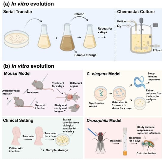
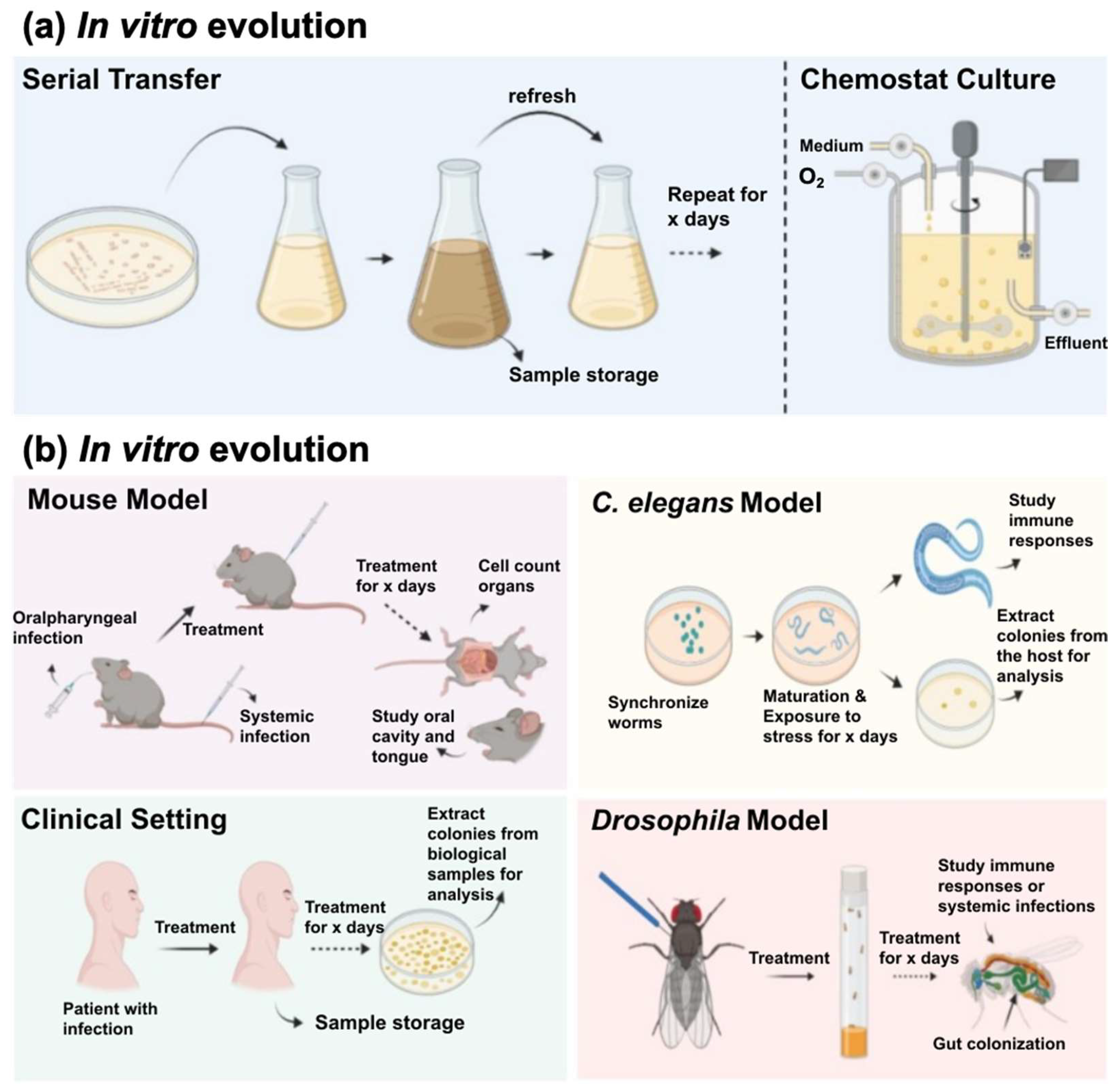
Disclaimer/Publisher’s Note: The statements, opinions and data contained in all publications are solely those of the individual author(s) and contributor(s) and not of MDPI and/or the editor(s). MDPI and/or the editor(s) disclaim responsibility for any injury to people or property resulting from any ideas, methods, instructions or products referred to in the content. |
© 2026 by the authors. Licensee MDPI, Basel, Switzerland. This article is an open access article distributed under the terms and conditions of the Creative Commons Attribution (CC BY) license.