


Abstract
Porcine reproductive and respiratory syndrome (PRRS) remains one of the most economically devastating diseases of the global swine industry, yet data on circulating PRRS virus (PRRSV) variants in Russia remain limited. This study aimed to isolate PRRSV from clinical specimens collected on six production sites in the Central Federal District of the Russian Federation, characterize the isolates molecularly and phylogenetically, and evaluate the pathogenicity of one isolate in an experimental piglet model. Virus isolation was performed on primary porcine alveolar macrophage (PAM) and porcine leukocyte (PL) cultures, with parallel assessment of the MARC-145 cell line. Pathogenicity was assessed in six 8-week-old piglets following combined intranasal and intramuscular inoculation, without a sham-inoculated control group. Five PRRSV type 1, subtype 2 isolates forming a monophyletic cluster within lineage L2 were recovered (GenBank accession numbers QB180257, QB180263, QB180264, QB180265, and QB180286). All five isolates replicated efficiently in PAM and PL cultures, producing a pronounced cytopathic effect, whereas no evidence of productive replication was detected in the MARC-145 cell line under the conditions tested. Experimental infection induced a moderate, non-fatal disease characterized by fever, apathy, and tremor; clinical signs resolved within the 21-day observation period, during which PRRSV RNA remained detectable in blood. The most severe histopathological lesions were observed in animals necropsied at 14 days post-inoculation. These findings provide an initial characterization of contemporary Russian PRRSV-1 subtype 2 isolates and support the use of primary macrophage-based cultures for field virus isolation, while underscoring the need for continuous molecular-epizootiological surveillance in Russia.
1. Introduction
Porcine reproductive and respiratory syndrome (PRRS) continues to rank among the most economically consequential and difficult-to-eradicate diseases affecting the global and Russian commercial swine industries. Financial losses associated with PRRS outbreaks are estimated in the hundreds of millions of dollars annually, borne by pork producers worldwide [1,2].
The causative agent, PRRS virus (PRRSV), is an enveloped, single-stranded, positive-sense RNA virus classified within the family Arteriviridae, genus Betaarterivirus [3,4]. Two principal clinical presentations are recognized: a respiratory syndrome in nursery and growing pigs and reproductive failure in gestating sows [5]. In vivo, the cellular tropism of PRRSV is largely restricted to cells of the mononuclear phagocyte system. Viral replication occurs predominantly in macrophage subpopulations residing in pulmonary tissue, the placenta, and lymphoid organs, with porcine alveolar macrophages (PAMs) serving as the primary target cell [6].
A defining feature of PRRSV that complicates eradication efforts is its exceptionally high genetic variability and evolutionary rate [7]. Since its emergence, the virus has diverged into two principal species (type 1 and type 2), each further subdivided into numerous genetic lineages and subtypes. This heterogeneity carries direct clinical significance. In recent decades, devastating outbreaks caused by newly emerged highly pathogenic variants—such as HP-PRRSV in Asia and virulent type 1 strains of the Lena-like cluster in Europe—have been documented [8,9]. Such pronounced genetic drift means that commercial vaccines based on “classical” strains often confer only partial cross-protection against contemporary field isolates, resulting in vaccine-breakthrough infections [10].
The high economic burden of PRRS necessitates continuous monitoring of the molecular-biological properties of the circulating virus. While the reduced permissiveness of continuous cell lines (such as MARC-145) to certain field PRRSV-1 isolates and the superior utility of primary alveolar macrophages for virus recovery are well-established concepts in virology [11,12], there remains a significant knowledge gap regarding the genetic diversity, biological properties, and pathogenicity of contemporary PRRSV variants circulating in the Russian Federation. Building an effective, science-based containment strategy requires the comprehensive characterization of these domestically circulating isolates. Although obtaining purified virus stocks from field samples presents methodological challenges related to cell system selection, it remains an obligatory prerequisite for subsequent experimental pathogenicity assessment and robust phylogenetic reconstruction.
Therefore, the primary novelty of the present study lies in the recovery and characterization of contemporary Russian PRRSV isolates, rather than the general demonstration of cell culture permissiveness. Specifically, this study aimed to isolate PRRSV from clinical specimens collected in the Central Federal District of the Russian Federation, perform molecular-genetic typing and phylogenetic analysis to determine their evolutionary positioning, and evaluate their pathogenic potential under experimental conditions in piglets. By providing these regional data, this work addresses a critical gap in the global molecular epidemiology of PRRSV-1.
2. Materials and Methods
2.1. Biological Material
Clinical specimens (whole blood and organ suspensions of tonsils, lungs, and lymph nodes) were obtained from pigs on six production sites (designated farms 1, 6, 7, 9, 10, and 13) in the Central Federal District of the Russian Federation where respiratory signs and reproductive disorders had been registered. A total of 15 specimens originating from 15 animals (sows and piglets aged 0–1 month; Table S1) were collected in February 2025 during an outbreak investigation. The vaccination status of the source herds against PRRSV was not documented at the time of specimen collection; consequently, it remains unknown whether the sampled animals had received PRRSV vaccination, and the potential influence of vaccine-induced immunity on viral shedding and isolation success cannot be assessed. This limitation is acknowledged in Section 4.4. Each specimen was frozen once at −20 °C and shipped to the laboratory in March 2025; no specimen underwent more than one freeze–thaw cycle before inoculation. The isolate nomenclature follows the format PRRSV/year/farm number/sample number (e.g., PRRSV/2025/1/2 indicates the virus isolated in 2025 from farm 1, specimen 2). Specimen-level metadata, including animal group, specimen type, original Ct values, storage history, and isolation outcomes, are provided in Table S1.
2.2. Virus Isolation and Cultivation
RT-qPCR-positive specimens were homogenized, centrifuged at 3000× g for 10 min, and the supernatant was filtered through 0.22 µm membrane filters before inoculation. For primary isolation, filtered specimens were inoculated in triplicate wells (0.5 cm3 per well) of 12-well plates containing PAM monolayers. To characterize the susceptibility of PL and MARC-145 cultures under the conditions tested, the five PAM-recovered isolates and the five corresponding original specimens were inoculated onto PL and MARC-145 monolayers; in addition, PL and MARC-145 cultures were inoculated with PAM passage-1 virus-containing material to assess propagation of PAM-recovered isolates (Table S3). Because not all RT-qPCR-positive specimens were inoculated in parallel onto all three cell systems, no quantitative comparison of primary-isolation sensitivity across the three substrates is made, and the observations for PL and MARC-145 cultures apply only to the specimens, isolates, and conditions actually tested. Inoculation volumes were 0.5 cm3 for 12- and 48-well plates and 1.0 cm3 for T25 flasks. Adsorption was performed for 60 min at (37.0 ± 0.5) °C, after which the inoculum was removed, monolayers were washed with serum-free medium, and maintenance medium containing 2% fetal bovine serum was added. Cultures were maintained at (37.0 ± 0.5) °C in a humidified atmosphere with 5% CO2 and examined daily by light microscopy; at the end of each 5–7-day passage interval, cultures were subjected to one freeze–thaw cycle before the next passage. Successful primary isolation was defined by the presence of a characteristic cytopathic effect (CPE) combined with a decrease in Ct values by RT-qPCR relative to the original material. For each cell culture system and each passage, uninfected control cultures were maintained in parallel and remained negative throughout the study. Infectious virus titers were determined by endpoint dilution (10−1–10−6) in 48-well plates with four wells per dilution in three independent experiments and expressed as TCID50/cm3 calculated by the Reed–Muench method; the endpoint was a CPE confirmed by immunocytochemistry. The lower limit of detection of the titration was 10^0.5 TCID50/cm3; variability across the three independent experiments is reported as the range (Tables S2 and S6).
Cell line characterization and quality control. The MARC-145 cell line (a continuous subclone of MA-104, African green monkey kidney) was obtained from the institutional cell culture collection, catalogue no. 59. Cells were used at passage 5 following recovery from cryopreservation; the prior passage history before cryopreservation was not documented. Cell viability upon thawing exceeded 90%, as determined by trypan blue exclusion. Mycoplasma testing of the MARC-145 stock was not performed; primary PAM and PL cultures used in parallel experiments were mycoplasma-negative by routine screening. The absence of productive replication in MARC-145 therefore cannot be attributed to cell-line contamination, but a formal QC of the stock remains a limitation (Section 4.4, item 8). The receptor phenotype (CD163, CD169 expression) of the MARC-145 stock was not characterized in this study. Authentication of the cell line by short-tandem-repeat (STR) profiling was not performed. Inoculation was performed using ten-fold serial dilutions (10−1–10−5) of virus-containing material, with adsorption for 60 min at (37.0 ± 0.5) °C. For each passage, matched PAM and PL cultures inoculated with the same inoculum served as positive biological controls to verify inoculum viability. All assays were performed in biological triplicate where applicable.
To prevent cross-contamination, each isolate was processed in separate wells and passage series with dedicated reagent sets, and uninfected control cultures for every cell system and passage remained negative throughout the study; the distinct identity of the five isolates was additionally confirmed by ORF5 sequencing performed directly on the original clinical specimens.
2.3. Preparation of Primary Cell Cultures
To minimize the confounding effect of donor-to-donor variability in primary cell susceptibility, a blocked experimental design was employed: within each passage or experimental batch, all isolates were tested in parallel on cells derived from a single donor. This matched design ensured that any observed differences in viral replication kinetics, CPE development, or isolation success reflected intrinsic properties of the isolates rather than variability in donor cell permissiveness.
Porcine alveolar macrophages (PAMs). PAMs were harvested by bronchoalveolar lavage from six individual 4–6-week-old specific-pathogen-free (SPF) piglets, each used for a separate passage level across all isolates. Euthanasia was performed in accordance with the Guide for the Care and Use of Laboratory Animals and AVMA Guidelines. The heart–lung block was aseptically excised. Lungs were initially flushed with 100 cm3 of Eagle’s MEM supplemented with heparin (5 IU/cm3) and a double concentration of antibiotics (penicillin 100 IU/cm3, monomycin 100 µg/cm3, polymyxin 50 IU/cm3, amphotericin B). Subsequently, three 100 cm3 aliquots of MEM with antibiotics were instilled, the lavage fluid was collected, and cells were pelleted by centrifugation at 300–650× g for 10–15 min at 4 °C. The cell pellet was washed at least four times to remove debris. Cell suspensions from individual donors were prepared separately and not pooled. The final viable cell concentration was adjusted to 1.5–2.0 × 10⁶ cells/mL, with cell viability exceeding 90% as determined by trypan blue exclusion. Donor piglets used for PAM preparation were confirmed PRRSV-negative by RT-qPCR and ELISA prior to euthanasia; beyond SPF status, donor cells were not screened for additional pathogens (see Section 4.4, Limitations).
Porcine leukocytes (PLs). Peripheral blood was collected from six individual 6–8-week-old SPF piglets via cranial vena cava puncture into heparinized vacuum tubes at two-week intervals. Each blood collection provided cells for a single experimental batch in which all isolates were tested simultaneously on the same donor-derived culture. After incubation at (37.0 ± 0.5) °C for 45–60 min, the upper layer (plasma with leukocytes) was aspirated. Cells were resuspended in Eagle’s MEM supplemented with 5% fetal bovine serum, heparin (5 IU/cm3), and the antibiotic complex described above. The seeding concentration was adjusted to 3–6 × 10⁶ cells/cm3. PL cultures represented the total adherent cell fraction obtained from peripheral blood without further separation into subpopulations. Cultures were incubated at (37.0 ± 0.5) °C, and the medium was replaced at 24 and 72 h. Donor piglets used for PL preparation were confirmed PRRSV-negative by RT-qPCR and ELISA prior to blood collection; no FACS-based composition analysis of PL cultures was performed (see Section 4.4, Limitations).
2.4. RT-qPCR, ORF5 Amplification, and Sequencing
RT-qPCR for PRRSV RNA detection. Total RNA extraction and real-time reverse transcription quantitative PCR (RT-qPCR) were performed using the commercial PCR-PRRS-FAKTOR kit (VET FAKTOR LLC, Troitsk, Russia), which includes the DNA/RNA-C-FACTOR extraction reagent kit (catalog number EDR001, VET FAKTOR LLC, Troitsk, Russia) for nucleic acid isolation. Briefly, total RNA was extracted from 100 µL of homogenized tissue suspension, serum, or culture supernatant using the DNA/RNA-C-FACTOR kit according to the manufacturer’s instructions, with a final elution volume of 50 µL. Amplification was performed on a CFX Touch Real-Time PCR Detection System (Bio-Rad Laboratories, Hercules, CA, USA) under the following cycling conditions: reverse transcription at 50 °C for 15 min, initial denaturation at 95 °C for 5 min, followed by 45 cycles of 95 °C for 15 s and 60 °C for 60 s (fluorescence acquisition in the FAM/Green channel). Each run included a no-template negative control, a kit-supplied positive control (inactivated PRRSV), and an internal extraction control (supplied with the kit) to monitor extraction efficiency and detect PCR inhibition. Three result categories were applied. (i) Validated positive: A characteristic sigmoidal amplification curve with a Ct value ≤ 35 at 45 total cycles together with valid internal extraction and amplification controls; this threshold follows the manufacturer’s instruction for routine veterinary diagnostics, where it is set to exclude false-positive late-cycle artifacts in samples of unknown status. (ii) Weak positive (equivocal): A sigmoidal amplification curve with Ct 35–40 without noise and with a discernible exponential phase, valid internal controls excluding inhibition, and reproducibility upon repeat testing; this category was applied only to samples from the experimentally infected piglets, i.e., animals of known infection status, in which low-level viremia near the limit of detection of the assay is expected and legitimately yields late-cycle amplification. (iii) Negative: Ct > 40, undetermined Ct, or discordant internal-control signals; such runs were considered invalid and the corresponding specimens were re-extracted and re-tested. Weak-positive results were interpreted descriptively and were not used as the basis for quantitative or temporal claims. The analytical sensitivity (limit of detection, LOD) declared by the manufacturer is 102 genome equivalents per mL. Results with discordant internal-control signals were considered invalid, and the corresponding specimens were re-extracted and re-tested.
ORF5 amplification, sequencing, and phylogenetic analysis. For phylogenetic characterization, the ORF5 gene fragment was amplified from the original clinical specimens corresponding to the five recovered isolates using conventional RT-PCR according to the WOAH protocol [13]. The amplification yielded an 848-bp product corresponding to genomic positions 13348–14196 of the PRRSV-1 reference strain Lelystad virus (GenBank accession no. M96262), using the primer pair forward 5′-TTCACAGATTATGTGGCCCATGTGAC-3′ and reverse 5′-CGYGACACCTTRAGGGCRTATATCAT-3′. Amplicons were resolved in 1.5% agarose gels containing 0.001% ethidium bromide in Tris-acetate buffer at 110 mA for 40–60 min and visualized on a GelDocXR imaging system (Bio-Rad Laboratories, Hercules, CA, USA). Amplification success was confirmed by comparing the size of the observed band in the sample lane with that of a DNA molecular-weight marker; a positive result was defined as a visible band migrating at the expected size corresponding to the calculated amplicon length.
Amplicons were purified from agarose gels using the MagPure Universal DNA Kit (MagenBiotechnology, Guangzhou, China) and subjected to bidirectional Sanger sequencing with BigDye Terminator v3.1 (Applied Biosystems, Foster City, CA, USA) under the following cycling conditions: 95 °C for 10 s, 50 °C for 20 s, and 60 °C for 4 min (25 cycles), using forward and reverse primers separately. Post-sequencing cleanup was performed with the iX Pure kit (NimaGen, Nijmegen, The Netherlands). Sequencing reactions (20 µL) were run on a Nanofor-05 capillary sequencer (DNA-Technology, Moscow, Russia) according to the manufacturer’s instructions.
Sequencing was performed directly on the original clinical specimens from which the five isolates were recovered; therefore, culture-adaptive mutations were excluded from the deposited sequences. Chromatograms were evaluated against predefined quality criteria: Phred quality score ≥ 30 across the ORF5 coding region, unambiguous peaks, and absence of “N” base calls in the core region. Consensus sequences were generated by assembling forward and reverse reads in BioEdit v7 with manual inspection of terminal discrepancies.
2.5. Phylogenetic Analysis
Nucleotide sequences were aligned using the MUSCLE algorithm implemented in MEGA12 (version 12.0.4); the final alignment comprised 510 nucleotide positions corresponding to the ORF5 coding region. The best-fit nucleotide substitution model was determined with the “Find Best DNA/Protein Models” feature of MEGA12 on the basis of the Bayesian Information Criterion and was identified as the General Time Reversible model with gamma-distributed rate variation and a proportion of invariant sites (GTR+G+I). Phylogenetic trees were reconstructed by the maximum-likelihood method with 1000 bootstrap replicates; gaps and missing data were treated by complete deletion. The tree was rooted with the PRRSV-2 strain VR-2332 (GenBank accession no. U87392.3) as an outgroup, and branch lengths are measured in substitutions per site. Assignment of the isolates to PRRSV-1 subtype 2 and lineage L2 was based on monophyletic clustering with established reference strains, with bootstrap support ≥ 70% at the terminal cluster level; support values at deeper lineage-defining nodes were lower and are reported in Section 3.2. This assignment follows the PRRSV-1 genetic classification schemes by Yim-Im et al. [14] and Stadejek et al. [15]. Reference sequences were retrieved from the GenBank database on 19 October 2026 using nucleotide BLAST2.17.0 with a similarity threshold > 85%; geographic and temporal coverage was restricted to contemporary strains (2000–2026) from Russia and neighboring countries, and known vaccine and laboratory-adapted strains were explicitly excluded. Near-identical sequences (>99.9% identity) from the same farm and year were collapsed into a single representative.
The ORF5 nucleotide sequences of the five isolates were deposited in the GenBank database under accession numbers QB180264 (PRRSV/2025/1/2), QB180263 (PRRSV/2025/1/3), QB180265 (PRRSV/2025/6/5), QB180286 (PRRSV/2025/10/11), and QB180257 (PRRSV/2025/13/10).
2.6. Immunocytochemistry (ICC)
Ten-fold serial dilutions (10−1–10−6) of virus-containing material in serum-free Eagle’s MEM were inoculated onto MARC-145 or PAM monolayers grown in 48-well culture plates (four wells per dilution). Plates were incubated at (37.0 ± 0.5) °C for 60 min, after which the inoculum was removed and replaced with Eagle’s MEM containing 2% fetal bovine serum. Upon CPE appearance in the lower dilutions (10−1–10−3), plates were dried at (20 ± 2) °C for 60 min, and cells were fixed with 30% acetone for 15 min.
After drying, an indirect immunocytochemical method was employed. Wells were incubated for 60 min at (37.0 ± 0.5) °C with an in-house-produced polyclonal swine anti-PRRSV antiserum diluted 1:40 in PBS. Following three washes with PBS containing Tween 80, the wells were incubated for 30 min at (37.0 ± 0.5) °C with a peroxidase-conjugated anti-swine IgG. After three additional washes, the substrate solution (3-amino-9-ethylcarbazole + N,N-dimethylformamide + 0.05 M sodium acetate) was added for 15 min, and results were recorded.
Results were evaluated qualitatively using brightfield microscopy on a ZOE cell imager (Bio-Rad, USA) by assessing the presence or absence of specific cytoplasmic staining. Each assay included (i) a positive control consisting of PAM cultures infected with a PRRSV vaccine strain, and (ii) a negative control consisting of uninfected PAM (or MARC-145) cultures processed with the complete antiserum–conjugate cocktail to exclude non-specific binding. A secondary antibody-only control was not included in the assay; consequently, non-specific binding of the conjugate itself cannot be formally excluded, although the absence of staining in the uninfected negative control argues against substantial background under the conditions used. Staining was scored qualitatively; no quantitative counting of antigen-positive cells was performed.
2.7. Histopathology
Tissue samples were collected from the following organs at necropsy: mandibular and inguinal lymph nodes, tonsils, lungs, thymus, and kidneys. For organs with visible gross lesions, samples were taken from the affected areas; for organs without apparent lesions, representative sections were collected from standardized anatomical locations. A minimum of three to four tissue sections per organ were prepared from each animal.
Tissue samples were fixed in 10% neutral buffered formalin, processed through a standard histological tissue processor, embedded in paraffin, and sectioned at 4–5 µm thickness on a rotary microtome. Sections were stained with hematoxylin and eosin (H&E) for light-microscopic examination.
Histopathological assessment was performed using light microscopy Olympus BX53(Evident Corporation, formerly Olympus Corporation, Tokyo, Japan) in a descriptive format without application of a formal scoring system. The nature and severity of lesions were evaluated according to standard morphological criteria, including presence and degree of interstitial pneumonia, extent of lymphoid infiltration, presence of hemorrhages, and dystrophic or destructive changes in parenchymal organs. Histopathological assessment was not performed in a blinded manner, as the examiner was aware of the necropsy time point for each animal (see Section 4.4, Limitations).
2.8. Experimental Infection of Piglets
The isolate PRRSV/2025/1/2, exhibiting the highest infectious titer (5.00-5.25 lg TCID50/cm3) and lowest Ct, was selected for pathogenicity assessment. Six clinically healthy 8-week-old male piglets (Large White breed) weighing 18–20 kg were obtained from the Experimental Animal Preparation Unit of the Federal Research Center for Virology and Microbiology (FRCVM). The SPF colony is maintained under controlled biosecurity conditions with regular health monitoring. Prior to the experiment, each piglet was tested for the absence of PRRSV, African swine fever virus (ASFV), classical swine fever virus (CSFV), and porcine circovirus type 2 (PCV2) on days −7, −3, and 0 (day of inoculation). PRRSV status was assessed by RT-qPCR and ELISA; ASFV, CSFV, and PCV2 were tested by RT-qPCR/PCR. All animals tested negative for all four pathogens at all three time points. No broader pathogen-exclusion panel (e.g., swine influenza virus, Mycoplasma hyopneumoniae, porcine parvovirus) was applied, and this limitation is acknowledged in Section 4.4.
Piglets were housed in individual pens in a temperature-controlled facility (22–24 °C) with a 12-h light/dark cycle and had ad libitum access to commercial pig feed and water. Animals were randomly allocated to three euthanasia time points (days 7, 14, and 21 post-inoculation; two piglets per time point).
Animals were inoculated with a third-passage virus suspension with a nominal concentration of 4.0 lg TCID50/mL. Each piglet received a total volume of 5 mL, comprising 4 mL delivered intranasally (equivalent to a dose of 4.6 lg TCID50) and 1 mL delivered intramuscularly (equivalent to a dose of 4.0 lg TCID50), resulting in a total infectious dose of approximately 4.7 lg TCID50 per animal. The actual infectious titer of the inoculum was confirmed by back-titration of the residual suspension, which yielded 4.0 lg TCID50/mL, consistent with the nominal concentration.
The combined intranasal and intramuscular route was selected to balance biological relevance with experimental reliability in this small exploratory study: intranasal inoculation models the natural oropharyngeal exposure route, while the intramuscular component serves to guarantee systemic infection across all animals in the absence of a sham-inoculated control group, thereby minimizing the risk that individual variability in mucosal absorption would result in uninfected animals.
Animals were monitored daily for 21 days. Rectal temperature was measured daily, and fever was defined as a rectal temperature ≥ 40.0 °C. Clinical signs (apathy and reduced appetite, muscular tremor and other CNS signs, respiratory distress, skin lesions) were recorded daily per animal as present/absent, with the duration and severity of each episode noted on visual examination; feed intake was not measured quantitatively, and refusal of feed was recorded as part of the clinical examination; serial weighing was not performed. No composite clinical score was applied; individual daily data are provided in Tables S4–S6. Blood samples for RT-qPCR and virus titration were collected on days 3, 7, 10, 14, and 21 post-inoculation.
Euthanasia on days 7, 14, and 21 post-inoculation was performed using a captive bolt pistol (Stunner) followed by exsanguination, in accordance with AVMA Guidelines. Predefined humane endpoints were established to prevent unnecessary suffering and included any of the following criteria: (i) rectal temperature exceeding 41.5 °C persisting for more than 48 h despite symptomatic assessment; (ii) complete refusal of feed for more than 72 h; (iii) severe depression or apathy, unresponsive to external stimuli; (iv) marked dyspnea, tachypnea at rest, or cyanosis of the skin and mucous membranes; (v) inability to stand or move; or (vi) any clinical signs indicative of secondary bacterial infection or sepsis. Animals were assessed daily by unblinded clinical observers for the presence of these criteria; had any endpoint been reached, the affected animal would have been euthanized immediately outside the scheduled time points. No early humane endpoints were triggered during the study, and all animals survived to their scheduled euthanasia. Clinical observers and pathologists were not blinded to the inoculation status, as all animals belonged to a single infected group; histopathological assessment was also performed without blinding (see Section 4.4).
2.9. Ethical Statement
All animal procedures were conducted in compliance with the Guide for the Care and Use of Laboratory Animals and AVMA Guidelines on euthanasia. Two separate ethical approvals were obtained from the Ethics Committee of the Federal Research Center for Virology and Microbiology: (i) Protocol No. 1, approved on 20 January 2025, covering the collection of clinical specimens from pigs on commercial farms for virus isolation purposes, and (ii) Protocol Code 2, approved on 9 February 2026, covering the experimental infection of piglets for pathogenicity assessment. Donor piglets for PAM and PL preparation performed in 2025 for virus isolation were covered by Protocol No. 1 (20 January 2025), which also authorized laboratory animal procedures for isolation purposes; donor piglets used for blood titration during the 2026 challenge study were covered by Protocol Code 2 (9 February 2026).
2.10. Statistical Analysis
Given the exploratory design and the small numbers of animals (n = 6; two per necropsy time point) and cell donors, all data are presented descriptively as individual values, ranges, and means ± standard deviation where applicable; no inferential statistical testing was performed. The experimental unit was the individual animal for in vivo data and the individual donor cell culture (well or flask) for in vitro data. Biological and technical replicates for in vitro experiments are specified in Section 2.2 (triplicate wells for primary isolation; four wells per dilution in three independent experiments for titrations). Repeated measurements (daily temperatures, serial Ct values) are reported per animal or per specimen without aggregation. Non-detectable RT-qPCR results are reported as negative and were not imputed.
3. Results
3.1. Virus Isolation in Primary PAM Cultures
Of 15 clinical specimens screened by RT-qPCR, 14 tested positive for PRRSV RNA, with threshold cycle (Ct) values ranging from 16.53 to 34.67 (Table S1); the single RT-qPCR-negative specimen (no. 13) was not inoculated into cell cultures. Specimens with high Ct values approaching the threshold line may have been compromised by inhibitors, suboptimal transport or storage conditions, or freeze–thaw cycles; these possible explanations were not experimentally tested.
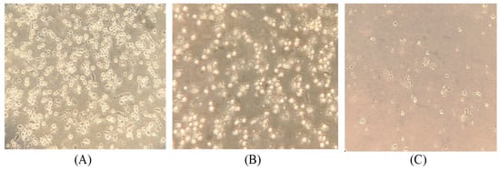
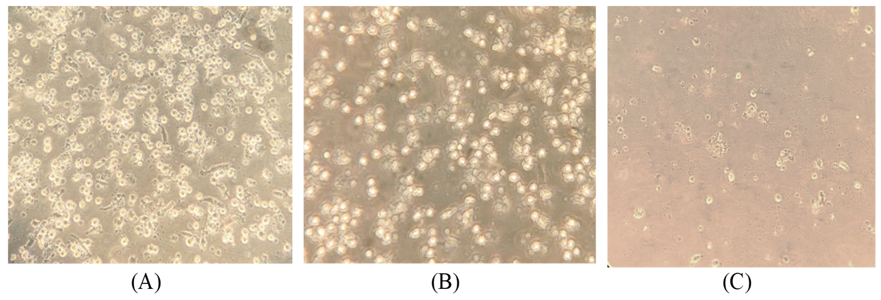
All RT-qPCR-positive specimens were inoculated onto primary PAM cultures in triplicate wells. After three sequential passages, five PRRSV isolates were recovered from specimen no. 2, 3, 5, 10, and 11 (all whole blood), corresponding to PRRSV/2025/1/2, PRRSV/2025/1/3, PRRSV/2025/6/5, PRRSV/2025/13/10, and PRRSV/2025/10/11, respectively. Isolation success was not determined by the original Ct value alone: successful specimens had original Ct values of 17.11–20.58, whereas PCR-positive specimens that did not yield virus spanned Ct 16.53–34.67 (Table S2). By specimen type, isolates were recovered from 5 of 9 blood specimens, whereas none of the five RT-qPCR-positive organ suspensions produced virus. Viral replication was accompanied by CPEs evident from passage 1–2. Initial CPE signs—rounding of infected macrophages followed by detachment from the substrate—appeared at 36–48 h post-inoculation; by 72–96 h, extensive monolayer destruction was observed (Figure 1).
Figure 1.
Cytopathic effect of PRRSV isolate PRRSV/2025/1/2 in primary PAM culture (passage 1; cells obtained from a single SPF donor piglet). (A) Intact control PAM monolayer; (B) PAM culture 48 h post-inoculation, showing rounding and partial detachment of infected macrophages; and (C) PAM culture 72 h post-inoculation, showing extensive monolayer destruction. Light microscopy, magnification ×200. Representative fields were selected from triplicate wells; images are representative of the CPEs observed across all five isolates. Inoculum: original specimen filtrate.
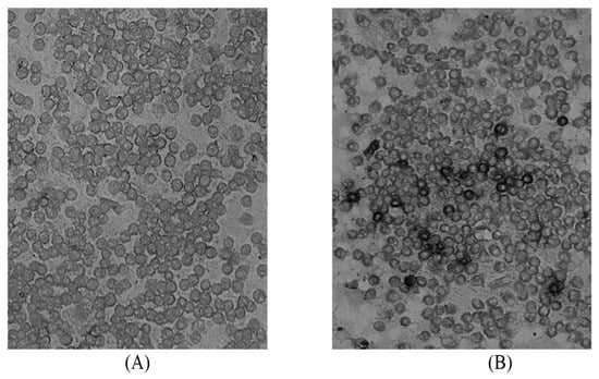
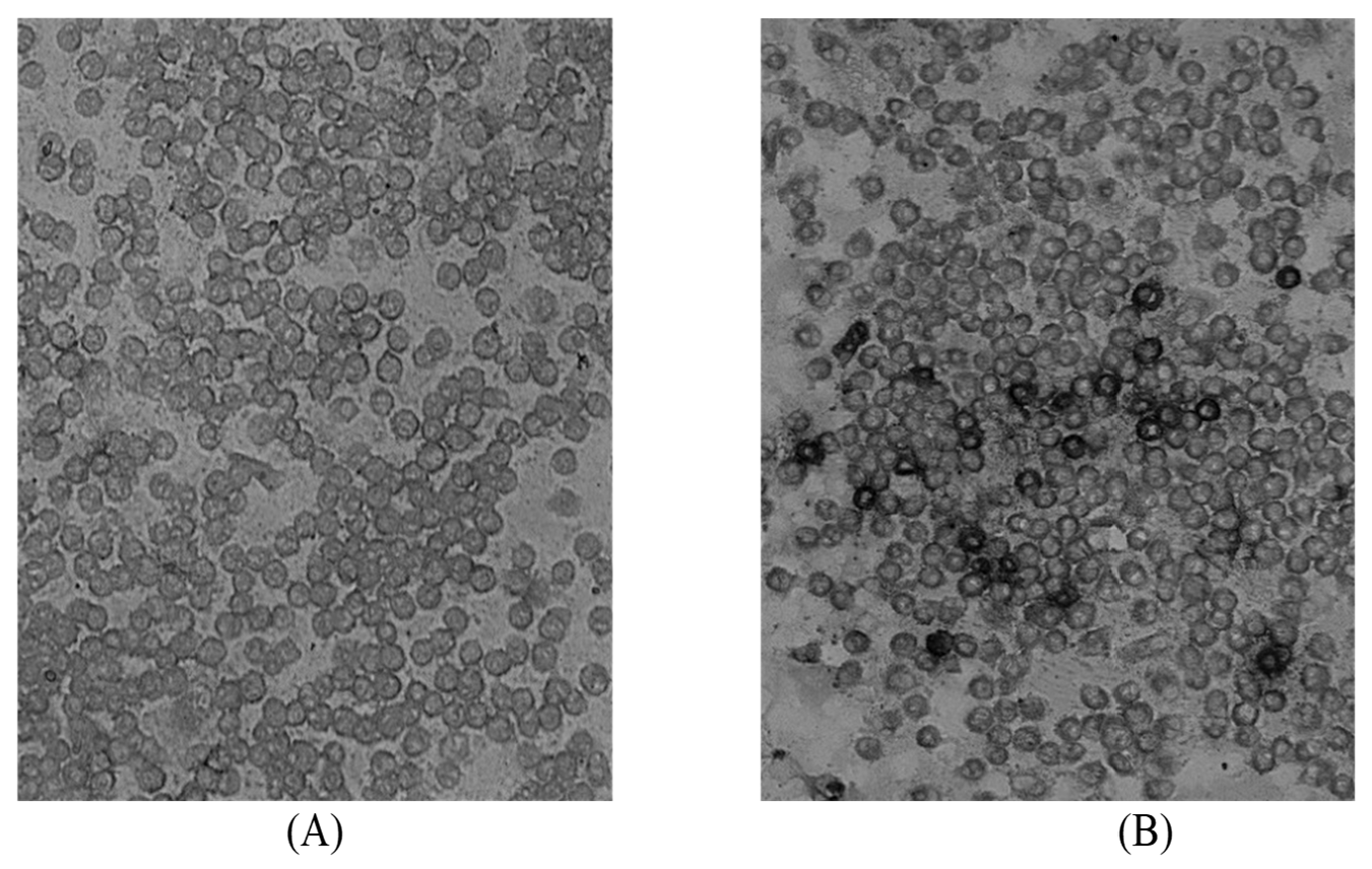
Virus replication was confirmed by RT-qPCR: Ct values decreased by a mean of 6.9 cycles (range 4.3–9.6) between the original material and passage 1 and by a mean of 4.9 cycles (range 1.0–8.6) between passage 1 and passage 3 (Table S2), yielding passage-3 titers of 4.75–5.25 lg TCID50/cm3 (Table S2). Because no quantitative standard curve was used, these Ct shifts are reported as descriptive indicators of viral replication rather than absolute viral-load measurements. ICC staining of infected PAM cultures revealed specific cytoplasmic labeling, confirming the presence of viral antigens (Figure 2).
Figure 2.
Immunocytochemical detection of PRRSV in primary PAM culture inoculated with isolate PRRSV/2025/1/2 (passage 1; cells from a single SPF donor piglet). (A) Uninfected control monolayer processed with polyclonal swine anti-PRRSV antiserum and peroxidase-conjugated anti-swine IgG (negative control); (B) infected monolayer at 48 h post-inoculation showing specific cytoplasmic staining (AEC chromogen, dark brown-to-black precipitate (grayscale images)). Indirect ICC method: in-house polyclonal swine anti-PRRSV serum (1:40), followed by peroxidase-conjugated anti-swine IgG. Brightfield microscopy, ZOE cell imaging system (Bio-Rad, USA), magnification ×200. Results were evaluated qualitatively by assessing the presence or absence of specific cytoplasmic staining.
3.2. Molecular-Genetic and Phylogenetic Characterization
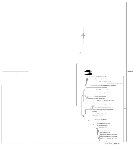
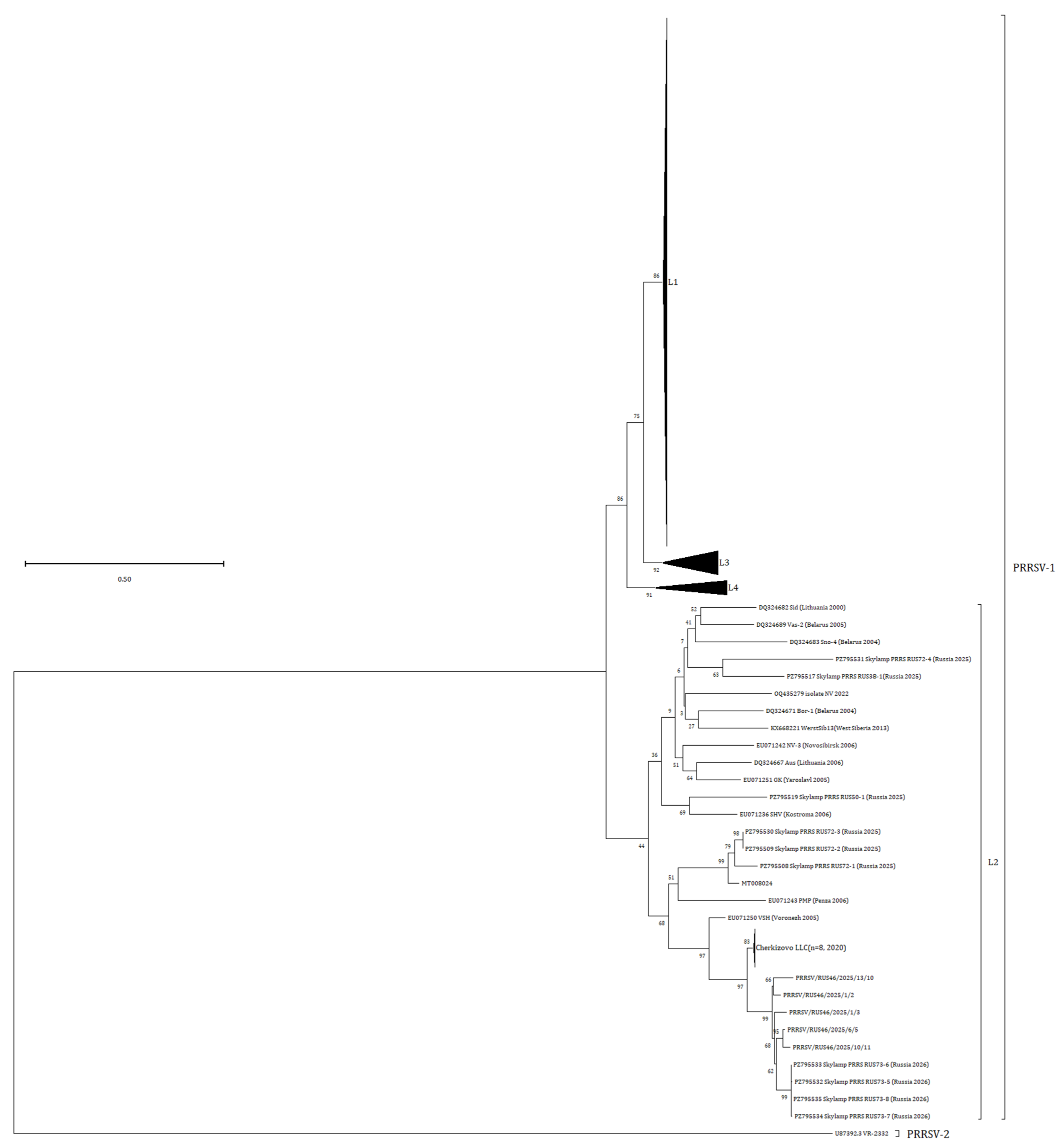
ORF5 sequencing and phylogenetic analysis assigned all five isolates to PRRSV type 1, subtype 2 (Figure 3), in accordance with the classifications of Yim-Im et al. [14] and Stadejek et al. [15]. The isolates formed a strongly supported monophyletic cluster within lineage L2 (bootstrap 99%), distinct from an older, more widely distributed clade comprising sequences from Belarus, Lithuania, and various Russian regions. Within the studied-isolate clade, internal resolution was limited: only the pairing of PRRSV/2025/6/5 and PRRSV/2025/10/11 received robust bootstrap support (95%), whereas the remaining internal nodes (66–68%) fell below the 70% threshold adopted for cluster interpretation; accordingly, no stable sub-cluster structure was resolved, which is consistent with the limited phylogenetic resolution of ORF5 at this genetic depth. A recent local expansion is one possible explanation of the poorly resolved internal topology but remains an untested possibility that cannot be distinguished from alternative scenarios on the basis of these data. Support for the node joining the studied-isolate cluster with the sister clade of contemporary Russian strains (PZ795532–PZ795535, Russia 2026) was moderate (62%), and support at the basal node of lineage L2 was low (44%). The weak support at deep nodes is consistent with the known limited phylogenetic resolution of the ORF5 fragment for ancient divergences; accordingly, subtype and lineage assignment relied on monophyletic clustering with established reference strains rather than on deep-node bootstrap values. This pattern is compatible with epidemiological linkage and limited local ORF5 diversification; inter-farm spread and introduction history cannot be confirmed from ORF5 data alone and require whole-genome sequencing and farm-level epidemiological documentation.
Figure 3.
Maximum-likelihood phylogenetic tree based on ORF5 nucleotide sequences (final alignment length 510 nt; General Time Reversible model with gamma-distributed rate variation and a proportion of invariant sites [GTR+G+I]; 1000 bootstrap replicates). The tree is rooted with the PRRSV-2 strain VR-2332 (GenBank accession no. U87392.3) as an outgroup; branch lengths are drawn to scale in substitutions per site (scale bar = 0.50). Bootstrap values are shown at the nodes (no cutoff applied). Bootstrap support for the studied-isolate clade was 99%; within this clade, only the PRRSV/2025/6/5 + PRRSV/2025/10/11 pairing received robust support (95%), whereas the remaining internal nodes (66–68%) fell below the 70% threshold. Support for the node uniting the studied isolates with the sister clade of Russian 2026 strains (PZ795532–PZ795535) was 62%, and support at the basal node of lineage L2 was 44%. The five studied isolates are indicated bold by their isolate names with GenBank accession numbers PRRSV/2025/1/2 (QB180264), PRRSV/2025/1/3 (QB180263), PRRSV/2025/6/5 (QB180265), PRRSV/2025/10/11 (QB180286), and PRRSV/2025/13/10 (QB180257). Collapsed reference lineages L1, L3, and L4 are shown as filled triangles; the collapsed cluster of near-identical sequences from Cherkizovo LLC (2020; n = 8) is shown as a labeled bar. Reference sequences were retrieved from GenBank on 19 October 2026 (BLAST similarity > 85%; strains from 2000–2026 originating from Russia and neighboring countries; vaccine and laboratory-adapted strains excluded); labels indicate country/region and year of collection.
Pairwise ORF5 nucleotide identity was 94.12–98.14% among the five isolates and 93.54–95.87% to the closest reference sequence (GenBank accession no. PZ795533.1), which falls within the sister clade of Russian 2026 strains. The ORF5 nucleotide sequences of the five isolates have been deposited in the GenBank database under the following accession numbers: PRRSV/2025/1/2 (QB180264), PRRSV/2025/1/3 (QB180263), PRRSV/2025/6/5 (QB180265), PRRSV/2025/10/11 (QB180286), and PRRSV/2025/13/10 (QB180257).
3.3. Cultivation of Isolates in Primary Porcine Leukocyte Cultures
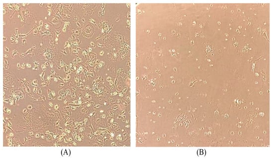
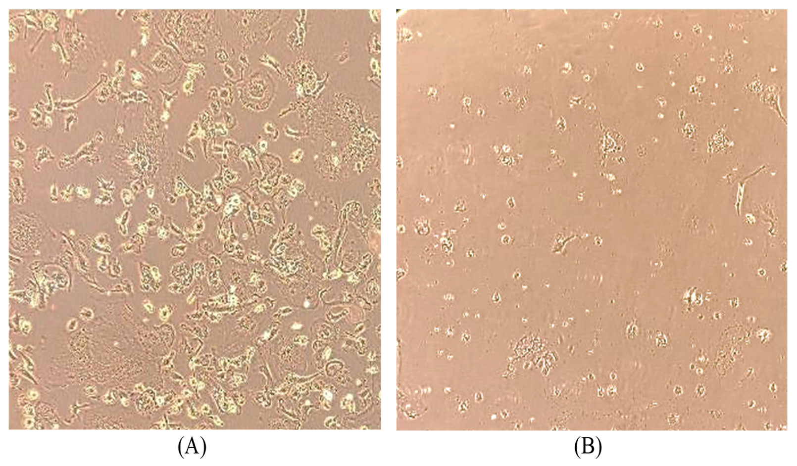
To determine whether PL cultures support primary isolation as well as propagation of PAM-recovered isolates, PL monolayers were inoculated both with the original filtered specimens and with PAM passage-1 virus-containing material (dilutions 10−1–10−2). Cultivation was carried out over 96–120 h per passage across three sequential passages. With PAM-derived inocula, all five isolates produced CPEs commencing at 36–48 h (rounding of infected cells, loss of adhesion, detachment); by 72–96 h, extensive monolayer destruction affecting up to 90% of cells was observed by visual estimation of the destroyed monolayer area (no quantitative scoring was applied) (Figure 4). RT-qPCR confirmed replication: Ct values decreased from 12.53–18.11 in PAM passage-1 inocula to 8.55–12.44 at PL passage 3. When original specimens were used as inoculum, CPEs and Ct decreases (from 20.71–25.34 in the inoculum to 10.13–15.12 at passage 3) demonstrated that PL cultures also support primary isolation from clinical material for the five blood specimens tested (Table S3).
Figure 4.
Cytopathic effect of PRRSV isolate PRRSV/2025/1/2 in primary PL culture (passage 1; cells obtained from a single SPF donor piglet, total adherent fraction without subpopulation separation). (A) Intact control PL monolayer; (B) PL culture 72 h post-inoculation, showing rounding, loss of adhesion, and detachment of infected cells affecting up to 90% of the monolayer area (visual estimation; no quantitative scoring was applied). Light microscopy, magnification ×200. Representative fields were selected from triplicate wells. Inoculum: PAM passage-1 virus-containing material (dilutions 10−1–10−2), adsorption 60 min at 37 °C.
3.4. Assessment of Susceptibility of the MARC-145 Continuous Cell Line
To assess the susceptibility of the continuous MARC-145 cell line to the isolated field strains, cultivation was performed over five sequential passages. A confluent monolayer of MARC-145 cells was inoculated with ten-fold serial dilutions of virus-containing material (10−1–10−5) and incubated in a CO2 incubator at (37 ± 0.5) °C for 7 days. The results demonstrated the absence of CPE development in MARC-145 cell cultures at all passage levels. Visual inspection revealed no characteristic morphological changes in the cells, such as rounding, cytoplasmic vacuolization, or detachment from the monolayer. RT-qPCR analysis revealed a progressive increase in Ct values of 3–5 cycles between sequential passages, with complete loss of detection by passages 4–5. Additional verification by immunocytochemistry (ICC) using PRRSV-specific antisera did not result in staining of the fixed cells. Thus, no evidence of productive replication of the field PRRSV isolates under investigation was detected in the continuous MARC-145 cell line under the conditions tested. These findings are consistent with published data on the reduced susceptibility of MARC-145 cells to European PRRSV type 1 strains. No MARC-145-permissive PRRSV positive control was included in these experiments; therefore, the absence of productive replication is reported for the tested cell stock and conditions only, and a qualitative block to replication cannot be firmly established (see Section 4.4).
3.5. Assessment of Pathogenic Properties of the PRRSV Isolate
Fever (rectal temperature ≥ 40.0 C, maximum 40.0–41.3 °C across animals) was recorded as early as day 1 post-inoculation in all six piglets, whereas reduced appetite and activity developed from days 4–6; given the absence of a sham-inoculated control group and the usual incubation period of PRRSV, these earliest observations cannot be confidently attributed to the virus and may partly reflect handling and adaptation stress. Muscular tremor was observed in two animals. The duration of clinical manifestations varied among animals (fever on 3–13 days and reduced appetite on 2–11 days, depending on the animal; Table S5); individual daily temperatures and clinical signs are provided in Tables S4 and S5.
PRRSV RNA in blood was detected from day 3 up to day 21 post-inoculation; all positive samples at days 3, 7, 10 and 21 were validated positives (Ct ≤ 35), with a single exception: the sample from piglet 4 at day 14 yielded a weak-positive result (Ct 37.24; sigmoidal curve above the routine diagnostic threshold with valid internal controls, confirmed upon repeat testing), which is reported as weak positive in Table S6 and was culture-negative. The lowest Ct values (23.08–26.30) were recorded on day 7; because no quantitative standard curve was used, results are reported as Ct values rather than viral-load equivalents. According to titration in primary PAM and PL cultures, infectious virus was recovered from blood between days 3 and 14 post-inoculation in at least one culture system in all six piglets (individual values in Table S6), with titers of 2.0–3.75 lg TCID50/cm3 and the highest values on days 7–10; at day 21, both remaining animals were RT-qPCR-positive with validated Ct values (30.42 and 28.04) but culture-negative. Individual animal values for each sampling day are provided in Table S6.
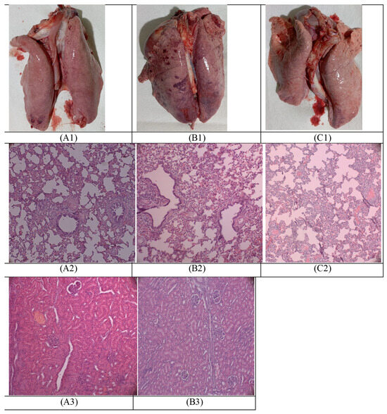
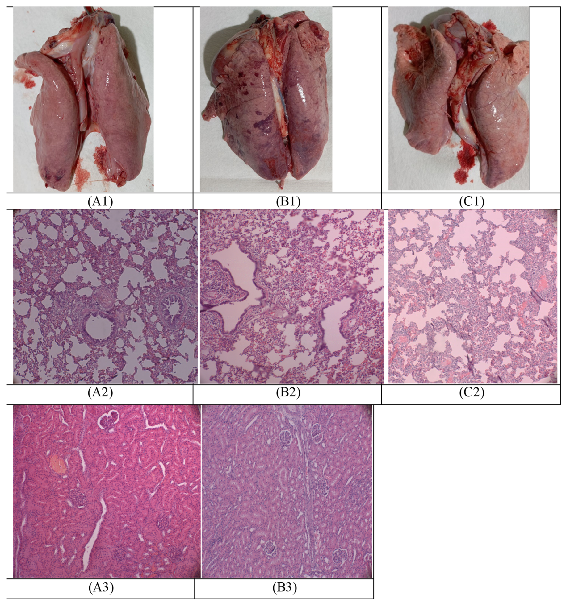
At necropsy, gross changes were registered in the lungs, inguinal lymph nodes, and kidneys. No gross or histological changes were observed in mandibular lymph nodes and tonsils in any animal. The lesions were most pronounced in the animals examined at 14 dpi (Figure 5(A1,B1,C1)). Histological examination of the inguinal lymph nodes revealed multiple petechial hemorrhages in the cortical parenchyma beneath the capsule. In the lungs, thickening of the interalveolar septa, congestive hyperemia, peribronchial lymphocytic infiltration (interstitial pneumonia), and atelectasis and dystelectasis were identified (Figure 5(A2,B2,C2)). In individual animals, marked depletion of thymic lobules, cellular depopulation, cortical depletion with predominance of the medullary zone, and formation of Hassall’s corpuscles were observed. In the kidneys, partial disruption of the renal corpuscle architecture in the cortical substance with apparent absence of the Bowman’s capsule was registered; because this finding may reflect sectioning or processing artifact, representative micrographs are provided (Figure 5(A3,B3)).
Figure 5.
Gross and histopathological changes in piglets experimentally infected with PRRSV isolate PRRSV/2025/1/2 at different time points post-inoculation (dpi). (A1,B1,C1) Gross appearance of the lungs at 7, 14, and 21 dpi, respectively, from piglets #1, #3, and #5. (A2,B2,C2) Histological examination of the lungs at 7, 14, and 21 dpi showing interstitial pneumonia with thickened interalveolar septa, peribronchial lymphocytic infiltration, and atelectasis. (A3,B3) Histology of the kidneys at 14 and 21 dpi from piglets #3 and #5, respectively, showing partial disruption of renal corpuscle architecture with apparent absence of Bowman’s capsule. Hematoxylin and eosin staining, magnification ×200. Each panel is representative of the findings observed in both animals necropsied at the corresponding time point (piglets #1–2 at 7 dpi, #3–4 at 14 dpi, #5–6 at 21 dpi). Lesion severity was qualitatively comparable between the two animals within each time point, although formal quantitative scoring was not applied (see Section 4.4, Limitations).
4. Discussion
The present study provides a preliminary characterization of five PRRSV-1 subtype 2 isolates circulating in the Central Federal District of the Russian Federation, encompassing virus isolation, molecular-genetic typing, phylogenetic reconstruction, and experimental pathogenicity assessment.
4.1. Cell System Dependency for Virus Isolation
The successful recovery of all five isolates in primary PAM and PL cultures, contrasted with the absence of detectable productive replication in the MARC-145 cell line over five sequential passages, corroborates and extends earlier observations on the limited permissiveness of this continuous cell line to European-type PRRSV-1 field strains [11,12,16]. PAMs remain the natural target cells for PRRSV in vivo and are reported in the literature to express the necessary entry receptors (e.g., CD163, Sn/CD169) [17]. Receptor expression was not measured in the present study and is invoked here solely as background interpretation of the observed susceptibility differences. The demonstrated efficacy of primary PL cultures as an alternative substrate—obtained via a less invasive cranial vena cava puncture—offers a pragmatic compromise for laboratories with limited access to SPF animals, as these cells of the monocytic lineage also support PRRSV replication for the five blood specimens and five isolates tested [18]. The progressive increase in Ct values (3–5 cycles per passage) and the absence of CPE or ICC staining indicate that, under the conditions tested and in the absence of a MARC-145-permissive positive control, no productive replication of the field isolates was detected in the tested MARC-145 stock; a qualitative block to replication cannot be firmly established on this basis alone. This lack of permissiveness without prior adaptation is typical for European PRRSV-1 strains and underscores the significant risk of false-negative results and systematic underestimation of viral diversity if diagnostic monitoring relies solely on MARC-145 [15,17]. From a practical standpoint, our findings suggest that PRRSV-1 surveillance in Russia would benefit from incorporating primary PAM or PL cultures for virus isolation, particularly when working with European type 1 strains that may not replicate efficiently in MARC-145 cells. However, broader validation across more isolates, donors, and laboratories is warranted before establishing standardized national protocols.
4.2. Phylogenetic Positioning and Epidemiological Implications
Phylogenetic analysis based on ORF5 sequences assigned all five isolates to PRRSV type 1, subtype 2, lineage L2. The isolates formed a strongly supported monophyletic cluster (bootstrap 99%) separated from an older, more widely distributed clade comprising sequences from Belarus, Lithuania, and various Russian regions [14,15,19]. This genetic separation is compatible with two untested alternative scenarios—a relatively recent independent introduction event or a locally sustained transmission chain that has accumulated sufficient microevolution to become phylogenetically distinguishable—and neither scenario can be favored on the basis of ORF5 data alone; the weak support at the basal node of lineage L2 (44%) further underscores that lineage assignment in this study rests on terminal clustering with reference strains rather than on a deep phylogenetic signal.
Within the studied-isolate clade, internal resolution was limited: only the pairing of PRRSV/2025/6/5 and PRRSV/2025/10/11 received robust bootstrap support (95%), whereas the remaining internal nodes (66–68%) fell below the 70% threshold adopted for cluster interpretation. The resulting poorly resolved, near star-like topology is reported descriptively; a recent, rapid local expansion is mentioned only as an untested possibility, and no inference about the tempo or spatial dynamics of spread is drawn from the five ORF5 sequences. The ORF5 differences observed among the five isolates (pairwise identity 94.12–98.14%) reflect the high mutation rate characteristic of RNA viruses; however, given the small number of sequences and the single collection window, they should not be interpreted as evidence of fine-scale diversification dynamics and are reported descriptively. Likewise, the sister relationship between the studied cluster and contemporary Russian strains of 2026 (PZ795532–PZ795535; closest reference PZ795533.1) rests on moderate support (62%) and a pairwise identity of 93.54–95.87%, and therefore provides only a tentative anchor for the ancestry of the studied cluster. Definitive confirmation of inter-farm spread, introduction history, or ancestral relationships would require whole-genome sequencing supported by documented farm-level epidemiological links. The observed genetic differences may also carry potential implications for the efficacy of molecular diagnostics and for the design of regionally matched vaccines; however, because primer mismatch, assay sensitivity, antigenicity, neutralization, and cross-protection were not evaluated in this study, these implications remain hypotheses that require direct testing.
These observations highlight the inadequacy of relying on a single ORF5-based classification for long-term epidemiological tracking. Whole-genome sequencing of future isolates would provide higher resolution for reconstructing transmission pathways, identifying recombination events, and detecting the emergence of novel variants with altered antigenic or pathogenic properties.
4.3. Pathogenicity Profile in Experimental Piglets
Experimental infection of 8-week-old piglets with the PRRSV/2025/1/2 isolate produced a moderate, non-fatal disease; clinical signs resolved within the 21-day observation period, although PRRSV RNA remained detectable in blood at the final time point. The ORF5-based placement of the isolate outside both the highly pathogenic PRRSV-2 (HP-PRRSV) [8,20,21] and the highly virulent East European subtype 3 (Lena-like) strains [22] is provided as contextual comparison only, since virulence cannot be inferred from ORF5 classification alone. Clinical manifestations—fever (40.0–41.3 °C), apathy, partial anorexia, and intermittent muscular tremor—persisted for 2–13 days. Viremia kinetics, with RNA detection from 3 to 21 dpi and peak viremia (lowest Ct values and highest infectious titers) at 7–10 dpi, align with published PRRSV-1 pathogenesis data [5]. Although the absence of lethal outcomes might suggest a benign infection, even moderate PRRS courses can cause substantial economic losses due to reduced growth performance and compromised reproductive efficiency [23]. In contrast, experimental infection with highly pathogenic PRRSV-2 variants can produce markedly more severe disease, underscoring that pathogenicity is strain-dependent and cannot be extrapolated from the single isolate tested here [24].
The most pronounced gross and histopathological lesions—interstitial pneumonia, lymphocytic infiltration, atelectasis, and lymph node and renal damage—were observed at 14 dpi, coinciding with the period immediately following peak viremia. The detection of PRRSV RNA in blood by RT-qPCR, combined with the characteristic histopathological lesions consistent with PRRSV infection observed in multiple organs, suggests active systemic viral replication and associated tissue damage. While the marked lesions at 14 dpi emphasize the tissue tropism and pathogenic potential of the isolate, it is important to consider that the immune response and overall disease severity can vary significantly depending on host age, immune status, and the presence of concurrent secondary infections, which were partially excluded by testing for PRRSV, ASFV, CSFV and PCV2, although a broader pathogen-exclusion panel was not applied (Section 4.4).
4.4. Limitations and Future Directions
Several limitations of the present study should be acknowledged. First, the experimental infection was performed with a single isolate (PRRSV/2025/1/2) selected for its highest in vitro titer; this selection criterion may introduce bias, and the pathogenicity findings cannot be generalized to all five isolated variants or to Russian PRRSV-1 subtype 2 viruses in general. Second, the absence of a sham-inoculated control group limits our ability to definitively attribute all observed clinical and pathological changes to PRRSV infection, although the experimental design was constrained by ethical considerations (3Rs principle) and the exploratory nature of the study. Third, with only two animals per necropsy time point, the study lacks statistical power to establish robust temporal patterns and observed peaks should be interpreted as descriptive rather than definitive. Fourth, while ORF5 gene sequencing is a standard and appropriate tool for the preliminary genetic classification and lineage assignment of PRRSV, it represents only a small fraction (approximately 4%) of the viral genome. It is therefore insufficient to reliably reconstruct precise inter-farm transmission routes, identify specific introduction events, or exclude homologous recombination, which is a common evolutionary mechanism in PRRSV. Consequently, all statements in this study regarding the direction of viral spread or introduction events between farms should be interpreted strictly as epidemiological hypotheses rather than definitive conclusions. Whole-genome sequencing is strongly recommended in future studies to provide the resolution necessary to trace transmission dynamics and to assess recombination. Fifth, the study was limited to 8-week-old piglets; susceptibility and disease severity may differ markedly in younger (suckling/nursery) or older (finishing) animals, as well as in gestating sows. Sixth, the absence of a vaccination-challenge component precludes assessment of cross-protection afforded by currently available commercial vaccines against these specific isolates. Seventh, histopathological assessment was not performed in a blinded manner, which may introduce observer bias. Eighth, the MARC-145 cell line used in this study was not fully characterized: the passage history prior to cryopreservation was unknown, mycoplasma status was not independently confirmed for this specific cell stock, authentication by STR profiling was not performed, and receptor phenotype (CD163/CD169) was not assessed. Consequently, the absence of detectable productive replication applies strictly to the tested MARC-145 stock under the conditions used and cannot be generalized to all MARC-145 variants or to all PRRSV-1 subtype 2 isolates. A MARC-145-permissive positive control was not included in the MARC-145 susceptibility assays, further limiting the strength of the qualitative conclusion regarding the block to replication. Ninth, primary cell cultures (PAM and PL) were each derived from six individual donor piglets, but cells were not pooled; each donor provided cells for a separate passage level or experimental batch. Consequently, within each passage or batch, all isolates were tested in parallel on cells from the same donor (blocked design), which controls for donor-to-donor variability within that comparison. However, because different passages used different donors, donor-to-donor variability across passages could not be quantified. Donor piglets were characterized only by SPF status, PRRSV negativity (RT-qPCR and ELISA), and cell viability; broader pathogen screening of donor cells and FACS-based composition analysis of PL cultures were not performed. Similarly, experimental piglets were tested only for PRRSV, ASFV, CSFV, and PCV2; the absence of other subclinical co-infections (e.g., swine influenza virus, Mycoplasma hyopneumoniae, porcine parvovirus) was not verified, and the contribution of such agents to the observed lesions cannot be entirely excluded. Furthermore, the vaccination status of the source herds from which clinical specimens were collected was not documented; consequently, the potential influence of vaccine-induced immunity on viral shedding patterns and isolation success cannot be assessed. Tenth, the reference sequence set used for phylogenetic comparison may not exhaustively represent contemporary regional PRRSV-1 diversity, and reference-selection bias cannot be excluded. Finally, the immunocytochemical assay relied on internal positive and negative controls rather than an independently validated reagent panel; a secondary antibody-only control was not included, and the specificity of the in-house antiserum is supported by the control pattern observed within this study rather than by a formal cross-reactivity validation.
5. Conclusions
Under the conditions tested, primary PAM and PL cultures effectively supported the isolation of field PRRSV-1 isolates, whereas no evidence of productive replication was detected in the specific MARC-145 cell stock evaluated. These findings suggest a potential risk of false-negative isolation results when MARC-145 cells are used without prior virus adaptation, although this conclusion is limited to the tested cell stock and experimental conditions and would require broader validation across additional MARC-145 variants and PRRSV-1 isolates. All five isolates were recovered from whole-blood specimens (5 of the 9 blood specimens tested), whereas no isolates were recovered from the five RT-qPCR-positive organ suspensions examined. All five characterized isolates belong to PRRSV type 1, subtype 2, and form a genetically distinct monophyletic cluster within lineage L2. This genetic clustering is compatible with epidemiological linkage and limited ORF5 diversification among the sampled isolates; however, reconstruction of introduction history and inter-farm transmission routes cannot be confirmed from ORF5 data alone and requires whole-genome sequencing supported by farm-level epidemiological documentation. Experimental infection with the single isolate PRRSV/2025/1/2 produced a moderate, non-fatal phenotype in a small uncontrolled exploratory trial, characterized by fever, transient viremia peaking at 7–10 dpi, and the most pronounced histopathological changes in the animals necropsied at 14 dpi, without mortality. These findings support the incorporation of primary macrophage-based isolation into PRRSV surveillance in the Russian Federation, pending broader validation, and underscore the need for future antigenic and cross-protection studies as a prerequisite for regionally matched vaccine design.
Supplementary Materials
The following supporting information can be downloaded at https://www.mdpi.com/article/10.3390/microorganisms14112685/s1. Table S1: Specimen-level metadata and isolation outcomes (15 clinical specimens, Central Federal District, February 2025); Table S2: RT-qPCR Ct values and infectious titers of original specimens and PAM passages; Table S3: RT-qPCR Ct values of PL cultures inoculated with original filtered specimens (dilutions 10−1–10−2) and with PAM passage-1 material; Table S4: Individual daily rectal temperatures (°C) of the six experimentally infected piglets; Table S5: Individual clinical signs (days post-inoculation); Table S6: Individual viremia data by sampling day.
Author Contributions
Conceptualization, M.E.V., I.P.S., S.G.Y. and D.V.K.; methodology, M.E.V. and I.P.S.; validation, O.L.K. and V.M.L.; formal analysis, O.L.K. and S.G.Y.; investigation, M.E.V.,D.A.K., I.P.S., A.A.G., O.S.P. and S.Y.M.; resources, S.G.Y. and D.V.K.; data curation, M.E.V. and I.P.S.; writing—original draft preparation, T.A.S., M.E.V., I.P.S. and A.A.G.; writing—review and editing, T.A.S., S.G.Y. and D.V.K.; visualization, M.E.V., D.A.K. and A.A.G.; supervision, S.G.Y. and D.V.K.; project administration, D.V.K.; funding acquisition, D.V.K. All authors have read and agreed to the published version of the manuscript.
Funding
This study was performed with the support of the Federal Research Center for Virology and Microbiology, budgetary program number FGNM-2026-0003.
Institutional Review Board Statement
Two separate ethical approvals were obtained from the Ethics Committee of the Federal Research Center for Virology and Microbiology: (i) Protocol No. 1, approved on 20 January 2025, covering the collection of clinical specimens from pigs on commercial farms for virus isolation purposes, and (ii) Protocol Code 2, approved on 9 February 2026, covering the experimental infection of piglets for pathogenicity assessment. Both protocols were approved prior to the commencement of the respective experimental procedures. All procedures were conducted in accordance with the Guide for the Care and Use of Laboratory Animals and AVMA Guidelines on euthanasia.
Informed Consent Statement
Not applicable.
Data Availability Statement
The ORF5 nucleotide sequences of the five PRRSV-1 isolates characterized in this study have been deposited in the GenBank database under accession numbers QB180257, QB180263, QB180264, QB180265, and QB180286. All other data supporting the findings of this study are available from the corresponding author upon reasonable request.
Acknowledgments
The authors thank the staff of the Laboratory of Molecular Virology and the Department of Biological Safety for their technical assistance in cell culture preparation and animal handling. During the preparation of this manuscript, the authors used a large language model (AI-assisted translation and editing tool, Qwen 3.8 Max) for the purposes of translating the manuscript from Russian into English and improving the readability and style of the text. The authors have reviewed and edited all AI-generated output and take full responsibility for the content of this publication.
Conflicts of Interest
The authors declare no conflicts of interest.
Abbreviations
The following abbreviations are used in this manuscript:
| AVMA | American Veterinary Medical Association |
| CPE | cytopathic effect |
| Ct | threshold cycle |
| DNA | deoxyribonucleic acid |
| dpi | days post-inoculation |
| FBS | fetal bovine serum |
| FRCVM | Federal Research Center for Virology and Microbiology |
| H&E | hematoxylin and eosin |
| HP-PRRSV | highly pathogenic porcine reproductive and respiratory syndrome virus |
| ICC | immunocytochemistry |
| IgG | immunoglobulin G |
| MEM | minimum essential medium |
| ORF | open reading frame |
| PAM | porcine alveolar macrophage |
| PBS | phosphate-buffered saline |
| PCR | polymerase chain reaction |
| PL | porcine leukocyte |
| PRRS | porcine reproductive and respiratory syndrome |
| PRRSV | porcine reproductive and respiratory syndrome virus |
| PRRSV-1 | porcine reproductive and respiratory syndrome virus type 1 |
| RF | Russian Federation |
| RNA | ribonucleic acid |
| RT-qPCR | real-time quantitative reverse transcription polymerase chain reaction |
| SPF | specific-pathogen-free |
| TCID50 | 50% tissue culture infectious dose |
| WOAH | World Organisation for Animal Health |
References
- Holtkamp, D.; Kliebenstein, J.; Neumann, E.; Zimmerman, J.; Rotto, H.; Yoder, T.; Wang, C.; Yeske, P.; Mowrer, C.; Haley, C. Assessment of the Economic Impact of Porcine Reproductive and Respiratory Syndrome Virus on United States Pork Producers. JSHAP 2013, 21, 72–84. [Google Scholar] [CrossRef] [Scilit]
- Zhang, Z.; Li, Z.; Li, H.; Yang, S.; Ren, F.; Bian, T.; Sun, L.; Zhou, B.; Zhou, L.; Qu, X. The Economic Impact of Porcine Reproductive and Respiratory Syndrome Outbreak in Four Chinese Farms: Based on Cost and Revenue Analysis. Front. Vet. Sci. 2022, 9, 1024720. [Google Scholar] [CrossRef] [Scilit]
- American Association of Swine Veterinarians Swine Disease Manual: Updated Guide to Swine Pathogens and Parasites. Available online: https://www.aasv.org/2020/07/swine-disease-manual-updated-guide-to-swine-pathogens-and-parasites-3/ (accessed on 25 March 2025).
- Brinton, M.A.; Gulyaeva, A.A.; Balasuriya, U.B.R.; Dunowska, M.; Faaberg, K.S.; Goldberg, T.; Leung, F.C.C.; Nauwynck, H.J.; Snijder, E.J.; Stadejek, T.; et al. ICTV Virus Taxonomy Profile: Arteriviridae 2021. J. Gen. Virol. 2021, 102, 001632. [Google Scholar] [CrossRef] [Scilit]
- Zimmerman, J.J.; Dee, S.A.; Holtkamp, D.J.; Murtaugh, M.P.; Stadejek, T.; Stevenson, G.W.; Torremorell, M.; Yang, H.; Zhang, J. Porcine Reproductive and Respiratory Syndrome Viruses (Porcine Arteriviruses). In Diseases of Swine; Zimmerman, J.J., Karriker, L.A., Ramirez, A., Schwartz, K.J., Stevenson, G.W., Zhang, J., Eds.; Wiley: Hoboken, NJ, USA, 2019; pp. 685–708. ISBN 978-1-119-35085-9. [Google Scholar]
- Ma, X.; Wang, P.; Zhang, R.; Zhao, Y.; Wu, Y.; Luo, C.; Zeshan, B.; Yang, Z.; Qiu, L.; Zhou, Y.; et al. A NADC30-like PRRSV Causes Serious Intestinal Infections and Tropism in Piglets. Vet. Microbiol. 2022, 268, 109397. [Google Scholar] [CrossRef] [Scilit]
- Zhou, L.; Han, J.; Yang, H. The Evolution and Diversity of Porcine Reproductive and Respiratory Syndrome Virus in China. Vet. Microbiol. 2024, 298, 110252. [Google Scholar] [CrossRef] [Scilit]
- Liu, J.-K.; Zhou, X.; Zhai, J.-Q.; Li, B.; Wei, C.-H.; Dai, A.-L.; Yang, X.-Y.; Luo, M.-L. Emergence of a Novel Highly Pathogenic Porcine Reproductive and Respiratory Syndrome Virus in China. Transbound. Emerg. Dis. 2017, 64, 2059–2074. [Google Scholar] [CrossRef] [Scilit]
- Wang, G.; Yu, Y.; Tu, Y.; Tong, J.; Liu, Y.; Zhang, C.; Chang, Y.; Wang, S.; Jiang, C.; Zhou, E.-M.; et al. Highly Pathogenic Porcine Reproductive and Respiratory Syndrome Virus Infection Induced Apoptosis and Autophagy in Thymi of Infected Piglets. PLoS ONE 2015, 10, e0128292. [Google Scholar] [CrossRef] [Scilit]
- Hu, W.; Tang, D.; Zeng, Z.; Wang, B.; Zhou, M.; Mao, Y.; Zhou, P.; He, S. Research Progress on the Molecular Mechanism of Immune Escape of Porcine Reproductive and Respiratory Syndrome Virus. Virology 2025, 602, 110298. [Google Scholar] [CrossRef] [Scilit]
- Yim-im, W.; Huang, H.; Zheng, Y.; Li, G.; Rawal, G.; Gauger, P.; Krueger, K.; Main, R.; Zhang, J. Characterization of PRRSV in Clinical Samples and the Corresponding Cell Culture Isolates. Transbound. Emerg. Dis. 2022, 69, E3045–E3059. [Google Scholar] [CrossRef] [Scilit]
- Wu, X.; Qi, J.; Cong, X.; Chen, L.; Hu, Y.; Yoo, D.; Wang, G.; Tian, F.; Li, F.; Sun, W.; et al. Establishment and Characterization of a High and Stable Porcine CD163-Expressing MARC-145 Cell Line. BioMed Res. Int. 2018, 2018, 1–9. [Google Scholar] [CrossRef] [Scilit]
- World Organisation for Animal Health. Manual of Diagnostic Tests and Vaccines for Terrestrial Animals, 12th ed.; World Organisation for Animal Health (WOAH): Paris, France, 2023. [Google Scholar]
- Yim-im, W.; Anderson, T.K.; Böhmer, J.; Baliellas, J.; Stadejek, T.; Gauger, P.C.; Krueger, K.M.; Vermeulen, C.J.; Buter, R.; Kazlouski, A.; et al. Refining Genetic Classification of Global Porcine Reproductive and Respiratory Syndrome Virus Type 1 (PRRSV-1) and Investigating Their Geographic and Temporal Distributions. Vet. Microbiol. 2025, 302, 110413. [Google Scholar] [CrossRef] [Scilit]
- Stadejek, T.; Stankevicius, A.; Murtaugh, M.P.; Oleksiewicz, M.B. Molecular Evolution of PRRSV in Europe: Current State of Play. Vet. Microbiol. 2013, 165, 21–28. [Google Scholar] [CrossRef] [Scilit]
- de Abin, M.F.; Spronk, G.; Wagner, M.; Fitzsimmons, M.; Abrahante, J.E.; Murtaugh, M.P. Comparative Infection Efficiency of Porcine Reproductive and Respiratory Syndrome Virus Field Isolates on MA104 Cells and Porcine Alveolar Macrophages. Can. J. Vet. Res. 2009, 73, 200–204. [Google Scholar]
- Van Breedam, W.; Delputte, P.L.; Van Gorp, H.; Misinzo, G.; Vanderheijden, N.; Duan, X.; Nauwynck, H.J. Porcine Reproductive and Respiratory Syndrome Virus Entry into the Porcine Macrophage. J. Gen. Virol. 2010, 91, 1659–1667. [Google Scholar] [CrossRef] [Scilit]
- Frydas, I.S.; Verbeeck, M.; Cao, J.; Nauwynck, H.J. Replication Characteristics of Porcine Reproductive and Respiratory Syndrome Virus (PRRSV) European Subtype 1 (Lelystad) and Subtype 3 (Lena) Strains in Nasal Mucosa and Cells of the Monocytic Lineage: Indications for the Use of New Receptors of PRRSV (Lena). Vet. Res. 2013, 44, 73. [Google Scholar] [CrossRef] [Scilit]
- Stadejek, T.; Oleksiewicz, M.B.; Scherbakov, A.V.; Timina, A.M.; Krabbe, J.S.; Chabros, K.; Potapchuk, D. Definition of Subtypes in the European Genotype of Porcine Reproductive and Respiratory Syndrome Virus: Nucleocapsid Characteristics and Geographical Distribution in Europe. Arch. Virol. 2008, 153, 1479–1488. [Google Scholar] [CrossRef] [Scilit]
- Tong, G.-Z.; Zhou, Y.-J.; Hao, X.-F.; Tian, Z.-J.; An, T.-Q.; Qiu, H.-J. Highly Pathogenic Porcine Reproductive and Respiratory Syndrome, China. Emerg. Infect. Dis. 2007, 13, 1434–1436. [Google Scholar] [CrossRef] [Scilit]
- Zhou, L.; Yang, H. Porcine Reproductive and Respiratory Syndrome in China. Virus Res. 2010, 154, 31–37. [Google Scholar] [CrossRef] [Scilit]
- Karniychuk, U.U.; Geldhof, M.; Vanhee, M.; Van Doorsselaere, J.; Saveleva, T.A.; Nauwynck, H.J. Pathogenesis and Antigenic Characterization of a New East European Subtype 3 Porcine Reproductive and Respiratory Syndrome Virus Isolate. BMC Vet. Res. 2010, 6, 30. [Google Scholar] [CrossRef] [Scilit]
- Montaner-Tarbes, S.; Del Portillo, H.A.; Montoya, M.; Fraile, L. Key Gaps in the Knowledge of the Porcine Respiratory Reproductive Syndrome Virus (PRRSV). Front. Vet. Sci. 2019, 6, 38. [Google Scholar] [CrossRef] [Scilit]
- Yuan, L.; Zhu, Z.; Fan, J.; Liu, P.; Li, Y.; Li, Q.; Sun, Z.; Yu, X.; Lee, H.S.; Tian, K.; et al. High Pathogenicity of a Chinese NADC34-like PRRSV on Pigs. Microbiol. Spectr. 2022, 10, e01541-22. [Google Scholar] [CrossRef] [Scilit]
Disclaimer/Publisher’s Note: The statements, opinions and data contained in all publications are solely those of the individual author(s) and contributor(s) and not of MDPI and/or the editor(s). MDPI and/or the editor(s) disclaim responsibility for any injury to people or property resulting from any ideas, methods, instructions or products referred to in the content. |
© 2026 by the authors. Licensee MDPI, Basel, Switzerland. This article is an open access article distributed under the terms and conditions of the Creative Commons Attribution (CC BY) license.