


Abstract
Viral metagenomic sequencing was used to characterize viral sequences in six pools comprising 90 ticks collected in Tibet in 2026. Viruses belonging to 35 families were identified based on read-level taxonomic assignments. Chuviridae, Phenuiviridae, and Rhabdoviridae were prominent RNA viral families in the pooled profiles. Five representative RNA viral sequences were examined by annotation and phylogenetic analysis: one Phenuiviridae-associated partial L-region sequence, two Chuviridae-associated sequences, and two Rhabdoviridae-associated partial L-region sequences. Their recovered L-protein regions were closely related to previously reported tick-associated viruses, with amino acid identities of 98.67–99.95% over the compared regions. The Phenuiviridae-associated sequence grouped with Uukuvirus-related sequences, the Chuviridae-associated sequences with Mivirus-related references, and the Rhabdoviridae-associated sequences near Deltarhabdovirinae references. Chu4909 has read coverage across the assembled sequence, but its circular configuration remains putative, and its topology and genome completeness remain unconfirmed. No novel viruses were identified in this study. These observations provide additional sequence data from Tibetan tick pools. The sequence detections were not independently confirmed by PCR/RT-PCR and do not establish individual-tick infection prevalence, active infection, or viral replication.
1. Introduction
Ticks are obligate hematophagous ectoparasites that act as crucial vectors and hosts for a variety of pathogens. They participate in the natural circulation and transmission of viruses, bacteria, and protozoa, significantly impacting both human and animal health [1,2]. In recent years, driven by changes in tick distribution ranges, host composition, and environmental conditions, the emergence and re-emergence of tick-associated diseases have drawn increasing attention [1,3].
Traditional research on tick-associated viruses has largely relied on virus isolation, serological testing, and targeted molecular assays, which have certain limitations when it comes to discovering highly divergent or unknown viruses. With the advent of next-generation high-throughput sequencing, viral metagenomics and metatranscriptomics have significantly broadened the scope of viral discovery, unveiling a highly diverse RNA virosphere within arthropods [4,5]. Large-scale tick virome studies have demonstrated that ticks harbor rich RNA viral communities. For instance, a metatranscriptomic analysis by Ni et al. on 31 tick species across mainland China identified 724 RNA viruses and assembled 1801 complete or near-complete viral genomes [6]. Furthermore, studies focusing on different regions and tick species indicate that the composition and abundance of tick viromes exhibit distinct ecological variations [7,8,9,10].
RNA viral groups such as Phenuiviridae, Chuviridae, and Rhabdoviridae have been repeatedly detected in ticks across various geographical regions, displaying high genetic diversity [7,8,9,11,12]. Metatranscriptomic studies of ticks in northwestern China have further revealed an extensive diversity of tick-associated RNA viruses [9], suggesting that tick viromes from different ecological environments remain incompletely characterized and warrant further investigation.
The Qinghai–Tibet Plateau provides a high-altitude setting for investigating tick-associated viral sequences. Previous surveys of ticks and fleas from Himalayan marmots and of Dermacentor nuttalli ticks in Qinghai Province have already demonstrated RNA viral diversity on the plateau [13,14]. The present study adds an independent dataset from six Tibetan tick pools, linking read-level taxonomic profiles with annotation, read-coverage assessment, and L-protein phylogenetic comparisons of five representative assemblies related to previously reported RNA viruses. This contribution addresses the need for additional sequence-supported descriptive datasets from Tibetan tick pools. The study does not claim a previously unsampled locality or tick species, and no novel viruses were identified.
We therefore performed viral metagenomic sequencing of six pooled libraries comprising 90 ticks collected in Tibet. We described the detected viral family profiles and analyzed the assembled sequences and phylogenetic relationships of five representative RNA viral sequences associated with Phenuiviridae, Chuviridae, and Rhabdoviridae. The analysis provides descriptive sequence information for comparison with previously reported tick-associated viruses; it does not estimate individual infection prevalence or identify ecological drivers of viral community composition.
2. Materials and Methods
2.1. Sample Collection
A total of 90 wild, free-living ticks were collected from vegetation at six sampling sites in Tibet in 2026 using cloth-based flagging/dragging. The sites were Bayi District and Gongbujiangda, Lang, Bomi, Motuo, and Chayu counties. These sites corresponded to Xizang-35-1, Xizang-35-2, Xizang-35-3, Xizang-35-4, Xizang-S-2, and Xizang-22, respectively. The five available collection dates were in July 2026; the collection date for Bomi was unavailable. Locality records included coordinates reported as WGS84 and approximate reference elevations of 1200–3410 m. The provenance of the coordinates and whether they represented exact collection points were not documented, and the elevations were not reported as field measurements. Sampling metadata are summarized in Table S1 (Pool_metadata). No ticks were collected directly from vertebrate hosts. The specimens appeared morphologically similar, but formal specimen-level species identification was not performed; developmental stage and sex were not recorded. The samples were transported to the laboratory under dry-ice cold-chain conditions and stored at −80 °C before homogenization. Retrospective morphological identification was not possible with the remaining material. Tick taxonomic associations were therefore investigated using COI sequences recovered from the pooled sequencing reads; these associations were not treated as identification of every individual tick.
For retrospective tick classification, quality-controlled R1 and R2 reads before host depletion were screened independently against 137 COI reference sequences extracted from annotated mitochondrial RefSeq records retrieved on 11 October 2026 using the query Ixodidae[Organism] AND mitochondrion[filter] AND refseq[filter]. Screening used Bowtie2 v2.5.0 with —very-sensitive-local and 16 threads. An initial screen used the first 1,000,000 reads from each mate for each library, without random subsampling. Because this screen did not yield contigs for Xizang-35-2 and Xizang-35-3, all available quality-controlled reads were subsequently screened for those two libraries. Selected R1 and R2 reads were assembled as unpaired inputs using MEGAHIT v1.2.9 with k-mer sizes of 21, 33, 55, and 77, a minimum contig length of 200 nt, and a memory limit of 4,000,000,000 bytes. Assembly was attempted when at least three individual reads were selected; this operational setting was not a validated taxonomic detection threshold.
Recovered contigs were first compared against the COI reference panel using local BLASTn v2.12.0+ with -task blastn, an E-value threshold of 1 × 10⁻¹⁰, and up to 20 target sequences. All nine recovered contigs were subsequently searched against the unrestricted NCBI core_nt database using web nucleotide BLAST (BLASTN v2.17.0+) on 11 October 2026, with an E-value threshold of 1 × 10⁻¹⁰ and 100 reported target records per query. Taxonomic associations were assessed from the highest-scoring matches, nucleotide identity, aligned length, query coverage, and competing reference labels; no universal percentage-identity cutoff was used to establish formal species assignments. Table S1 (Tick_identification worksheet) reports contig lengths, representative highest-scoring reference accessions, reference taxon names, nucleotide identities, query coverage, and screening scope. These comparisons provide provisional associations at the pooled-library level. The initial Ixodidae-only reference panel may underrepresent soft ticks, and the different screening scopes do not support comparisons of tick abundance, species richness, or complete species composition.
2.2. Sample Preparation and Library Construction
Individual ticks were mechanically homogenized under low-temperature conditions, suspended in Dulbecco’s phosphate-buffered saline (DPBS), vortexed, and centrifuged at 15,000× g for 10 min at 4 °C. Equal volumes of supernatant from the individual homogenates were combined into six pools: Xizang-35-1 (16 ticks), Xizang-35-2 (16 ticks), Xizang-35-3 (17 ticks), Xizang-35-4 (16 ticks), Xizang-S-2 (15 ticks), and Xizang-22 (10 ticks). Pool allocation was based on sampling site: ticks collected at the same site were combined into one pool, without combining specimens from different sites. Equal-volume pooling refers to the volume of supernatant added from each individual homogenate; it does not establish equal contributions of tick biomass, extracted nucleic acids, or viral material.
The pooled samples were centrifuged again at 13,000× g for 5 min at 4 °C. The resulting supernatants were collected and filtered through a 0.45-µm pore-size membrane (Millipore, Darmstadt, Germany) to remove eukaryotic cells and host tissue debris and reduce bacterial contamination. The filtrate was treated with a DNase/RNase mixture at 37 °C for 60 min to reduce unprotected nucleic acids [15,16]. This enrichment step may affect the recovery of different viral groups and does not by itself establish that all retained nucleic acids originated from intact virions.
Nucleic acids were extracted using the QIAamp Viral RNA Mini Kit (QIAGEN, Hilden, Germany), whose procedure can co-purify DNA present in the sample together with RNA. The DNase/RNase pretreatment preceded nucleic acid extraction; no post-extraction DNase treatment or other DNA-depletion step was applied. Co-extracted DNA remained in the same reaction mixture during reverse transcription and subsequent DNA synthesis. RNA was reverse-transcribed using SuperScript IV (Invitrogen, Carlsbad, CA, USA) and 100 pmol random hexamer primers. Klenow fragment polymerase (New England BioLabs, Ipswich, MA, USA) was then used to generate double-stranded DNA templates, including the second strand of RNA-derived cDNA. This procedure therefore allowed retained DNA and RNA-derived cDNA to contribute to the DNA library; their relative recovery efficiencies were not determined. Six libraries were prepared using the Nextera XT DNA Library Preparation Kit (Illumina, San Diego, CA, USA) and sequenced on an Illumina NovaSeq 6000 platform with paired-end 150-bp sequencing and dual indexing [17,18,19].
2.3. Bioinformatics Analysis
To reduce the amount of host-derived sequences, trimmed reads were aligned against the Ixodes scapularis reference genome (GCF_016920785.2) using Bowtie2 v2.5.0 in its default alignment mode [20,21]. At the time of the original analysis, the tick specimens had not been identified to species. The available Ixodes scapularis reference genome was therefore used as a general reference for computational subtraction of tick-derived sequences. Paired reads were retained using --un-conc-gz, which selects pairs that fail to align concordantly to the reference. The reference was used for computational subtraction and does not identify the sampled tick species. Retrospective COI comparisons suggested associations with Rhipicephalus, Hyalomma, and Argas at the pooled-library level. A single Ixodes reference does not represent this inferred taxonomic breadth and may leave residual tick-derived sequences or endogenous viral elements; the subsequent COI analysis did not change the original host-depletion workflow.
Raw sequencing reads were processed using Trim Galore v0.6.7 before reference filtering with the following parameters: -q 25 --phred33 --clip_R1 10 --clip_R2 10 --length 35 --stringency 3 --paired [22]. Adapters were detected automatically. These settings were verified from the six Tibetan library trimming reports. Reads retained after tick-reference filtering were additionally aligned against lab_contaminate, an in-house sequence collection compiled by the laboratory for reference-based background filtering, using Bowtie2 --very-sensitive-local --no-mixed and retaining the --un-conc-gz output. This retention rule does not establish the absence of all background sequences. This computational reference filter was not informed by matched negative-control sequencing and does not replace experimental negative controls.
The reads retained after these two reference-filtering steps were assembled separately for each library with MEGAHIT v1.2.9, using --min-contig-len 200 and otherwise the recorded default assembly settings [23]. Assembled contigs were searched using DIAMOND v2.1.13 BLASTx against the local viral protein database 2025-9-virus.dmnd, with DAA output (-f 100), a maximum of 25 target sequences per query (-k 25), and 18 threads (-p 18) [24,25]. The local viral database was downloaded on 9 May 2026; its source release or separate build version was not documented in the retained records. DIAMOND searches used the default E-value threshold of 0.001, with no additional explicitly specified minimum alignment-length or percentage-identity filters.
Assembled contigs were selected and exported from specific viral-family nodes in MEGAN for downstream sequence annotation [26]. For read-based taxonomic profiles, only R1 reads from the post-reference-filtering files were queried against the local nr database nr250731.dmnd (downloaded on 31 July 2025) with DIAMOND BLASTx --taxonlist 10239 -f 6 -k 25. Candidate read identifiers were deduplicated, and the corresponding R1 sequences were extracted and searched against the full nr database without the viral taxonomic restriction, using -f 100 -k 25. These comparisons used the default E-value threshold of 0.001. DAA files were classified using MEGAN v7.1.1 daa2rma and the megan-nr-r2.mdb mapping database. The mapping database was downloaded on 25 June 2025; a separate release or build identifier was not documented in the retained records. Reads and contigs were classified using the same Naive LCA settings: minimum bit score, 50; maximum E-value, 0.01; top percent, 10%; minimum support percentage, 0.01%; and minimum support setting, 1. Read assignment used readCount mode, with paired-read handling disabled. The minimum percentage identity, minimum read length, minimum read-coverage percentage, and minimum reference-coverage percentage were all set to 0, and the configured Percent to cover parameter was 100%. The MEGAN contaminant filter was disabled. Assignments to Viruses (NCBI taxonomy ID 10239) or its descendants were retained for the read-based viral inventory; these assignments were interpreted as viral taxa (taxonomic assignment labels) rather than formally validated species. MEGAN v6.22.2 was used to export sequences for phylogenetic analysis, whereas all other MEGAN operations were performed using v7.1.1. The zero-valued MEGAN identity, length, and coverage settings indicate that these filters were not additionally imposed; they are not validated detection cutoffs. The available contig-processing scripts implement the viral-protein BLASTx search followed by MEGAN classification using daa2rma and megan-nr-r2.mdb. They do not specify a viral-versus-non-viral bit-score comparison, handling of equal scores, or additional contig-retention or exclusion rules against a non-viral non-redundant (NVNR) database. A separate NVNR screening step could not be verified from the retained records. The unrestricted nr search described above applies to read classification and does not establish competitive filtering of the assembled contigs.
2.4. Viral Community Analysis
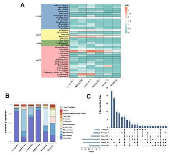
Viral assignments from MEGAN v7.1.1 were used for descriptive profiles of the six pooled libraries. R v4.4.2 was used for the stacked bar and UpSet plots; the heatmap was generated using ChiPlot (https://www.chiplot.online/ (accessed on 20 September 2026)). The supplied family-level export contains non-integer assignment values and is not treated as an unnormalized raw-read count table. Figure 1A displays log10-transformed family-level profile values. For Figure 1B, each family-level profile value was divided by the sum of all family-level values within the same library. The resulting within-library percentages are provided in Table S1 (Family_proportions worksheet); these values retain the processing represented in the supplied MEGAN export and are not reconstructed raw-read counts. Families were ranked by their arithmetic mean relative proportion across the six libraries; the ten highest-ranked families were displayed separately, and all remaining families were combined as others. The table was reshaped using reshape2 (v1.4.4) and plotted using ggpubr (v0.6.0) and ggplot2 (v4.0.3). For the UpSet plot, the first six sample columns of the MEGAN taxonomic assignment table exported at the species level were converted to binary presence/absence, scoring positive assignment values (>0) as present and zero as absent. UpSetR (v1.4.0) displayed up to 30 intersections ranked by frequency, with the six libraries kept in their specified order. This >0 criterion defines detection in the exported table for the descriptive overlap analysis; it does not validate species assignments or infections. The upstream DIAMOND and MEGAN thresholds are reported in Section 2.3. No additional minimum assignment-value filter was applied when converting the exported table to presence/absence. Unequal pool sizes and sequencing depths limit quantitative comparisons of richness and abundance. Each pooled library was treated as one analytical unit, and the six pools were not treated as replicate ecological or experimental groups. Between-pool profiles were described without claims of statistically significant differences. Relative proportions and log transformation do not by themselves correct for unequal pool size or detection sensitivity. A viral taxon detected in only one library was described as detected only in that library under the applied criteria; non-detection in another library was not interpreted as biological absence or true exclusivity. For Table S2, names and available genus and family annotations were taken from the original MEGAN taxonomic paths. Representative protein accessions for read-classification labels were selected from the exported BLASTx matches, using the most frequent accession among the highest-bit-score matches for each read and restricting the reported identifiers to INSDC or RefSeq protein accessions. These accessions document read-level reference matches, not a nearest nucleotide-genome comparison. Read-classification labels without an eligible representative INSDC or RefSeq protein accession were retained in Table S2 with the reference accession marked as “Not available”. Where the original MEGAN taxonomic path did not provide a named genus or family, the corresponding field was marked as “Not assigned in source taxonomy”. Reference accessions for the five assembled sequences are those used in their sequence comparisons; their study-generated nucleotide accessions are reported separately as GenBank and GenBase accessions. The within-library percentages describe composition among the exported family-level assignments; their denominator is not the total number of sequencing reads and they are not estimates of viral copies per tick. Unnormalized counts from different pools were not used to infer differences in individual viral burden.
Figure 1.
Descriptive viral taxonomic profiles of six Tibetan tick pools. (A) Heatmap of log10-transformed family-level profile values exported from MEGAN. Because the supplied export contains fractional values, these are not designated raw-read counts. The genome-type categories shown in the original analysis are indicated on the left. (B) Within-library relative proportions of family-level assignments. The ten families with the highest mean relative proportion across the six libraries are shown separately; the remaining families are combined as others. (C) UpSet plot of viral taxa (MEGAN taxonomic assignment labels) detected in the six libraries, with presence defined as an assignment value > 0. Up to 30 intersections are displayed in descending frequency, with library order retained. Set sizes give detected labels per library; intersection sizes give labels detected in the exact combination of libraries indicated by the connected dots. These labels do not represent formally validated species assignments. Detection in only one library does not establish biological exclusivity; unequal pool sizes and sequencing depths can affect detection probabilities and the observed overlap.
2.5. Viral Sequence Extension and Annotation
Contigs longer than 500 bp were compared with related references in Geneious Prime v2024.0.5 to inspect and annotate the recovered viral sequence regions [27]. For each of the five representative sequences, the corresponding raw R1 and R2 reads were mapped to the assembled reference using the Geneious mapper in Geneious Prime v2024.0.5 with the Low Sensitivity/Fastest preset. The reads were not assigned paired-read relationships using Set Paired Reads and were therefore mapped as unpaired reads. Fine Tuning was set to None (fast/read mapping), and Do not trim was selected. Structural-variant and additional insertion/deletion searches were disabled. The recorded preset settings were: word length, 24; index word length, 14; ignore words repeated more than 8 times; gaps allowed, with a maximum of 10% per read and a maximum gap size of 3; maximum mismatches per read, 10%; and maximum ambiguity, 4. The minimum mapping-quality, minimum-overlap, and minimum-overlap-identity options were unchecked; the inactive displayed values were not applied as additional filtering thresholds. The setting for multiple equally best matches was Randomly. Read mapping was used to assess support for the final assembled references. No new consensus sequences were exported from this mapping step, and no minimum-depth or allele-frequency thresholds were applied to generate a replacement consensus. Apart from the Chu4909 joining and retention of one copy of the repeated sequence described below, no manual nucleotide substitutions, insertions, or deletions were made. Inspection of the five final submitted FASTA sequences found no N bases or other nucleotide ambiguity codes; this does not independently validate base-call accuracy. Coverage at contig ends was not assessed separately, and no terminal coverage statistics are reported. Overall read-coverage breadth was not treated as verification of genome termini or the proposed circular junction. Mapped reads in Table S3 are counts of individual read sequences from R1 and R2, not counts of read pairs or independent fragments. Mean read depth was calculated over the ungapped reference nucleotide positions, and read-coverage breadth was the percentage of those positions covered by at least one mapped read. Mapping statistics were available for all five representative sequences. These sequence-specific mapping counts are distinct from the post-reference-filtering R1-only taxonomic counts in Table S1; they were not divided by those counts or expected to match them. This mapping step is not treated as sufficient evidence of complete genomes. For Chu4909, the 9-bp repeated sequence GTTGGGGAG was used during manual editing in Geneious, with one copy retained in the 11,234-bp assembled sequence. To assess read coverage and the proposed junction, the Xizang-35-4 raw R1 and R2 reads were mapped as unpaired reads to this reference using the same preset with fine-tuning disabled. The reference was set to circular for this mapping test. Coverage was calculated over the 11,234 ungapped reference positions, and potential end-to-start junction-crossing alignments were inspected for alignment extent and mismatches on both sides of the repeated sequence. Neither manual editing nor the circular reference setting was treated as evidence of circular genome topology.
The Geneious Prime v2024.0.5 Find ORFs function was used with the Standard genetic code, a minimum predicted length of 400 bp, and ATG as the required start codon. The 400-bp minimum length and ATG requirement were operational settings applied in Geneious for a restricted ORF annotation screen, rather than universal criteria for viral coding sequences or genome completeness. Predicted coding regions were compared with the Conserved Domain Database (CDD v3.21; https://www.ncbi.nlm.nih.gov/Structure/cdd/wrpsb.cgi (accessed on 20 September 2026)) [28] and the NCBI protein database using BLASTx. This screen excludes ORFs shorter than 400 bp and may miss coding sequences with alternative initiation codons or truncated coding regions. Failure to detect an ORF under this screen was not interpreted as evidence that the corresponding coding region was absent. Conserved domains were used to support functional annotation, not genome completeness. Homology- or domain-supported regions are reported as partial coding regions where protein completeness was not established. An ATG-initiated ORF within an assembled contig does not by itself establish recovery of a complete viral protein or genome. Amino acid identities refer to the homologous L-protein regions compared here and are not whole-genome nucleotide identities. The reported identities were taken from NCBI BLASTx comparisons and calculated as identical amino acid residues divided by the aligned amino acid length. Identity values are retained to two decimal places to distinguish the highest-identity comparison from exact identity. Reference-protein coverage was calculated from the aligned reference span divided by the total reference-protein length. Coverage of the submitted query region was not interpreted as coverage of the entire assembled contig or viral genome. The reported accessions identify the selected comparison references. Recovered sequence lengths, source pools, predicted coding-region labels, reference comparisons, read-mapping statistics for all five sequences, and study-generated nucleotide accession numbers are summarized in Table S3. Nucleotide sequences encoding the recovered L-protein regions were submitted as queries for the retained NCBI BLASTx comparisons. The NCBI results displayed 100% query coverage, rounded to whole percentages, for all five selected comparisons. Query coverage refers to coverage of the submitted L-region nucleotide query and is distinct from reference-protein coverage and read-coverage breadth across the assembled contig; it does not establish recovery of a full-length L protein or a complete viral genome. The listed references are reported as comparison references rather than uniformly designated best database matches.
Completeness terminology was based on the expected genome length and organization of the related virus, representation of the expected segments and coding regions, terminal or junction evidence, and read depth and breadth across the recovered sequence. A complete genome would require recovery of all expected genomic components, including the relevant termini or a supported circular junction; for a segmented virus, this refers to the entire set of genomic segments. A near-complete designation would require evidence that essentially all expected genomic components were represented, with missing regions explicitly documented. A complete genome segment refers to one resolved component of a segmented genome and does not imply recovery of the other segments. Read coverage of an assembled contig was assessed separately from genome completeness: even coverage over the entire contig does not show that all genomic regions have been recovered. Expected length, a conserved motif, a predicted ORF, or read coverage alone was insufficient to establish completeness. Because termini, the proposed circular junction, or the full complement of segments were not established, none of the five representative sequences was designated a complete or near-complete viral genome.
2.6. Phylogenetic Analysis
Recovered L-protein amino acid regions were compared with closely related tick-associated viral L proteins and representative homologous proteins from related genera, families, or subfamilies, retrieved from the NCBI Protein database (GenBank and RefSeq). Alignments were performed with MUSCLE in MEGA v11, and columns with more than 50.0% gaps were excluded before phylogenetic inference [29,30]. The reference set was selected for sequence comparison and was not designed as a systematic geographic sample.
Maximum-likelihood trees were inferred using IQ-TREE v2.3.6 with substitution models selected by ModelFinder according to the Bayesian information criterion (BIC) [31]. Branch support was assessed using 1000 ultrafast bootstrap (UFBoot) replicates with NNI optimization of bootstrap trees. For visualization, the displayed trees were rooted on the branches separating Abras virus (Figure 2), Wenling fish chu-like virus (Figure 3), and the Gammarhabdovirinae reference clade (Figure 4) from their respective remaining references. Reference protein accession numbers, including those used for rooting, are provided in Table S5. After gap filtering, the final alignment for Figure 2 contained 37 sequences and 4738 amino-acid positions (Q.pfam+F+R6); Figure 3 contained 24 sequences and 2336 positions (LG+F+I+R4); and Figure 4 contained 31 sequences and 2491 positions (rtREV+F+R5). Table S4 summarizes the sequence counts, alignment lengths, selected models, bootstrap replicates, displayed support statistics, and rooting references for all three analyses. Single values displayed at nodes represent UFBoot support percentages; SH-aLRT values are not displayed.
Figure 2.
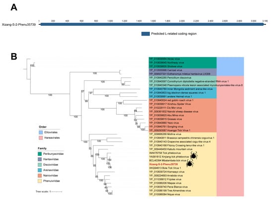
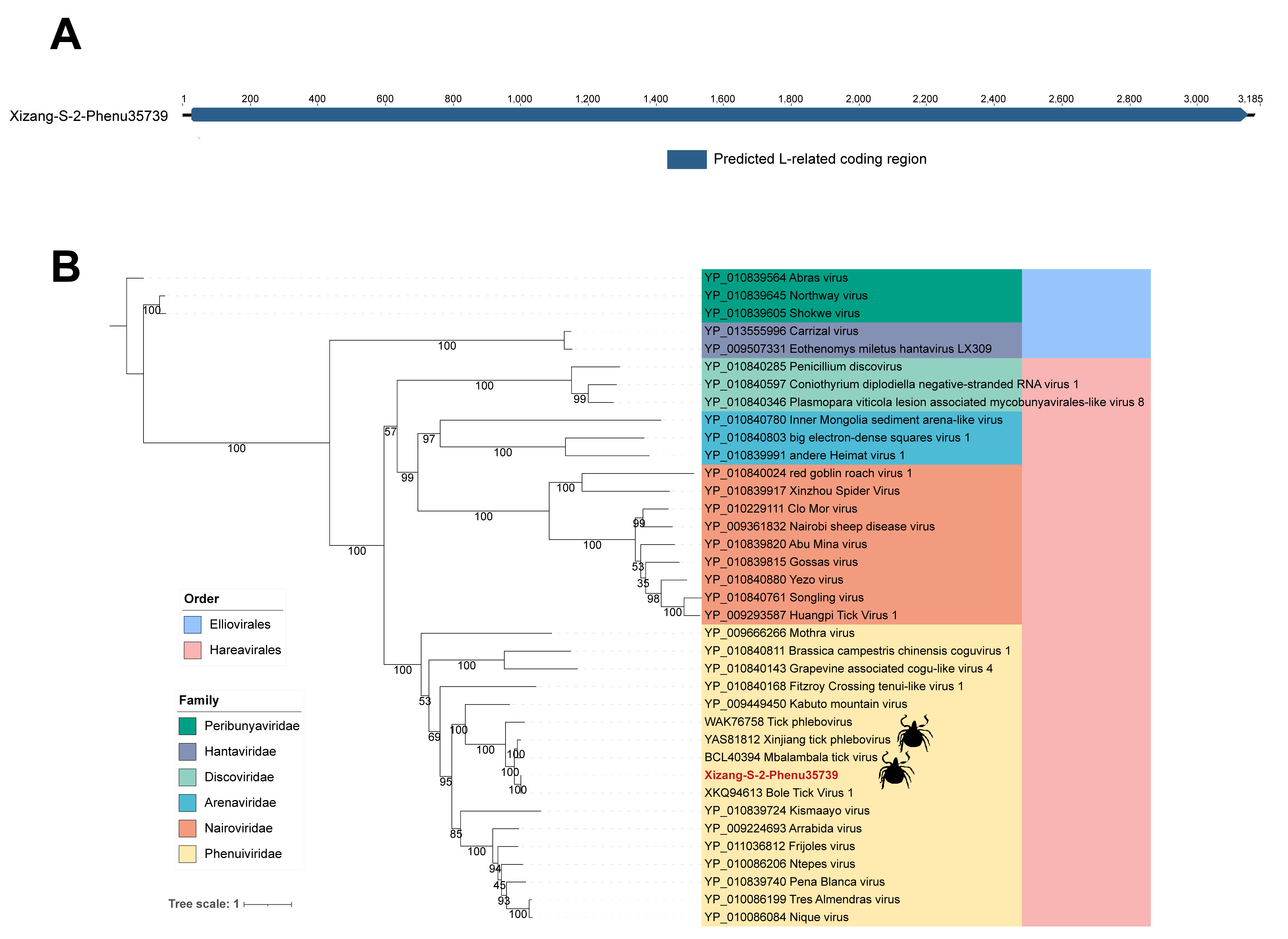
Sequence annotation and phylogenetic comparison of Xizang-S-2-Phenu35739. (A) Organization of the recovered 3185-bp Phenuiviridae-associated partial L-region sequence. The blue region denotes the predicted L-related coding region and does not indicate a complete segmented viral genome. (B) Maximum-likelihood comparison of recovered L-protein amino acid regions and representative Bunyaviricetes references using IQ-TREE v2.3.6. The study sequence is marked in red; colors indicate the groups shown in the legend, and tick silhouettes indicate the reported association of reference sequences with ticks. The scale bar shows substitutions per site. Each node label represents ultrafast bootstrap support (UFBoot, %) from 1000 replicates; SH-aLRT values are not displayed. The alignment after gap filtering contained 37 sequences and 4738 amino-acid positions; the selected model was Q.pfam+F+R6.
Figure 3.
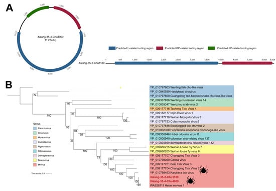
Sequence annotation and phylogenetic comparison of two Chuviridae-associated sequences. (A) Schematic organization of the recovered sequences. Blue, red, and green regions indicate predicted L-, GP-, and NP-related coding regions, respectively. Chu4909 has read coverage across the assembled sequence, but its circular configuration remains putative, and its topology and genome completeness remain unconfirmed. The circular depiction is a schematic representation. Chu1189 is a partial genomic sequence. (B) Maximum-likelihood comparison of recovered L-protein amino acid regions and Chuviridae references using IQ-TREE v2.3.6. Study sequences are marked in red, and colors indicate the groups in the legend. Tick silhouettes indicate reported tick-associated reference sequences. The scale bar shows substitutions per site. Each node label represents ultrafast bootstrap support (UFBoot, %) from 1000 replicates; SH-aLRT values are not displayed. The alignment after gap filtering contained 24 sequences and 2336 amino-acid positions; the selected model was LG+F+I+R4.
Figure 4.
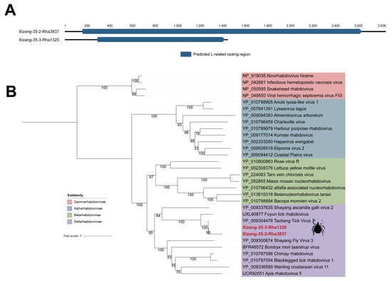
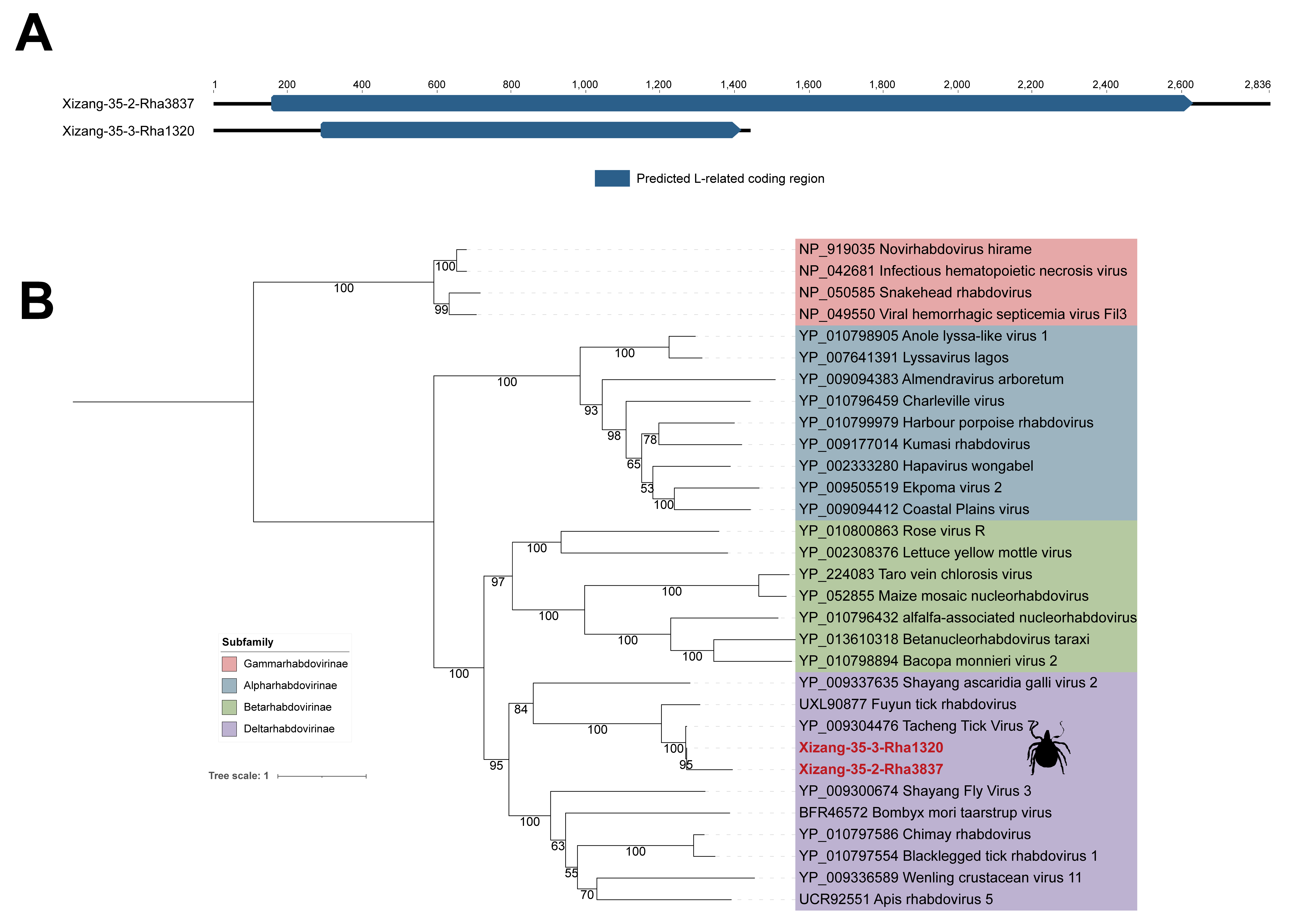
Sequence annotation and phylogenetic comparison of two Rhabdoviridae-associated sequences. (A) Organization of Xizang-35-2-Rha3837 (2836 bp) and Xizang-35-3-Rha1320 (1440 bp), with predicted L-related coding regions in blue at nt 157–2631 and nt 290–1417, respectively. Both are partial L-region sequences; neither represents a complete L gene or a complete or near-complete viral genome. (B) Maximum-likelihood comparison of recovered L-protein amino acid regions and selected Rhabdoviridae references using IQ-TREE v2.3.6. Study sequences are marked in red, and colors indicate the subfamilies shown in the legend. Tick silhouettes denote reported tick-associated reference sequences. The scale bar shows substitutions per site. Each node label represents ultrafast bootstrap support (UFBoot, %) from 1000 replicates; SH-aLRT values are not displayed. The alignment after gap filtering contained 31 sequences and 2491 amino-acid positions; the selected model was rtREV+F+R5.
The resulting phylogenetic trees were imported into the iTOL online tool (https://itol.embl.de/ (accessed on 20 September 2026)) [32] for visualization. Post-editing and aesthetic refinements were completed using Adobe Illustrator 2024 v28.7.1, and the host silhouette vectors referenced in the trees were all sourced from the PhyloPic platform (https://www.phylopic.org/ (accessed on 20 September 2026)).
2.7. Quality Control
No batch-matched extraction or library-preparation blanks were included, and no matched negative-control libraries were sequenced for the six tick pools. Aerosol-barrier tips and RNase/DNase-free consumables were used to reduce cross-contamination. No contamination-exclusion threshold or decision rule based on matched negative-control sequencing was applied. Reference-based background filtering is described in Section 2.3.
3. Results
3.1. Characterization of the Tick Virome Community
Six pooled libraries were constructed from the 90 ticks. The raw FASTQ files contained 282,318,532 reads in total (R1 + R2), representing 141,159,266 paired-end fragments, with a mean read length of 125.4 bp and an overall GC content of 47.8%. Per-library R1 + R2 counts were 36,178,020 for Xizang-35-1, 48,835,112 for Xizang-35-2, 53,472,932 for Xizang-35-3, 42,428,730 for Xizang-35-4, 62,860,462 for Xizang-S-2, and 38,543,276 for Xizang-22. These quantities describe the raw sequencing data and are distinct from downstream viral-assigned read counts. Adapter and quality trimming retained 254,892,258 reads (R1 + R2). The tick-reference-filtered output comprised 245,298,542 reads, as reconstructed from paired-input totals in the subsequent background-reference-filtering logs; 245,298,528 reads remained after both reference-filtering steps. Final MEGAN assignments to Viruses or its descendants comprised 69,526 unique R1 read identifiers, equivalent to 0.057% of the 122,649,264 R1 reads entering the viral-screening workflow after both reference filters. Per-library raw, post-trimming, and reference-filtered read counts, viral-assigned R1 counts, and their denominators and percentages are summarized in Table S1 (Read_statistics worksheet). These percentages are read-classification proportions and do not measure individual-tick infection prevalence. Viral read percentages are reported to three decimal places to preserve information on small proportions and avoid rounding low values to 0.0%. The MEGAN export of unassembled-read classifications used for the UpSet plot contained 101 distinct viral taxonomic assignment labels across the six pooled libraries. The family-level export contained assignments to 35 viral families (Figure 1A,B). Table S2 lists all 101 read-based taxonomic assignment labels, three additional families represented only in the family-level profile, and the five representative assembled sequences. Four labels—Crifsvirus GAP52, Cytomegalovirus humanbeta5, Enterovirus A, and Risingsunvirus risingsun—had no eligible representative INSDC or RefSeq protein accession in the supplied match records; they are retained in Table S2 with the reference accession marked as “Not available”. The assembled-sequence records are listed separately and are not added to the count of read-classification labels. Because the export included higher-rank and unclassified labels, the total is reported as taxonomic assignment labels rather than confirmed viral species. These assignments do not imply recovery of a corresponding assembled viral genome for every label.
Retrospective COI screening and assembly recovered nine contigs, ranging from 258 to 901 nt, across the six libraries (Table S1, Tick_identification worksheet). Contigs from Xizang-35-1, Xizang-35-4, and Xizang-22 had representative highest-scoring matches labelled Rhipicephalus turanicus, with 99.6–100.0% nucleotide identity and 93.4–100.0% query coverage. The two contigs from Xizang-S-2 matched Hyalomma asiaticum references at 99.8–99.9% identity and 97.2–100.0% query coverage. Xizang-35-2 and Xizang-35-3 yielded 552- and 366-nt contigs, respectively, matching Argas persicus references at 99.8% and 100.0% identity, both with 100.0% query coverage. These Argas-associated sequences indicate that the material should not be described exclusively as hard ticks; Argas persicus belongs to Argasidae. These are provisional COI-based sequence associations, and short fragments and closely related reference labels limit definitive species assignment. They do not establish that every tick within a pool belongs to the matched reference taxon, exclude additional taxa, or identify which individual ticks contributed the viral sequences.
Five representative RNA viral sequences were selected for annotation and phylogenetic comparison. They comprise one Phenuiviridae-associated partial L-region sequence, two Chuviridae-associated sequences, and two Rhabdoviridae-associated partial L-region sequences. The presence of a conserved motif was used as annotation evidence, not as a criterion for a near-complete genome. The prominence of RNA-virus assignments is consistent with previous tick virome surveys [6,8,33]. The five characterized sequences were related to previously reported viruses; no novel viruses were identified in this study.
Phenuiviridae, Chuviridae, and Rhabdoviridae were prominent RNA viral families in the pooled profiles. Members of these groups have also been reported in invertebrate and tick virome studies [4,6,8]. Their sequence detection in the present pools does not by itself demonstrate replication in ticks or identify the biological source of every assigned read.
Figure 1 summarizes the family-level profile values and within-library composition. The reported 35 families comprised 12 dsDNA, 5 ssDNA, 3 dsRNA, and 15 ssRNA families. Chuviridae accounted for large proportions of the viral assignments in Xizang-35-1, Xizang-35-3, and Xizang-35-4. These proportions describe the sequenced pools and do not estimate the abundance of viruses per individual tick.
Rhabdoviridae accounted for a large proportion of assignments in Xizang-35-2, whereas Phenuiviridae was prominent in Xizang-S-2. Microviridae- and Papillomaviridae-associated reads were also detected. Previous studies have described differences in pooled tick virome profiles [6,7], but the present pools differ in size and may differ in unrecorded biological and technical factors [8,34]. Consequently, these observations are reported descriptively and are not attributed to particular ecological conditions. These DNA-virus-associated assignments are reported as sequence detections; they were not verified as infections of the sampled ticks.
The UpSet plot (Figure 1C) summarizes detection overlap among pooled libraries, with viral taxa defined as MEGAN taxonomic assignment labels and detection defined as an exported assignment value >0. Xizang-35-3 and Xizang-35-2 contained 49 and 46 detected viral taxa, respectively. Nine taxa were detected in exactly these two libraries and in none of the other four; this intersection excludes taxa also detected in additional libraries and is not the total overlap between the two libraries. Seven taxa were detected in all six libraries. Twenty-three taxa were detected only in Xizang-35-3 and 19 only in Xizang-35-2 under the applied criteria. These observations describe library-level detection patterns, not taxa biologically unique to a pool. Pool size, sequencing depth, and assignment thresholds can affect detection probabilities, so non-detection in another library does not establish absence or a difference in individual-tick viral richness.
The seven viral taxa detected across all six pools provide candidates for further sequence-level examination. As discussed in previous tick virome studies, detection across pools requires cautious biological interpretation [6,11]. Shared detection does not establish a core resident virome, enhanced adaptation, or active infection in all ticks.
3.2. Genomic and Phylogenetic Characterization of a Phenuiviridae Sequence Identified in This Study
Phenuiviridae comprises viruses with diverse hosts and segmented negative-sense or ambisense RNA genomes [35,36]. Typical genomes contain multiple segments encoding proteins including L, glycoproteins, and nucleocapsid proteins. In the genus Uukuvirus, the ICTV species-demarcation criterion is L-protein amino acid identity below 95.0% [36]. Tick-associated phenuivirus sequences have been reported in multiple settings [37], and their medical significance varies among viruses [35,38]. Xizang-S-2-Phenu35739 (GenBank QB150285) is a 3185-bp recovered sequence containing a predicted L-related coding region (Figure 2A). Read mapping yielded 2855 mapped reads, a mean depth of 125.3×, and 100.0% read-coverage breadth across the assembled reference (Table S3). It is described as a partial L-region sequence; the available assembly evidence does not establish a near-complete L segment or a complete segmented genome. The identity of this partial L region was interpreted as evidence of relationship to a reference virus; the L-protein species criterion was not used to assign a new species from this fragment.
A maximum-likelihood tree was inferred from the recovered L-related amino acid region and representative Bunyaviricetes reference sequences, including Phenuiviridae and other selected families (Figure 2B). Xizang-S-2-Phenu35739 grouped with the Phenuiviridae-associated references in the displayed analysis.
Xizang-S-2-Phenu35739 grouped with Xinjiang tick phlebovirus (YAS81812), Tick phlebovirus (WAK76758), and Mbalambala tick virus (BCL40394), with displayed ultrafast bootstrap support of 100%. The recovered L-related region had 99.81% amino acid identity to Bole Tick Virus 1 (XKQ94613.1; 1038/1040 aligned amino acids). The comparison spanned reference residues 1096–2135 of the 2148-aa protein, corresponding to 48.4% reference-protein coverage. These observations support a close relationship to the Uukuvirus-related references in this analysis. The assembly is reported as a Bole Tick Virus 1-related sequence detection in a Tibetan tick pool, without designation of a distinct species or strain. High identity over this partial region and phylogenetic proximity do not independently establish a species assignment. The selected reference set includes close relatives reported from China and Africa. Its limited and geographically uneven sampling, together with the presence of closely related African references, does not support statistically significant regional clustering, geographic movement, or host-specific conservation.
3.3. Genomic and Phylogenetic Characterization of Chuviridae Sequences Identified in This Study
Chuviridae includes arthropod-associated negative-sense RNA viruses with diverse genomic organizations [38,39]. Mivirus-related sequences may have linear or circular configurations and encode proteins including L, GP, and NP. ICTV distinguishes Mivirus species using amino acid identity below 90.0% between complete L-protein sequences [39]. Tick-associated chuvirus sequences have been described previously [11,33,38]. Two sequences were recovered here: Xizang-35-4-Chu4909 (11,234 bp; GenBank QB150284), with predicted L-, GP-, and NP-related ORFs, and Xizang-35-2-Chu1189 (5829 bp; GenBank QB150288), with predicted L- and GP-related regions (Figure 3A). Mapping of the Xizang-35-4 raw R1 and R2 reads yielded 29,131 mapped reads covering all 11,234 reference positions, with a mean read depth of 369.1×. Potential junction-crossing alignments had insufficient flanking alignment or substantial mismatches and did not provide reliable support for the proposed end-to-start junction. Chu4909 has read coverage across the assembled sequence, but its circular configuration remains putative, and its topology and genome completeness remain unconfirmed. Chu1189 is described as a partial genomic sequence. Mapping to Chu1189 yielded 474 mapped reads, a mean depth of 11.1×, and 99.9% read-coverage breadth across the assembled reference (Table S3).
In the selected Chuviridae comparison (Figure 3B), both Tibetan sequences grouped with Mivirus-related references, including Hebei mivirus 1 (WAS28118.1), with displayed ultrafast bootstrap support of 100%. The compared L-protein regions of Xizang-35-2-Chu1189 and Xizang-35-4-Chu4909 had amino acid identities of 99.92% (1197/1198 aligned amino acids) and 99.95% (2155/2156), respectively, to Hebei mivirus 1 (WAS28118.1). The Chu1189 comparison spanned reference residues 959–2156, representing 55.6% of the 2156-aa reference protein. The Chu4909 comparison spanned all 2156 reference amino acids and contained one amino acid mismatch. Full reference-protein coverage does not establish viral genome completeness or validate the nucleotide termini. The two assemblies are therefore reported as Hebei mivirus 1-related detections from Tibetan tick pools, with phylogenetic associations to Mivirus. The reported identities describe the compared L regions and do not establish two new species, geographic variants, or new strains. The reference virus name is not used as a formal species assignment for these assemblies.
Other neighboring references included Changping Tick Virus 2/3 (YP_009177704/YP_009177707), Bole Tick Virus 3 (YP_009177701), and Karukera tick virus (YP_010798463). The topology describes relationships among the sequences included in this comparison; it does not demonstrate host adaptation or sustained replication in ticks.
3.4. Genomic and Phylogenetic Characterization of Rhabdoviridae Sequences Identified in This Study
Rhabdoviridae includes negative-sense RNA viruses with diverse hosts and genomic organizations, with genomes generally about 10–16 kb in length. ICTV subfamily and genus demarcation uses phylogenetic relationships based on full-length L sequences together with genome and biological characteristics; species criteria depend on the genus [40]. Tick-associated rhabdovirus sequences have been described in previous virome studies [11,41]. Xizang-35-2-Rha3837 (2836 bp; GenBank QB150287) and Xizang-35-3-Rha1320 (1440 bp; GenBank QB150286) were recovered as partial L-region sequences (Figure 4A). Both contigs are substantially shorter than typical rhabdovirus genomes. The predicted regions span nt 157–2631 and nt 290–1417, respectively (1-based inclusive coordinates, including the terminal stop codon); these coordinates describe predicted coding regions within the recovered contigs. They do not represent complete L-protein genes or complete or near-complete rhabdovirus genomes [40]. Mapping to Rha3837 yielded 604 mapped reads, a mean depth of 30.4×, and 99.0% read-coverage breadth; mapping to Rha1320 yielded 251 mapped reads, a mean depth of 25.1×, and 100.0% read-coverage breadth. These statistics describe support across the recovered references and do not establish recovery of complete L genes or viral genomes. Sequence lengths, source pools, and mapping statistics are summarized in Table S3.
The Rhabdoviridae comparison included references assigned to Gammarhabdovirinae, Alpharhabdovirinae, Betarhabdovirinae, and Deltarhabdovirinae (Figure 4B). Both recovered Tibetan sequences were placed near Deltarhabdovirinae references. This placement based on partial L regions supports only a tentative phylogenetic association at the subfamily level; it does not constitute a formal subfamily assignment or resolve genus, species, strain, or variant classification.
The two Tibetan sequences formed a clade with displayed ultrafast bootstrap support of 95% and were placed with Tacheng tick virus 7 (YP_009304476.1). The recovered L-protein regions of Xizang-35-2-Rha3837 and Xizang-35-3-Rha1320 had amino acid identities of 99.15% (817/824 aligned amino acids) and 98.67% (370/375), respectively, to Tacheng tick virus 7 (YP_009304476.1). The comparisons spanned reference residues 1392–2215 and 1841–2215, covering 37.2% and 16.9%, respectively, of the 2215-aa reference protein. Reported amino acid identities of the recovered L-protein regions of Xizang-35-2-Rha3837 and Xizang-35-3-Rha1320 to Fuyun tick rhabdovirus (UXL90877.1) were 51.03% and 46.13%, respectively. These comparisons document similarity of the recovered L regions to Tacheng tick virus 7. Placement near the subfamily references is interpreted separately as a tentative phylogenetic association. No genus, species, strain, or variant designation is assigned to either partial sequence. Closely related L-region sequences have therefore been detected in geographically separated regions, Tibet and Xinjiang; these partial-region comparisons do not establish widespread distribution or occurrence in intervening regions. Tick-associated rhabdovirus diversity has also been described previously [11,41].
4. Discussion
This study provides descriptive viral taxonomic profiles from six Tibetan tick pools and sequence comparisons of representatives associated with Phenuiviridae, Chuviridae, and Rhabdoviridae. The prominence of RNA-virus assignments is consistent with previous tick and plateau arthropod surveys [6,9,13]. The added contribution is a dataset linking read-level profiles with annotation, coverage assessment, and comparisons of five known-virus-related assemblies. Pooling and incomplete specimen metadata limit ecological and epidemiological interpretation; one library per sampling site does not provide within-site biological replication.
RNA viral diversity has been repeatedly documented in invertebrates and ticks [4,6,11]. The prominent RNA viral families detected here have also appeared in other tick virome surveys [6,9,11,13,33,42,43]. Differences in the relative profiles of the six pools are descriptive. Pool sizes and sequencing depths differed, and each pool represented a different sampling site. All specimens were collected from the environment rather than directly from vertebrate hosts, but individual-tick species and complete within-pool species composition were not established. Such variation can affect detection, as recognized in previous studies [7,8,34,44,45]; without replicate libraries within sampling sites or other defined ecological strata, the data primarily support descriptive differences in library composition. They cannot establish strong ecological heterogeneity, identify its drivers, or support quantitative comparisons of individual infection burden. These proportions are compositional: a larger share for one family can result from a smaller share for other families and does not demonstrate an increase in absolute viral quantity.
Interpretation of the recovered sequences is limited by the regions represented in the assemblies. For segmented viruses, recovery of a single L-related region does not establish a complete genome [36]. Sequence similarity within a recovered coding region supports annotation and comparison, but does not by itself establish species novelty, a host association, or genome completeness. Chu4909 has read coverage across the assembled sequence, but its circular configuration remains putative, and its topology and genome completeness remain unconfirmed. Geographic connectivity, host movement, tick dispersal, and transport by migratory birds were not measured in this study. Their possible contributions to similarities between geographically separated sequences remain hypotheses for future testing, rather than transmission mechanisms established by the reference-tree topology.
Sequence surveys can inform further investigation of tick-associated viruses, including medically important groups [1,33,35,40], but the present detections do not establish disease risk. Relevant regional RNA-virus reports include detection of a proposed Himalayan subtype of tick-borne encephalitis virus in Himalayan marmots in Qinghai Province on the Qinghai–Tibet Plateau [46] and Gagosa Mountain virus in Ornithodoros lahorensis collected from sheep in Shigatse, Tibet [47]. These animal or tick sequence detections must be distinguished from demonstrated clinical disease in humans or livestock. No clinical cases, serology, or disease outcomes were examined in the present study, and it cannot estimate the local burden of tick-borne viral disease.
Several limitations constrain interpretation. Sampling sites and the site-based pooling criterion were documented, but formal specimen-level tick identification, developmental stage, and sex were not recorded. The collection date for Bomi was unavailable. Coordinate provenance was not documented, and locality-level reference elevations do not establish the elevation of each exact collection point. Retrospective COI comparisons provided provisional tick taxonomic associations for each library, but complete within-pool species composition was not established. The short recovered fragments, limited initial reference panel, and unequal screening scopes preclude definitive identification of every tick or quantitative comparison of tick taxa. Although the collected specimens appeared morphologically similar, this observation was not treated as evidence that all ticks belonged to a single species. One pooled library per sampling site and the absence of individual-level viral validation prevent reliable species-specific or replicated site-level ecological analysis and assignment of a viral sequence to a particular tick species within a pool.
Individual-tick prevalence and co-infection could not be determined from these pooled data. Each pooled library was the analytical unit. Shared and single-library detections were interpreted descriptively using the >0 assignment-value criterion, because pool sizes varied (10–17 ticks), sequencing depths differed, and specimen metadata were incomplete. These factors can affect the probability of detecting a taxon in each library and therefore the observed numbers of shared and single-library taxa. Non-detection does not establish biological absence, and detection across pools does not constitute independent confirmation in individual ticks.
The limitations of pooled detection have also been discussed in previous tick virome studies [9]. Residual tick-derived reads may remain after subtraction against a single reference, and endogenous viral elements cannot be excluded by that step alone. The documented contig search used a viral protein database and therefore did not itself compare viral with non-viral matches; a separate competitive screening step could not be verified. This limits the assessment of candidate specificity and the reproducibility of candidate selection. Read-level taxonomic assignments do not independently validate the specificity of every assembled candidate.
Technical factors, including nuclease treatment, extraction, amplification, and library preparation, can affect viral recovery [15,16]. DNA-virus and bacteriophage assignments may reflect blood meals, tick-associated microorganisms, or environmental material; reagent-associated background also remains a possible source. For reporting the family-level profile, retrovirus-related read assignments are grouped under the label “Endogenous retroviruses (ERVs)” in Figure 1. These assignments describe sequences detected in pooled tick material and do not establish active infection or viral replication. Biological sources were not resolved by the present pooled sequencing, reference filtering, or taxonomic assignments. Accordingly, tick-associated denotes detection in a tick-derived pool and does not establish infection or replication in ticks. The absence of matched extraction and library-preparation blanks prevents direct assessment of background introduced during sample processing and sequencing. Reference-based filtering cannot exclude all reagent- or laboratory-derived sequences. This limitation is particularly relevant to low-abundance taxonomic assignments, whose biological origin remains uncertain.
Finally, the representative sequence detections were not independently confirmed by targeted PCR/RT-PCR, virus isolation, or individual-tick assays. Read mapping assesses support for recovered sequence regions but does not substitute for independent confirmation or demonstrate active infection or replication. The findings are therefore reported as detected sequences in tick-derived pools. Chu4909 has read coverage across the assembled sequence, but its circular configuration remains putative, and its topology and genome completeness remain unconfirmed. Reliable support for the proposed end-to-start junction was not obtained. Coverage at the ends of the recovered contigs was not assessed separately, and genome termini have not been independently validated. The absence of N or other ambiguity codes in the submitted sequences does not establish that every nucleotide was supported by high-confidence reads. Accordingly, the study reports sequence detections and phylogenetic relationships without asserting active infection, replication, circularity, genome completeness, or novel species status.
5. Conclusions
Metagenomic analysis of six pools comprising 90 ticks from Tibet yielded read-level taxonomic assignments to 35 viral families. The pooled profiles describe sequence-level taxonomic diversity, and analyses of five representative sequences associated with Phenuiviridae, Chuviridae, and Rhabdoviridae document phylogenetic relationships to previously reported tick-associated viruses over the recovered L-protein regions. No novel viruses were identified. Genome completeness was not established; Chu4909 has read coverage across the assembled sequence, but its circular configuration remains putative and its topology remains unconfirmed. Unequal pool sizes and sequencing depths, incomplete specimen metadata, and the absence of biological replication within defined ecological strata restrict comparisons to descriptive library composition. The absence of matched negative controls limits assessment of laboratory-derived background. Without independent confirmation, these detections do not establish active infection, replication, individual-tick infection patterns, ecological drivers, or transmission processes. The dataset adds sequence information for comparison with other tick virome surveys.
Supplementary Materials
The following supporting information can be downloaded at: https://www.mdpi.com/article/10.3390/microorganisms14112568/s1, Table S1: Tick pool metadata and sequencing library information. Worksheets provide pool metadata (Pool_metadata), sequencing-stage statistics and viral read proportions (Read_statistics), within-library relative proportions of viral family assignments (Family_proportions), and COI-based taxonomic associations of tick sequences (Tick_identification); Table S2: Inventory of read-based viral taxonomic assignments and representative assembled sequences; Table S3: Characteristics of the five representative viral sequences; Table S4: Summary of phylogenetic analyses; Table S5: Reference protein sequences used for phylogenetic analyses.
Author Contributions
S.N. and W.Z. conceived and designed the study. C.Z., Z.Z. and P.W. performed the experiments and analyzed the data. Z.Z. prepared the figures and wrote the original manuscript. C.Z., P.W., W.Z. and S.N. reviewed and revised the manuscript. W.Z. and S.N. supervised the study and acquired funding. All authors have read and agreed to the published version of the manuscript.
Funding
This research was funded by the National Key Research and Development Program of China, grant number 2023YFD1801300.
Institutional Review Board Statement
This study involved only the collection of free-living ticks from the environment. No vertebrate animals were captured, restrained, maintained, or dissected, and no ticks were collected directly from vertebrate hosts. Environmental tick collection was included in the ethics review framework of a National Key R&D Program project on disease prevention and control in plateau animals (approval No. JSDX20251211001, approval date: 11 December 2025). No human participants were involved.
Informed Consent Statement
Not applicable.
Data Availability Statement
Raw sequencing reads from the six specimen libraries were deposited in the Genome Sequence Archive (GSA) at the National Genomics Data Center under accession CRA049867 and BioProject PRJCA074065, with BioSample accessions SAMC8505619–SAMC8505624 and GSA Run accessions CRR3542369–CRR3542374. Five representative assembled nucleotide sequences were deposited in GenBase under accessions C_AA501328.1 (Xizang-35-2-Chu1189), C_AA501329.1 (Xizang-S-2-Phenu35739), C_AA501330.1 (Xizang-35-2-Rha3837), C_AA501331.1 (Xizang-35-3-Rha1320), and C_AA501332.1 (Xizang-35-4-Chu4909). The same five assembled nucleotide sequences have also been submitted to GenBank and assigned accessions QB150284 (Xizang-35-4-Chu4909), QB150285 (Xizang-S-2-Phenu35739), QB150286 (Xizang-35-3-Rha1320), QB150287 (Xizang-35-2-Rha3837), and QB150288 (Xizang-35-2-Chu1189). Accession numbers have been assigned; public release remains subject to completion of GenBank processing. Table S2 distinguishes reference protein accessions associated with BLASTx comparisons from nucleotide accessions assigned to assembled sequences generated in this study. Read-based classification labels without a corresponding characterized and deposited assembly have no study-generated sequence accession.
Acknowledgments
The authors thank Gongga of Tibet Agricultural and Animal Husbandry University for assistance with tick sample collection, and all personnel who assisted with laboratory processing.
Conflicts of Interest
The authors declare no conflicts of interest.
References
- Madison-Antenucci, S.; Kramer, L.D.; Gebhardt, L.L.; Kauffman, E. Emerging Tick-Borne Diseases. Clin. Microbiol. Rev. 2020, 33, e00083-18. [Google Scholar] [CrossRef] [Scilit]
- Shi, J.; Hu, Z.; Deng, F.; Shen, S. Tick-Borne Viruses. Virol. Sin. 2018, 33, 21–43. [Google Scholar] [CrossRef] [Scilit]
- Ye, R.-Z.; Li, Y.-Y.; Wang, X.-Y.; Cao, W.-C. Tick-Borne Viruses: Discovery, Clinical Aspects, Influencing Factors and Control. Nat. Rev. Microbiol. 2026, 24, 654–672, Erratum in Nat. Rev. Microbiol. 2026, 24, 673.. [Google Scholar] [CrossRef] [Scilit]
- Shi, M.; Lin, X.-D.; Tian, J.-H.; Chen, L.-J.; Chen, X.; Li, C.-X.; Qin, X.-C.; Li, J.; Cao, J.-P.; Eden, J.-S.; et al. Redefining the Invertebrate RNA Virosphere. Nature 2016, 540, 539–543. [Google Scholar] [CrossRef] [Scilit]
- Pérez-Sautu, U.; Wiley, M.R.; Prieto, K.; Chitty, J.A.; Haddow, A.D.; Sánchez-Lockhart, M.; Klein, T.A.; Kim, H.-C.; Chong, S.-T.; Kim, Y.-J.; et al. Novel Viruses in Hard Ticks Collected in the Republic of Korea Unveiled by Metagenomic High-Throughput Sequencing Analysis. Ticks Tick.-Borne Dis. 2021, 12, 101820. [Google Scholar] [CrossRef] [Scilit]
- Ni, X.-B.; Cui, X.-M.; Liu, J.-Y.; Ye, R.-Z.; Wu, Y.-Q.; Jiang, J.-F.; Sun, Y.; Wang, Q.; Shum, M.H.-H.; Chang, Q.-C.; et al. Metavirome of 31 Tick Species Provides a Compendium of 1801 RNA Virus Genomes. Nat. Microbiol. 2023, 8, 162–173. [Google Scholar] [CrossRef] [Scilit]
- Pettersson, J.H.-O.; Shi, M.; Bohlin, J.; Eldholm, V.; Brynildsrud, O.B.; Paulsen, K.M.; Andreassen, Å.; Holmes, E.C. Characterizing the Virome of Ixodes Ricinus Ticks from Northern Europe. Sci. Rep. 2017, 7, 10870. [Google Scholar] [CrossRef] [Scilit]
- Xu, L.; Guo, M.; Hu, B.; Zhou, H.; Yang, W.; Hui, L.; Huang, R.; Zhan, J.; Shi, W.; Wu, Y. Tick Virome Diversity in Hubei Province, China, and the Influence of Host Ecology. Virus Evol. 2021, 7, veab089, Erratum in Virus Evol. 2022, 7, veab108.. [Google Scholar] [CrossRef] [Scilit]
- Kong, Y.; Zhang, G.; Jiang, L.; Wang, P.; Zhang, S.; Zheng, X.; Li, Y. Metatranscriptomics Reveals the Diversity of the Tick Virome in Northwest China. Microbiol. Spectr. 2022, 10, e01115-22. [Google Scholar] [CrossRef] [Scilit]
- Yang, Z.; Zhang, J.; Yang, S.; Wang, X.; Shen, Q.; Sun, G.; Wang, H.; Zhang, W. Virome Analysis of Ticks in a Forest Region of Liaoning, China: Characterization of a Novel Hepe-like Virus Sequence. Virol. J. 2021, 18, 163. [Google Scholar] [CrossRef] [Scilit]
- Harvey, E.; Rose, K.; Eden, J.-S.; Lo, N.; Abeyasuriya, T.; Shi, M.; Doggett, S.L.; Holmes, E.C. Extensive Diversity of RNA Viruses in Australian Ticks. J. Virol. 2019, 93, e01358-18. [Google Scholar] [CrossRef] [Scilit]
- Tokarz, R.; Williams, S.H.; Sameroff, S.; Sanchez Leon, M.; Jain, K.; Lipkin, W.I. Virome Analysis of Amblyomma Americanum, Dermacentor Variabilis, and Ixodes Scapularis Ticks Reveals Novel Highly Divergent Vertebrate and Invertebrate Viruses. J. Virol. 2014, 88, 11480–11492. [Google Scholar] [CrossRef] [Scilit]
- Zhu, W.; Pu, J.; Jin, D.; Lu, S.; Yang, J.; Xu, J. RNA Virome of Ticks and Fleas Infesting Marmota himalayana from the Qinghai-Tibet Plateau. Anim. Microbiome 2025, 7, 131. [Google Scholar] [CrossRef] [Scilit]
- Fang, Y.; Wang, J.; Sun, J.; Su, Z.; Chen, S.; Xiao, J.; Ni, J.; Hu, Z.; He, Y.; Shen, S.; et al. RNA Viromes of Dermacentor nuttalli Ticks Reveal a Novel Uukuvirus in Qinghai Province, China. Virol. Sin. 2024, 39, 537–545. [Google Scholar] [CrossRef] [Scilit]
- Conceição-Neto, N.; Zeller, M.; Lefrère, H.; De Bruyn, P.; Beller, L.; Deboutte, W.; Yinda, C.K.; Lavigne, R.; Maes, P.; Ranst, M.V.; et al. Modular Approach to Customise Sample Preparation Procedures for Viral Metagenomics: A Reproducible Protocol for Virome Analysis. Sci. Rep. 2015, 5, 16532. [Google Scholar] [CrossRef] [Scilit]
- Zhang, W.; Li, L.; Deng, X.; Kapusinszky, B.; Delwart, E. What Is for Dinner? Viral Metagenomics of US Store Bought Beef, Pork, and Chicken. Virology 2014, 468–470, 303–310. [Google Scholar] [CrossRef] [Scilit]
- Lu, X.; Ji, L.; Wang, H.; Zhang, Q.; Wang, X.; Liu, Y.; Shen, Q.; Yang, S.; Ma, X.; Zhang, W.; et al. Highly Diverse RNA Viruses and Phage Sequences Concealed within Birds. Microbiol. Spectr. 2024, 12, e00802-24. [Google Scholar] [CrossRef] [Scilit]
- Dai, Z.; Wang, H.; Xu, J.; Lu, X.; Ni, P.; Yang, S.; Shen, Q.; Wang, X.; Li, W.; Wang, X.; et al. Unveiling the Virome of Wild Birds: Exploring CRESS-DNA Viral Dark Matter. Genome Biol. Evol. 2024, 16, evae206. [Google Scholar] [CrossRef] [Scilit]
- Zhang, W.; Yang, S.; Shan, T.; Hou, R.; Liu, Z.; Li, W.; Guo, L.; Wang, Y.; Chen, P.; Wang, X.; et al. Virome Comparisons in Wild-Diseased and Healthy Captive Giant Pandas. Microbiome 2017, 5, 90. [Google Scholar] [CrossRef] [Scilit]
- Langmead, B.; Salzberg, S.L. Fast Gapped-Read Alignment with Bowtie 2. Nat. Methods 2012, 9, 357–359. [Google Scholar] [CrossRef] [Scilit]
- Langmead, B.; Wilks, C.; Antonescu, V.; Charles, R. Scaling Read Aligners to Hundreds of Threads on General-Purpose Processors. Bioinformatics 2019, 35, 421–432. [Google Scholar] [CrossRef] [Scilit]
- Schmieder, R.; Edwards, R. Quality Control and Preprocessing of Metagenomic Datasets. Bioinformatics 2011, 27, 863–864. [Google Scholar] [CrossRef] [Scilit]
- Li, D.; Liu, C.-M.; Luo, R.; Sadakane, K.; Lam, T.-W. MEGAHIT: An Ultra-Fast Single-Node Solution for Large and Complex Metagenomics Assembly via Succinct de Bruijn Graph. Bioinformatics 2015, 31, 1674–1676. [Google Scholar] [CrossRef] [Scilit]
- Buchfink, B.; Xie, C.; Huson, D.H. Fast and Sensitive Protein Alignment Using DIAMOND. Nat. Methods 2015, 12, 59–60. [Google Scholar] [CrossRef] [Scilit]
- Buchfink, B.; Reuter, K.; Drost, H.-G. Sensitive Protein Alignments at Tree-of-Life Scale Using DIAMOND. Nat. Methods 2021, 18, 366–368. [Google Scholar] [CrossRef] [Scilit]
- Deng, X.; Naccache, S.N.; Ng, T.; Federman, S.; Li, L.; Chiu, C.Y.; Delwart, E.L. An Ensemble Strategy That Significantly Improves de Novo Assembly of Microbial Genomes from Metagenomic Next-Generation Sequencing Data. Nucleic Acids Res. 2015, 43, e46. [Google Scholar] [CrossRef] [Scilit]
- Bergholm, J.; Tessema, T.S.; Blomström, A.-L.; Berg, M. Metagenomic Insights into the Complex Viral Composition of the Enteric RNA Virome in Healthy and Diarrheic Calves from Ethiopia. Virol. J. 2025, 22, 188. [Google Scholar] [CrossRef] [Scilit]
- Marchler-Bauer, A.; Derbyshire, M.K.; Gonzales, N.R.; Lu, S.; Chitsaz, F.; Geer, L.Y.; Geer, R.C.; He, J.; Gwadz, M.; Hurwitz, D.I.; et al. CDD: NCBI’s Conserved Domain Database. Nucleic Acids Res. 2015, 43, D222–D226. [Google Scholar] [CrossRef] [Scilit]
- Tamura, K.; Stecher, G.; Kumar, S. MEGA11: Molecular Evolutionary Genetics Analysis Version 11. Mol. Biol. Evol. 2021, 38, 3022–3027. [Google Scholar] [CrossRef] [Scilit]
- Kumar, S.; Stecher, G.; Li, M.; Knyaz, C.; Tamura, K. MEGA X: Molecular Evolutionary Genetics Analysis across Computing Platforms. Mol. Biol. Evol. 2018, 35, 1547–1549. [Google Scholar] [CrossRef] [Scilit]
- Minh, B.Q.; Schmidt, H.A.; Chernomor, O.; Schrempf, D.; Woodhams, M.D.; von Haeseler, A.; Lanfear, R. IQ-TREE 2: New Models and Efficient Methods for Phylogenetic Inference in the Genomic Era. Mol. Biol. Evol. 2020, 37, 1530–1534, Erratum in Mol. Biol. Evol. 2021, 37, 2461. [Google Scholar] [CrossRef] [Scilit]
- Letunic, I.; Bork, P. Interactive Tree of Life (iTOL) v5: An Online Tool for Phylogenetic Tree Display and Annotation. Nucleic Acids Res. 2021, 49, W293–W296. [Google Scholar] [CrossRef] [Scilit]
- Vandegrift, K.J.; Kapoor, A. The Ecology of New Constituents of the Tick Virome and Their Relevance to Public Health. Viruses 2019, 11, 529. [Google Scholar] [CrossRef] [Scilit]
- Sameroff, S.; Tokarz, R.; Charles, R.A.; Jain, K.; Oleynik, A.; Che, X.; Georges, K.; Carrington, C.V.; Lipkin, W.I.; Oura, C. Viral Diversity of Tick Species Parasitizing Cattle and Dogs in Trinidad and Tobago. Sci. Rep. 2019, 9, 10421. [Google Scholar] [CrossRef] [Scilit]
- Sun, M.-H.; Ji, Y.-F.; Li, G.-H.; Shao, J.-W.; Chen, R.-X.; Gong, H.-Y.; Chen, S.-Y.; Chen, J.-M. Highly Adaptive Phenuiviridae with Biomedical Importance in Multiple Fields. J. Med. Virol. 2022, 94, 2388–2401. [Google Scholar] [CrossRef] [Scilit]
- Sasaya, T.; Palacios, G.; Briese, T.; Di Serio, F.; Groschup, M.H.; Neriya, Y.; Song, J.-W.; Tomitaka, Y. ICTV Virus Taxonomy Profile: Phenuiviridae 2023. J. Gen. Virol. 2023, 104, 001893. [Google Scholar] [CrossRef] [Scilit]
- López, Y.; Miranda, J.; Mattar, S.; Gonzalez, M.; Rovnak, J. First Report of Lihan Tick Virus (Phlebovirus, Phenuiviridae) in Ticks, Colombia. Virol. J. 2020, 17, 63. [Google Scholar] [CrossRef] [Scilit]
- Bratuleanu, B.E.; Temmam, S.; Munier, S.; Chrétien, D.; Bigot, T.; van der Werf, S.; Savuta, G.; Eloit, M. Detection of Phenuiviridae, Chuviridae Members, and a Novel Quaranjavirus in Hard Ticks from Danube Delta. Front. Vet. Sci. 2022, 9, 863814. [Google Scholar] [CrossRef] [Scilit]
- Kuhn, J.H.; Dheilly, N.M.; Junglen, S.; Paraskevopoulou, S.; Shi, M.; Di Paola, N. ICTV Virus Taxonomy Profile: Jingchuvirales 2023. J. Gen. Virol. 2023, 104, 001924. [Google Scholar] [CrossRef] [Scilit]
- Walker, P.J.; Freitas-Astúa, J.; Bejerman, N.; Blasdell, K.R.; Breyta, R.; Dietzgen, R.G.; Fooks, A.R.; Kondo, H.; Kurath, G.; Kuzmin, I.V.; et al. ICTV Virus Taxonomy Profile: Rhabdoviridae 2022. J. Gen. Virol. 2022, 103, 001689. [Google Scholar] [CrossRef] [Scilit]
- Temmam, S.; Monteil-Bouchard, S.; Robert, C.; Baudoin, J.-P.; Sambou, M.; Aubadie-Ladrix, M.; Labas, N.; Raoult, D.; Mediannikov, O.; Desnues, C. Characterization of Viral Communities of Biting Midges and Identification of Novel Thogotovirus Species and Rhabdovirus Genus. Viruses 2016, 8, 77. [Google Scholar] [CrossRef] [Scilit]
- Liu, Z.; Li, L.; Xu, W.; Yuan, Y.; Liang, X.; Zhang, L.; Wei, Z.; Sui, L.; Zhao, Y.; Cui, Y.; et al. Extensive Diversity of RNA Viruses in Ticks Revealed by Metagenomics in Northeastern China. PLoS Negl. Trop. Dis. 2022, 16, e0011017. [Google Scholar] [CrossRef] [Scilit]
- Guo, L.; Ma, J.; Lin, J.; Chen, M.; Liu, W.; Zha, J.; Jin, Q.; Hong, H.; Huang, W.; Zhang, L.; et al. Virome of Rhipicephalus Ticks by Metagenomic Analysis in Guangdong, Southern China. Front. Microbiol. 2022, 13, 966735. [Google Scholar] [CrossRef] [Scilit]
- Tian, D.; Ye, R.-Z.; Li, Y.-Y.; Wang, N.; Gao, W.-Y.; Wang, B.-H.; Lin, Z.-T.; Zhu, W.-J.; Wang, Q.-S.; Liu, Y.-T.; et al. Virome Specific to Tick Genus with Distinct Ecogeographical Distribution. Microbiome 2025, 13, 57. [Google Scholar] [CrossRef] [Scilit]
- Li, C.; Li, J.; Ma, R.; Zhang, Y.; Miao, Y.; Liu, M.; Jiang, N.; Li, J.; Luo, X.; Hu, Z.; et al. Landscape and Ecological Correlates of Tick-Associated Viruses in Inner Mongolia, China. J. Infect. 2026, 93, 106802. [Google Scholar] [CrossRef] [Scilit]
- Dai, X.; Shang, G.; Lu, S.; Yang, J.; Xu, J. A New Subtype of Eastern Tick-Borne Encephalitis Virus Discovered in Qinghai-Tibet Plateau, China. Emerg. Microbes Infect. 2018, 7, 74. [Google Scholar] [CrossRef] [Scilit]
- Tu, Y.; Wang, C.; Zhu, W.; Danba, T.; Feng, S.; Ren, M.; Niu, G.; Zhu, Y. Gagosa Mountain Virus, a Novel Arbovirus Identified in Ornithodoros Lahorensis Ticks from the Shigatse Region of the Tibetan Plateau. Ticks Tick.-Borne Dis. 2025, 16, 102571. [Google Scholar] [CrossRef] [Scilit]
Disclaimer/Publisher’s Note: The statements, opinions and data contained in all publications are solely those of the individual author(s) and contributor(s) and not of MDPI and/or the editor(s). MDPI and/or the editor(s) disclaim responsibility for any injury to people or property resulting from any ideas, methods, instructions or products referred to in the content. |
© 2026 by the authors. Licensee MDPI, Basel, Switzerland. This article is an open access article distributed under the terms and conditions of the Creative Commons Attribution (CC BY) license.