


Abstract
The objective of this study was to characterize four clonal Enterococcus faecalis strains isolated from human breast milk through in vitro and in silico analyses and to evaluate their microbiological, antagonistic, molecular, genomic, and potential probiotic properties. The four isolates were Gram-positive cocci, negative for catalase, oxidase, and hemolytic activity, and showed phenotypic resistance to cephalothin, gentamicin, and tetracycline. The strains showed antagonistic and lytic activity against Staphylococcus aureus, Escherichia coli, and Listeria monocytogenes. Molecular identification by 16S rRNA sequencing and average nucleotide identity (ANI) analysis confirmed species assignment and genomic similarity among the isolates, with ANI values exceeding 99%. Whole-genome analysis identified bacteriocin-associated genes, including Enterocin_96 and Enterolysin_A, as well as ireK, efrA, and dfrE, with no evidence of vancomycin resistance genes. The analysis also revealed two virulence-associated genes (asa1 and gelE) and few mobile genetic elements. The strains showed tolerance under simulated gastrointestinal conditions, maintaining counts of ≥7 log10 CFU/mL after 4 h from an initial inoculum of approximately 9 log10 CFU/mL, in addition to exhibiting mucin adherence and biofilm-forming capacity. This study provides further evidence for the presence of E. faecalis in the human breast milk microbiota. In addition, the results provide phenotypic, functional, and genomic information relevant to the evaluation of human breast milk-derived E. faecalis strains in the context of probiotic research.
1. Introduction
International organizations, including the Food and Agriculture Organization of the United Nations (FAO) and the World Health Organization (WHO), have emphasized the need for coordinated strategies to address priority global health challenges, particularly antimicrobial resistance, zoonoses, and foodborne diseases [1]. Antimicrobial resistance represents a major global health challenge, with an estimated 4.95 million deaths associated with infections caused by multidrug-resistant bacteria in 2019. The Review on Antimicrobial Resistance warned that, by 2050, there will be more deaths associated with multidrug-resistant “superbugs” than those caused by cancer, and that the accumulated economic losses could reach 100 trillion USD over that period [2]. In consequence, resistance to antibiotics in pathogenic bacteria represents one of the current and future most alarming threats. In this context, Staphylococcus aureus is regarded as one of the main pathogenic bacteria worldwide, responsible for over a million deaths yearly, and it is the leading agent in 135 countries with an attributable burden of multidrug-resistant strains [3].
According to the World Health Organization, emerging infectious diseases of zoonotic origin represent 60% of cases registered worldwide [4]. Thirty new pathogens have been reported during the last 30 years, out of which approximately 75% derived from animals, which highlights the importance of the One Health approach. Regarding foodborne diseases, Escherichia coli has been identified as a causal agent [5], possessing high genetic plasticity that allows it to acquire genes resistant to antibiotics via mobile genetic elements, such as plasmids and phages [6]. L. monocytogenes is the causal agent of listeriosis, one of the foodborne diseases with a high death rate, from 20 to 30% [7]. In 2022, the European Centre for Disease Prevention and Control (ECDC) reported that 2770 listeriosis cases were confirmed by 30 European Union countries, with Germany, France and Spain being the ones that reported the most cases (548, 451 and 437, respectively), representing 51.8% [8]. In the United States, the Centers for Disease Control and Prevention (CDC) estimates that listeriosis affects approximately 1600 people every year, with 16.25% of cases resulting in death [9].
Considering the above-mentioned, it is important to isolate, identify and characterize strains from lactic acid bacteria (LAB) from different niches that meet the criteria from FAO/WHO [10]. LAB have been isolated from the gastrointestinal tract in humans and animals, vegetables and fruits [11,12], and are widely used as probiotics, highlighting their use as probiotics from the Lactococcus, Lactobacillus, Streptococcus, Enterococcus, Bifidobacterium, Pediococcus and Propionibacteria genera [13]. Several studies have described their mechanisms of action against bacterial antagonism, such as competitive exclusion [14], competition for nutrients or direct damage to another microorganism [15], which supports their use as probiotics, which are defined as live microorganisms that confer health benefits to the host when administered in adequate quantities [16]. Within this group, Enterococcus strains, particularly E. faecalis, have attracted interest as potential probiotic candidates because some strains exhibit functional characteristics associated with probiotic activity, including antagonistic activity against pathogenic microorganisms, gastrointestinal tolerance and adhesion [17,18]. An interesting example is the E. faecalis strain used in Symbioflor 1, which has been commercially used as a probiotic [19]. However, because E. faecalis is also recognized as an opportunistic pathogen and may harbor antimicrobial resistance and virulence-associated genes, its probiotic potential must be assessed at the strain level through functional and genomic safety characterization [20].
Human breast milk is the niche of a diverse microbial community that plays an important role in the creation of the intestinal microbiome in a newborn, as well as the growth of the immunologic system. It is estimated that an infant that consumes approximately 800 mL of human breast milk per day receives 105 to 107 CFU/mL of these bacteria [21].
Several authors have isolated and confirmed the identity of E. faecalis in human and animal intestines via molecular techniques [22,23]. In addition, this species has been identified in human breast milk samples, which has originated doubts about their origin and the mechanisms that are involved in colonizing this niche. In this regard, the entero-mammary pathway theory proposes a biologic model whereby intestinal lumen bacteria can translocate to the mammary gland during lactation due to the increase of intestine permeability, the activation of mucosal immune system, internalization of bacteria by dendritic cells or macrophage, and their transportation through mesenteric lymphatic glands to mammary glands [24,25,26]. During human lactation, colostrum, transition and mature milk are produced. Colostrum is rich in immunological components and contains relatively low concentrations of lactose and considerable amounts of proteins [27]. Transition milk represents a period of accelerated production of milk to support nutritional needs; meanwhile, mature milk is rich in lactose [28]. In this niche, microbiota can differ. Colostrum presents a larger bacterial diversity, where Weissella, Leuconostoc, Staphylococcus and Streptococcus genera prevail. On the other hand, Bifidobacterium, Enterococcus, and Lactobacillus genera prevail in transition and mature milk [29]. During the last decades Reis et al. [30] isolated Enterococcus thailandicus and Weissella confusa from human breast milk, and these strains exhibited antagonistic activity against Salmonella enterica serotype enteritidis, E. faecalis 29212 and L. monocytogenes. E. faecalis is considered a controversial bacterium, recognized as an opportunistic pathogen [31,32]. Consequently, its potential application as a probiotic requires careful evaluation of strain-specific safety characteristics. Recent genomic studies of E. faecalis isolates from human breast milk have identified both probiotic-associated traits and genes associated with antimicrobial resistance and virulence, highlighting the importance of comprehensive genomic characterization for the strain-level assessment of potential probiotic candidates [33]. Nevertheless, specific E. faecalis strains have also demonstrated functional characteristics associated with probiotic potential. For example, E. faecalis SN21-3 isolated from healthy chicken intestine exhibited antagonistic potential against S. typhimurium, S. aureus and E. coli [18]. In this regard, Ling et al. [34] reported that the E. faecalis TMBC 10513 strain was able to improve the growth of yellow-feathered chickens and maintain their intestines’ health while they faced an avian E. coli infection.
There is a classification of Enterococcus spp. strains based on a designated genetic subtype called sequence type (ST) and a group of conserved housekeeping genes that allows the identification of evolutionary relationships. Some of these STs are associated with antibiotic resistance, nosocomial infections, and high pathogenicity [35]. In addition, Gawryszewska et al. [36] identified E. faecalis strains isolated from clinical environments (ST2, ST6, ST9, ST28, ST87, and ST103), and non-clinical environments (ST4, ST16, ST21, and ST40) that were related to high-risk enterococcal clonal complexes (HiRECC).
This study aimed to isolate lactic acid bacteria from human breast milk and integrate in vitro and in silico approaches to identify and characterize strains with antimicrobial activity and promising probiotic potential. Molecular identification based on 16S rRNA gene sequencing was first performed to determine the genus and species, followed by whole-genome sequencing and analysis to assess bacteriocinogenic potential and characterize antimicrobial resistance, virulence, and mobile genetic elements, in accordance with selected FAO/WHO criteria [10]. Despite previous studies on lactic acid bacteria (LAB) and E. faecalis isolated from human breast milk, functional and genomic characterization at the strain level remains essential because characteristics related to antagonistic activity, safety, and probiotic potential may vary even among isolates sharing the same taxonomic identity within a given niche.
2. Materials and Methods
2.1. Bacterial Strains and Culture Conditions
A total of 360 mL of human breast milk was extracted by a healthy adult lactating woman on day 13 of lactation, using a breast pump under aseptic conditions, and donated for research purposes. The milk sample was divided into two sterile bags (Philips AVENT, Drachten, The Netherlands), with 180 mL in each, and ultra-frozen at −80 ± 2 °C. Subsequently, the samples were freeze-dried using a Labconco FreeZone 4.5 (Labconco Corporation, Kansas City, MO, USA) at −47 ± 2 °C and 0.160 mBar for 36 h. The samples were stored at −20 ± 2 °C until use. One gram of freeze-dried human breast milk was reactivated in 30 mL BHI (Brain Heart Infusion broth, BD Difco™, Franklin Lakes, NJ, USA) acidified to pH 5.0 and non-acidified to pH 6.5 under sterile conditions in a laminar airflow cabinet (NuAire, Inc., Plymouth, MN, USA) and incubated at 37 ± 2 °C (Quincy Lab, Inc., Chicago, IL, USA) for 24 h in a jar anaerobic system (BD GasPakTM 100, BD, Franklin Lakes, NJ, USA). The reactivated samples were plated on 1.5% BHI agar and incubated at 37 ± 2 °C for 18 h. The cells from isolated colonies that were Gram-positive were resuspended in 5 mL BHI and incubated at 37 ± 2 °C for 18 h and collected by centrifugation at 6000 rpm for 20 min at 4 °C using a Centurion Scientific K2015 refrigerated centrifuge (Centurion Scientific Ltd., Chichester, West Sussex, UK); the pellet was resuspended in 3 mL BHI for cryopreservation with glycerol at 25% and storage at −20 ± 2 °C until used. To evaluate antibacterial activity in spot-on-the-lawn tests, strains of S. aureus ATCC 6538P (FDA 209P) and Listeria monocytogenes ATCC 15313, associated with foodborne outbreaks, and E. coli O103 [37], associated with the development of hemolytic uremic syndrome (HUS), were selected. These indicator microorganisms were also used for the in situ activity zymograms.
2.2. Hemolytic Activity, Catalase, and Oxidase Test
The hemolytic activity of the isolates recovered from human breast milk was determined following the method described by Diguță et al. [38]. All strains were streak-plated onto blood agar base plates (BBL™, BD, Franklin Lakes, NJ, USA) supplemented with 5% (v/v) defibrinated bovine blood, and incubated at 37 ± 2 °C for 24 h, after which the hemolysis pattern was recorded. A clear zone around the colonies indicated β-hemolysis (complete erythrocyte lysis), a greenish zone indicated α-hemolysis (partial lysis), and the absence of any visible change in the medium was recorded as γ-hemolysis (no hemolytic activity). S. aureus ATCC 6538P was used as the positive control. In parallel, catalase activity was assessed using 3% (v/v) hydrogen peroxide (Jaloma, Guadalajara, Jalisco, Mexico), and oxidase activity was determined using Bactident™ Oxidase test strips (Merck, Darmstadt, Germany) according to the manufacturer’s instructions, with P. aeruginosa ATCC 9027 as the positive control. Ligilactobacillus murinus B1 [11] was used as the negative control for all three tests: hemolytic activity, catalase, and oxidase tests.
2.3. Antimicrobial Susceptibility Testing
Antimicrobial susceptibility was evaluated with a panel of 10 antibiotics for Gram-positive bacteria (PT-34 Multibac I.D., Mexico City, Mexico), using the Kirby-Bauer disk diffusion method [39]. Preparation of the cultures of the four isolated E. faecalis strains and inoculation of the agar plates were performed according to the supplier’s instructions for the PT-34 Multibac I.D. system. Each strain was tested against ampicillin (10 µg, AM), cephalothin (30 µg, CF), cefotaxime (30 µg, CFX), ciprofloxacin (5 µg, CPF), erythromycin (15 µg, E), gentamicin (10 µg, GE), penicillin (10 U, PE), sulfamethoxazole/trimethoprim (25 µg, SXT), tetracycline (30 µg, TE), and vancomycin (30 µg, VA) [40]. Inhibition zone diameters were measured in millimeters and interpreted according to the criteria provided by the manufacturer, which were based on the CLSI M100-Ed30 guidelines [41]. Results were reported as inhibition zone diameter (mm) and classified as susceptible (S), intermediate (I), or resistant (R).
2.4. Assessment of Antagonistic Activity
The antagonistic activity of the isolated E. faecalis human breast milk strains against S. aureus ATCC 6538P, E. coli O103 and L. monocytogenes ATCC 15313 was evaluated by the spot-on-lawn assay [42] in double-layer agar plates. The base layer contained BHI agar at 1.5%, and the upper layer contained 0.6% agar. For S. aureus ATCC 6538P (FDA 209P) and L. monocytogenes ATCC 15313 the BHI medium was used, and the Luria–Bertani medium (Condalab, Madrid, Spain) was used for E. coli O103 [37]. A 5 µL drop of log-base culture of each E. faecalis strain was placed in the base layer. The pathogenic microorganisms in log phase (7 log10 CFU/mL) were incorporated at 1% in the second layer, which contained 0.6% agar. The inhibition zones were measured after incubation at 37 ± 2 °C for 18 h.
2.5. Lytic Activity of Cell-Free Supernatants
To determine the lytic activity of cell-free supernatants from E. faecalis strains isolated from human breast milk, the protocol described by Llorente et al. [43] was used. Each E. faecalis strain was cultured twice in sterile BHI broth at 37 ± 2 °C, the first culture was incubated for 24 h, and the second for 4 h. Cell-free supernatants were collected by centrifugation at 6000 rpm and 4 °C for 15 min, sterilized by membrane filtration using a 0.22 µm filter (Merck MilliporeTM, Ireland), and stored at −20 ± 2 °C until use. Proteins from 10 mL of each supernatant were precipitated using the methanol-chloroform method described by Wessel and Flügge [44]. The resulting pellets were resuspended in 200 µL of PBS (pH 7.4; Immuno Concepts, Cat. No. 1011), and protein content was determined using the Quick Start™ Bradford assay [45]. The electrophoretic profile was resolved in 16.5% Tris-tricine gels [46], which were either run conventionally or copolymerized with target strains (S. aureus ATCC 6538P, E. coli O103 and L. monocytogenes ATCC 15313) for in situ activity zymograms [11].
2.6. Molecular Characterization by 16S rRNA Gene Sequencing
2.6.1. Extraction of Genomic DNA
Genomic DNA from E. faecalis strains isolated from human breast milk was extracted from 1.5 mL of a 12-h culture using the phenol-chloroform method [47]. Lactococcus lactis ATCC 11454 and Enterococcus faecium ATCC 6569 were used as positive controls for genus identification. DNA concentration and purity were quantified by spectrophotometry using a NanoDrop ND-1000 spectrophotometer (Thermo Fisher Scientific, Wilmington, DE, USA), considering an acceptable A260/A280 ratio between 1.6 and 2.0 [48]. DNA integrity was assessed on 1.5% agarose gels (Certified™ Molecular Biology Agarose; Cat. No. 1613101) stained with ethidium bromide (1 µg/µL; Sigma-Aldrich, St. Louis, MO, USA; Cat. No. E7637). Electrophoresis was performed using a Mini-Sub Cell GT horizontal electrophoresis system (Bio-Rad, Hercules, CA, USA) for 70 min at 90 V. DNA bands were visualized using a UV transilluminator (witeg Labortechnik GmbH, Wertheim, Germany).
2.6.2. Primer Design and PCR Conditions
Primers targeting the 16S rRNA gene sequences of Enterococcus spp. (accession numbers MZ540312 and OR502236) and Lactococcus lactis (accession number NR_040955) registered in GenBank-NCBI were designed by the authors using BioEdit, PrimerQuest, and OligoAnalyzer, and synthesized by T4 Oligo (Accesolab, Ciudad de México, Mexico) (Table 1). The primers designed for Enterococcus were intended to amplify a conserved region at the genus level, whereas species-level identification was subsequently confirmed by sequencing and sequence similarity searches using the NCBI BLAST online tool. PCR reactions were performed in a Techne thermocycler (Bibby Scientific Ltd., Stone, Staffordshire, UK) using a protocol adapted from Javed et al. [49], with some modifications: initial denaturation at 94 °C for 5 min, followed by 30 cycles of denaturation at 94 °C for 45 s, annealing at 55 °C (based on primer Ta) for 30 s, and extension at 72 °C for 30 s, with a final extension step at 72 °C for 5 min. L. lactis ATCC 11454 and E. faecium ATCC 6569 were used as positive controls. Amplicons were resolved in 1.5% agarose gels (Mini-Sub Cell GT chamber, Bio-Rad, USA) using 100 bp molecular markers (100 bp PCR Molecular Ruler, Cat. No. 1708206, Bio-Rad) and loading dye (Blue/Orange Loading Dye 6X, Promega™, Madison, WI, USA), and visualized using a UV transilluminator.
Table 1.
Primers designed with the PrimerQuest Tool for the identification of Enterococcus spp. and Lactococcus lactis.
2.6.3. Phylogenetic Tree Construction
The 16S rRNA gene amplicons from the isolated strains were sequenced at the National Biodiversity Laboratory (LANABIO), Institute of Biology, Biodiversity Annex. Sequences were aligned using the ClustalW algorithm and trimmed to 660 bp with BioEdit v7.7.1, together with reference sequences of E. faecalis (MG850852, OM913066, OK326547, KT260655, EU547773, OP420701, OQ452919, OK326129), E. faecium (ON377335, MG850844, MT421820), E. lactis (KM922590, OP359261), E. hirae (KT261037), and E. mundtii (KJ888137) retrieved from GenBank-NCBI. Phylogenetic analysis was performed using the Maximum Likelihood method based on the Tamura-Nei model [50] in MEGA v12 [51]. Node support was assessed by bootstrap analysis with 1000 replicates [52], and tree topology was inferred using the Neighbor-Joining method [53].
2.6.4. Pairwise Evolutionary Distances
Pairwise evolutionary distances of the 16S rRNA gene sequences were estimated using the Maximum Composite Likelihood method [54] in MEGA v12. Transitions and transversions were considered under uniform rates among sites and a homogeneous substitution pattern among lineages. Missing data were treated by pairwise deletion, and the reliability of the estimates was evaluated by bootstrap analysis with 1000 replicates.
2.7. Illumina Shotgun Sequencing
2.7.1. Genomic DNA Extraction
Sample preparation was performed following the protocol described by Zeden and Gründling [55]. Genomic DNA was extracted using the Bacteria DNA Preparation kit (Jena Bioscience, Cat. No. PP-206L, Jena, Germany). Prior to library preparation, DNA quality was assessed through the following controls: (1) Concentration (50–500 ng total), measured using a Qubit® 2.0 Fluorometer (Thermo Fisher Scientific, Waltham, MA, USA); QubitTM dsDNA HS Assay Kit, Cat. No. Q32852, Thermo Fisher Scientific); (2) Purity, measured with a NanoDrop 2000 (Thermo Scientific, Wilmington, DE, USA); and (3) Integrity, assessed on a 1% (w/v) agarose gel (UltraPure™ Agarose, Invitrogen, Carlsbad, CA, USA, Cat. No. 16500-100).
2.7.2. Library Preparation
Two kits were used for library preparation: the Illumina® DNA Prep Tagmentation (M) Beads kit (Cat. No. 20060060, Illumina, San Diego, CA, USA) and the Nextera® XT DNA Library Prep kit (Cat. No. FC-131-1096, Illumina), following the manufacturer’s protocols. Genomic DNA was fragmented using magnetic Bead-Linked Transposomes (BLT), and the resulting fragments were tagged with indexes (Nextera® XT Index Kit v2 Set A, Cat. No. FC-131-2001, Illumina). Library cleanup was performed using magnetic beads, and the resulting libraries were assessed for size and quantity using an Agilent 2100 Bioanalyzer (Agilent Technologies, Santa Clara, CA, USA) and Qubit® 2.0 Fluorometer (Invitrogen, Thermo Fisher Scientific, Waltham, MA, USA), respectively. A 100 pM pool was prepared and loaded onto the iSeq™ 100 Flow Cell within the cartridge (iSeq™ 100 i1 Reagent Cartridge v2, Illumina®, Cat. No. 20031371), which was then loaded into the Illumina iSeq™ 100 system. Paired-end reads of 151 bp were generated with a Q30 quality score and stored in Illumina BaseSpace.
2.7.3. Genome Assembly and Analysis
Raw reads from the genome sequencing of the four isolated E. faecalis strains were processed with Trimmomatic v0.39 [56] to remove adapters and low-quality sequences (Phred score ≤ 20) (Supplementary Figures S1–S4). Processed reads were then used for de novo assembly with One Codex v0.16.0 (https://www.onecodex.com/) and SPAdes v4.2.0 [57]. Assembly quality was assessed using RagTag v2.1.0 and QUAST v5.2.0. Average genome coverage was estimated by calculating per-position depth using SAMtools v1.10 (samtools depth) in a Linux (Ubuntu) environment; the resulting whole-genome sequences were deposited in GenBank-NCBI. Average nucleotide identity (ANI) analysis was performed using pyani v0.2.12 in Ubuntu 20.04 LTS. To assess genomic similarity among the E. faecalis strains and analyze their phylogenetic relationships at the genome level, a heatmap and dendrogram were generated using BLAST-based alignment (ANIb). Functional annotation of the whole-genome sequences was performed using the NCBI Prokaryotic Genome Annotation Pipeline (PGAP) v6.10 [58,59], enabling the identification of coding sequences (CDS), transfer RNAs (tRNAs) and ribosomal RNA genes (rRNAs). For the construction of the circular genome map, a complementary annotation was generated using Prokka v1.14.6 [60], which was used as input for Proksee (https://proksee.ca/; accessed on 20 November 2024) [61].
2.8. In Silico Assessment of Bacteriocinogenic Potential
The potential for bacteriocin production of the E. faecalis C2-7, C4-7, C5-7 and C6-7 strains was evaluated in silico using assembled genomes of each strain. Genomic archives were analyzed in the BAGEL4 platform (http://bagel4.molgenrug.nl/; accessed on 5 December 2024) [62] to identify genes and biosynthetic clusters associated with bacteriocins and ribosomally synthesized and post-translationally modified peptides.
2.9. Genomic Screening for Microbiological Safety
In silico analysis of the assembled genomes was used to identify antimicrobial resistance genes, virulence-associated genes, sequence type, and mobile genetic elements. Antimicrobial resistance genes were identified using the Comprehensive Antibiotic Resistance Database (CARD; accessed on 25 November 2024) [63] implemented in the Proksee platform. In parallel, sequence type (ST) was assigned by multilocus sequence typing (MLST) using the PubMLST database for E. faecalis (https://pubmlst.org/organisms/enterococcus-faecalis; accessed on 18 January 2025), based on seven housekeeping genes (aroE, gdh, gki, gyd, pstS, xpt, and yqiL) [64]. Additionally, virulence-associated genes were identified through analysis of protein sequences derived from genome annotation, using BLASTp v2.9.0+ against the Virulence Factor Database (VFDB; accessed on 28 May 2025) [65]; results were filtered according to an identity threshold of ≥90%, a coverage of ≥70%, and an E-value ≤ 1 × 10⁻⁵. E. faecalis MMH594 (PRJNA89033; GenBank-NCBI) was used as a positive control, as it harbours the five target virulence factors (asa1, gelE, cylA, cylL and esp), and proteins associated with virulence were extracted using seqkit for functional confirmation. Finally, mobile genetic elements were identified using mobileOG-db [66] on the Proksee platform (Accessed on 3 July 2025). The sequences identified were subsequently stored in BioEdit, and nucleotide BLAST (BLASTn) searches were performed using the NCBI online tool (Accessed on 10 July 2025) to identify homologous sequences and examine their reported genomic locations.
2.10. Assessment of Functional Probiotic Properties
2.10.1. Evaluation of Tolerance to Simulated Gastrointestinal Conditions
The ability of the four E. faecalis strains isolated from human breast milk to survive simulated gastrointestinal conditions was evaluated. Pediococcus acidilactici ATCC 8042 was used as a positive control [67]. Tolerance to gastric conditions was assessed using the method described by Fernández et al. [68] and Diguță et al. [38]. Cells from each E. faecalis culture, incubated at 37 ± 2 °C for 18 h, were harvested by centrifugation at 6000 rpm and 4 °C for 10 min. Pellets were washed twice under aseptic conditions with sterile PBS (pH 7.4) and resuspended to an OD600 of 0.4 (9 log10 CFU/mL) in simulated gastric juice containing 125 mM NaCl, 7 mM KCl, 125 mM NaHCO3, and 3 g/L pepsin (Sigma-Aldrich, St. Louis, MO, USA). The pH of the gastric juice was adjusted to 2.0 or 4.0 with 1 N HCl before use. Tolerance to intestinal conditions was assessed using the method described by Adetoye et al. [69], with some modifications. Cells were harvested as described above, and the OD600 was adjusted to 0.4 in simulated intestinal medium containing 0.1% (w/v) pancreatin (Sigma-Aldrich, St. Louis, MO, USA) and 0.15% (w/v) bile salts (Ox-bile, Sigma-Aldrich, St. Louis, MO, USA) in sterile distilled water, adjusted to pH 8.0 with 5 M NaOH. Bacterial suspensions were incubated at 37 ± 2 °C under aerobic conditions, and cell viability was determined at 0, 2, and 4 h by serial decimal dilutions in PBS (pH 7.4) and plate counts on 1.5% BHI agar, incubated for 48 h.
2.10.2. Evaluation of Mucin Adherence
The adherence of the four isolated E. faecalis strains to immobilised mucin was evaluated on 96-well polystyrene plates (Sarstedt Inc., Nümbrecht, Germany) under sterile conditions, in accordance with Dhanani and Bagchi [70], with some modifications. Plates were coated with 300 µL of a type III porcine mucin solution (1 mg/mL; Sigma-Aldrich, St. Louis, MO, USA) in sterile PBS (pH 7.4) and incubated at 4 ± 2 °C for 18 h. Wells were washed twice with a 1% (w/v) bovine serum albumin (Sigma-Aldrich, St. Louis, MO, USA) solution in sterile PBS (pH 7.4) at room temperature for 2 h. In parallel, each of the isolated E. faecalis strains was cultured at 37 ± 2 °C for 18 h. Cells were harvested by centrifugation at 6000 rpm and 4 °C for 5 min, washed twice with sterile PBS, and adjusted to a cell density of 9 log10 CFU/mL in sterile PBS (OD600). A 200 µL volume of each culture was added to the mucin-coated wells and incubated at 37 ± 2 °C for 90 min. Non-adherent bacterial cells were then removed, and wells were washed five times with 300 µL of sterile PBS. Adherent cells were released with 300 µL of 0.05% (v/v) Triton X-100 (Bio-Rad Laboratories, Hercules, CA, USA) in sterile PBS at 37 ± 2 °C for 20 min. Decimal dilutions were prepared in PBS (pH 7.4) for plating on 1.5% BHI agar, incubated at 37 ± 2 °C for 24 h. P. acidilactici ATCC 8042, cultured in modified MRS [43], was used as a positive control. Adherence capacity was expressed as the percentage of log10 CFU adhered relative to the initial inoculum, according to the following formula:
where %A is the percentage of adherence, log10 CFU final is the number of adhered cells for each strain, and log10 CFU initial represents 9 log10 CFU.
% A = (log10 CFU final/log10 CFU initial) × 100
2.10.3. Biofilm Formation Assay
The ability to form biofilms was evaluated on abiotic surfaces of polystyrene plates and glass tubes, using a modified method based on Khan et al. [71]. Each of the E. faecalis strains isolated from human breast milk was cultured twice in sterile BHI at 37 ± 2 °C; the first culture was incubated for 24 h, and the second for 18 h. OD600 was measured using a BioPhotometer (Eppendorf AG, Hamburg, Germany). Cells from 10 mL of culture were harvested by centrifugation at 6000 rpm and 4 °C for 10 min and adjusted to an OD600 of 4 (9 log10 CFU/mL, confirmed by plate count on 1.5% BHI agar). This adjusted culture was used to inoculate both surfaces, 200 µL per well on polystyrene plates and 1 mL per glass tube, followed by incubation at 37 ± 2 °C for 24 h. Five washes with sterile PBS (pH 7.4) were performed to remove non-adherent cells. Once dried, samples were stained with 0.1% crystal violet solution for 20 min, washed with sterile deionized water, and de-stained with 95% ethanol for 30 min.
Biofilm quantification was performed on polystyrene plates, and absorbance was measured at 595 nm using an iMark™ Microplate Absorbance Reader (Bio-Rad Laboratories, Hercules, CA, USA). E. faecium ATCC 6569 and P. acidilactici ATCC 8042 were included as reference strains for the biofilm formation assay. Biofilm formation was classified according to the scheme proposed by Wang et al. [72], as follows:
Weak biofilm formation: OD595 blank < OD595 ≤ 2 × OD595 blank
Moderate biofilm formation: 2 × OD595 blank < OD595 ≤ 4 × OD595 blank
Strong biofilm formation: OD595 > 4 × OD595 blank
The corresponding blank was used for each strain according to the culture medium, with BHI used for all strains except P. acidilactici ATCC 8042, for which modified MRS was used. The thresholds were derived from the corresponding blank OD595 values according to the classification scheme described above.
2.11. Statistical Analysis
Assays for antagonistic activity, tolerance to simulated gastrointestinal conditions, mucin adherence, and biofilm formation capacity were performed in three independent experiments, each carried out in duplicate. Data were expressed as mean ± standard deviation and analyzed using one-way or three-way analysis of variance, followed by Tukey’s post hoc test, and statistical significance was set at p < 0.05. Statistical analyses were performed in OriginPro 2019b (OriginLab Corporation, Northampton, MA, USA).
3. Results
3.1. Isolation and Characterization of E. faecalis Strains
In this study, the Gram-positive isolates C1-7, C2-7, C3-7, C4-7, C5-7, and C6-7 were initially isolated from the human breast milk of a healthy woman. During the preliminary in vitro evaluation, hemolytic activity was assessed, as it has been associated with the virulence of several pathogenic bacteria due to its capacity to lyse erythrocytes and cause adverse effects such as anemia and tissue damage in the host. S. aureus ATCC 6538P (positive control) displayed type-β hemolysis on agar (Blood Agar Base, BD BBL™) supplemented with bovine blood, confirming its characteristic hemolytic behavior. In contrast, L. murinus B1 and four of the isolated E. faecalis strains exhibited type-γ hemolysis (absence of hemolysis), suggesting a non-hemolytic profile under the evaluated conditions. This finding is particularly relevant, as the absence of hemolytic activity is considered a favorable criterion for selecting strains with biotechnological or probiotic potential. C2-7, C4-7, C5-7, and C6-7 tested negative for catalase and oxidase activity, consistent with the typical characteristics of lactic acid bacteria [18,73]. The negative control, L. murinus B1, also showed negative results for both tests. Conversely, C1-7 and C3-7, as well as P. aeruginosa ATCC 9027 (positive control), showed positive catalase and oxidase activity. These enzymes have been associated with mechanisms that promote bacterial survival against host oxidative stress and contribute to the virulence of several pathogens [74,75]. Consequently, strains C1-7 and C3-7 were excluded from subsequent assays because they did not meet the initial microbiological screening criteria established for further characterization.
3.2. Antimicrobial Susceptibility
Antimicrobial susceptibility results of the four E. faecalis isolated strains showed resistance to cephalosporins (cephalothin and cefotaxime), gentamicin and tetracycline, as well as intermediate susceptibility or resistance to ampicillin and erythromycin. Susceptibility to ciprofloxacin, penicillin, sulfamethoxazole/trimethoprim and vancomycin was observed (Table 2).
Table 2.
Antimicrobial susceptibility pattern of E. faecalis strains isolated from human breast milk.
3.3. Antagonistic Activity
The culture of the four selected E. faecalis strains, which were catalase- and oxidase-negative, in logarithmic phase (4 h) (Growth kinetics are presented in Supplementary Figure S5), showed antagonistic activity against the pathogen strains. The highest antagonistic activity was observed in E. faecalis C2-7, C4-7, and C5-7 against E. coli O103, with inhibition zones ≥ 15.17 mm, and a significant difference was observed between C5-7 and C6-7 (p = 0.00527). This was followed by the inhibitory activity of E. faecalis C2-7 against S. aureus ATCC 6538P, with an inhibition zone of 14.17 mm, which was significantly different from those of C4-7, C5-7 and C6-7 (p < 0.01). The smallest inhibition zone (10.43 mm) was observed when E. faecalis C6-7 was challenged against L. monocytogenes ATCC 15313, which differed significantly from the inhibition zone produced by C2-7 (p = 0.04443) (Table 3).
Table 3.
Antagonistic activity (mm) of E. faecalis against pathogen strains.
3.4. Lytic Activity of Cell-Free Supernatants from E. faecalis Culture
The protein concentration in the cell-free supernatants of a 4 h culture of the isolated E. faecalis strains was 386 ± 5.29 µg/mL (C2-7), 373 ± 3 µg/mL (C4-7), 368 ± 1.73 µg/mL (C5-7), and 352 ± 2 µg/mL (C6-7). The electrophoretic profile obtained using 16.5% Tris-tricine gels (49.5% T, 6% C) (Figure 1) showed 12 protein bands with relative molecular masses ranging from 9.54 ± 0.28 to 103.02 ± 0.48 kDa (Supplementary Table S1). Two protein bands with relative molecular masses of 17.39 ± 0.15 and 25.60 ± 0.45 kDa were observed, with positions overlapping those of two lytic activity bands with relative molecular masses of 19.71 ± 7.77 and 29.67 ± 9.33 kDa in gels copolymerized with S. aureus ATCC 6538P (Figure 2A), E. coli O103 (Figure 2B), and L. monocytogenes ATCC 15313 (Figure 2C) after 12 h of incubation in renaturation solution.
Figure 1.
Electrophoretic profile of cell-free supernatants from a 4 h culture of E. faecalis strains in BHI. Tris-tricine gel at 16.5% (49.5% T, 6% C), stained with 0.025% Coomassie Blue. Lane 1: Molecular Weight Marker (Low Range SDS-PAGE Standards, Cat. No. 1610305, Bio-Rad); Lane 2: C2-7; Lane 3: C4-7; Lane 4: C5-7; and Lane 5: C6-7.
Figure 2.
In situ activity zymograms in 16.5% Tris-tricine gels (49.5% T, 6% C) copolymerized with: (A) S. aureus ATCC 6538P, (B) E. coli O103, and (C) L. monocytogenes ATCC 15313, stained with 1% (w/v) methylene blue (M9140, Sigma-Aldrich) in 0.1% KOH. Lane 1: Molecular weight marker (Low Range SDS-PAGE Standards, Cat. No. 1610305, Bio-Rad). Cell-free supernatant proteins (5 µg) from a 4 h culture of E. faecalis strains. Lane 2: C2-7; Lane 3: C4-7; Lane 4: C5-7; and Lane 5: C6-7. Translucent bands indicate lytic activity.
3.5. Molecular Characterization by 16S rRNA Gene Sequencing
3.5.1. Molecular Identification of E. faecalis Strains
The molecular characterization results based on the 16S rRNA gene showed that the designed primers were efficient, amplifying the target regions. The four strains isolated from human breast milk did not amplify products with the Lactococcus lactis species-specific primers, while the positive control strain, L. lactis ATCC 11454, amplified a product of 431 bp (Figure 3A). In contrast, all four strains amplified products for the Enterococcus spp. genus with both designed primer pairs, yielding product sizes of 545 bp (Figure 3B) and 752 bp (Figure 3C). The homology analysis of the 545 bp and 752 bp amplicon sequences, performed using BLAST-NCBI, identified all four isolated strains as E. faecalis, with a similarity percentage of 99–100% when compared with E. faecalis reference strains from GenBank-NCBI (Supplementary Tables S2 and S3).
Figure 3.
1.5% agarose gels showing products amplified via 16S rRNA gene PCR of the LAB. (A) 431 bp for L. lactis. (Lane 1: 100 bp ladder; Lane 2: sterile H2O; Lane 3: L. lactis ATCC 11454; Lane 4: C2-7; Lane 5: C4-7; Lane 6: C5-7; and Lane 7: C6-7). (B) 545 bp and (C) 752 bp for Enterococcus spp. (Lane 1: 100 bp ladder; Lane 2: E. faecium ATCC 6569; Lane 3: C2-7; Lane 4: C4-7; Lane 5: C5-7; Lane 6: C6-7; and Lane 7: sterile H2O).
3.5.2. Phylogenetic Analysis
Figure 4 presents the phylogenetic tree constructed using the Maximum Likelihood method with the 752 bp amplicon sequences of the 16S rRNA gene from the four isolated E. faecalis strains, which grouped into different clades, indicating the existence of differences in their nucleotide sequences. Notably, none of them clustered in clades with E. faecalis reference strains from GenBank-NCBI, which were isolated from various niches, including fruits, rodent intestinal microbiota, and nosocomial infections, among others.
Figure 4.
A phylogenetic tree was constructed using the Maximum Likelihood method (Tamura-Nei model) with 1000 bootstrap replicates in MEGA12. Based on the V4–V7 (752 bp) hypervariable region sequences of the 16S rRNA gene, the four E. faecalis strains isolated from human breast milk are highlighted in blue, compared with the E. faecium ATCC 6569 reference strain and with sequences reported in GenBank-NCBI.
3.5.3. Paired Evolutionary Distances
The analysis of paired evolutionary distances among the isolated E. faecalis strains C2-7, C4-7, C5-7, and C6-7, performed using the Maximum Composite Likelihood method in MEGA12, showed high genetic similarity and a very low divergence, with substitution values ranging between 0.0014 and 0.0035, suggesting a possible clonal origin. In contrast, higher divergence values were observed when comparing these isolates with the reference strain E. faecium ATCC 6569 (0.0076–0.0091) (Supplementary Table S4), supporting its taxonomic differentiation at the species level.
3.6. Analysis of Assembled Whole-Genome Sequences
The general characteristics of the whole genomes of the four isolated E. faecalis strains are summarized in Table 4. Genome assembly yielded an average of 2,870,020 ± 741 bp for the four strains, distributed across 3 contigs. The contig N50 values were 346,244 bp for C2-7, C4-7, and C5-7, with corresponding contig L50 values of 4, whereas C6-7 showed a contig N50 of 382,114 bp and a contig L50 of 3. Coverage ranged from 88X to 129.59X, and a guanine-cytosine (G + C) content of 37.5%. Subsequent annotation, performed using the NCBI Prokaryotic Genome Annotation Pipeline (PGAP), identified an average of 2756.5 ± 1.29 genes in the four strains, including 2664.25 ± 0.96 protein-coding sequences (CDS) and 67 RNA genes; among the latter, 12 rRNAs (5S, 16S, and 23S, four copies each), 51 tRNAs, and 4 ncRNAs were detected. Whole-genome sequences were deposited in GenBank-NCBI. To assess genomic relatedness, an ANI analysis was performed, revealing high similarity among the E. faecalis strains C2-7, C4-7, C5-7, and C6-7 isolated from human breast milk, with ANI values exceeding 99% and forming a compact cluster within the dendrogram. These strains were also tightly grouped with the genomes of E. faecalis reference strains from the GenBank-NCBI database, confirming their taxonomic assignment, whereas genomes belonging to E. faecium and E. mundtii formed separate clusters with lower ANI values; this separation was consistent across both the heatmap and the hierarchical clustering analysis (Supplementary Figure S6). Finally, the annotated genomic features described above, together with the results of the antagonistic activity and some microbiological safety assessments, are illustrated in the whole circular genome map of E. faecalis strain C2-7 (Figure 5).
Table 4.
Attributes of the assembled whole genomes of the E. faecalis strains isolated from human breast milk.
Figure 5.
Circular map of the whole genome of E. faecalis C2-7 isolated from human breast milk. Genome annotation was performed using Prokka (v1.14.6); genes encoding bacteriocins were identified with BAGEL4, antimicrobial resistance genes with the Comprehensive Antibiotic Resistance Database (CARD), and genes associated with mobile genetic elements using mobileOG-db.
3.7. Bacteriocinogenic Potential
In silico analysis of the bacteriocinogenic potential of the whole genomes of the isolated E. faecalis strains, performed with BAGEL4, identified two genes associated with bacteriocin production: a 249 bp gene encoding an 8.9 kDa enterocin (Enterocin_96) (Figure 6A) and a 1029 bp gene encoding a 37.2 kDa enterolysin (Enterolysin_A) (Figure 6B). Both genes were located within genomic regions associated with bacteriocin biosynthetic clusters.
Figure 6.
Bacteriocin gene clusters predicted in the E. faecalis C2-7 genome, identified with BAGEL4. Two regions of interest are shown, associated with genes encoding Enterocin_96 (A) and Enterolysin_A (B), related to biosynthesis, immunity, transport, and regulation functions.
3.8. Genomic Assessment of Microbiological Safety
3.8.1. Genotypic Analysis of Antimicrobial Resistance
In silico analysis of the whole genomes from the four isolated E. faecalis strains, performed using the Comprehensive Antibiotic Resistance Database (CARD) on the Proksee platform, identified three genes associated with antimicrobial resistance: ireK (2157 bp), efrA (1728 bp), and dfrE (495 bp). In addition, no vancomycin resistance was identified in either the phenotypic or genotypic assays.
3.8.2. Multilocus Sequence Typing (MLST)
The in silico MLST analysis, based on the whole-genome sequences of the four E. faecalis strains isolated from human breast milk, showed exact matches with the analyzed housekeeping genes, allowing the assignment of the following alleles: aroE (10), gdh (6), gki (5), gyd (2), pstS (11), xpt (1), and yqiL (22). This allelic profile corresponded to sequence type (ST) 68, according to the PubMLST database.
3.8.3. Identification of Virulence-Associated Genes
In silico analysis of the whole genomes of the isolated E. faecalis strains, using BLASTp alignments against the VFDB database, identified two virulence-associated genes, asa1 and gelE, with similarities greater than 97% and highly significant E-values (≤1 × 10⁻⁵). These genes encode aggregation substance and the extracellular metalloprotease gelatinase, respectively, which have been associated with virulence in E. faecalis. In contrast, the E. faecalis MMH594 control strain showed a broader set of virulence-associated genes, including cylA, cylL, and esp (Table 5). Comparative analysis revealed a high level of conservation of the identified virulence-associated genes among the four isolated E. faecalis strains, with the same genes detected in all four strains. This pattern suggests a highly conserved genomic profile, consistent with a clonal relationship among the strains.
Table 5.
Profile of virulence-associated genes in isolated E. faecalis strains, identified by BLASTp analysis (VFDB).
3.8.4. Identification of Mobile Genetic Elements
In the four E. faecalis strains isolated from human breast milk, analysis with mobileOG-db identified 22 sequences related to genetic transfer and 42 related to phages (Figure 5). BLASTn analysis of the 22 sequences identified homologous sequences, with 20 having homologs reported at chromosomal locations, whereas two prgZ sequences of 519 and 837 bp, respectively, had homologs reported on plasmids and were predicted to encode a pheromone-binding protein. Genes of the oligopeptide transport system (oppB–oppF) were also identified, as well as phage-associated proteins including gp44, gp60, gp34, and erf, all showing >99% identity to E. faecalis reference sequences (GenBank-NCBI).
3.9. Functional Probiotic Properties
3.9.1. Tolerance to Simulated Gastrointestinal Conditions
The survival results of the four isolated E. faecalis strains and P. acidilactici ATCC 8042 under simulated gastric conditions with 0.3% pepsin showed significant effects of pH and exposure time on viability (p < 0.0001 for both factors), as well as a significant pH × time interaction (p < 0.0001). At pH 4.0, strains survived at ≥7 log10 CFU/mL after 4 h, whereas a decrease from 8.30 ± 1.18 to 1.78 ± 1.06 log10 CFU/mL was observed at pH 2.0 after 2 h, with no detectable viability at 4 h (Table 6). No significant effects of strain or interactions involving strain were detected. Under simulated intestinal conditions at pH 8.0, with 0.1% (w/v) pancreatin and 0.3% or 0.6% bile salts, viability was significantly affected by exposure time (p < 0.0001), whereas neither bile concentration (p = 0.11937) nor the bile concentration × time interaction (p = 0.49081) was significant. Viability decreased from 9.012 log10 CFU/mL at 0 h to 8.478 and 7.618 log10 CFU/mL after 2 and 4 h, respectively, with values remaining ≥7 log10 CFU/mL after 4 h (Table 7).
Table 6.
Cell viability (log10 CFU/mL) of E. faecalis strains isolated from human breast milk under gastrointestinal tract conditions at pH 2.0 and 4.0.
Table 7.
Cell viability (log10 CFU/mL) of E. faecalis strains isolated from human breast milk under simulated intestinal digestion conditions at pH 8.0.
3.9.2. Mucin Adherence Assay
The adherence assay of E. faecalis isolates obtained from human breast milk and P. acidilactici ATCC 8042 (positive control) to porcine gastric mucin (Type III mucin, 1 mg/mL) immobilized on plates showed a significant overall difference among groups by one-way ANOVA (p = 0.04814). E. faecalis isolates C2-7, C5-7, and C6-7, together with P. acidilactici ATCC 8042, showed adherence percentages of 62.32%, 59.92%, 61.18%, and 62.05%, respectively, corresponding to approximately 5.6–5.8 log10 CFU/mL/well relative to the initial inoculum of 9 log10 CFU/mL/well after incubation at 37 ± 2 °C for 90 min. E. faecalis isolate C4-7 showed an adherence percentage of 58.82%, corresponding to 4.4 log10 CFU/mL/well. Although the one-way ANOVA indicated an overall significant difference among groups, Tukey’s multiple-comparison test did not identify significant differences between any pair of groups (p > 0.05). Therefore, no statistically significant pairwise differences in mucin adherence were detected among the groups evaluated.
3.9.3. Biofilm Formation Capacity
The biofilm formation capacity, evaluated on polystyrene plates and glass tubes using 0.1% crystal violet and Wang’s scheme, was classified as weak, moderate, or strong according to the surface-specific optical density thresholds for each of the four E. faecalis strains isolated from human breast milk and the reference strains (E. faecium ATCC 6569 and P. acidilactici ATCC 8042). Results showed no significant differences (p > 0.05) among the isolated E. faecalis strains (C2-7, C4-7, C5-7, and C6-7) for either surface, with strong biofilm formation on polystyrene (OD595 0.35–0.49) and moderate formation on glass (OD595 1.05–1.30). In contrast, P. acidilactici ATCC 8042 exhibited strong biofilm formation on both polystyrene (OD595 0.439) and glass (OD595 2.37); however, significant differences were observed compared with the other strains on glass (p < 0.01). E. faecium ATCC 6569 showed moderate and weak biofilm formation on polystyrene and glass, respectively (Table 8). These results indicate differences in biofilm formation according to the surface evaluated.
Table 8.
Biofilm formation capacity of LAB strains.
4. Discussion
In this study, four E. faecalis strains (C2-7, C4-7, C5-7, and C6-7) were isolated from a single human breast milk sample and characterized by phenotypic and genomic analyses. The isolates were identified as Gram-positive cocci that tested negative for catalase and oxidase and showed no hemolytic activity. They showed antagonistic activity against S. aureus ATCC 6538P, E. coli O103, and L. monocytogenes ATCC 15313, while lytic activity was detected by in situ activity zymograms against these indicator microorganisms. These results provided the basis for further genomic and functional characterization of the isolates, including the assessment of antimicrobial resistance genes, virulence-associated genes, mobile genetic elements, and tolerance under simulated gastrointestinal conditions. The results were interpreted in the context of criteria established by the FAO/WHO for evaluating candidate probiotic microorganisms [10].
Previous research has documented the presence of E. faecalis in the microbiome of human breast milk from healthy lactating women [76]. In addition, several Enterococcus strains have been isolated from non-clinical environments. Javed et al. [49] reported that the E. faecalis HM-Q31 strain isolated from breast milk exhibited antagonistic activity against P. aeruginosa ATCC 27853, with inhibition zones of 12.00 ± 0.58 mm. Similarly, Sonei et al. [77] reported a different antagonistic spectrum among E. faecium and E. faecalis strains isolated from different types of cheese. In their study, none of the strains produced inhibition zones against S. aureus when overnight cultures were evaluated, whereas inhibition zones of 6–10 mm were observed against E. coli. In the present study, all four E. faecalis strains cultured in the log phase showed antagonistic activity against S. aureus ATCC 6538P, E. coli O103 and L. monocytogenes ATCC 15313, with inhibition zones reaching up to 16.00 ± 0.13 mm.
To investigate whether lytic activity could contribute to the antagonistic activity of the cell-free culture supernatants of the four isolated E. faecalis strains, the methodology proposed by Mora et al. [78] was used to detect proteins exhibiting lytic activity through electrophoresis in copolymerized gels with the cells of the target microorganisms. When Sandoval et al. [11] applied this methodology, they demonstrated that 16 Ligilactobacillus murinus strains isolated from the intestinal tract of suckling mice contained a 24 kDa protein with aminopeptidase and lytic activity against Campylobacter jejuni ATCC 49943.
In this study, the zymogram analysis revealed two lytic activity bands that corresponded approximately to protein bands with estimated molecular masses of 17.39 ± 0.15 and 25.60 ± 0.45 kDa. Previous studies provide relevant comparisons. Swe et al. [79] characterized EF1097 from E. faecalis, an antimicrobial protein of approximately 17 kDa with activity against several Gram-positive bacteria, but without causing lysis. To our knowledge, no previous studies have reported a protein of approximately 17 kDa with lytic activity in E. faecalis, highlighting the need for further characterization of the protein detected in the present study. In contrast, the protein with a relative molecular mass of 25.60 ± 0.45 kDa was comparable in size to LysEF-P10, a phage-derived endolysin from E. faecalis consisting of 238 amino acids and having an approximate molecular mass of 26 kDa. LysEF-P10 contains a CHAP catalytic domain and exhibits bactericidal activity associated with cell wall degradation [80]. Future studies should include the identification of these proteins by peptide mass fingerprinting using mass spectrometry to determine their molecular identity.
The primary and secondary microbiological characteristics relative to LAB, as well as the antagonistic and lytic activity shown by the human breast milk E. faecalis strains isolated, led to the application of molecular techniques that included primer design, PCR with DNA extracted from the four strains, and the purification and sequencing of amplicons for their in silico analysis, allowing molecular identification at the genus and species levels based on the 16S rRNA gene. Identity percentages from 99 to 100% were obtained by comparison with E. faecalis reference strains from GenBank (Accession numbers MG850852, AP031218, and OM913066), with paired evolutionary distances ranging from 0.0014 to 0.0035 substitutions per site. In contrast, E. faecium ATCC 6569 was used for taxonomic differentiation at the species level, and divergence values of 0.0076–0.0091 were observed, supporting this differentiation. Kim et al. [81] performed molecular identification by sequencing the 16S rRNA gene in 39 isolates of the Enterococcus genus obtained from dairy product samples. E. faecalis (23%) and E. hirae (46%) were predominant, followed by E. durans, E. faecium and E. gallinarum, with similarity values greater than 99% compared with reference strains.
Following the sequencing and assembly of the whole genomes from the four isolated strains, genomic analysis was essential for their in silico characterization. The whole genomes of the human breast milk E. faecalis strains isolated in this study were obtained through de novo assembly using the One Codex platform, which implements the Shovill assembly pipeline based on SPAdes. The resulting assemblies showed comparable assembly metrics to those obtained through the independent execution of SPAdes v4.2.0-Linux. The consistency observed in genome size, number of contigs, GC content, and N50 values suggested that the two assembly approaches produced consistent results for the four E. faecalis strains. In this sense, assemblies consisting of three contigs were obtained, with an average genome size of 2,870,020 ± 741 bp and a GC content of 37.5%. The N50 values were identical in three strains (C2-7, C4-7, and C5-7: 346,244 bp), whereas the value for C6-7 was 382,114 bp, with L50 values ranging from 3 to 4, consistent with low fragmentation. The annotations were obtained with the NCBI Prokaryotic Genome Annotation Pipeline (PGAP), and some genomic attributes were comparable to those of the E. faecalis SN21-3 genome reported by Lou et al. [18], with a size of 2.9 Mb and a GC content of 37.5%. Furthermore, the assembled genomes were used for ANI analysis. This information further supported the taxonomic assignment of the strains at the species level and their affiliation with the Enterococcus genus based on comparisons with GenBank reference genomes. High ANI values (>99%) observed among the whole genomes of the C2-7, C4-7, C5-7, and C6-7 strains in the heatmap (Supplementary Figure S6) indicated a high degree of genomic similarity and supported a possible clonal relationship among the isolates. This level of nucleotide similarity is consistent with isolates derived from a common ecological niche [82]. The clear separation between E. faecalis, E. faecium and E. mundtii was consistent with the use of ANI for species-level differentiation, with a 95% threshold commonly applied for distinguishing bacterial species within the Enterococcus genus.
The in silico analysis of the bacteriocinogenic potential of the whole genomes of the four E. faecalis strains isolated from human breast milk revealed two genes associated with bacteriocin production: 1) Enterocin_96, encoding a peptide of 83 amino acids with a predicted molecular weight of 8.9 kDa (Class II), typically associated with Enterococcus and described as exerting its activity through alterations of the cytoplasmic membrane that promote pore formation and the collapse of the transmembrane potential [83]; and 2) Enterolysin_A, encoding a protein of 343 amino acids with a predicted molecular weight of 37.2 kDa (Class III), with functional similarity to endopeptidase proteins and peptidoglycan-hydrolyzing domains and described as being associated with lytic activity against Gram-positive bacteria through mechanisms involving cell wall degradation [84].
Microbiological safety-related characteristics were evaluated in E. faecalis strains isolated from human breast milk through in vitro phenotypic antimicrobial susceptibility assays and in silico analysis of the sequenced genomes to evaluate the presence or absence of antimicrobial resistance and virulence-associated genes, as well as mobile genetic elements. The analysis of the antimicrobial susceptibility results for the isolated E. faecalis strains in this study showed that, in vitro, all four strains were resistant to cephalosporins (cephalothin and cefotaxime), gentamicin, and tetracycline, with intermediate susceptibility (I) or resistance (R) to ampicillin (I: C2-7 and C6-7; R: C4-7 and C5-7) and erythromycin (I: C2-7, C5-7 and C6-7; R: C4-7), whereas they were susceptible to ciprofloxacin, penicillin, sulfamethoxazole/trimethoprim and vancomycin. In silico analysis of the complete genomes of the four isolated strains identified ireK (cephalosporins), dfrE (diaminopyrimidine), and efrA (ABC efflux pump), whereas no vancomycin resistance genes were detected. The presence of vanA and vanB genes has been commonly reported in Enterococcus strains [85]. Resistance of enterococci to clinically relevant antibiotics, such as cephalosporins, antifolates, and aminoglycosides, has been described previously. Djorić & Kristich [86] demonstrated at the genomic level that cephalosporin resistance in E. faecalis is intrinsic. Beegum et al. [87] identified eight genes associated with multidrug resistance in the whole genomes of E. faecalis strains isolated from foods of animal origin, including diaminopyrimidine resistance genes (dfrE and dfrG). Lavilla et al. [88] reported that EfrAB efflux pump expression in E. faecalis strain Ac 8-2 was induced by gentamicin and streptomycin and repressed by 3 mM EDTA.
In the strains analyzed in this study, a discrepancy was observed between phenotypic resistance to gentamicin and tetracycline and the genomic findings, since neither aph(2″) nor tet genes were detected. In this regard, a similar discrepancy between phenotype and genotype has been reported by Cisneros et al. [89], who demonstrated that LAB resistant to ampicillin and tetracycline did not show transferable genetic determinants associated with this resistance, suggesting that these phenotypes could be related to intrinsic tolerance mechanisms rather than to the acquisition of resistance genes through mobile genetic elements.
It is worth noting that ST68 was identified in all four E. faecalis strains isolated from breast milk, according to Gawryszewska et al. [36], this sequence type was not included among the high-risk enterococcal clonal complexes. Yao et al. [35] identified ST68 in E. faecium strains associated with vancomycin resistance.
In silico BLASTp analysis against the VFDB database of the E. faecalis strains isolated from breast milk revealed the presence of genes encoding asa1 and gelE, with identity values greater than 98%. These genes encode aggregation substance and the extracellular metalloproteinase gelatinase, respectively [90]. However, some studies have suggested potential links between these factors and traits that may contribute indirectly to bacterial persistence and interactions with host-associated environments. For instance, Süßmuth et al. [91] indicated that asa1 promotes cell aggregation and adhesion to host tissues, thereby facilitating colonization. Meanwhile, Pirbonyeh et al. [92] proposed that gelE may support the persistence of enterocin-producing Enterococcus strains by contributing to biofilm dynamics. Importantly, the control strain E. faecalis MMH594 (GenBank-NCBI) possessed all five target virulence-associated genes (asa1, gelE, cylA, cylL, and esp).
As a final safety criterion in the in silico characterization of E. faecalis strains isolated from human breast milk, the presence of mobile genetic elements, including plasmid- and phage-associated sequences, was analyzed in the whole-genome sequences using mobileOG-db, as these elements may contribute to the dissemination of antimicrobial resistance and virulence genes. This analysis revealed 22 sequences related to genetic transfer, for which homologous sequences were identified by BLASTn; 20 had homologs reported at chromosomal locations, whereas two prgZ sequences of 519 and 837 bp, respectively, had homologs reported on plasmids and were predicted to encode a pheromone-binding protein. Leonard et al. [93] demonstrated that prgZ is involved in the response to the cCF10 pheromone peptide during conjugative transfer of plasmid pCF10 in E. faecalis. However, detection of this gene alone does not demonstrate horizontal gene transfer or resistance gene dissemination, as conjugation requires the coordinated participation of multiple regulatory and structural genes, in addition to a functional transfer system.
A total of 42 phage-associated sequences were identified in the whole genomes of the four isolated strains, all located on the chromosome, including gp44, gp60, g34, and erf. Their chromosomal localization is consistent with the presence of prophage regions in the genomes of these E. faecalis strains isolated from human breast milk. Previous studies have reported that phages contribute to bacterial genomic plasticity [94].
Because resistance (ireK, efrA, and dfrE) and virulence (asa1 and gelE) genes can reside on mobile genetic elements, such as plasmids and phages, the identification of these elements in the sequenced genomes warrants consideration when assessing the genomic safety profile of the strains. In this context, Huo et al. [95] suggested that a functional CRISPR-Cas system in E. faecalis strains limits the acquisition of plasmid pAM714 (carrying erythromycin resistance) by acting as a barrier against horizontal gene transfer. Although the presence and functionality of the CRISPR-Cas system were not evaluated in the sequenced genomes in this study, the identification of mobile genetic elements together with antimicrobial resistance and virulence-associated genes highlights the need for further genomic and functional analyses to determine whether these features are physically linked or potentially mobile. Taken together, these results indicate that while the genomes contain components associated with the bacterial mobilome, their presence alone does not demonstrate horizontal transfer or co-transfer of antimicrobial resistance and virulence-associated genes. Nonetheless, these observations should be considered preliminary and require confirmation through genomic context analysis and functional assays to assess the potential mobilizability and transferability of these sequences.
The evaluation of the probiotic potential of the E. faecalis strains isolated from breast milk was performed to assess their survival under gastrointestinal stress conditions and their mucin-adhesion capacity, both considered relevant criteria for the selection of candidate probiotic microorganisms [10]. In vitro survival assays of the E. faecalis isolates and P. acidilactici ATCC 8042 under simulated gastrointestinal conditions revealed differences in bacterial survival according to pH and exposure time. Under highly acidic gastric conditions at pH 2.0, all four E. faecalis strains showed a reduction of 6–7 log10 CFU/mL after 2 h and complete loss of viable cells after 4 h. Conversely, at pH 4.0, survival remained at 7 log10 CFU/mL after 4 h. These findings are consistent with those reported by Han et al. [96], who also documented a marked reduction in the viability of E. faecalis strains under acidic gastric conditions. Likewise, under simulated intestinal conditions with 0.1% pancreatin and 0.3% or 0.6% bile salts at pH 8.0, approximately 7 log10 CFU/mL were maintained after 4 h of incubation. In agreement with the present results, Sonei et al. [77] reported that E. faecalis LR71 and KR24 isolated from Lighvan and Koozeh cheeses, respectively, maintained viable cells of 6 and 7 log10 CFU/mL after 3 h under simulated intestinal conditions.
In vitro assessment of the adherence capacity of the four E. faecalis isolates to porcine gastric mucin showed adherence percentages ranging from 58.82% to 62.32%, with no significant differences between any of the groups according to Tukey’s multiple-comparison test. In a different experimental approach, Hanifeh et al. [97] evaluated the adherence of E. faecalis and E. faecium to duodenal mucosa using an ex vivo model and reported greater adherence of E. faecalis than E. faecium. These observations highlight the relevance of adherence in the interaction of E. faecalis with gastrointestinal surfaces.
Although biofilm formation is not included in the guidelines for probiotic evaluation [10], this property has dual implications: on the one hand, it represents a potential risk factor for invasive infection; on the other hand, biofilm formation has been described as a potentially desirable trait in some probiotic strains, as it may contribute to mucosal adherence, persistence in the gastrointestinal tract, and the production of exopolysaccharides with potential prebiotic effects [98,99].
In the present study, the E. faecalis strains showed different values across the target surfaces, with higher values observed on glass than on polystyrene. This pattern may reflect variations in surface-associated development, as reported by Medison et al. [100], who confirmed glass-surface biofilm formation by Bacillus licheniformis YZCUO202005 and reported that this process was strongly dependent on bacterial cell density, and Capri et al. [101], who demonstrated biofilm formation on polystyrene (OD600 < 0.5) by goat raw milk isolates E. faecalis RGM25, RGM157, and RGM180, consistent with the observations in the present study. Taken together, these traits, along with simulated gastrointestinal survival and mucin adhesion, provide additional evidence of properties relevant to the interaction of the E. faecalis strains with gastrointestinal environments; however, they do not demonstrate intestinal persistence or interaction with the mucosa-associated immune system. These results can be considered in the context of the entero-mammary pathway hypothesis, which proposes a potential link between gut microbiota and the mammary glands through bacterial translocation and activation of gut-associated lymphoid tissue (GALT) [25].
Overall, the four clonal E. faecalis strains isolated from a single human breast milk sample exhibited a combination of antagonistic, lytic, gastrointestinal tolerance, mucin-adhesion, and genomic characteristics of interest. However, the presence of antimicrobial resistance and virulence-associated genes, together with mobile genetic elements, emphasizes the need for a cautious assessment of their safety and functional properties. Additional genomic, functional, and in vivo evaluations will be necessary to further assess their safety and suitability as potential probiotic strains.
5. Conclusions
The integrated phenotypic, functional, and genomic characterization of four clonal E. faecalis strains isolated from human breast milk revealed a combination of antagonistic and lytic activities, gastrointestinal tolerance, mucin adhesion, and strain-specific genomic features relevant to functionality and safety assessment. These findings are preclinical and support human breast milk as a source of E. faecalis strains with traits of interest for probiotic research, while underscoring the importance of comprehensive safety assessment at the strain level. The whole-genome sequences deposited in GenBank-NCBI provide an additional resource for comparative and functional genomic studies of breast milk-derived E. faecalis. Further in-depth studies integrating safety, probiotic functionality, and host-interaction assessments will be essential to determine the suitability of selected strains as potential novel probiotic candidates.
6. Study Limitations
This study has some limitations that should be considered when interpreting the findings. All four characterized E. faecalis strains were obtained from a single human breast milk sample donated by one healthy lactating woman at a single lactation time point. Therefore, the findings should not be generalized to E. faecalis populations from human breast milk from other donors or lactation stages. In addition, the functional characterization was performed exclusively through in vitro culture-based assays, and no animal or clinical validation was conducted. Further studies involving multiple independent donors and lactation stages, as well as in vivo and clinical approaches, are warranted to confirm the reproducibility, safety, and potential probiotic properties of these strains.
Supplementary Materials
The following supporting information can be downloaded at: https://www.mdpi.com/article/10.3390/microorganisms14112505/s1. Table S1. Relative molecular masses of the bands identified in the electrophoretic profiles of proteins from cell-free supernatants of E. faecalis cultures in logarithmic phase. Table S2. BLAST-NCBI homology analysis results for the molecular identification of lactic acid bacteria strains isolated from human breast milk, based on the 545 bp amplicon sequences. Table S3. BLAST-NCBI homology analysis results for the molecular identification of lactic acid bacteria strains isolated from human breast milk, based on the 752 bp amplicon sequences. Table S4. Pairwise evolutionary distances among the E. faecalis strains isolated from human breast milk. Figures S1–S4. FastQC analysis for the quality assessment of sequencing reads of E. faecalis strains isolated from human breast milk: C2-7 (Figure S1), C4-7 (Figure S2), C5-7 (Figure S3), and C6-7 (Figure S4). Figure S5. Growth kinetics of the four E. faecalis strains isolated from human breast milk, C2-7 (A), C4-7 (B), C5-7 (C), and C6-7 (D), in BHI broth, constructed to identify the logarithmic phase (4 h) for subsequent cell-free supernatant collection. Figure S6. Heatmap of the Average Nucleotide Identity (ANI) and hierarchical clustering of the E. faecalis whole genomes.
Author Contributions
Conceptualization, J.P.-C. and A.L.-B.; methodology, J.P.-C., A.L.-B. and S.F.; validation, J.P.-C. and A.L.-B.; formal analysis, J.P.-C., A.L.-B., C.G.G.-T. and S.F.; investigation, J.P.-C. and A.L.-B.; resources, J.P.-C. and A.L.-B.; data curation, J.P.-C., A.L.-B., C.G.G.-T. and S.F.; writing—original draft preparation, J.P.-C. and A.L.-B.; writing—review and editing, J.P.-C., A.L.-B., C.G.G.-T. and S.F.; visualization, J.P.-C. and A.L.-B.; supervision, A.L.-B., C.G.G.-T. and S.F.; project manager, A.L.-B.; funding acquisition, A.L.-B. All authors have read and agreed to the published version of the manuscript.
Funding
This research was funded by a scholarship originally awarded by the Consejo Nacional de Ciencia y Tecnología (CONACYT), now the Secretaría de Ciencia, Humanidades, Tecnología e Innovación (SECIHTI), grant number 833086; by the Programa de Apoyo a Proyectos de Investigación e Innovación Tecnológica (PAPIIT), Dirección General de Asuntos del Personal Académico (DGAPA), Universidad Nacional Autónoma de México (UNAM), Project Code IN223925; and by the Cátedra de Investigación CI2479, Facultad de Estudios Superiores Cuautitlán, UNAM.
Institutional Review Board Statement
The research protocol was reviewed and approved retrospectively by the Multidisciplinary Committee on Research and Teaching Ethics (Comité Multidisciplinario de Ética en la Investigación y Docencia, CoMEID) of the Faculty of Higher Studies Cuautitlán, National Autonomous University of Mexico (UNAM) (protocol code C 26_14; approval date: 16 October 2026). The human breast milk sample used in this study was collected on 28 May 2019, before the establishment of the CoMEID on 21 May 2022. As explained in the official clarification issued by the committee, retrospective approval was granted as an exceptional measure after the committee reviewed the specific circumstances of the case, since the sample had been collected before the committee was established.
Informed Consent Statement
Written informed consent was obtained from the donor at the time of sample collection. The consent form was signed by the donor and witnesses.
Data Availability Statement
Whole genome sequences of Enterococcus faecalis strains C2-7, C4-7, C5-7, and C6-7, isolated in this study, have been deposited in the GenBank-NCBI database under BioProject accession numbers PRJNA1285914, PRJNA1400960, PRJNA1401271, and PRJNA1401798. Corresponding BioSample accessions are SAMN49783781, SAMN54558830, SAMN54559362, and SAMN54572053. Genome assemblies are available under accession numbers JBQPVY000000000, JBTRAV000000000, JBTRAW000000000, and JBTSWG000000000, respectively.
Acknowledgments
Laura Margarita Márquez Valdelamar and Nelly María López Ortiz, of the Laboratorio Nacional de la Biodiversidad (LANABIO), Instituto de Biología, Pabellón de la Biodiversidad, CU, for their support with the sequencing service for the assembled genomes.
Conflicts of Interest
The authors declare no conflicts of interest.
References
- FAO; UNEP; WHO; WOAH. One Health Joint Plan of Action, 2022–2026; WHO: Geneva, Switzerland, 2022; ISBN 978-92-5-136957-9. [Google Scholar]
- O’Neill, J. Review on Antimicrobial Resistance Tackling Drug-Resistant Infections Globally: Final Report and Recommendations. Available online: https://www.cabidigitallibrary.org/doi/full/10.5555/20173071720 (accessed on 9 August 2024).
- Ikuta, K.S.; Swetschinski, L.R.; Robles Aguilar, G.; Sharara, F.; Mestrovic, T.; Gray, A.P.; Davis Weaver, N.; Wool, E.E.; Han, C.; Gershberg Hayoon, A.; et al. Global mortality associated with 33 bacterial pathogens in 2019: A systematic analysis for the Global Burden of Disease Study 2019. Lancet 2022, 400, 2221–2248. [Google Scholar] [CrossRef] [Scilit] [PubMed]
- WHO. One Health. Available online: https://www.who.int/es/news-room/fact-sheets/detail/one-health (accessed on 10 April 2024).
- Paton, A.W.; Paton, J.C. Detection and characterization of Shiga toxigenic Escherichia coli by using multiplex PCR assays for stx1, stx2, eaeA, enterohemorrhagic E. coli hlyA, rfbO111, and rfbO157. J. Clin. Microbiol. 1998, 36, 598–602. [Google Scholar] [CrossRef] [Scilit] [PubMed]
- Cho, S.; Ramadan, H.; Hiott, L.M.; Frye, J.G.; Jackson, C.R. Antimicrobial Resistance Genotypes and Mobile Genetic Elements of Poultry-Derived Escherichia coli: A Retrospective Genomic Study from the United States. Pathogens 2025, 14, 726. [Google Scholar] [CrossRef] [Scilit] [PubMed]
- FDA. Get the Facts About Listeria. FDA, 2020. Available online: https://www.fda.gov/animal-veterinary/animal-health-literacy/get-facts-about-listeria (accessed on 11 July 2024).
- ECDC Listeriosis—Annual Epidemiological Report for 2022. Available online: https://www.ecdc.europa.eu/en/publications-data/listeriosis-annual-epidemiological-report-2022 (accessed on 15 November 2025).
- CDC. How to Prevent Listeria Infection. Available online: https://www.cdc.gov/listeria/es/prevention/prevention.html (accessed on 20 October 2025).
- FAO; WHO. Probiotics in Food Health and Nutritional Properties and Guidelines for Evaluation; FAO: Rome, Italy, 2002. [Google Scholar]
- Sandoval-Mosqueda, I.L.; Llorente-Bousquets, A.; Soto, C.; Márquez, C.M.; Fadda, S.; Del Río García, J.C. Ligilactobacillus murinus Strains Isolated from Mice Intestinal Tract: Molecular Characterization and Antagonistic Activity against Food-Borne Pathogens. Microorganisms 2023, 11, 942. [Google Scholar] [CrossRef] [Scilit] [PubMed]
- Vitali, B.; Minervini, G.; Rizzello, C.G.; Spisni, E.; Maccaferri, S.; Brigidi, P.; Gobbetti, M.; Di Cagno, R. Novel probiotic candidates for humans isolated from raw fruits and vegetables. Food Microbiol. 2012, 31, 116–125. [Google Scholar] [CrossRef] [Scilit] [PubMed]
- Kothari, D.; Patel, S.; Kim, S.-K. Probiotic supplements might not be universally-effective and safe: A review. Biomed. Pharmacother. 2019, 111, 537–547. [Google Scholar] [CrossRef] [Scilit] [PubMed]
- Fujiwara, S.; Hashiba, H.; Hirota, T.; Forstner, J.F. Inhibition of the binding of enterotoxigenic Escherichia coli Pb176 to human intestinal epithelial cell line HCT-8 by an extracellular protein fraction containing BIF of Bifidobacterium longum SBT2928: Suggestive evidence of blocking of the binding receptor gangliotetraosylceramide on the cell surface. Int. J. Food Microbiol. 2001, 67, 97–106. [Google Scholar] [CrossRef] [Scilit] [PubMed]
- O’Shea, E.F.; Cotter, P.D.; Stanton, C.; Ross, R.P.; Hill, C. Production of bioactive substances by intestinal bacteria as a basis for explaining probiotic mechanisms: Bacteriocins and conjugated linoleic acid. Int. J. Food Microbiol. 2012, 152, 189–205. [Google Scholar] [CrossRef] [Scilit] [PubMed]
- Villena, J.; Kitazawa, H. Modulation of Intestinal TLR4-Inflammatory Signaling Pathways by Probiotic Microorganisms: Lessons Learned from Lactobacillus jensenii TL2937. Front. Immunol. 2014, 4, 512. [Google Scholar] [CrossRef] [Scilit] [PubMed]
- Elnar, A.G.; Kim, G.-B. Probiotic potential and safety assessment of bacteriocinogenic Enterococcus faecalis CAUM157. Front. Microbiol. 2025, 16, 1563444. [Google Scholar] [CrossRef] [Scilit] [PubMed]
- Lou, H.; Wang, J.; Wang, Y.; Gao, Y.; Wang, W. Comprehensive assessment of Enterococcus faecalis SN21-3: Probiotic features and safety evaluation for potential animal use. Food Biosci. 2024, 58, 103688. [Google Scholar] [CrossRef] [Scilit]
- Fritzenwanker, M.; Kuenne, C.; Billion, A.; Hain, T.; Zimmermann, K.; Goesmann, A.; Chakraborty, T.; Domann, E. Complete Genome Sequence of the Probiotic Enterococcus faecalis Symbioflor 1 Clone DSM 16431. Genome Announc. 2013, 1, e00165-12. [Google Scholar] [CrossRef] [Scilit] [PubMed]
- Anjum, J.; Zaidi, A.; Barrett, K.; Tariq, M. A potentially probiotic strain of Enterococcus faecalis from human milk that is avirulent, antibiotic sensitive, and nonbreaching of the gut barrier. Arch. Microbiol. 2022, 204, 158. [Google Scholar] [CrossRef] [Scilit] [PubMed]
- Martín, R.; Langa, S.; Reviriego, C.; Jiménez, E.; Marín, M.L.; Olivares, M.; Boza, J.; Jiménez, J.; Fernández, L.; Xaus, J.; et al. The commensal microflora of human milk: New perspectives for food bacteriotherapy and probiotics. Trends Food Sci. Technol. 2004, 15, 121–127. [Google Scholar] [CrossRef] [Scilit]
- Bagci, U.; Ozmen Togay, S.; Temiz, A.; Ay, M. Probiotic characteristics of bacteriocin-producing Enterococcus faecium strains isolated from human milk and colostrum. Folia Microbiol. 2019, 64, 735–750. [Google Scholar] [CrossRef] [Scilit] [PubMed]
- Notarbartolo, V.; Giuffrè, M.; Montante, C.; Corsello, G.; Carta, M. Composition of Human Breast Milk Microbiota and Its Role in Children’s Health. Pediatr. Gastroenterol. Hepatol. Nutr. 2022, 25, 194. [Google Scholar] [CrossRef] [Scilit] [PubMed]
- Fernández, L.; Langa, S.; Martín, V.; Maldonado, A.; Jiménez, E.; Martín, R.; Rodríguez, J.M. The human milk microbiota: Origin and potential roles in health and disease. Pharmacol. Res. 2013, 69, 1–10. [Google Scholar] [CrossRef] [Scilit] [PubMed]
- Rodríguez, J.M. The Origin of Human Milk Bacteria: Is There a Bacterial Entero-Mammary Pathway during Late Pregnancy and Lactation? Adv. Nutr. 2014, 5, 779–784. [Google Scholar] [CrossRef] [Scilit] [PubMed]
- Selma-Royo, M.; Calvo Lerma, J.; Cortés-Macías, E.; Collado, M.C. Human milk microbiome: From actual knowledge to future perspective. Semin. Perinatol. 2021, 45, 151450. [Google Scholar] [CrossRef] [Scilit] [PubMed]
- Munblit, D.; Treneva, M.; Peroni, D.; Colicino, S.; Chow, L.; Dissanayeke, S.; Pampura, A.; Boner, A.; Geddes, D.; Boyle, R.; et al. Immune Components in Human Milk Are Associated with Early Infant Immunological Health Outcomes: A Prospective Three-Country Analysis. Nutrients 2017, 9, 532. [Google Scholar] [CrossRef] [Scilit] [PubMed]
- Bhatia, J.; Shamir, R.; Vandenplas, Y. (Eds.) Protein in Neonatal and Infant Nutrition: Recent Updates: 86th Nestlé Nutrition Institute Workshop, Beijing, May 2015; Nestlé Nutrition Institute Workshop Series; S. Karger AG: Basel, Switzerland, 2016; Volume 86, ISBN 978-3-318-05482-8. [Google Scholar]
- Mantziari, A.; Rautava, S. Factors influencing the microbial composition of human milk. Semin. Perinatol. 2021, 45, 151507. [Google Scholar] [CrossRef] [Scilit] [PubMed]
- Reis, N.A.; Saraiva, M.A.F.; Duarte, E.A.A.; De Carvalho, E.A.; Vieira, B.B.; Evangelista-Barreto, N.S. Probiotic properties of lactic acid bacteria isolated from human milk. J. Appl. Microbiol. 2016, 121, 811–820. [Google Scholar] [CrossRef] [Scilit] [PubMed]
- Kao, P.H.N.; Kline, K.A. Dr. Jekyll and Mr. Hide: How Enterococcus faecalis Subverts the Host Immune Response to Cause Infection. J. Mol. Biol. 2019, 431, 2932–2945. [Google Scholar] [CrossRef] [Scilit] [PubMed]
- Elashiry, M.M.; Bergeron, B.E.; Tay, F.R. Enterococcus faecalis in secondary apical periodontitis: Mechanisms of bacterial survival and disease persistence. Microb. Pathog. 2023, 183, 106337. [Google Scholar] [CrossRef] [Scilit] [PubMed]
- Badr, L.; Yasir, M.; Alkhaldy, A.A.; Soliman, S.A.; Ganash, M.; Turkistani, S.A.; Jiman-Fatani, A.A.; Al-Zahrani, I.A.; Azhar, E.I. Genomic evaluation of the probiotic and pathogenic features of Enterococcus faecalis from human breast milk and comparison with the isolates from animal milk and clinical specimens. PeerJ 2024, 12, e18392. [Google Scholar] [CrossRef] [Scilit] [PubMed]
- Ling, C.; Yan, F.; Lin, K.; Ye, H.; Cao, Q.; Dong, Z.; Zhang, C.; Zuo, J.; Wang, W. A native probiotic strain Enterococcus faecalis TMBC 10513 alleviates avian pathogenic Escherichia coli-induced growth impairment and intestinal disruptions in yellow-feathered broilers at the early stage of infection. Poult. Sci. 2025, 104, 105725. [Google Scholar] [CrossRef] [Scilit] [PubMed]
- Yao, H.; Wang, J.; Dong, R.; Yi, W.; Zhang, M.; Zhu, N.; Jia, S.; Wu, R.; Guo, X.; Dong, T.; et al. Genomic characterization of vancomycin-resistant Enterococcus faecium and van-carrying mobile genetic elements in a tertiary hospital in northeastern China. Front. Microbiol. 2026, 17, 1804495. [Google Scholar] [CrossRef] [Scilit] [PubMed]
- Gawryszewska, I.; Malinowska, K.; Kuch, A.; Chrobak-Chmiel, D.; Trokenheim, Ł.Ł.; Hryniewicz, W.; Sadowy, E. Distribution of antimicrobial resistance determinants, virulence-associated factors and clustered regularly interspaced palindromic repeats loci in isolates of Enterococcus faecalis from various settings and genetic lineages. Pathog. Dis. 2017, 75, ftx021. [Google Scholar] [CrossRef] [Scilit] [PubMed]
- Oliver-Gonzalez, R.; García-Tovar, C.; Juárez-Mosqueda, L.; Navarro-Garcia, F. Infection of rabbit kidney cells (RK13) by enteropathogenic Escherichia coli as a model to study the dynamics of actin cytoskeleton. Can. J. Microbiol. 2008, 54, 748–757. [Google Scholar] [CrossRef] [Scilit] [PubMed]
- Diguță, C.F.; Nițoi, G.D.; Matei, F.; Luță, G.; Cornea, C.P. The Biotechnological Potential of Pediococcus spp. Isolated from Kombucha Microbial Consortium. Foods 2020, 9, 1780. [Google Scholar] [CrossRef] [Scilit] [PubMed]
- Bauer, A.W.; Kirby, W.M.M.; Sherris, J.C.; Turck, M. Antibiotic Susceptibility Testing by a Standardized Single Disk Method. Am. J. Clin. Pathol. 1966, 45, 493–496. [Google Scholar] [CrossRef] [Scilit]
- El Zowalaty, M.E.; Lamichhane, B.; Falgenhauer, L.; Mowlaboccus, S.; Zishiri, O.T.; Forsythe, S.; Helmy, Y.A. Antimicrobial resistance and whole genome sequencing of novel sequence types of Enterococcus faecalis, Enterococcus faecium, and Enterococcus durans isolated from livestock. Sci. Rep. 2023, 13, 18609, Erratum in Sci. Rep. 2023, 13, 19879. https://doi.org/10.1038/s41598-023-42838-z. [Google Scholar] [CrossRef] [Scilit] [PubMed]
- CLSI M100-Ed30; Performance Standards for Antimicrobial Susceptibility Testing, 30th Ed. Clinical and Laboratory Standards Institute: Wayne, PA, USA, 2020.
- M02-A11; Performance Standards for Antimicrobial Disk Susceptibility Tests. Approved Standard—Eleventh Edition. CLSI—Clinical and Laboratory Standards Institute: Wayne, PA, USA, 2012.
- Llorente, A.; Pérez-Munguía, S.; Farrés, A. Novel extracellular proteolytic activity in Pediococcus acidilactici ATCC 8042. Can. J. Microbiol. 2008, 54, 694–699. [Google Scholar] [CrossRef] [Scilit] [PubMed]
- Wessel, D.; Flügge, U.I. A method for the quantitative recovery of protein in dilute solution in the presence of detergents and lipids. Anal. Biochem. 1984, 138, 141–143. [Google Scholar] [CrossRef] [Scilit] [PubMed]
- Bradford, M.M. A rapid and sensitive method for the quantitation of microgram quantities of protein utilizing the principle of protein-dye binding. Anal. Biochem. 1976, 72, 248–254. [Google Scholar] [CrossRef] [PubMed]
- Schägger, H.; Von Jagow, G. Tricine-sodium dodecyl sulfate-polyacrylamide gel electrophoresis for the separation of proteins in the range from 1 to 100 kDa. Anal. Biochem. 1987, 166, 368–379. [Google Scholar] [CrossRef] [Scilit] [PubMed]
- Sambrook, J.; Russell, D.W. Detection of DNA in Agarose Gels. Cold Spring Harb. Protoc. 2006, 2006, pdb.prot4022. [Google Scholar] [CrossRef] [Scilit] [PubMed]
- Wilfinger, W.W.; Mackey, K.; Chomczynski, P. Effect of pH and Ionic Strength on the Spectrophotometric Assessment of Nucleic Acid Purity. BioTechniques 1997, 22, 474–481. [Google Scholar] [CrossRef] [Scilit] [PubMed]
- Javed, G.A.; Arshad, N.; Munir, A.; Khan, S.Y.; Rasheed, S.; Hussain, I. Signature probiotic and pharmacological attributes of lactic acid bacteria isolated from human breast milk. Int. Dairy J. 2022, 127, 105297. [Google Scholar] [CrossRef] [Scilit]
- Tamura, K.; Nei, M. Estimation of the number of nucleotide substitutions in the control region of mitochondrial DNA in humans and chimpanzees. Mol. Biol. Evol. 1993, 10, 512–526. [Google Scholar] [CrossRef] [Scilit] [PubMed]
- Kumar, S.; Stecher, G.; Suleski, M.; Sanderford, M.; Sharma, S.; Tamura, K. MEGA12: Molecular Evolutionary Genetic Analysis Version 12 for Adaptive and Green Computing. Mol. Biol. Evol. 2024, 41, msae263. [Google Scholar] [CrossRef] [Scilit]
- Felsenstein, J. Confidence limits on phylogenies: An approach using the bootstrap. Evolution 1985, 39, 783–791. [Google Scholar] [CrossRef] [Scilit] [PubMed]
- Saitou, N.; Nei, M. The neighbor-joining method: A new method for reconstructing phylogenetic trees. Mol. Biol. Evol. 1987, 44, 406–425. [Google Scholar] [CrossRef] [Scilit] [PubMed]
- Tamura, K.; Nei, M.; Kumar, S. Prospects for inferring very large phylogenies by using the neighbor-joining method. Proc. Natl. Acad. Sci. USA 2004, 101, 11030–11035. [Google Scholar] [CrossRef] [Scilit] [PubMed]
- Zeden, M.S.; Gründling, A. Preparation of Staphylococcus aureus Genomic DNA Using a Chloroform Extraction and Ethanol Precipitation Method, Followed by Additional Cleanup and Quantification Steps. Cold Spring Harb. Protoc. 2023, 2023, pdb.prot107899. [Google Scholar] [CrossRef] [Scilit] [PubMed]
- Bolger, A.M.; Lohse, M.; Usadel, B. Trimmomatic: A flexible trimmer for Illumina sequence data. Bioinformatics 2014, 30, 2114–2120. [Google Scholar] [CrossRef] [Scilit] [PubMed]
- Bankevich, A.; Nurk, S.; Antipov, D.; Gurevich, A.A.; Dvorkin, M.; Kulikov, A.S.; Lesin, V.M.; Nikolenko, S.I.; Pham, S.; Prjibelski, A.D.; et al. SPAdes: A New Genome Assembly Algorithm and Its Applications to Single-Cell Sequencing. J. Comput. Biol. 2012, 19, 455–477. [Google Scholar] [CrossRef] [Scilit] [PubMed]
- Tatusova, T.; DiCuccio, M.; Badretdin, A.; Chetvernin, V.; Nawrocki, E.P.; Zaslavsky, L.; Lomsadze, A.; Pruitt, K.D.; Borodovsky, M.; Ostell, J. NCBI prokaryotic genome annotation pipeline. Nucleic Acids Res. 2016, 44, 6614–6624. [Google Scholar] [CrossRef] [Scilit] [PubMed]
- Haft, D.H.; Badretdin, A.; Coulouris, G.; DiCuccio, M.; Durkin, A.S.; Jovenitti, E.; Li, W.; Mersha, M.; O’Neill, K.R.; Virothaisakun, J.; et al. RefSeq and the prokaryotic genome annotation pipeline in the age of metagenomes. Nucleic Acids Res. 2024, 52, D762–D769. [Google Scholar] [CrossRef] [Scilit] [PubMed]
- Seemann, T. Prokka: Rapid prokaryotic genome annotation. Bioinformatics 2014, 30, 2068–2069. [Google Scholar] [CrossRef] [Scilit] [PubMed]
- Grant, J.R.; Enns, E.; Marinier, E.; Mandal, A.; Herman, E.K.; Chen, C.; Graham, M.; Van Domselaar, G.; Stothard, P. Proksee: In-depth characterization and visualization of bacterial genomes. Nucleic Acids Res. 2023, 51, W484–W492. [Google Scholar] [CrossRef] [Scilit] [PubMed]
- van Heel, A.J.; de Jong, A.; Song, C.; Viel, J.H.; Kok, J.; Kuipers, O.P. BAGEL4: A user-friendly web server to thoroughly mine RiPPs and bacteriocins. Nucleic Acids Res. 2018, 46, W278–W281. [Google Scholar] [CrossRef] [Scilit] [PubMed]
- Alcock, B.P.; Huynh, W.; Chalil, R.; Smith, K.W.; Raphenya, A.R.; Wlodarski, M.A.; Edalatmand, A.; Petkau, A.; Syed, S.A.; Tsang, K.K.; et al. CARD 2023: Expanded curation, support for machine learning, and resistome prediction at the Comprehensive Antibiotic Resistance Database. Nucleic Acids Res. 2023, 51, D690–D699. [Google Scholar] [CrossRef] [Scilit] [PubMed]
- Jolley, K.A.; Bray, J.E.; Maiden, M.C.J. Open-access bacterial population genomics: BIGSdb software, the PubMLST.org website and their applications. Wellcome Open Res. 2018, 3, 124. [Google Scholar] [CrossRef] [Scilit] [PubMed]
- Chen, L.; Yang, J.; Yu, J.; Yao, Z.; Sun, L.; Shen, Y.; Jin, Q. VFDB: A reference database for bacterial virulence factors. Nucleic Acids Res. 2004, 33, D325–D328. [Google Scholar] [CrossRef] [Scilit] [PubMed]
- Brown, C.L.; Mullet, J.; Hindi, F.; Stoll, J.E.; Gupta, S.; Choi, M.; Keenum, I.; Vikesland, P.; Pruden, A.; Zhang, L. mobileOG-db: A Manually Curated Database of Protein Families Mediating the Life Cycle of Bacterial Mobile Genetic Elements. Appl. Environ. Microbiol. 2022, 88, e00991-22. [Google Scholar] [CrossRef] [Scilit] [PubMed]
- Sandoval-Mosqueda, I.; Llorente-Bousquets, A.; Montiel-Sosa, J.F.; Corona, L.; Guadarrama-Álvarez, Z. Encapsulation of Lactobacillus plantarum ATCC 8014 and Pediococcus acidilactici ATCC 8042 in a freeze-dried alginate-gum arabic system and its in vitro testing under gastrointestinal conditions. J. Microencapsul. 2019, 36, 591–602. [Google Scholar] [CrossRef] [Scilit] [PubMed]
- Fernandez, M.F.; Boris, S.; Barbes, C. Probiotic properties of human lactobacilli strains to be used in the gastrointestinal tract. J. Appl. Microbiol. 2003, 94, 449–455. [Google Scholar] [CrossRef] [Scilit] [PubMed]
- Adetoye, A.; Pinloche, E.; Adeniyi, B.A.; Ayeni, F.A. Characterization and anti-salmonella activities of lactic acid bacteria isolated from cattle faeces. BMC Microbiol. 2018, 18, 96. [Google Scholar] [CrossRef] [Scilit] [PubMed]
- Dhanani, A.S.; Bagchi, T. The expression of adhesin EF-Tu in response to mucin and its role in Lactobacillus adhesion and competitive inhibition of enteropathogens to mucin. J. Appl. Microbiol. 2013, 115, 546–554. [Google Scholar] [CrossRef] [Scilit] [PubMed]
- Khan, R.; Zakir, M.; Khanam, Z.; Shakil, S.; Khan, A.U. Novel compound from Trachyspermum ammi (Ajowan caraway) seeds with antibiofilm and antiadherence activities against Streptococcus mutans: A potential chemotherapeutic agent against dental caries: Antibiofilm activity of Trachyspermum ammi active compound. J. Appl. Microbiol. 2010, 109, 2151–2159. [Google Scholar] [CrossRef] [Scilit] [PubMed]
- Wang, J.; Wang, Z.; Zhang, J.; Ding, Y.; Ma, Z.; Jiang, F.; Nie, X.; Tang, S.; Chen, M.; Wu, S.; et al. Prevalence, antibiotic susceptibility and genetic diversity of Campylobacter jejuni isolated from retail food in China. LWT 2021, 143, 111098. [Google Scholar] [CrossRef] [Scilit]
- Yadav, R.; Puniya, A.K.; Shukla, P. Probiotic Properties of Lactobacillus plantarum RYPR1 from an Indigenous Fermented Beverage Raabadi. Front. Microbiol. 2016, 7, 1683. [Google Scholar] [CrossRef] [Scilit] [PubMed]
- Eason, M.M.; Fan, X. The role and regulation of catalase in respiratory tract opportunistic bacterial pathogens. Microb. Pathog. 2014, 74, 50–58. [Google Scholar] [CrossRef] [Scilit] [PubMed]
- McKay, L.S.; Camilli, A.; Cotter, P.A. The role of cytochrome oxidases in bacterial virulence. Infect. Immun. 2026, 94, e00609-24. [Google Scholar] [CrossRef] [Scilit] [PubMed]
- Kim, S.Y.; Yi, D.Y. Analysis of the human breast milk microbiome and bacterial extracellular vesicles in healthy mothers. Exp. Mol. Med. 2020, 52, 1288–1297. [Google Scholar] [CrossRef] [Scilit] [PubMed]
- Sonei, A.; Edalatian Dovom, M.R.; Yavarmanesh, M. Evaluation of probiotic, safety, and technological properties of bacteriocinogenic Enterococcus faecium and Enterococcus faecalis strains isolated from lighvan and koozeh cheeses. Int. Dairy J. 2024, 148, 105807. [Google Scholar] [CrossRef] [Scilit]
- Mora, D.; Musacchio, F.; Fortina, M.G.; Senini, L.; Manachini, P.L. Autolytic activity and pediocin-induced lysis in Pediococcus acidilactici and Pediococcus pentosaceus strains. J. Appl. Microbiol. 2003, 94, 561–570. [Google Scholar] [CrossRef] [Scilit] [PubMed]
- Swe, P.M.; Heng, N.C.K.; Ting, Y.-T.; Baird, H.J.; Carne, A.; Tauch, A.; Tagg, J.R.; Jack, R.W. ef1097 and ypkK encode enterococcin V583 and corynicin JK, members of a new family of antimicrobial proteins (bacteriocins) with modular structure from Gram-positive bacteria. Microbiology 2007, 153, 3218–3227. [Google Scholar] [CrossRef] [Scilit] [PubMed]
- Cheng, M.; Zhang, Y.; Li, X.; Liang, J.; Hu, L.; Gong, P.; Zhang, L.; Cai, R.; Zhang, H.; Ge, J.; et al. Endolysin LysEF-P10 shows potential as an alternative treatment strategy for multidrug-resistant Enterococcus faecalis infections. Sci. Rep. 2017, 7, 10164. [Google Scholar] [CrossRef] [Scilit] [PubMed]
- Kim, S.-H.; Chon, J.-W.; Jeong, H.-W.; Song, K.-Y.; Kim, D.-H.; Bae, D.; Kim, H.; Seo, K.-H. Identification and phylogenetic analysis of Enterococcus isolates using MALDI-TOF MS and VITEK 2. AMB Express 2023, 13, 21. [Google Scholar] [CrossRef] [Scilit] [PubMed]
- Jain, C.; Rodriguez-R, L.M.; Phillippy, A.M.; Konstantinidis, K.T.; Aluru, S. High throughput ANI analysis of 90K prokaryotic genomes reveals clear species boundaries. Nat. Commun. 2018, 9, 5114. [Google Scholar] [CrossRef] [Scilit] [PubMed]
- Izquierdo, E.; Wagner, C.; Marchioni, E.; Aoude-Werner, D.; Ennahar, S. Enterocin 96, a Novel Class II Bacteriocin Produced by Enterococcus faecalis WHE 96, Isolated from Munster Cheese. Appl. Environ. Microbiol. 2009, 75, 4273–4276. [Google Scholar] [CrossRef] [Scilit] [PubMed]
- Fujii, A.; Kawada-Matsuo, M.; Le, M.N.-T.; Suzuki, Y.; Nishihama, S.; Shiba, H.; Aikawa, T.; Komatsuzawa, H. Comprehensive analysis of bacteriocins produced by clinical enterococcal isolates and their antibacterial activity against Enterococci including VRE. Sci. Rep. 2025, 15, 4846. [Google Scholar] [CrossRef] [Scilit] [PubMed]
- Ranotkar, S.; Kumar, P.; Zutshi, S.; Prashanth, K.S.; Bezbaruah, B.; Anand, J.; Lahkar, M. Vancomycin-resistant enterococci: Troublemaker of the 21st century. J. Glob. Antimicrob. Resist. 2014, 2, 205–212. [Google Scholar] [CrossRef] [Scilit] [PubMed]
- Djorić, D.; Kristich, C.J. Extracellular SalB Contributes to Intrinsic Cephalosporin Resistance and Cell Envelope Integrity in Enterococcus faecalis. J. Bacteriol. 2017, 199, 10-1128. [Google Scholar] [CrossRef] [Scilit] [PubMed]
- Beegum, M.; Waseem Mirsab, P.T.; Malla, B.A.; Dubal, Z.B.; Vpps, R.K.G.; VinodhKumar, O.R. Whole genome sequencing and comparative genomics analysis of Enterococcus faecalis isolates from animal source foods. Microbe 2026, 11, 100733. [Google Scholar] [CrossRef] [Scilit]
- Lavilla Lerma, L.; Benomar, N.; Sánchez Valenzuela, A.; Casado Muñoz, M.D.C.; Gálvez, A.; Abriouel, H. Role of EfrAB efflux pump in biocide tolerance and antibiotic resistance of Enterococcus faecalis and Enterococcus faecium isolated from traditional fermented foods and the effect of EDTA as EfrAB inhibitor. Food Microbiol. 2014, 44, 249–257. [Google Scholar] [CrossRef] [Scilit] [PubMed]
- Cisneros, L.; Cattelan, N.; Villalba, M.I.; Rodriguez, C.; Serra, D.O.; Yantorno, O.; Fadda, S. Lactic acid bacteria biofilms and their ability to mitigate Escherichia coli O157:H7 surface colonization. Lett. Appl. Microbiol. 2021, 73, 247–256. [Google Scholar] [CrossRef] [Scilit] [PubMed]
- Strateva, T.; Atanasova, D.; Savov, E.; Petrova, G.; Mitov, I. Incidence of virulence determinants in clinical Enterococcus faecalis and Enterococcus faecium isolates collected in Bulgaria. Braz. J. Infect. Dis. 2016, 20, 127–133. [Google Scholar] [CrossRef] [Scilit] [PubMed]
- Süßmuth, S.D.; Muscholl-Silberhorn, A.; Wirth, R.; Susa, M.; Marre, R.; Rozdzinski, E. Aggregation Substance Promotes Adherence, Phagocytosis, and Intracellular Survival of Enterococcus faecalis within Human Macrophages and Suppresses Respiratory Burst. Infect. Immun. 2000, 68, 4900–4906. [Google Scholar] [CrossRef] [Scilit] [PubMed]
- Pirbonyeh, N.; Emami, A.; Javanmardi, F. The Science behind Biofilm: Unraveling Enterococcus Genus’ Remarkable Ability to Produce Microbial Communities. In Infectious Diseases; Tellez-Isaias, G., Graham, D., El-Ashram, S., Eds.; IntechOpen: London, UK, 2024; Volume 32, ISBN 978-0-85014-351-5. [Google Scholar]
- Leonard, B.A.; Podbielski, A.; Hedberg, P.J.; Dunny, G.M. Enterococcus faecalis pheromone binding protein, PrgZ, recruits a chromosomal oligopeptide permease system to import sex pheromone cCF10 for induction of conjugation. Proc. Natl. Acad. Sci. USA 1996, 93, 260–264. [Google Scholar] [CrossRef] [Scilit] [PubMed]
- Shen, J.; Zhou, J.; Xu, Y.; Xiu, Z. Prophages contribute to genome plasticity of Klebsiella pneumoniae and may involve the chromosomal integration of ARGs in CG258. Genomics 2020, 112, 998–1010. [Google Scholar] [CrossRef] [Scilit] [PubMed]
- Huo, W.; Price, V.J.; Sharifi, A.; Zhang, M.Q.; Palmer, K.L. Enterococcus faecalis Strains with Compromised CRISPR-Cas Defense Emerge under Antibiotic Selection for a CRISPR-Targeted Plasmid. Appl. Environ. Microbiol. 2023, 89, e00124-23. [Google Scholar] [CrossRef] [Scilit] [PubMed]
- Han, S.-K.; Shin, M.-S.; Park, H.-E.; Kim, S.-Y.; Lee, W.-K. Screening of Bacteriocin-producing Enterococcus faecalis Strains for Antagonistic Activities against Clostridium perfringens. Korean J. Food Sci. Anim. Resour. 2014, 34, 614–621. [Google Scholar] [CrossRef] [Scilit] [PubMed]
- Hanifeh, M.; Spillmann, T.; Huhtinen, M.; Sclivagnotis, Y.S.; Grönthal, T.; Hynönen, U. Ex-Vivo Adhesion of Enterococcus faecalis and Enterococcus faecium to the Intestinal Mucosa of Healthy Beagles. Animals 2021, 11, 3283. [Google Scholar] [CrossRef] [Scilit] [PubMed]
- Fernández Ramírez, M.D.; Smid, E.J.; Abee, T.; Nierop Groot, M.N. Characterisation of biofilms formed by Lactobacillus plantarum WCFS1 and food spoilage isolates. Int. J. Food Microbiol. 2015, 207, 23–29. [Google Scholar] [CrossRef] [Scilit] [PubMed]
- Pannella, G.; Lombardi, S.J.; Coppola, F.; Vergalito, F.; Iorizzo, M.; Succi, M.; Tremonte, P.; Iannini, C.; Sorrentino, E.; Coppola, R. Effect of Biofilm Formation by Lactobacillus plantarum on the Malolactic Fermentation in Model Wine. Foods 2020, 9, 797. [Google Scholar] [CrossRef] [Scilit] [PubMed]
- Medison, R.G.; Jiang, J.; Medison, M.B.; Tan, L.-T.; Kayange, C.D.M.; Sun, Z.; Zhou, Y. Evaluating the potential of Bacillus licheniformis YZCUO202005 isolated from lichens in maize growth promotion and biocontrol. Heliyon 2023, 9, e20204. [Google Scholar] [CrossRef] [Scilit] [PubMed]
- Capri, F.C.; Gaglio, R.; Botta, L.; Settanni, L.; Alduina, R. Selection of starter lactic acid bacteria capable of forming biofilms on wooden vat prototypes for their future application in traditional Sicilian goat’s milk cheese making. Int. J. Food Microbiol. 2024, 419, 110752. [Google Scholar] [CrossRef] [Scilit] [PubMed]
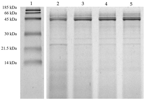
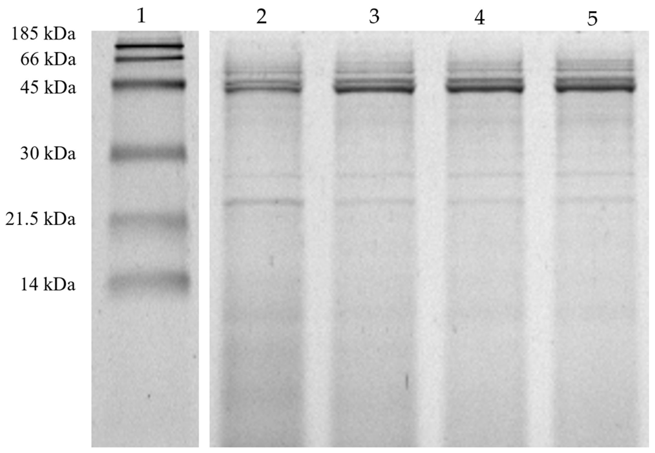
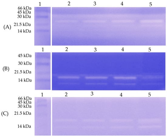
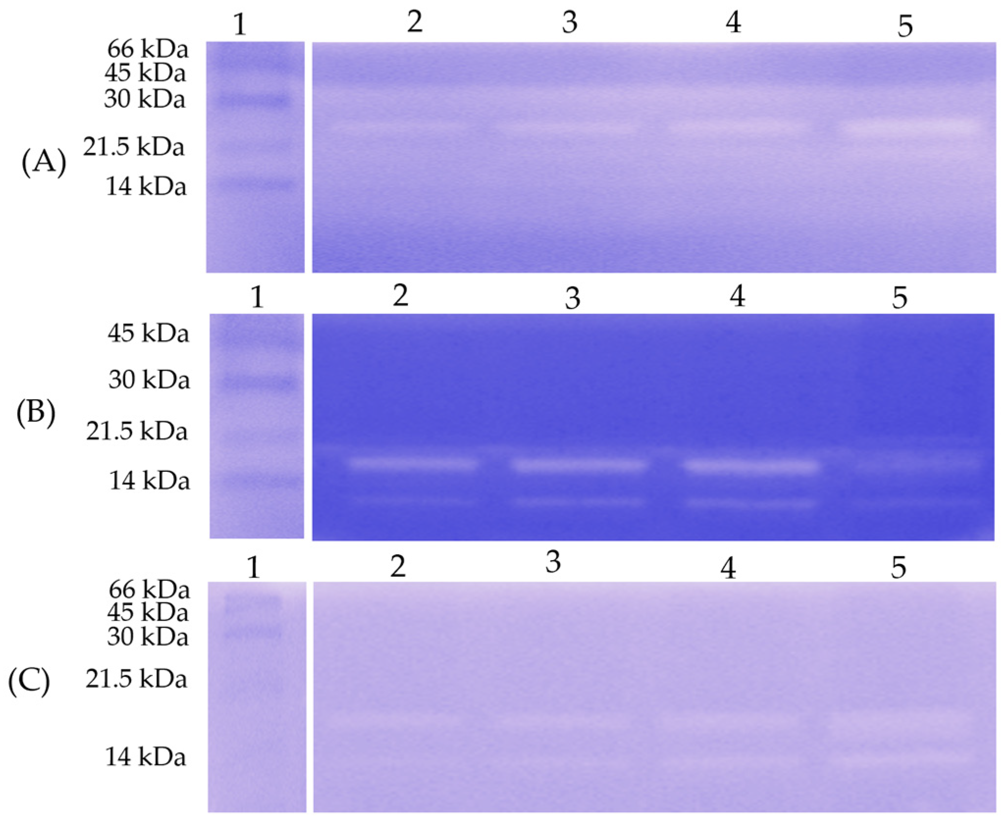
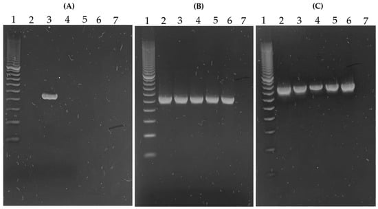
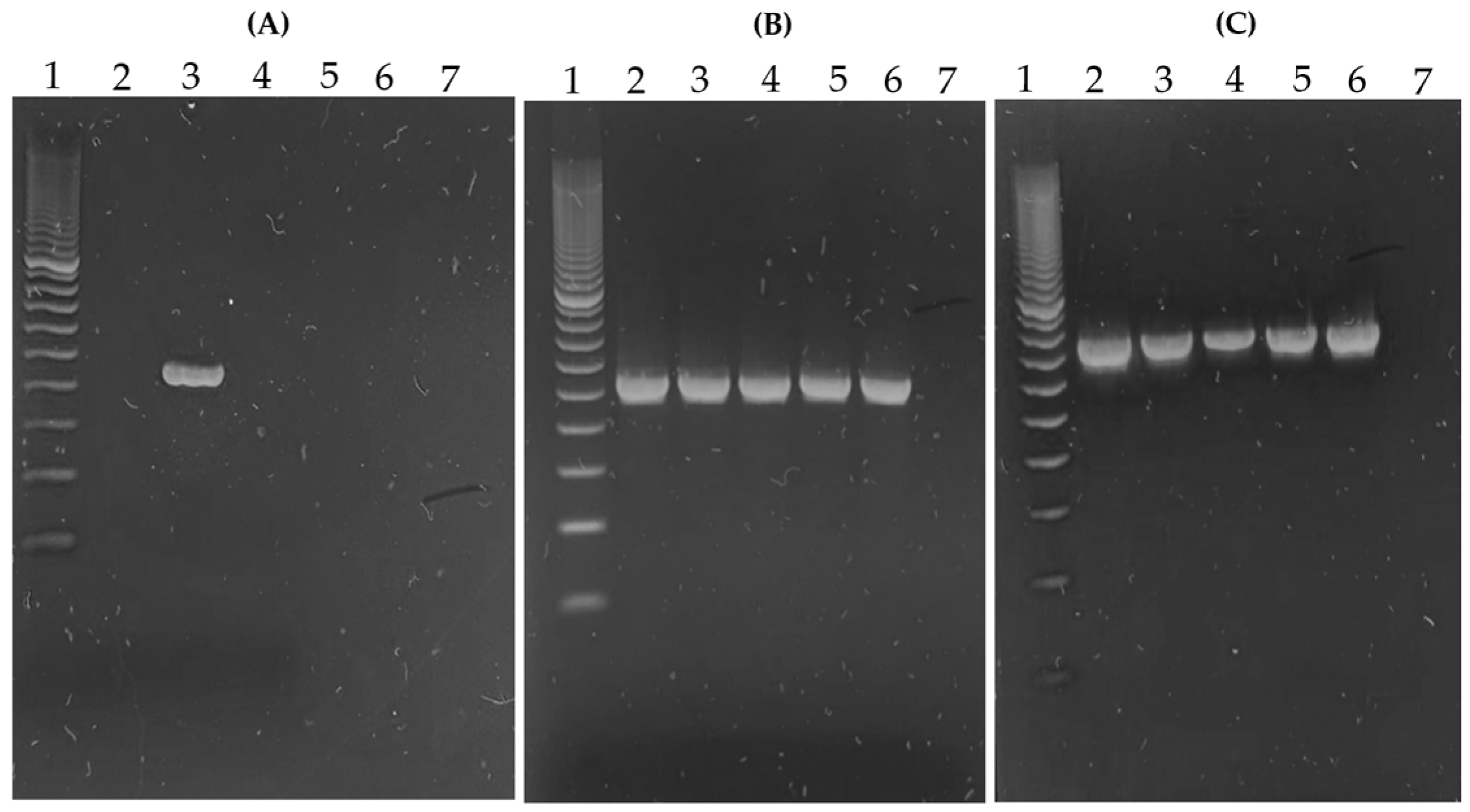
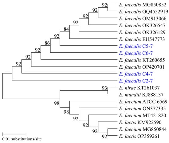
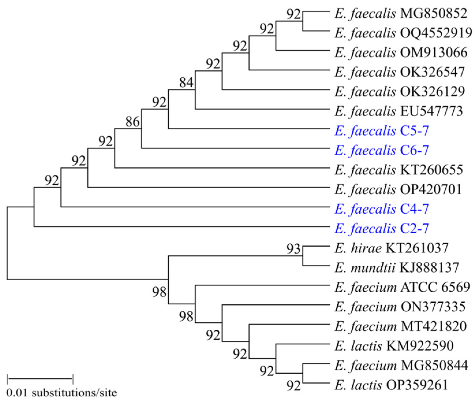
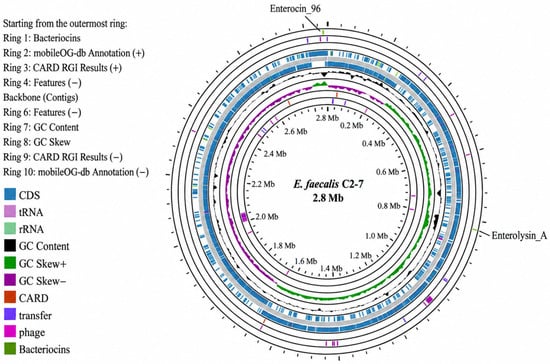
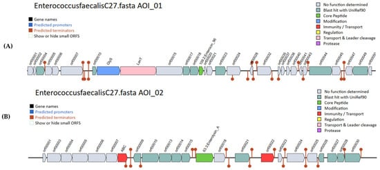

Disclaimer/Publisher’s Note: The statements, opinions and data contained in all publications are solely those of the individual author(s) and contributor(s) and not of MDPI and/or the editor(s). MDPI and/or the editor(s) disclaim responsibility for any injury to people or property resulting from any ideas, methods, instructions or products referred to in the content. |
© 2026 by the authors. Licensee MDPI, Basel, Switzerland. This article is an open access article distributed under the terms and conditions of the Creative Commons Attribution (CC BY) license.