


Abstract
Synovial fluid leucocyte count has long been regarded as a cornerstone in the diagnosis of paediatric septic arthritis, with a threshold of 50,000 cells/mm3 widely adopted in clinical practice. This narrative review critically appraised the available evidence regarding its diagnostic utility and interpretation in paediatric septic arthritis. To enhance the transparency and representativeness of the narrative synthesis, rather than to achieve the exhaustive and reproducible study retrieval required for a systematic review, relevant studies were identified through a structured literature search. Fourteen paediatric studies evaluating synovial fluid leucocyte count in the diagnostic assessment of septic arthritis were identified. Whilst several studies reported markedly elevated leucocyte counts in children with septic arthritis, others demonstrated microbiologically confirmed infections below the traditional cut-off, with substantial overlap observed with alternative diagnoses, particularly Lyme arthritis and juvenile idiopathic arthritis. Pathogen characteristics, host inflammatory responses, and prior antibiotic exposure were identified as important factors influencing synovial fluid findings. Current evidence suggests that synovial leucocyte count is best regarded as a marker of intra-articular inflammation rather than a definitive diagnostic test for septic arthritis. Its value lies in contributing to an integrated diagnostic assessment incorporating clinical presentation, imaging findings, microbiological investigations, and emerging molecular diagnostic techniques.
1. Introduction
Paediatric septic arthritis is an orthopaedic emergency requiring prompt diagnosis and treatment to prevent irreversible joint damage, growth disturbance, functional impairment, and systemic complications [1]. However, diagnosis remains challenging in children because clinical manifestations are often non-specific, particularly during the early stages of disease, and definitive microbiological confirmation of infection is not immediately available during the initial diagnostic work-up [2,3,4].
To facilitate the early diagnosis of septic arthritis, numerous clinical prediction algorithms have been proposed in the literature, combining clinical findings with serum inflammatory markers [5,6,7]. Nevertheless, synovial fluid analysis remains the cornerstone of the diagnostic evaluation of suspected septic arthritis [8,9,10]. In addition to microbiological investigations, synovial fluid is routinely assessed for its macroscopic appearance, Gram stain, total leucocyte count, and differential cell count, all of which contribute to diagnostic and therapeutic decision-making. Among these parameters, synovial leucocyte count has historically occupied a central role in the diagnostic assessment of septic arthritis. However, growing paediatric evidence has challenged the reliability of interpreting synovial leucocyte count according to a single diagnostic threshold [2,11,12,13,14,15,16,17].
The purpose of this narrative review is to critically evaluate the role of synovial fluid leucocyte count in the diagnosis of acute bacterial septic arthritis in children, hereafter referred to as septic arthritis unless otherwise specified. Specifically, this review examines the biological basis of synovial leucocytosis, the historical development of diagnostic thresholds, contemporary paediatric evidence regarding their diagnostic performance, and the pathogen-, treatment-, and host-related factors that influence synovial leucocyte count, with the aim of defining how this parameter should be interpreted in contemporary paediatric clinical practice.
2. Literature Search and Study Selection
This narrative review critically appraised the available evidence regarding the interpretation and diagnostic utility of synovial fluid leucocyte count in paediatric septic arthritis. Relevant studies were identified through a structured literature search.
A structured search of PubMed/MEDLINE, Embase, and the Cochrane Library was conducted from database inception to 31 August 2026. Search terms included combinations of “septic arthritis”, “acute bacterial arthritis”, “paediatric”, “children”, “native joint”, “joint aspiration”, “synovial fluid”, “synovial leucocyte count”, “synovial white blood cell count”, “polymorphonuclear neutrophils”, and “diagnostic threshold”. Reference lists of relevant articles and reviews were also screened to identify additional studies.
Eligible studies included paediatric populations with suspected or confirmed septic arthritis and reported synovial fluid leucocyte counts, diagnostic performance measures, or comparative data from clinically relevant differential diagnoses. Prospective and retrospective cohort studies and case series providing original clinical data were considered. Studies exclusively involving adults, implant- or prosthesis-related joint infections, those not reporting synovial fluid leucocyte counts, and those without relevant diagnostic data were excluded. Non-English publications were excluded unless an English-language version or translation was available through a journal, database, or other reliable academic source. Titles and abstracts of retrieved records were screened for relevance, followed by full-text assessment of potentially eligible articles according to these criteria. Following screening and full-text assessment, fourteen studies met the eligibility criteria and were retained for the narrative synthesis; their main characteristics and findings are summarised in Table 1.
Microbiological confirmation was not required for inclusion, and both microbiologically confirmed and clinically presumed culture-negative cases were considered. Cases in which a bacterial pathogen, including Kingella kingae, was identified by pathogen-specific PCR despite negative cultures were considered microbiologically confirmed. Where available, the reference standard and definition of septic arthritis used by each study were recorded and are reported in Table 1. Lyme and brucellar arthritis were considered clinically relevant differential diagnoses rather than primary target conditions, given their distinct clinical and microbiological characteristics and potential overlap in synovial leucocyte counts. Importantly, their epidemiological relevance is strongly geographically dependent, with the pre-test probability of these conditions varying substantially according to regional endemicity and exposure.
As this is a narrative review, the literature search and study-selection process was not designed as a formal systematic or scoping review, and no PRISMA-compliant protocol was applied. The objective was to provide a transparent and representative synthesis of the available paediatric evidence relevant to the interpretation of synovial leucocyte count. Accordingly, no formal risk-of-bias assessment or quantitative evidence synthesis was performed. The heterogeneity of the included studies, particularly with respect to study populations, diagnostic definitions, causative pathogens, joints examined, and reported outcomes, further limited the appropriateness of quantitative pooling.
3. Biological Basis of Synovial Leucocytosis
3.1. Mechanisms of Leucocyte Recruitment During Joint Infection
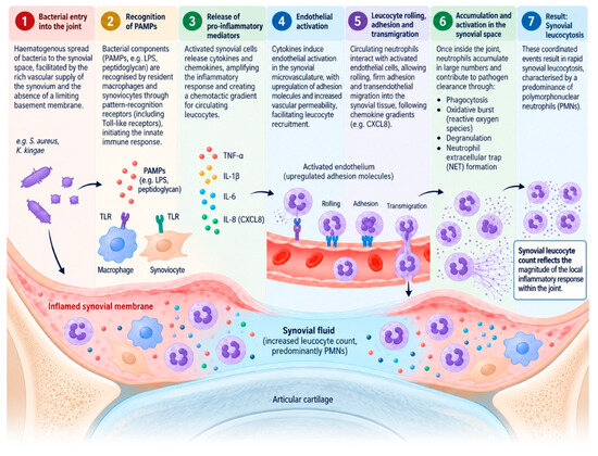
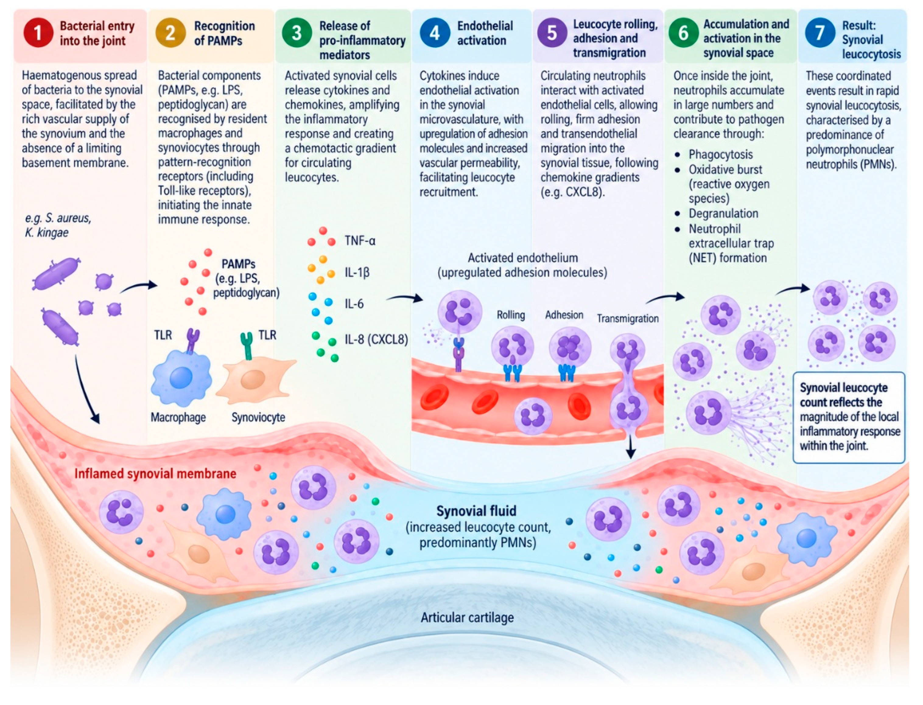
Septic arthritis is characterised by the presence of bacteria within the synovial space, followed by activation of a rapid and intense inflammatory response. In children, the synovium is particularly susceptible to haematogenous bacterial invasion because of its rich vascular supply and the absence of a limiting basement membrane, facilitating the translocation of microorganisms into the joint cavity [18,19,20,21]. Once bacteria enter the synovial space, pathogen-associated molecular patterns (PAMPs) are recognised by resident macrophages and synoviocytes through pattern-recognition receptors, including Toll-like receptors, initiating the innate immune response [22,23].
This early inflammatory phase is characterised by the release of pro-inflammatory cytokines, including tumour necrosis factor-α (TNF-α), interleukin (IL)-1β, IL-6, and IL-8 [20,22,24]. These mediators promote endothelial activation, upregulation of adhesion molecules, increased vascular permeability, and leucocyte chemotaxis, thereby facilitating the recruitment of inflammatory cells from the circulation into the infected synovium [22]. Among them, IL-8 (also known as CXCL8) is a potent neutrophil chemoattractant and plays a central role in the recruitment and activation of PMNs within the infected joint [25]. Activated endothelial cells express adhesion molecules that facilitate leucocyte rolling, adhesion, and transmigration into the synovial compartment [22,26,27]. Once within the joint, leucocytes, especially neutrophils, contribute to pathogen clearance through phagocytosis, oxidative burst activity, degranulation, and the formation of neutrophil extracellular traps [28].
Collectively, these coordinated inflammatory events result in rapid synovial leucocytosis, characterised by a predominance of PMNs. Synovial leucocyte count therefore reflects the magnitude of the local inflammatory response, providing the biological basis for its longstanding use as a diagnostic marker of septic arthritis (Figure 1).
Figure 1.
Mechanism of leucocyte recruitment during joint infection. Bacterial entry and recognition trigger pro-inflammatory signalling, endothelial activation, and neutrophil recruitment into the synovial space, resulting in PMN-predominant synovial leucocytosis. Coloured dots represent the different inflammatory mediators indicated in the figure (TNF-α, IL-1β, IL-6, and IL-8/CXCL8).
3.2. Inflammation-Mediated Joint Damage
Beyond their role in pathogen clearance, the inflammatory cells recruited into the synovial compartment contribute directly to articular tissue injury [29]. Activated neutrophils release reactive oxygen species (ROS) and proteolytic enzymes that contribute to bacterial killing but may also cause collateral tissue injury when local antioxidant defences are overwhelmed [30]. In parallel, the inflammatory environment promotes the activation of matrix-degrading enzymes, including matrix metalloproteinases (MMPs), leading to degradation of proteoglycans and collagen within the extracellular matrix (ECM) [31]. Progressive matrix disruption, together with oxidative injury and chondrocyte dysfunction and death, compromises the structural integrity of articular cartilage and may ultimately result in irreversible cartilage loss [32,33]. Importantly, cartilage degradation may begin within the first hours of joint infection, with experimental evidence demonstrating loss of cartilage glycosaminoglycans as early as eight hours after joint infection [34,35].
Articular damage may be further amplified by synovial inflammation and the accumulation of inflammatory exudate within the joint cavity. Increased vascular permeability and cellular infiltration promote synovial oedema and joint effusion, raising intra-articular pressure and potentially compromising local perfusion. Reduced blood supply may contribute to tissue ischaemia and further chondrocyte injury and, particularly in anatomically vulnerable joints such as the shoulder or the hip, may contribute to substantial local morbidity [36]. If infection and inflammation persist, the combined effects of enzymatic degradation, oxidative injury, and impaired perfusion may extend to the subchondral bone and ultimately lead to progressive and potentially irreversible joint destruction [37].
These mechanisms can also be interpreted within the Damage-Response Framework (DRF), which conceptualises disease manifestations and tissue damage as the result of the dynamic interaction between pathogen-associated factors and the host immune response. Within this framework, host damage may arise from microbial virulence factors, an insufficient immune response that permits pathogen persistence, an excessive inflammatory response that itself contributes to tissue injury, or a combination of these mechanisms [38]. This interplay is particularly relevant in septic arthritis, in which immune activation is essential for pathogen control but may simultaneously contribute to articular damage. Collectively, these mechanisms demonstrate that synovial leucocytosis is not merely a diagnostic finding but also reflects an intra-articular inflammatory response capable of contributing to progressive articular damage.
3.3. Factors Influencing Synovial Leucocyte Count
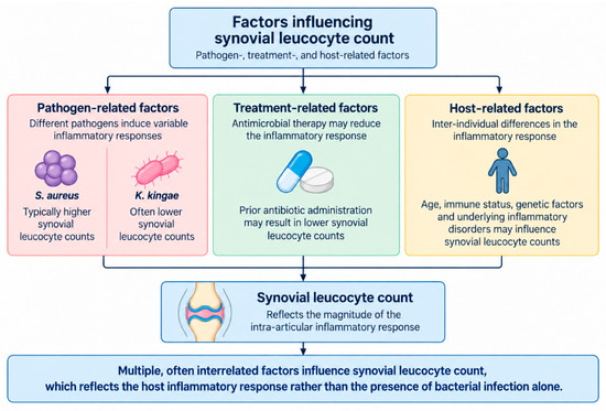
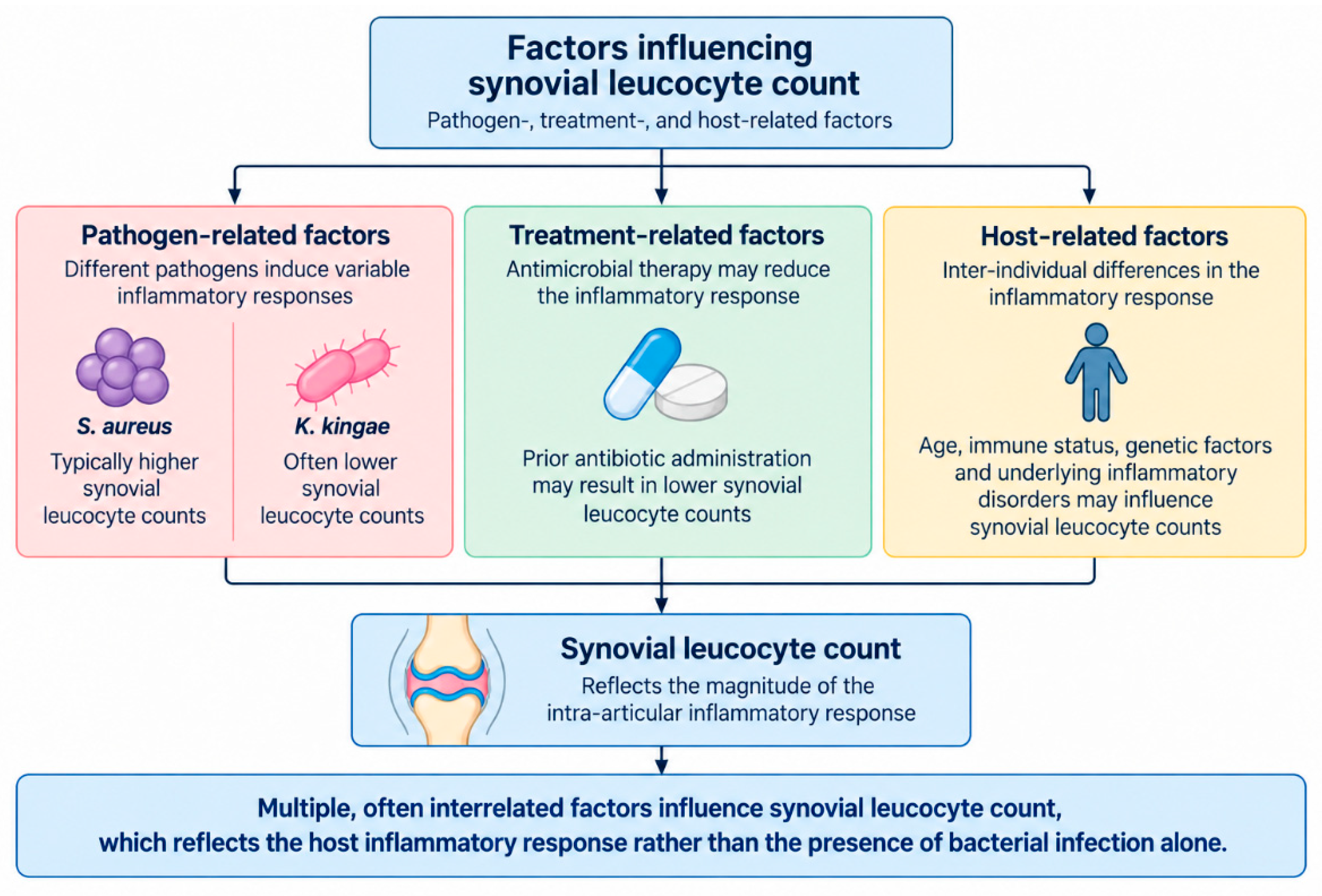
Although synovial leucocyte count reflects the intensity of intra-articular inflammation, its magnitude varies considerably between patients owing to a complex interplay of pathogen-, treatment-, and host-related factors (Figure 2). These factors are frequently interrelated; for example, age influences the epidemiological distribution of causative pathogens in paediatric septic arthritis and may therefore indirectly affect the magnitude of the synovial inflammatory response. This heterogeneity helps explain why no single synovial leucocyte threshold can reliably distinguish septic arthritis from alternative diagnoses.
Figure 2.
Factors influencing synovial leucocyte count. Synovial leucocyte count is influenced by a complex interplay of pathogen-, treatment-, and host-related factors. These factors contribute to substantial variability in synovial leucocytosis, reinforcing its interpretation as a marker of the magnitude of intra-articular inflammation rather than of bacterial infection alone.
One of the principal determinants of synovial fluid cellularity is the causative pathogen. Infections caused by highly pyogenic organisms, such as S. aureus, are generally associated with marked neutrophilic inflammation and substantial synovial leucocytosis. In contrast, K. kingae, now recognised as a leading cause of osteoarticular infection in children younger than four years, often produces a milder clinical and biological inflammatory response [12]. Several studies have reported relatively modest synovial leucocyte counts in children with K. kingae septic arthritis, consistent with its generally less pronounced inflammatory phenotype [2,18,39]. However, exceptions have been reported. Basmaci et al. reported a higher median synovial leucocyte count in children with K. kingae arthritis than in those with S. aureus arthritis, despite the milder clinical presentation associated with K. kingae [11]. Consequently, differences in synovial leucocyte count between paediatric cohorts may partly reflect differences in age distribution and the associated pathogen spectrum, rather than differences in the intrinsic diagnostic performance of synovial leucocyte count itself.
K. kingae possesses several virulence and immune-evasion mechanisms that may contribute to its comparatively attenuated inflammatory phenotype. Type IV pili mediate initial adherence to host cells, while subsequent pilus retraction facilitates Knh-mediated close attachment [40]. The polysaccharide capsule and galactan exopolysaccharide promote immune evasion by reducing neutrophil recognition, phagocytosis, and killing and protecting against complement-mediated lysis [41]. In addition, the RtxA toxin exerts cytotoxic effects on epithelial, synovial, and macrophage-like cells and may contribute to immune evasion through cytotoxicity towards recruited phagocytic cells [42]. K. kingae also carries the phase-variable Type III DNA methyltransferase ModK; ModK1 “ON” variants have been associated with reduced macrophage production of pro-inflammatory cytokines, including IL-8, IL-1β, and TNF [43]. Collectively, these mechanisms may facilitate persistence while attenuating host inflammatory responses, providing a plausible biological basis for the comparatively mild inflammatory phenotype observed in K. kingae infection [40,41,42,43].
Treatment-related factors, particularly antibiotic administration before arthrocentesis, may also contribute to variability in synovial fluid findings. Empirical antimicrobial therapy initiated before joint aspiration may attenuate the local inflammatory response and alter synovial fluid findings [44]. However, much of the evidence supporting this effect derives from adult populations and should therefore be regarded as indirect evidence when considering paediatric septic arthritis. Puzzitiello et al. reported lower synovial leucocyte counts following pre-aspiration antibiotic administration in adults with native-joint septic arthritis, while Massey et al. similarly demonstrated that the optimal diagnostic leucocyte threshold was lower in a predominantly adult cohort receiving antibiotics before arthrocentesis [44,45].
Paediatric-specific evidence remains considerably more limited. Obana et al. found no significant association between prior antibiotic exposure and synovial leucocyte count or PMN proportion in children with septic arthritis [10]. Other paediatric studies have included relatively small numbers of pretreated patients or have not reported antibiotic exposure in sufficient detail to permit robust conclusions [46,47]. Interpretation of these apparently conflicting findings is limited by the inconsistent reporting of antibiotic exposure across studies, particularly regarding the antimicrobial agent administered, duration of treatment, and interval between antibiotic administration and joint aspiration. Differences in these factors, together with variation in study populations and sample sizes, may contribute to the observed discrepancies. Future paediatric studies should therefore report pre-aspiration antibiotic exposure in a standardised manner, including the agent administered, duration of therapy, and interval between administration and synovial fluid collection. Whenever clinically feasible, synovial fluid should be obtained before the initiation of antimicrobial therapy.
Host-related factors also contribute to variability in synovial fluid findings through differences in the inflammatory response. Immune status and underlying inflammatory disorders may modulate this response [48,49] such that synovial leucocyte counts may vary even among patients with the same infection. Moreover, because intense intra-articular inflammation is not specific to bacterial infection, conditions such as Lyme arthritis and juvenile idiopathic arthritis may also produce marked synovial leucocytosis despite the absence of pyogenic pathogens [50,51]. These observations reinforce that synovial leucocyte count primarily reflects the magnitude of the local inflammatory response rather than the presence of bacterial infection itself.
4. Historical Development of Diagnostic Thresholds
Historically, interpretation of synovial fluid in suspected septic arthritis has relied heavily on numerical thresholds intended to distinguish bacterial infection from non-infectious inflammatory joint disease. Among these, a synovial leucocyte count exceeding 50,000 cells/mm3 [45,52,53,54,55] and a PMN proportion greater than 90% became widely used indicators of septic arthritis [56,57,58].
These cut-offs were largely informed by studies demonstrating higher synovial leucocyte counts and neutrophil proportions in septic arthritis than in non-infectious joint conditions. As readily available parameters in the absence of immediate microbiological confirmation, they became incorporated into the diagnostic assessment of suspected septic arthritis.
However, the evidence underlying these conventional thresholds was derived predominantly from adult or mixed-age populations, with limited validation in children. Their diagnostic applicability to paediatric septic arthritis therefore requires consideration in the context of contemporary paediatric evidence.
Table 1.
Summary of paediatric studies assessing the diagnostic utility of synovial leucocyte count in septic arthritis.
5. Contemporary Paediatric Evidence
Recent paediatric studies have challenged the diagnostic reliability of the traditional synovial leucocyte threshold of 50,000 cells/mm3 for septic arthritis. As summarised in Table 1, higher synovial leucocyte counts are generally associated with bacterial infection; however, microbiologically confirmed infections may occur below this cut-off, while inflammatory and other infectious arthritides may produce values above it [3,5,10].
Several paediatric studies have documented septic arthritis despite synovial leucocyte counts below 50,000 cells/mm3. Williams et al. reported a median synovial leucocyte count of 21,800 cells/mm3 in children with PCR-confirmed K. kingae septic arthritis [12]. Low counts have also been reported in other infectious arthritides within the differential diagnosis; Press et al. similarly found a median count of 9500 cells/mm3 in children with microbiologically confirmed brucellar arthritis [13]. Thomas et al. found that 9 of 51 (17.6%) children with true septic arthritis had synovial leucocyte counts below 50,000 cells/mm3, with a mean count of 22,000 cells/mm3 in this subgroup [50]. In the diagnostic ‘grey zone’ examined by Heyworth et al., septic arthritis was diagnosed in 17% of children with synovial leucocyte counts below 50,000 cells/mm3, compared with 48% of those with counts ≥50,000 cells/mm3. Although a count ≥50,000 cells/mm3 was associated with significantly higher odds of septic arthritis, these findings demonstrate that increasing leucocytosis raises the probability of septic arthritis without providing an absolute diagnostic boundary [2,14,50,61].
This variability persists even in cohorts with generally high synovial leucocyte counts. Dart et al. observed a median count of 126,130 cells/mm3 in septic arthritis, yet 19% of confirmed cases had values below 50,000 cells/mm3 [47]. Similarly, Illán-Ramos et al. reported a median of 61,322 cells/mm3 in K. kingae septic arthritis, while one of four patients with available synovial leucocyte counts (25%) had a value below the traditional cut-off [60].
Conversely, values above 50,000 cells/mm3 are not specific for pyogenic infection. Baldwin et al. reported a mean synovial leucocyte count of 53,640 cells/mm3 in children with Lyme arthritis, exceeding the conventional threshold [59]. Similarly, Dart et al. found that 56% of children with Lyme arthritis had counts >50,000 cells/mm3 [47]. Comparable overlap has been observed in inflammatory rheumatological disease, with Aupiais et al. reporting values above 50,000 cells/mm3 in approximately half of children with juvenile idiopathic arthritis [3].
Interpretation of these findings is also influenced by differences in the reference standards used across studies. Some studies defined septic arthritis exclusively by microbiological confirmation, whereas others included clinically presumed culture-negative cases or incorporated synovial leucocyte count itself into the diagnostic definition. The latter introduces incorporation bias, because the index test forms part of the reference standard against which its diagnostic performance is evaluated, potentially inflating its apparent diagnostic accuracy. Conversely, reliance on microbiological confirmation alone may misclassify true culture-negative infections, particularly following prior antibiotic exposure or in infections caused by fastidious organisms. Such differences in reference standards may therefore contribute to the heterogeneity in reported diagnostic performance and limit direct comparison of synovial leucocyte thresholds across studies.
Taken together, these findings support interpreting synovial leucocyte count as a continuous diagnostic variable rather than a binary test. Increasing synovial leucocytosis is associated with a greater likelihood of septic arthritis, but no single threshold reliably confirms or excludes the diagnosis in children.
6. Clinical Implications and Future Perspectives
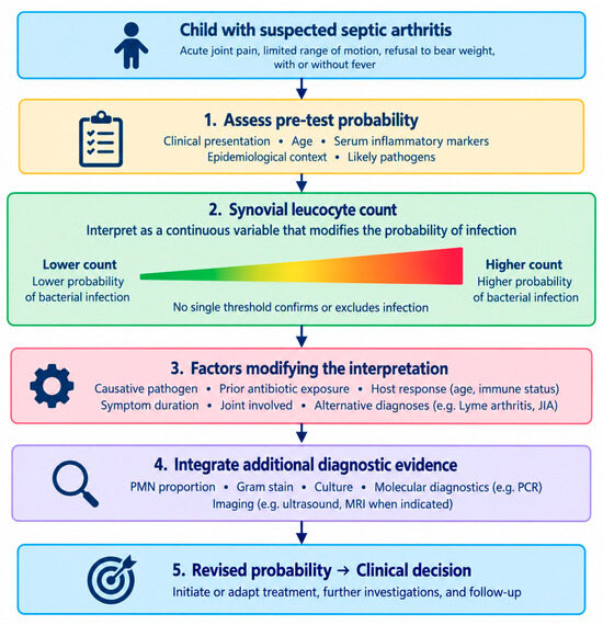
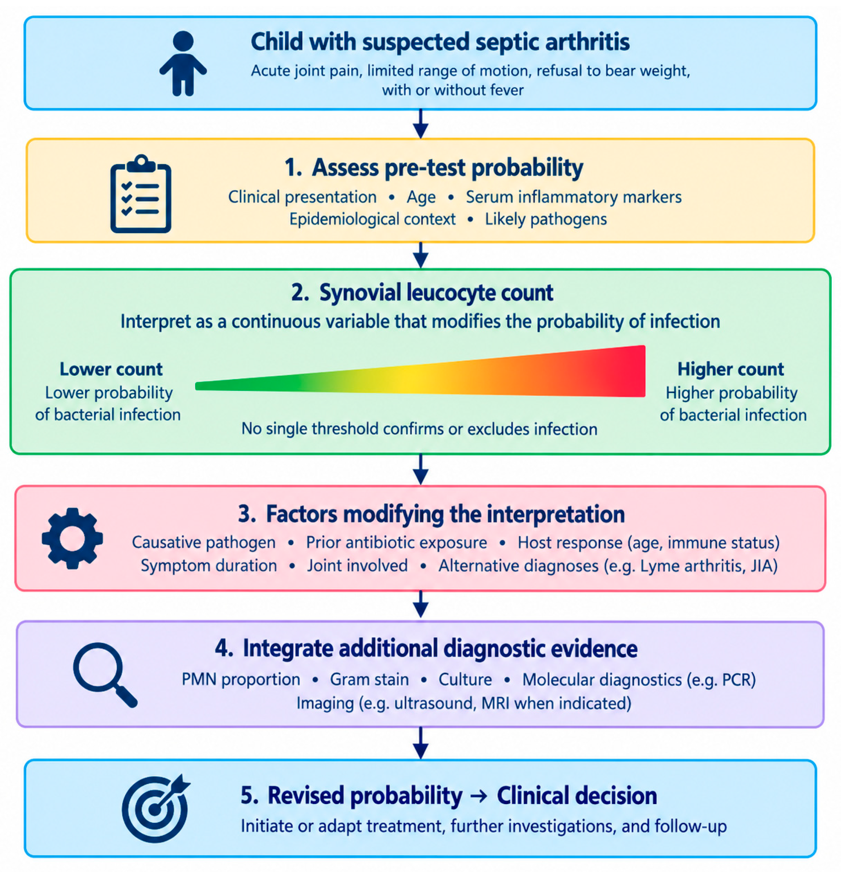
From a clinical perspective, synovial leucocyte count should no longer be interpreted as an isolated diagnostic criterion but rather as one component of an integrated and probabilistic diagnostic assessment. Its diagnostic significance depends on the pre-test probability of acute bacterial arthritis, which is determined by the clinical presentation, age, epidemiological context, serum inflammatory markers, imaging findings, and the likelihood of specific pathogens. The synovial leucocyte count should therefore be interpreted as a continuous variable that modifies this initial probability rather than as a binary test that independently confirms or excludes infection.
Accordingly, the same synovial leucocyte count may have different diagnostic implications depending on the clinical context. For example, a count of 40,000 cells/mm3 should not substantially reduce concern for acute bacterial arthritis in a febrile, systemically unwell child who refuses weight bearing and has markedly elevated inflammatory markers. Conversely, the same value in an otherwise well child presenting with isolated knee swelling in a Lyme-endemic region may carry a different diagnostic weight. Conceptually, the diagnostic process can therefore be considered as a sequence from pre-test probability, through interpretation of the synovial leucocyte count and PMN proportion, to a revised post-test probability, which is subsequently refined by microbiological, clinical, and imaging findings (Figure 3).
Figure 3.
Integrated diagnostic interpretation of synovial leucocyte count in suspected paediatric septic arthritis. Synovial leucocyte count should be interpreted as a continuous variable that modifies the pre-test probability of infection. Its interpretation should integrate clinical context and additional diagnostic evidence to refine the probability of infection and guide clinical decision-making.
This approach is broadly consistent with the 2023 PIDS/IDSA guideline for acute bacterial arthritis in children, which recommends synovial leucocyte count and differential as part of the initial evaluation while emphasising that these findings should be interpreted alongside the clinical presentation and other laboratory findings [9]. The guideline also supports molecular testing when routine microbiological investigations fail to identify a pathogen, particularly in preschool-aged children at risk of K. kingae infection. Decisions regarding joint drainage are likewise based on the overall clinical context, including the severity of infection, the affected joint, and the gross appearance and cellular profile of the synovial fluid, rather than on a single synovial leucocyte threshold [9]. The present review therefore largely aligns with current guideline recommendations while further emphasising synovial leucocyte count as a continuous, probability-modifying variable rather than a fixed diagnostic threshold.
As molecular diagnostic techniques continue to improve pathogen detection, future diagnostic strategies are likely to rely increasingly on this integrated approach. Conventional Gram staining and culture remain fundamental, but the increasing availability of molecular diagnostic techniques has substantially expanded the microbiological investigation of culture-negative infections.
Since the early 2000s, the increasing use of nucleic acid amplification tests (NAATs) has substantially improved the microbiological diagnosis of paediatric osteoarticular infections and has contributed to a better understanding of their bacterial epidemiology. Targeted PCR-based testing represents an important advance by allowing the rapid detection of pathogen-specific nucleic acid directly in clinical specimens, including in infections caused by fastidious organisms that may remain undetected by conventional culture [62]. Advances in NAAT technology, particularly the development of species-specific real-time PCR assays, have improved diagnostic performance while reducing some of the limitations associated with broad-range amplification techniques.
This evolution is particularly relevant to K. kingae, which is now recognised as a leading cause of osteoarticular infection in children younger than four years [63]. Species-specific PCR has substantially increased its detection, including in culture-negative cases, and has consequently revealed a larger population of microbiologically confirmed infections associated with a comparatively mild clinical and biological inflammatory response [64]. Importantly, however, more than 20% of paediatric osteoarticular infections may remain microbiologically undocumented despite contemporary molecular testing, suggesting that additional fastidious or as-yet unrecognised organisms may remain undetected with currently available targeted assays [65,66]. Molecular diagnostic advances have also improved the identification of other infectious arthritides, including those caused by Borrelia burgdorferi and Brucella spp., although these remain clinically relevant differential diagnoses rather than the primary diagnostic target of this review [67,68].
These developments may have important implications for the interpretation of synovial leucocyte count. The increasing use of targeted molecular assays allows microbiological confirmation of infections that might previously have remained culture-negative, microbiologically undocumented, or diagnostically uncertain, including infections caused by less pyogenic organisms [62,63,64,65,66]. Such cases may present with less pronounced synovial leucocytosis and may therefore partly explain why contemporary evidence increasingly reports microbiologically confirmed infections below the traditional threshold of 50,000 cells/mm3. Thus, the increasing recognition of septic arthritis presenting with lower synovial leucocyte counts should not necessarily be interpreted as a change in the biological behaviour of the disease, but may partly reflect improved microbiological detection across a broader spectrum of pathogen virulence, bacterial burden, and disease stage.
Rather than replacing synovial fluid analysis, these techniques should be regarded as complementary investigations that further refine the probability of infection, particularly in diagnostically challenging cases.
Additional biochemical parameters may also provide complementary information when interpreted alongside synovial leucocyte count. Changes in synovial glucose, lactate, and total protein have been investigated as potential indicators of bacterial joint infection, although their diagnostic performance and clinical utility remain insufficiently established, particularly in children [69,70]. Procalcitonin has also emerged as a potential biomarker of bacterial arthritis. Aggarwal et al. reported significantly higher serum and synovial procalcitonin concentrations in patients with pyogenic compared with non-pyogenic arthritis, with synovial procalcitonin demonstrating slightly greater diagnostic accuracy than serum procalcitonin (AUC 0.914 vs. 0.895) [71]. However, the available evidence remains limited and is predominantly derived from adult or mixed-age populations. These parameters should therefore currently be regarded as complementary rather than standalone diagnostic markers, and further paediatric-specific studies are required.
Imaging should similarly be interpreted as a complementary component of this diagnostic framework rather than as an independent discriminator of infection or a substitute for synovial fluid analysis [72,73]. Conventional MRI can contribute to the differentiation of septic from non-septic arthritis through features such as synovial enhancement, periarticular soft-tissue abnormalities, and bone marrow changes; however, joint effusion itself remains non-specific, and substantial overlap exists between septic and reactive effusions [72]. Emerging techniques, including DWI and T2-relaxometry, may provide additional information on joint-fluid characteristics, but their role in paediatric septic arthritis remains investigational [74,75]. Whether these approaches provide diagnostic value beyond established clinical, synovial, and microbiological parameters requires further investigation.
Future studies should prospectively evaluate synovial leucocyte count in large, multicentre paediatric cohorts using standardised definitions of acute bacterial arthritis and systematic microbiological testing. Rather than seeking another universal cut-off, future research should assess synovial leucocyte count as a continuous variable and determine its incremental diagnostic value when combined with clinical findings, serum inflammatory markers, PMN proportion, culture, and molecular diagnostics. Future studies should also account for the diagnostic method used for pathogen identification, as increasing use of targeted molecular assays may alter the spectrum of microbiologically confirmed disease and, consequently, the observed distribution of synovial leucocyte counts. Stratification by age, causative pathogen, joint involved, and prior antibiotic exposure would further clarify the factors influencing its diagnostic performance.
7. Conclusions
Synovial fluid leucocyte count remains an important component of the diagnostic evaluation of children with suspected septic arthritis. Although increasing synovial leucocytosis is associated with a greater probability of bacterial infection, contemporary paediatric evidence demonstrates that no single synovial leucocyte threshold can reliably confirm or exclude the diagnosis.
Rather than representing an absolute diagnostic criterion, synovial leucocyte count should be regarded as a marker of intra-articular inflammation. In clinical practice, a value below 50,000 cells/mm3 should not exclude septic arthritis when clinical suspicion remains high, while a value above this threshold should not be considered diagnostic of bacterial infection in isolation. Its interpretation must therefore be integrated with the clinical presentation, serum inflammatory markers, imaging findings, microbiological investigations, and, increasingly, molecular diagnostic techniques.
Author Contributions
Conceptualization, V.S. and D.C.; writing—original draft preparation, V.S. and D.C.; writing—review and editing, V.S., A.R., A.T., S.E.H., P.A.D., G.d.M., O.V., C.S., R.D. and D.C.; visualization, V.S., A.R. and A.T.; supervision, D.C. All authors have read and agreed to the published version of the manuscript.
Funding
This research received no external funding.
Institutional Review Board Statement
Not applicable.
Informed Consent Statement
Not applicable.
Data Availability Statement
No new data were created or analysed in this study. Data sharing is not applicable to this article.
Acknowledgments
During the preparation of this manuscript, the authors used ChatGPT (OpenAI, GPT-5.6) to improve the readability and language of the manuscript and to assist in the preparation of schematic figures. All AI-assisted graphical content was critically reviewed and revised by the authors to ensure scientific accuracy and consistency with the manuscript. The authors have reviewed and edited all AI-assisted output and take full responsibility for the content of this publication.
Conflicts of Interest
The authors declare no conflicts of interest.
Abbreviations
The following abbreviations are used in this manuscript:
| AUC | Area under the curve |
| C-SA | Confirmed septic arthritis |
| CXCL8 | C-X-C motif chemokine ligand 8 |
| DRF | Damage-Response Framework |
| DWI | Diffusion-weighted imaging |
| ECM | Extracellular matrix |
| IL | Interleukin |
| IL-1β | Interleukin-1 beta |
| IL-6 | Interleukin-6 |
| IL-8 | Interleukin-8 |
| IQR | Interquartile range |
| JIA | Juvenile idiopathic arthritis |
| K. kingae | Kingella kingae |
| MMPs | Matrix metalloproteinases |
| MRI | Magnetic resonance imaging |
| MRSA | Methicillin-resistant Staphylococcus aureus |
| MSSA | Methicillin-susceptible Staphylococcus aureus |
| NAATs | Nucleic acid amplification tests |
| OM | Osteomyelitis |
| PAMPs | Pathogen-associated molecular patterns |
| PCR | Polymerase chain reaction |
| PMN/PMNs | Polymorphonuclear neutrophil(s) |
| ROS | Reactive oxygen species |
| rRNA | Ribosomal RNA |
| S. aureus | Staphylococcus aureus |
| SA | Septic arthritis |
| S-SA | Suspected septic arthritis |
| TNF-α | Tumour necrosis factor alpha |
| WBC | White blood cell |
References
- Brown, D.W.; Sheffer, B.W. Pediatric Septic Arthritis: An Update. Orthop. Clin. N. Am. 2019, 50, 461–470. [Google Scholar] [CrossRef] [Scilit]
- Thomas, M.; Bonacorsi, S.; Simon, A.-L.; Mallet, C.; Lorrot, M.; Faye, A.; Dingulu, G.; Caseris, M.; Boneca, I.G.; Aupiais, C.; et al. Acute Monoarthritis in Young Children: Comparing the Characteristics of Patients with Juvenile Idiopathic Arthritis versus Septic and Undifferentiated Arthritis. Sci. Rep. 2021, 11, 3422. [Google Scholar] [CrossRef] [Scilit]
- Aupiais, C.; Basmaci, R.; Ilharreborde, B.; Blachier, A.; Desmarest, M.; Job-Deslandre, C.; Faye, A.; Bonacorsi, S.; Alberti, C.; Lorrot, M. Arthritis in Children: Comparison of Clinical and Biological Characteristics of Septic Arthritis and Juvenile Idiopathic Arthritis. Arch. Dis. Child. 2017, 102, 316–322. [Google Scholar] [CrossRef] [Scilit]
- Paton, E.A. Evaluating the Child With a Limp. Adv. Emerg. Nurs. J. 2007, 29, 68–72. [Google Scholar] [CrossRef] [Scilit]
- Caird, M.S.; Flynn, J.M.; Leung, Y.L.; Millman, J.E.; D’Italia, J.G.; Dormans, J.P. Factors Distinguishing Septic Arthritis from Transient Synovitis of the Hip in Children. A Prospective Study. J. Bone Jt. Surg. Am. 2006, 88, 1251–1257. [Google Scholar] [CrossRef] [Scilit]
- Kocher, M.S.; Zurakowski, D.; Kasser, J.R. Differentiating between Septic Arthritis and Transient Synovitis of the Hip in Children: An Evidence-Based Clinical Prediction Algorithm. J. Bone Jt. Surg. Am. 1999, 81, 1662–1670. [Google Scholar] [CrossRef] [Scilit]
- Kocher, M.S.; Mandiga, R.; Zurakowski, D.; Barnewolt, C.; Kasser, J.R. Validation of a Clinical Prediction Rule for the Differentiation between Septic Arthritis and Transient Synovitis of the Hip in Children. J. Bone Jt. Surg. Am. 2004, 86, 1629–1635. [Google Scholar] [CrossRef] [Scilit]
- Miller, J.M.; Binnicker, M.J.; Campbell, S.; Carroll, K.C.; Chapin, K.C.; Gonzalez, M.D.; Harrington, A.; Jerris, R.C.; Kehl, S.C.; Leal, S.M.; et al. Guide to Utilization of the Microbiology Laboratory for Diagnosis of Infectious Diseases: 2024 Update by the Infectious Diseases Society of America (IDSA) and the American Society for Microbiology (ASM). Clin. Infect. Dis. 2018, 67, e1–e94. [Google Scholar] [CrossRef] [Scilit]
- Woods, C.R.; Bradley, J.S.; Chatterjee, A.; Kronman, M.P.; Arnold, S.R.; Robinson, J.; Copley, L.A.; Arrieta, A.C.; Fowler, S.L.; Harrison, C.; et al. Clinical Practice Guideline by the Pediatric Infectious Diseases Society (PIDS) and the Infectious Diseases Society of America (IDSA): 2023 Guideline on Diagnosis and Management of Acute Bacterial Arthritis in Pediatrics. J. Pediatr. Infect. Dis. Soc. 2024, 13, 1–59, Erratum in J. Pediatr. Infect. Dis. Soc. 2024, 13, 443. https://doi.org/10.1093/jpids/piae065. [Google Scholar] [CrossRef] [Scilit] [PubMed]
- Obana, K.K.; Murgai, R.R.; Schur, M.; Broom, A.M.; Hsu, A.; Kay, R.M.; Pace, J.L. Synovial Fluid Cell Counts and Its Role in the Diagnosis of Paediatric Septic Arthritis. J. Child. Orthop. 2019, 13, 417–422. [Google Scholar] [CrossRef] [Scilit]
- Basmaci, R.; Lorrot, M.; Bidet, P.; Doit, C.; Vitoux, C.; Penneçot, G.; Mazda, K.; Bingen, E.; Ilharreborde, B.; Bonacorsi, S. Comparison of Clinical and Biologic Features of Kingella kingae and Staphylococcus aureus Arthritis at Initial Evaluation. Pediatr. Infect. Dis. J. 2011, 30, 902–904. [Google Scholar] [CrossRef] [Scilit]
- Williams, N.; Cooper, C.; Cundy, P. Kingella kingae Septic Arthritis in Children: Recognising an Elusive Pathogen. J. Child. Orthop. 2014, 8, 91–95. [Google Scholar] [CrossRef] [Scilit]
- Press, J.; Peled, N.; Buskila, D.; Yagupsky, P. Leukocyte Count in the Synovial Fluid of Children with Culture-Proven Brucellar Arthritis. Clin. Rheumatol. 2002, 21, 191–193. [Google Scholar] [CrossRef] [Scilit]
- Heyworth, B.E.; Shore, B.J.; Donohue, K.S.; Miller, P.E.; Kocher, M.S.; Glotzbecker, M.P. Management of Pediatric Patients with Synovial Fluid White Blood-Cell Counts of 25,000 to 75,000 Cells/Mm3 After Aspiration of the Hip. J. Bone Jt. Surg. 2015, 97, 389–395. [Google Scholar] [CrossRef] [Scilit]
- Spyridakis, E.; Gerber, J.S.; Schriver, E.; Grundmeier, R.W.; Porsch, E.A.; St Geme, J.W.; Downes, K.J. Clinical Features and Outcomes of Children with Culture-Negative Septic Arthritis. J. Pediatr. Infect. Dis. Soc. 2019, 8, 228–234. [Google Scholar] [CrossRef] [Scilit]
- Otsubo, Y.; Shibata, M.; Hataya, H.; Ota, N.; Horikoshi, Y. Joint Fluid Cell Count in Acute Bacterial Arthritis Patients Proven Positive Culture in Children. Pediatr. Infect. Dis. J. 2024, 43, 640–642. [Google Scholar] [CrossRef] [Scilit]
- Dechnik, A.; Kahane, C.G.; Nigrovic, L.E.; Lyons, T.W. Utility of Synovial Fluid Biomarkers for Culture-Positive Septic Arthritis in a Lyme Disease-Endemic Region. Pediatr. Emerg. Care 2024, 40, e82–e88. [Google Scholar] [CrossRef] [Scilit]
- Yagupsky, P. Native Joint Arthritis in Children. In Bone and Joint Infections; Zimmerli, W., Ed.; Wiley: Hoboken, NJ, USA, 2021; pp. 117–137. [Google Scholar]
- Thakolkaran, N.; Shetty, A.K. Acute Hematogenous Osteomyelitis in Children. Ochsner J. 2019, 19, 116–122. [Google Scholar] [CrossRef] [Scilit]
- Shirtliff, M.E.; Mader, J.T. Acute Septic Arthritis. Clin. Microbiol. Rev. 2002, 15, 527–544. [Google Scholar] [CrossRef] [Scilit]
- Iliadis, A.D.; Ramachandran, M. Paediatric Bone and Joint Infection. EFORT Open Rev. 2017, 2, 7–12. [Google Scholar] [CrossRef] [Scilit]
- Zec, K.; Schonfeldova, B.; Ai, Z.; Van Grinsven, E.; Pirgova, G.; Eames, H.L.; Berthold, D.L.; Attar, M.; Compeer, E.B.; Arnon, T.I.; et al. Macrophages in the Synovial Lining Niche Initiate Neutrophil Recruitment and Articular Inflammation. J. Exp. Med. 2023, 220, e20220595. [Google Scholar] [CrossRef] [Scilit]
- Knab, K.; Chambers, D.; Krönke, G. Synovial Macrophage and Fibroblast Heterogeneity in Joint Homeostasis and Inflammation. Front. Med. 2022, 9, 862161. [Google Scholar] [CrossRef] [Scilit]
- Earwood, J.S.; Walker, T.R.; Sue, G.J.C. Septic Arthritis: Diagnosis and Treatment. Am. Fam. Physician 2021, 104, 589–597. [Google Scholar]
- Pichert, A.; Schlorke, D.; Franz, S.; Arnhold, J. Functional Aspects of the Interaction between Interleukin-8 and Sulfated Glycosaminoglycans. Biomatter 2012, 2, 142–148. [Google Scholar] [CrossRef] [Scilit]
- Mitroulis, I.; Alexaki, V.I.; Kourtzelis, I.; Ziogas, A.; Hajishengallis, G.; Chavakis, T. Leukocyte Integrins: Role in Leukocyte Recruitment and as Therapeutic Targets in Inflammatory Disease. Pharmacol. Ther. 2015, 147, 123–135. [Google Scholar] [CrossRef] [Scilit]
- Langer, H.F.; Chavakis, T. Leukocyte-Endothelial Interactions in Inflammation. J. Cell. Mol. Med. 2009, 13, 1211–1220. [Google Scholar] [CrossRef] [Scilit]
- Gierlikowska, B.; Stachura, A.; Gierlikowski, W.; Demkow, U. Phagocytosis, Degranulation and Extracellular Traps Release by Neutrophils-The Current Knowledge, Pharmacological Modulation and Future Prospects. Front. Pharmacol. 2021, 12, 666732. [Google Scholar] [CrossRef] [Scilit]
- Schmal, H.; Bernstein, A.; Feucht, M.J.; Erdle, B.; Pestka, J.M.; Pham, T.M.; Kubosch, E.J. Prospective Clinical Trial for Septic Arthritis: Cartilage Degradation and Inflammation Are Associated with Upregulation of Cartilage Metabolites. Mediat. Inflamm. 2016, 2016, 5491971. [Google Scholar] [CrossRef] [Scilit]
- Carmona-Rivera, C.; Kaplan, M.J.; O’Neil, L.J. Neutrophils in Inflammatory Bone Diseases. Curr. Osteoporos. Rep. 2024, 22, 280–289. [Google Scholar] [CrossRef] [Scilit]
- Kanangat, S.; Postlethwaite, A.; Hasty, K.; Kang, A.; Smeltzer, M.; Appling, W.; Schaberg, D. Induction of Multiple Matrix Metalloproteinases in Human Dermal and Synovial Fibroblasts by Staphylococcus aureus: Implications in the Pathogenesis of Septic Arthritis and Other Soft Tissue Infections. Arthritis Res. Ther. 2006, 8, R176. [Google Scholar] [CrossRef] [Scilit]
- Deng, Y.; Liu, J.; Bamahel, A.S.; Zhang, P.; Yu, A.; Cui, H.; Xu, H.; Fang, Y. Chondrocyte Inflammatory-Immune Responses in Osteoarthritis: From Pathogenesis to Therapeutic Intervention. Int. Immunopharmacol. 2026, 173, 116270. [Google Scholar] [CrossRef] [Scilit]
- Sun, H.B. Mechanical Loading, Cartilage Degradation, and Arthritis. Ann. N. Y Acad. Sci. 2010, 1211, 37–50. [Google Scholar] [CrossRef] [Scilit]
- Donders, C.M.; Spaans, A.J.; van Wering, H.; van Bergen, C.J. Developments in Diagnosis and Treatment of Paediatric Septic Arthritis. World J. Orthop. 2022, 13, 122–130. [Google Scholar] [CrossRef] [Scilit]
- Smith, R.L.; Schurman, D.J.; Kajiyama, G.; Mell, M.; Gilkerson, E. The Effect of Antibiotics on the Destruction of Cartilage in Experimental Infectious Arthritis. J. Bone Jt. Surg. Am. 1987, 69, 1063–1068. [Google Scholar] [CrossRef] [Scilit]
- Vidigal Júnior, E.C.; Vidigal, E.C.; Fernandes, J.L. Avascular Necrosis as a Complication of Septic Arthritis of the Hip in Children. Int. Orthop. 1997, 21, 389–392. [Google Scholar] [CrossRef] [Scilit]
- Sherwood, J.C.; Bertrand, J.; Eldridge, S.E.; Dell’Accio, F. Cellular and Molecular Mechanisms of Cartilage Damage and Repair. Drug Discov. Today 2014, 19, 1172–1177. [Google Scholar] [CrossRef] [Scilit]
- Pruksaphon, K.; Amsri, A.; Jeenkeawpieam, J.; Thammasit, P.; Nosanchuk, J.D.; Youngchim, S. The Microbial Damage and Host Response Framework: Lesson Learned from Pathogenic Survival Trajectories and Immunoinflammatory Responses of Talaromyces Marneffei Infection. Front. Immunol. 2024, 15, 1448729. [Google Scholar] [CrossRef] [Scilit]
- Gouveia, C.; Subtil, A.; Norte, S.; Arcangelo, J.; Santos, M.A.; Corte-Real, R.; Simões, M.J.; Canhão, H.; Tavares, D. Distinguishing Kingella kingae from Pyogenic Acute Septic Arthritis in Young Portuguese Children. Microorganisms 2022, 10, 1233. [Google Scholar] [CrossRef] [Scilit]
- Porsch, E.A. Kingella kingae Virulence Factors and Insights into Pathogenicity. Microorganisms 2022, 10, 997. [Google Scholar] [CrossRef] [Scilit]
- Muñoz, V.L.; Porsch, E.A.; St Geme, J.W. Kingella kingae Surface Polysaccharides Promote Resistance to Neutrophil Phagocytosis and Killing. mBio 2019, 10, e00631-19. [Google Scholar] [CrossRef] [Scilit]
- Kehl-Fie, T.E.; St Geme, J.W. Identification and Characterization of an RTX Toxin in the Emerging Pathogen Kingella kingae. J. Bacteriol. 2007, 189, 430–436. [Google Scholar] [CrossRef] [Scilit]
- Srikhanta, Y.N.; Fung, K.Y.; Pollock, G.L.; Bennett-Wood, V.; Howden, B.P.; Hartland, E.L. Phasevarion-Regulated Virulence in the Emerging Pediatric Pathogen Kingella kingae. Infect. Immun. 2017, 85, e00319-17. [Google Scholar] [CrossRef] [Scilit]
- Puzzitiello, R.N.; Lipson, S.E.; Michaud, R.G.; York, B.R.; Finch, D.J.; Menendez, M.E.; Ryan, S.P.; Wurcel, A.G.; Salzler, M.J. Effect of Antibiotic Administration Before Joint Aspiration on Synovial Fluid White Blood Cell Count in Native Joint Septic Arthritis. Open Forum Infect. Dis. 2024, 11, ofad600. [Google Scholar] [CrossRef] [Scilit]
- Massey, P.A.; Clark, M.D.; Walt, J.S.; Feibel, B.M.; Robichaux-Edwards, L.R.; Barton, R.S. Optimal Synovial Fluid Leukocyte Count Cutoff for Diagnosing Native Joint Septic Arthritis After Antibiotics: A Receiver Operating Characteristic Analysis of Accuracy. J. Am. Acad. Orthop. Surg. 2021, 29, e1246–e1253. [Google Scholar] [CrossRef] [Scilit]
- Nyaaba, I.; Zambelli, P.-Y.; Chaouch, A.; Bregou, A.; Uçkay, İ.; Samara, E. Diagnostic Utility of Synovial Fluid Cell Counts and CRP in Pediatric Knee Arthritis: A 10-Year Monocentric, Retrospective Study. Children 2022, 9, 1367. [Google Scholar] [CrossRef] [Scilit]
- Dart, A.H.; Michelson, K.A.; Aronson, P.L.; Garro, A.C.; Lee, T.J.; Glerum, K.M.; Nigrovic, P.A.; Kocher, M.S.; Bachur, R.G.; Nigrovic, L.E. Hip Synovial Fluid Cell Counts in Children From a Lyme Disease Endemic Area. Pediatrics 2018, 141, e20173810. [Google Scholar] [CrossRef] [Scilit]
- Dehority, W.; Plaster, S.; Helmig, K.C.; Huff, N.; Parsons, A.; Tigert, S.L.; Silva, S. Assessment of Synovial Fluid and Serum Cytokine Levels in Children with Septic Arthritis. J. Investig. Med. 2021, 69, 1059–1062. [Google Scholar] [CrossRef] [Scilit]
- Gonzalez-Chapa, J.A.; Peña-Martinez, V.M.; Vílchez-Cavazos, J.F.; Salinas-Carmona, M.C.; Rosas-Taraco, A.G. Systemic and Local Cytokines Profile Determine Severity and Prognosis in Human Septic Arthritis. Arch. Med. Res. 2022, 53, 170–178. [Google Scholar] [CrossRef] [Scilit]
- Thomas, C.S.; Schiffman, C.J.; Faino, A.; Bompadre, V.; Schmale, G.A. Diagnostic Criteria for the Painful Swollen Pediatric Knee: Distinguishing Septic Arthritis From Aseptic Effusion in a Non-Lyme Endemic Area. Front. Surg. 2021, 8, 740285. [Google Scholar] [CrossRef] [Scilit]
- Mathews, C.J.; Weston, V.C.; Jones, A.; Field, M.; Coakley, G. Bacterial Septic Arthritis in Adults. Lancet 2010, 375, 846–855. [Google Scholar] [CrossRef] [Scilit]
- Ross, J.J. Septic Arthritis of Native Joints. Infect. Dis. Clin. N. Am. 2017, 31, 203–218. [Google Scholar] [CrossRef] [Scilit]
- Margaretten, M.E.; Kohlwes, J.; Moore, D.; Bent, S. Does This Adult Patient Have Septic Arthritis? JAMA 2007, 297, 1478–1488. [Google Scholar] [CrossRef] [Scilit]
- Horowitz, D.L.; Katzap, E.; Horowitz, S.; Barilla-LaBarca, M.-L. Approach to Septic Arthritis. Am. Fam. Physician 2011, 84, 653–660. [Google Scholar]
- Couderc, M.; Pereira, B.; Mathieu, S.; Schmidt, J.; Lesens, O.; Bonnet, R.; Soubrier, M.; Dubost, J.-J. Predictive Value of the Usual Clinical Signs and Laboratory Tests in the Diagnosis of Septic Arthritis. Can. J. Emerg. Med. 2015, 17, 403–410. [Google Scholar] [CrossRef] [Scilit]
- Clerc, O.; Prod’hom, G.; Greub, G.; Zanetti, G.; Senn, L. Adult Native Septic Arthritis: A Review of 10 Years of Experience and Lessons for Empirical Antibiotic Therapy. J. Antimicrob. Chemother. 2011, 66, 1168–1173. [Google Scholar] [CrossRef] [Scilit]
- Butler, B.A.; Fitz, D.W.; Lawton, C.D.; Li, D.D.; Balderama, E.S.; Stover, M.D. Early Diagnosis of Septic Arthritis in Immunocompromised Patients. J. Orthop. Sci. 2018, 23, 542–545. [Google Scholar] [CrossRef] [Scilit]
- Jung, S.-W.; Kim, D.-H.; Shin, S.-J.; Kang, B.-Y.; Eho, Y.-J.; Yang, S.-W. Septic Arthritis Associated with Systemic Sepsis. Int. Orthop. 2018, 42, 1–7. [Google Scholar] [CrossRef] [Scilit]
- Baldwin, K.D.; Brusalis, C.M.; Nduaguba, A.M.; Sankar, W.N. Predictive Factors for Differentiating Between Septic Arthritis and Lyme Disease of the Knee in Children. J. Bone Jt. Surg. 2016, 98, 721–728. [Google Scholar] [CrossRef] [Scilit]
- Illán-Ramos, M.; Guillén-Martín, S.; Prieto-Tato, L.M.; Cacho-Calvo, J.B.; González-Romo, F.; Francisco-González, L.; Ramos-Amador, J.T. Kingella kingae as a common cause of arthritis septic in children. Rev. Esp. Quimioter. 2018, 31, 439–442. [Google Scholar]
- Ruzbarsky, J.J.; Gladnick, B.P.; Dodwell, E. Diagnosing Septic Arthritis in the Synovial White Cell Count “Gray Zone.”. HSS J. Musculoskelet. J. Hosp. Spec. Surg. 2016, 12, 190–192. [Google Scholar] [CrossRef] [Scilit]
- Sbampato, V.; De Marco, G.; Tsoupras, A.; Khan, A.; Beaugé, A.; Dayer, R.; Tabard-Fougère, A.; Ceroni, D. Next-Generation Sequencing for Diagnosing Primary Osteoarticular Infections Compared to Conventional Culture: A Systematic Review and Meta-Analysis. Int. J. Infect. Dis. 2026, 164, 108380. [Google Scholar] [CrossRef] [Scilit]
- Fortunato, M.; Tabard-Fougère, A.; De Marco, G.; Vazquez, O.; Steiger, C.; Paris, E.; Ramadani, A.; Tsoupras, A.; Dayer, R.; Ceroni, D. An Evaluation of Novel Inflammatory Biomarkers in Children with Kingella kingae Osteoarticular Infections. Pathogens 2026, 15, 763. [Google Scholar] [CrossRef] [Scilit]
- Yagupsky, P. The Past, Present, and Future of Kingella kingae Detection in Pediatric Osteoarthritis. Diagnostics 2022, 12, 2932. [Google Scholar] [CrossRef] [Scilit]
- Khan, A.A.; De Marco, G.; Paris, E.; Vazquez, O.; Steiger-Tuk, C.; Dayer, R.; Ceroni, D. Reflections on So-Called Culture-Negative Osteoarticular Infections in Pediatric Populations. Front. Pediatr. 2026, 14, 1775415. [Google Scholar] [CrossRef] [Scilit]
- Samara, E.; Spyropoulou, V.; Tabard-Fougère, A.; Merlini, L.; Valaikaite, R.; Dhouib, A.; Manzano, S.; Juchler, C.; Dayer, R.; Ceroni, D. Kingella kingae and Osteoarticular Infections. Pediatrics 2019, 144, e20191509. [Google Scholar] [CrossRef] [Scilit]
- Nocton, J.J.; Dressler, F.; Rutledge, B.J.; Rys, P.N.; Persing, D.H.; Steere, A.C. Detection of Borrelia burgdorferi DNA by Polymerase Chain Reaction in Synovial Fluid from Patients with Lyme Arthritis. N. Engl. J. Med. 1994, 330, 229–234. [Google Scholar] [CrossRef] [Scilit]
- Yagupsky, P.; Morata, P.; Colmenero, J.D. Laboratory Diagnosis of Human Brucellosis. Clin. Microbiol. Rev. 2019, 33, e00073-19. [Google Scholar] [CrossRef] [Scilit]
- Sacco, I.; Saadan, C.; Pina Vegas, L.; Chevalier, X.; Souhail, B.; Lepeule, R.; Oubaya, N.; Bastard, J.-P.; Fellahi, S.; Eymard, F. A New Biochemical Score from Synovial Fluid for Diagnosing Septic Arthritis. Jt. Bone Spine 2026, 93, 105980. [Google Scholar] [CrossRef] [Scilit]
- Omar, M.; Ettinger, M.; Reichling, M.; Petri, M.; Lichtinghagen, R.; Guenther, D.; Suero, E.M.; Jagodzinski, M.; Krettek, C. Preliminary Results of a New Test for Rapid Diagnosis of Septic Arthritis with Use of Leukocyte Esterase and Glucose Reagent Strips. J. Bone Jt. Surg. 2014, 96, 2032–2037. [Google Scholar] [CrossRef] [Scilit]
- Aggarwal, P.; Mahapatra, S.; Avasthi, S.; Aslam, A.; Kumar, V. Role of Serum and Synovial Procalcitonin in Differentiating Septic from Non-Septic Arthritis- a Prospective Study. J. Clin. Orthop. Trauma 2022, 31, 101948. [Google Scholar] [CrossRef] [Scilit]
- Adam, M.; Ibrahim, B.; Khidir, R.; Elmahdi, E.; Ahmed, S.; Ahmed, A. Usefulness of MRI Findings in Differentiating between Septic Arthritis and Transient Synovitis of Hip Joint in Children: A Systematic Review and Meta-Analysis. Eur. J. Radiol. Open 2022, 9, 100439. [Google Scholar] [CrossRef] [Scilit]
- Chan, B.Y.; Crawford, A.M.; Kobes, P.H.; Allen, H.; Leake, R.L.; Hanrahan, C.J.; Mills, M.K. Septic Arthritis: An Evidence-Based Review of Diagnosis and Image-Guided Aspiration. Am. J. Roentgenol. 2020, 215, 568–581. [Google Scholar] [CrossRef] [Scilit]
- Habre, C.; Botti, P.; Laurent, M.; Ceroni, D.; Toso, S.; Hanquinet, S. Benefits of Diffusion-Weighted Imaging in Pediatric Acute Osteoarticular Infections. Pediatr. Radiol. 2022, 52, 1086–1094. [Google Scholar] [CrossRef] [Scilit]
- Van Leeuwen, F.H.P.; Lena, B.; Zwanenburg, J.J.M.; Van Vulpen, L.F.D.; Bartels, L.W.; Fischer, K.; Nap, F.J.; De Jong, P.A.; Bos, C.; Foppen, W. Detecting Low Blood Concentrations in Joints Using T1 and T2 Mapping at 1.5, 3, and 7 T: An in Vitro Study. Eur. Radiol. Exp. 2021, 5, 51. [Google Scholar] [CrossRef] [Scilit]
Disclaimer/Publisher’s Note: The statements, opinions and data contained in all publications are solely those of the individual author(s) and contributor(s) and not of MDPI and/or the editor(s). MDPI and/or the editor(s) disclaim responsibility for any injury to people or property resulting from any ideas, methods, instructions or products referred to in the content. |
© 2026 by the authors. Licensee MDPI, Basel, Switzerland. This article is an open access article distributed under the terms and conditions of the Creative Commons Attribution (CC BY) license.