


Abstract
This study proposes a safe, seated-use training device designed to strengthen the ankle dorsiflexor muscles, which are essential for maintaining walking stability and preventing falls. In addition to being safe and easy to use, the device enables ‘multitasking exercise’, allowing users to train while sitting and performing daily activities such as watching television. Furthermore, a muscle strength measurement system was developed to quantitatively evaluate the dorsiflexor force. To evaluate its effectiveness, electromyographic analysis examined the proposed device alongside commercially available steppers, demonstrating that only the proposed device provided stable and periodic activation of the ankle dorsiflexor muscle group. A three-week training test with healthy adults revealed increased dorsiflexor strength, confirming the device’s effectiveness following seated training. Exercise using the proposed device is expected to improve muscle strength around the ankle and toes, thereby enhancing ankle stability and helping to prevent falls caused by tripping. Safe and stable walking contributes to improved activities of daily living and extends the range of life activities for the elderly, preventing bedridden conditions and maintaining or improving quality of life.
1. Introduction
According to a 2021 white paper on Japan’s aging society, more than 25% of situations in which elderly individuals require long-term care are attributable to fractures, falls, and frailty. Patients who have suffered a stroke often experience falls during walking, usually due to tripping. It is frequently stated that insufficient strength in the muscles required for dorsiflexion leads to an inability to maintain dorsiflexion of the ankle joint.
Although Little et al. reported that hip flexion plays an important role after stroke, and that the influence of insufficient ankle dorsiflexion is smaller than previously assumed [1], various studies have shown that delayed and reduced ankle dorsiflexion, and reduced ankle dorsiflexion strength, play important roles in falls among elderly individuals [2,3,4]. Simulations have also shown that dorsiflexion failure increases the likelihood of falling [5]. Furthermore, Barrett et al. [6] reported that elderly individuals with a history of falls exhibit larger variability in minimum foot clearance (MFC) compared with those without a history of falls, and Khandoker et al. reported that individuals at high risk of falling exhibit greater variability in toe clearance [7]. Given the above findings, ankle dorsiflexion function can be considered to contribute to postural and walking stability, and maintaining it is important for older adults.
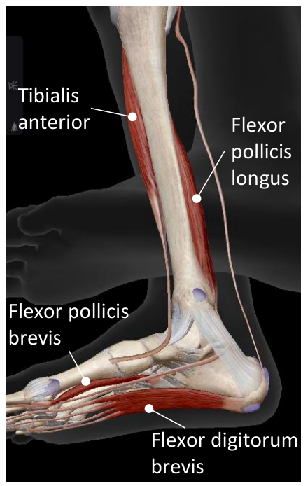
When elderly populations are targeted, general strength training that affects broad areas of the body or lower limbs does not clearly demonstrate a significant fall-prevention effect [8,9,10]. Therefore, intensive training of the ankle dorsiflexor muscle group is considered more effective for preventing tripping. Such training can be expected to strengthen the tibialis anterior muscle (the primary dorsiflexor located on the anterior side of the lower leg from the knee to ankle), as shown in Figure 1. Moreover, ankle stability will be improved if the training simultaneously activates the flexor hallucis (the primary dorsiflexor located on the side of the arch of the foot toward the big toe) and the flexor digitorum brevis (the primary dorsiflexor for the second toe to the little toe located on the top of the foot). In other words, strengthening muscles related to the toes can also enhance toe grip strength, contributing to balance stability and preventing falling due to tripping [11,12,13,14].
Weight-bearing exercises such as squats can target the ankle dorsiflexor group, but improper posture may lead to injuries around the knees and tendons. For example, a review by Bonilla et al. noted that bodyweight training performed with insufficient technical proficiency may increase the risk of injury, including knee injury, and therefore requires expert supervision as well as careful consideration of load intensity, posture, and biomechanical factors [15]. There are training methods that load the tibialis anterior while leaning against a wall in a standing posture, but for post-stroke patients or those with hemiplegia, concerns regarding trunk stability and fall risk make seated methods preferable. According to guidelines issued by the American Heart Association/American Stroke Association, balance ability is often impaired after stroke, resulting in an increased risk of falls [16]. In addition, rehabilitation guidelines issued by the Mongolian Ministry of Health and the World Health Organization state that the five main recommended postures for patients after stroke are the unaffected-side lying position, affected-side lying position, supine position, sitting on a bed, and sitting on a chair, and emphasize that measures should be taken to minimize fall risk because falls may cause serious complications [17]. However, the need to attend gyms with specialized equipment for safe training can incur high costs, and thus, for sustainable exercise, it is beneficial to have simple, safe, and easily accessible devices for home use.
Figure 1.
Diagram of muscles involved when using the ‘Wavy Stepper’. Image adapted from Visible Body: Human Anatomy Atlas 2025 [18]; annotations added by the authors.
Therefore, we developed a muscle-strengthening device that we have termed the Wavy Stepper, which enables seated training of the ankle dorsiflexor muscle group while watching TV or performing other tasks. One of the authors, a stroke survivor, was strongly motivated by their personal rehabilitation experience with coping with hemiplegia and contributed the original concept for this device.
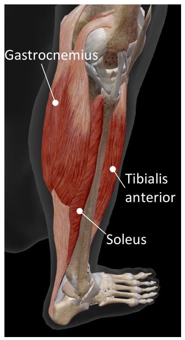
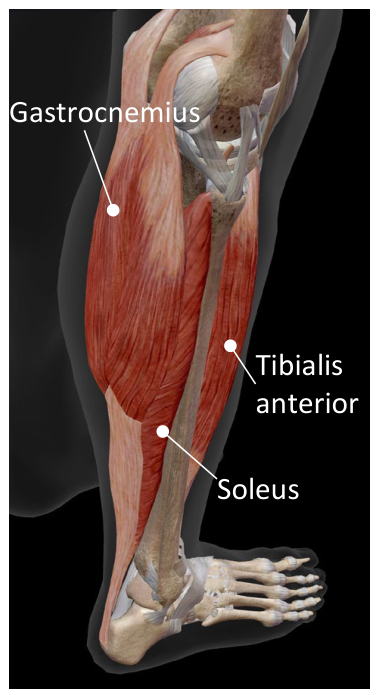
The developed training device is lightweight, has a simple mechanism, and can be used indoors while seated. Unlike existing devices, it allows training of the ankle dorsiflexor group while performing other tasks. Existing robotic devices such as Biodex, can train ankle dorsiflexors but are large and expensive. Ankle-foot orthoses with rotational joints and springs or brakes have also been studied, but they require custom fitting and must be worn each time the user walks, making casual use difficult. Small steppers sold in sports stores primarily claim to strengthen plantarflexor muscles, such as the gastrocnemius (Figure 2), and are mainly intended for standing use, which is less safe for elderly or post-stroke patients.
Figure 2.
Diagram of calf muscles. Image adapted from Visible Body: Human Anatomy Atlas 2025 [18]; annotations added by the authors.
Our device is newly developed and lacks prior validation, and we therefore first performed experiments with healthy adults to determine its effects and reveal points requiring improvement, before proceeding to trials with elderly or hemiplegic individuals. In this report, we introduce the mechanism of the proposed device and present preliminary experimental results with healthy adults. Experiments were performed comparing electromyography (EMG) recordings using different steppers. Muscle strength after three weeks of training was assessed by measuring the tibialis anterior and toe grip strength.
2. Proposed Equipment ‘Wavy Stepper’
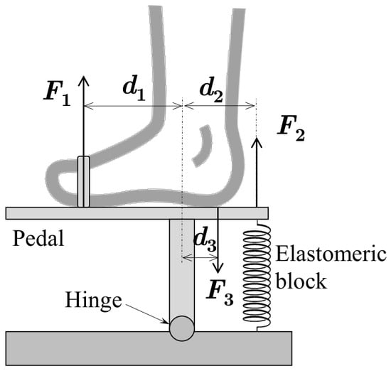
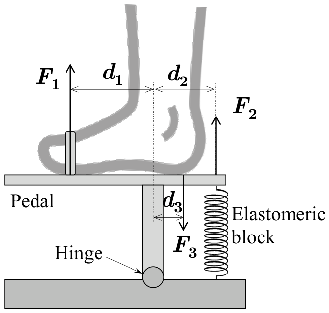
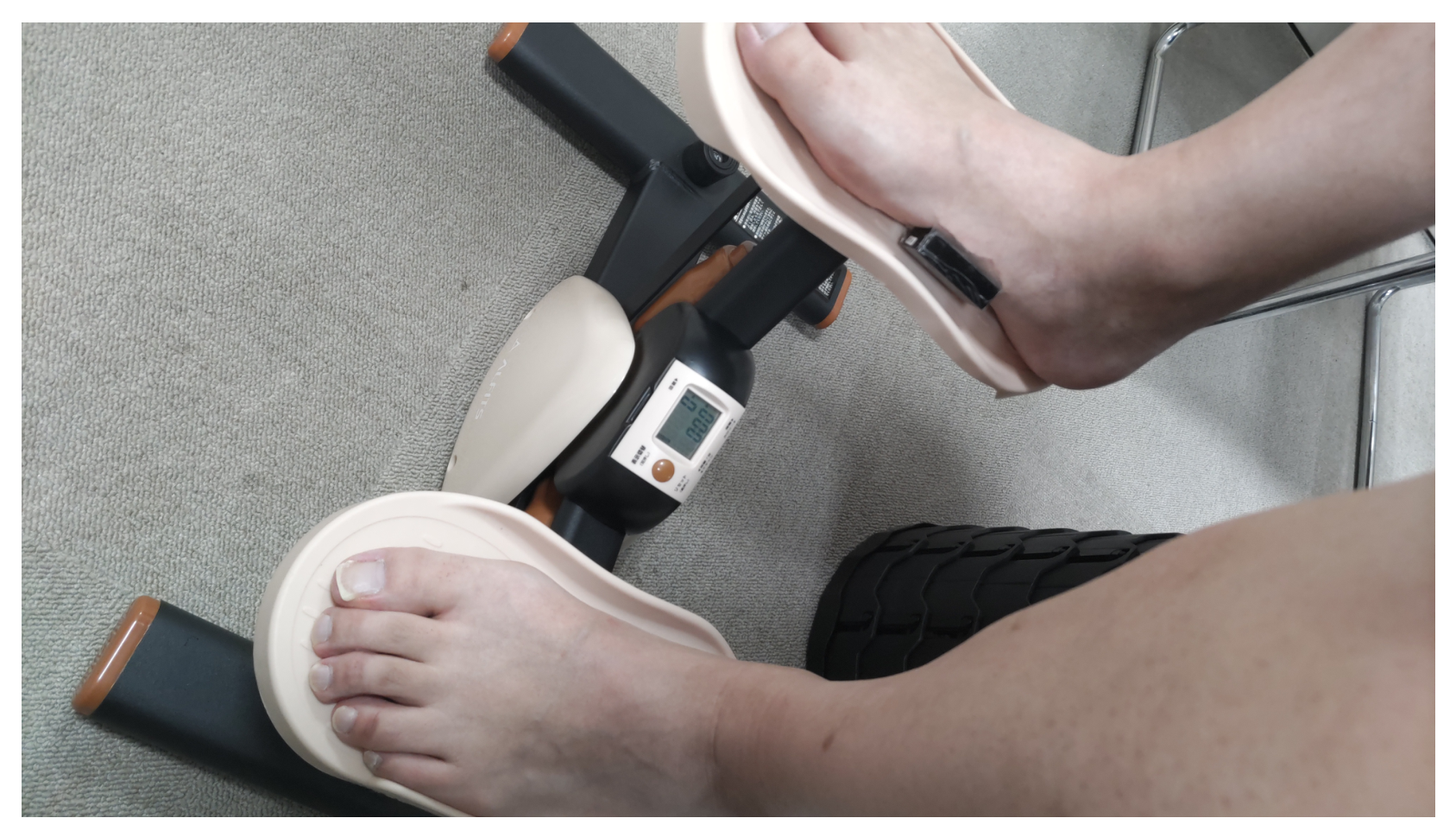
A schematic of the proposed training device, the Wavy Stepper, is shown in Figure 3. Table 1 defines the parameters shown in the figure. The user places one foot on the pedal and secures it near the metatarsal region with a strap. With the heel remaining in contact with the end of the pedal, a dorsiflexion motion is performed, lifting the toes at a stepping-like pace. To compensate for differences in foot size, a foam pad can be attached to the end of the pedal, allowing for adjustment of the heel position. The pedal is connected to a base hinge via a column allowing rotational movement, while the elastic force of an elastomeric block installed on the heel side provides resistance to dorsiflexion, serving as the training load.
Figure 3.
Diagram of the strengthening exercise equipment ‘Wavy Stepper’.
Table 1.
Kinematic parameters shown in Figure 3.
Parameters and shown in Figure 3 vary depending on foot size, but for approximation, we assume and . The moment balance around the hinge can then be calculated as
yielding
By appropriately selecting , can be made to be sufficiently larger than . Furthermore, the small vertical displacement of the ankle rotation minimizes contributions from hip and knee movements (gluteal and hamstring muscles). In actual operation, the distance from the hinge to the pedal affects the vertical displacement; a shorter distance allows more natural dorsiflexion but reduces motion stroke and reactive force, so an appropriate distance must be selected. In contrast, common steppers have a rotation center near the toes, causing large ankle displacement and greater involvement of the gluteal and hamstring muscles.
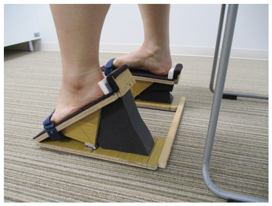
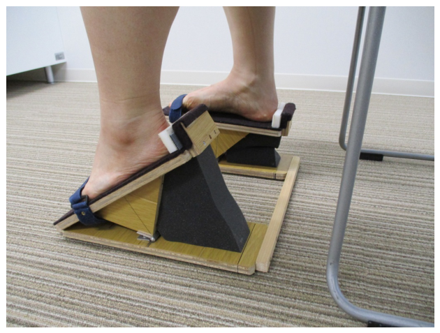
Since the device is intended for seated multitasking use at home and in places such as adult day services and hospital rooms, it is desirable that it is lightweight, portable, and comfortable against the skin. A wooden prototype was produced, as shown in Figure 4. In this prototype, the structural parts are wooden, the hinge is metal, and the strap is non-stretchable and soft to avoid injury. The dimensions were chosen for manufacturability while considering the points discussed above.
Figure 4.
Prototype of the exercise equipment.
Because the Wavy Stepper is designed for simple everyday use, it is a passive device and the intensity of the load cannot be controled. Future developments could involve creating a training device that allows the load to be actively controlled using actuators. Such load control could be achieved by placing a very small foldable pneumatic actuator (as proposed by Lu et al. [19]) on the toe side, or by placing a balloon actuator (using a bicycle inner tube as proposed by the authors [20]) on the heel side.
3. Muscle Strength Measurement System
Because the proposed device trains the ankle dorsiflexor muscle group, evaluation of its effects requires measuring the strength of these muscles. This involves performing isometric dorsiflexion of the ankle and measuring the force output via the top of the foot. Handheld dynamometers are often used clinically, but muscle activity around the hip and knee may influence measurements, and skilled operation is required for accurate measurement. Indeed, Moraux et al. noted issues with joint position standardization and operator strength dependency [21], and Siddiqi et al. recommended toe and ankle fixation to reduce variability [22]. However, misalignment of belts or instruments, or the leverage effect caused by the center of rotation being moved to the contact point of the heel with the floor, may cause errors. We therefore developed a portable and highly accurate system for measuring ankle dorsiflexor strength. Mauracher et al. reported a similar device [23], but it also fixes the heel, and is thus also subject to the issues reported above.
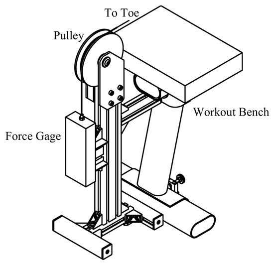
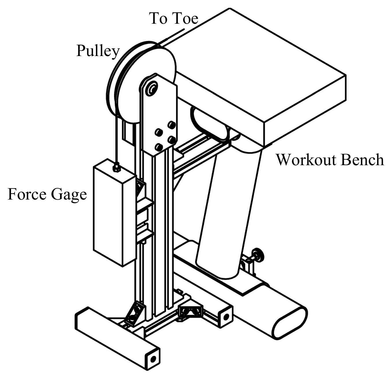
The system we designed consists of a bench and measurement apparatus (Figure 5). The bench is a standard training bench, while the apparatus comprises a force gauge, wire, pulley, and structural components. A 200-N-rated digital force gauge (general-purpose type, Imada, Toyohashi, Japan) was used for the measurements. Preliminary verification indicated that the 200-N sensor meets the expected measurement range; for subjects with higher strength, a 500-N-rated sensor may be substituted. When the subject exerts dorsiflexion force, it is transmitted via a wire to the force gauge. The apparatus is fixed to the bench to prevent motion.
Figure 5.
Diagram of the measurement equipment for ankle dorsal muscle groups.
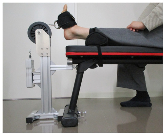
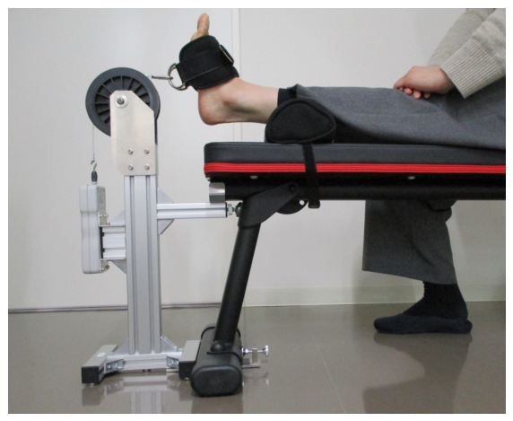
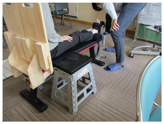
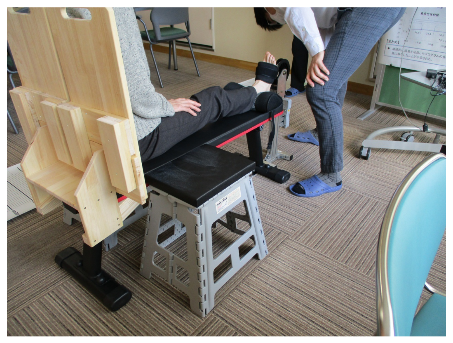
A manufactured system is shown in Figure 6. The subject sits on the bench, extends one leg, and places a cuff near the metatarsal region. The wire must remain horizontal, and the heel should not touch the bench. A foam block may be placed under the calf to adjust height as necessary. As in Figure 7, the subject leans slightly against the backrest and keeps their back straight, placing their hands on the thigh of the side being measured. Safety and durability were confirmed by the authors’ experience.
Figure 6.
Measurement equipment for ankle dorsal muscle groups.
Figure 7.
The measurement equipment in use.
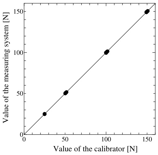
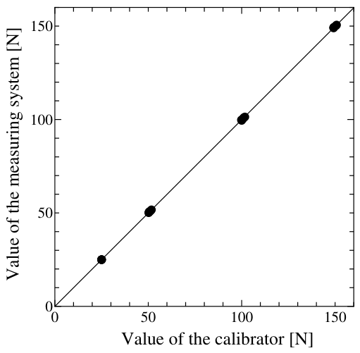
The measurement system was calibrated by pulling with another force gauge of the same type (0.1-N resolution, approximately 1-N accuracy). Target values of 25, 50, 100, and 150 N were used, and measurements were performed five times at each target value, giving a total of 20 measurements. Calibration data are shown in Figure 8, and can be approximated by a straight line through the origin with a slope of 1 (). Errors depend solely on gauge accuracy and are approximately 0.2% at 200 N, indicating a sufficiently small error for correct measurement.
Figure 8.
Calibration of the measurement equipment.
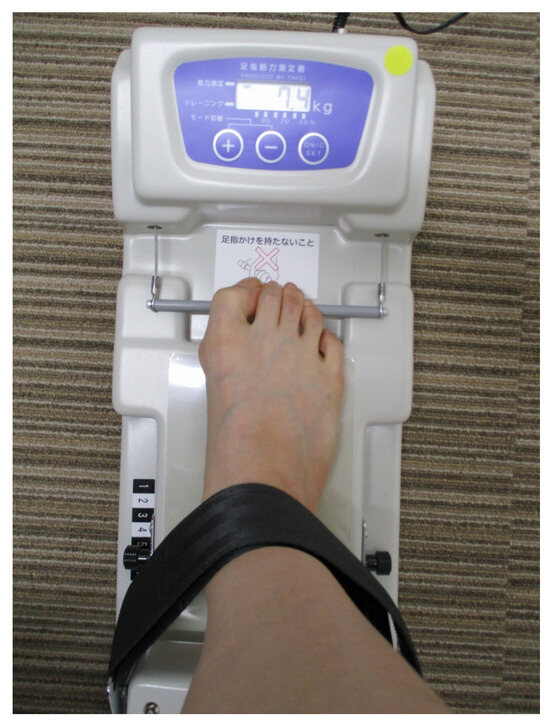
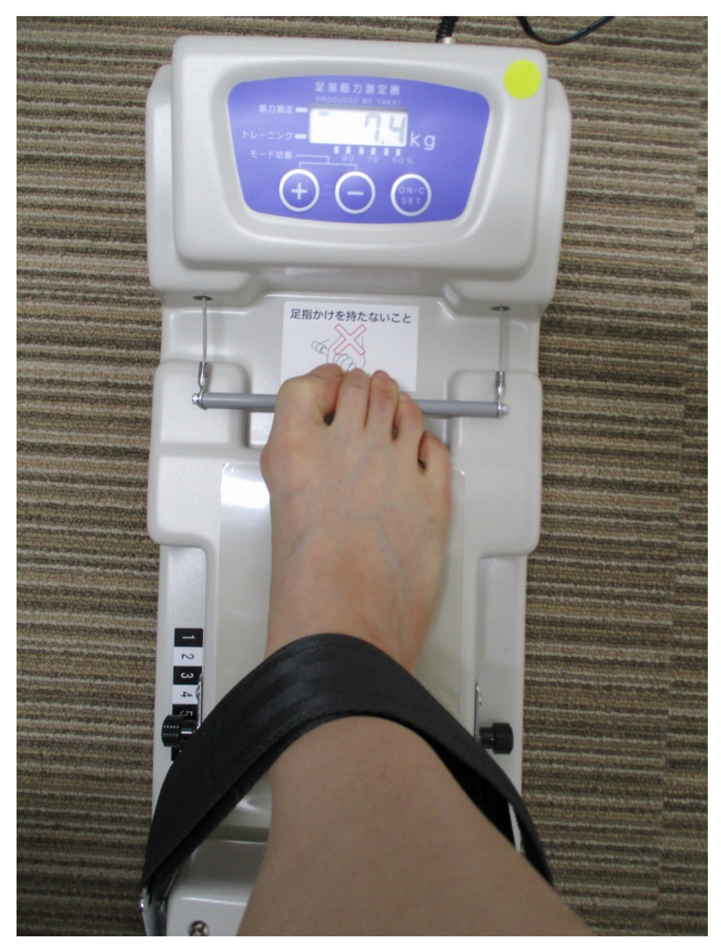
Toe grip strength was also measured to comprehensively evaluate muscle strength around the ankle. As mentioned in the Introduction section, there is a significant relationship between toe strength and muscle strength around the ankle joint and balance ability. A toe grip strength meter (Toe Muscle Strength Meter II, Takei Instruments, Kamo, Japan) was therefore also used (Figure 9). For this, the subject sat on a chair, lightly gripped a bar with four or more toes, adjusted their heel position, and pulled the bar maximally to record the force.
Figure 9.
Toe muscle strength meter.
4. Preliminary Experiment 1
4.1. Objective
Prior to the planned subject experiments for verifying the training effects, a preliminary experiment was conducted to investigate whether the developed training device could effectively load the ankle dorsiflexor muscles in comparison with other types of training devices.
4.2. Method
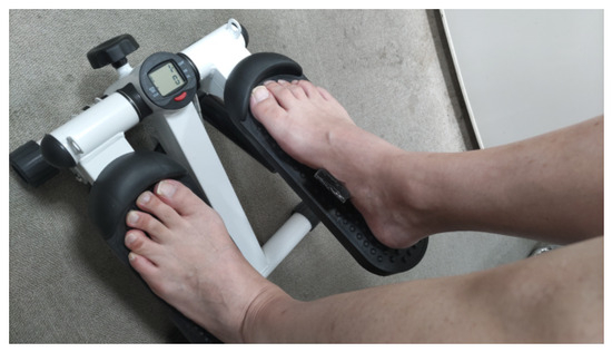
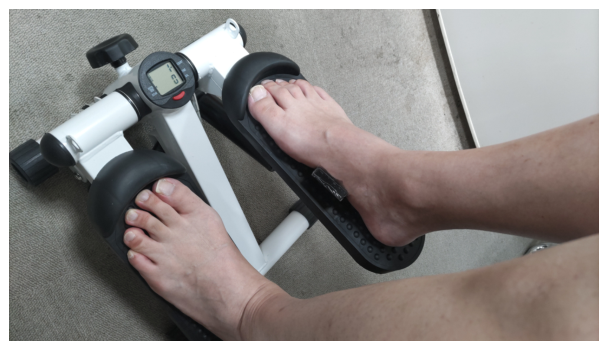
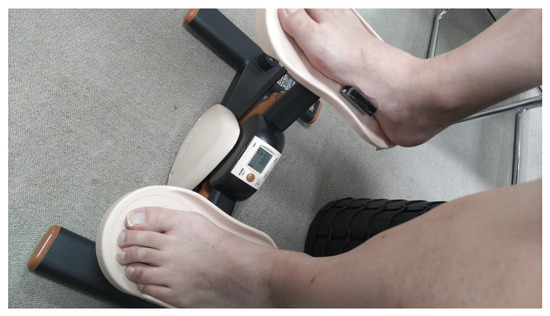
To evaluate the degree of muscle activity, electromyographic (EMG) signals were measured. First, an exploratory screening was conducted to determine which muscles were the most meaningful to evaluate. Because this screening was exploratory, the subject was one of the authors (a male in his 50s). Two commercially available steppers were used as comparison devices: the heel-up-and-down type stepper (Fitness Stepper SR-FT028, SunRuck, Okawara-cho, Japan) shown in Figure 10, and the side-step type stepper (Side Stepper VIER, ALINCO FITNESS, Osaka, Japan) shown in Figure 11.
Figure 10.
Fitness Stepper.
Figure 11.
Side Stepper.
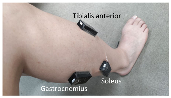
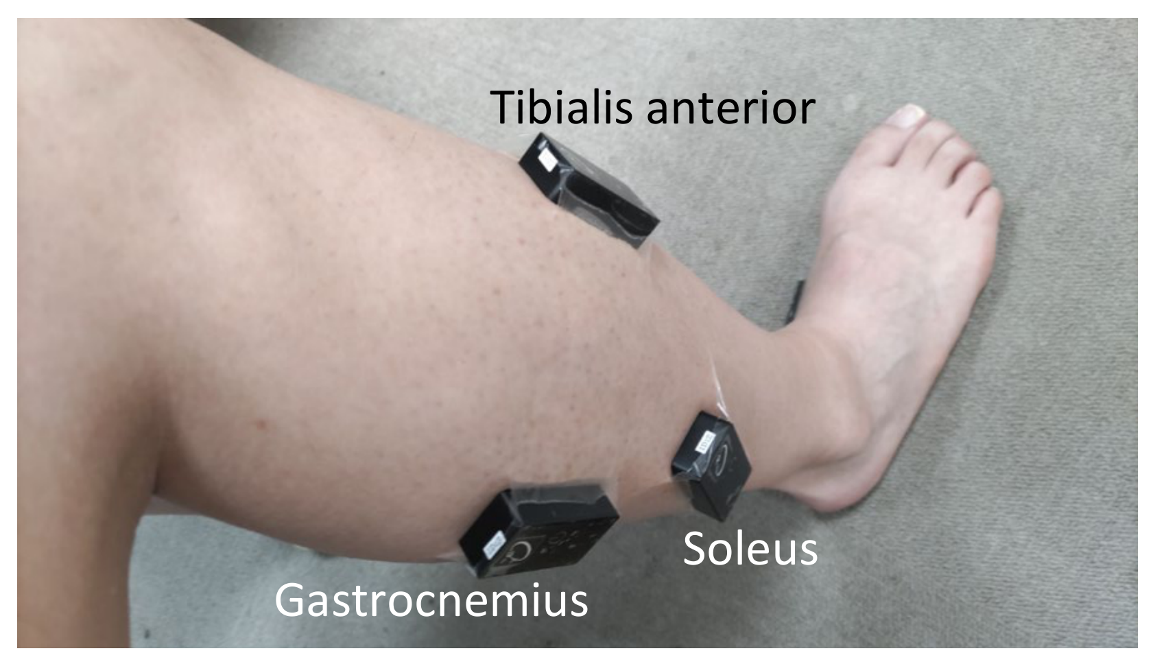
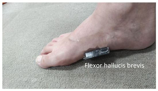
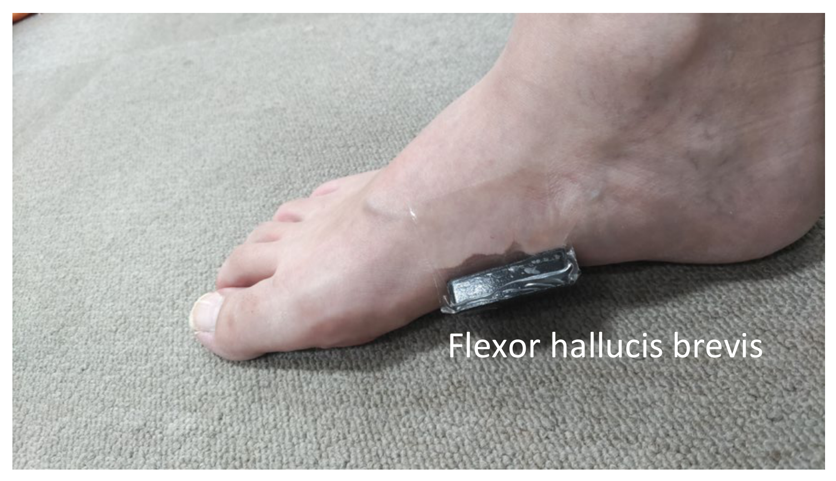
Wireless EMG sensors (OE-WS12, Oisaka Electronic Equipment Ltd., Fukuyama, Japan) were attached to the right tibialis anterior, gastrocnemius, and soleus muscles (the surface muscles contributing to ankle motion), as illustrated in Figure 12. Although it is difficult to measure the surface EMG of muscles involved in toe gripping because of their complex anatomical paths inside the foot, one sensor was attached to the arch, as shown in Figure 13, to record EMG activity mainly from the flexor hallucis brevis.
Figure 12.
EMG sensors on the right leg.
Figure 13.
EMG sensor on the right foot.
The EMG signals were sampled at 10 ms intervals, and a hardware-implemented band-pass filter of 5.3–442 Hz was applied. Each EMG signal was rectified and integrated over 100 ms intervals to obtain the integrated EMG (iEMG). Before the trials, the maximum voluntary contraction (MVC) values were obtained by measuring the EMG in the following standardized manner for all subjects. For all muscles, measurements were performed with the subject seated. For the tibialis anterior, subjects performed maximal ankle dorsiflexion with the hip, knee, and ankle flexed at , while the examiner applied manual resistance to the dorsum of the foot. For the flexor hallucis brevis, subjects performed maximal toe flexion in the same posture. For the soleus, the chair position was adjusted so that the sole of the foot made contact with a wall with the knee extended, and subjects performed maximal ankle plantarflexion against resistance. Each measurement lasted 10 s. To reduce the influence of spike noise, the MVC value was defined as the maximum of the averaged values over 10 iEMG samples. In the following analyses, iEMG values are normalized by these MVC values and expressed as %MVC.
During the experiments, the subject stepped approximately once per second for 10 s, resulting in five or six load cycles on the tested leg. The first and last 1 s intervals were excluded from the analysis because of posture instability.
Exploratory screening results showed that although the tibialis anterior, flexor hallucis brevis, and soleus showed activation, the gastrocnemius exhibited very little activation. Therefore, in Preliminary Experiment 1, the tibialis anterior, soleus, and flexor hallucis brevis were selected for evaluation. The two commercial steppers provided strong resistance, making it impossible to maintain a 1 Hz stepping cycle in the seated posture; thus, the stepping interval was extended to one step every 2–3 s. For this reason, the tempo was not controlled using a metronome or similar device in Preliminary Experiment 1. Additionally, because the Fitness Stepper and Side Stepper were originally intended for use in a standing posture, the results obtained during standing exercise were also recorded for reference.
The experimental procedures and equipment used in Preliminary Experiment 1 were the same as those used in the exploratory screening. However, EMG signals were recorded at 1 ms intervals and the integration window for iEMG was set to 50 ms.
The subjects were five healthy young adults aged 20–25 years, all of whom reported their dominant leg to be their right leg. Four subjects habitually performed strength training for approximately 1 h per session: one had done so for less than 6 months, one for 6 months to less than 1 year, and two for between 1 and 3 years. One subject was not currently performing strength training but had a previous training history of between 1 and 3 years. All subjects reported that they had no current injuries or pain that might affect the training. This study was approved by the Ethical Review Board for Human Subject Research at Toyama Prefectural University (R7-25).
The statistical analysis was performed as follows. For the tibialis anterior (TA), soleus (SOL), and flexor hallucis brevis (FHB), the mean muscle activity level (Mean iEMG) was calculated from the iEMG data of each trial. To evaluate periodic characteristics, power spectral density (PSD) analysis using Welch’s method was performed, and the peak frequency () in the range of 0.3–5.0 Hz was determined. The deviation from 1 Hz, , and the band power ratio around the peak frequency were also calculated. To evaluate the relative activity levels among the muscles, and were calculated. In addition, to evaluate the relationship between the temporal changes in the tibialis anterior and flexor hallucis brevis, the Pearson correlation coefficient between their iEMG time series, CTA-FHB, was calculated. For all of these metrics, the mean values across subjects were calculated.
For the statistical analysis, linear mixed-effects models were used, with the subject as a random effect and the device type evaluated as a fixed effect for both the inter-device comparison and the comparison including the standing-condition data recorded for reference. Since the Wavy Stepper is designed for use while seated, the main comparisons between devices were limited to data obtained while seated. However, commercially available steppers are designed for use while standing, so data obtained while standing was also used as reference data. In addition, one-sample t-tests were performed to test whether and differed significantly from a value of 1.0, which indicates equal activity between the target muscles TA and FH and the comparison muscle SOL, and whether the correlation coefficient CTA-FHB differed significantly from 0, which indicates no linear relationship between the iEMG time series of TA and FHB.
4.3. Results
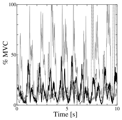
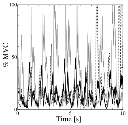
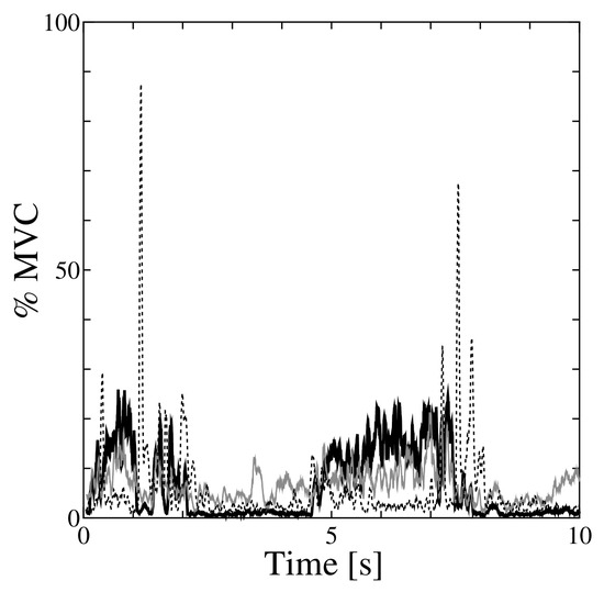
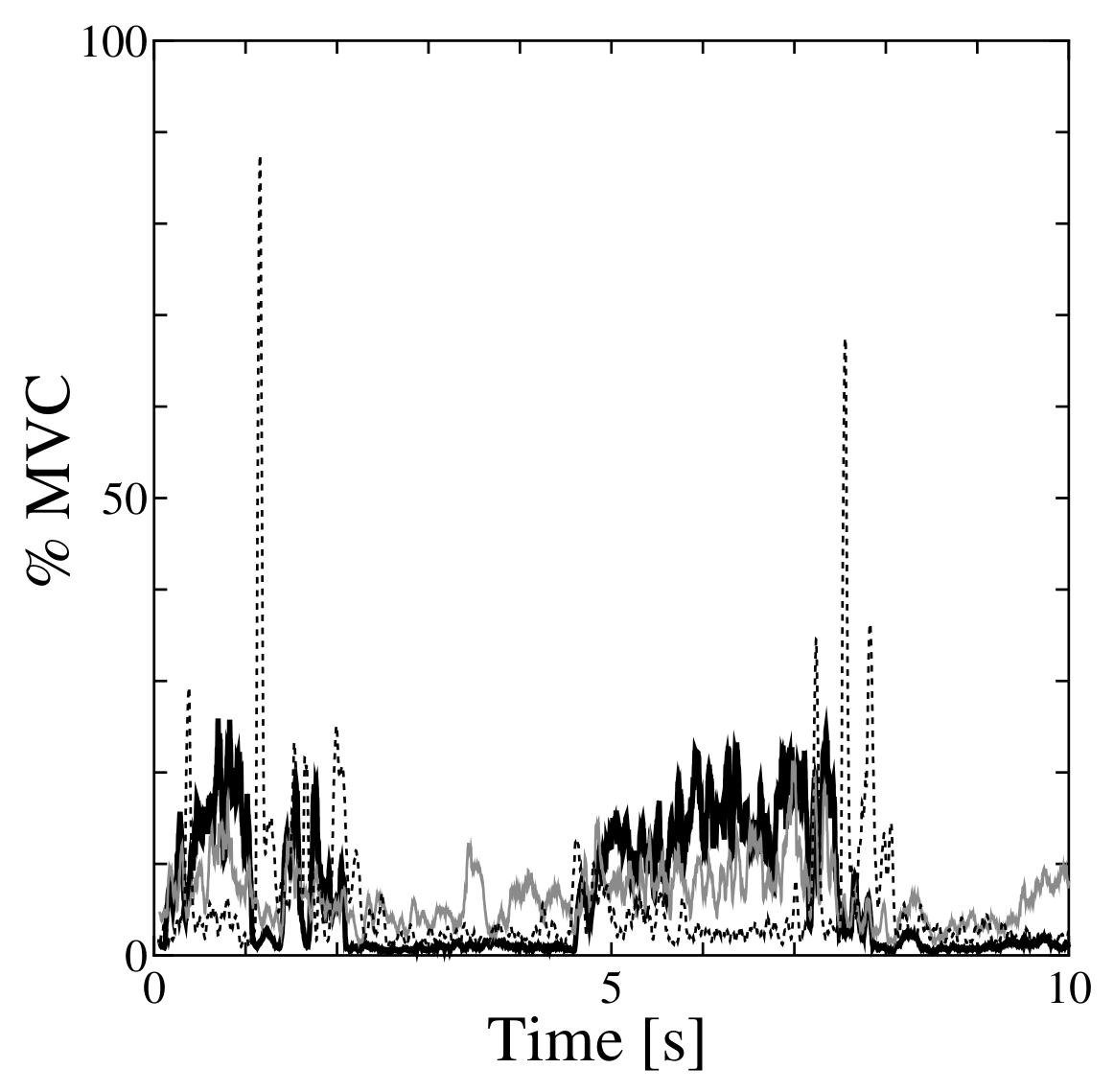
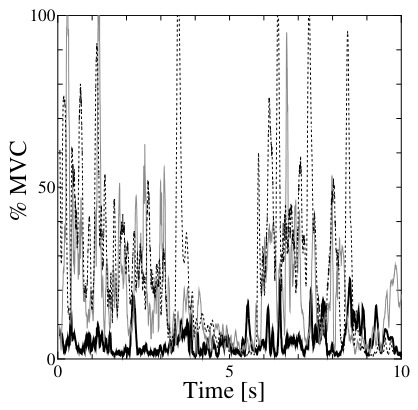
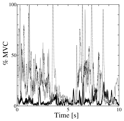
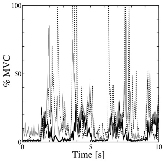
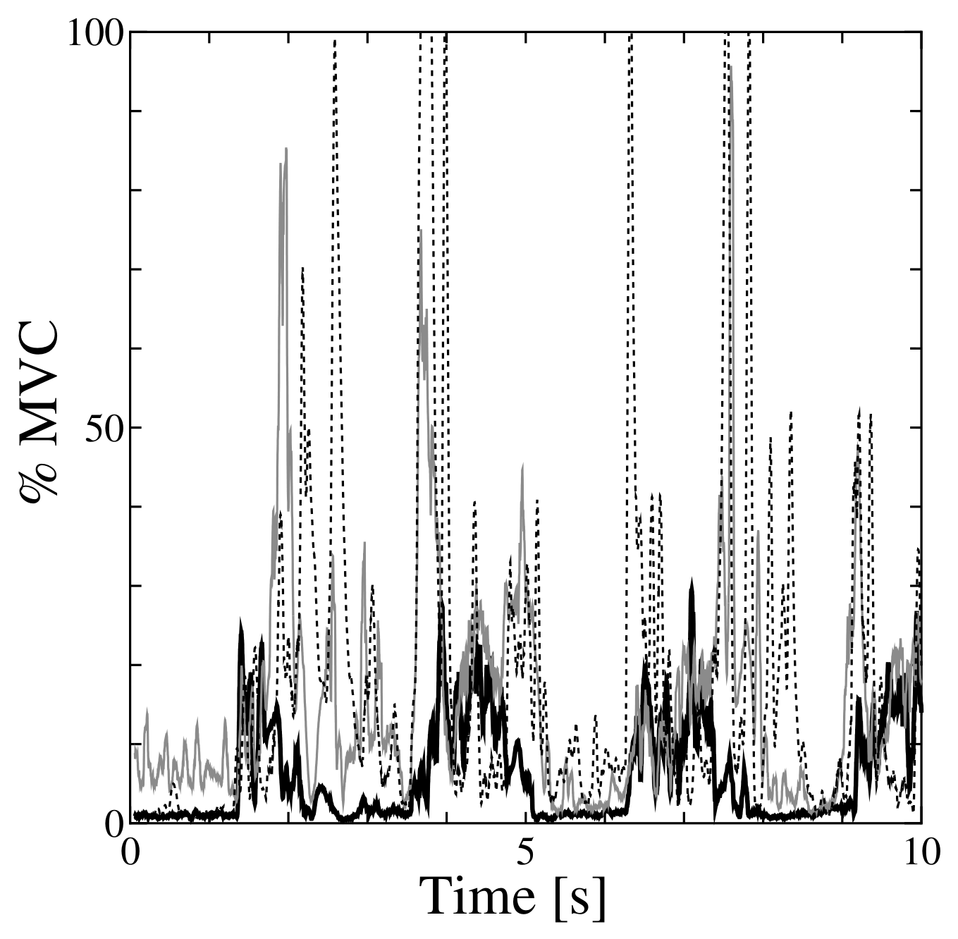
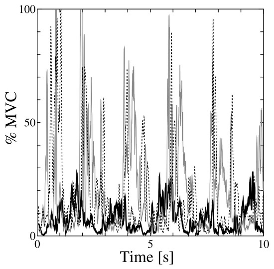
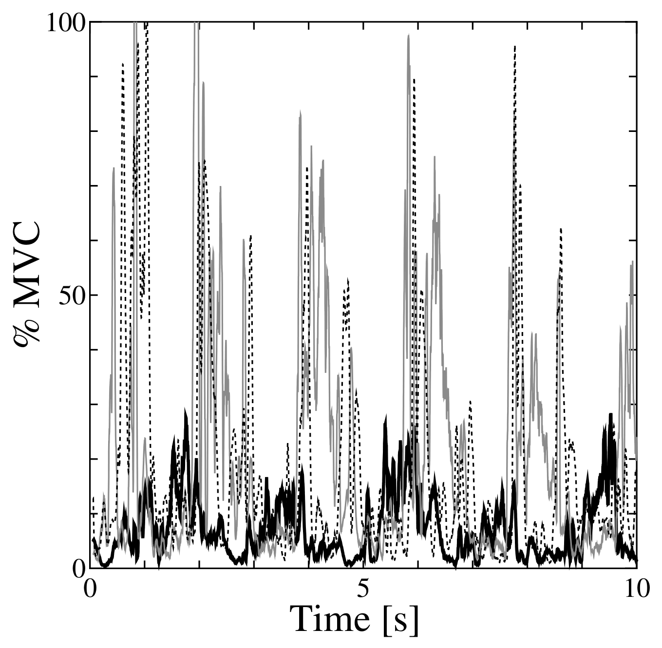
The iEMG results from Subject 2 are shown in Figure 14 and Figure 15 as representative examples. In each EMG graph, the iEMG of the tibialis anterior is plotted as a bold solid line, that of the soleus as a gray solid line, and that of the flexor hallucis brevis as a dashed line.
Figure 14.
The iEMG of Subject 2 with the Wavy Stepper (seated). Black solid line: tibialis anterior; gray solid line: soleus; dashed line: flexor hallucis brevis.
Figure 15.
The iEMG of Subject 2 with the Fitness Stepper (seated). Black solid line: tibialis anterior; gray solid line: soleus; dashed line: flexor hallucis brevis.
Figure 14 shows the results obtained with the Wavy Stepper. The tibialis anterior exhibited two peaks during each movement cycle, with the larger peak reaching approximately 40% of MVC. The soleus also showed periodic activation, with a greater activity level than that of the tibialis anterior. The flexor hallucis brevis likewise showed periodic behavior, although the magnitude of its activity varied considerably.
Figure 15 shows the results obtained using the Fitness Stepper in a seated position. Although the load was greater than that occurring the Wavy Stepper, the activity levels of all muscles were low and none of the muscles showed periodic activation. In fact, the subject was hardly able to move the stepper.
Figure 16 shows the results obtained using the Side Stepper in a seated position. In this situation, the subject was again hardly able to move the stepper. Although the load was greater than that of the Wavy Stepper, as was the case with the Fitness Stepper, the tibialis anterior showed very little activation. The soleus occasionally showed very large values, but in most cases its activity was approximately 40% of MVC. The flexor hallucis brevis showed a similar tendency, and none of the muscles showed periodic activation.
Figure 16.
The iEMG of Subject 2 with the Side Stepper (seated). Black solid line: tibialis anterior; gray solid line: soleus; dashed line: flexor hallucis brevis.
The results of the exercise measurements performed with the subject standing, acquired as reference comparisons, are shown in Figure 17 and Figure 18. Figure 17 shows the results obtained when using the Fitness Stepper in a standing posture. In this case, the subject was able to perform stepping movements at a pace of approximately eight steps in 10 s. Accordingly, the tibialis anterior showed periodic activation of approximately 20% of MVC. The soleus and flexor hallucis brevis also appeared to show periodic activation, but their activity was unstable.
Figure 17.
The iEMG of Subject 2 with the Fitness Stepper (standing). Black solid line: tibialis anterior; gray solid line: soleus; dashed line: flexor hallucis brevis.
Figure 18.
The iEMG of Subject 2 with the Side Stepper (standing). Black solid line: tibialis anterior; gray solid line: soleus; dashed line: flexor hallucis brevis.
Figure 18 shows the results obtained when using the Side Stepper in a standing posture. The subject was able to perform stepping movements at a pace of approximately 10 steps in 10 s. The tibialis anterior showed periodic activation, with its activity level being approximately 20% of MVC. The soleus showed periodic activation with a phase shift relative to that of the tibialis anterior, and its activity occasionally exceeded 80% of MVC. The flexor hallucis brevis showed a similar tendency.
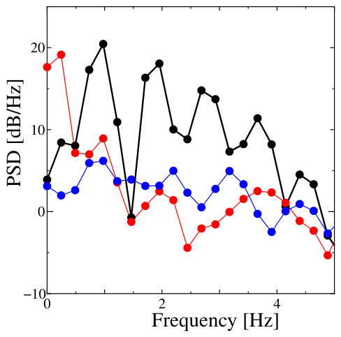
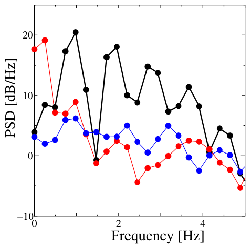
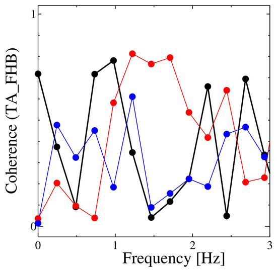
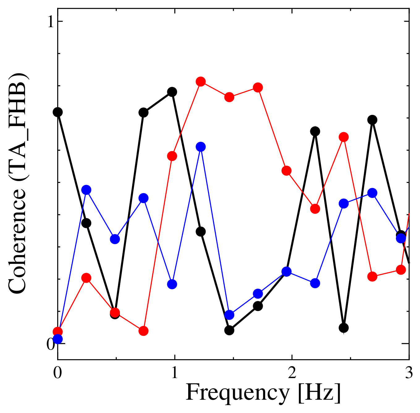
The results of the PSD analysis of the example iEMG data from Subject 2 are shown in Figure 19. The coherence between the iEMG data of the tibialis anterior and the flexor hallucis brevis was calculated, and a graph showing the region around 1 Hz, which corresponds to the target movement frequency, is presented in Figure 20. In both figures, the black line represents the Wavy Stepper, the red line the Fitness Stepper, and the blue line the Side Stepper. All lines are also marked with symbols indicating the data points. As shown in Figure 19, exercise using the Wavy Stepper at frequencies around 1 Hz and lower exhibited higher PSD values than exercise using the other devices. As shown in Figure 20, exercise using the Wavy Stepper exhibited the highest coherence around 1 Hz.
Figure 19.
Power spectral density from Subject 2. Black line: Wavy Stepper; red line: Fitness Stepper; blue line: Side Stepper. All lines are marked with symbols indicating the data points.
Figure 20.
Coherence between TA and FHB around 1 Hz. Black line: Wavy Stepper; red line: Fitness Stepper; blue line: Side Stepper. All lines are marked with symbols indicating the data points.
The results of the statistical analyses for the five subjects are shown in Table 2, Table 3 and Table 4. Note that these tables show only the results obtained using the Wavy Stepper, Fitness Stepper, and Side Stepper in a seated position. Table 2 summarizes the descriptive statistics for the tibialis anterior, which was the primary muscle of interest for each device. Table 3 shows the results of comparisons of the tibialis anterior metrics using linear mixed-effects models. In particular, it presents the test results for the overall effect of device and the post hoc comparisons using the Wavy Stepper as the reference. Table 4 shows the results of one-sample t-tests for the muscle activity ratios and correlation coefficients for each device. For , the test value was 0, whereas for the other metrics the test value was 1.
Table 2.
Descriptive statistics of the main outcome measures for the tibialis anterior in the inter-device comparison.
Table 3.
Linear mixed-effects model results for the inter-device comparison for the tibialis anterior. Reported are the p-values for comparison of all devices and the post hoc contrasts against the Wavy Stepper. Negative differences indicate smaller values than obtained with the Wavy Stepper. Fitness: Fitness Stepper; Wavy: Wavy Stepper; Side: Side stepper.
Table 4.
One-sample t-tests for ratio and correlation metrics in the inter-device comparison. and were tested against 1.0, and CTA-FHB was tested against 0.0. The column t indicates the one-sample t-statistic.
These tables indicate the following findings:
- The tibialis anterior showed a significantly greater mean iEMG with the Wavy Stepper than with the other two devices.
- The tibialis anterior showed a significantly higher peak frequency with the Wavy Stepper than with the other two devices.
- The correlation between the tibialis anterior and flexor hallucis brevis was significantly higher during use of the Wavy Stepper than during use of the Fitness Stepper.
- For all devices, the TA/SOL ratio did not significantly exceed 1, suggesting that TA tended to be comparable to or lower than SOL.
- For all devices, the FHB/SOL ratio did not significantly exceed 1, although the mean values tended to indicate greater FHB activity than SOL activity.
For reference, comparisons with the measurement results obtained using the Fitness Stepper and Side Stepper in a standing position were also made. For the tibialis anterior, the only significant difference found concerned the peak frequency of the movement, which was higher with the Wavy Stepper than with the Fitness Stepper.
4.4. Discussion
Compared with the Fitness Stepper and Side Stepper used in a seated position, the Wavy Stepper produced significantly greater muscle activity in the tibialis anterior. In other words, the Wavy Stepper may be more effective than these other devices in eliciting activation of the tibialis anterior. However, the TA/SOL ratio did not significantly exceed 1, suggesting that the tibialis anterior activity was not clearly greater than soleus activity in relative terms.
In addition, periodic components were observed in the tibialis anterior activity during use of the Wavy Stepper, and in some trials, temporal coordination with the flexor hallucis brevis was suggested. In the representative trial, muscle activities including those of the tibialis anterior and flexor hallucis brevis tended to appear around a common peak frequency.
These results suggest that the Wavy Stepper can provide load to the tibialis anterior while enabling repetitive movement with a consistent rhythm when the user is in a seated position.
5. Preliminary Experiment 2
5.1. Objective
In studies examining to strength training, increases in muscle strength have been confirmed after short training periods ranging from 3 days to 4 weeks [24,25,26,27]; therefore, we expected training effects to appear within approximately 3 weeks.
To verify whether this duration was sufficient and to investigate the usability of the developed training device and muscle strength measurement system, a preliminary test was conducted.
5.2. Method
The preliminary test involved two male university students in their 20s, with no injuries or pain in the lower limbs and no difficulties in walking or exercising. This study was approved by the Ethical Review Board for Human Subject Research at Toyama Prefectural University (Nursing No. R4-26). Both participants were provided with written and verbal explanations and gave informed consent. The preliminary test was conducted according to the following procedure:
- Measure basic attributes (sex, age group, habitual exercise) and pre-training muscle strength.
- Conduct training using the Wavy Stepper three times per week for three weeks.
- Each training session lasted 30 min, including breaks.
- The subject sat on a chair with their knee at , placed their feet on the Wavy Stepper, and performed ankle dorsiflexion exercises. The degree of dorsiflexion required to be achieved was demonstrated in advance.
- Training was performed at the subject’s own pace within a pain-free range.
- The training hour was chosen freely by the subjects.
- Subjects could listen to music, read books, use their computer, or perform similar activities during training.
- Muscle strength was remeasured after three weeks, using the same procedure as used in step 1.
5.3. Results
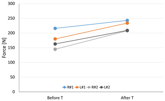
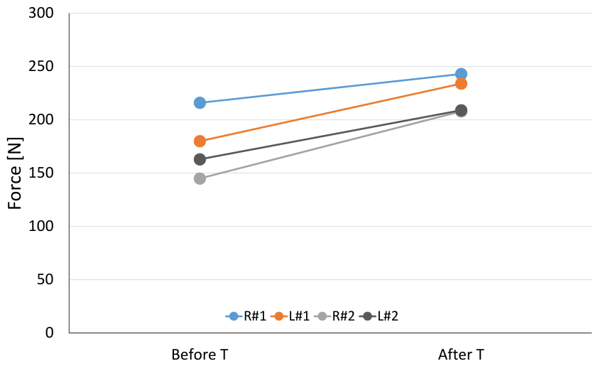
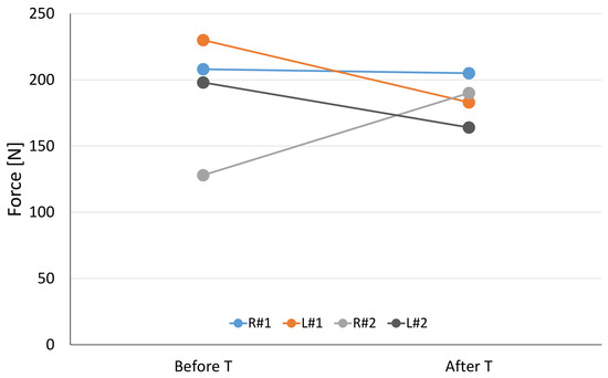
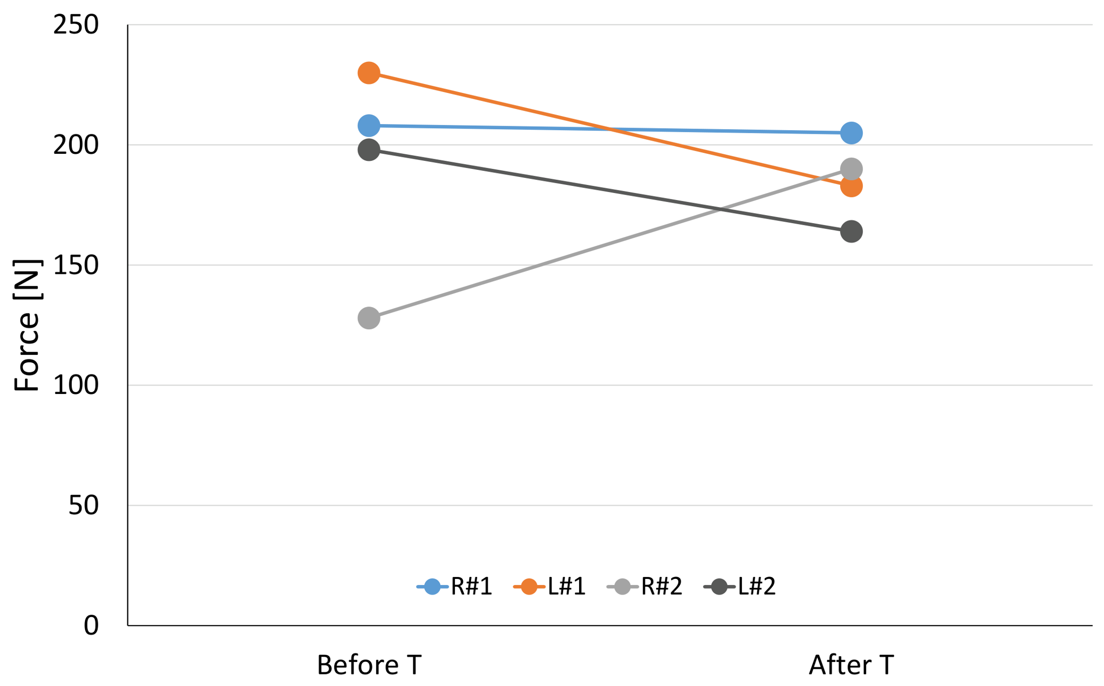
The results of the preliminary test are shown in Table 5 and Figure 21 and Figure 22. Each measurement was performed twice under the same conditions, and the larger value (in N) was recorded. Figure 21 and Figure 22 show the ankle dorsiflexor and toe grip strengths, respectively. In the figures, R#1 and L#1 represent the right and left feet, respectively, of Subject 1, and R#2 and L#2 represent the right and left feet of Subject 2.
Table 5.
Preliminary test results: changes in ankle dorsiflexor and toe grip strength before and after three weeks of training.
Figure 21.
Preliminary test results: ankle dorsiflexor muscles.
Figure 22.
Preliminary test results: toe grip strength.
These results indicate that the muscle strength of the ankle dorsiflexors increased in both feet of both subjects, whereas toe grip strength showed variable changes, with values increasing, decreasing, or remaining unchanged.
5.4. Discussion
Although this was a preliminary test and the results should be interpreted cautiously, the three-week training appears to have increased the strength of the ankle dorsiflexor muscles. During an interview, Subject 2 reported that he initially needed breaks during the 30 min session, but that toward the end, he was able to complete the session without resting.
Conversely, toe flexor strength did not appear to increase. As mentioned for Preliminary Experiment 1, a certain amount of load is also applied to the muscles that contribute to toe flexion. However, because results other than those for the flexor pollicis brevis could not be confirmed, it is possible that the load on other toes was insufficient. Of course, because the number of subjects was small, it is also possible that the observed results were influenced by simple random effects. Further investigation is needed to clarify this point.
Using the developed muscle strength measurement system, reproducible measurements could be obtained even by non-experts. However, consistent cuff placement at the same position on the instep of the foot is required to avoid variability in measurements, and this placement should be carefully adjusted during testing.
6. Conclusions
This paper proposes a simple and safe training device that allows convenient exercising of the ankle dorsiflexor muscle whilst the subject is seated, and potentially performing other tasks. The main contributions of this study include not only the design of this device, but also its preliminary validation from an engineering perspective through to EMG-based comparisons with commercially available steppers and muscle strength measurements. The results of the preliminary experiments suggested that the proposed device can effectively load the tibialis anterior during seated exercise and may therefore contribute to strengthening the ankle dorsiflexor muscles. In addition, the results of the three-week training suggested a tendency toward increased ankle dorsiflexor strength, indicating the expected effect.
A limitation of this study is that the training test involving human subjects remained only a preliminary evaluation conducted with a small number of healthy young adults. The variability in measurements can be expected to be greater if subjects are elderly or have paralysis. In addition, because the device is intended to allow casual training even while performing other tasks, exercise intensity could not be strictly controlled, and the results may therefore have included substantial variability in exercise intensity. Because the training period was relatively short, it is also possible that neural adaptation, such as increased motor neuron output, had a greater influence than muscle hypertrophy [24,27]. Future studies will involve more subjects and further investigate the effects of training with this device, including effects on toe muscle strength.
We expect that training with this device can improve muscle strength around the ankle and toes, strengthening that can enhance ankle stability and help prevent falls caused by tripping. Performing standing training immediately after a cerebrovascular incident carries a risk of imbalance and falling, and by facilitating initial muscle training in a seated position, our device allows training to be initiated safely. However, indications for use should be determined carefully, for example, whether the user is able to perform dorsiflexion voluntarily. Since the resistance of the Wavy Stepper cannot be adjusted after manufacturing, it is necessary to adjust either the number of steps or the exercise time so that the overall load gradually increases. Gradual training also builds patient confidence, enabling safe transition to standing and walking training. Consequently, we expect that the training device can contribute to improving activities of daily living and quality of life.
Author Contributions
Conceptualization, I.K.; methodology, all authors; mechanical design, K.K.; software, K.K.; validation, all authors; investigation, K.K., H.W. and T.K.; resources, K.K., H.W. and T.K.; writing—original draft preparation, K.K.; writing—review and editing, K.K.; visualization, K.K. and H.W.; supervision, K.K.; project administration, K.K. All authors have read and agreed to the published version of the manuscript.
Funding
This research received no external funding.
Institutional Review Board Statement
The study was conducted in accordance with the Declaration of Helsinki, and approved by the Ethical Review Board for Human Subject Research at Toyama Prefectural University (R7-25).
Informed Consent Statement
Informed consent was obtained from all subjects involved in the study.
Data Availability Statement
The original data contributions presented in the study are included in the article; further inquiries can be directed to the corresponding author.
Conflicts of Interest
The funders had no role in the design of the study; in the collection, analyses, or interpretation of data; in the writing of the manuscript; or in the decision to publish the results. I.K. is an employee of I-CAPS Co., Japan, who provided the Wavy Stepper.
References
- Little, V.L.; McGuirk, T.E.; Patten, C. Impaired limb shortening following stroke: What’s in a name? PLoS ONE 2014, 9, e110140. [Google Scholar] [CrossRef] [Scilit] [PubMed]
- Sorock, G.S.; Labiner, D.M. Peripheral neuromuscular dysfunction and falls in an elderly cohort. Am. J. Epidemiol. 1992, 136, 584–591. [Google Scholar] [CrossRef] [Scilit]
- Kemoun, G.; Thoumie, P.; Boisson, D.; Guieu, J. Ankle dorsiflexion delay can predict falls in the elderly. J. Rehabil. Med. 2002, 34, 278–283. [Google Scholar] [CrossRef] [Scilit]
- Abd El-Kader, S.M.; Ashmawy, E.M. Ankle Dorsiflexors Strength Improves Balance Performance in Elderly: A Corelational Study. Eur. J. Gen. Med. 2014, 11, 60–65. [Google Scholar] [CrossRef] [Scilit]
- Gefen, A. Simulations of foot stability during gait characteristic of ankle dorsiflexor weakness in the elderly. IEEE Trans. Neural Syst. Rehabil. Eng. 2001, 9, 333–337. [Google Scholar] [CrossRef] [Scilit] [PubMed]
- Barrett, R.; Mills, P.; Begg, R. A systematic review of the effect of ageing and falls history on minimum foot clearance characteristics during level walking. Gait Posture 2010, 32, 429–435. [Google Scholar] [CrossRef] [Scilit]
- Khandoker, A.H.; Taylor, S.B.; Karmakar, C.K.; Begg, R.K.; Palaniswami, M. Investigating Scale Invariant Dynamics in Minimum Toe Clearance Variability of the Young and Elderly During Treadmill Walking. IEEE Trans. Neural Syst. Rehabil. Eng. 2008, 16, 380–389. [Google Scholar] [CrossRef] [Scilit]
- Ishigaki, E.Y.; Ramos, L.G.; Carvalho, E.S.; Lunardi, A.C. Effectiveness of muscle strengthening and description of protocols for preventing falls in the elderly: A systematic review. Braz. J. Phys. Ther. 2014, 18, 111–118. [Google Scholar] [CrossRef] [Scilit]
- Sherrington, C.; Fairhall, N.J.; Wallbank, G.K.; Tiedemann, A.; Michaleff, Z.A.; Howard, K.; Clemson, L.; Hopewell, S.; Lamb, S.E. Exercise for preventing falls in older people living in the community. Cochrane Database Syst. Rev. 2019, CD012424. [Google Scholar] [CrossRef] [Scilit]
- Claudino, J.G.; Afonso, J.; Sarvestan, J.; Lanza, M.B.; Pennone, J.; Filho, C.A.C.; Serrão, J.C.; Espregueira-Mendes, J.; Vasconcelos, A.L.V.; de Andrade, M.P.; et al. Strength Training to Prevent Falls in Older Adults: A Systematic Review with Meta-Analysis of Randomized Controlled Trials. J. Clin. Med. 2021, 10, 3184. [Google Scholar] [CrossRef] [Scilit]
- Kojima, K.; Kamai, D.; Yamamoto, A.; Tsuchitani, Y.; Kataoka, H. The effect of toe-grasping exercises on balance ability in home-based rehabilitation: A randomized controlled trial by block randomization. Phys. Ther. Res. 2021, 24, 272–279. [Google Scholar] [CrossRef] [Scilit]
- Arai, R.; Fuchigami, M.; Hatamura, K.; Yamamoto, K.; Yoshimatsu, T. Relationship between toe grip strength and dynamic balance in older adult patients with femoral neck fracture. J. Phys. Ther. Sci. 2023, 35, 384–388. [Google Scholar] [CrossRef] [Scilit]
- Mawarikado, Y.; Inagaki, Y.; Fujii, T.; Kubo, T.; Kido, A.; Tanaka, Y. Relationship between fall history and toe grip strength in older adults with knee osteoarthritis in Japan: A cross-sectional study. PLoS ONE 2023, 18, e0282944. [Google Scholar] [CrossRef] [Scilit]
- Kawai, T.; Kanamori, M.; Washizuka, H.; Yoshii, M.; Teranishi, K. The Effectiveness of Toe Flexion and Extension Exercise for Fall Prevention in Community-Dwelling Older Females. Toyama Med. J. 2023, 34, 27–36. [Google Scholar] [CrossRef]
- Bonilla, D.A.; Cardozo, L.A.; Vélez-Gutiérrez, J.M.; Arévalo-Rodríguez, A.; Vargas-Molina, S.; Stout, J.R.; Kreider, R.B.; Petro, J.L. Exercise selection and common injuries in fitness centers: A systematic integrative review and practical recommendations. Int. J. Environ. Res. Public Health 2022, 19, 12710. [Google Scholar] [CrossRef] [Scilit]
- Winstein, C.J.; Stein, J.; Arena, R.; Bates, B.; Cherney, L.R.; Cramer, S.C.; Deruyter, F.; Eng, J.J.; Fisher, B.; Harvey, R.L.; et al. Guidelines for adult stroke rehabilitation and recovery: A guideline for healthcare professionals from the American Heart Association/American Stroke Association. Stroke 2016, 47, e98–e169. [Google Scholar] [CrossRef] [Scilit] [PubMed]
- Ministry of Health of Mongolia. Clinical Guideline on Stroke Rehabilitation; Management of Patients with Stroke: Rehabilitation, Prevention and Management of Complications, and Discharge Planning. 2013. Available online: https://extranet.who.int/ncdccs/Data/MNG_D1_2.%20Rehabilitation%20guideline%20of%20Stroke.pdf (accessed on 30 March 2026).
- Visible Body 2025; Cengage Learning, Inc.: Independence, KY, USA, 2025.
- Lu, J.; Lv, X.; Liu, J.; Liu, H.; Huang, T. Fully flexible wearable pouch pneumatic artificial muscle based on asymmetric expansion. Chin. J. Mech. Eng. 2026, 39, 100085. [Google Scholar] [CrossRef] [Scilit]
- Koyanagi, K.; Murabayashi, T.; Oshima, T.; Kagechika, K.; Tamamoto, T.; Sawai, K.; Motoyoshi, T.; Masuta, H. Multi-balloon actuator for advanced hand rehabilitation after stroke. In Proceedings of the 2017 IEEE/SICE International Symposium on System Integration; IEEE: New York, NY, USA, 2017; pp. 930–935. [Google Scholar]
- Moraux, A.; Canal, A.; Ollivier, G.; Ledoux, I.; Doppler, V.; Payan, C.; Hogrel, J.Y. Ankle dorsi-and plantar-flexion torques measured by dynamometry in healthy subjects from 5 to 80 years. BMC Musculoskelet. Disord. 2013, 14, 104. [Google Scholar] [CrossRef] [Scilit]
- Siddiqi, A.; Arjunan, S.P.; Kumar, D. Improvement of isometric dorsiflexion protocol for assessment of tibialis anterior muscle strength. MethodsX 2015, 2, 107–111. [Google Scholar] [CrossRef] [Scilit] [PubMed]
- Mauracher, M.E.; Asmussen, M.J.; Nigg, S.R.; Omu, O.; Jarvis, S.E. Portable fixed dynamometry to quantify ankle dorsiflexion force. Muscle Nerve 2019, 60, 56–61. [Google Scholar] [CrossRef] [Scilit]
- Prevost, M.C.; Nelson, A.G.; Maraj, B.K. The effect of two days of velocity-specific isokinetic training on torque production. J. Strength Cond. Res. 1999, 13, 35–39. [Google Scholar]
- Costa, P.B.; Herda, T.J.; Herda, A.A.; Cramer, J.T. Effects of short-term dynamic constant external resistance training and subsequent detraining on strength of the trained and untrained limbs: A randomized trial. Sports 2016, 4, 7. [Google Scholar] [CrossRef] [Scilit] [PubMed]
- Saeterbakken, A.H.; Olsen, A.; Behm, D.G.; Bardstu, H.B.; Andersen, V. The short-and long-term effects of resistance training with different stability requirements. PLoS ONE 2019, 14, e0214302. [Google Scholar] [CrossRef] [Scilit] [PubMed]
- Del Vecchio, A.; Casolo, A.; Negro, F.; Scorcelletti, M.; Bazzucchi, I.; Enoka, R.; Felici, F.; Farina, D. The increase in muscle force after 4 weeks of strength training is mediated by adaptations in motor unit recruitment and rate coding. J. Physiol. 2019, 597, 1873–1887. [Google Scholar] [CrossRef] [Scilit]

Disclaimer/Publisher’s Note: The statements, opinions and data contained in all publications are solely those of the individual author(s) and contributor(s) and not of MDPI and/or the editor(s). MDPI and/or the editor(s) disclaim responsibility for any injury to people or property resulting from any ideas, methods, instructions or products referred to in the content. |
© 2026 by the authors. Licensee MDPI, Basel, Switzerland. This article is an open access article distributed under the terms and conditions of the Creative Commons Attribution (CC BY) license.