


Abstract
Over the past decade, the incidence of infectious syphilis has been on the rise in the general Chinese population. Consequently, Treponema Pallidum (TP) testing has been proposed as a surrogate marker for sexually transmitted pathogens and for monitoring risky sexual behaviors among blood donors globally. In addition, sexual contact with individuals chronically infected with hepatitis B virus (HBV) is recognized as one of the primary routes of HBV transmission. Blood donors may acquire HBV infection through sexual contact with chronically infected partners, particularly with occult hepatitis B infections (OBIs), which are characterized by intermittent and extremely low viral loads. Therefore, the prevalence of OBIs among syphilis-positive blood donations and the corresponding risks to blood safety require further investigation. This study aimed to investigate the prevalence of OBIs among syphilis-positive blood donors and assess the surrogate value of TP testing for evaluating OBI-related risks to blood supply. After routine screening using serological assays and nucleic acid testing (NAT), blood donation samples with positive anti-TP enzyme-linked immunosorbent assay (ELISA) results were collected and further confirmed by the Treponema Pallidum Particle Agglutination Assay (TPPA). For blood donations confirmed positive for syphilis, further tests were performed to characterize whether the donations had HBV co-infection, including electrochemiluminescence immunoassay (ECLI) for the detection of hepatitis B surface antigen (HBsAg), anti-hepatitis B surface antibody (anti-HBs), hepatitis B e antigen (HBeAg), anti-hepatitis B e antibody (anti-HBe), and anti-hepatitis B core antibody (anti-HBc). Additionally, quantitative real-time polymerase chain reaction (qPCR) was used for HBV DNA quantification, and nested PCRs for the S and basal core promoter/precore (BCP/PC) region were conducted in combination with high-volume nucleic acid extraction. Subsequently, molecular characterization of HBV DNA in these co-infected samples was carried out by DNA sequencing to analyze the viral genetic features. Of 252 anti-TP ELISA+ donations screened from 64,871 blood samples, 138 (138/250, 55.2%) donations were confirmed syphilis-positive but NAT−, among which 78 (78/138, 56.5%) were anti-HBc-positive, and 88 (88/138, 63.7%) had anti-HBs. Notably, seven donations (7/138, 5.1%) were diagnosed as OBI co-infections, and available sequence analysis revealed that three cases were genotype B and one case was genotype C. In addition, several mutations in the S region of the HBV genome were identified, including Q101R, K122R, Q129H, T131N, M133T, G145R, and Y161F mutations. Furthermore, nucleotide mutations such as T1719G, A1752T, G1896A, and A1762T/G1764A in the BCP/PC regions were also detected in these OBI donations. These mutations may contribute to the extremely low HBV viral loads and/or failure in HBsAg detection, collectively leading to OBIs. These data indicate that syphilis screening of blood donors has potential to serve as an additional safeguard measure for excluding donations co-infected with OBIs. The high prevalence of undetected OBIs in syphilis-positive blood donors further supports that syphilis screening has the potential to serve as a surrogate marker for HBV-related risks in the blood supply.
1. Introduction
Syphilis is an infectious disease caused by TP infection, and unprotected sexual contact and trans-placental spread during pregnancy are its main spreading routes [1]. It is one of the mandatory serological screening tests for transfusion-transmitted infections (TTIs) recommended by the World Health Organization (WHO) [2]. Syphilis is estimated to affect 36 million people worldwide, with 11 million new cases each year and more than 90% occurring in developing nations [3,4]. It was reported that syphilis accounts for approximately 10% of the sexually transmitted diseases [5]. According to the Chinese Health Statistical Digest by the Chinese Ministry of Health, the incidence of syphilis is ranked third following viral hepatitis and tuberculosis in Chinese class A and B communicable diseases [6].
In recent years, syphilis infection has been on the rise in China. The total incidence of syphilis increased from 1.0 to 32.2 per 100,000 between 1995 and 2016 in China [7]. Among blood donors, it has been reported that the seroprevalence of TP antibody ranged from 0.31 to 0.70% in different regions of China [8,9]. This relatively high prevalence of TP infection may endanger blood safety as some of these infected individuals may go to donate blood without realizing that they were actually infected. Therefore, it is necessary to identify the high-risk groups in the blood donor population to further ensure blood safety.
Our previous study observed that the prevalence of occult hepatitis B infections (OBIs) in the partners of chronically infected HBV individuals was significantly higher than that in the general donor population, and the majority of these OBIs could not be detected by routine screening with ID NAT and HBsAg, indicating a potential risk to blood safety [10]. As both are sexually transmitted pathogens, blood donors may be co-infected with HBV and TP. To clarify the epidemiological profile of TP and hepatitis B virus co-infection in blood donors and to assess the associated risk to blood safety, we enrolled a large cohort of Chinese blood donors in Shenzhen Blood Center. All participants were screened for HBV and TP using serological and molecular assays. For TP-seropositive donors, high-volume nucleic acid extraction was applied to detect HBV DNA as previously reported [10], so as to evaluate the potential risk of screening failure to blood safety.
2. Materials and Methods
2.1. Blood Samples and Routine Screening
A total of 64,871 blood donations were collected by the Shenzhen Blood Center between August 2023 and January 2024. Demographic data, including gender, age, educational background, and occupation, were routinely collected from donors. Before donation, informed consent, the donation registration form was signed, and all donors passed the physical examination and health questionnaire, as well as rapid tests for HBsAg, ALT, and hemoglobin (Minray Bio-Medical Electronics, Ltd., Shenzhen, China) [11]. Routine screening with dual ELISA assays for HBsAg and anti-HIV (Bio-Rad Laboratories, Inc., Hercules, CA, USA and Zhuhai LivZon Diagnostics Inc., Zhuhai, China); anti-HCV and TP (WanTai Diagnostics Inc., Beijing, China and Zhuhai LivZon Diagnostics Inc., Zhuhai, China) was performed. If both results for TP screening were reactive, the sample was classified as TP-negative. Conversely, if one test yielded a negative result while the other was reactive, the sample was retested in duplicate using the same assay kits. If one or two of the repeated results were positive (repeat reactive), the sample was defined as TP ELISA-positive. All TP ELISA-positive samples were collected and stored for further testing. In addition, all donations were screened by NAT for HBV DNA, HCV RNA, and HIV RNA using the Procleix® Ultrio Elite® Assay (Novartis Diagnostics, Emeryville, CA, USA) on the Elite platform and discriminatory strategies were implemented as reported in our previous study [12]. The routine screening data of blood donors, covering the entire workflow from donor recruitment to the issuance of test results, were archived in the electronic system to facilitate retrospective analysis. Further study-specific laboratory testing, data extraction, and statistical analysis were initiated after 7 February 2024.
2.2. TP Confirmation
To confirm TP infection in ELISA anti-TP-positive donations, confirmatory tests by TPPA and Toluidine Red Unheated Serum Test (TRUST) were performed by the Shenzhen Center for Chronic Disease Control and Prevention. Donations with TPPA+ and TRUST + were confirmed as TP-positive.
2.3. Supplemental Serologic Testing and NAT Testing
TP-positive donations were further analyzed by HBsAg II Quant, along with anti-HBs, HBeAg, anti-HBe, and anti-HBc by ECLI (Roche Elecsys, Indianapolis, IN, USA) [12]. Additionally, TP-positive samples were further tested individually by MPX2.0 ID HBV NAT (Roche Diagnostics, Pleasanton, CA, USA; LOD: 2.3 IU/mL; input volume 1 mL). Molecular assays for donations with confirmed TP-positive results were performed using a combination of qPCR and nested PCRs that amplify the BCP/PC and the S regions with a large extraction volume of 2.5–5 mL plasma in order to detect very low-level OBIs. HBV DNA sequencing, serotyping, and genotyping were analyzed as previously reported [13,14]. The sensitivity and specificity of the tests used in this study were summarized in Table 1.
Table 1.
The sensitivity and specificity of the tests used in this study.
2.4. OBI-Associated Residual Risk (RR) in Blood Donations Without Anti-TP Screening
According to a previous study [15], the RR of OBI without anti-TP screening was calculated as follows:
RR = p (OBI without anti-TP screening) × p (transmission rate)
2.5. Statistical Analysis
The statistical software STATA 16.0 was used for data analysis. Categorical variables and continuous variables were analyzed using Fisher’s exact test and the non-parametric Mann-Whitney test, respectively. The 95% confidence intervals (CIs) for the residual-risk estimate were calculated using the Wilson score method. A p-value < 0.05 was defined as statistically significant.
3. Results
3.1. Screening and Detection of Syphilis
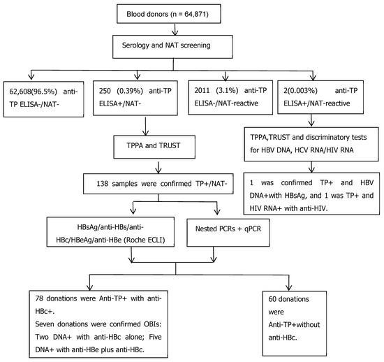
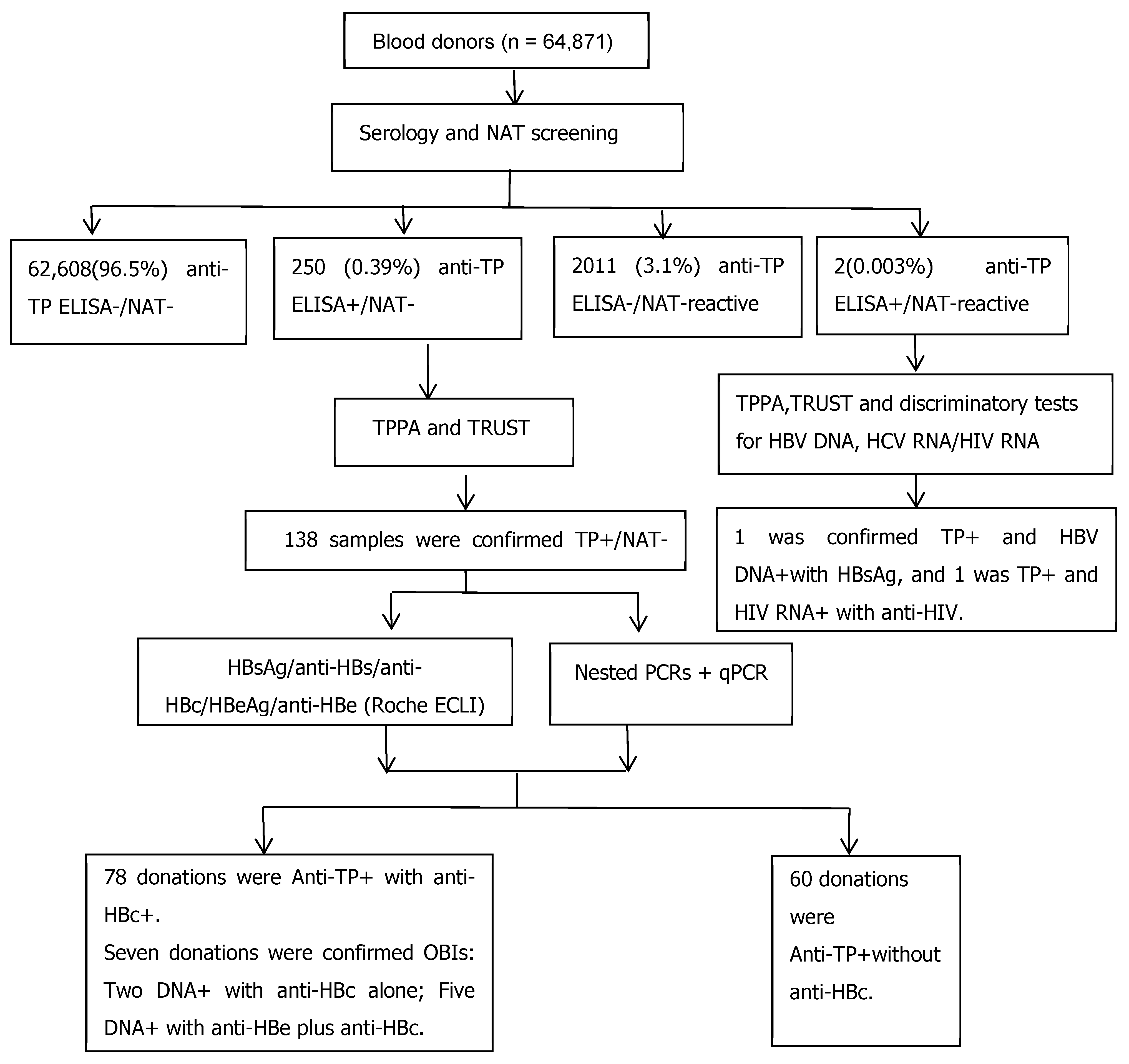
A total of 64,871 blood donations were screened by the dual ELISA assays for HBsAg, anti-HCV, anti-HIV, and anti-TP, in combination with NAT for HBV DNA, HCV RNA, and HIV RNA; 62,543 donations were qualified and released, and 252 anti-TP ELISA+ samples were identified, among which 250 were NAT−, one was HBV NAT-reactive with HBsAg ELISA+, and one was HIV NAT-reactive with anti-HIV ELISA+. After anti-TP confirmatory tests and the discriminatory assay for NAT-reactive donations, 138/250 (55.2%) anti-TP ELISA+/NAT− samples were confirmed TP-positive, and one HBV/TP co-infection and one HIV/TP co-infection were identified. Totally, 140 donations were confirmed as TP-positive. Of 138 confirmed TP-positive donations with NAT-negative results, anti-HBc was detected in 78 cases (78/138, 56.5%), while anti-HBs positivity was observed in 88 cases (88/138, 63.7%), as detailed in Figure 1. Notably, seven (7/138, 5.1%) were diagnosed as occult HBV co-infections by various serological and molecular assays.
Figure 1.
Flow chart of the study design. Dual ELISA assays and NAT were employed as initial screening, while TPPA and TRUST were performed for TP confirmatory tests. Additional HBV serological testing (ECLI) and molecular testing (qPCR and nested PCRs) were used to further clarify OBIs.
The demographic, serologic, and molecular characteristics of 138 TP-confirmed positive donations with NAT-negative results stratified by sex are shown in Table 2. For these 138 TP-confirmed positive donations, seropositive rate of anti-TP in first-time donors was higher than that in repeated donors. The prevalence of syphilis was lower (0.19%) in male donors than in female donors (0.25%). We also found that the majority of the TP-confirmed positive donations were in the 45–60 age group, accounting for 43.48%. Our data also indicated that donors with a lower education level (below high school) had a higher proportion of TP infection.
Table 2.
Socio-demographic characteristics of 138 TP-confirmed positive donations stratified by sex.
3.2. OBI and Anti-HBc Detection in Different Age Groups of TP-Confirmed Positive Donations
A total of 138 confirmed TP-positive donations were screened from 64,871 donations. Specifically, 82 were detected in male donors (82/42,101, 0.19%), and 56 cases were identified in female donors (56/22,770, 0.25%) (Table 2). There were significant differences in gender distribution across different age groups (χ2 = 31.1, p < 0.0001). Seventy-eight donations were anti-HBc-positive (56.5%; 95% CI: 47.8 to 64.9%) (Table 3). There was a clear increase in anti-HBc prevalence with age, ranging from 9.53% in donors aged under 30 years to 85.19% in those over 50 years (χ2 = 31.2, p < 0.0001). Six OBIs were detected in the 41–50 age group, and OBI prevalence was significantly higher in this age group than in other age groups (χ2 = 8.49, p < 0.05).
Table 3.
Frequency of HBV serological markers and HBV DNA in different age groups of 138 confirmed TP-positive donations.
3.3. OBI-Associated RR in Blood Donations Without Anti-TP Screening
In this study, p (OBI without anti-TP screening) was seven cases in 62,543 donations, and p (transmission rate) can be estimated as 2/11 [16], so the RR was calculated as follows:
RR = p (OBI without anti-TP screening) × p (transmission rate)
RR = 7/62,543 × 2/11 = 1:49,141 [95% CI: 1:29,291–1:88,495]
3.4. Serologic and Molecular Characteristics of Seven Donations Co-Infected with OBI and TP
The HBV s gene was successfully amplified from four co-infected donations, and sequence analysis revealed that three cases were genotype B and one case was genotype C. The viral loads of these donations were less than 10 IU/mL or undetectable. In addition, several mutations in the S region of HBV were identified, including Q101R, K122R, Q129H, T131N, M133T, G145R and Y161F mutations. Furthermore, the BCP/PC region of the HBV genome was successfully amplified from five co-infected donations, and nucleotide mutations such as T1719G, A1752T, G1896A, and A1762T/G1764A in the BCP/PC regions were also detected (Table 4).
Table 4.
Serologic and molecular characteristics of 7 donations co-infected with OBI and TP.
4. Discussion
Blood transfusion is critical for saving the lives of patients. Nevertheless, it also bears the risk for transmission of blood-borne pathogens such as HBV, hepatitis C virus (HCV), human immunodeficiency virus (HIV) and TP. In this study, we particularly focused on the prevalence of TP in blood donors and co-infection of HBV and TP, given the fact that both are sexually transmitted pathogens. Our data showed that the overall prevalence of TP infection in blood donors was 0.21% in Southern China (138 out of 64,871 screened donations), which was slightly lower when compared to the results from Jinan, China (0.33%) [11]. In a previous study from the US, the prevalence of donations categorized as consensus positive of syphilis was 28.4/100,000 [17]. A study from Colombia reported the prevalence of donations with positive anti-TP tests was 1.86% [18]. The prevalence of TP infection among blood donors in Yemen and Kyrgyzstan was 2.4% and 3.6%, respectively [19,20]. In African countries, Zambia exhibited the highest TP prevalence at 40.5% [21], followed by Equatorial Guinea at 21.51% [22] and Angola at 20% [23] while relatively lower prevalence was identified in four nations: Cameroon at 8.1% [24], Ghana at 7.5% [25], Tanzania at 4.7% [26], and Ethiopia at 1.5% [27]. Meta-analysis revealed the pooled prevalence of syphilis among blood donors in Thailand was 0.42% [28]. Collectively, these data indicate that although the prevalence of TP varies largely in blood donors from different countries/regions, TP screening serves as a reliable surrogate marker to ensure blood safety by preventing transfusion-related syphilis transmission.
Our current study demonstrated that the confirmed positive rate of TP infection in female blood donors was higher than that in male donors. In each age subgroup, the seroprevalence of anti-TP remained higher in female donors compared to male donors, which was consistent with the results of a previous study [11]. Furthermore, the prevalence increased significantly with age suggesting that older donors bear a higher risk in terms of blood safety. One of the reasons why women bear a higher risk of being infected with TP is that unprotected sexual behavior renders them more susceptible to infection. It has been reported that females were more likely to be infected with sexually transmitted diseases (STDs) partly because of the different physiology and anatomy of the genital organs between both sexes [29]. Studies have confirmed that the male-to-female transmission rate was higher than the female-to-male transmission rate in certain STDs, such as HIV [29,30]. Our findings further demonstrated that the seroprevalence of TP infection was higher in individuals with a lower educational level, which is consistent with the result from previous investigation [31]. This phenomenon indicated that a higher educational level contributes to the enhanced awareness of health knowledge leading to lower risk of infection [32]. Given ongoing social development and increased population mobility, public health authorities should prioritize the dissemination of STD knowledge among populations with lower educational levels.
TP remains viable for several years at −78 °C, and blood samples from syphilis patients may still retain infectivity for up to 4 days when stored at 4 °C [33]. Previous studies have demonstrated that TP screening is not an effective surrogate marker for assessing the risk of other TTIs, given that the majority of TTI-marker-positive blood donations were not positive for TP. In addition, the co-infection rate of HBV/TP among US blood donors was as low as 1.5 per 100,000 donations [34,35]. However, risk factors for syphilis infection in blood donors are also shared by other blood-borne pathogens such as HBV [36,37]. A Chinese blood center reported that approximately 0.8% of HBsAg-positive blood donations were also tested reactive for TP [38]. In the present study, among 64,871 screened blood donations, 0.21% were confirmed as TP-positive but NAT-negative. Of these TP-positive donations, 56.5% were anti-HBc-positive, and 5.1% were diagnosed with OBIs. Additionally, sequence analysis revealed that various mutations of the HBV genome may contribute to the extremely low HBV viral loads and/or detection failure of HBsAg, collectively leading to these un-identified OBIs by routine screening in blood establishments, even by the most sensitive individual nucleic acid testing (ID-NAT). Our data also demonstrated that without anti-TP screening, the residual risk of OBI remained at a high level of 1:49,141, posing a potential threat to blood safety. These findings suggest that TP screening in blood donors may serve as an additional safeguard measure to exclude donations with undetectable OBIs. The observed co-infection of TP and HBV among blood donors is not unexpected, as these sexually transmitted infections share common risk factors.
Sexual transmission has become an important mode of HBV transmission [39,40]. A study by the American Red Cross reported that four out of nine recipients (44.4%) contracted HBV infection via blood donations from the sexual partners of HBV-positive donors [41]. Existing research demonstrates that anti-HBc prevalence increases with age, reflecting a higher cumulative risk of HBV exposure via sexual contact among blood donors [42,43]. In addition, 15 observational studies conducted in Western and Pacific nations have demonstrated that sexual risk factors, such as contact with a hepatitis virus-infected partner, correlate with elevated HBV/HCV infection risk in blood donors [44]. In the present study, among 138 confirmed TP-positive blood donations, 56.5% were anti-HBc-positive, and 5.1% were identified as OBI/TP co-infections. Such a high prevalence of HBV–TP co-infection and elevated anti-HBc seropositivity indicate that sexual contact may be one of the origins for the occurrence of OBIs. Our previous investigation of donor couples with chronic HBV infection found that seven out of 10 infected couples shared HBV sequence homology over 99%, supporting the hypothesis that HBV transmission occurs between partners predominantly via sexual contact [10]. Notably, three of these seven cases were classified as OBIs. Collectively, these findings, in combination with results from previous studies, could suggest that sexual exposure to HBV-infected individuals constitutes one of the primary sources of OBIs.
Our study has limitations. No epidemiological or behavioral information was collected from the enrolled donors, comparative analyses (e.g., TP-positive versus TP-negative donors), appropriate statistical measures and/or evaluation of the diagnostic or predictive performance of TP screening were not performed in the present study. These limitations render our findings mainly descriptive in nature and future study should focus on direct comparisons between TP-positive and TP-negative in the prevalence of HBV infections, particularly OBIs.
5. Conclusions
In summary, of 138 (138/64,871, 0.21%) TP-positive but NAT-negative donors screened from 64,871 blood samples, 78 (78/138, 56.5%) were anti-HBc-positive, and seven (7/138, 5.1%) exhibited OBIs that were not detected by routine screening assays. Several mutations were observed in S and the BCP/PC gene of the HBV genome, which may contribute to the extremely low HBV viral loads and/or failure in HBsAg detection by routine blood screening, collectively leading to OBIs. These findings demonstrate that TP screening has the potential to serve as an additional safeguard to exclude donations co-infected with TP/OBI, as indicated by a relatively higher prevalence of undetectable OBIs in TP-positive blood donors in our current study.
Author Contributions
X.Y. designed the experiments and wrote and reviewed the manuscript. L.C. and B.H. reviewed, revised, and edited the manuscript. X.X., J.Z., H.X. and J.S. participated in the study design, performed the experiments, collected/analyzed data, or provided feedback on the original manuscript. All authors have read and agreed to the published version of the manuscript.
Funding
The authors declared that financial support was received from the Sanming Project of Medicine in Shenzhen, Shenzhen Municipal Health Commission (No. SZSM202311032).
Institutional Review Board Statement
This study was conducted in accordance with the Declaration of Helsinki, and the protocol was approved by the Ethics Committee of Shenzhen Blood Center (SZBCMEC-2024-055) on [7 February 2024]. The ethics approval authorized the use of specimens collected before February 2024. Specimens collected during routine blood donation procedures were stored as archived samples and subsequently analyzed retrospectively after approval had been granted.
Informed Consent Statement
The written informed consent was obtained from each donor before blood donation.
Data Availability Statement
The data sets used and/or analyzed during the current study are available from the corresponding author on reasonable request.
Acknowledgments
The authors declare that no Generative AI was used in the creation of this manuscript, and reasonable efforts have been made to ensure accuracy, including review by the authors wherever possible.
Conflicts of Interest
The authors declare that this work was conducted in the absence of any commercial or financial relationships that could be construed as a potential conflicts of interest.
Abbreviations
HBV = hepatitis B virus; HBsAg, hepatitis B surface antigen; anti-HBc, hepatitis B core antibody; anti-HBs, hepatitis B surface antibody; OBI = occult hepatitis B infection; CHB = chronic hepatitis B; NAT = nucleic acid testing; TP = Treponema Pallidum; TPPA = Treponema Pallidum Particle Agglutination Assay; TRUST = Toluidine Red Unheated Serum Test; ELISA(s) = enzyme-linked immuno-absorbent assay(s); ECLI = electrochemiluminescence immunoassay; STDs = sexually transmitted diseases; WHO = World Health Organization; ID = individual donation; MP = mini pool; PCR = polymerase chain reaction; LOD = limit of detection; BCP/PC = basic core promoter/pre-core; MHR = major hydrophilic region; STD = sexually transmitted disease; RR = residual risk.
References
- Singh, A.E.; Romanowski, B. Syphilis: Review with emphasis on clinical, epidemiologic, and some biologic features. Clin. Microbiol. Rev. 1999, 12, 187–209. [Google Scholar] [CrossRef] [Scilit] [PubMed]
- World Health Organization (WHO). Screening Donated Blood for Transfusion-Transmissible Infections: Recommendations. Available online: https://www.who.int/publications/i/item/9789241547888 (accessed on 1 January 2009).
- Lafond, R.E.; Lukehart, S.A. Biological basis for syphilis. Clin. Microbiol. Rev. 2006, 19, 29–49. [Google Scholar] [CrossRef] [Scilit] [PubMed]
- Kitayama, K.; Segura, E.R.; Lake, J.E.; Perez-Brumer, A.G.; Oldenburg, C.E.; Myers, B.A.; Pourjavaheri, P.; Okorie, C.N.; Cabello, R.L.; Clark, J.L. Syphilis in the Americas: A protocol for a systematic review of syphilis prevalence and incidence in four high-risk groups, 1980–2016. Syst. Rev. 2017, 6, 195–201. [Google Scholar] [CrossRef] [Scilit] [PubMed]
- Kaur, G.; Kaur, P. Syphilis testing in blood donors: An update. Blood Transfus. 2015, 13, 197–204. [Google Scholar] [CrossRef] [Scilit] [PubMed]
- Hesketh, T.; Ye, X.; Zhu, W. Syphilis in China: The great comeback. Emerg. Health Threat. J. 2008, 1, e6. [Google Scholar] [CrossRef] [Scilit] [PubMed]
- Tao, Y.; Chen, M.Y.; Tucker, J.D.; Ong, J.J.; Tang, W.; Wong, N.S.; Chu, M.; Zhuang, X.; Fairley, C.K.; Zhang, L. A Nationwide Spatiotemporal Analysis of Syphilis over 21 Years and Implications for Prevention and Control in China. Clin. Infect. Dis. 2020, 70, 136–139. [Google Scholar] [CrossRef] [Scilit] [PubMed]
- Ji, Z.H.; Li, C.Y.; Lv, Y.G.; Cao, W.; Chen, Y.Z.; Chen, X.P.; Tian, M.; Li, J.H.; An, Q.X.; Shao, Z.J. The prevalence and trends of transfusion-transmissible infectious pathogens among first-time, voluntary blood donors in Xi’an, China between 1999 and 2009. Int. J. Infect. Dis. 2013, 17, e259–e262. [Google Scholar] [CrossRef] [Scilit] [PubMed]
- Song, Y.; Bian, Y.; Petzold, M.; Ung, C.O. Prevalence and trend of major transfusion-transmissible infections among blood donors in Western China, 2005 through 2010. PLoS ONE 2014, 9, e94528. [Google Scholar] [CrossRef] [Scilit] [PubMed]
- Ye, X.; Li, T.; Yu, B.; Zeng, J.; Shi, Y.; Xie, H.; Branch, D.R.; Loriamini, M.; Li, B.; Chen, L. The high prevalence of occult hepatitis B infections among the partners of chronically infected HBV blood donors emphasizes the potential residual risk to blood safety. J. Med. Virol. 2023, 95, e29006. [Google Scholar] [CrossRef] [Scilit] [PubMed]
- Chen, X.; Liu, Q.; Sun, P.; Yuan, S.; Liao, H.; Zhang, X. Prevalence of Syphilis Infections Among Volunteer Blood Donors in Jinan Blood Center, China: A 15-Year Retrospective Study. Infect. Drug Resist. 2022, 15, 6431–6440. [Google Scholar] [CrossRef] [Scilit] [PubMed]
- Ye, X.; Li, T.; Zhang, R.; Liu, H.; Zeng, J.; Hong, W.; Lu, L.; Zhu, W.; Li, S.; Xu, M.; et al. Comprehensive Analysis of Hepatitis B Virus Infections in Blood Donors in Southern China That Are Surface Antigen Positive but Nucleic Acid Testing Negative. Transfusion 2020, 7, 1476–1482. [Google Scholar] [CrossRef] [Scilit] [PubMed]
- Allain, J.P.; Candotti, D.; Soldan, K.; Sarkodie, F.; Phelps, B.; Giachetti, C.; Shyamala, V.; Yeboah, F.; Anokwa, M.; Owusu-Ofori, S.; et al. The risk of hepatitis B virus infection by transfusion in Kumasi, Ghana. Blood 2003, 101, 2419–2425. [Google Scholar] [CrossRef] [Scilit] [PubMed]
- Ye, X.; Xu, X.; Dang, Y.; Zeng, J.; Xie, H.; Li, B.; He, B.; Chen, L. High Genetic Variability of Hepatitis B Virus in Blood Donors with Surface Antigen Positive But DNA Negative in Southern China. J. Med. Virol. 2025, 97, e70396. [Google Scholar] [CrossRef] [Scilit] [PubMed]
- Seed, C.R.; Kiely, P.; Hoad, V.C.; Keller, A.J. Refining the risk estimate for transfusion-transmission of occult hepatitis B virus. Vox Sang. 2017, 112, 3–8. [Google Scholar] [CrossRef] [Scilit] [PubMed]
- Spreafico, M.; Berzuini, A.; Foglieni, B.; Candotti, D.; Raffaele, L.; Guarnori, I.; Colli, A.; Maldini, F.F.; Allain, J.-P.; Prati, D. Poor Efficacy of Nucleic Acid Testing in Identifying Occult HBV Infection and Consequences for Safety of Blood Supply in Italy. J. Hepatol. 2015, 63, 1068–1076. [Google Scholar] [CrossRef] [Scilit] [PubMed]
- Conti, G.; Notari, E.P.; Dodd, R.Y.; Kessler, D.; Custer, B.; Reik, R.; Lanteri, M.C.; Hailu, B.; Yang, H.; Stramer, S.L.; et al. Syphilis seroprevalence and incidence in US blood donors from 2020 to 2022. Transfusion 2024, 64, 325–333. [Google Scholar] [CrossRef] [Scilit] [PubMed]
- Martínez-Garcés, J.C.; Macías-Vidal, M.; Maestre-Serrano, R.; Ávila-De la Hoz, R.; Navarro-Jiménez, E.; Bula-Viecco, J.; Ricaurte-Barrera, L. Seroreactivity and prevalence of syphilis in donors at a blood bank in Barranquilla, Colombia. Biomedica 2019, 39, 163–171. [Google Scholar] [CrossRef] [Scilit] [PubMed]
- Alharazi, T.; Alzubiery, T.K.; Alcantara, J.C.; Qanash, H.; Bazaid, A.S.; Altayar, M.A.; Aldarhami, A. Prevalence of Transfusion-Transmitted Infections (HCV, HIV, Syphilis and Malaria) in Blood Donors: A Large-Scale Cross-Sectional Study. Pathogens 2022, 11, 726. [Google Scholar] [CrossRef] [Scilit] [PubMed]
- Karabaev, B.B.; Beisheeva, N.J.; Satybaldieva, A.B.; Ismailova, A.D.; Pessler, F.; Akmatov, M.K. Seroprevalence of hepatitis B, hepatitis C, human immunodeficiency virus, Treponema pallidum, and co-infections among blood donors in Kyrgyzstan: A retrospective analysis (2013–2015). Infect. Dis. Poverty 2017, 6, 45. [Google Scholar] [CrossRef] [Scilit] [PubMed]
- Katamba, C.; Chungu, T.; Lusale, C. HIV, syphilis and hepatitis B coinfections in Mkushi, Zambia: A cross-sectional study. F1000 Res. 2019, 8, 562–569. [Google Scholar] [CrossRef] [Scilit] [PubMed]
- Xie, D.D.; Li, J.; Chen, J.T.; Eyi, U.M.; Matesa, R.A.; Obono, M.M.; Ehapo, C.S.; Yang, L.Y.; Yang, H.; Yang, H.T.; et al. Seroprevalence of Human Immunodeficiency Virus, Hepatitis B Virus, Hepatitis C Virus, and Treponema pallidum Infections among Blood Donors on Bioko Island, Equatorial Guinea. PLoS ONE 2015, 10, e0139947. [Google Scholar] [CrossRef] [Scilit] [PubMed]
- Quintas, A.E.; Dias, C.C.; Cogle, A.D.C.; Cordeiro, L.; Sarmento, A. Seroprevalence of viral transfusion transmissible infections (HBsAg, anti-HCV, anti-HIV, Syphilis) and coinfection among healthy volunteer blood donors during 5-years in Luanda, Angola. Braz. J. Infect. Dis. 2023, 27, 103704. [Google Scholar] [CrossRef] [Scilit] [PubMed]
- Eboumbou Moukoko, C.E.; Ngo Sack, F.; Essangui Same, E.G.; Mbangue, M.; Lehman, L.G. HIV, HBV, HCV and T. pallidum infections among blood donors and Transfusion-related complications among recipients at the Laquintinie hospital in Douala, Cameroon. BMC Hematol. 2014, 14, 5–11. [Google Scholar] [CrossRef] [Scilit] [PubMed]
- Kuznik, A.; Habib, A.G.; Manabe, Y.C.; Lamorde, M. Estimating the Public Health Burden Associated with Adverse Pregnancy Outcomes Resulting from Syphilis Infection Across 43 Countries in Sub-Saharan Africa. Sex. Transm. Dis. 2015, 42, 369–375. [Google Scholar] [CrossRef] [Scilit] [PubMed]
- Sakala, J.; Chizuni, N.; Nzala, S. A study on usefulness of a set of known risk factors in predicting maternal syphilis infections in three districts of Western Province, Zambia. Pan Afr. Med. J. 2016, 24, 75–81. [Google Scholar] [CrossRef] [Scilit] [PubMed]
- Melku, M.; Ambachew, S.; Enawgaw, B.; Abebe, M.; Abebe, Z.; Deressa, T.; Damtie, D.; Biadgo, B.; Tessema, B.; Geremew, D.; et al. Sero-epidemiology and associated factors of HIV, HBV, HCV and syphilis among blood donors in Ethiopia: A systematic review and meta-analysis. BMC Infect. Dis. 2021, 21, 778–782. [Google Scholar] [CrossRef] [Scilit] [PubMed]
- Rattanatham, R.; Mala, W.; Kotepui, K.U.; Masangkay, F.R.; Rattanawan, C.; Lasom, S.; Wangdi, K.; Kotepui, M. A systematic review and meta-analysis of the prevalence and risk of syphilis among blood donors in Thailand. Sci. Rep. 2025, 15, 9316. [Google Scholar] [CrossRef] [Scilit] [PubMed]
- Wu, X.; Guan, Y.; Ye, J.; Fu, H.; Zhang, C.; Lan, L.; Wu, F.; Tang, F.; Wang, F.; Cai, Y.; et al. Association between syphilis seroprevalence and age among blood donors in Southern China: An observational study from 2014 to 2017. BMJ Open 2019, 9, e024393. [Google Scholar] [CrossRef] [Scilit] [PubMed]
- Kim, J.H. HIV transmissions by stage and sex role in long-term concurrent sexual partnerships. Acta Biotheor. 2015, 63, 33–54. [Google Scholar] [CrossRef] [Scilit] [PubMed]
- Lin, J.T.; Wu, W.J.; Yang, F.Y.; Yu, Y.-T.; Li, Y.-H.; Yu, J.-L. Trend of syphilis infection among voluntary blood donors in Foshan City from 2008 to 2017. J. Exp. Hematol. 2020, 28, 641–645. [Google Scholar] [CrossRef]
- Zheng, X.; Ding, W.; Ling, X.; Shi, J.; Dong, J.; Yu, G.; Chen, Y.; Li, R.; Xu, L.; Li, X.; et al. Prevalence of Treponema Pallidum Antibody among Volunteer Blood Donors in China. Can. J. Infect. Dis. Med. Microbiol. 2022, 2022, 1668703. [Google Scholar] [CrossRef] [Scilit] [PubMed]
- Liu, S.; Luo, L.; Xi, G.; Wan, L.; Zhong, L.; Chen, X.; Gong, T.; Li, S.; He, Y.; Li, N. Seroprevalence and risk factors on Syphilis among blood donors in Chengdu, China, from 2005 to 2017. BMC Infect. Dis. 2019, 19, 509–516. [Google Scholar] [CrossRef] [Scilit] [PubMed]
- Zou, S.; Notari, E.P.; Fang, C.T.; Stramer, S.L.; Dodd, R.Y. Current value of serologic test for syphilis as a surrogate marker for blood-borne viral infections among blood donors in the United States. Transfusion 2009, 49, 655–661. [Google Scholar] [CrossRef] [Scilit] [PubMed]
- Conti, G.; Notari, E.P., 4th; Dodd, R.Y.; Gorlin, J.; Custer, B.; Reik, R.; Hailu, B.; Whitaker, B.I.; Stramer, S.L.; U.S. Transfusion-Transmissible Infections Monitoring System (TTIMS). Transfusion-transmissible coinfections among US blood donors. Transfusion 2024, 64, 2241–2246. [Google Scholar] [CrossRef] [Scilit] [PubMed]
- O’Brien, S.F.; Drews, S.J.; Yi, Q.L.; Osmond, L.; Tran, V.; Zhou, H.Y.; Goldman, M. Monitoring syphilis serology in blood donors: Is there utility as a surrogate marker of early transfusion transmissible infection behavioral risk? Transfusion 2023, 63, 1195–1203. [Google Scholar] [CrossRef] [Scilit] [PubMed]
- Yang, T.; Chen, Q.; Li, D.; Wang, T.; Gou, Y.; Wei, B.; Tao, C. High prevalence of syphilis, HBV, and HCV co-infection, and low rate of effective vaccination against hepatitis B in HIV-infected patients in West China hospital. J. Med. Virol. 2018, 90, 101–108. [Google Scholar] [CrossRef] [Scilit] [PubMed]
- Liu, J.; Huang, Y.; Wang, J.; Guo, N.; Li, J.; Dong, X.; Ma, H.; Tiemuer, M.; Huang, M.; Wright, D.J.; et al. NHLBI Retrovirus Epidemiology Donor Study-II (REDS-II), International Component. The increasing prevalence of serologic markers for syphilis among Chinese blood donors in 2008 through 2010 during a syphilis epidemic. Transfusion 2012, 52, 1741–1749. [Google Scholar] [CrossRef] [Scilit] [PubMed]
- Zhang, L.; Wang, Y.Y.; Huang, Y.J.; Wang, Q.M.; Nelson, K.E.; Wang, A.Q.; Shen, H.-P.; Liu, X.-L.; Zhang, Y.-P.; Yan, D.-H.; et al. Status of HBsAg seroprevalence in 15 million rural couples in China: A cross-sectional study. Sci. Rep. 2017, 21, 7–12. [Google Scholar] [CrossRef] [Scilit] [PubMed]
- Liu, J.; Zhang, S.; Wang, Q.; Shen, H.; Zhang, M.; Zhang, Y.; Yan, D.; Liu, M. Prevalence of HBsAg/HBeAg amongst 1 936 801 couples preparing for pregnancy in rural China: An observational study. J. Viral Hepat. 2017, 24, 679–686. [Google Scholar] [CrossRef] [Scilit] [PubMed]
- Stramer, S.L.; Wend, U.; Candotti, D.; Foster, G.A.; Hollinger, F.B.; Dodd, R.Y.; Allain, J.P.; Gerlich, W. Nucleic acid testing to detect HBV infection in blood donors. N. Engl. J. Med. 2011, 364, 236–247. [Google Scholar] [CrossRef] [Scilit] [PubMed]
- Ye, X.; Li, T.; Xu, X.; Du, P.; Zeng, J.; Zhu, W.; Yang, B.; Li, C.; Allain, J.P. Characterization and follow-up study of occult hepatitis B virus infection in anti-HBc-positive qualified blood donors in southern China. Blood Transfus. 2017, 15, 6–12. [Google Scholar] [CrossRef] [Scilit] [PubMed]
- Zheng, X.; Ye, X.; Du, P.; Zeng, J.; Zhu, W.; Yang, B.; Li, C.; Allain, J.P. High prevalence of anti-hepatitis B core antigen in hepatitis B virus-vaccinated Chinese blood donors suggests insufficient protection but little threat to the blood supply. Transfusion 2015, 55, 890–897. [Google Scholar] [CrossRef] [Scilit] [PubMed]
- Van Remoortel, H.; Matthysen, W.; Avau, B.; Compernolle, V.; Vandekerckhove, P.; De Buck, E. Is sexual risk behaviour associated with an increased risk of transfusion-transmissible infections in blood donors from Western and Pacific countries? A systematic review and meta-analysis. Vox Sang. 2020, 115, 107–123. [Google Scholar] [CrossRef] [Scilit] [PubMed]

Disclaimer/Publisher’s Note: The statements, opinions and data contained in all publications are solely those of the individual author(s) and contributor(s) and not of MDPI and/or the editor(s). MDPI and/or the editor(s) disclaim responsibility for any injury to people or property resulting from any ideas, methods, instructions or products referred to in the content. |
© 2026 by the authors. Licensee MDPI, Basel, Switzerland. This article is an open access article distributed under the terms and conditions of the Creative Commons Attribution (CC BY) license.