


Abstract
Dermatophytosis caused by Trichophyton spp. represents a growing public health concern, exacerbated by the increasing prevalence of difficult-to-treat infections and the emergence of resistance to standard antifungal agents, such as terbinafine. In this study, we evaluated the in vitro antifungal activity of a thiosemicarbazide derivative, 1-[(1-methyl-4-nitroimidazol-2-yl)carbonyl]-4-(3-methylphenyl)thiosemicarbazide, against a panel of anthropophilic and zoophilic Trichophyton spp. dermatophytes. Susceptibility testing was performed via the broth microdilution method to determine the minimum inhibitory concentrations (MICs) and minimum fungicidal concentrations (MFCs). The compound demonstrated antifungal efficacy against all the strains tested, with MIC values ranging from 31.25 to 125 µg/mL. Crucially, low MFC/MIC ratios (≤4) confirmed a fungicidal effect, which was further corroborated by microscopic analysis revealing severe hyphal damage in treated dermatophytes. The derivative exhibited consistent activity against both T. rubrum and T. mentagrophytes, including clinical isolates. To our knowledge, this is one of the first detailed evaluations of a nitroimidazole–thiosemicarbazide hybrid against Trichophyton spp. that combines quantitative MIC/MFC testing with morphological assessment. The tested thiosemicarbazide derivative noticeably decreased the viability of VERO and A375 cells, but even at the highest tested concentration, after 72 h of incubation, the cellular viability remained at 65%. These findings suggest that the investigated compound is a promising candidate for the development of new fungicidal drugs or disinfectants for the effective prophylaxis or management of superficial mycoses in human and veterinary medicine.
1. Introduction
Dermatophytes are filamentous, keratinophilic, and keratinolytic fungi that cause dermatophytoses, that is, fungal infections of the skin, scalp, and nails [1,2,3,4,5]. These fungi may be of human origin (anthropophilic, e.g., T. rubrum) or animal origin (zoophilic, e.g., T. mentagrophytes). Superficial infections of glabrous skin typically present as localized inflammation caused by opportunistic fungi such as dermatophytes. In tinea corporis, the lesions are usually round or oval, pink, single or multiple, clearly demarcated from the surrounding healthy skin, and sometimes show a paler central area. As the lesions enlarge, they may merge into extensive, irregular foci. These are often accompanied by pruritus [1,2,3,4,6,7,8].
The clinical manifestations of onychomycosis include changes in nail color and structure, increased brittleness and crumbling, subungual hyperkeratosis, and onycholysis, that is, separation of the nail plate from the nail bed. These changes largely result from the ability of dermatophytes to degrade keratin, the main structural protein of these tissues, through keratinolytic enzymes [2,3,5,8]. Most fungal infections of the skin, mucous membranes, and nails are caused by dermatophytes of the genus Trichophyton, mainly T. rubrum and T. mentagrophytes, and less frequently T. interdigitale and T. tonsurans. Individuals at particular risk include older adults, patients with congenital or iatrogenic immune dysfunction, those receiving long-term antibiotic therapy or other drugs that impair host defenses, and patients with chronic comorbidities (such as diabetes or thyroid disease). Additional predisposing factors include impaired peripheral circulation, occupational exposure (participation in sports, use of swimming pools, residence in dormitories or barracks, and other crowded facilities and environments), and chronic pressure and maceration associated with nonbreathable footwear [1,3,4,5,7].
Dermatophytes are currently the most common fungal pathogens worldwide. They spread efficiently through direct contact with infected individuals or animals or indirectly via fomites such as towels and clothing, posing a major public health challenge [4,5,8,9]. In Europe and Asia, T. rubrum infections have increased [6,10] in recent years, driven by lifestyle changes, population aging, and an increasing prevalence of onychomycosis and tinea pedis. In recent decades, shifts in the etiology of dermatomycosis, T. tonsurans, T. interdigitale/T. mentagrophyte complex [11,12,13,14] and the newly identified terbinafine-resistant T. indotineae, which often plays greater roles [11,12,13,14,15,16]. These species have fuelled outbreaks worldwide.
In southwestern Poland, adults account for >70% of dermatophytosis cases, with T. tonsurans causing >17% of cases [17]; in contrast, a 2019–2024 study of >1200 pediatric patients in northern Poland identified the T. rubrum complex as predominant [18]. Tinea capitis and tinea corporis were more prevalent among younger children, whereas tinea pedis and tinea unguium were more prevalent among adolescents. Local data from the University Clinic in Gdańsk (2011–2016) revealed that dermatophytes are responsible for >12,000 infections (7324 patients from Gdańsk, Poland, and 4729 from Grodno, Belarus), with T. rubrum (55% of cases) followed by the T. mentagrophytes/interdigitale complex [19]. These findings align with global trends, including a Chinese study of >30,000 clinical cultures (2018–2023), in which T. rubrum reached an approximately 90% prevalence [20]. Dermatophytosis affects 20–25% of the population worldwide [6,11,12,13,14,16,21,22]. T. indotineae, closely related to T. mentagrophytes and T. interdigitale, is spreading rapidly beyond South Asia [12,22], accounting for nearly 40% of UK (>157 cases, 2017–2024) reference laboratory dermatophyte isolates [11] and exhibiting terbinafine resistance [11,12,14,15,16,21].
Given these trends, developing new, more effective prevention and treatment strategies, alongside innovative antifungal therapies, has become an urgent priority for contemporary pharmacy and medicine. Despite advances in topical and systemic antifungals (particularly those active against dermatophytes), the global emergence and spread of terbinafine-resistant T. indotineae and other Trichophyton spp. underscore the critical need for novel fungicides with alternative mechanisms of action [14,23]. Hybrid molecules combining nitroimidazole and thiosemicarbazide moieties have recently been recognized as promising platforms for broad-spectrum antifungal drug candidates [24,25].
Despite the wide availability of topical and systemic antifungal agents, treating dermatophytoses remains challenging. Therapeutic failure, relapses, prolonged treatment durations, and adverse effects are still common, with increasing concerns about emerging resistance, particularly to terbinafine, in Trichophyton spp., including T. indotineae and certain T. rubrum isolates [4,6,7,10,12,20,21,26,27,28]. These limitations highlight the need for novel fungicidal compounds with reliable activity against dermatophytes and potential applications in both human and veterinary medicine.
Thiosemicarbazide derivatives and nitroimidazole-based compounds have garnered attention for their broad antimicrobial effects, including those against pathogenic fungi. Hybrid molecules that combine these pharmacophores into a single scaffold may offer enhanced antifungal potency and selectivity toward dermatophytes [26,27]. These agents could serve not only to treat and prevent superficial mycoses but also as active ingredients in disinfectants and antiseptic formulations to reduce the number of environmental reservoirs of dermatophytes.
Therefore, this study evaluated the antifungal and fungicidal activities of 1-[(1-methyl-4-nitroimidazol-2-yl)carbonyl]-4-(3-methylphenyl)thiosemicarbazide against Trichophyton spp. dermatophytes. We assessed its in vitro activity profile and potential as a lead candidate for novel antifungal formulations in human and veterinary applications.
2. Materials and Methods
2.1. Thiosemicarbazide Derivative
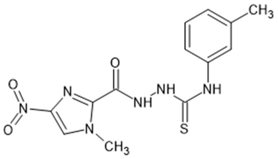
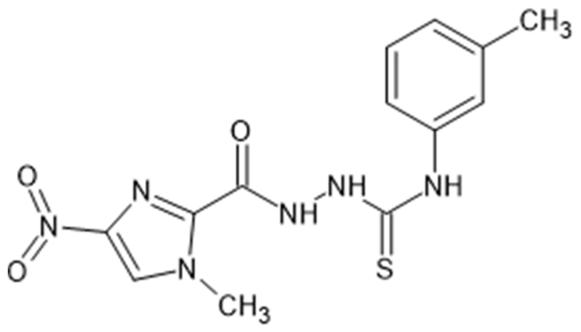
The thiosemicarbazide derivative was synthesized by reacting 1-methyl-4-nitroimidazole-2-carboxylic acid hydrazide with 3-methylphenyl isothiocyanate in anhydrous ethanol at a temperature just below the boiling point for 1 h (Figure 1). The precipitate was filtered after cooling, washed with hot distilled water and diethyl ether, air-dried, and recrystallized from 96% ethanol. Full synthetic details are available in prior publications [25,29,30], where this compound was designated no. 16 [31].
Figure 1.
Chemical structure of the 1-[(1-methyl-4-nitroimidazole-2-yl)carbonyl]-4-(3-methylphenyl)thiosemicarbazide derivative investigated against Trichophyton spp. dermatophytes.
2.2. Antifungal Activity Assay
The antifungal activity of 1-[(1-methyl-4-nitroimidazole-2-yl)carbonyl]-4-(3-methylphenyl)thiosemicarbazide was evaluated via the broth microdilution method in accordance with the European Committee on Antimicrobial Susceptibility Testing (EUCAST) guidelines [31,32,33,34]. Fungal cultures were grown aerobically on solid Sabouraud agar (Biomaxima, Lublin, Poland) at 25 ± 2 °C for 5–10 days prior to testing. Reference strains from the American Type Culture Collection (ATCC) included T. interdigitale ATCC 9533 (formerly T. mentagrophytes) and T. rubrum ATCC 28188, along with clinical isolates (T. mentagrophytes complex, T. rubrum, and T. tonsurans) from diverse body sites (e.g., toenails, facial/neck/groin/hand/foot skin) found in the museum collection of the Department of Pharmaceutical Microbiology at the Medical University of Lublin, Poland.
A stock solution of the tested compound (50 mg/mL) was prepared in 1 mL of dimethyl sulfoxide (DMSO, POCH, Gliwice, Poland). A working solution (2000 µg/mL) was then prepared in sterile Mueller–Hinton broth (MHB, Biomaxima, Lublin, Poland) enriched with 2% glucose (Novo Nordisk, Warsaw, Poland). This solution was used to test the sensitivity of fungi to antifungal substances. To determine antifungal activity via the minimum inhibitory concentration (MIC), 96-well microtiter plates (Medlab, Raszyn, Poland) were used. Briefly, 200 µL of the 2000 µg/mL solution was added to the first row; 100 µL of sterile MHB + 2% glucose was added to subsequent rows. Serial twofold dilutions were prepared by transferring 100 µL from each row of wells to the next row (with thorough mixing), yielding final concentrations ranging from 3.91 to 2000 µg/mL. Fluconazole (Glentham Life Sciences, Corsham, UK) was tested at 0.049–100 µg/mL. All tests were performed in triplicate across three independent experiments, with interexperimental variability <2-fold dilution, per EUCAST guidelines. Representative data are shown.
Suspensions of the tested dermatophytes were prepared in an aseptic environment in accordance with the EUCAST E. Def 9.4 standard [32,35]. Sterile equipment and reagents were used for this purpose. A small amount of material was collected from a fresh fungal culture via a moistened swab dipped in 0.85% NaCl solution and transferred to tubes containing 5 mL of distilled water supplemented with 0.1% Tween-20 (Chemland, Stargard, Poland). After thorough mixing, the suspension was vortexed for 15 s and then transferred to a sterile syringe fitted with an 11 µm pore-diameter filter (Bionovo, Legnica, Poland) for filtration. The filtrate was collected in a sterile test tube to remove hyphal fragments. The suspension was adjusted to a density of 0.5 on the McFarland scale (2–5 × 10⁶ CFU/mL) in distilled water. The suspensions of the tested fungi obtained in this way were then diluted in MHB + 2% glucose medium to a final density of 2–5 × 10⁵ CFU/mL. This mixture was then introduced into all the wells of the plate in a 100 µL volume via an automatic pipette. While determining the activity of the compound against the tested Trichophyton spp. strains, the following steps were carried out: (a) checking the viability of each of the tested fungal strains by adding 100 µL of the suspension to three wells containing pure MHB + 2% glucose medium without the tested compound; (b) checking the purity of the MHB + 2% glucose medium by placing 100 µL of sterile medium without the tested compound or fungi in the wells of a titration plate; and (c) checking the tested compound at the same series of dilutions in MHB + 2% glucose medium without adding the fungal suspension [33].
The microplates were prepared and incubated in a humid chamber at 25 ± 2 °C under aerobic conditions for 3–4 days. After this time, the growth of the dermatophytes in the wells was analyzed via a Bio-Tek ELx800 spectrophotometer (Biokom, Janki, Poland) at 490 nm and via macroscopic observation of turbidity. The endpoints were defined as the lowest concentration of the compound that completely inhibited growth, compared with growth in control wells containing only the quality control agent and no test compounds. All evaluations were performed in triplicate.
The measurements were then processed via the Gen5 version. 3.03.14 (Biokom, Janki, Poland). Fungal growth was indicated by visible turbidity in the microplate wells. The effects of 1-[(1-methyl-4-nitroimidazol-2-yl)carbonyl]-4-(3-methylphenyl)thiosemicarbazide against Trichophyton spp. dermatophytes were assessed on the basis of the minimum inhibitory concentration (MIC) and minimum fungicidal concentration (MFC). The MIC was defined as the lowest concentration of the test compound in wells that inhibited visible fungal growth by ≥90%. The lowest concentration of the test compound was 50% (MIC50—for endpoints determined spectrophotometrically with a 50% growth inhibition endpoint) or 90% (MIC90—for endpoints determined spectrophotometrically with a 90% growth inhibition endpoint) [36]. A reduction in the growth of the test fungi was observed after 5–7 days of incubation compared with the growth of the control fungi at 25 ± 2 °C under aerobic conditions. The MFC was defined as the lowest compound concentration that did not change or exhibited approximately 99.0–99.5% killing activity, as confirmed by microscopic observation and subculture on drug-free media [37]. Control wells containing fungi but no test compound were used to verify normal fungal growth. On the basis of the obtained results and the MFC/MIC ratio, the fungicidal (MFC/MIC ≤ 4) or fungistatic (MFC/MIC > 4) activity of the tested compound against dermatophytes was determined [32,35].
2.3. Cytotoxicity Screening
Cytotoxicity was tested in the BSL-2 laboratory following previously described experimental procedures [38]. The noncancerous VERO (ATCC, CCL-81) and cancer-derived A375 (ATCC, CRL-1619) and MDA-MB-231 (ATCC, HTB-26) cells were used in this study. The media used for in vitro culture included Dulbecco’s modified Eagle’s medium (DMEM; Corning, Tewksbury, MA, USA) for VERO and A375 cells and modified Eagle’s medium (MEM; Corning) for MDA-MB-231 cells. The media were supplemented with antibiotics (penicillin–streptomycin solution, Corning) and fetal bovine serum (FBS, Corning) at 10% (passaging) and 2% (maintenance/experiments). Phosphate-buffered saline (PBS) and trypsin were obtained from Corning, while 3-(4,5-dimethylthiazol-2-yl)-2,5-diphenyltetrazolium bromide (MTT) was purchased from Sigma-Aldrich (Merck KGaA, Darmstadt, Germany). The mixture was incubated at 37 °C in 5% CO2 (Panasonic Healthcare Co., Tokyo, Japan). The cells were subcultured in T25 flasks (EasYFlasks, Nunc, Thermo Fisher Scientific, Waltham, MA, USA) and then passaged into 96-well plates (Falcon, Corning) to produce cell monolayers. The number of passages for all the cell lines used in this research did not exceed 20. The thiosemicarbazide derivative was dissolved (50 mg/mL) in DMSO (cell culture grade, PanReac Applichem, Darmstadt, Germany) to produce stock solutions, which were stored frozen (−23 °C) until analysis. Cytotoxicity was assessed via a microculture tetrazolium (MTT) assay. Briefly, monolayers of appropriate cells in 96-well plates were incubated for 72 h with serial dilutions of the thiosemicarbazide derivative in culture media. Afterward, the media were removed, the cell monolayers were washed with PBS, and MTT-supplemented medium was added. Following a 3 h incubation, the formazan product was dissolved, and the solution was incubated overnight. Moreover, the toxicity of the use of DMSO as a solvent for stock solutions was evaluated. The absorbance was measured (at 540 and 620 nm) via a Synergy H1 Multi-Mode Microplate Reader (BioTek Instruments, Inc., Winooski, VT, USA) with Gen5 software (ver. 3.09.07; BioTek Instruments, Inc., Winooski, VT, USA), and the results were exported to GraphPad Prism (version 10.2.0, San Diego, CA, USA) for analysis. The experiments were performed in triplicate across 3 independent experiments. The statistical significance of differences between means (mean cellular viability) was determined via two-way ANOVA followed by Tukey’s post hoc tests. A p value of <0.05 was considered to indicate statistical significance.
3. Results
3.1. Antifungal Activity Against Trichophyton spp. Dermatophytes
The antifungal activity of 1-(1-methyl-4-nitroimidazol-2-yl)carbonyl-4-(3-methylphenyl)thiosemicarbazide was evaluated against a panel of Trichophyton spp. strains, including both anthropophilic (T. rubrum, T. tonsurans) and zoophilic (T. mentagrophytes complex) species.
The obtained MIC values ranged from 31.25 to 125 µg/mL (Table 1), indicating antifungal inhibitory activity. Notably, the compound was highly effective against clinical isolates of T. rubrum, the most common etiological agent of onychomycosis, with MICs consistently lower than 125 µg/mL.
Table 1.
Fungicidal activity of 1-[(1-methyl-4-nitroimidazol-2-yl)carbonyl]-4-(3-methylphenyl)thiosemicarbazide on the basis of MIC and MFC values against reference strains and clinical isolates of dermatophytes belonging to the genus Trichophyton.
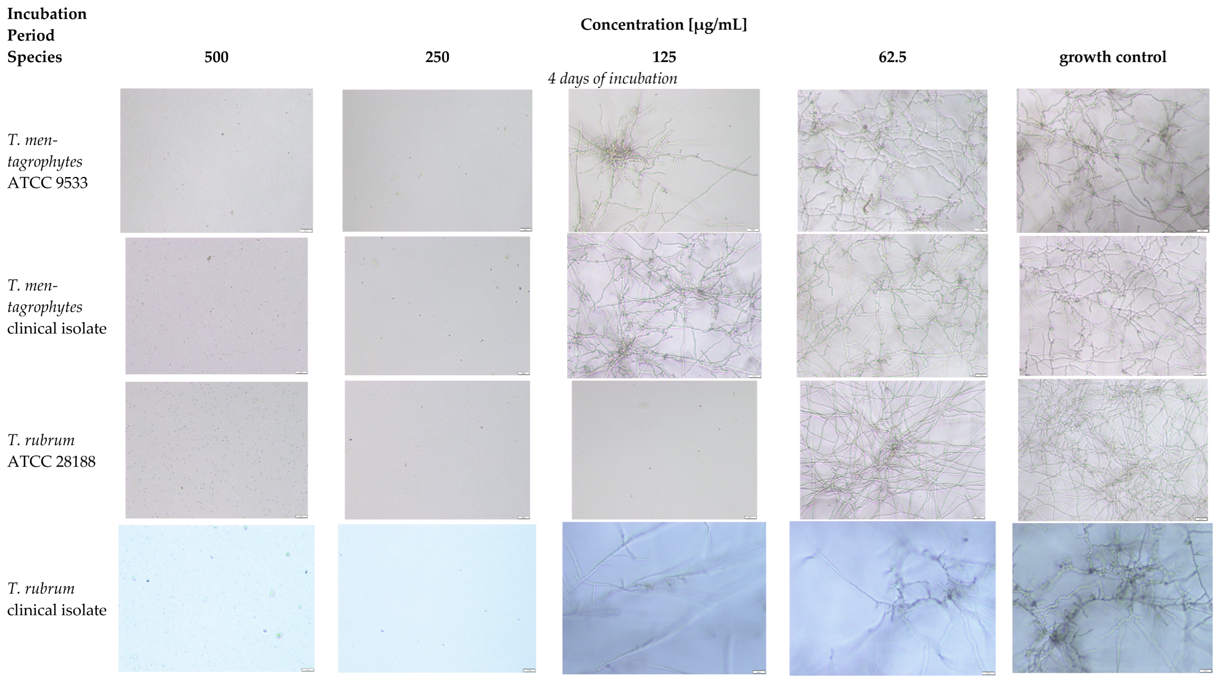
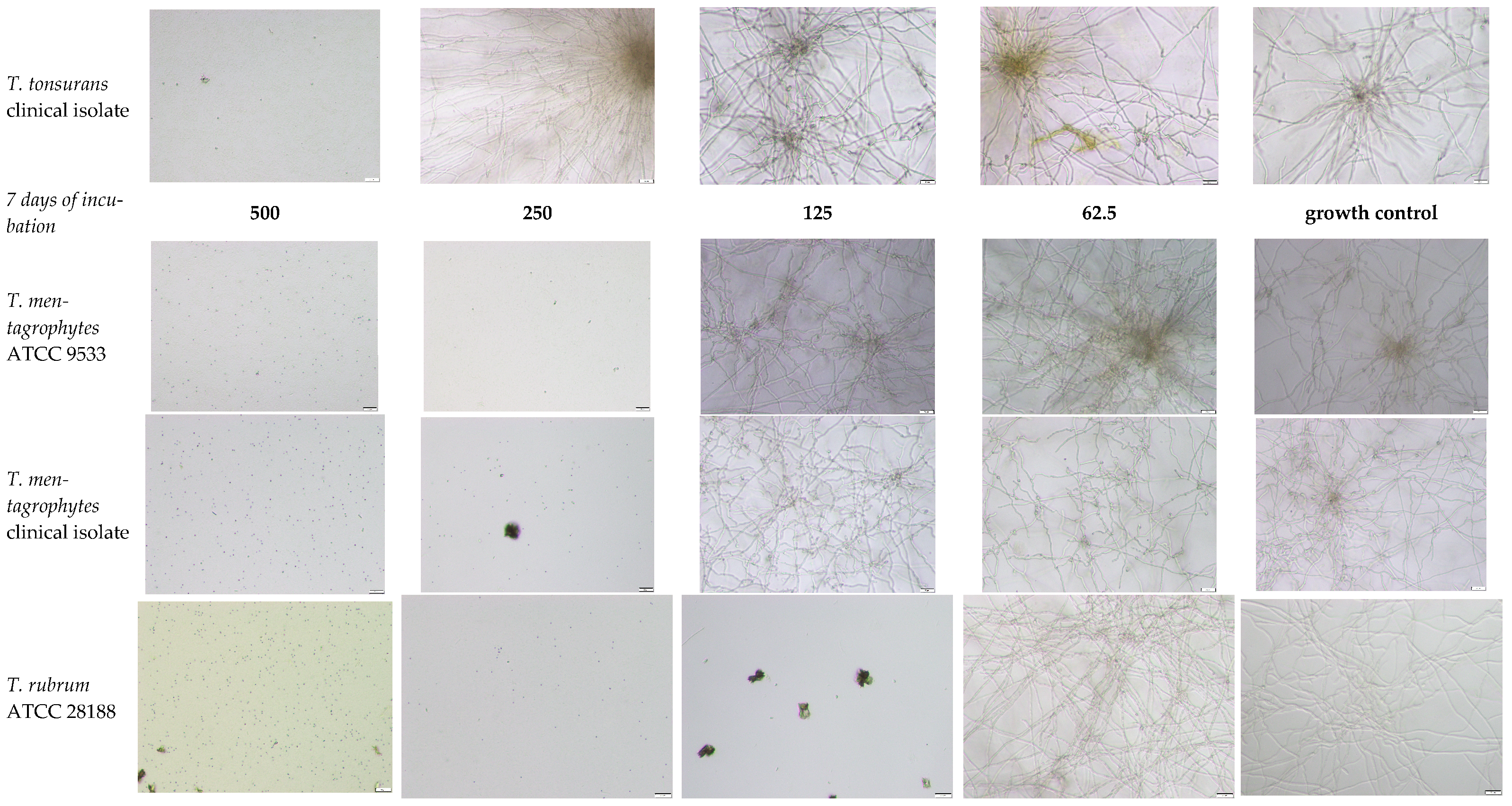
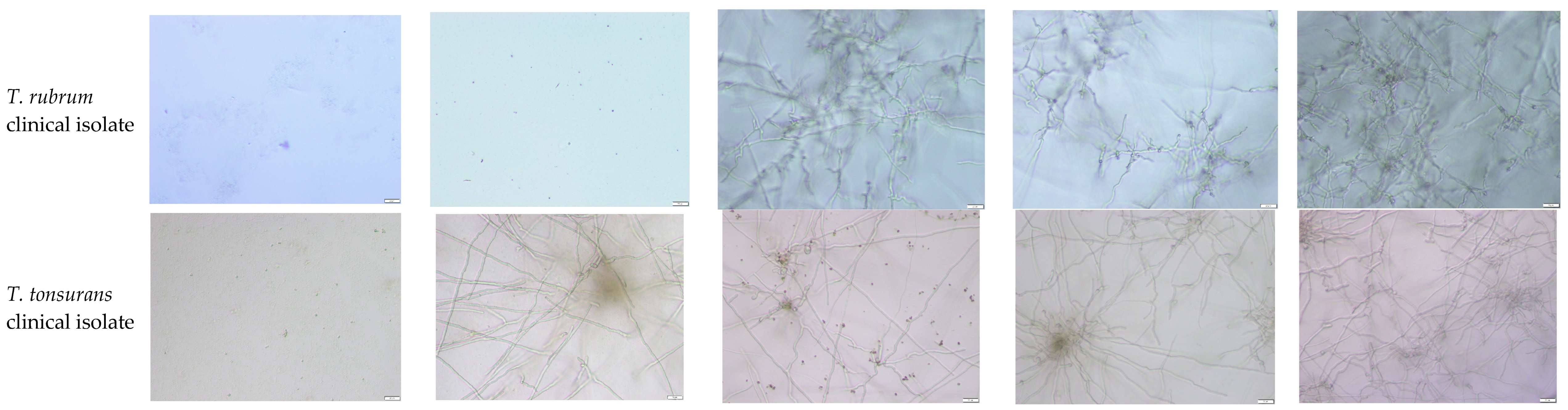
To determine the nature of the antifungal effect, the MFCs were assessed. On the basis of the MIC50 values (ranging from 31.25 to 250 µg/mL), the MIC90 values (ranging from 62.5 to 250 µg/mL), and the calculated MFC/MIC ratios (MFC/MIC = 1), the compound was fungicidal rather than fungistatic toward the majority of the dermatophyte strains tested. The most favorable activity was observed against the T. rubrum ATCC 28188 reference strain (MIC50 = 31.25 µg/mL, MIC90 = 62.5 µg/mL, MFC/MIC = 2). Overall, the compound demonstrated consistent fungicidal properties (MFC/MIC ≤ 4) against both reference and clinical isolates, including T. rubrum strains, which are the leading epidemiological cause of onychomycosis worldwide. This activity was confirmed microscopically by an inhibitory effect on fungal growth and an absence of hyphae and/or spores produced by the tested reference strains and clinical isolates.
3.2. Microscopic Analysis of Trichophyton spp. Morphology Under the Influence of the Compound Tested
To further elucidate the antifungal effects of 1-(1-methyl-4-nitroimidazol-2-yl)carbonyl-4-(3-methylphenyl)thiosemicarbazide, microscopic observations of Trichophyton spp. hyphal growth were performed following exposure to the compound. Following the MIC readings, the incubation process continued for up to 7 days in a humid chamber under aerobic conditions at 25 ± 2 °C. After 4 and 7 days of incubation, a microscopic evaluation of the impact of the test compound on the growth and formation of hyphae and spores of dermatophytes in the genus Trichophyton (Figure A1) was performed. Following incubation, the presence of hyphae and spores in individual wells of a multiwell plate was examined under 2000× magnification via an Olympus DP22 inverted automatic light microscope equipped with CellSens Dimensions 2.3 software (Olympus Corporation, Tokio, Japan). Examples of the microscopic evaluation of the effects of selected concentrations of 1-[(1-methyl-4-nitroimidazol-2-yl)carbonyl]-4-(3-methylphenyl)thiosemicarbazide on the presence of hyphae and spores of dermatophytes are presented in Figure A1.
The control samples cultivated in the tested compound- or fluconazole-free media retained the typical morphology of dermatophytes, with long, regular, branched hyphae and smooth cell walls. In contrast, fungal cells exposed to the tested thiosemicarbazide derivative presented pronounced structural alterations, including irregular branching, cytoplasmic granulation, and marked vacuolization, which was particularly evident in the T. rubrum ATCC 28188 strain. At concentrations equal to or exceeding the MIC of the tested derivative, only single, nongerminating conidia or no conidia at all were observed in the microscopic field, and hyphae were also absent.
3.3. Influence on the Cell Lines
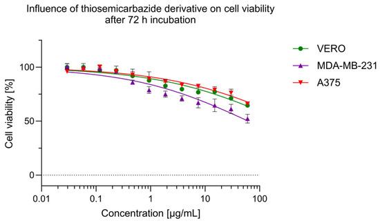
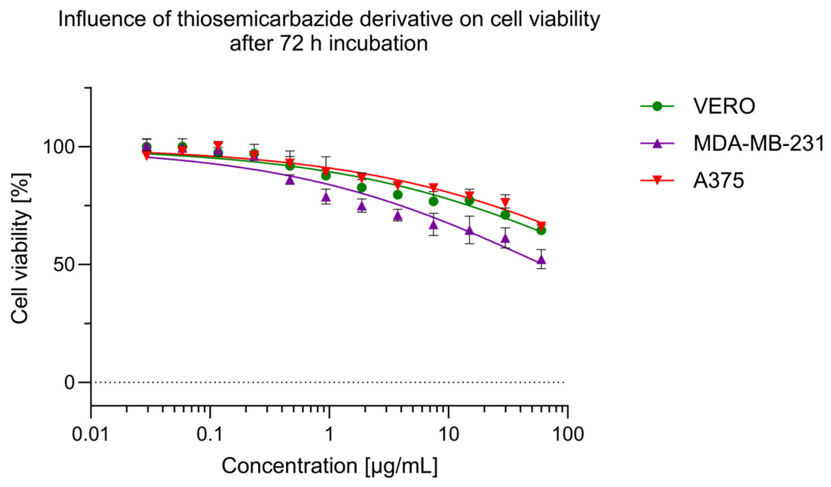
When 1-[(1-methyl-4-nitroimidazol-2-yl)carbonyl]-4-(3-methylphenyl)thiosemicarbazide was tested at 125 µg/mL, a small amount of crystals were observed in the culture medium. It was not possible to increase the solubility of the thiosemicarbazide derivative by increasing the DMSO concentration because of potential toxic effects on cell lines. Thus, cytotoxicity was assessed at concentrations up to 62.5 µg/mL. The thiosemicarbazide derivative showed a dose-dependent cytotoxic effect on the tested cell lines, as shown in Figure 2, with VERO and A375 cells exhibiting a similar pattern of cytotoxicity, decreasing the mean cellular viability to 65% (±2 pp) at 62.5 µg/mL. Compared with VERO and A375 cells, MDA-MB-231 cells were more sensitive to the test compounds, with a mean cellular viability ranging from 0.98 to 62.5 µg/mL, which was significantly lower (p < 0.05). At the highest tested concentration, the mean viability of MDA-MB-231 cells was 52% (±4 pp).
Figure 2.
Dose-response influence of the 1-[(1-methyl-4-nitroimidazole-2-yl)carbonyl]-4-(3-methylphenyl)thiosemicarbazide derivative on VERO, A375 and MDA-MB-231 cell lines.
4. Discussion
When the literature on this topic is analyzed, several studies have described nitrogen-containing heterocyclic compounds with antifungal activity [25,31,39,40,41]. This is not surprising, given that many clinically used drugs contain nitrogen heterocycles. Güzeldemirci et al. [39] reported the synthesis of new or modified antifungal and antibacterial agents on the basis of a core structure comprising two heterocyclic rings, imidazole and thiazole. Among these derivatives, only a single compound exhibited high activity against T. rubrum and Microsporum audouini, comparable to that of ketoconazole. The same group subsequently developed new thiosemicarbazide derivatives, as well as cyclic analogs of 1,2,4-triazole and 1,3,4-thiadiazole containing imidazothiazole moieties [40]. All of these compounds were subjected to biological evaluation, and two thiosemicarbazides displayed antifungal activity against yeast-like fungi (Candida parapsilosis ATCC 22019 and Candida krusei ATCC 6258) and dermatophytes of the genus Trichophyton, including T. erinacei NCPF 375 and T. tonsurans NCPF 245, with MIC values of 64 μg/mL. In another study, Terzioğlu Klip et al. [31] obtained S-(4,5-disubstituted-1,2,4-triazolo)acetic acid hydrazide and converted it into the corresponding thiosemicarbazide derivatives by reacting it with aliphatic and aromatic isothiocyanates. The resulting compounds were tested for antifungal activity against Microsporum gypseum (NCPF 580), Microsporum canis, T. mentagrophytes, and Candida albicans (ATCC 10231). Two derivatives, one bearing a 4-bromophenyl substituent and the other bearing a 4-nitrophenyl group at the 4-position of the thiosemicarbazide moiety, exhibited broad-spectrum antifungal activity, with MIC values of 4–8 μg/mL. In this case, the antifungal effect resulted from the combined influence of the triazole/thiosemicarbazide framework and strongly electron-withdrawing para substituents. In contrast, the compound investigated in the present study contains a 1-methyl-4-nitroimidazole pharmacophore linked to a 3-methylphenyl-substituted thiosemicarbazide moiety. The presence of the 3-methyl group may influence antifungal activity through several mechanisms. As a weak electron-donating substituent, the methyl group increases the electron density of the aromatic ring and may influence the acidity of adjacent NH groups within the thiosemicarbazide fragment. Furthermore, the meta-methyl substituent increases lipophilicity and steric bulk, which may enhance penetration through the lipid-rich fungal cell envelope and affect interactions with biological targets. Unlike para-substituted electron-withdrawing groups such as nitro or bromo groups, the meta-methyl group does not exhibit strong resonance effects, suggesting that the observed activity of the present compound is more likely attributable to a favorable balance among lipophilicity, steric effects, and the contribution of the nitroimidazole moiety.
It is therefore plausible that the antifungal activity observed in this study arises from the synergistic combination of the nitroimidazole pharmacophore and the thiosemicarbazide scaffold rather than from the isolated effect of the 3-methyl substituent alone. Further structure–activity relationship studies involving direct comparisons of meta-methyl, para-bromo, and para-nitro analogs will be needed to clarify the precise role of electronic and steric effects in determining antifungal potency. Yamaguchi et al. [41] further demonstrated that introducing a thiosemicarbazide system into a molecule can increase its antifungal activity by up to 15-fold. Taken together, these findings indicate that the incorporation of a thiosemicarbazide scaffold and appropriately substituted aromatic rings is a promising strategy for enhancing antifungal potency against dermatophytes and yeast-like fungi.
Although no dedicated mechanistic assay was performed in the present work, the observed fungicidal activity and morphological alterations suggest that the tested nitroimidazole–thiosemicarbazide derivative affects fungal cellular integrity. However, the precise molecular mechanism remains unknown and needs further research [42,43]. Previous studies on structurally related thiosemicarbazide/thiosemicarbazone and nitroimidazole derivatives proposed mechanisms involving membrane perturbation, oxidative stress induction, and redox imbalance, although these effects have not yet been verified for the present compound [24,41,44]. Such effects have been reported for related antimicrobial scaffolds, although they were not tested here [45]. Yamaguchi et al. [41] demonstrated that a camphene–thiosemicarbazide derivative active against T. mentagrophytes induced morphological alterations and impaired Calcofluor White staining, suggesting effects on chitin-containing structures, whereas ergosterol did not appear to be the primary target. Thiosemicarbazide-based scaffolds are increasingly recognized as privileged structures in the design of antifungal drugs, with several series demonstrating low-to-moderate minimum inhibitory concentrations (MICs) against dermatophytes and yeast-like fungi [24]. In this context, the activity profile of our compound, including its fungicidal effect on Trichophyton spp. dermatophytes, is at least comparable to and, in some cases, more favorable than the MIC values of 31.25–500 µg/mL reported for previously described thiosemicarbazide derivatives [25].
A comparison of our results with literature data provides valuable insights into the structure-activity relationships within this class of compounds. While previous studies on thiosemicarbazide derivatives reported MIC values against dermatophytes ranging from 125 to 2000 µg/mL for T. mentagrophytes and from 31.25 to 2000 µg/mL for T. rubrum [25], the 1-[(1-methyl-4-nitroimidazol-2-yl)carbonyl]-4-(3-methylphenyl)thiosemicarbazide investigated in this study demonstrated good potency, with MIC values in the range of 31.25–125 µg/mL against both species (with clearly lower MIC values against T. rubrum). This enhanced activity may be attributed to the specific combination of the 1-methyl-4-nitroimidazole moiety and the 3-methylphenyl substituent. Unlike the 4-bromophenyl and 4-nitrophenyl derivatives described by Terzioğlu Klip et al. [31], which contain strong electron-withdrawing groups, the presence of a methyl group in the meta position of the phenyl ring appears to favorably influence the interaction of the molecule with fungal targets. Furthermore, the nitroimidazole core itself is known to contribute to antimicrobial efficacy, potentially through mechanisms involving nitro-group reduction and the subsequent generation of toxic intermediates, a pathway well established for nitroimidazole drugs but less explored in the context of dermatophyte inhibition.
A critical finding of this study is the fungicidal nature of the tested compound against Trichophyton spp., as evidenced by the low MFC/MIC ratios. In clinical practice, fungicidal agents are generally preferred over fungistatic agents for the treatment of dermatophytoses, particularly in immunocompromised patients or in cases of onychomycosis where eradication of the pathogen from the keratinized matrix is difficult [2,3,7,22,27,46,47]. While many azoles primarily exert fungistatic effects, our ability to kill dermatophyte cells at concentrations near the inhibitory level suggests the potential for higher cure rates and a lower risk of relapse [1,7,27]. This profile is particularly promising in the context of the increasing prevalence of difficult-to-treat infections caused by T. indotineae and terbinafine-resistant T. rubrum strains [10,11,12,13,14,15,21,47,48]. The clinical importance of a fungicidal profile is further emphasized by the rapid worldwide emergence of T. rubrum strains resistant to terbinafine, which are often associated with squalene epoxidase mutations and elevated MICs. This results in recalcitrant cases of tinea corporis and cruris [14,23,49]. Novel agents that target alternative pathways and retain activity against Trichophyton spp., regardless of the mechanism of terbinafine resistance, could be valuable options for the management of difficult-to-treat dermatophytoses and for antifungal stewardship [14,23].
A noncancerous VERO cell line was selected to study the cytotoxicity of the derivative 1-[(1-methyl-4-nitroimidazol-2-yl)carbonyl]-4-(3-methylphenyl)thiosemicarbazide. VERO cells were selected because they can be maintained and passaged without signs of senescence, provide reliable, repeatable results, and are commonly used in cytotoxicity studies and in vitro efficacy testing [50,51,52]. Moreover, VERO cells are endorsed by ISO experts as suitable for in vitro cytotoxicity testing [53]. For cytotoxicity comparisons, two cancer cell lines—MDA-MB-231 and A375—were selected, originating from breast adenocarcinoma and malignant melanoma, respectively. The evaluation of the impact of the 1-[(1-methyl-4-nitroimidazol-2-yl)carbonyl]-4-(3-methylphenyl)thiosemicarbazide derivative on the tested cell lines revealed a potential cytotoxic influence, reducing the viability of the MDA-MB-231 or 65% VERO, and A375 cells to 52% at the highest tested concentration. When fluconazole was tested in VERO cells, cytotoxicity after 24 h of incubation was observed at 1306 μM fluconazole (approx. 400 µg/mL), reducing viability to 86%. At 2612.1 μM, viability was reduced to 35%. This effect was likely due to fluconazole-induced cell necrosis, increased reactive oxygen species (ROS), and DNA damage [43]. Fluconazole showed greater cytotoxicity against L929 cells (murine fibroblasts), with reduced cell viability observed at 24 and 48 h of incubation starting at 62.5 µg/mL, and at 250 µg/mL, it was reduced by more than 90%. Interestingly, miconazole showed greater cytotoxicity at 15.6 µg/mL, reducing L929 viability to less than 60% [54] Miconazole also showed greater cytotoxicity against HaCaT cells (human keratinocytes) than terbinafine, with noticeable cytotoxicity at a concentration of 6.25 µg/mL. Both miconazole and terbinafine stimulate ROS in keratinocytes, potentially inducing oxidative stress and cell death [55]. Considering the reported cytotoxicity of approved antifungals such as fluconazole, miconazole, and terbinafine, the observed effects of the 1-[(1-methyl-4-nitroimidazol-2-yl)carbonyl]-4-(3-methylphenyl)thiosemicarbazide derivative on VERO, A375, and MDA-MB-231 do not preclude its potential antifungal applications. However, the authors acknowledge the limitations of the applied cytotoxicity methodology, the limited selection of cell lines, and the need for further testing to evaluate the safety of the new antifungal, 1-[(1-methyl-4-nitroimidazol-2-yl)carbonyl]-4-(3-methylphenyl)thiosemicarbazide, including in vitro skin irritation and other bioassays. Importantly, future studies should include a human keratinocyte cell line to better assess skin cytotoxicity associated with potential topical application.
From a practical perspective, the broad spectrum of activity and fungicidal mechanism of the investigated thiosemicarbazide derivative open several potential avenues for application. Its high efficacy against zoophilic and anthropophilic dermatophytes suggests that it could be developed not only as a therapeutic agent for superficial mycoses in humans but also for veterinary use. Furthermore, given the environmental stability of dermatophyte arthrospores, the compound could serve as an active ingredient in surface disinfectants for high-risk areas such as swimming pools, gyms, and veterinary clinics. Future studies should focus on evaluating the safety profile of this molecule in mammalian cells, assessing its skin permeation properties, and developing optimal topical formulations, such as lacquers or creams, to fully exploit its therapeutic potential.
Current topical therapies for onychomycosis, such as efinaconazole and tavaborole, exhibit variable fungicidal versus fungistatic behavior against Trichophyton spp., with efinaconazole demonstrating potent fungicidal activity in keratin-containing media, whereas tavaborole predominantly exhibits fungistatic properties [56,57]. Collectively, these data indicate that the ability to exhibit fungicidal activity in a keratin-rich environment is an important factor in achieving a clinical cure. These findings suggest that nitroimidazole–thiosemicarbazide derivatives with confirmed fungicidal activity could be promising candidates for topical formulations to treat nails or skin [56].
This study has several strengths, including the use of both reference and clinical Trichophyton isolates, determination of MIC and MFC values according to standard microdilution protocols, and a combination of quantitative susceptibility testing with microscopic assessment of hyphal damage. The observed morphological alterations should therefore be interpreted only as indicators of the cellular response to the compound and not as definitive evidence of a specific antifungal mechanism. The precise molecular mechanism and proposed effects, such as membrane perturbation or oxidative stress, should be considered in future studies. However, this work has several limitations. These include the lack of biofilm- or keratin-related models and the need for in vivo evaluation; mechanistic studies are necessary to determine the exact cellular targets involved. These issues should be addressed in future studies before any drug is developed.
5. Conclusions
The tested compound, 1-(1-methyl-4-nitroimidazol-2-yl)carbonyl-4-(3-methylphenyl)thiosemicarbazide, demonstrated potent antifungal activity, exhibiting lethal effects against dermatophytes of the genus Trichophyton. The data obtained identify this molecule as a fungicidal agent, offering a distinct advantage over purely fungistatic alternatives. Consequently, it represents a promising candidate for the development of novel pharmaceuticals to prevent and treat superficial mycoses caused by anthropophilic and zoophilic dermatophytes in both humans and animals. In addition to clinical therapy, its biological activity provides grounds for potential applications in the medical, pharmaceutical, agricultural, and veterinary industries, including its use as an effective disinfectant, antiseptic, or preservative. Future research will focus on evaluating its safety profile and in vivo efficacy to further validate its versatility and clinical utility. Owing to its consistent fungicidal activity against Trichophyton spp., the tested nitroimidazole–thiosemicarbazide derivative shows promise as a lead compound for the further preclinical development of topical antifungals or disinfectants targeting dermatophytoses in humans and/or animals.
Author Contributions
Conceptualization, U.K. and S.A.; methodology, S.A. and U.K.; software, S.A.; validation, S.A., U.K., Ł.Ś. and M.W.; formal analysis, S.A.; investigation, S.A. and Ł.Ś.; resources, S.A., U.K., Ł.Ś. and M.W.; data curation, S.A. and U.K.; writing—original draft preparation, S.A.; writing—review and editing, S.A., U.K. and M.W.; visualization, S.A.; supervision, U.K. and M.W.; project administration, U.K. and M.W.; funding acquisition, U.K. and M.W. All authors have read and agreed to the published version of the manuscript.
Funding
This research was funded by the Medical University of Lublin, Poland, grant numbers DS15 (Monika Wujec) and DS31 (Urszula Kosikowska).
Institutional Review Board Statement
Not applicable.
Informed Consent Statement
Not applicable.
Data Availability Statement
The raw data supporting the conclusions of this article will be made available by the authors upon request.
Conflicts of Interest
The authors declare that they have no conflicts of interest.
Abbreviations
The following abbreviations are used in this manuscript:
| MIC | minimal inhibitory concentration |
| MFC | minimal fungicidal concentration |
| MFC50 | the lowest concentration that establishes a predetermined reduction of 50% of fungal growth after 5–7 days of incubation, as compared to the growth in the control |
| MFC90 | the lowest concentration that establishes a predetermined reduction of 90% of fungal growth after 5–7 days of incubation, as compared to the growth in the control |
Appendix A
Figure A1.
Microscopic observation of the thiosemicarbazide derivative against Trichophyton spp. dermatophytes investigated (4 and 7 days at 25 ± 2 °C under aerobic conditions). Abbreviations: 2000× magnification, 20 µm scale bar, Olympus DP22 automated inverted light microscope, CellSens Dimensions 2.3 software, Olympus Corporation; growth control refers to the growth of fungi without the presence of the tested thiosemicarbazide derivative.
References
- Martinez-Rossi, N.M.; Peres, N.T.A.; Bitencourt, T.A.; Martins, M.P.; Rossi, A. State-of-the-Art Dermatophyte Infections: Epidemiology Aspects, Pathophysiology, and Resistance Mechanisms. J. Fungi 2021, 7, 629. [Google Scholar]
- Barac, A.; Stjepanovic, M.; Krajisnik, S.; Stevanovic, G.; Paglietti, B.; Milosevic, B. Dermatophytes: Update on Clinical Epidemiology and Treatment. Mycopathologia 2024, 189, 101. [Google Scholar] [CrossRef] [Scilit] [PubMed]
- Wang, R.; Huang, C.; Zhang, Y.; Li, R. Invasive Dermatophyte Infection: A Systematic Review. Mycoses 2021, 64, 340–348. [Google Scholar] [PubMed]
- Dellière, S.; Jabet, A.; Abdolrasouli, A. Current and Emerging Issues in Dermatophyte Infections. PLoS Pathog. 2024, 20, e1012258. [Google Scholar] [CrossRef] [Scilit] [PubMed]
- Deng, R.; Wang, X.; Li, R. Dermatophyte Infection: From Fungal Pathogenicity to Host Immune Responses. Front. Immunol. 2023, 14, 1285887. [Google Scholar] [CrossRef] [Scilit] [PubMed]
- Ha, N.G.; Bang, Y.J.; Lee, W.J. Epidemiological Trends and Clinical Features of Trichophyton Rubrum Infections: A 10-Year Retrospective Review of 38,391 Cases (2014–2023). J. Korean Med. Sci. 2025, 40, e264. [Google Scholar] [CrossRef] [Scilit] [PubMed]
- Gnat, S.; Łagowski, D.; Nowakiewicz, A. Major Challenges and Perspectives in the Diagnostics and Treatment of Dermatophyte Infections. J. Appl. Microbiol. 2020, 129, 212–232. [Google Scholar] [CrossRef] [Scilit] [PubMed]
- Gupta, C.; Das, S.; Gaurav, V.; Singh, P.K.; Rai, G.; Datt, S.; Tigga, R.A.; Pandhi, D.; Bhattacharya, S.N.; Ansari, M.A.; et al. Review on Host-Interaction in Dermatophyte Infections. J. Med. Mycol. 2023, 33, 101331. [Google Scholar]
- Kruithoff, C.; Gamal, A.; McCormick, T.S.; Ghannoum, M.A. Dermatophyte Infections Worldwide: Increase in Incidence and Associated Antifungal Resistance. Life 2024, 14, 1. [Google Scholar]
- Mazzantini, D.; Celandroni, F.; Calvigioni, M.; Lupetti, A.; Ghelardi, E. In Vitro Resistance and Evolution of Resistance to Tavaborole in Trichophyton Rubrum. Antimicrob. Agents Chemother. 2021, 65, e02324-20. [Google Scholar] [CrossRef] [Scilit] [PubMed]
- Abdolrasouli, A.; Barton, R.C.; Borman, A.M. Spread of Antifungal-Resistant Trichophyton indotineae, United Kingdom, 2017–2024. Emerg. Infect. Dis. 2025, 31, 192. [Google Scholar] [CrossRef] [Scilit] [PubMed]
- Prunglumpoo, S.; Krongboon, K.; Kazushi, A.; Bunnag, T.; Paunrat, W. Emergence of Resistant Dermatophytosis Caused by Trichophyton indotineae: First Case Series in Thailand. Med. Mycol. Case Rep. 2025, 48, 100713. [Google Scholar] [CrossRef] [Scilit] [PubMed]
- Tóth, Z.; Ványai, B.; Kovács, R.; Jakab, Á.; Szegedi, A.; Balázs, B.; Majoros, L. First Report of Trichophyton indotineae Infection in Hungary. J. Fungi 2025, 11, 248. [Google Scholar] [CrossRef] [Scilit]
- McTaggart, L.R.; Cronin, K.; Ruscica, S.; Patel, S.N.; Kus, J.V. Emergence of Terbinafine-Resistant Trichophyton indotineae in Ontario, Canada, 2014–2023. J. Clin. Microbiol. 2025, 63, e0153524. [Google Scholar] [CrossRef] [Scilit] [PubMed]
- Bhuiyan, M.S.I.; Verma, S.B.; Illigner, G.-M.; Uhrlaß, S.; Klonowski, E.; Burmester, A.; Noor, T.; Nenoff, P. Trichophyton Mentagrophytes ITS Genotype VIII/Trichophyton indotineae Infection and Antifungal Resistance in Bangladesh. J. Fungi 2024, 10, 768. [Google Scholar] [CrossRef] [Scilit]
- Cañete-Gibas, C.F.; Mele, J.; Patterson, H.P.; Sanders, C.J.; Ferrer, D.; Garcia, V.; Fan, H.; David, M.; Wiederhold, N.P. Terbinafine-Resistant Dermatophytes and the Presence of Trichophyton indotineae in North America. J. Clin. Microbiol. 2023, 61, e0056223. [Google Scholar] [CrossRef] [Scilit] [PubMed]
- Gawdzik, A.; Nowogrodzka, K.; Hryncewicz-Gwóźdź, A.; Maj, J.; Szepietowski, J.; Jankowska-Konsur, A. Epidemiology of Dermatomycoses in Southwest Poland, Years 2011–2016. Adv. Dermatol. Allergol./Postępy Dermatol. Alergol. 2019, 36, 604–608. [Google Scholar] [CrossRef] [Scilit]
- Rychlik, K.; Sternicka, J.; Zabłotna, M.; Nowicki, R.J.; Bieniaszewski, L.; Purzycka-Bohdan, D. Superficial Fungal Infections in the Pediatric Dermatological Population of Northern Poland. J. Fungi 2025, 11, 533. [Google Scholar] [CrossRef] [Scilit]
- Petranyuk, A.; Bykowska, B.; Wilkowska, A.; Yarmolik, E.; Chworyk, D.; Nowicki, R. Factors Influencing Epidemiology of Dermatomycoses at Gdansk, Poland and Grodno, Belarus. Adv. Dermatol. Allergol./Postępy Dermatol. Alergol. 2022, 39, 788–792. [Google Scholar] [CrossRef] [Scilit]
- Li, Q.; Li, J.; Zhi, H.; Lv, W.; Sang, B.; Zhong, Y.; Chen, X.; Xia, X.; Liu, Z. Epidemiological Survey of 32,786 Culture-Positive Dermatophytosis Cases in Hangzhou from 2018 to 2023. Mycopathologia 2024, 189, 98. [Google Scholar] [CrossRef] [Scilit] [PubMed]
- Kano, R.; Kimura, U.; Kakurai, M.; Hiruma, J.; Kamata, H.; Suga, Y.; Harada, K. Trichophyton indotineae Sp. Nov.: A New Highly Terbinafine-Resistant Anthropophilic Dermatophyte Species. Mycopathologia 2020, 185, 947–958. [Google Scholar] [CrossRef] [Scilit] [PubMed]
- Kong, X.; Xie, W.; Fu, M.; Feng, P.; Li, Z.; Liu, H.; Tong, Z.; Abliz, P.; Jiang, Y.; Yang, L.; et al. Antifungal Resistance of the Trichophyton Mentagrophytes/Trichophyton Interdigitale Species Complex: Insights from the China Antifungal Resistance Dermatophytes Surveillance Network Study (CARDS). J. Eur. Acad. Dermatol. Venereol. 2026, 40, 199–209. [Google Scholar] [PubMed]
- Lockhart, S.R.; Smith, D.J.; Gold, J.A.W. Trichophyton indotineae and Other Terbinafine-Resistant Dermatophytes in North America. J. Clin. Microbiol. 2023, 61, e0090323. [Google Scholar] [CrossRef] [Scilit] [PubMed]
- Bajaj, K.; Buchanan, R.M.; Grapperhaus, C.A. Antifungal Activity of Thiosemicarbazones, Bis(Thiosemicarbazones), and Their Metal Complexes. J. Inorg. Biochem. 2021, 225, 111620. [Google Scholar] [CrossRef] [Scilit] [PubMed]
- Andrzejczuk, S.; Kosikowska, U.; Wujec, M. In Vitro Antifungal Activity of Thiosemicarbazide Derivatives with a Nitroimidazole Moiety Against Trichophyton Spp. Dermatophytes. Molecules 2025, 30, 4439. [Google Scholar] [CrossRef] [Scilit] [PubMed]
- Sardana, K.; Mathachan, S.R. Super Bioavailable Itraconazole and Its Place and Relevance in Recalcitrant Dermatophytosis: Revisiting Skin Levels of Itraconazole and Minimum Inhibitory Concentration Data. Indian Dermatol. Online J. 2021, 12, 1–5. [Google Scholar] [PubMed]
- Poddar, S.; Das, A.; Hay, R.J.; Wollina, U. Newer Therapies in Dermatophytosis. Indian J. Dermatol. 2023, 68, 515–519. [Google Scholar] [CrossRef] [Scilit] [PubMed]
- Kolarczyková, D.; Lysková, P.; Švarcová, M.; Kuklová, I.; Dobiáš, R.; Mallátová, N.; Kolařík, M.; Hubka, V. Terbinafine Resistance in Trichophyton Mentagrophytes and Trichophyton Rubrum in the Czech Republic: A Prospective Multicentric Study. Mycoses 2024, 67, e13708. [Google Scholar] [CrossRef] [Scilit] [PubMed]
- Dziduch, K.; Kołodziej, P.; Paneth, A.; Bogucka-Kocka, A.; Wujec, M. Synthesis and Anthelmintic Activity of New Thiosemicarbazide Derivatives—A Preliminary Study. Molecules 2020, 25, 2770. [Google Scholar] [CrossRef] [Scilit] [PubMed]
- Dziduch, K.; Janowska, S.; Andrzejczuk, S.; Strzyga-Łach, P.; Struga, M.; Feldo, M.; Demchuk, O.; Wujec, M. Synthesis and Biological Evaluation of New Compounds with Nitroimidazole Moiety. Molecules 2024, 29, 3023. [Google Scholar] [CrossRef] [Scilit] [PubMed]
- Terzioğlu Klip, N.; Çapan, G.; Gürsoy, A.; Uzun, M.; Satana, D. Synthesis, Structure, and Antifungal Evaluation of Some Novel 1,2,4-Triazolylmercaptoacetylthiosemicarbazide and 1,2,4-Triazolylmercaptomethyl-1,3,4-Thiadiazole Analogs. J. Enzym. Inhib. Med. Chem. 2010, 25, 126–131. [Google Scholar]
- Arendrup, M.C.; Kahlmeter, G.; Guinea, J.; Meletiadis, J.; Subcommittee on Antifungal Susceptibility Testing (AFST) of the ESCMID European Committee for Antimicrobial Susceptibility Testing (EUCAST). How to: Perform Antifungal Susceptibility Testing of Microconidia-Forming Dermatophytes Following the New Reference EUCAST Method E.Def 11.0, Exemplified by Trichophyton. Clin. Microbiol. Infect. 2021, 27, 55–60. [Google Scholar] [PubMed]
- Espinel-Ingroff, A.; Chaturvedi, V.; Fothergill, A.; Rinaldi, M.G. Optimal Testing Conditions for Determining MICs and Minimum Fungicidal Concentrations of New and Established Antifungal Agents for Uncommon Molds: NCCLS Collaborative Study. J. Clin. Microbiol. 2002, 40, 3776–3781. [Google Scholar] [CrossRef] [Scilit] [PubMed]
- European Committee on Antimicrobial Susceptibility Testing. Breakpoint Tables for Interpretation of MICs for Antifungal Agents, Version 10.0, 2020. Available online: https://www.eucast.org/bacteria/clinical-breakpoints-and-interpretation/clinical-breakpoint-tables/ (accessed on 1 December 2025).
- European Committee on Antimicrobial Susceptibility Testing. EUCAST Definitive Document E. Def 9.4 Method for the Determination of Broth Dilution Minimum Inhibitory Concentrations of Antifungal Agents for Conidia Forming Molds, 2022. Available online: https://www.eucast.org/fungi-afst/methodology-and-instructions/ast-of-moulds/ (accessed on 13 November 2025).
- Arendrup, M.C.; Jørgensen, K.M.; Guinea, J.; Lagrou, K.; Chryssanthou, E.; Hayette, M.-P.; Barchiesi, F.; Lass-Flörl, C.; Hamal, P.; Dannaoui, E.; et al. Multicenter Validation of a EUCAST Method for the Antifungal Susceptibility Testing of Microconidia-Forming Dermatophytes. J. Antimicrob. Chemother. 2020, 75, 1807–1819. [Google Scholar] [PubMed]
- Ouf, S.A.; Gomha, S.M.; Eweis, M.; Ouf, A.S.; Sharawy, I.A.A.; Alharbi, S.A. Antidermatophytic Activity of Some Newly Synthesized Arylhydrazonothiazoles Conjugated with Monoclonal Antibody. Sci. Rep. 2020, 10, 20863. [Google Scholar] [CrossRef] [Scilit] [PubMed]
- Hryć, B.; Ak, G.; Yagi, S.; Sieniawska, E.; Świątek, Ł.; Salwa, K.; Cetiz, M.V.; Yildiztugay, E.; Dall’Acqua, S.; Zengin, G. Exploring the Therapeutic Potential of Cousinia Iconica Hub.-Mor Extracts through Chemical Profiling, in Vitro and in-Silico Assays. J. Agric. Food Res. 2026, 26, 102678. [Google Scholar]
- Ulusoy Güzeldemirci, N.; Şatana, D.; Küçükbasmacı, Ö. Synthesis, Characterization, and Antimicrobial Evaluation of Some New Hydrazinecarbothioamide, 1,2,4-Triazole and 1,3,4-Thiadiazole Derivatives. J. Enzym. Inhib. Med. Chem. 2013, 28, 968–973. [Google Scholar] [CrossRef] [Scilit] [PubMed]
- Dincel, E.D.; Kuran, E.D.; Dinç, H.Ö.; Başoğlu, F.; Güler, E.; Güzeldemirci, N.U. Synthesis, Characterization, and Antimicrobial Evaluation of Some Novel Hydrazinecarbothioamides. Turk. J. Pharm. Sci. 2025, 22, 226. [Google Scholar] [CrossRef] [Scilit] [PubMed]
- Yamaguchi, M.U.; Barbosa da Silva, A.P.; Ueda-Nakamura, T.; Dias Filho, B.P.; Conceição da Silva, C.; Nakamura, C.V. Effects of a Thiosemicarbazide Camphene Derivative on Trichophyton Mentagrophytes. Molecules 2009, 14, 1796–1807. [Google Scholar] [CrossRef] [Scilit] [PubMed]
- ISO 10993-5:2009; Biological Evaluation of Medical Devices—Part 5: Tests for In Vitro Cytotoxicity. International Organization for Standardization: Geneva, Switzerland, 2009.
- dos Santos Correa, R.M.; Mota, T.C.; Guimarães, A.C.; Bonfim, L.T.; Burbano, R.R.; de Oliveira Bahia, M. Cytotoxic and Genotoxic Effects of Fluconazole on African Green Monkey Kidney (Vero) Cell Line. BioMed Res. Int. 2018, 2018, 6271547. [Google Scholar] [CrossRef] [Scilit] [PubMed]
- de Araújo Neto, L.N.; do Carmo Alves de Lima, M.; de Oliveira, J.F.; de Souza, E.R.; Buonafina, M.D.S.; Vitor Anjos, M.N.; Brayner, F.A.; Alves, L.C.; Neves, R.P.; Mendonça-Junior, F.J.B. Synthesis, Cytotoxicity and Antifungal Activity of 5-Nitro-Thiophene-Thiosemicarbazones Derivatives. Chem. Biol. Interact. 2017, 272, 172–181. [Google Scholar] [CrossRef] [Scilit] [PubMed]
- Xiaoming, W.; Ting, L.; Xi, L.; Naiyan, S.; Fan, L.; Lei, L.; Xiangkai, Z.; Jinhu, H.; Liping, W. A Potential Nontraditional Approach to Combat TmexCD1-ToprJ1-Mediated Tigecycline Resistance: Melatonin as a Synergistic Adjuvant of Tigecycline. Antimicrob. Agents Chemother. 2023, 67, e00047-23. [Google Scholar] [CrossRef] [Scilit] [PubMed]
- Durdu, M.; Ilkit, M. Strategies to Improve the Diagnosis and Clinical Treatment of Dermatophyte Infections. Expert Rev. Anti. Infect. Ther. 2023, 21, 29–40. [Google Scholar] [PubMed]
- Gold, J.A.W.; Lipner, S.R. The Rise of Antifungal-Resistant Dermatophyte Infections: What Dermatologists Need to Know. Cutis 2025, 115, 151. [Google Scholar] [CrossRef] [Scilit] [PubMed]
- Futatsuya, T.; Anzawa, K.; Mochizuki, T.; Shimizu, A. Trichophyton Tonsurans Infection Diagnosis, Molecular Epidemiology, Drug Susceptibility and Infection Control. Med. Mycol. J. 2023, 64, 49–54. [Google Scholar] [PubMed]
- Jabet, A.; Normand, A.-C.; Brun, S.; Dannaoui, E.; Bachmeyer, C.; Piarroux, R.; Hennequin, C.; Moreno-Sabater, A. Trichophyton indotineae, from Epidemiology to Therapeutic. J. Med. Mycol. 2023, 33, 101383. [Google Scholar] [CrossRef] [Scilit]
- Godoy, M.G.; Fernandes, K.V.; Gutarra, M.L.E.; Melo, E.J.T.; Castro, A.M.; MacHado, O.L.T.; Freire, D.M.G. Use of Vero Cell Line to Verify the Biodetoxification Efficiency of Castor Bean Waste. Process Biochem. 2012, 47, 578–584. [Google Scholar] [CrossRef] [Scilit]
- Maheswaran, H.; Djearamane, S.; Tanislaus Antony Dhanapal, A.C.; Wong, L.S. Cytotoxicity of Green Synthesized Zinc Oxide Nanoparticles Using Musa Acuminata on Vero Cells. Heliyon 2024, 10, e31316. [Google Scholar] [CrossRef] [Scilit] [PubMed]
- Modimola, M.S.; Green, E.; Njobeh, P.; Senabe, J.; Fouche, G.; McGaw, L.; Nkadimeng, S.M.; Mathiba, K.; Mthombeni, J. Investigating the Toxicity of Compounds Yielded by Staphylococci on Vero Cells. Toxins 2022, 14, 712. [Google Scholar] [CrossRef] [Scilit] [PubMed]
- Caldeirão, A.C.M.; Araujo, H.C.; Arias, L.S.; Ramírez Carmona, W.; Miranda, G.P.; Oliveira, S.H.P.; Pessan, J.P.; Monteiro, D.R. Nanocarriers of Miconazole or Fluconazole: Effects on Three-Species Candida Biofilms and Cytotoxic Effects in Vitro. J. Fungi 2021, 7, 500. [Google Scholar] [CrossRef] [Scilit]
- Lam, P.-L.; Wong, M.-M.; Hung, L.-K.; Yung, L.-H.; Tang, J.-O.; Lam, K.-H.; Chung, P.-Y.; Wong, W.-Y.; Ho, Y.-W.; Wong, R.-M.; et al. Miconazole and Terbinafine Induced Reactive Oxygen Species Accumulation and Topical Toxicity in Human Keratinocytes. Drug Chem. Toxicol. 2022, 45, 834–838. [Google Scholar] [PubMed]
- Tachibana, H.; Kumagai, N.; Tatsumi, Y. Fungicidal Activity in the Presence of Keratin as an Important Factor Contributing to in Vivo Efficacy: A Comparison of Efinaconazole, Tavaborole, and Ciclopirox. J. Fungi 2017, 3, 58. [Google Scholar] [CrossRef] [Scilit]
- Poulakos, M.; Grace, Y.; Machin, J.D.; Dorval, E. Efinaconazole and Tavaborole: Emerging Antifungal Alternatives for the Topical Treatment of Onychomycosis. J. Pharm. Pract. 2017, 30, 245–255. [Google Scholar] [PubMed]
- Del Rosso, J.Q. The Role of Topical Antifungal Therapy for Onychomycosis and the Emergence of Newer Agents. J. Clin. Aesthet. Dermatol. 2014, 7, 10. [Google Scholar] [PubMed]

Disclaimer/Publisher’s Note: The statements, opinions and data contained in all publications are solely those of the individual author(s) and contributor(s) and not of MDPI and/or the editor(s). MDPI and/or the editor(s) disclaim responsibility for any injury to people or property resulting from any ideas, methods, instructions or products referred to in the content. |
© 2026 by the authors. Licensee MDPI, Basel, Switzerland. This article is an open access article distributed under the terms and conditions of the Creative Commons Attribution (CC BY) license.