


Abstract
Candida albicans is a ubiquitous commensal fungus that may be lethal once it gains access to the bloodstream, following a breach in protective barriers such as skin or gut lining. Intravenous injection of C. albicans (4.5 × 10⁴ yeasts/gm of mouse) leads reproducibly to systemic infection with a median survival of about 75 h. We studied the effects of two human innate immune effectors on the course of systemic infections. The soluble human pentraxin serum amyloid P component (hSAP) retards death in murine disseminated candidiasis. In contrast, another soluble pentraxin, human C-reactive protein (hCRP), hastens death. To examine the pathological basis for these differences, necropsies were performed, and the right kidney was removed for study. Candidiasis caused abundant collagen deposition (the precursor to fibrosis) and loss of contrast between the kidney medulla and cortex. Daily administration of subcutaneous hSAP following the intravenous injection of C. albicans preserved the discrete histological difference between cortex and medulla and lessened host collagen deposition. Yeasts and hyphae within abscesses were decorated with hSAP. Contrastingly, kidneys from animals administered C. albicans and hCRP showed extensive collagen deposition and loss of the boundary between the cortex and the medulla of the kidney. hCRP did not bind to fungi but bound to damaged tissue surrounding abscesses, leading to a more destructive infection with loss of tissue. Staining cells with antibodies to CD45 (to detect T-lymphocytes, myelocytes, monocytes, and macrophages) and antibodies to Ly-6G (neutrophils, and granulocytes) showed that hSAP retarded infiltration of inflammatory cells into diseased areas. The results are consistent with the hypothesis that early administration of hSAP represses the migration of inflammatory cells, dampens the production of collagen by fibroblasts, and dampens the overall immune response of the host to infection. In doing so, hSAP prolonged life, whereas hCRP facilitated the infectious process and hastened death.
1. Introduction
Candida albicans is an opportunistic pathogen, causing potentially fatal systemic disease if it gains access to the bloodstream after a breach in protective barriers such as the skin or gut lining. In both commensal and disease states, adherence is a crucial first step in colonization [1,2]. Among the C. albicans adhesins, the ALS gene family includes eight paralogs. Als1 and Als3 are expressed at high levels in biofilm formation and in systemic infections. These adhesins are among the proteins that form functional amyloids on the C. albicans surface, and the amyloids alter the course of pathogenesis [3,4]. Als5 (formerly Ala1) was the first Candida adhesin discovered, and it is important for commensal interactions [5]. For adherence to occur with Als1, Als3, or Als5p, a threonine-rich segment in the cell surface glycoprotein capable of undergoing amyloid formation must be present [6]. Functional amyloid nanodomains potentiate substrate adhesion through catch-bonding and facilitate fungal aggregation [7]. The Als amyloids also bind the soluble pattern recognition receptor (PRR) serum amyloid P component (SAP) [8]. This binding skews macrophages to the anti-inflammatory M2 state and modulates the immune response [4]. In human disease, C. albicans expresses surface functional amyloids, both in superficial infections and in deep abscesses [9]. We showed that other pathogenic fungi display surface amyloids during systemic infection as well [9]. Fungi with these surface amyloid nanodomains bind the soluble pattern recognition receptor (PRR) [10] serum amyloid P component (SAP) but not the related pentraxin, CRP [4]. SAP binds to all endogenous and exogenous amyloid in the human body, whether intra- or extra-cellular [11]. Consequently, we have been interested in the role of SAP and its effects on the pathogenesis of disseminated candidiasis.
SAP is a member of the pentraxin family of immune effectors. In humans, SAP is constitutively expressed, and when bound to pattern targets, it greatly affects innate immune response. Human SAP-coated C. albicans passivates the innate immune response to fungi: for example, macrophages that bind SAP-coated C. albicans are skewed toward the non-inflammatory M2 state. Such macrophages secrete the anti-inflammatory cytokine IL-10 and decrease secretion of the pro-inflammatory cytokines, TNF-a, IF-γ, IL-6, and IL-17 [4]. We have shown that, in mice challenged with a lethal dose of C. albicans, the administration of hSAP prolongs survival, whereas hCRP shortens lifespan [12]. Thus, these two PRRs have apparently opposite effects on the course of infection.
To confirm this hypothesis, we report here on the histology of kidneys from mice killed by disseminated candidiasis after treatment with SAP or CRP [12]. The results were consistent with an anti-inflammatory role for hSAP and a pro-inflammatory role for hCRP.
2. Materials and Methods
Preparation of Fungi. C. albicans SC5413 was maintained on YPD agar (RPI, Mount Pleasant, IL, USA). Yeasts were prepared for intravenous injection by removing a loopful of fungi from the plate and added to 50 mL of YPD liquid media (Life Technologies Corp., Carlsbad, CA, USA) maintained at room temperature (26 °C) in a shaking water bath. After 24 h growth, 1 mL of overnight growth was placed in 50 mL fresh YPD and cultured for 24 h. Yeasts were washed by centrifugation in TRIS buffer with Ca2+ pH 7.8. Yeast cells were counted, and dilutions were performed to achieve the desired yeast cell concentration for intravenous injection (4.5 × 10⁴ yeasts/gm of mouse).
Experimental Protocol. The role of SAP and CRP in a murine model of disseminated candidiasis was studied in the following manner. Male BALBcJ mice (20–25 gms) were administered 2 mg human SAP or CRP SQ, one day before the yeast administration (denoted as day −1). Yeast cells were administered intravenously on day 0, injected in the lateral tail vein (volumes of buffer were 90 to 130 µL). Additionally, SAP or CRP, 1 mg, was injected subcutaneously. Mice were checked 3 times/day and sacrificed when morbidly ill. Following death or sacrifice of animals by CO2 inhalation, the right kidney was removed, weighed and inspected visually, then placed in 10% formalin overnight followed by 70% ethanol. The right kidney is a reproducible target for adherence of intravenous C. albicans followed by infection [13]. Purified, authenticated, pathogen-free hSAP and hCRP were gifts from MB Pepys, University College, London.
Animal Use. Animal use protocol #19-587 was reviewed and approved by the University of Arizona Institutional Animal Care and Use Committee.
Histology and Immunohistochemistry. Tissue Acquisition Cellular/Molecular Analysis Share Resources at the University of Arizona performed the histology and immunohistochemistry. All kidney samples were embedded in paraffin, sectioned at 4 µm and stained with hematoxylin and eosin. A published protocol for the Masson Trichrome stain was followed [14]. Immunoreactivity was detected using the automatic platform Leica Bond RXm. Heat-induced antigen retrieval was done using the Bond Epitope Retrieval Solution 1 or 2 antibody (citrate-based pH6 Leica Biosystems, Catalog No. AR9961 or EDTA-based pH9, (Leica Biosystems, Catalog No. AR9640)). Immunoreactivity was detected using the Bond Polymer Refine Detection Kit (Leica Biosystems, Catalog No. DS9800). Since the polymer in the kit only detects rabbit and mouse IgG antibodies, a secondary antibody, rabbit anti-rat IgG (H+L) mouse absorbed (Vector, Catalog No. AI-4001) and rabbit anti-Goat IgG (H+L) mouse absorbed (Vector, Catalog No. AI-5000) were applied at 1:1000 and 1:3000, respectively, and incubated for 8 min at room temperature before the polymer application. See Table 1.
Table 1.
Stains and antibodies utilized in this investigation.
Microscopy. Microscopy was performed using the slide scanner, Leica Aperio AT2 (Leica Biosystems, Danvers, MA, USA) and photomicroscopy performed using the QuPath program [15]. Thin tissue slices were prepared and stained with the various tissue and antibody stains outlined in Table 1.
The end point for each mouse was death. The right kidney was removed, observed, and weighed prior to processing for histopathology. Each experiment consisted of 3 to 5 groups of 5 animals each; one set of five were controls (received only C. albicans); another set of 5 received hSAP and C. albicans; and another set of five received hCRP and C. albicans. Experiments were repeated a minimum of three times. All animals died of renal failure, an invariable accompaniment of intravenous injection of yeasts [13]. The photomicrographs are representative of 5 animals at death from each experimental group.
3. Results
Preliminary Comments. We focused solely on the histology of the mouse kidney. The kidney is the most reproducible target for adherence and infection following intravenous administration of yeast cells [12]. The intravenous injection of yeasts was followed with subcutaneous injection of human pentraxins. SAP and CRP serum levels differ in man and mouse. Although SAP is a constitutive glycoprotein in man, remaining constant from day to day with a mean concentration in men of ~32 mg/L and in women ~21 mg/L [11]. However, in the mouse, SAP behaves as the acute phase reactant, and CRP is constitutive. In humans, CRP is an acute phase reactant, rising from <1 mg/L to as much as 500 mg/L [11]. Therefore, its measurement is a valuable clinical tool, providing a measure of the severity of inflammation or infection [11]. The present results complement a prior report by us on the survival of mice given C. albicans intravenously, with or without subcutaneous hSAP or hCRP [12]. For all photomicrographs, the following applies: the end point for each mouse was death and the right kidney was removed and processed for histopathology. Each experiment consisted of three to five groups of five animals each; one set of five were controls (received only C. albicans); another set of five received hSAP and C. albicans; and another set of five received hCRP and C. albicans. Experiments were repeated a minimum of three times. All animals died of renal failure, an invariable accompaniment of intravenous injection of yeasts [13]. The photomicrographs are representative of five animals at death from each experimental group.
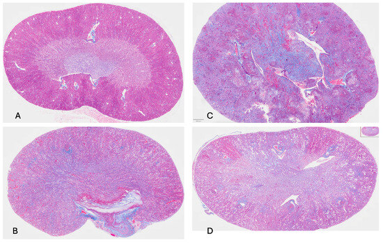
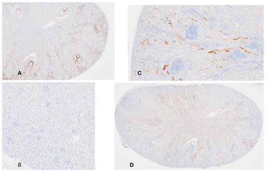
Damage to Kidney Following Intravenous Administration of C. albicans and Daily SQ hSAP or hCRP. Blood filtration in the kidney occurs across basement membranes within the glomeruli, located in the cortex of the kidney. Urine collects and drains within adjacent tubules and then flows to collecting ducts found in the medulla of the kidney. Following intravenous administration of C. albicans to mice, blood cultures remained sterile for days [13]. Nevertheless, by day 2, kidney invasion was extensive, as demonstrated in Figure 1. In uninfected mice, the kidney, when stained with trichrome, displays a sharp contrast between the cortex and medulla (Figure 1A). In response to C. albicans, infection fibroblasts laid down collagen, particularly in the medulla and parts of the cortex (Figure 1B). These characteristics were exaggerated in mice injected SQ with hCRP and fungi (Figure 1C). In this circumstance, the kidney was swollen and lost its normal bean shape. There was abundant collagen deposition as well as C. albicans proliferation throughout the organ. Figure 1D shows the kidney from a mouse administered with C. albicans and daily hSAP. Surprisingly, the delineation between the medulla and cortex remained. There was reduced collagen deposition compared to the infected kidney (Figure 1B). Fungi are evident, particularly in the cortex of the kidney (scarlet red). Administration of hSAP spared much of the inflammatory destruction of the kidney as well as reduced collagen deposition, features that are seen most dramatically with the administration of hCRP and C. albicans alone (Figure 1C).
Figure 1.
(A) Uninfected mouse right kidney (trichrome stain; 40×); no pentraxin was administered; the trichrome stain demonstrates the sharp distinction between the medulla (the central part of the kidney) and the cortical region of the kidney. (B) Right kidney from mouse given C. albicans only (trichrome; 40×); changes detectable 2 days after receiving C. albicans yeast cells intravenously. The distinction between medulla and cortex was lost and collagen deposition was extensive in the medulla and cortex (blue hue denotes collagen). (C) Right kidney from mouse given C. albicans intravenously and hCRP daily SQ. C. albicans was evident as a stringy, pinkish-red-stained infiltrate especially prominent in the cortex and medullary regions (trichrome stain; 40×). (D) Right kidney from mouse given C. albicans intravenously and hSAP daily (trichrome stain; 40×). Note that the characteristic histological difference between the cortex and medulla was maintained even though fungi (stringy pinkish material) were present in the medulla and less so in the medulla; the amount of collagen deposition was less than that in Figure 1B or Figure 1C.
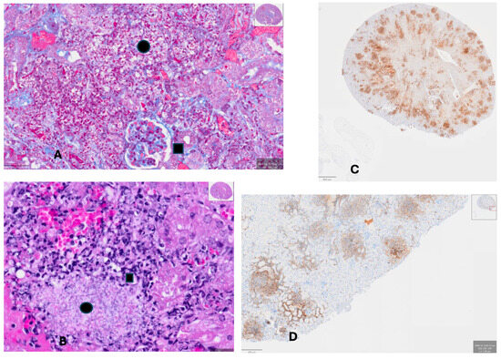
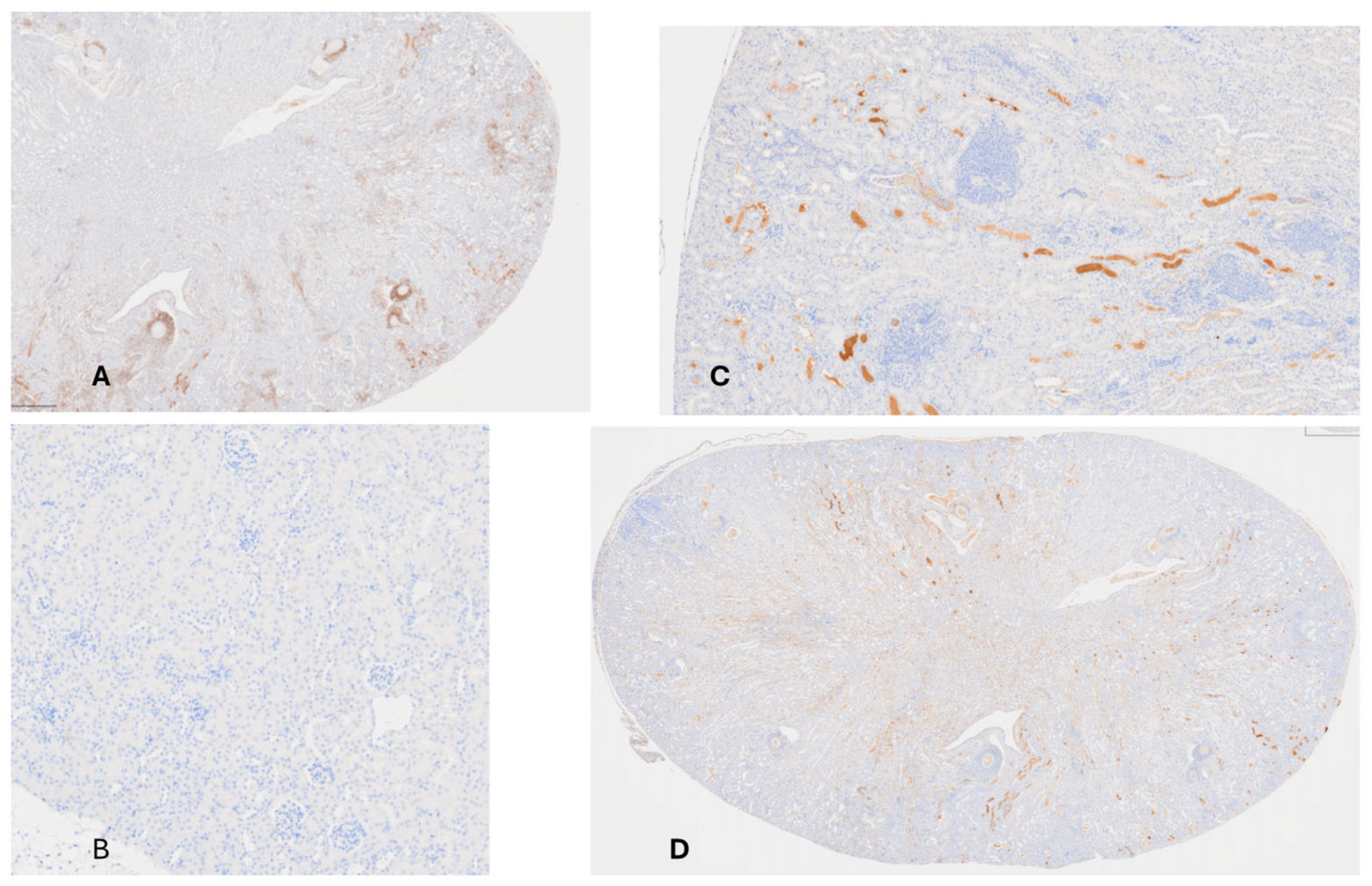
Histology of Kidney from hCRP-Treated Mouse along with C. albicans. Figure 2A,B show developing abscesses of a mouse administered with C. albicans and daily hCRP. There was extensive proliferation of fungi (black circle) and fibrosis (blue hue) within an abscess near a glomerulus (black square) in Figure 2A, and an abscess (black circle) surrounded by inflammatory cells (black square) in Figure 2B. Figure 2C shows the same CRP-treated kidney stained with anti-Ly-G6 antibody, showing the antigen expressed on bone marrow-derived cells (e.g., myelocytes and macrophages). Figure 2D shows staining with anti-CD45 antibody which stains leukocytes, demonstrating marked infiltration of the kidney parenchyma of the medulla and cortex tissue with these inflammatory cells, especially adjacent to or within abscesses. hCRP treatment was associated with exuberant fungal growth and was permissive of inflammatory cell migration into the infected tissue.
Figure 2.
(A) Kidney of mouse that received C. albicans and hCRP. Trichrome stain demonstrates a glomerulus (black square) with proliferation of collagen and development of an abscess (pink yeasts and hyphae visible, black dot); there was a paucity of normal-appearing kidney parenchyma (~400×). (B) Trichrome stain of same kidney shows yeast and hyphae at the edge of the abscess (black square) with pink staining of fungi in the abscess proper (black dot, ~400×). (C) Same kidney of mouse given C. albicans and hCRP stained with anti-LyG-6 (staining neutrophils, monocytes and granulocytes; 40×); (D) same kidney of mouse given C. albicans and hCRP stained with anti-CD45 which stains leukocytes; (40×).
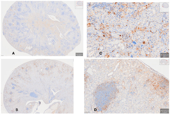
Histology of Kidney from hSAP-Treated Mouse. Figure 3A demonstrates that mice had a greatly diminished presence of leukocytes (stained by anti-CD45) in infected areas compared to the mouse given hCRP (cf. Figure 2D). T-lymphocyte distribution was faint and widespread, not concentrated around abscesses. Figure 3B–D show staining with anti-CD3e antibody. In the uninfected kidney (Figure 3B), there is homogeneous distribution of T-lymphocytes; in Figure 3C, uptake of anti-CD3e staining T cells is concentrated at the portal of entry of the fungus in the glomeruli and their tubules and collecting ducts extending into the medulla; Figure 3D shows widespread distribution of T-lymphocytes, perhaps greatest in the medulla for a mouse given C. albicans and hSAP. Compared to mice given C. albicans and hCRP, it is apparent that hSAP-treated mice have a muted cellular host response to infection.
Figure 3.
(A) Kidney of mouse given C. albicans and hSAP stained with anti-CD45. There is a muted invasion of leucocytes into inflamed/infected areas throughout the kidney at sites of abscesses (compare to Figure 2D) (40×). (B) Uninfected mouse kidney stained with anti-CD3e (stains all T cells) showing no uptake in cortex or medulla C (100×). (C) Kidney of mouse given C. albicans alone stained with anti-CD3e showing concentration of staining in glomeruli and their tubules and collecting ducts; there is no staining in adjacent abscesses (100×); (D) Kidney of mouse given C. albicans and hSAP and stained with anti-CD3e (same animal as Figure 3A stained with a different antibody); diffuse uptake in cortex and medulla indicating the presence of T cells throughout the kidney (as opposed to spotty areas shown in Figure 3C) (40×).
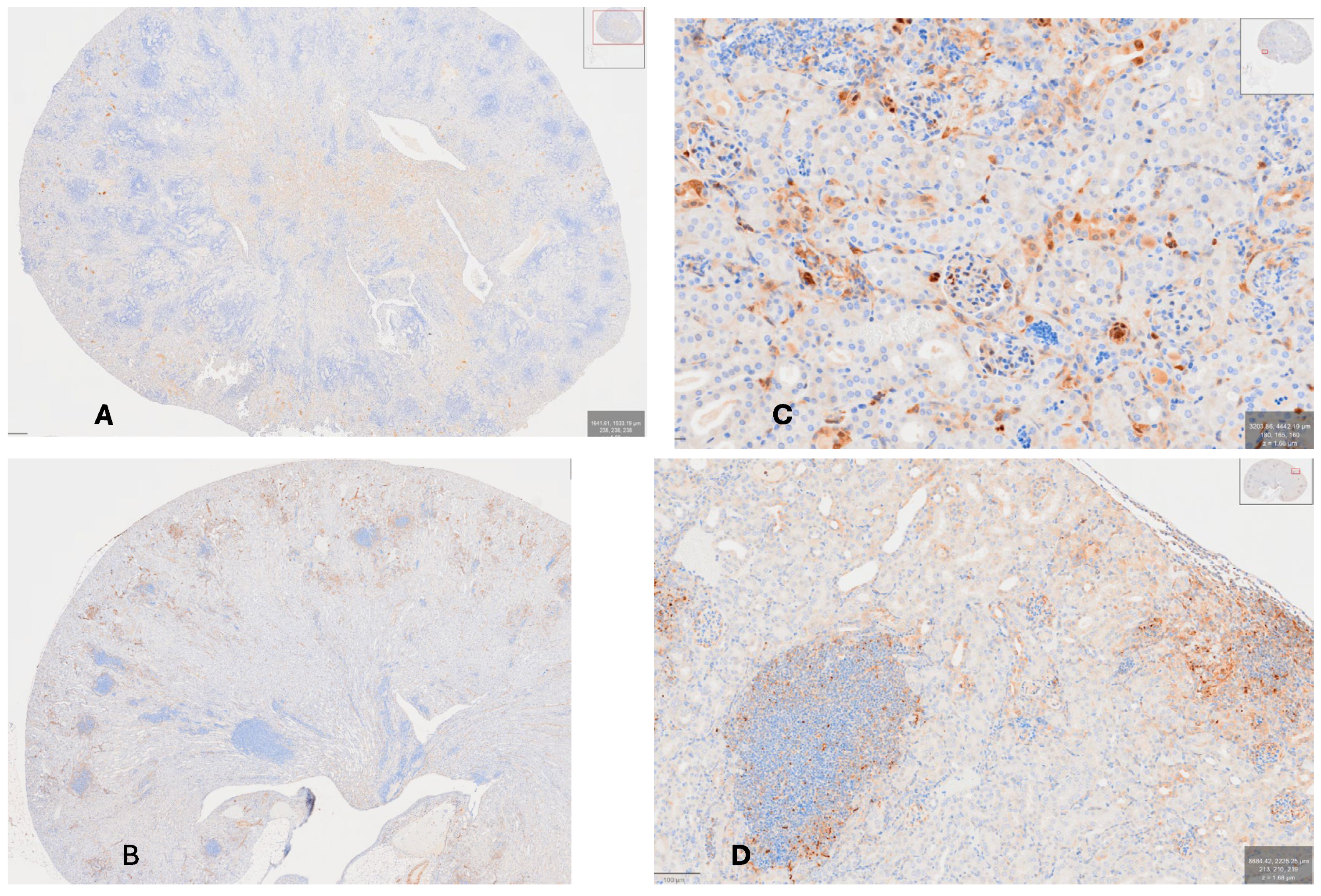
hCRP Binds to Damaged Kidney Tissue Whereas, hSAP Binds to Fungi. Figure 4A shows the distribution of T cells in a mouse given C. albicans and hCRP with widespread development of abscesses. Figure 4B shows that the binding of hCRP occurs to damaged tissue surrounding abscess development, whereas hSAP (Figure 4C,D) binds to tissues with intact remnants of basement membrane as well as to yeasts and hyphae, especially at the outer edge of a large abscess (Figure 4D).
Figure 4.
(A) Kidney of mouse given C. albicans and hCRP stained with anti-CD3e (same animal as Figure 2C stained with different antibody) showing the distribution of T cells in the medulla and cortex with an un-countable number of developing abscesses and do not contain any T cells (40×); (B) Kidney of mouse given C. albicans and hCRP stained with anti-CRP-1C (40×); (C) kidney of mouse given C. albicans and hSAP stained with anti-amyloid P (400×) showing staining of SAP to yeasts and hyphae on the periphery of a well-developed abscess; 400×; (D) staining with anti-amyloid P showing fungi scattered within an abscess (lower left) and a developing abscess near the cortical surface (on right of photograph).
4. Discussion
Candida albicans, once disseminated in the human body, remains a highly lethal disease despite appropriate antifungal therapy [16]. We show here that SQ hSAP preserved kidney histology and concomitantly reduced infiltration of immune cells in an animal model of disseminated candidiasis. Thus, in this mouse model of disseminated candidiasis, SAP injections early in the infectious process ameliorated disease [12]. SAP reduces inflammatory cytokine production, upregulates IL-10 and binds to DCSign [17]. This sequence of events reduces the number of circulating fibrocytes and, thus, post infectious fibrosis. Exacerbation of the infectious process seen in disseminated candidiasis is similar to findings in the COVID-19 pneumonia, where the cytokine storm is a major contributor to the morbidity and mortality of hospitalized patients [18]. Fibrosis of pulmonary tissue due to infection is part of the normal inflammatory response to infection. When the response is dysregulated, as it often is in very ill patients with COVID-19, and occasionally in disseminated candidiasis, fibrosis due to collagen deposition is often the deleterious residue. We have discovered that SAP administration, when added early in the disease process, prolongs the survival of mice with disseminated candidiasis [12]. Nouresadeghi et al. found the opposite to be true in bacterial sepsis [19]. They found that SAP bound to bacteria and, in doing so, inhibited phagocytosis, which led to a lethal outcome for mice. We found that C. albicans binding SAP reduced the inflammatory response and enhanced survival [4,12]. In line with the salutary effect of SAP in infections, recombinant SAP was administered to patients for years, which resulted in amelioration and retardation of idiopathic pulmonary fibrosis [20].
Recently, Karhadkar et al. studied the COVID-19 pneumonia in a mouse model and found that recombinant SAP ameliorated the inflammatory response of the host, reducing cytokine production and fibrosis in the lung [21]. These results are similar to the results of our murine candidiasis study. Exacerbation of the infectious process seen in disseminated candidiasis is similar to findings in the COVID-19 pneumonia where the cytokine storm is a major contributor to the morbidity and mortality of hospitalized patients [18]. Fibrosis of pulmonary tissue due to infection is part of the normal inflammatory response to infection. When the response is dysregulated, as it often is in patients with COVID-19, and occasionally in disseminated candidiasis, fibrosis due to collagen deposition is often the deleterious residue. Interestingly, a study of COVID-19 patients in Intensive Care Units revealed that SAP blood levels had a negative association with fibrosis, indicating that high SAP levels corresponded with less fibrosis [18]. SAP thus appears to trigger an anti-inflammatory response and decreased tissue damage in several disorders. For example, it reduces keloid formation in individuals subject to the dysregulation of fibrocytes in wound healing [22]. Pending further study, perhaps employing immunomodulators [23], like SAP, judiciously in cases with dysregulated host immune responses, such as what occurs in COVID-19 and disseminated candidiasis (along with the standard agents such as antivirals and antifungals), might achieve better outcomes.
Our findings with CRP in the disseminated candidiasis model deserves further comment as well. The SQ hCRP in this animal model exacerbates the inflammatory response to C. albicans infections and led to pathological loss of kidney architecture and the additional infiltration of inflammatory markers and immune cells. Although purified hCRP is harmless when injected into humans [11], the protein, when bound to damaged tissue, can accelerate destruction. Consequently, removal of high levels of CRP associated with inflammation and infections shows some promise of disease improvement. For example, apheresis of CRP has been studied with patients with myocardial infarction [24]. Investigators found clinical improvement and less myocardial damage [24]. Other methods of reducing CRP post myocardial infarction include protein inhibitors of CRP that block its ability to bind to autologous ligands [9,25].
Author Contributions
Conceptualization, S.A.K.; Methodology, S.A.K.; Software, S.A.K.; Validation, S.A.K., R.E.S. and P.N.L.; Formal analysis, S.A.K., R.E.S. and P.N.L.; Investigation, S.A.K.; Resources, S.A.K.; Data curation, S.A.K.; Writing—original draft, S.A.K.; Writing—review and editing, S.A.K., R.E.S. and P.N.L.; Visualization, S.A.K.; Supervision, S.A.K.; Project administration, S.A.K.; Funding acquisition, S.A.K. All authors have read and agreed to the published version of the manuscript.
Funding
This research received no external funding.
Institutional Review Board Statement
The animal study protocol was approved by the University of Arizona Institutional Animal Care and Use Committee (protocol #: 19-587, approval date: 23 January 2020).
Informed Consent Statement
Not applicable.
Data Availability Statement
The original contributions presented in this study are included in the article. Further inquiries can be directed to the corresponding author.
Acknowledgments
The authors thank MB Pepys for the purified, pathogen-free hSAP and hCRP provided for these experiments, which would not have been possible without his generous gift. We also wish to thank Maga Sanchez at the Tissue Acquisition Cellular/Molecular Analysis Shared Resource at the University of Arizona Cancer Center for the preparation of tissue samples and immunostaining and Melanie Miranda, Department of Pathology, University of Arizona for preparation of the microphotographs.
Conflicts of Interest
The authors declare no conflicts of interest.
References
- Klotz, S.A. Fungal adherence to the vascular compartment: A critical step in the pathogenesis of disseminated candidiasis. Clin. Infect. Dis. 1992, 14, 340–347. [Google Scholar] [CrossRef] [Scilit] [PubMed]
- Klotz, S.A. Plasma and extracellular matrix proteins mediate in the fate of Candida albicans in the human host. Med. Hypotheses 1994, 42, 328–334. [Google Scholar] [CrossRef] [Scilit] [PubMed]
- Lipke, P.N.; Klotz, S.A.; Dufrene, Y.F.; Jackson, D.N.; Garcia-Sherman, M.C. Amyloid-Like β-Aggregates as Force-Sensitive Switches in Fungal Biofilms and Infections. Microbiol. Mol. Biol. Rev. 2017, 82, e00035-17. [Google Scholar] [CrossRef] [Scilit] [PubMed] [PubMed Central]
- Behrens, N.E.; Lipke, P.N.; Pilling, D.; Gomer, R.H.; Klotz, S.A. Serum Amyloid P Component Binds Fungal Surface Amyloid and Decreases Human Macrophage Phagocytosis and Secretion of Inflammatory Cytokines. mBio 2019, 10, e00218-19. [Google Scholar] [CrossRef] [Scilit] [PubMed] [PubMed Central]
- Gaur, N.K.; Klotz, S.A. Expression, cloning, and characterization of a Candida albicans gene, ALA1, that confers adherence properties upon Saccharomyces cerevisiae for extracellular matrix proteins. Infect. Immun. 1997, 65, 5289–5294. [Google Scholar] [CrossRef] [Scilit] [PubMed] [PubMed Central]
- Frank, A.T.; Ramsook, C.B.; Otoo, H.N.; Tan, C.; Soybelman, G.; Rauceo, J.M.; Gaur, N.K.; Klotz, S.A.; Lipke, P.N. Structure and function of glycosylated tandem repeats from Candida albicans Als adhesins. Eukaryot. Cell 2010, 9, 405–414. [Google Scholar] [CrossRef] [Scilit] [PubMed] [PubMed Central]
- Lipke, P.N. Not gently down the stream: Flow induces amyloid bonding in environmental and pathological fungal biofilms. mBio 2025, 16, e0020325. [Google Scholar] [CrossRef] [Scilit]
- Gilchrist, K.B.; Garcia, M.C.; Sobonya, R.; Lipke, P.N.; Klotz, S.A. New features of invasive candidiasis in humans: Amyloid formation by fungi and deposition of serum amyloid P component by the host. J. Infect. Dis. 2012, 206, 1473–1478. [Google Scholar] [CrossRef] [Scilit] [PubMed] [PubMed Central]
- Garcia-Sherman, M.C.; Lundberg, T.; Sobonya, R.E.; Lipke, P.N.; Klotz, S.A. A unique biofilm in human deep mycoses: Fungal amyloid is bound by host serum amyloid P component. npj Biofilms Microbiomes 2015, 1, 15009. [Google Scholar] [CrossRef] [Scilit] [PubMed] [PubMed Central]
- Agrawal, A.; Singh, P.P.; Bottazzi, B.; Garlanda, C.; Mantovani, A. Pattern recognition by pentraxins. Adv. Exp. Med. Biol. 2009, 653, 98–116. [Google Scholar] [CrossRef] [Scilit] [PubMed] [PubMed Central]
- Pepys, M.B. The Pentraxins 1975–2018: Serendipity, Diagnostics and Drugs. Front. Immunol. 2018, 9, 2382. [Google Scholar] [CrossRef] [Scilit] [PubMed] [PubMed Central]
- Klotz, S.A.; Lipke, P.N. The Paradoxical Effects of Serum Amyloid-P Component on Disseminated Candidiasis. Pathogens 2022, 11, 1304. [Google Scholar] [CrossRef] [Scilit] [PubMed] [PubMed Central]
- MacCallum, D.M.; Odds, F.C. Temporal events in the intravenous challenge model for experimental Candida albicans infections in female mice. Mycoses 2005, 48, 151–161. [Google Scholar] [CrossRef] [Scilit]
- Carson, L.F.; Hladik, C. Histotechnology: A Self-Instructional Text, 3rd ed.; Chicago, III; American Society of Clinical Pathologists: Chicago, IL, USA, 2009; pp. 148–149. [Google Scholar]
- Bankhead, P.; Loughrey, M.B.; Fernández, J.A.; Dombrowski, Y.; McArt, D.G.; Dunne, P.D.; McQuaid, S.; Gray, R.T.; Murray, L.J.; Coleman, H.G.; et al. QuPath: Open source software for digital pathology image analysis. Sci. Rep. 2017, 7, 16878. [Google Scholar] [CrossRef] [Scilit]
- Lionakis, M.S.; Fischer, B.G.; Lim, J.K.; Swamydas, M.; Wan, W.; Richard Lee, C.C.; Cohen, J.I.; Scheinberg, P.; Gao, J.L.; Murphy, P.M. Chemokine receptor Ccr1 drives neutrophil-mediated kidney immunopathology and mortality in invasive candidiasis. PLoS Pathog. 2012, 8, e1002865. [Google Scholar] [CrossRef] [Scilit] [PubMed] [PubMed Central]
- Cox, N.; Pilling, D.; Gomer, R.H. DC-SIGN activation mediates the differential effects of SAP and CRP on the innate immune system and inhibits fibrosis in mice. Proc. Natl. Acad. Sci. USA 2015, 112, 8385–8390. [Google Scholar] [CrossRef] [Scilit] [PubMed] [PubMed Central]
- Ghanem, M.; Homps-Legrand, M.; Garnier, M.; Morer, L.; Goletto, T.; Frija-Masson, J.; Wicky, P.H.; Jaquet, P.; Bancal, C.; Hurtado-Nedelec, M.; et al. Blood fibrocytes are associated with severity and prognosis in COVID-19 pneumonia. Am. J. Physiol. Lung Cell. Mol. Physiol. 2021, 321, L847–L858. [Google Scholar] [CrossRef] [Scilit] [PubMed] [PubMed Central]
- Noursadeghi, M.; Bickerstaff, M.C.; Gallimore, J.R.; Herbert, J.; Cohen, J.; Pepys, M.B. Role of serum amyloid P component in bacterial infection: Protection of the host or protection of the pathogen. Proc. Natl. Acad. Sci. USA 2000, 97, 14584–14589. [Google Scholar] [CrossRef] [Scilit] [PubMed] [PubMed Central]
- Raghu, G.; Hamblin, M.J.; Brown, A.W.; Golden, J.A.; Ho, L.A.; Wijsenbeek, M.S.; Vasakova, M.; Pesci, A.; Antin-Ozerkis, D.E.; Meyer, K.C.; et al. Long-term evaluation of the safety and efficacy of recombinant human pentraxin-2 (rhPTX-2) in patients with idiopathic pulmonary fibrosis (IPF): An open-label extension study. Respir. Res. 2022, 23, 129. [Google Scholar] [CrossRef] [Scilit] [PubMed] [PubMed Central]
- Karhadkar, T.R.; Pilling, D.; Gomer, R.H. Serum Amyloid P inhibits single stranded RNA-induced lung inflammation, lung damage, and cytokine storm in mice. PLoS ONE 2021, 16, e0245924. [Google Scholar] [CrossRef] [Scilit] [PubMed] [PubMed Central]
- Naylor, M.C.; Lazar, D.A.; Zamora, I.J.; Mushin, O.P.; Yu, L.; Brissett, A.E.; Olutoye, O.O. Increased in vitro differentiation of fibrocytes from keloid patients is inhibited by serum amyloid P. Wound Repair Regen. 2012, 20, 277–283. [Google Scholar] [CrossRef] [Scilit] [PubMed]
- Cox, N.; Pilling, D.; Gomer, R.H. Serum amyloid P: A systemic regulator of the innate immune response. J. Leukoc. Biol. 2014, 96, 739–743. [Google Scholar] [CrossRef] [Scilit] [PubMed] [PubMed Central]
- Kayser, S.; Brunner, P.; Althaus, K.; Dorst, J.; Sheriff, A. Selective Apheresis of C-Reactive Protein for Treatment of Indications with Elevated CRP Concentrations. J. Clin. Med. 2020, 9, 2947. [Google Scholar] [CrossRef] [Scilit] [PubMed] [PubMed Central]
- Pepys, M.B.; Hirschfield, G.M.; Tennent, G.A.; Gallimore, J.R.; Kahan, M.C.; Bellotti, V.; Hawkins, P.N.; Myers, R.M.; Smith, M.D.; Polara, A.; et al. Targeting C-reactive protein for the treatment of cardiovascular disease. Nature 2006, 440, 1217–1221. [Google Scholar] [CrossRef] [Scilit]

Disclaimer/Publisher’s Note: The statements, opinions and data contained in all publications are solely those of the individual author(s) and contributor(s) and not of MDPI and/or the editor(s). MDPI and/or the editor(s) disclaim responsibility for any injury to people or property resulting from any ideas, methods, instructions or products referred to in the content. |
© 2026 by the authors. Licensee MDPI, Basel, Switzerland. This article is an open access article distributed under the terms and conditions of the Creative Commons Attribution (CC BY) license.