


Abstract
Background: Persistent infection with high-risk human papillomavirus (HPV) genotypes is the principal etiological factor in cervical carcinogenesis. However, HPV genotype distribution and co-infection patterns differ across populations and clinical settings. The present study aimed to characterize HPV genotypes and co-infection patterns among clinically selected Iraqi women with suspected HPV-associated disease. Methods: In this cross-sectional study, 100 clinically selected women attending clinics in Ramadi and Baghdad, Iraq (December 2025–July 2026), each contributed one specimen, collected by swab or superficial lesion scraping from a cervical, vulval, anal, or perianal site based on clinical assessment. HPV genotyping was performed by polymerase chain reaction followed by microarray hybridization, targeting 35 HPV genotypes. Results: HPV DNA was detected in 65 of 100 women (65.0%; 95% confidence interval, 55.3–73.6%). Of these, 48 had low-risk (LR)-only, 9 high-risk/potentially high-risk (HR/PHR)-only, and 8 mixed LR/HR/PHR infections. HPV-6 and HPV-11 were the most frequently detected genotypes (36 and 27 of 88 genotype detections, respectively), and multiple-genotype infections were identified in 20 women. Conclusions: HPV-6 and HPV-11 predominated in this clinically selected cohort, although HR/PHR and multiple-genotype infections were also identified. These preliminary, descriptive data highlight the need for larger, more representative studies of HPV genotype distribution in Iraq.
1. Introduction
Human papillomavirus (HPV) is the most common sexually transmitted viral infection worldwide and the necessary cause of cervical cancer, which remains a major global health burden. According to GLOBOCAN 2024 estimates, approximately 604,196 new cervical cancer cases and 279,583 deaths occurred worldwide in 2024 [1]. HPV-associated disease imposes a substantial burden that is largely preventable through vaccination and screening; however, these measures remain underutilized [2]. They underpin the World Health Organization (WHO) cervical cancer elimination strategy, whose 90-70-90 targets address vaccination, screening, and treatment [3].
In Iraq, the same GLOBOCAN 2024 release estimated approximately 496 new cervical cancer cases and 303 cervical cancer deaths in 2024, corresponding to age-standardized incidence and mortality rates of 3.0 and 1.9 per 100,000 women, respectively [4]. These population-level estimates provide clinical context but do not reflect the outcomes assessed in the present study, which focuses on HPV genotype distribution and co-infection patterns in a clinically selected cohort.
The carcinogenic potential of HPV genotypes varies widely. Carcinogenic HPV genotypes are classified as Group 1 carcinogens by the International Agency for Research on Cancer (IARC) based on epidemiological and mechanistic data. In contrast, other genotypes are placed in the lower certainty classifications: Group 2A (probably carcinogenic) and Group 2B (possibly carcinogenic) [5]. These genotypes can be classified into high-risk (HR) or potentially high-risk (PHR) groups in clinical and epidemiological studies, but these risk classifications are not equivalent to IARC classifications [6]. Low-risk (LR) genotypes, particularly HPV-6 and HPV-11, are mainly associated with anogenital warts [7,8]. Importantly, in some cases apparently innocuous lesions may coexist with HR/PHR genotypes or multiple infections, indicating that clinical appearance alone and general risk-group classification may be insufficient [9,10].
The shift toward primary HPV DNA testing in cervical cancer screening has been driven by the shortcomings of conventional cytology, and HPV DNA testing is the primary screening test recommended by the WHO for the general population of women [11]. Genotype-resolved assays additionally support epidemiological research by identifying individual HPV types and co-infections, and post-polymerase chain reaction (PCR) microarray hybridization enables the concurrent detection of multiple genotypes in a single assay [12,13].
However, HPV genotyping data from clinically selected women sampled at different anogenital sites remain limited, especially in Iraq. This gap is clinically relevant because HR/PHR co-infections may occur alongside apparently benign lesions, while the nonavalent HPV vaccine covers only a subset of oncogenic genotypes. HPV genotype distributions also vary by population, clinical context, and anatomical site. A cohort from Chengdu, China, reported a high-risk-dominant distribution, with HPV-52, HPV-16, and HPV-58 predominating [9]. In Iraq, a large retrospective cohort using the same microarray platform identified HPV-18, HPV-11, and HPV-16 as the most frequently detected genotypes [14].
Although previous Iraqi studies have reported HPV prevalence and genotype distributions, further characterization of genotype diversity and co-infection patterns in clinically selected women remains valuable. This study aimed to characterize the distribution of low-risk and high-risk human papillomavirus genotypes and their co-infection patterns among clinically selected Iraqi women using post-PCR microarray genotyping.
2. Materials and Methods
2.1. Study Design and Participant Selection
This prospective cross-sectional study included 100 clinically selected women evaluated between December 2025 and July 2026 at a private gynecological clinic and the outpatient clinic of Ramadi General Hospital, Ramadi, Iraq (n = 90), and at hospitals and private gynecological clinics in Baghdad, Iraq (n = 10). All participants were enrolled under the same study protocol, eligibility criteria, and consent procedure at both sites, using clinically based non-probability sampling. At each site, the attending specialist gynecologist assessed each woman, obtained signed written informed consent for HPV testing and study participation, selected the sampling site according to clinical findings, and collected one specimen. The investigator maintained direct communication with the participating gynecologists throughout the study to ensure consistent protocol application. Enrollment was not documented as consecutive, and the number of eligible women who declined participation was not recorded.
Women aged 18–50 years presenting with clinically suspected HPV-associated lesions or other suspicious cervical or anogenital findings, as judged by the attending gynecologist, were eligible. Pregnant women and women with a documented history of systemic autoimmune disease were excluded. Standardized participant-level clinical indication, cytology results, and immunosuppressive treatment status were not systematically recorded. The study is reported in accordance with the STROBE guidelines (Supplementary Table S4).
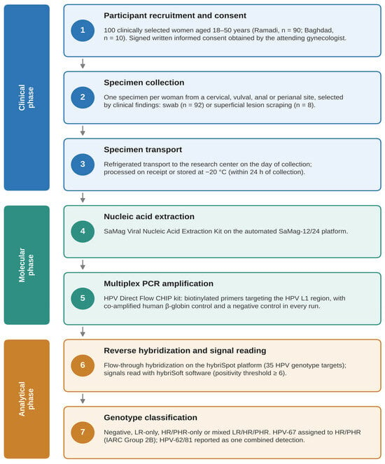
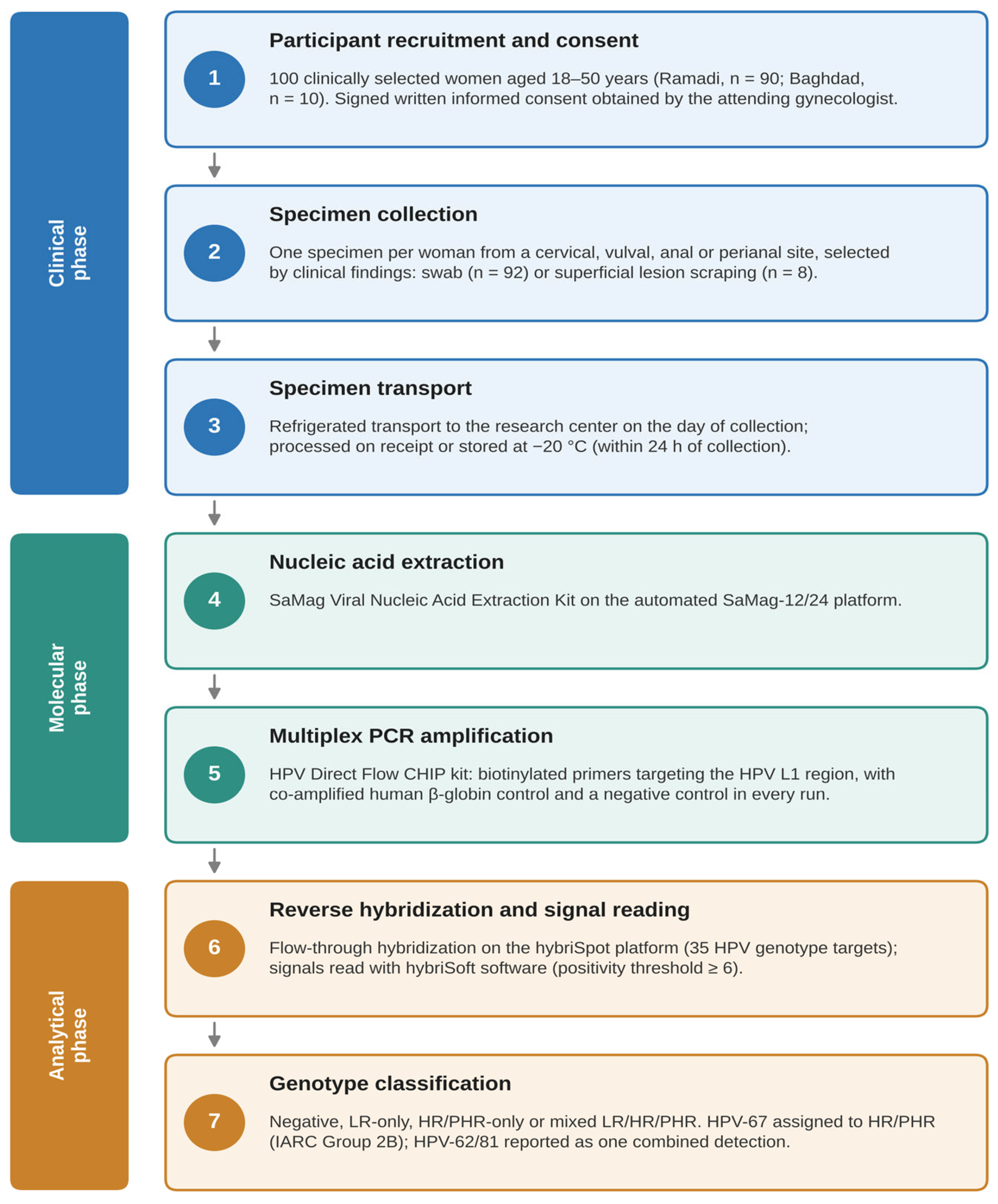
One specimen per participant was obtained from one of four anatomical regions: cervical, vulval, anal, or perianal. The seven specimen categories were cervical swabs (n = 44), vulval swabs (n = 45), superficial vulval lesion scrapings (n = 4), anal swabs (n = 2), superficial anal lesion scrapings (n = 3), perianal swabs (n = 1), and superficial perianal lesion scrapings (n = 1). The eight lesion-derived specimens were superficial scrapings of lesions diagnosed clinically as anogenital warts by the attending gynecologist based on their macroscopic appearance; they were collected for HPV DNA analysis, and no tissue biopsies or histopathological examinations were performed. All specimens were transported under refrigeration to the research center on the day of collection and, on receipt, were either processed immediately or stored at −20 °C until nucleic acid extraction; all specimens were therefore processed or frozen within 24 h of collection. All specimens underwent the same DNA extraction, PCR amplification, and microarray genotyping workflow (Figure 1).
Figure 1.
Study workflow for HPV detection and microarray-based genotyping.
2.2. Automated Nucleic Acid Extraction
Total nucleic acids were extracted from the collected specimens using the SaMag Viral Nucleic Acid Extraction Kit (Ref: SM003; Sacace Biotechnologies Srl, Como, Italy) on the automated SaMag-12/24 platform (Sacace Biotechnologies Srl). The same extraction workflow was applied to all 100 specimens before HPV amplification. Because the SaMag system is not among the extraction systems listed in the HPV Direct Flow CHIP instructions, its suitability for downstream amplification was monitored in every run using the co-amplified human β-globin control and a negative control; no specimen was classified as invalid (Section 2.5).
In brief, 400 μL of each sample was lysed with Proteinase K solution (30 μL) plus Lysis Buffer 1 (720 μL). Carrier RNA was added at 20 μL/400 μL to the sample, following the manufacturer’s instructions. The automated system was used to process the lysate using Binding Buffer 1 (1000 μL) and magnetic-bead capture (800 μL), followed by washing in Washing Buffers 1, 2, and 3 (1000 μL each). The purified nucleic acids were eluted in RNase-free water and either quantified immediately or stored at −20 °C for amplification [15].
2.3. Multiplex PCR Amplification
Multiplex PCR was performed using the HPV Direct Flow CHIP Kit (Vitro S.A., Seville, Spain) with purified DNA in a final reaction volume of 30 μL. Biotinylated primers amplified a region of the HPV L1 gene. The resulting amplicons were used for subsequent reverse hybridization and were not interpreted as a separate diagnostic assay.
Thermal cycling was performed on an Esco Swift Maxi Thermal Cycler (Esco Micro Pte. Ltd., Singapore): 25 °C for 10 min; 94 °C for 3 min; 15 cycles of 94 °C, 47 °C, and 72 °C for 30 s each; 35 cycles of 94 °C, 65 °C, and 72 °C for 30 s each, followed by 72 °C for 5 min and a hold at 8 °C [16]. Each run included a negative control.
Contamination prevention included physically separated pre- and post-amplification areas, dedicated equipment, aerosol-resistant filter tips, and the uracil-N-glycosylase/dUTP carryover-prevention system incorporated into the assay [16].
2.4. Microarray-Based Genotyping
HPV genotyping was performed by post-PCR flow-through reverse hybridization using the HPV Direct Flow CHIP assay on the hybriSpot 12 platform (Vitro S.A., Seville, Spain). The assay targets 35 HPV genotypes. Biotinylated PCR products were hybridized to membrane-bound probes, and colorimetric signals were interpreted using the manufacturer’s hybriSoft software v2.2 (Supplementary Figure S1). The assay output was categorized using the study-defined risk groups described below.
Flow diagram of the study procedures, from participant recruitment, consent, and specimen collection, through nucleic acid extraction, multiplex PCR and microarray hybridization, to genotype classification.
2.5. Interpretation Thresholds
A gray intensity threshold of at least 6 was used for positive genotype-specific signals according to the assay interpretation protocol [16]. HPV genotype detection and interpretation were performed according to the manufacturer’s instructions, including the recommended assay quality-control procedures. Co-amplified human β-globin served as the endogenous amplification control, as specified by the manufacturer; no specimen was classified as invalid, and all 100 specimens yielded interpretable final results and were included in the analysis.
Genotype risk classification was based on three distinct classification frameworks. The IARC carcinogenicity categories—Group 1 (carcinogenic to humans), Group 2A (probably carcinogenic to humans), Group 2B (possibly carcinogenic to humans), and Group 3 (not classifiable as to carcinogenicity to humans)—were treated as the reference carcinogenicity framework. Manufacturer-assigned risk categories were retained separately according to the classification provided by the assay manufacturer. For statistical analysis, the study used a predefined operational classification of low-risk (LR) and high-risk/probably or potentially high-risk (HR/PHR) genotypes. The HR/PHR designation used in this study should therefore be interpreted as an analytical grouping and not as a formal IARC classification.
Although the manufacturer’s software classifies HPV-67 within the low-risk panel, HPV-67 was prospectively assigned to the study’s HR/PHR category before data analysis based on its classification by IARC as Group 2B (possibly carcinogenic to humans) [5]. This study-applied classification was predefined and applied uniformly to all specimens. Accordingly, the manufacturer-provided risk category and the study-applied classification were retained as separate classifications. A complete list of genotype classifications used in the study is provided in Supplementary Table S1.
Because HPV-62 and HPV-81 share a single probe on this platform, positive signals at this probe position were reported using the combined designation “HPV-62/81” and were not interpreted as individual genotype detections. This reporting approach was consistent with the documented probe configuration and cross-reactivity characteristics of the multiplex chip [16], as previously reported in a regional cohort using the same platform [14]. All statistical analyses were performed using the predefined study-applied genotype classifications.
2.6. Statistical Analysis
Statistical analyses were performed using GraphPad Prism v9.0 and Python (SciPy v1.17). Age was summarized using descriptive statistics, and categorical outcomes were reported as counts and percentages. Age distribution was assessed using the Shapiro–Wilk test; the Kruskal–Wallis test was used to compare age across the four HPV risk categories [17], and its effect size was expressed as epsilon-squared (ε2 = H/(n − 1)). Genotype frequencies, specimen categories, and pairwise genotype co-occurrences were summarized descriptively. No inferential comparisons between anatomical sites were performed because sampling sites were selected based on clinical presentation and several subgroups were small. Two-sided 95% confidence intervals (CIs) for proportions were calculated using the Wilson score method; statistical significance was evaluated at a two-sided threshold of p < 0.05.
3. Results
3.1. Participant Characteristics, HPV Prevalence, and Infection Multiplicity
Among the 100 enrolled participants, HPV DNA was detected in 65 (65.0%; 95% CI, 55.3–73.6%), whereas 35 (35.0%) tested negative. Among HPV-positive participants, LR-only infections predominated (48/65, 73.8%), whereas HR/PHR-only and mixed LR/HR/PHR infections were less frequent (Table 1). The mean participant age was 31.6 ± 6.5 years (range, 20–50 years). Participant characteristics by recruitment site, anatomical region, and collection method are summarized in Table 2.
Table 1.
Distribution of HPV risk categories among study participants, with counts and 95% confidence intervals (n = 100).
Table 2.
Characteristics of study participants by recruitment site, anatomical region, and collection method (n = 100).
Age was not normally distributed (Shapiro–Wilk W = 0.969, p = 0.017). Median age was 32.0 years (interquartile range [IQR], 28.5–36.0) among HPV-negative participants, 30.0 years (IQR, 26.8–33.0) among those with LR-only infections, 33.0 years (IQR, 29.0–35.0) among those with HR/PHR-only infections, and 31.5 years (IQR, 26.8–33.5) among those with mixed infections; the difference across the four HPV risk categories was negligible (ε2 = 0.02; Kruskal–Wallis H = 2.05, df = 3, p = 0.563). A complete summary of age distributions, including minimum and maximum values, is provided in Supplementary Table S2.
Among the 65 HPV-positive participants, 45 (69.2%; 95% CI, 57.2–79.1%) harbored a single HPV genotype, whereas 20 (30.8%; 95% CI, 20.9–42.8%) harbored multiple genotypes. Multiple-genotype infections accounted for 20.0% of the total cohort (20/100; 95% CI, 13.3–28.9%).
3.2. Genotype-Specific Prevalence and Vaccine-Targeted Genotypes
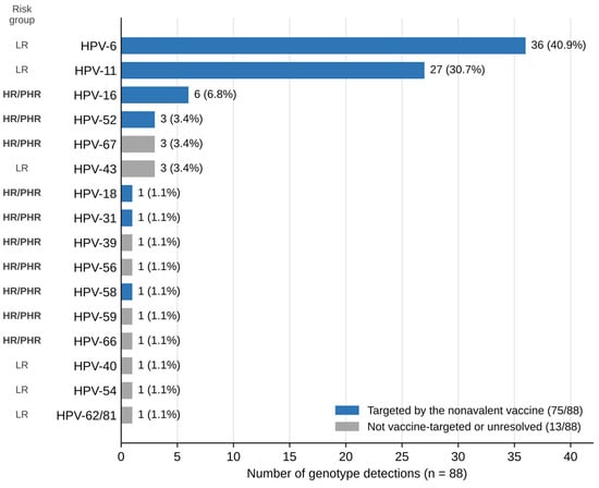
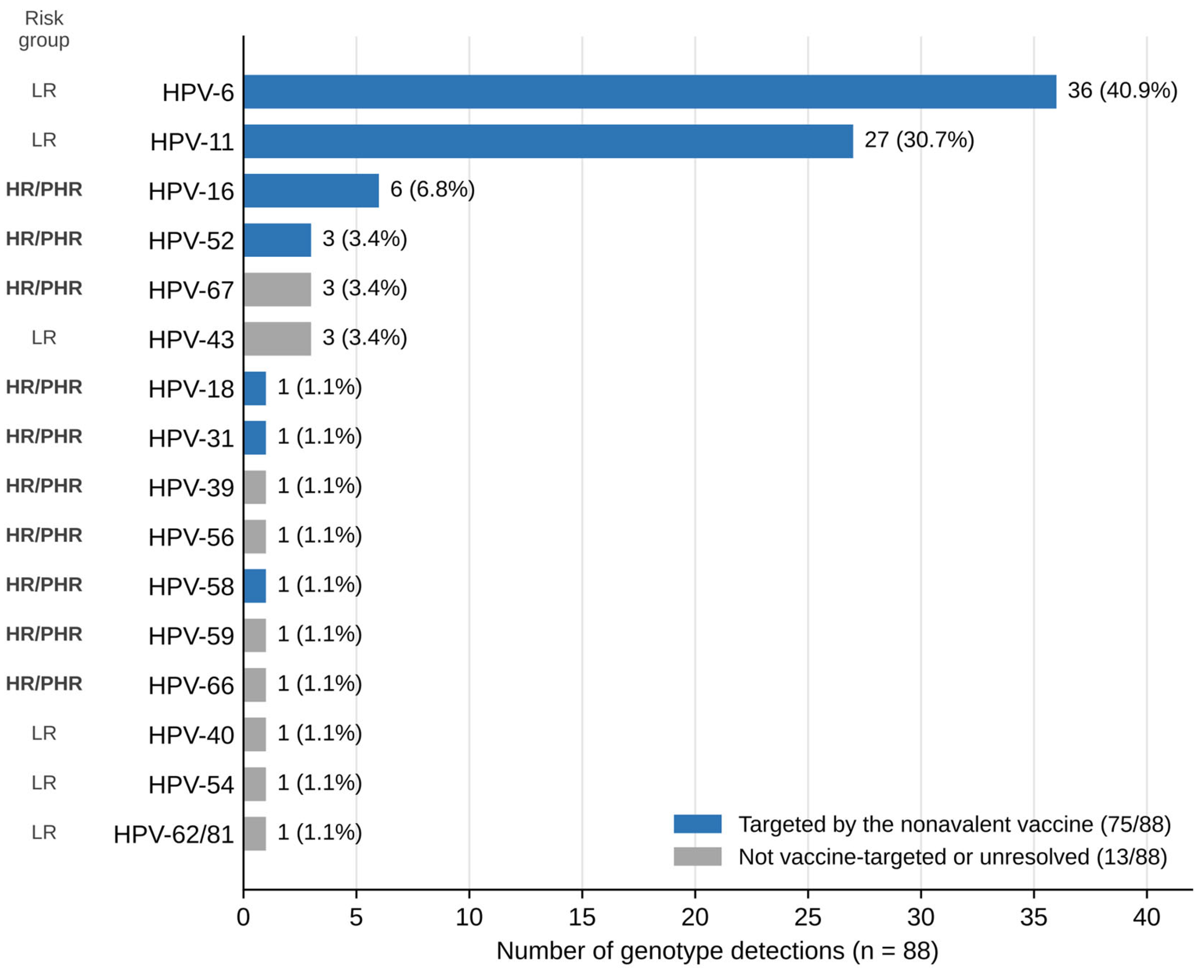
A total of 88 genotype detections were recorded among the 65 HPV-positive participants, counting the shared HPV-62/81 probe signal as one unresolved detection rather than two separately identified genotypes. HPV-6 and HPV-11 were most frequent (36/88, 40.9%; 27/88, 30.7%, respectively; Figure 2). The nine genotypes targeted by the nonavalent vaccine accounted for 75/88 detections (85.2%; 95% CI, 76.3–91.2%). At the participant level, 62/100 women (62.0%; 95% CI, 52.2–70.9%), or 62/65 HPV-positive women (95.4%; 95% CI, 87.3–98.4%), carried at least one vaccine-targeted genotype. Counts for each genotype are given in Supplementary Table S3.
Figure 2.
Genotype-specific detection frequencies among the 65 HPV-positive participants (n = 88 genotype detections). Blue bars indicate the nine genotypes targeted by the nonavalent HPV vaccine (75/88 detections, 85.2%), including 12 of the 19 HR/PHR detections (63.2%); grey bars indicate other genotypes or the unresolved HPV-62/81 shared-probe detection. The left-hand column shows the study-applied risk group (LR or HR/PHR). Percentages use genotype detections, not participants, as the denominator.
Among 19 HR/PHR genotype detections, 12 (63.2%; 95% CI, 41.0–80.9%) corresponded to genotypes targeted by the nonavalent HPV vaccine. This proportion uses HR/PHR detections not participants as its denominator.
3.3. Specimen Type and HPV Risk Category
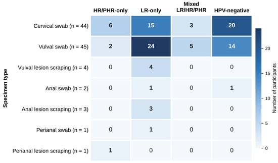
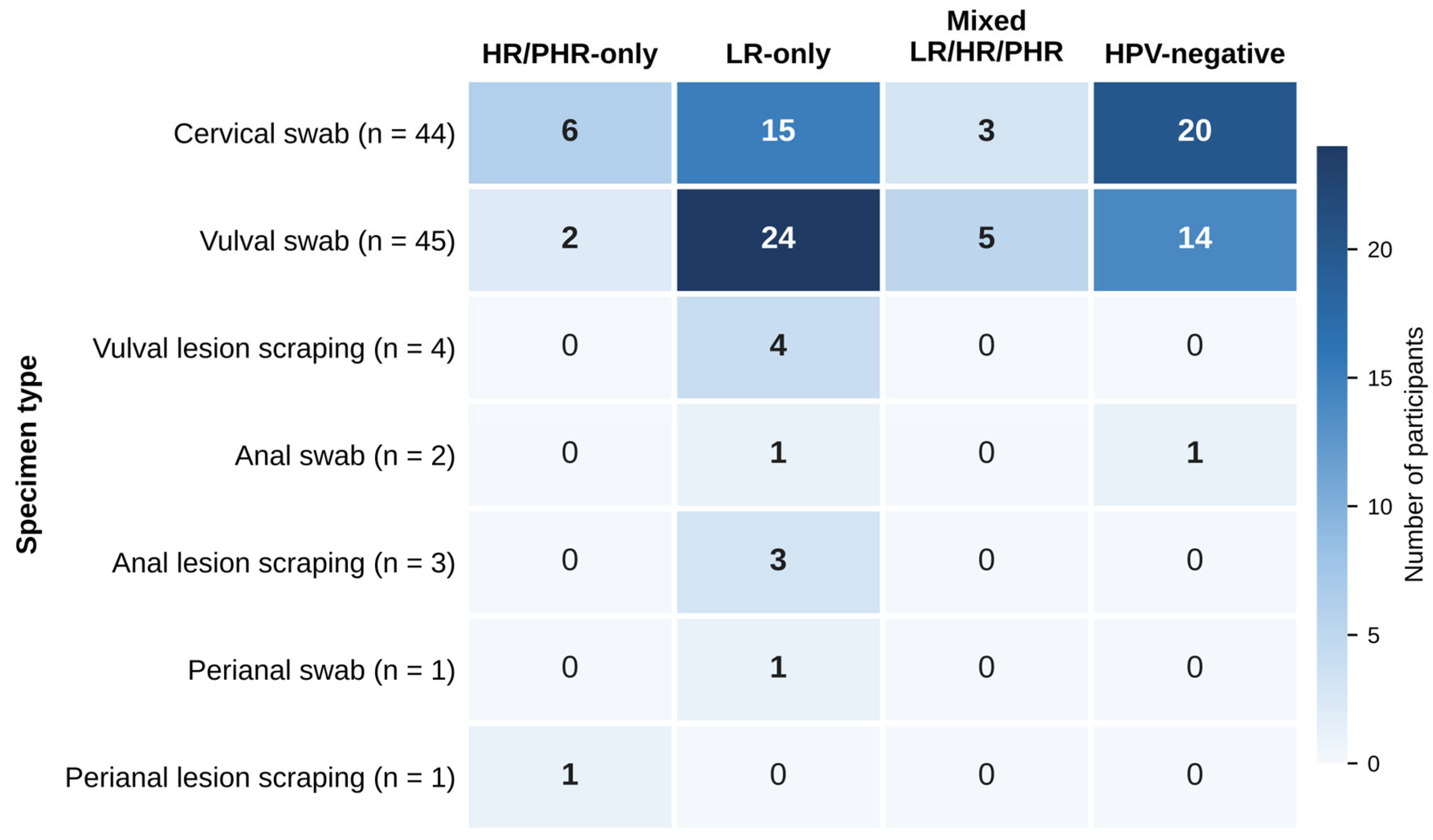
Across the seven specimen categories, the distributions of HR/PHR-only, LR-only, mixed, and HPV-negative results were summarized descriptively (Figure 3). Cervical swabs (n = 44) comprised 6 HR/PHR-only, 15 LR-only, 3 mixed, and 20 negative results; vulval swabs (n = 45) comprised 2, 24, 5, and 14, respectively. The remaining specimen categories had small counts and are displayed in Figure 3.
Figure 3.
Distribution of HPV risk categories among the seven specimen types (n = 100). Cell values are participant counts; darker shading indicates higher counts. Lesion-derived specimens were superficial scrapings of clinically visible lesions.
3.4. Genotype Co-Occurrence in Multiple Infections
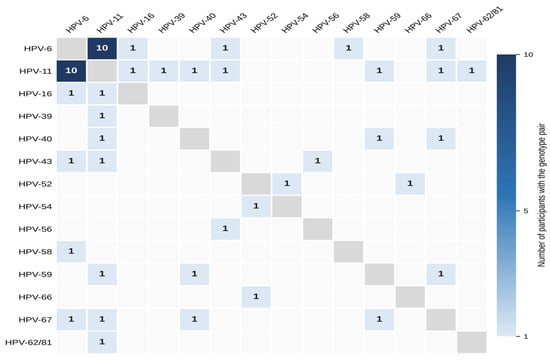
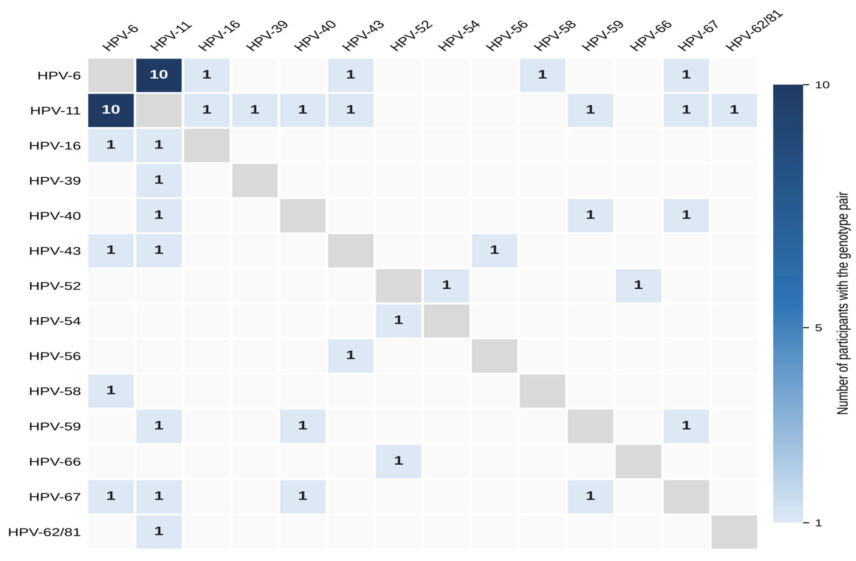
Multiple-genotype infections were identified in 20 participants, yielding 27 pairwise genotype co-occurrences after decomposing multiple-genotype infections into all possible genotype pairs. Each unique genotype pair was counted once per participant. The HPV-6/HPV-11 pair was the most frequently observed combination, accounting for 10 of the 27 recorded pairwise co-occurrences (37.0%; Figure 4).
Figure 4.
Symmetric heatmap of HPV genotype co-occurrence among 20 participants with multiple-genotype infections (27 pairwise co-occurrences; 18 distinct genotype pairs). Each off-diagonal cell shows the number of participants with the indicated pair; empty cells indicate pairs that were not observed, and diagonal cells are not applicable. HPV-62/81 was treated as one unresolved shared-probe detection. The matrix is descriptive and does not establish biological interactions between genotypes.
4. Discussion
This study characterized HPV genotype distribution and co-occurrence in a clinically selected cohort of 100 Iraqi women, each contributing one specimen from an anatomical site selected on clinical grounds. Low-risk genotypes, particularly HPV-6 and HPV-11, predominated. HR/PHR genotypes were also detected, both alone and alongside low-risk genotypes.
An earlier Iraqi study using a microarray-based approach reported HPV positivity of 12.7% [14], compared with 65% in the present cohort. These percentages should not be interpreted as directly comparable estimates because the populations, recruitment indications, and sampling contexts may differ. The higher observed positivity in this clinically selected cohort may partly reflect selection of women with suspected HPV-associated disease; no formal between-study statistical comparison was performed.
HPV genotype distributions reported in other settings vary with the populations examined and their indications for testing. Earlier Iraqi work on women with benign and malignant cervical lesions has focused on high- and intermediate-risk genotypes [18]. Studies from the Gulf region, China, and Sulaymaniyah have reported differing high-risk genotype profiles [9,19,20]. Conversely, studies of clinically recognized anogenital warts have frequently identified HPV-6 and HPV-11 [10,21]. These reports provide context for the low-risk predominance observed here, but differences in recruitment, specimen types, and assay methods limit direct comparisons. The predominance of HPV-6 and HPV-11 is likely to reflect, at least in part, the clinical selection of women with suspected HPV-associated disease, and should not be generalized to population-based cervical screening.
The lesion-derived samples were superficial scrapings collected for HPV DNA testing rather than excisional tissue biopsies, and no histopathological evaluation was available. Detection of HPV-16 as the only genotype in one perianal lesion clinically diagnosed as a wart is noteworthy, because anogenital warts are mainly associated with HPV-6 and HPV-11 [10,21]; this finding raises the possibility of an HPV-16-associated lesion other than a benign condyloma, but without histopathological confirmation it cannot establish dysplasia, Bowenoid papulosis, or any other histological diagnosis.
Multiple-genotype infection was identified in 20 of 100 participants (20.0%), or 20 of 65 HPV-positive participants (30.8%). Previous studies have reported different frequencies of multiple-genotype infection [10,22]. With only 20 multiple-genotype infections, the co-occurrence patterns detected here are descriptive; this cross-sectional study cannot establish biological interactions among genotypes, increased lesion risk, or causal relationships. Some cohorts have linked multiple high-risk HPV infections to greater cervical lesion risk [23]; the present data cannot address this question.
Seventeen women had HR/PHR-only (n = 9) or mixed LR/HR/PHR (n = 8) infections. A validated high-risk HPV assay would be expected to detect the HR component of these infections when the genotype lies within its target panel, and many such assays also report HPV-16 and HPV-18 individually. Extended genotype-resolved testing adds information by identifying the remaining individual genotypes and their co-occurring low-risk types. Two of the 17 women carried HPV-67 (IARC Group 2B) as their only HR/PHR genotype; this genotype lies outside the high-risk panels, usually comprising 13–14 genotypes, of clinically validated high-risk HPV assays [24]. The vaccine-targeted proportions reported in Section 3.2, whether expressed per genotype detection or per participant, describe genotype representation in this clinically selected cohort and do not establish vaccine effectiveness or the fraction of disease preventable by vaccination.
The post-PCR microarray assay provided genotype-resolved information across its defined detection panel, enabling description of the specific HPV combinations identified in this cohort. The assay also has inherent limitations: detection is restricted to its 35-genotype panel, HPV-62 and HPV-81 cannot be distinguished, and results are qualitative, providing no information on viral load. Other molecular approaches, including validated high-risk HPV assays, genotype-specific PCR, and sequencing, have different analytical scopes and clinical applications. As no direct comparison with alternative HPV assays was performed, the present study does not provide evidence of superior diagnostic sensitivity, clinical utility, concordance, throughput, or cost-effectiveness of the microarray platform. Further comparative studies are needed to evaluate its potential application in broader HPV surveillance and its economic implications.
The study used a consistent laboratory workflow and assay-based quality-control procedures. These measures supported interpretation of the observed genotyping results, but they do not eliminate potential pre-analytical variation across specimen types or recruitment settings.
Clinically based non-probability recruitment may introduce selection bias and overrepresent women with symptoms or clinically suspicious findings; therefore, the observed genotype frequencies should not be interpreted as population prevalence. Additional limitations include heterogeneity of clinical indications and sampling regions, small anal and perianal subgroups, and the absence of standardized participant-level data on testing indication, cervical cytology, previous HPV-related disease, immunosuppressive treatment, vaccination, and behavioral risk factors, which may have influenced the observed genotype distribution. No histopathological confirmation was performed for superficial lesion scrapings. Consequently, the clinical diagnosis of anogenital warts could not exclude other HPV-associated lesions with similar appearances, limiting the clinicopathological interpretation of the genotyping findings. Differences in cellular yield and viral load between swabs and superficial lesion scrapings may affect specimen comparability; because only eight lesion-derived specimens were collected, differences between collection methods could not be evaluated. Furthermore, the cross-sectional design and absence of longitudinal follow-up prevent causal inference, while the clinically selected sample limits the generalizability of the findings to the broader population of Iraqi women. Future studies using paired sampling of multiple anogenital sites within the same women, together with standardized clinical data, would allow site-specific variation in HR/PHR and multiple-genotype infections to be examined formally.
5. Conclusions
This cross-sectional study describes HPV genotype distribution and co-infection patterns among 100 clinically selected Iraqi women. Low-risk HPV-6 and HPV-11 predominated, although HR/PHR and mixed infections were also identified. Genotype-resolved microarray analysis demonstrated its utility in identifying the specific HPV genotype combinations within the study cohort; however, its performance was not directly compared with other HPV assays. The results are preliminary and descriptive and should not be interpreted as representative national estimates or as validation of a population screening or vaccination strategy.
Supplementary Materials
The following supporting information can be downloaded at: https://www.mdpi.com/article/10.3390/pathogens15111193/s1, Table S1. Complete genotype classification, including manufacturer-assigned risk category, study-applied classification, and detection frequency among the 65 HPV-positive participants. HPV-67 was assigned to the study-applied HR/PHR category based on its IARC Group 2B classification. HPV-62 and HPV-81 share a single probe and are reported as HPV-62/81. Percentages use HPV-positive participants (n = 65) as the denominator, whereas Figure 2 uses total genotype detections (n = 88). Manufacturer-assigned categories followed the assay classification; study-applied categories followed the predefined framework described in the Methods. Table S2. Age distribution by HPV risk category, expressed as median, interquartile range (IQR), and range. Table S3. Aggregated frequency distribution of individual HPV genotypes detected among the 65 HPV-positive participants (n = 88 genotype detections total). This table reports summary counts only; no individual-level participant data are included, consistent with the Data Availability Statement. Table S4. STROBE checklist for the cross-sectional study, showing reporting items and corresponding manuscript sections. Figure S1. Schematic representation of microarray-based HPV genotype detection and result interpretation. The schematic illustrates the interpretation pathway from genotype-specific microarray hybridization signals to HPV genotype identification, risk classification, and categorization of single- versus multiple-genotype infections. Detected genotypes were classified as low-risk (LR) or high-risk/potentially high-risk (HR/PHR) according to the predefined study classification. HPV-62/81 represents a combined assay signal recorded as a single genotype detection because the assay did not distinguish between the two genotypes.
Author Contributions
D.M.A.-K.: laboratory analysis and investigation, formal statistical analysis, and writing of the original draft; M.T.S.A.-O.: study conceptualization and design, supervision, and writing—review and editing; R.M.M.: clinical supervision of specimen collection, data analysis; T.W.Y.: writing—original draft and editing, funding. All authors have read and agreed to the published version of the manuscript.
Funding
This research was funded by Universiti Kuala Lumpur, Institute of Medical Science Technology (UniKL MESTECH), A1-1, Jalan TKS 1, Taman Kajang Sentral, 43000 Kajang, Selangor, Malaysia.
Institutional Review Board Statement
The study protocol was reviewed and approved by the Ethical Approval Committee, University of Anbar, College of Medicine (approval Ref. No. 127, dated 12 November 2025), in accordance with the ethical principles of the Declaration of Helsinki. This manuscript reports the molecular genotyping component of the approved research project. The associated immunological analyses are outside the scope of the present report.
Informed Consent Statement
Written informed consent was obtained from all participants before specimen collection.
Data Availability Statement
Aggregated genotype frequency data are provided in Supplementary Table S3. De-identified participant-level data are available from the corresponding author upon reasonable request.
Conflicts of Interest
The authors declare no conflicts of interest.
Abbreviations
The following abbreviations are used in this manuscript:
| HPV | Human papillomavirus |
| LR | Low-risk |
| HR | High-risk |
| PHR | Probably/potentially high-risk |
| IARC | International Agency for Research on Cancer |
| PCR | Polymerase chain reaction |
| DNA | Deoxyribonucleic acid |
| L1 | Late protein 1 |
| CI | Confidence interval |
| IQR | Interquartile range |
| dUTP | Deoxyuridine triphosphate |
| SD | Standard deviation |
| STROBE | Strengthening the Reporting of Observational Studies in Epidemiology |
| WHO | World Health Organization |
References
- Sung, H.; Filho, A.M.; Laversanne, M.; Ferlay, J.; Siegel, R.L.; Soerjomataram, I.; Jemal, A.; Bray, F. Global cancer statistics 2024: GLOBOCAN estimates of incidence and mortality worldwide for 34 cancers in 186 countries. CA Cancer J. Clin. 2026, 76, e70090. [Google Scholar] [CrossRef] [Scilit]
- Ilic, I.; Ilic, M. Human papillomavirus vaccination coverage estimates among the primary target cohort (9–14-year-old girls) in the world (2010–2024). Vaccines 2025, 13, 1010. [Google Scholar] [CrossRef] [Scilit]
- Wilailak, S.; Kengsakul, M.; Kehoe, S. Strategic approaches for global cervical cancer elimination: An update review and call for national action. Int. J. Gynaecol. Obstet. 2025, 171, 120–128. [Google Scholar] [CrossRef] [Scilit]
- International Agency for Research on Cancer. Iraq: GLOBOCAN 2024 Fact Sheet. Global Cancer Observatory. Available online: https://gco.iarc.who.int/media/globocan/factsheets/populations/368-iraq-fact-sheet.pdf (accessed on 30 October 2026).
- Bouvard, V.; Baan, R.; Straif, K.; Grosse, Y.; Secretan, B.; El Ghissassi, F.; Benbrahim-Tallaa, L.; Guha, N.; Freeman, C.; Galichet, L.; et al. A review of human carcinogens—Part B: Biological agents. Lancet Oncol. 2009, 10, 321–322. [Google Scholar] [CrossRef] [Scilit]
- Camero-Ortiz, E.A.; Delgado-García, S.K.; Bendezu-Quispe, G.; Grandez-Castillo, G.A. Diagnostic criteria for HPV infection classification: A comment on Wen et al. Virol. J. 2025, 22, 369. [Google Scholar] [CrossRef] [Scilit]
- You, X.; Valente, S.; Machalek, D.A.; Felsher, M.; Pieniazek, I.; Nowicka, K.; Tota, J.E.; Chen, Y.-T.; Niccolai, L.M.; Cui, F. Epidemiologic, clinical, and economic burden of anogenital warts among adults: A global systematic review. Int. J. Infect. Dis. 2025, 161, 108029. [Google Scholar] [CrossRef] [Scilit]
- Giuliani, E.; Donà, M.G.; Zaccarelli, M.; Stingone, C.; Gianserra, L.; Capodieci, S.; Cafaro, V.; Fulgenzio, C.; Latini, A.; Giuliani, M. Trends of anogenital warts: A 32-year retrospective observational study (Italy, 1991–2022). J. Clin. Med. 2025, 14, 3962. [Google Scholar] [CrossRef] [Scilit]
- Wang, W.; Zhang, J.; Mei, C.; Dai, Q.; Cai, Y.; Wei, X.; Li, Y. Prevalence and genotype distribution of human papillomavirus infection among women in Chengdu, China between 2020 and 2024. Front. Public Health 2026, 13, 1740177. [Google Scholar] [CrossRef] [Scilit]
- Rideg, O.; Oszter, A.; Makk, E.; Kálmán, E.; Farkas, K.; Tornóczky, T.; Kovács, K. Wide spectrum analysis of human papillomavirus genotypes in external anogenital warts. Vaccines 2021, 9, 604. [Google Scholar] [CrossRef] [Scilit]
- World Health Organization. WHO Guideline for Screening and Treatment of Cervical Pre-Cancer Lesions for Cervical Cancer Prevention: Use of Human Papillomavirus (HPV) DNA Genotyping; World Health Organization: Geneva, Switzerland, 2026; Available online: https://www.who.int/publications/i/item/9789240121744 (accessed on 30 October 2026).
- Herraez-Hernandez, E.; Alvarez-Perez, M.; Navarro-Bustos, G.; Esquivias, J.; Alonso, S.; Aneiros-Fernandez, J.; Lacruz-Pelea, C.; Sanchez-Aguera, M.; Santamaria, J.S.; de Antonio, J.C.; et al. HPV Direct Flow CHIP: A new human papillomavirus genotyping method based on direct PCR from crude-cell extracts. J. Virol. Methods 2013, 193, 9–17. [Google Scholar] [CrossRef] [Scilit]
- Bellosillo, B.; Ibáñez, R.; Roura, E.; Monfil, L.; Asensio-Puig, L.; Álvarez, I.; Muset, M.; Florencia, Y.; Paytubi, S.; de Andrés-Pablo, Á.; et al. Clinical validation of the Vitro HPV Screening Assay for its use in primary cervical cancer screening. Cancers 2024, 16, 1322. [Google Scholar] [CrossRef] [Scilit]
- Tahir, M.; Mohammed, D.; Sadiq, C. Prevalence and genotypic characterization of human papillomavirus in Iraqi women: A retrospective study of 2193 cases. Virol. J. 2025, 22, 394. [Google Scholar] [CrossRef] [Scilit]
- Sacace Biotechnologies Srl. SaMag Viral Nucleic Acid Extraction Kit (SM003)—Instructions for Use; Sacace Biotechnologies: Como, Italy, 2021. [Google Scholar]
- Vitro S.A. HPV Direct Flow CHIP Kit—Instructions for Use; Vitro S.A.: Seville, Spain, 2020. [Google Scholar]
- Yang, H.J.; Lee, H.S. Common statistical methods used in medical research. Kosin Med. J. 2025, 40, 21–30. [Google Scholar] [CrossRef] [Scilit]
- Al-Ouqaili, M.T.S.; Al-Kubaisy, S.H.M. Molecular genetics study on high and intermediate risk genotypes of human papillomavirus among patients with benign and malignant cervical lesions. Int. J. Pharm. Sci. Res. 2019, 10, 2062–2070. [Google Scholar] [CrossRef] [Scilit]
- AlMesbah, N.; Maatoug, J.; Selim, N.; Bougmiza, I. Human papillomavirus prevalence and genotypes in Gulf Cooperation Council countries: A scoping review 2017–2024. Qatar Med. J. 2024, 2024, 33. [Google Scholar] [CrossRef] [Scilit]
- Sleman, S. Prevalence and genotype distribution of human papillomavirus in Sulaymaniyah, Kurdistan Region, Iraq. Front. Cell. Infect. Microbiol. 2026, 16, 1830236. [Google Scholar] [CrossRef] [Scilit]
- Sadati, M.S.; Jahangiri, S.; Azizi, A.; Parvar, S.Y.; Akbarzadeh Jahromi, M.; Dehghan, Y. Prevalence and distribution of HPV genotypes in patients with genital warts: A cross-sectional study from South of Iran. Health Sci. Rep. 2025, 8, e71345. [Google Scholar] [CrossRef] [Scilit]
- Finney, D.; Hashim, A.L.O.; Bylappa, S.K.; Mugar, A.E.; Nandhagopal, M.; Jabbar, M.A.; Rahamathullah, N. Genotypic distribution and molecular epidemiology of HPV in women in the UAE using PNA-based RT PCR. PLoS ONE 2026, 21, e0346052. [Google Scholar] [CrossRef] [Scilit]
- Jing, Y.; Chen, J.; Lin, F.; Huang, X.; Liu, Y.; Zhao, M.; Ye, C.; Zhao, L.; Liu, X.; Yang, J. Multiple high-risk human papillomavirus infections exacerbate cervical lesion risk: Epidemiological evidence from Suining, Sichuan. Virol. J. 2025, 22, 268. [Google Scholar] [CrossRef] [Scilit]
- Arbyn, M.; Simon, M.; Peeters, E.; Xu, L.; Meijer, C.J.L.M.; Berkhof, J.; Cuschieri, K.; Bonde, J.; Vanlencak, A.O.; Zhao, F.-H.; et al. 2020 list of human papillomavirus assays suitable for primary cervical cancer screening. Clin. Microbiol. Infect. 2021, 27, 1083–1095. [Google Scholar] [CrossRef] [Scilit]

Disclaimer/Publisher’s Note: The statements, opinions and data contained in all publications are solely those of the individual author(s) and contributor(s) and not of MDPI and/or the editor(s). MDPI and/or the editor(s) disclaim responsibility for any injury to people or property resulting from any ideas, methods, instructions or products referred to in the content. |
© 2026 by the authors. Licensee MDPI, Basel, Switzerland. This article is an open access article distributed under the terms and conditions of the Creative Commons Attribution (CC BY) license.