Next Issue
Volume 12, January
Previous Issue
Volume 11, November


Pathogens, Volume 11, Issue 12 (December 2022) – 168 articles

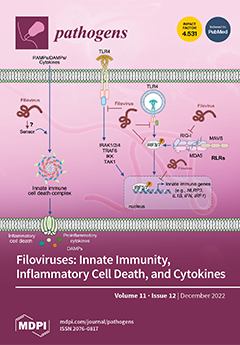
Cover Story (view full-size image): Innate immune pattern recognition receptors (PRRs), such as toll-like receptors (TLRs), retinoic acid-inducible gene-I-like receptors (RLRs), and NOD-like receptors (NLRs), detect pathogens such as filoviruses and induce the production of proinflammatory cytokines and interferons as well as initiate cell death for host defense. Filoviruses modulate this response, causing increased viral replication, morbidity, and mortality. RLRs are the most well-characterized PRRs in filovirus infection; ebolaviruses and marburgviruses inhibit molecules in the RLR pathway and dampen the early immune response, allowing viral spread. Evidence suggests that other PRRs sense filovirus infection, though these interactions are not well defined. PRRs and cell death mechanisms may serve as therapeutic targets to eliminate the virus and reduce deadly inflammation. View this paper
  • Issues are regarded as officially published after their release is announced to the table of contents alert mailing list.
  • You may sign up for e-mail alerts to receive table of contents of newly released issues.
  • PDF is the official format for papers published in both, html and pdf forms. To view the papers in pdf format, click on the "PDF Full-text" link, and use the free Adobe Reader to open them.
Order results
Result details
Section
Select all
Export citation of selected articles as:
13 pages, 3753 KiB  
Article
Hexameric Aggregation Nucleation Core Sequences and Diversity of Pathogenic Tau Strains
by Ling Wu, Sidharth S. Madhavan, Christopher Tan and Bin Xu
Pathogens 2022, 11(12), 1559; https://doi.org/10.3390/pathogens11121559 - 19 Dec 2022
Cited by 3 | Viewed by 1562
Abstract
Tau aggregation associates with multiple neurodegenerative diseases including Alzheimer’s disease and rare tauopathies such as Pick’s disease, progressive supranuclear palsy, and corticobasal degeneration. The molecular and structural basis of tau aggregation and related diverse misfolded tau strains are not fully understood. To further [...] Read more.
Tau aggregation associates with multiple neurodegenerative diseases including Alzheimer’s disease and rare tauopathies such as Pick’s disease, progressive supranuclear palsy, and corticobasal degeneration. The molecular and structural basis of tau aggregation and related diverse misfolded tau strains are not fully understood. To further understand tau-protein aggregation mechanisms, we performed systematic truncation mutagenesis and mapped key segments of tau proteins that contribute to tau aggregation, where it was determined that microtubule binding domains R2 and R3 play critical roles. We validated that R2- or R3-related hexameric PHF6 and PHF6* peptide sequences are necessary sequences that render tau amyloidogenicity. We also determined that the consensus VQI peptide sequence is not sufficient for amyloidogenicity. We further proposed single- and dual-nucleation core-based strain classifications based on recent cryo-EM structures. We analyzed the structural environment of the hexameric peptide sequences in diverse tau strains in tauopathies that, in part, explains why the VQI consensus core sequence is not sufficient to induce tau aggregation. Our experimental work and complementary structural analysis highlighted the indispensible roles of the hexameric core sequences, and shed light on how the interaction environment of these core sequences contributes to diverse pathogenic tau-strains formation in various tauopathy brains. Full article
(This article belongs to the Special Issue Prions and Prion-Like Transmissible Protein Pathogens)
Show Figures

Figure 1

13 pages, 2086 KiB  
Article
An Insight into the Genome of Pathogenic and Non-Pathogenic Acanthamoeba
by Chayan Sharma, Sumeeta Khurana, Amit Arora, Alka Bhatia and Amit Gupta
Pathogens 2022, 11(12), 1558; https://doi.org/10.3390/pathogens11121558 - 19 Dec 2022
Cited by 2 | Viewed by 1650
Abstract
Background: Acanthamoeba are amphizoic amoeba majorly responsible for causing Acanthamoeba keratitis (AK) and Granulomatous amoebic encephalitis (GAE). Despite its ubiquitous nature, the frequency of infections is not high, probably due to the existence of non-pathogenic isolates. The whole-genome sequencing and an annotated genome [...] Read more.
Background: Acanthamoeba are amphizoic amoeba majorly responsible for causing Acanthamoeba keratitis (AK) and Granulomatous amoebic encephalitis (GAE). Despite its ubiquitous nature, the frequency of infections is not high, probably due to the existence of non-pathogenic isolates. The whole-genome sequencing and an annotated genome assembly can unravel the biological functions and help in identifying probable genes related to pathogenicity. Methods: Illumina and Nanopore sequencing were performed for keratitis, encephalitis, and non-pathogenic environmental isolates. Hybrid assembly was prepared for the AK and GAE isolates, while only the Illumina reads were utilized for a non-pathogenic environmental isolate. Protein coding genes were identified using the GeneMark-ES program and BLASTx module of Diamond used for gene prediction. Additionally, the Kyoto Encyclopedia of Genes and Genomes annotation and cluster of orthologous group’s annotation using RPS-blast against the CDD database was performed. The subsequent data analysis and validation will help identify probable pathogenic genes. Results: The genome assemblies of 9.67, 8.34, and 8.89 GBs were reported for GAE, AK, and non-pathogenic isolate, respectively. KEGG reported 22,946 in GAE, 24,231 in keratitis, and 9367 genes in the environmental isolate. The COG annotation revealed 3232 in GAE, 3403 in keratitis, and 1314 genes in the non-pathogenic isolate. Conclusion: The present study has attempted to generate de novo hybrid genome assemblies of Acanthamoeba that would help decode the genome of free-living amoeba and will provide genomic data for a better understanding of virulence-related factors. Full article
(This article belongs to the Special Issue ICOPA 2022 Free Living Amoebae Symposia Special Issue)
Show Figures

Figure 1

11 pages, 4797 KiB  
Article
Detection and Molecular Characterization of Canine Distemper Virus in Wildlife from Northern Italy
by Tiziana Trogu, Anna Castelli, Sabrina Canziani, Clara Tolini, Maya Carrera, Enrica Sozzi, Davide Lelli, Giovanni Tosi, Laura Fiorentini, Alessandra Di Donato, Gianluca Rugna, Danilo Lanci, Antonio Lavazza and Ana Moreno
Pathogens 2022, 11(12), 1557; https://doi.org/10.3390/pathogens11121557 - 19 Dec 2022
Cited by 2 | Viewed by 1655
Abstract
Canine distemper virus (CDV) is a fatal, highly contagious disease found in wild and domestic carnivores. Several outbreaks have occurred in wildlife in Italy in recent years. This study aims to detect CDV in wildlife following the increasing mortality of foxes (Vulpes [...] Read more.
Canine distemper virus (CDV) is a fatal, highly contagious disease found in wild and domestic carnivores. Several outbreaks have occurred in wildlife in Italy in recent years. This study aims to detect CDV in wildlife following the increasing mortality of foxes (Vulpes vulpes) in the Emilia-Romagna region (northern Italy) observed in 2021. Sixty-seven foxes and one badger (Meles meles) were subjected to necropsy followed by histological examination and were analyzed with molecular techniques to detect the presence of CDV. Of the tested animals, 16% (nine foxes and one badger) were positive for CDV. Phylogenetic analysis showed two different lineages based on complete H gene sequences. The Europe/South America-1 lineage was detected in one fox from Modena, which resembled the CDV variant associated with a previous outbreak in northern Italy in 2018, while the European Wildlife lineage was detected in animals from the Rimini province. Amino acid analysis highlighted a Y549H mutation in all sequences collected, which is commonly associated with increased virulence. Full article
(This article belongs to the Special Issue Canine Distemper Virus Infection)
Show Figures

Figure 1

18 pages, 3157 KiB  
Article
Evolution of Rabies Virus Isolates: Virulence Signatures and Effects of Modulation by Neutralizing Antibodies
by Juliana Amorim Conselheiro, Gisely Toledo Barone, Sueli Akemi Taniwaki Miyagi, Sheila Oliveira de Souza Silva, Washington Carlos Agostinho, Joana Aguiar and Paulo Eduardo Brandão
Pathogens 2022, 11(12), 1556; https://doi.org/10.3390/pathogens11121556 - 19 Dec 2022
Cited by 2 | Viewed by 2006
Abstract
Lyssavirus rabies (RABV) is an RNA virus and, therefore, is subject to mutations due to low RNA polymerase replication fidelity, forming a population structure known as a viral quasispecies, which is the core of RNA viruses’ adaptive strategy. Under new microenvironmental conditions, the [...] Read more.
Lyssavirus rabies (RABV) is an RNA virus and, therefore, is subject to mutations due to low RNA polymerase replication fidelity, forming a population structure known as a viral quasispecies, which is the core of RNA viruses’ adaptive strategy. Under new microenvironmental conditions, the fittest populations are selected, and the study of this process on the molecular level can help determine molecular signatures related to virulence. Our aim was to survey gene signatures on nucleoprotein and glycoprotein genes that might be involved in virulence modulation during the in vitro evolution of RABV lineages after serial passages in a neuronal cell system with or without the presence of neutralizing antibodies based on replicative fitness, in vivo neurotropism and protein structure and dynamics. The experiments revealed that amino acids at positions 186 and 188 of the glycoprotein are virulence factors of Lyssavirus rabies, and site 186 specifically might allow the attachment to heparan as a secondary cell receptor, while polymorphism at position 333 might allow the selection of escape mutants under suboptimal neutralizing antibodies titers. Full article
(This article belongs to the Special Issue Emerging and Re-emerging Viral Infectious Diseases)
Show Figures

Figure 1

9 pages, 1723 KiB  
Brief Report
A Systemic Study of Subcellular Localization of Porcine Epidemic Diarrhea Virus Proteins
by Huixin Zhu, Zitong Li, Juan Bai, Ping Jiang, Xianwei Wang and Xing Liu
Pathogens 2022, 11(12), 1555; https://doi.org/10.3390/pathogens11121555 - 17 Dec 2022
Cited by 1 | Viewed by 1410
Abstract
Porcine epidemic diarrhea virus (PEDV), a highly pathogenic enteric coronavirus, is regarded as one of the most severe porcine pathogens. To date, there are still no commercial vaccines or drugs that can provide full protection against the epidemic strains. A better understanding of [...] Read more.
Porcine epidemic diarrhea virus (PEDV), a highly pathogenic enteric coronavirus, is regarded as one of the most severe porcine pathogens. To date, there are still no commercial vaccines or drugs that can provide full protection against the epidemic strains. A better understanding of the subcellular location of individual proteins could benefit from studying the protein functions and mechanisms of how the virus regulates key cellular processes, finally leading to the development of antiviral agents. In this study, we characterized the subcellular localization of PEDV proteins using multi-labeled fluorescent immunocytochemistry. As a result, 11 proteins showed cytoplasmic distribution and 10 proteins showed both cytoplasmic and nuclear distribution. Furthermore, we demonstrated that four proteins (Nsp3, Nsp4, Nsp6, and S1) were co-localized in the endoplasmic reticulum (ER), while four proteins (Nsp2, S2, N, and ORF3) were partially observed in the ER, two proteins (E and M) were co-localized in the Golgi apparatus, and two proteins (Nsp2 and E) were partially co-localized with the mitochondria. These viral proteins may perform specific functions at specific cellular locations. Together, these results describe a subcellular localization map of PEDV proteins, which will help to characterize the functions of these proteins in the future. Full article
(This article belongs to the Section Viral Pathogens)
Show Figures

Figure 1

15 pages, 2289 KiB  
Article
Type I Interferon Signaling Controls Gammaherpesvirus Latency In Vivo
by Johannes Schwerk, Lucas Kemper, Kendra A. Bussey, Stefan Lienenklaus, Siegfried Weiss, Luka Čičin-Šain, Andrea Kröger, Ulrich Kalinke, Christopher M. Collins, Samuel H. Speck, Martin Messerle, Dagmar Wirth, Melanie M. Brinkmann, Hansjörg Hauser and Mario Köster
Pathogens 2022, 11(12), 1554; https://doi.org/10.3390/pathogens11121554 - 17 Dec 2022
Cited by 7 | Viewed by 1685
Abstract
Gammaherpesviruses, such as Epstein-Barr virus and Kaposi’s sarcoma-associated herpesvirus, are important human pathogens involved in lymphoproliferative disorders and tumorigenesis. Herpesvirus infections are characterized by a biphasic cycle comprised of an acute phase with lytic replication and a latent state. Murine gammaherpesvirus 68 (MHV-68) [...] Read more.
Gammaherpesviruses, such as Epstein-Barr virus and Kaposi’s sarcoma-associated herpesvirus, are important human pathogens involved in lymphoproliferative disorders and tumorigenesis. Herpesvirus infections are characterized by a biphasic cycle comprised of an acute phase with lytic replication and a latent state. Murine gammaherpesvirus 68 (MHV-68) is a well-established model for the study of lytic and latent life cycles in the mouse. We investigated the interplay between the type I interferon (IFN)-mediated innate immune response and MHV-68 latency using sensitive bioluminescent reporter mice. Adoptive transfer of latently infected splenocytes into type I IFN receptor-deficient mice led to a loss of latency control. This was revealed by robust viral propagation and dissemination of MHV-68, which coincided with type I IFN reporter induction. Despite MHV-68 latency control by IFN, the continuous low-level cell-to-cell transmission of MHV-68 was detected in the presence of IFN signaling, indicating that IFN cannot fully prevent viral dissemination during latency. Moreover, impaired type I IFN signaling in latently infected splenocytes increased the risk of virus reactivation, demonstrating that IFN directly controls MHV-68 latency in infected cells. Overall, our data show that locally constrained type I IFN responses control the cellular reservoir of latency, as well as the distribution of latent infection to potential new target cells. Full article
(This article belongs to the Special Issue Animal Models for Human Viruses)
Show Figures

Figure 1

10 pages, 1153 KiB  
Article
Assessment of Risk of Exposure to Leishmania Parasites among Renal Disease Patients from a Renal Unit in a Sri Lankan Endemic Leishmaniasis Focus
by Chandrani Menike, Rajeewa Dassanayake, Renu Wickremasinghe, Maheeka Seneviwickrama, Indika De Alwis, Ahmed Abd El Wahed and Shalindra Ranasinghe
Pathogens 2022, 11(12), 1553; https://doi.org/10.3390/pathogens11121553 - 16 Dec 2022
Viewed by 1105
Abstract
Leishmania donovani causes both cutaneous and visceral leishmaniasis (CL and VL) in Sri Lanka, where chronic kidney disease (CKD) and kidney transplant recipients’ (KTR) geographical areas overlap. This study aimed to determine the risk of exposure to Leishmania infection among renal patients. This [...] Read more.
Leishmania donovani causes both cutaneous and visceral leishmaniasis (CL and VL) in Sri Lanka, where chronic kidney disease (CKD) and kidney transplant recipients’ (KTR) geographical areas overlap. This study aimed to determine the risk of exposure to Leishmania infection among renal patients. This cross-sectional study in a renal unit assessed clinical symptoms and signs of CL and VL in recipients of blood/kidney or immunosuppressives. Sera were tested with Leishmania-specific DAT and rK-39 ELISA. There were 170 participants. A total of 84.1% (n = 143) were males (CKD: 101, KTR; 42, mean age 45) and 27 were females (females: CKD: 23, KTR: 4, mean age 39 years). Recipients of blood transfusion/s within last 2 years: 75.9% (CKD: 115, KTR: 14), on immunosuppressive therapy: 34.1% (CKD: 13, KTR: 45). Two CKD patients repeatedly showed clear positive titres (1: 12,800 and 1: 3200) with Leishmania-DAT and another two (CKD) became marginally positive with rK39-ELISA. Prevalence of anti-Leishmania antibodies: 2.4% (4/170). All four patients were clinically asymptomatic and were recipients of recent blood transfusions. Attributable risk of exposure to Leishmania infection through blood transfusions was 0.032, OR 2.99 (95% CI = 0.16 to 56.45, p = 0.47). Therefore, routine screening of kidney/blood donors and CKD and KTR patients in Sri Lanka may not be necessary. Full article
Show Figures

Figure 1

14 pages, 297 KiB  
Review
Landscape of TB Infection and Prevention among People Living with HIV
by Anca Vasiliu, Rebecca Abelman, Yousra Kherabi, Antonia Morita Iswari Saktiawati and Alexander Kay
Pathogens 2022, 11(12), 1552; https://doi.org/10.3390/pathogens11121552 - 16 Dec 2022
Cited by 2 | Viewed by 1559
Abstract
Tuberculosis (TB) is one of the leading causes of mortality in people living with HIV (PLHIV) and contributes to up to a third of deaths in this population. The World Health Organization guidelines aim to target early detection and treatment of TB among [...] Read more.
Tuberculosis (TB) is one of the leading causes of mortality in people living with HIV (PLHIV) and contributes to up to a third of deaths in this population. The World Health Organization guidelines aim to target early detection and treatment of TB among PLHIV, particularly in high-prevalence and low-resource settings. Prevention plays a key role in the fight against TB among PLHIV. This review explores TB screening tools available for PLHIV, including symptom-based screening, chest radiography, tuberculin skin tests, interferon gamma release assays, and serum biomarkers. We then review TB Preventive Treatment (TPT), shown to reduce the progression to active TB and mortality among PLHIV, and available TPT regimens. Last, we highlight policy-practice gaps and barriers to implementation as well as ongoing research needs to lower the burden of TB and HIV coinfection through preventive activities, innovative diagnostic tests, and cost-effectiveness studies. Full article
15 pages, 2762 KiB  
Article
Differential Role of Type 2 Diabetes as a Risk Factor for Tuberculosis in the Elderly versus Younger Adults
by Blanca I. Restrepo, Julia M. Scordo, Génesis P. Aguillón-Durán, Doris Ayala, Ana Paulina Quirino-Cerrillo, Raúl Loera-Salazar, America Cruz-González, Jose A. Caso, Mateo Joya-Ayala, Esperanza M. García-Oropesa, Alejandra B. Salinas, Leonardo Martinez, Larry S. Schlesinger, Jordi B. Torrelles and Joanne Turner
Pathogens 2022, 11(12), 1551; https://doi.org/10.3390/pathogens11121551 - 16 Dec 2022
Cited by 1 | Viewed by 1449
Abstract
The elderly are understudied despite their high risk of tuberculosis (TB). We sought to identify factors underlying the lack of an association between TB and type 2 diabetes (T2D) in the elderly, but not adults. We conducted a case–control study in elderly (≥65 [...] Read more.
The elderly are understudied despite their high risk of tuberculosis (TB). We sought to identify factors underlying the lack of an association between TB and type 2 diabetes (T2D) in the elderly, but not adults. We conducted a case–control study in elderly (≥65 years old; ELD) vs. younger adults (young/middle-aged adults (18–44/45–64 years old; YA|MAA) stratified by TB and T2D, using a research study population (n = 1160) and TB surveillance data (n = 8783). In the research study population the adjusted odds ratio (AOR) of TB in T2D was highest in young adults (AOR 6.48) but waned with age becoming non-significant in the elderly. Findings were validated using TB surveillance data. T2D in the elderly (vs. T2D in younger individuals) was characterized by better glucose control (e.g., lower hyperglycemia or HbA1c), lower insulin resistance, more sulphonylureas use, and features of less inflammation (e.g., lower obesity, neutrophils, platelets, anti-inflammatory use). We posit that differences underlying glucose dysregulation and inflammation in elderly vs. younger adults with T2D, contribute to their differential association with TB. Studies in the elderly provide valuable insights into TB-T2D pathogenesis, e.g., here we identified insulin resistance as a novel candidate mechanism by which T2D may increase active TB risk. Full article
(This article belongs to the Special Issue Pathogenicity of Mycobacterium tuberculosis)
Show Figures

Graphical abstract

16 pages, 5939 KiB  
Article
Dysbiosis in Head and Neck Cancer: Determining Optimal Sampling Site for Oral Microbiome Collection
by Dheeraj Pandey, Michal Szczesniak, Julia Maclean, Howard Chi Ho Yim, Fan Zhang, Peter Graham, Emad M. El-Omar and Peter Wu
Pathogens 2022, 11(12), 1550; https://doi.org/10.3390/pathogens11121550 - 16 Dec 2022
Cited by 3 | Viewed by 1769
Abstract
Recent research suggests that dysbiosis of the oral microbial community is associated with head and neck cancer (HNC). It remains unclear whether this dysbiosis causes chemo-radiotherapy (CRT)-related complications. However, to address this question, it is essential to determine the most representative oral site [...] Read more.
Recent research suggests that dysbiosis of the oral microbial community is associated with head and neck cancer (HNC). It remains unclear whether this dysbiosis causes chemo-radiotherapy (CRT)-related complications. However, to address this question, it is essential to determine the most representative oral site for microbiome sampling. In this study, our purpose was to determine the optimal site for oral sample collection and whether the presence of HNC is associated with altered oral microbiome from this site. In 21 newly diagnosed HNC patients and 27 healthy controls, microbiome samples were collected from saliva, swabs from buccal mucosa, tongue, hard palate, faucial pillars and all mucosal sites combined. Microbial DNA was extracted and underwent 16S rRNA amplicon gene sequencing. In healthy controls, analysis of observed taxonomic units detected differences in alpha- and beta-diversity between sampling sites. Saliva was found to have the highest intra-community microbial diversity and lowest within-subject (temporal) and between-subject variance. Feature intersection showed that most species were shared between all sites, with saliva demonstrating the most unique species as well as highest overlap with other sites. In HNC patients, saliva was found to have the highest diversity but differences between sites were not statistically significant. Across all sites, HNC patients had lower alpha diversity than healthy controls. Beta-diversity analysis showed HNC patients’ microbiome to be compositionally distinct from healthy controls. This pattern was confirmed when the salivary microbiome was considered alone. HNC patients exhibited reduced diversity of the oral microbiome. Salivary samples demonstrate temporal stability, have the richest diversity and are sufficient to detect perturbation due to presence of HNC. Hence, they can be used as representative oral samples for microbiome studies in HNC patients. Full article
(This article belongs to the Special Issue The Role of the Gut Microbiome in Health and Disease)
Show Figures

Figure 1

10 pages, 497 KiB  
Article
A Comparison of Doxycycline and Amoxicillin Containing Quadruple Eradication Therapy for Treating Helicobacter pylori-Infected Duodenal Ulcers: A Multicenter, Opened, Randomized Controlled Trial in China
by Jingshu Chi, Canxia Xu, Xiaoming Liu, Hao Wu, Xiaoran Xie, Peng Liu, Huan Li, Guiying Zhang, Meihua Xu, Chaomin Li, Chunlian Wang, Fengqian Song, Ming Yang and Jie Wu
Pathogens 2022, 11(12), 1549; https://doi.org/10.3390/pathogens11121549 - 16 Dec 2022
Cited by 2 | Viewed by 3305
Abstract
Background: Increased antibiotic resistance is one of the major factors contributing to the failure of H. pylori eradication. This study aimed to compare the efficacy and safety of doxycycline and amoxicillin, both critical components for bismuth-based quadruple therapy, for the first-line treatment of [...] Read more.
Background: Increased antibiotic resistance is one of the major factors contributing to the failure of H. pylori eradication. This study aimed to compare the efficacy and safety of doxycycline and amoxicillin, both critical components for bismuth-based quadruple therapy, for the first-line treatment of H. pylori-infected duodenal ulcers. Methods: An open, randomized case-controlled, multicenter trial was conducted in seven hospitals in China. A total of 184 eligible participants were divided into an IDFB (ilaprazole 5 mg, doxycycline 100 mg, furazolidone 100 mg, and bismuth 220 mg bid) or IAFB (ilaprazole 5 mg, amoxicillin 1000 mg, furazolidone 100 mg, and bismuth 220 mg bid) group for 14 days. Both groups were administrated with ilaprazole 5 mg qd for another 14 days. The main outcome was an H. pylori eradication rate; secondary outcomes were ulcer healing, relief of symptoms, and incidence of adverse effects. Results: The H. pylori eradication rates were 85.9% (95% CI 78.6–93.9) in the IDFB vs. 84.8% (95% CI 77.3–92.3) in the IAFB group in ITT analysis (p > 0.05), and 92.9% (95% CI 87.4–98.5) vs. and 91.8% (95% CI 85.8–97.7) in PP analysis (p > 0.05). The overall ulcer healing rates of IDFB and IAFB were 79.1% and 84.7% (p > 0.05), both effective in relieving symptoms. Only nine participants had adverse reactions in this trial (4/92 in IDFB and 5/92 in IAFB). Conclusion: A bismuth quadruple regimen containing doxycycline or amoxicillin could be an effective and safe treatment for H. pylori eradication, while doxycycline replacement is an alternative for participants with penicillin allergy. Full article
(This article belongs to the Section Bacterial Pathogens)
Show Figures

Figure 1

11 pages, 2162 KiB  
Article
rCsHscB Derived from Clonorchis sinensis: A Carcinogenic Liver Fluke Ameliorates LPS-Induced Acute Hepatic Injury by Repression of Inflammation
by Bo Zhang, Chunyang Fan, Qi Tan, Yuzhao Zhang, Qing Jiang, Qian Yu, Beibei Zhang, Kuiyang Zheng and Chao Yan
Pathogens 2022, 11(12), 1548; https://doi.org/10.3390/pathogens11121548 - 15 Dec 2022
Cited by 1 | Viewed by 1374
Abstract
Sepsis-associated acute liver injury caused by spillovers of bacteria and endotoxins (lipopolysaccharide, LPS) into the liver remains a public health issue due to the lack of specific therapeutic approaches. Previous studies showed that the recombinant protein HscB (rCsHscB) of Clonorchis sinensis, a [...] Read more.
Sepsis-associated acute liver injury caused by spillovers of bacteria and endotoxins (lipopolysaccharide, LPS) into the liver remains a public health issue due to the lack of specific therapeutic approaches. Previous studies showed that the recombinant protein HscB (rCsHscB) of Clonorchis sinensis, a carcinogenic liver fluke, had an anti-inflammatory effect and could alleviate inflammatory diseases such as enteritis; however, whether it can prevent sepsis-associated acute liver injury induced by LPS is still unknown. In our current study, the therapeutic effects and the potential mechanisms of rCsHscB on LPS-induced acute liver injury were investigated both in vivo and in vitro. The data showed that rCsHscB prevented LPS-induced liver damage, as demonstrated by histopathological observation and hepatic damage markers (the activities of serum ALT and AST) in a murine model of sepsis-associated acute liver injury. rCsHscB also significantly reversed the high levels of serum IL-6 and MCP-1 induced by LPS. In addition, rCsHscB attenuated the production of LPS-induced proinflammatory cytokines, including IL-6 and TNF-α, in a macrophage cell line-RAW264.7, through possible mediation by the MAPK signaling pathway in vitro. In conclusion, the present study demonstrates that rCsHscB derived from a fluke C. sinensis protects against sepsis-associated acute liver injury induced by LPS, which may be attributed to the inhibition of the MAPK signaling pathway. Our present study provides a potential therapeutic strategy for sepsis-associated acute liver injury. Full article
(This article belongs to the Special Issue State-of-Art and Perspectives of Parasites in China)
Show Figures

Figure 1

17 pages, 1232 KiB  
Review
The Double Game Played by Th17 Cells in Infection: Host Defense and Immunopathology
by Marino Paroli, Rosalba Caccavale, Maria Teresa Fiorillo, Luca Spadea, Stefano Gumina, Vittorio Candela and Maria Pia Paroli
Pathogens 2022, 11(12), 1547; https://doi.org/10.3390/pathogens11121547 - 15 Dec 2022
Cited by 16 | Viewed by 4695
Abstract
T-helper 17 (Th17) cells represent a subpopulation of CD4+ T lymphocytes that play an essential role in defense against pathogens. Th17 cells are distinguished from Th1 and Th2 cells by their ability to produce members of the interleukin-17 (IL-17) family, namely IL-17A and [...] Read more.
T-helper 17 (Th17) cells represent a subpopulation of CD4+ T lymphocytes that play an essential role in defense against pathogens. Th17 cells are distinguished from Th1 and Th2 cells by their ability to produce members of the interleukin-17 (IL-17) family, namely IL-17A and IL-17F. IL-17 in turn induces several target cells to synthesize and release cytokines, chemokines, and metalloproteinases, thereby amplifying the inflammatory cascade. Th17 cells reside predominantly in the lamina propria of the mucosa. Their main physiological function is to maintain the integrity of the mucosal barrier against the aggression of infectious agents. However, in an appropriate inflammatory microenvironment, Th17 cells can transform into immunopathogenic cells, giving rise to inflammatory and autoimmune diseases. This review aims to analyze the complex mechanisms through which the interaction between Th17 and pathogens can be on the one hand favorable to the host by protecting it from infectious agents, and on the other hand harmful, potentially generating autoimmune reactions and tissue damage. Full article
(This article belongs to the Special Issue 10th Anniversary of Pathogens: T Cells in Pathogenic Infections)
Show Figures

Figure 1

19 pages, 2429 KiB  
Systematic Review
Syphilis as Re-Emerging Disease, Antibiotic Resistance, and Vulnerable Population: Global Systematic Review and Meta-Analysis
by Yaneth Citlalli Orbe-Orihuela, Miguel Ángel Sánchez-Alemán, Adriana Hernández-Pliego, Claudia Victoria Medina-García and Dayana Nicté Vergara-Ortega
Pathogens 2022, 11(12), 1546; https://doi.org/10.3390/pathogens11121546 - 15 Dec 2022
Cited by 8 | Viewed by 4121
Abstract
Syphilis is a sexually transmitted disease that has become a public health problem, especially in vulnerable populations. A systematic review and time-free meta-analysis of the publications on the molecular detection of syphilis and mutations associated with antibiotic resistance, CORE group, and syphilis genotypes [...] Read more.
Syphilis is a sexually transmitted disease that has become a public health problem, especially in vulnerable populations. A systematic review and time-free meta-analysis of the publications on the molecular detection of syphilis and mutations associated with antibiotic resistance, CORE group, and syphilis genotypes in PubMed databases, Scielo, and Cochrane was performed, and the last search was conducted in June 2022. Proportions were calculated, and standard errors and confidence intervals were reported for all results of interest. We included 41 articles for quantitative extraction and data synthesis. An increase was observed in the proportion of subjects diagnosed with syphilis and the presence of the A2058G mutation during the 2018–2021 period compared to 2006 (70% 95%CI 50–87 vs. 58% 95%CI 12–78), and we observed that the greater the proportion of the population participating in men who have sex with men (MSM) (<50% and >50%) syphilis increased (78% 95%CI 65–90 vs. 33% 95%CI 19–49). In conclusion, we suggest that there are a set of characteristics that are contributing to the resurgence of syphilis and the selective pressure of bacteria. The MSM population could be a vulnerable factor for this scenario and the global presence of A2058G and A2059G mutations that confer resistance to macrolides. Full article
(This article belongs to the Special Issue Epidemiology of Pathogenic Agents)
Show Figures

Figure 1

13 pages, 2741 KiB  
Article
Enhanced Serum IgG Detection Potential Using 38KD-MPT32-MPT64, CFP10-Mtb81-EspC Fusion Protein and Lipoarabinomannan (LAM) for Human Tuberculosis
by Zhuohong Yan, Xiaojue Wang, Ling Yi, Bin Yang, Panjian Wei, Hongyun Ruan, Jinghui Wang, Xinting Yang and Hongtao Zhang
Pathogens 2022, 11(12), 1545; https://doi.org/10.3390/pathogens11121545 - 15 Dec 2022
Cited by 1 | Viewed by 1491
Abstract
For the rapid, reliable, and cost-effective methods of tuberculosis (TB) auxiliary diagnosis, antibody (Ab) detection to multiple antigens of Mycobacterium tuberculosis (Mtb) has great potential; however, this methodology requires optimization. We constructed 38KD-MPT32-MPT64, CFP10-Mtb81-EspC, and Ag85B-HBHA fusion proteins and evaluated the serum Ab [...] Read more.
For the rapid, reliable, and cost-effective methods of tuberculosis (TB) auxiliary diagnosis, antibody (Ab) detection to multiple antigens of Mycobacterium tuberculosis (Mtb) has great potential; however, this methodology requires optimization. We constructed 38KD-MPT32-MPT64, CFP10-Mtb81-EspC, and Ag85B-HBHA fusion proteins and evaluated the serum Ab response to these fusion proteins and to lipoarabinomannan (LAM) by ELISA in 50 TB patients and 17 non-TB subjects. IgG responses to the three fusion proteins and to LAM were significantly higher in TB patients, especially in Xpert Mtb-positive TB patients (TB-Xpert+), than in non-TB subjects. Only the anti-38KD-MPT32-MPT64 Ab showed higher levels in the Xpert Mtb-negative TB patients (TB-Xpert) than in the non-TB, and only the anti-LAM Ab showed higher levels in the TB-Xpert+ group than in the TB-Xpert group. Anti-Ag85B-HBHA Ab-positive samples could be accurately identified using 38KD-MPT32-MPT64. The combination of 38KD-MPT32-MPT64, CFP10-Mtb81-EspC, and LAM conferred definite complementarity for the serum IgG detection of TB, with relatively high sensitivity (74.0%) and specificity (88.2%). These data suggest that the combination of 38KD-MPT32-MPT64, CFP10-Mtb81-EspC, and LAM antigens provided a basis for IgG detection and for evaluation of the humoral immune response in patients with TB. Full article
Show Figures

Figure 1

14 pages, 3757 KiB  
Article
Antigenic Determinant of Helicobacter pylori FlaA for Developing Serological Diagnostic Methods in Children
by Hyun-Eui Park, Seorin Park, Damir Nizamutdinov, Ji-Hyeun Seo, Ji-Shook Park, Jin-Su Jun, Jeong-Ih Shin, Wongwarut Boonyanugomol, Jin-Sik Park, Min-Kyoung Shin, Seung-Chul Baik, Hee-Shang Youn, Myung-Je Cho, Hyung-Lyun Kang, Woo-Kon Lee and Myunghwan Jung
Pathogens 2022, 11(12), 1544; https://doi.org/10.3390/pathogens11121544 - 15 Dec 2022
Cited by 2 | Viewed by 1720
Abstract
The early diagnosis of Helicobacter pylori infection is important for gastric cancer prevention and treatment. Although endoscopic biopsy is widely used for H. pylori diagnosis, an accurate biopsy cannot be performed until a lesion becomes clear, especially in pediatric patients. Therefore, it is [...] Read more.
The early diagnosis of Helicobacter pylori infection is important for gastric cancer prevention and treatment. Although endoscopic biopsy is widely used for H. pylori diagnosis, an accurate biopsy cannot be performed until a lesion becomes clear, especially in pediatric patients. Therefore, it is necessary to develop convenient and accurate methods for early diagnosis. FlaA, an essential factor for H. pylori survival, shows high antigenicity and can be used as a diagnostic marker. We attempted to identify effective antigens containing epitopes of high diagnostic value in FlaA. Full-sized FlaA was divided into several fragments and cloned, and its antigenicity was investigated using Western blotting. The FlaA fragment of 1345–1395 bp had strong immunogenicity. ELISA was performed with serum samples from children by using the 1345–1395 bp recombinant antigen fragment. IgG reactivity showed 90.0% sensitivity and 90.5% specificity, and IgM reactivity showed 100% sensitivity and specificity. The FlaA fragment of 1345–1395 bp discovered in the present study has antigenicity and is of high value as a candidate antigen for serological diagnosis. The FlaA 1345–1395 bp epitope can be used as a diagnostic marker for H. pylori infection, thereby controlling various gastric diseases such as gastric cancer and peptic ulcers caused by H. pylori. Full article
(This article belongs to the Section Bacterial Pathogens)
Show Figures

Graphical abstract

20 pages, 4356 KiB  
Article
Immunopathology of Renal Tissue in Fatal Cases of Dengue in Children
by Lucca de Lima Siqueira Oliveira, Felipe de Andrade Vieira Alves, Kíssila Rabelo, Leandro Junqueira Moragas, Ronaldo Mohana-Borges, Jorge José de Carvalho, Carlos Basílio-de-Oliveira, Rodrigo Basílio-de-Oliveira, Fernando Colonna Rosman, Natália Gedeão Salomão and Marciano Viana Paes
Pathogens 2022, 11(12), 1543; https://doi.org/10.3390/pathogens11121543 - 15 Dec 2022
Cited by 3 | Viewed by 1433
Abstract
Dengue virus (DENV) infection represents a worldwide public health concern and can cause damage to multiple organs, including the kidney. In this work, we investigated the histopathological changes caused by dengue virus infection along with the detection of inflammatory mediators, cytokines, and cell [...] Read more.
Dengue virus (DENV) infection represents a worldwide public health concern and can cause damage to multiple organs, including the kidney. In this work, we investigated the histopathological changes caused by dengue virus infection along with the detection of inflammatory mediators, cytokines, and cell expression patterns in the renal tissue of three fatal cases in children. Hematoxylin and Eosin staining was performed to analyze these histopathological changes. Immunohistochemistry allowed for the detection of immunological inflammatory markers in renal tissues that were quantified and further analyzed. Vascular congestion, edema and glomerular infiltrate were observed in the three cases, in addition to the thickening of the matrix area around the glomerular capillaries and mononuclear infiltrate associated with vascular congestion in the medullary region. The renal tissues exhibited collagen deposition and high expression of CD68+ Mø, CD8+ T, CD56+ cells and MMP-9, and the cytokine profile was mainly characterized by the expression of IFN-γ and TNF-α. Additionally, the expression of RANTES, VEGFR-2 and VCAM-1 were observed. The replication of DENV was evidenced by the detection of the NS3 protein. These results contributed to clarifying the main factors that may be involved in changes in the renal tissue of fatal cases of dengue in children. Full article
(This article belongs to the Special Issue Emerging Infectious Diseases)
Show Figures

Figure 1

10 pages, 433 KiB  
Article
Changes in the Epidemiology of Respiratory Pathogens in Children during the COVID-19 Pandemic
by Asmae Lamrani Hanchi, Morad Guennouni, Toufik Ben Houmich, Mohamed Echchakery, Ghizlane Draiss, Noureddine Rada, Said Younous, Mohamed Bouskraoui and Nabila Soraa
Pathogens 2022, 11(12), 1542; https://doi.org/10.3390/pathogens11121542 - 15 Dec 2022
Cited by 9 | Viewed by 1697
Abstract
Since the outbreak of the COVID-19 pandemic, a significant decrease in non-COVID-19 respiratory illnesses were observed, suggesting that the implementation of measures against COVID-19 affected the transmission of other respiratory pathogens. The aim of this study was to highlight the changes in the [...] Read more.
Since the outbreak of the COVID-19 pandemic, a significant decrease in non-COVID-19 respiratory illnesses were observed, suggesting that the implementation of measures against COVID-19 affected the transmission of other respiratory pathogens. The aim of this study was to highlight the changes in the epidemiology of respiratory pathogens in children during the COVID-19 pandemic. All children with Severe Acute respiratory illness admitted to the pediatric departments between January 2018 and December 2021 with negative COVID-19 PCR, were enrolled. The detection of respiratory pathogens was made by the Film Array Respiratory Panel. A total of 902 respiratory specimens were tested. A significantly lower positivity rate during the COVID-19 period was found (p = 0.006), especially in infants under 6 months (p = 0.008). There was a substantial absence of detection of Respiratory Syncytial Virus and Influenza A during the winter season following the outbreak of the pandemic (p < 0.05; p = 0.002 respectively). An inter-seasonal resurgence of Respiratory Syncytial Virus was noted. Human Rhinovirus was detected throughout the year, and more prevalent in winter during COVID-19 (p = 0.0002). These changes could be explained by the impact of the implementation of preventive measures related to the COVID-19 pandemic on the transmission of respiratory pathogens in children. Full article
Show Figures

Figure 1

9 pages, 373 KiB  
Article
Loss of Caprine Arthritis Encephalitis Virus (CAEV) Herd Accreditation: Characteristics, Diagnostic Approach, and Specific Follow-Up Scenarios on Large Dairy Goat Farms
by Karianne Peterson, René van den Brom, Marian Aalberts, Carlijn ter Bogt-Kappert and Piet Vellema
Pathogens 2022, 11(12), 1541; https://doi.org/10.3390/pathogens11121541 - 15 Dec 2022
Cited by 3 | Viewed by 1361
Abstract
The retrovirus causing caprine arthritis encephalitis (CAE), a slowly progressive inflammatory disease in goats, belongs to the group of small ruminant lentiviruses (SRLVs) which cause lifelong infections that ought to be avoided for animal welfare as well as economic reasons. SRLV accreditation has [...] Read more.
The retrovirus causing caprine arthritis encephalitis (CAE), a slowly progressive inflammatory disease in goats, belongs to the group of small ruminant lentiviruses (SRLVs) which cause lifelong infections that ought to be avoided for animal welfare as well as economic reasons. SRLV accreditation has been in place for forty years in The Netherlands and is based on the screening of small ruminant sera for specific antibodies. This paper evaluates 38 dairy goat herds that lost CAEV accreditation between 2012 and 2022. The characteristics of these herds are discussed, and specific follow-up scenarios, depending on desired goals, are introduced. The herd size of the participating herds varies from approximately 400 to 4600 adult dairy goats. The larger herds tended to be more prone to lose herd accreditation and had more difficulties regaining accreditation. Possible routes of introduction are lined up. The Royal GD’s tailor-made approach and advice to support livestock farmers with herds that have lost CAE accreditation are discussed in detail. Specific emphasis is placed on the strategic deployment of various diagnostic tests (such as antibody ELISAs and PCR) in different media, such as (pooled) sera, (bulk)milk and tissue samples. Special attention is paid to the added value of retrospective bulk milk testing or the specific testing of groups based on housing and management, which enables the investigation of the moment of viral introduction and route of transmission into a herd. Furthermore, the prospective implementation of bulk milk and strategic pooled milk sample testing in the Dutch SRLV accreditation programs intensifies surveillance and enables the taking of swift action to prevent further transmission within and between herds. An appeal is made to share experiences to improve programs collectively, and to start research into the underlying mechanisms. Full article
Show Figures

Figure 1

13 pages, 3277 KiB  
Review
The History of the Intestinal Microbiota and the Gut-Brain Axis
by Zuzanna Lewandowska-Pietruszka, Magdalena Figlerowicz and Katarzyna Mazur-Melewska
Pathogens 2022, 11(12), 1540; https://doi.org/10.3390/pathogens11121540 - 15 Dec 2022
Cited by 5 | Viewed by 4102
Abstract
The gut-brain axis and the intestinal microbiota have been an area of an intensive research in the last few years. However, it is not a completely novel area of interest for physicians and scientists. From the earliest centuries, both professionals and patients turned [...] Read more.
The gut-brain axis and the intestinal microbiota have been an area of an intensive research in the last few years. However, it is not a completely novel area of interest for physicians and scientists. From the earliest centuries, both professionals and patients turned their attention to the gastrointestinal system in order to find the root of physical and mental disturbances. The approach to the gut-brain axis and the therapeutic methods have changed alongside the development of different medical approaches to health and illness. They often reflected the social changes. The authors of this article aim to provide a brief history of the gut-brain axis and the intestinal microbiota in order to demonstrate how important the study of these systems is for both scientists and medical professionals, as well as for the general public. We analysed the publications accessible through PubMed regarding the microbiota and gut-brain axis history. If available, we accessed the original historical sources. We conclude that although the history of this science might be long, there are still many areas that need to be researched, analysed, and understood in future projects. The interest in the subject is not diminishing, but rather it has increased throughout the years. Full article
(This article belongs to the Special Issue The Role of the Gut Microbiome in Health and Disease)
Show Figures

Figure 1

18 pages, 2263 KiB  
Article
Artificial Insemination as an Alternative Transmission Route for African Swine Fever Virus
by Virginia Friedrichs, Darwin Reicks, Tobias Hasenfuß, Elisabeth Gerstenkorn, Jeffrey J. Zimmerman, Eric A. Nelson, Tessa Carrau, Paul Deutschmann, Julia Sehl-Ewert, Hanna Roszyk, Martin Beer, Jane Christopher-Hennings and Sandra Blome
Pathogens 2022, 11(12), 1539; https://doi.org/10.3390/pathogens11121539 - 14 Dec 2022
Cited by 4 | Viewed by 6556
Abstract
The rapid spread of the African swine fever virus (ASFV), causing severe disease with often high fatality rates in Eurasian suids, prevails as a threat for pig populations and dependent industries worldwide. Although advancing scientific progress continually enhances our understanding of ASFV pathogenesis, [...] Read more.
The rapid spread of the African swine fever virus (ASFV), causing severe disease with often high fatality rates in Eurasian suids, prevails as a threat for pig populations and dependent industries worldwide. Although advancing scientific progress continually enhances our understanding of ASFV pathogenesis, alternative transmission routes for ASFV have yet to be assessed. Here, we demonstrate that ASFV can efficiently be transferred from infected boars to naïve recipient gilts through artificial insemination (AI). In modern pig production, semen from boar studs often supplies many sow herds. Thus, the infection of a boar stud presents the risk of rapidly and widely distributing ASFV within or between countries. Daily blood and semen collection from four boars after intramuscular inoculation with ASFV strain ‘Estonia 2014’ resulted in the detection of ASFV genomes in the semen as early as 2 dpi, in blood at 1 dpi while semen quality remained largely unaffected. Ultimately, after insemination with extended semen, 7 of 14 gilts were ASFV positive by 7 days post insemination, and all gilts were ASFV positive by 35 days post insemination. Twelve out of 13 pregnant gilts aborted or resorbed at the onset of fever. A proportion of fetuses originating from the remaining gilt showed both abnormalities and replication of ASFV in fetal tissues. Thus, we present evidence for the efficient transmission of ASFV to gilts via AI and also to implanted embryos. These results underline the critical role that boar semen could play in ASFV transmission. Full article
(This article belongs to the Special Issue An Update on African Swine Fever)
Show Figures

Graphical abstract

11 pages, 1027 KiB  
Systematic Review
Pulmonary Function in Post-Infectious Bronchiolitis Obliterans in Children: A Systematic Review and Meta-Analysis
by Eun Lee, Suyeon Park and Hyeon-Jong Yang
Pathogens 2022, 11(12), 1538; https://doi.org/10.3390/pathogens11121538 - 14 Dec 2022
Cited by 1 | Viewed by 1457
Abstract
Owing to the rarity of post-infectious bronchiolitis obliterans (PIBO), pulmonary function in children with PIBO has been mainly investigated in small-sample sized studies. This systematic review and meta-analysis investigated pulmonary function in children with PIBO, regardless of age at respiratory insult and PIBO [...] Read more.
Owing to the rarity of post-infectious bronchiolitis obliterans (PIBO), pulmonary function in children with PIBO has been mainly investigated in small-sample sized studies. This systematic review and meta-analysis investigated pulmonary function in children with PIBO, regardless of age at respiratory insult and PIBO diagnosis. A systematic literature search revealed 16 studies reporting pulmonary function data in 480 children with PIBO. Levels of key pulmonary function parameters were summarized by pooled mean difference (MD) only in children with PIBO, and a random effect model was used. Pooled MDs (95% confidence interval [CI]) for FEV1, FVC, and FEF25–75% were 51.4, (44.2 to 58.5), 68.4 (64.4 to 72.3), and 26.5 (19.3 to 33.6) % predicted, respectively, with FEV1/FVC of 68.8% (62.2 to 75.4). Pooled MDs (95% CI) of the z-scores for FEV1, FVC, and FEF25–75% were −2.6 (−4.2 to −0.9), −1.9 (−3.2 to −0.5), and −2.0 (−3.6 to −0.4). Pooled MD (95% CI) for the diffusion capacity of the lungs for carbon monoxide from two studies was 64.9 (45.6 to 84.3) % predicted. The post-bronchodilator use change in the FEV1 in three studies was 6.1 (4.9 to 7.2). There was considerable heterogeneity across the studies. PIBO is associated with moderately impaired pulmonary function, and this review facilitates an understanding of PIBO pathophysiology in children. Full article
Show Figures

Figure 1

15 pages, 1507 KiB  
Article
Surveillance, Diversity and Vegetative Compatibility Groups of Fusarium oxysporum f. sp. vasinfectum Collected in Cotton Fields in Australia (2017 to 2022)
by Duy P. Le, Chi P. T. Nguyen, Dinesh Kafle, Linda Scheikowski, Janelle Montgomery, Emma Lambeth, Amanda Thomas, Kieran O’Keeffe, Beth Shakeshaft, Alison Young, Andrew Mckay, Annabel Twine, Elsie Hudson, Rodney Jackson and Linda J. Smith
Pathogens 2022, 11(12), 1537; https://doi.org/10.3390/pathogens11121537 - 14 Dec 2022
Viewed by 1620
Abstract
Cotton (Gossypium hirsutum) is a billion-dollar crop in regional New South Wales (NSW) and Queensland, Australia. Fusarium wilt (FW) caused by Fusarium oxysporum f. sp. vasinfectum (Fov) is an economically important disease. Initial disease losses of up to 90% when the disease [...] Read more.
Cotton (Gossypium hirsutum) is a billion-dollar crop in regional New South Wales (NSW) and Queensland, Australia. Fusarium wilt (FW) caused by Fusarium oxysporum f. sp. vasinfectum (Fov) is an economically important disease. Initial disease losses of up to 90% when the disease was first detected resulted in fields being taken out of cotton production. The disease is now well-managed due to the adoption of highly resistant varieties. However, annual disease surveys recently revealed that the disease dynamic has changed in the past few seasons. With relatively mild and wet weather conditions during the 2021/22 growing season, FW was detected in eight surveyed valleys in NSW and Queensland, with the disease incidence as high as 44.5% and 98.5% in individual fields in early and late seasons, respectively. Fov is genetically distinct and evolved from local Fusarium oxysporum strains. Additionally, the pathogen was reported to evolve rapidly under continuous cotton cropping pressure. However, our knowledge of the genetic composition of the prevailing population is limited. Sequences of the translation elongation factor alpha 1 (TEF1) revealed that 94% of Fusarium isolates recovered from FW-infected cotton were clustered together with known Australian Fov and relatively distant related to overseas Fov races. All these isolates, except for nine, were further confirmed positive with a specific marker based on the Secreted in Xylem 6 (SIX6) effector gene. Vegetative compatibility group (VCG) analyses of 166 arbitrarily selected isolates revealed a predominance of VCG01111. There was only one detection of VCG01112 in the Border Rivers valley where it was first described. In this study, the exotic Californian Fov race 4 strain was not detected using a specific marker based on the unique Tfo1 insertion in the phosphate (PHO) gene. This study indicated that the prevalence and abundance of Fov across NSW and Queensland in the past five seasons was probably independent of its genetic diversity. Full article
(This article belongs to the Special Issue Current Research on Fusarium)
Show Figures

Figure 1

7 pages, 552 KiB  
Case Report
Campylobacter fetus Bacteremia Related to Vascular Prosthesis and Pseudoaneurysm Infection: A Case Report and Review
by Karolina Dobrović, Branko Fila, Andrea Janeš and Rok Civljak
Pathogens 2022, 11(12), 1536; https://doi.org/10.3390/pathogens11121536 - 14 Dec 2022
Cited by 4 | Viewed by 1505
Abstract
Background. Campylobacter fetus rarely causes gastrointestinal diseases but shows an affinity for the endovascular epithelium. Methods. We describe a case of C. fetus bacteremia related to vascular prosthesis and pseudoaneurysm infection, with a review of the literature. Results. A 67-year-old male was admitted [...] Read more.
Background. Campylobacter fetus rarely causes gastrointestinal diseases but shows an affinity for the endovascular epithelium. Methods. We describe a case of C. fetus bacteremia related to vascular prosthesis and pseudoaneurysm infection, with a review of the literature. Results. A 67-year-old male was admitted with a history of fever, weakness and painful swelling of the groin. After unsuccessful treatment with ciprofloxacin, the patient was transferred to our hospital, where he had been previously treated for aortoiliac occlusive disease including a prosthetic aortobifemoral and popliteal bypass with polyester graft placement. An angiography showed a pseudoaneurysm in the groin and, therefore, repair of the pseudoaneurysm, removal of the prosthesis and biologic graft placement were performed. Blood cultures and tissue samples of the vascular prosthesis and pseudoaneurysm yielded C. fetus resistant to ciprofloxacin. The patient was treated with meropenem for four weeks, followed by amoxicillin-clavulanate for another two weeks after discharge. Eight previously published cases of C. fetus bacteremia due to infected cardiovascular prosthetic devices (prosthetic heart valves, implantable cardioverter-defibrillators and a permanent pacemaker) were summarized in the review. Conclusions. To our knowledge, this is the first report of a C. fetus bacteremia related to post-surgical infection of a vascular prosthesis causing a pseudoaneurysm. Full article
Show Figures

Figure 1

17 pages, 9214 KiB  
Article
SARS-CoV-2 Exploits Non-Canonical Autophagic Processes to Replicate, Mature, and Egress the Infected Vero E6 Cells
by Juraj Koči, Marta Novotová, Monika Sláviková, Boris Klempa and Ivan Zahradník
Pathogens 2022, 11(12), 1535; https://doi.org/10.3390/pathogens11121535 - 14 Dec 2022
Cited by 1 | Viewed by 1531
Abstract
The coronavirus transforms the cytoplasm of susceptible cells to support virus replication. It also activates autophagy-like processes, the role of which is not well understood. Here, we studied SARS-CoV-2-infected Vero E6 cells using transmission electron microscopy and autophagy PCR array. After 6–24 h [...] Read more.
The coronavirus transforms the cytoplasm of susceptible cells to support virus replication. It also activates autophagy-like processes, the role of which is not well understood. Here, we studied SARS-CoV-2-infected Vero E6 cells using transmission electron microscopy and autophagy PCR array. After 6–24 h post-infection (hpi), the cytoplasm of infected cells only contained double-membrane vesicles, phagophores, and phagosomes engulfing virus particles and cytoplasmic debris, including damaged mitochondria. The phagosomes interacted with the viral nucleoprotein complex, virus particles, mitochondria, and lipid droplets. The phagosomes transformed into egress vacuoles, which broke through the plasmalemma and discharged the virus particles. The Vero E6 cells exhibited pronounced virus replication at 6 hpi, which stabilized at 18–24 hpi at a high level. The autophagy PCR array tests revealed a significant upregulation of 10 and downregulation of 8 autophagic gene markers out of 84. Altogether, these results underline the importance of autophagy-like processes for SARS-CoV-2 maturation and egress, and point to deviations from a canonical autophagy response. Full article
(This article belongs to the Special Issue SARS-CoV-2: From Virus Replication Cycle to Antiviral Strategies)
Show Figures

Figure 1

13 pages, 3082 KiB  
Article
Monitoring Wind-Borne Particle Matter Entering Poultry Farms via the Air-Inlet: Highly Pathogenic Avian Influenza Virus and Other Pathogens Risk
by Armin R. W. Elbers, José L. Gonzales, Miriam G. J. Koene, Evelien A. Germeraad, Renate W. Hakze-van der Honing, Marleen van der Most, Henk Rodenboog and Francisca C. Velkers
Pathogens 2022, 11(12), 1534; https://doi.org/10.3390/pathogens11121534 - 14 Dec 2022
Cited by 5 | Viewed by 3428
Abstract
Wind-supported transport of particle matter (PM) contaminated with excreta from highly pathogenic avian influenza virus (HPAIv)-infected wild birds may be a HPAIv-introduction pathway, which may explain infections in indoor-housed poultry. The primary objective of our study was therefore to measure the nature and [...] Read more.
Wind-supported transport of particle matter (PM) contaminated with excreta from highly pathogenic avian influenza virus (HPAIv)-infected wild birds may be a HPAIv-introduction pathway, which may explain infections in indoor-housed poultry. The primary objective of our study was therefore to measure the nature and quantity of PM entering poultry houses via air-inlets. The air-inlets of two recently HPAIv-infected poultry farms (a broiler farm and a layer farm) were equipped with mosquito-net collection bags. PM was harvested every 5 days for 25 days. Video-camera monitoring registered wild bird visits. PM was tested for avian influenza viruses (AIV), Campylobacter and Salmonella with PCR. Insects, predominantly mosquitoes, were tested for AIV, West Nile, Usutu and Schmallenberg virus. A considerable number of mosquitoes and small PM amounts entered the air-inlets, mostly cobweb and plant material, but no wild bird feathers. Substantial variation in PM entering between air-inlets existed. In stormy periods, significantly larger PM amounts may enter wind-directed air-inlets. PM samples were AIV and Salmonella negative and insect samples were negative for all viruses and bacteria, but several broiler and layer farm PM samples tested Campylobacter positive. Regular wild (water) bird visits were observed near to the poultry houses. Air-borne PM and insects—potentially contaminated with HPAIv or other pathogens—can enter poultry air-inlets. Implementation of measures limiting this potential introduction route are recommended. Full article
Show Figures

Figure 1

12 pages, 2963 KiB  
Article
Survey on Limnic Gastropods: Relationships between Human Health and Conservation
by Paulo R. S. Coelho, Fabricio T. O. Ker, Amanda D. Araujo, Hudson A. Pinto, Deborah A. Negrão-Corrêa, Roberta L. Caldeira and Stefan M. Geiger
Pathogens 2022, 11(12), 1533; https://doi.org/10.3390/pathogens11121533 - 13 Dec 2022
Cited by 2 | Viewed by 1510
Abstract
The present work aimed to study ecological aspects related to the distribution pattern of medically important and native freshwater mollusks, found in a rural municipality in the state of Minas Gerais, Brazil. Malacological captures were carried out in aquatic environments (lentic and lotic) [...] Read more.
The present work aimed to study ecological aspects related to the distribution pattern of medically important and native freshwater mollusks, found in a rural municipality in the state of Minas Gerais, Brazil. Malacological captures were carried out in aquatic environments (lentic and lotic) from 46 locations between October 2018 and September 2019. The collected specimens were subjected to taxonomic identification and evaluation for infection with trematode larvae. Qualitative data were used to analyze the similarity and the odds ratios between the environmental variables. In total, 1125 specimens were sampled, belonging to the following species: Biomphalaria glabrata, B. tenagophila, B. straminea, B. kuhniana, B. cousini, Biomphalaria sp., and Drepanotrema cimex (Planorbidae), Stenophysa marmorata (Physidae), Omalonyx sp. (Succineidae), Pseudosuccinea columella (Lymnaeidae), and Pomacea sp. (Ampullaridae). Echinostome, strigeocercaria, and xiphidiocercaria types of larval trematodes were detected in S. marmorata and D. cimex. Of note was the similarity in the distribution of S. marmorata, a supposedly endangered species, with that of the medically important Biomphalaria species, with the two sharing environments. This complex scenario led us to reflect on and discuss the need for the control of important intermediate hosts, as well as the conservation of endangered species. This relevant issue has not yet been discussed in detail, in Brazil or in other countries that recommend snail control. Full article
(This article belongs to the Special Issue Snail-Borne Diseases)
Show Figures

Figure 1

12 pages, 1012 KiB  
Article
Genetic Diversity of Candida spp. Isolates Colonizing Twins and Their Family Members
by Iwona Wojciechowska-Koszko, Paweł Kwiatkowski, Paulina Roszkowska, Barbara Krasnodębksa-Szponder, Michał Sławiński, Artur Gabrych, Stefania Giedrys-Kalemba, Barbara Dołęgowska, Edward Kowalczyk and Monika Sienkiewicz
Pathogens 2022, 11(12), 1532; https://doi.org/10.3390/pathogens11121532 - 13 Dec 2022
Cited by 2 | Viewed by 1448
Abstract
A wide range of options for studying Candida species are available through genetic methods. Twins, particularly monozygotic ones and their families may be fitting subjects for studying those microorganisms. The question is: How specific can yeast flora be in an individual? The study [...] Read more.
A wide range of options for studying Candida species are available through genetic methods. Twins, particularly monozygotic ones and their families may be fitting subjects for studying those microorganisms. The question is: How specific can yeast flora be in an individual? The study aimed to analyze the strain relatedness among commensal yeasts isolated from various parts of the bodies of healthy people and to compare correlations between the genotypes of the isolates. Yeasts were isolated from 63 twins and their family members (n = 25) from the oral cavity, anus, interdigital space and navel. After species identification, Candida albicans (n = 139), C. parapsilosis (n = 39), C. guilliermondii (n = 25), C. dubliniensis (n = 11) and C. krusei (n = 9) isolates were analyzed using the random amplified polymorphic DNA polymerase chain reaction (RAPD-PCR) optimization method. The similarities between the strains were calculated based on the Dice (Sab) coefficient and are displayed graphically as dendrograms. Using cluster analysis, the following relatedness was distinguished: 13 genotypes and three unique (Un) patterns among C. albicans; 10 genotypes and four Un patterns among C. parapsilosis; three genotypes and one Un pattern among C. guilliermondii and C. dubliniensis; and three genotypes among C. krusei isolates. The presence of identical, similar or both genotypes among the strains isolated from family members shows the transmission of yeasts between ontocenoses in the same person and between individuals. The similarity between the genotypes of C. albicans, C. guilliermondii, C. dubliniensis and C. krusei was more remarkable than between the genotypes of C. parapsilosis in the strains isolated from ontocenoses of the same individual and their family members. The degrees of genetic similarity between Candida spp. strains isolated from monozygotic twins and those obtained from their relatives did not differ. Full article
Show Figures

Figure 1

16 pages, 3923 KiB  
Article
Progress of the COVID-19: Persistence, Effectiveness, and Immune Escape of the Neutralizing Antibody in Convalescent Serum
by Dan Liang, Guanting Zhang, Mingxing Huang, Li Wang, Wenshan Hong, An’an Li, Yufeng Liang, Tao Wang, Jiahui Lu, Mengdang Ou, Zhongqiang Ren, Huiyi Lu, Rutian Zheng, Xionghui Cai, Xingfei Pan, Jinyu Xia and Changwen Ke
Pathogens 2022, 11(12), 1531; https://doi.org/10.3390/pathogens11121531 - 13 Dec 2022
Cited by 3 | Viewed by 1532
Abstract
Severe Acute Respiratory Syndrome Coronavirus 2 (SARS-CoV-2), a new coronavirus causing Coronavirus Disease 2019 (COVID-19), is a major topic of global human health concern. The Delta and Omicron variants have caused alarming responses worldwide due to their high transmission rates and a number [...] Read more.
Severe Acute Respiratory Syndrome Coronavirus 2 (SARS-CoV-2), a new coronavirus causing Coronavirus Disease 2019 (COVID-19), is a major topic of global human health concern. The Delta and Omicron variants have caused alarming responses worldwide due to their high transmission rates and a number of mutations. During a one-year follow-up (from June 2020 to June 2021), we included 114 patients with SARS-CoV-2 infection to study the long-term dynamics and the correlative factors of neutralizing antibodies (NAbs) in convalescent patients. The blood samples were collected at two detection time points (at 6 and 12 months after discharge). We evaluated the NAbs response of discharged patients by performing a micro-neutralization assay using a SARS-CoV-2 wild type. In addition, a total of 62 serum samples from discharged COVID-19 patients with Alpha, Beta, Delta, and Omicron variants of infection were enrolled to perform cross-neutralization tests using the original SARS-CoV-2 strain and VOCs variants (including Alpha, Beta, Gamma, Delta, and Omicron variants) and to assess the ability of NAbs against the SARS-CoV-2 variants. NAbs seroconversion occurred in 91.46% of patients (n = 82) in the first timepoint and in 89.29% of patients (n = 84) in the second detection point, and three kinds of NAbs kinetics curves were perceived. The NAbs levels in young patients had higher values than those in elder patients. The kinetics of disease duration was accompanied by an opposite trend in NAbs levels. Despite a declining NAbs response, NAbs activity was still detectable in a substantial proportion of recovered patients one year after discharge. Compared to the wild strain, the Omicron strain could lead to a 23.44-, 3.42-, 8.03-, and 2.57-fold reduction in neutralization capacity in “SAlpha”, “SBeta”, “SDelta”, and “SOmicron”, respectively, and the NAbs levels against the Omicron strain were significantly lower than those of the Beta and Delta variants. Remarkably, the NAbs activity of convalescent serum with Omicron strain infection was most obviously detectable against six SARS-CoV-2 strains in our study. The role of the vaccination history in NAbs levels further confirmed the previous study that reported vaccine-induced NAbs as the convincing protection mechanism against SARS-CoV-2. In conclusion, our findings highlighted the dynamics of the long-term immune responses after the disappearance of symptoms and revealed that NAbs levels varied among all types of convalescent patients with COVID-19 and that NAbs remained detectable for one year, which is reassuring in terms of protection against reinfection. Moreover, a moderate correlation between the duration of disease and Nabs titers was observed, whereas age was negatively correlated with Nabs titers. On the other hand, compared with other VOCs, the Omicron variant was able to escape the defenses of the immune system more significantly, and the convalescent serum infected with the Omicron variant played a critical part in protection against different SARS-CoV-2 variants. Recovery serum from individuals vaccinated with inactivated vaccine preceding infection with the Omicron strain had a high efficacy against the original strain and the VOCs variants, whereas the convalescent serum of persons vaccinated by inactivated vaccine prior to infection with the Delta variant was only potent against the wild-type strain. Full article
(This article belongs to the Section Emerging Pathogens)
Show Figures

Figure 1

22 pages, 7033 KiB  
Article
IL-10-Secreting CD8+ T Cells Specific for Human Cytomegalovirus (HCMV): Generation, Maintenance and Phenotype
by Sarah E. Jackson, George X. Sedikides, Veronika Romashova, Georgina Okecha, Ester B. M. Remmerswaal, Frederike J. Bemelman, John H. Sinclair and Mark R. Wills
Pathogens 2022, 11(12), 1530; https://doi.org/10.3390/pathogens11121530 - 13 Dec 2022
Cited by 3 | Viewed by 1701
Abstract
HCMV-specific CD8+ T-cells are potent anti-viral effector cells in HCMV infected individuals, but evidence from other viral infections suggests that CD8+ T-cells can also produce the immunomodulatory cytokine IL-10. In this work we show that there are HCMV-specific IL-10 CD8+ [...] Read more.
HCMV-specific CD8+ T-cells are potent anti-viral effector cells in HCMV infected individuals, but evidence from other viral infections suggests that CD8+ T-cells can also produce the immunomodulatory cytokine IL-10. In this work we show that there are HCMV-specific IL-10 CD8+ T-cell responses in a cohort of individuals aged 23–76 years of age, predominantly directed against the HCMV proteins known to be expressed during latent infections as well as towards the proteins US3 and pp71. The analysis of HCMV-specific responses established during primary infection has shown that the IL-10 responses to US3 and pp71 HCMV proteins are detectable in the first weeks post infection, but not the responses to latency-associated proteins, and this IL-10 response is produced by both CD8+ and CD4+ T-cells. Phenotyping studies of HCMV-specific IL-10+ CD8+ T-cells show that these are CD45RA+ effector memory cells and co-express CD28 and CD57, however, the expression of the inhibitory receptor PD-1 varied from 90% to 30% between donors. In this study we have described for the first time the HCMV-specific IL-10 CD8+ T-cell responses and have demonstrated their broad specificity and the potential immune modulatory role of the immune response to HCMV latent carriage and periodic reactivation. Full article
(This article belongs to the Special Issue Pathogenesis of Human Cytomegalovirus Infection)
Show Figures

Figure 1

Previous Issue
Next Issue
Back to TopTop