


Abstract
Microbiomes are defined as complex microbial communities, which are mainly composed of bacteria, fungi, and viruses residing in diverse regions of the human body. The human stomach consists of a unique and heterogeneous habitat of microbial communities owing to its anatomical and functional characteristics, that allow the optimal growth of characteristic bacteria in this environment. Gastric dysbiosis, which is defined as compositional and functional alterations of the gastric microbiota, can be induced by multiple environmental factors, such as age, diet, multiple antibiotic therapies, proton pump inhibitor abuse, H. pylori status, among others. Although H. pylori colonization has been reported across the world, chronic H. pylori infection may lead to serious consequences; therefore, the infection must be treated. Multiple antibiotic therapy improvements are not always successful because of the lack of adherence to the prescribed antibiotic treatment. However, the abuse of eradication treatments can generate gastric dysbiotic states. Dysbiosis of the gastric microenvironment induces microbial resilience, due to the loss of relevant commensal bacteria and simultaneous colonization by other pathobiont bacteria, which can generate metabolic and physiological changes or even initiate and develop other gastric disorders by non-H. pylori bacteria. This systematic review opens a discussion on the effects of multiple environmental factors on gastric microbial communities.
1. Introduction
The human microbiome is a collective of pathogenic and commensal microbial communities that colonize the human body [1]. These communities significantly influence human physiology in processes related to energy homeostasis, neurodevelopment, nutrition, immunologic activity, resistance to pathogens, and gut epithelial health [2,3]. It has been estimated that each person is colonized by a microbial density of approximately 1014 cells, showing that the number of microbial cells exceeds the total number of cells present in the human body [4]. In the stomach, it is assumed that a specific microbial composition coexists that is different from the established microbiota in other segments of the gastrointestinal tract [5], due to its unique compartmentalization, providing an acidic microenvironment in this organ, and inhibiting the growth of most opportunistic bacteria. These microenvironmental conditions allow only a few bacteria to optimally colonize the gastric epithelium [6], e.g., Helicobacter pylori.
Since the discovery of H. pylori in 1982, much has been uncovered about this microorganism, specifically relating to its association with several gastric diseases, both in the initiation and development [7]. The persistence of H. pylori in the stomach is usually associated with the development of gastroduodenal diseases [8]. However, this bacterium has colonized most of the world’s population, with most cases being asymptomatic [8]. The increasing rates of employment and the abuse of unnecessary antibiotic therapies for disease prevention or treatment have raised the question of the possible alterations in the native gastric microbiome and its consequences on human health. The purpose of this review is to update the awareness about the roles of H. pylori, non-H. pylori bacteria, and multiple environmental factors in the development of dysbiotic states in the gastric mucosa, and its related consequent disorders.
2. Methodology
2.1. Study Design
In this review, we attempted to assess the roles of H. pylori, non-H. pylori bacteria, and other environmental factors on the development of gastric dysbiosis, and its consequent gastric disorders. The present study was conducted using available data from worldwide published reports. Past studies were previously classified based on the selection of the study subjects by H. pylori status, disease status of the subjects (i.e., whether healthy subjects or patients diagnosed with gastric diseases caused by H. pylori infection), body site in the study (i.e., stomach), and the methodologies employed in the studies (such as culture-dependent methods, pyrosequencing, RT-PCR, and next-generation sequencing technologies). In this work, a special focus was given to H. pylori-positive and negative patients who developed gastric disorders probably caused by non-H. pylori microbiota.
2.2. Article Research
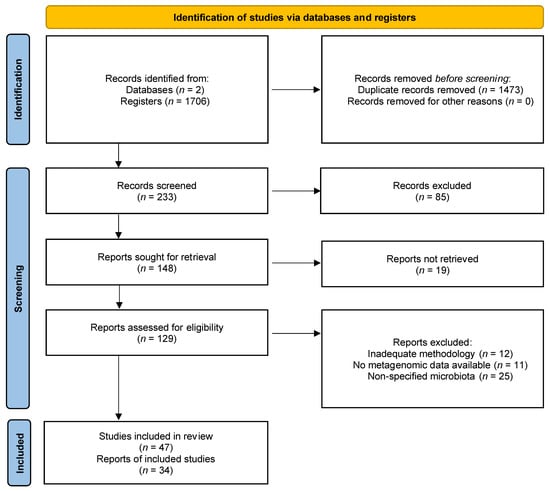
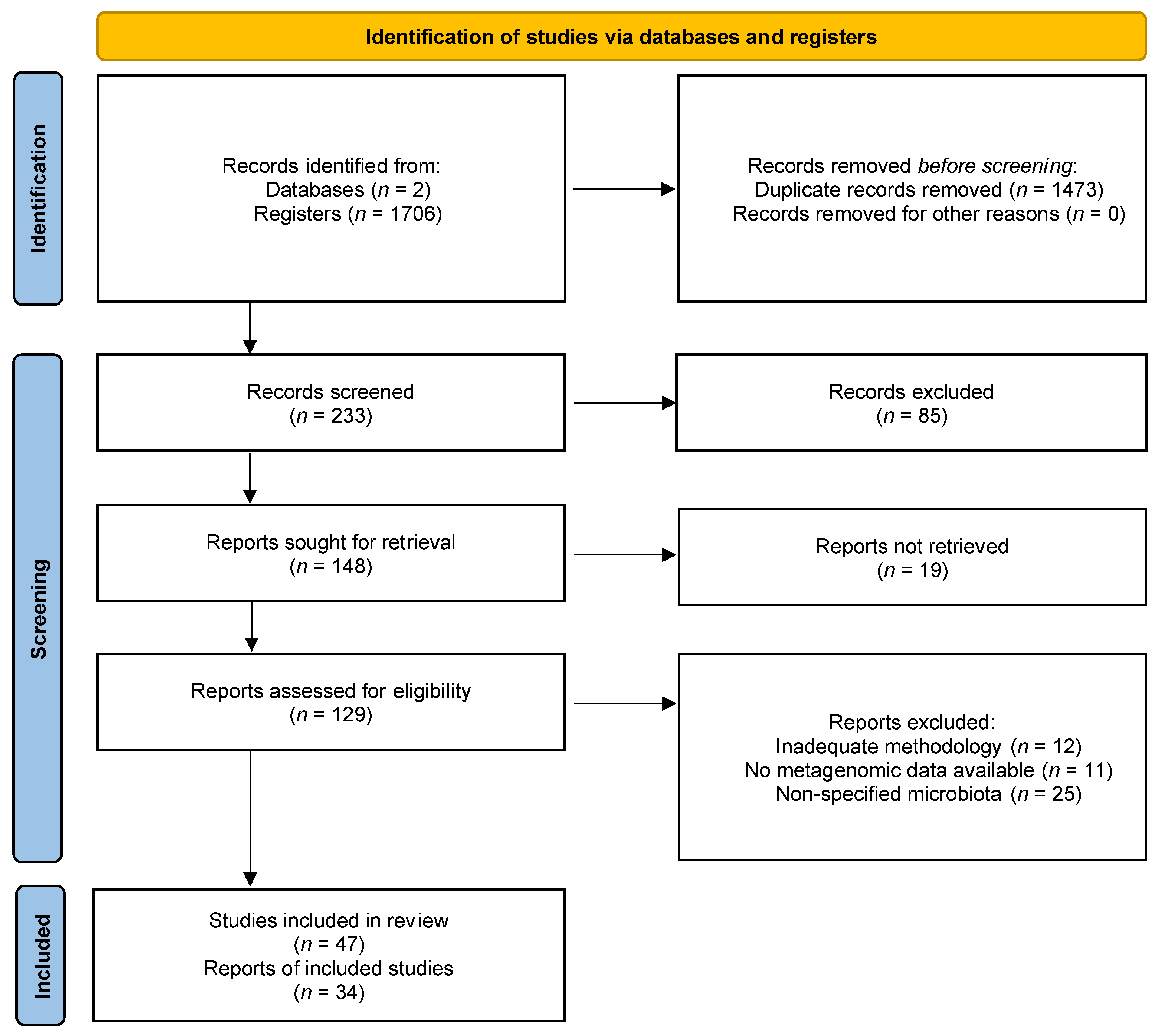
A review of several full-text articles and reviews was performed through two main databases (i.e., PubMed and Google Scholar), from February 2020 to July 2021 on the effects of H. pylori, non-H. pylori bacteria, and environmental factors in the gastric microbiota of patients and healthy subjects. This study identified 233 records after removing duplicates (n = 1473). After screening through titles and abstracts, 148 records were retained and assessed for eligibility.
2.3. Selection of Studies
Past studies that were previously identified in the literature were explored by their title and abstract. Articles and reviews with relevant abstracts were closely examined. The inclusion and exclusion criteria of the studies were determined by the investigators before the revision of the selected literature. The inclusion criteria were as follows: (1) original full-text articles that provided reproducible and sufficient original data about H. pylori colonization and/or infection and the employment of antibiotic treatments for the eradication of the infection; (2) original full-text articles that evaluated the effect of environmental factors, such as antibiotic therapies for bacterial infection eradication on the gastric microbiota of patients with different gastric disorders (including chronic gastritis, intestinal metaplasia, and gastric cancer), and healthy subjects; (3) original full-text articles employing methodologies included culture-dependent methods, pyrosequencing, RT-PCR, and next-generation sequencing technologies; (4) original full-text articles in English, and (5) reviews that involved human gastric metagenomic data, classified by the host health status. The articles that did not fulfil these criteria were excluded from this review.
2.4. Data Extraction
The authors confirmed the characteristics of each selected study, including title, first author’s surname, abstract, study subject conditions, H. pylori status, materials and methods, results, and the data obtained from stomach metagenomic analyses (Figure 1).
Figure 1.
PRISMA flow diagram for the identification and selection of articles on the effects of H. pylori, non-H. pylori bacteria, and multiple environmental factors on the gastric microbiota of patients and healthy subjects [9].
3. Main Findings and Discussion
3.1. Gastrointestinal Microbiota
The evolution of Homo sapiens sapiens has been irrevocably linked to the mutualistic relationships within the gastrointestinal microbiota [10]. Several methodologies have been used to identify the main genera residing in the gastrointestinal microbiota of healthy individuals such as culture-dependent methods; moreover, the employment of recent molecular technologies such as next-generation sequencing technologies has enabled the identification of more than 120 dominant phylotypes [1,11,12].
The human gastrointestinal microbiota contains a vast majority of bacterial species that belong to main phyla such as Bacteroidetes, Firmicutes, Verrucomicrobia, Proteobacteria, Actinobacteria, and Fusobacteria [13]. A study performed by Mailhe et al. included biopsy results obtained by endoscopy and colonoscopy procedures from the stomach (pH 1.8), duodenum (pH 2.5), ileum (pH 6.6), right (pH 6.9), and left (pH 7.1) colon. The study revealed relative abundances in the lower gastrointestinal microbiota than the microbiota present in the stomach and duodenum. Five main genera (Streptococcus, Escherichia, Bacteroides, Lachnoclostridium, and Blautia) and three species (Streptococcus salivarius, Escherichia coli, and Bacteroides uniformis) were identified and found colonizing different parts of the gastrointestinal tract [14]. Microbial composition and its abundance can significantly vary with diet and exposure to different compounds (e.g., drugs and pH), for instance, the acidic environment in the stomach (pH = 2–4) [15].
3.2. Gastric Microbiota
Microenvironmental characteristics such as pH, mucus density, bile, and peristaltic movements make the stomach a complex organ. This organ presents anatomical and histological characteristics, which are suitable for a specific and different microbial composition as compared to the other segments of the gastrointestinal tract [6]. The commensal and pathogenic bacterial growth is usually controlled by the prevailing conditions in the gastric region, due to its unique and heterogeneous habitat that harbors a specific number of microorganisms [6]. Predominant phyla in the stomach mainly include Proteobacteria, Firmicutes, Actinobacteria, Fusobacteria, and Bacteroidetes. Among these, the most abundant class is Bacilli (order Lactobacillales), the most prevailing family is Streptococcaceae (genus Streptococcus), the most prevailing genera are Prevotella, Rothia, Fusobacterium, and Veillonella (in decreasing order) [16]. Dekaboruah et al. reported that genera Neisseria, Haemophilus, Porphyromonas, and family Pasteurellaceae constituted approximately 70.5% of the total gastric microbiota [17].
The role of the gastric microbiota is mainly associated with metabolic functions, such as degradation of undigested carbohydrates and unabsorbed molecules, for the regulation of gastrointestinal homeostasis via the synthesis of short-chain fatty acids (SCFs) by gastrointestinal epithelial cells; control of the proliferation and differentiation of epithelial cells in addition to the development and maintenance of homeostasis by SCFs; maturation of the immune system through the development and regulation of the immune system and prevention of inappropriate inflammation development; energy balance, vulnerability to diseases (competition by the production of bacteriocins and inhibition of attachment and further invasion of pathogenic bacterial cells), and specific host behaviours [16,17], all of which contribute to the maintenance of a healthy microbiota.
3.3. Dysbiosis Generated in the Gastric Microenvironment by Helicobacter pylori Status Diseases
3.3.1. Dysbiosis
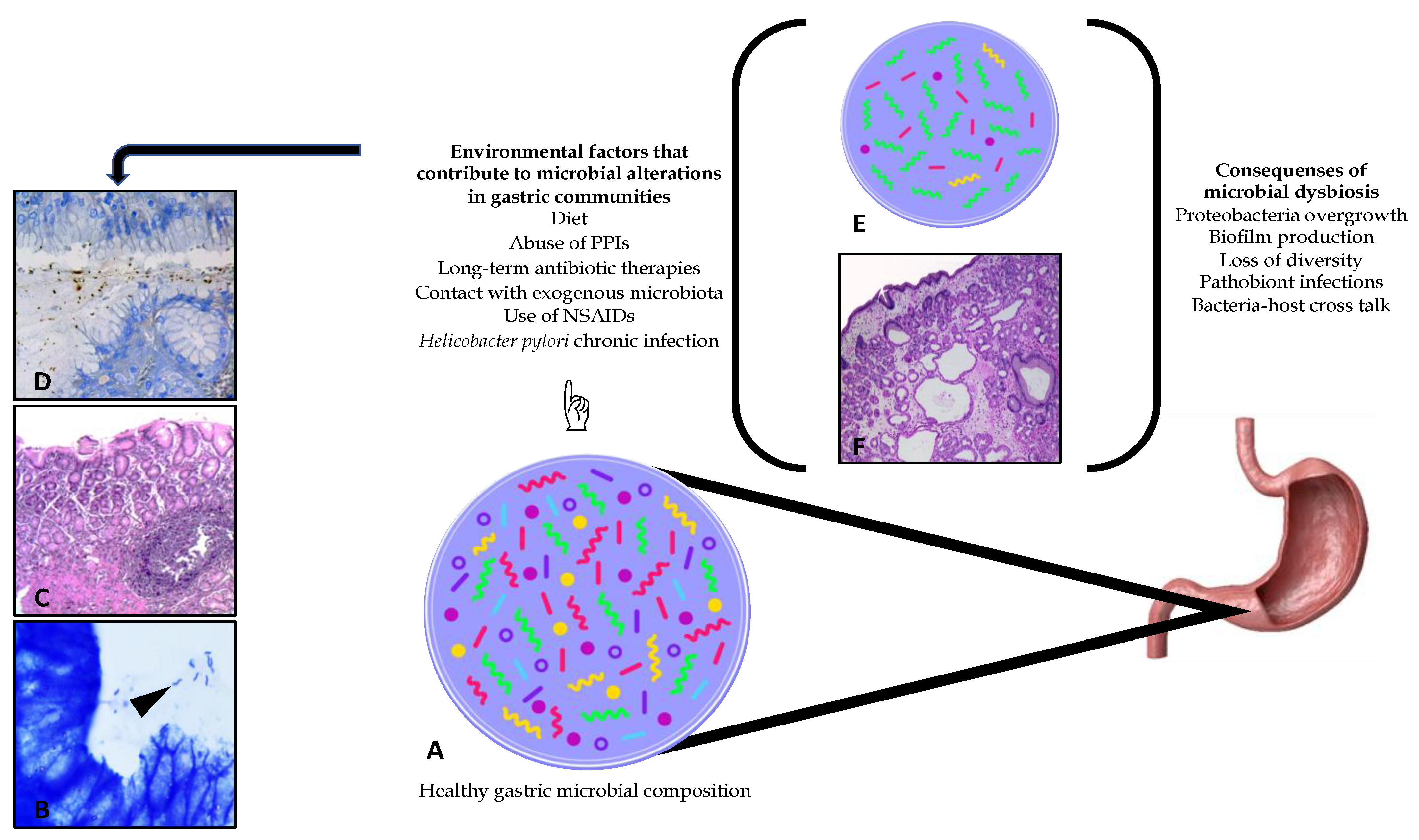
A healthy microbiota is defined as the coexistence of microbial communities, such as pathobionts and commensal bacteria with no harmful consequences. However, a person’s lifestyle and habits may lead to several significant alterations in the native composition and structure of the gastrointestinal microbiota [2]. In addition, the composition of native gastric microbiota is affected by several factors, such as host health status, H. pylori colonization, chronic infection and subsequent complications, the use and abuse of drugs and toxins, age, surgical interventions, inflammation, alcoholism, smoking, use of nonsteroidal anti-inflammatory drugs (NSAIDs), use of proton pump inhibitors (PPIs), and others [18], as shown in Figure 2. These conditions generate dysbiosis, defined as compositional and functional alterations of the microbiota of the host.
Figure 2.
Schematic of the gastric microbial composition and alterations generated by multiple environmental factors. (A) Indicates a schematic of a healthy gastric microbiota. A healthy microbiota is defined as the coexistence of microbial communities, such as pathogenic and symbiotic bacteria with no harmful consequences. Nevertheless, dysbiotic communities can be generated due to several environmental factors, especially by Helicobacter pylori chronic infection, which can be diagnosed through histopathological analysis and differential staining techniques; (B) Giemsa stain, 100×. Arrow highlight H. pylori in biopsy; (C) H&E stain, 40×; (D) anti-Hp stain, 100×. The presence of dysbiotic communities in the gastric microenvironment usually favours the continuous loss of bacterial diversity; (E) increasing infections caused by pathobionts and the growth of antibiotic-resistant gene bacteria. Histological damage can also be observed, caused by environmental factors, that is, the abuse of PPIs (F).
3.3.2. Helicobacter pylori
Since the discovery of Helicobacter pylori in the 20th century, metagenomic studies have focused on elucidating the role of H. pylori in the gastric microbiota of healthy subjects as well as on its role in the development of gastric disorders of infected patients [19,20]. There are some concepts related to the coevolution of H. pylori and its intimate association with humans that has enabled this bacterium to develop survival strategies and persist in the stomach. H. pylori is considered a member of the gastric and oral microbiota, with functions that are not yet fully understood [21,22]. Some studies suggest that the presence of this microbe may be beneficial to some people by reducing the risk of developing asthma during childhood; decreasing the frequency of inflammatory bowel and celiac diseases; interfering with obesity, among other issues [23].
Although it is estimated that 50% of the world’s population is colonized with H. pylori, only 1–2% develop several degrees of histological proinflammatory responses and evident complications of the gastric mucosa. Those responses have been attributed to host–environmental factors, such as continuous changes in the gastric pH, chronicity of the infection, specificity of the strain, genetic susceptibility of the host, hyperglycaemia, smoking, diet, and contact with exogenous microbiota (Figure 2) [24].
The interaction between virulent H. pylori strains, genetic background (cagA and vacA H. pylori genotypes), and host–environmental factors confined to the host, may influence the initiation and development of gastric diseases [22]. However, this bacterium is currently recognized as the main cause of peptic ulcers, chronic gastritis, and gastric adenocarcinoma, which represents 15.4% of cancers induced by infectious agents, and is the second leading cause of worldwide deaths related to cancer [25]. Maldonado-Contreras et al. found that members of the phylum Acidobacteria (class Acidobacteria), Proteobacteria (classes Alphaproteobacteria, Deltaproteobacteria, and Epsilonproteobacteria), and Spirochaetae (class Spirochetes) were correlated with the presence of H. pylori in infected patients [26]. By contrast, relative abundances of families Bradyhizobiaceae, Caulobacteraceae, Lactobacillaceae, and Burkholderiaceae were found to be significantly higher in H. pylori-negative patients when compared to H. pylori-positive patients [27].
3.3.3. Gastric Dysbiosis in Patients with Gastritis and H. pylori Variable Status
During childhood, infection with H. pylori usually begins with simple chronic mononuclear inflammation that progresses into varying degrees of acute neutrophilic inflammation; because of the progression of inflammation, gastritis evolves to atrophic gastritis over the years, which is characterized by the loss of normal mucosa glands in the body and/or antrum. Thus, chronic inflammation is intimately associated with neutrophilic inflammation and cytotoxicity of the infective H. pylori strains [28].
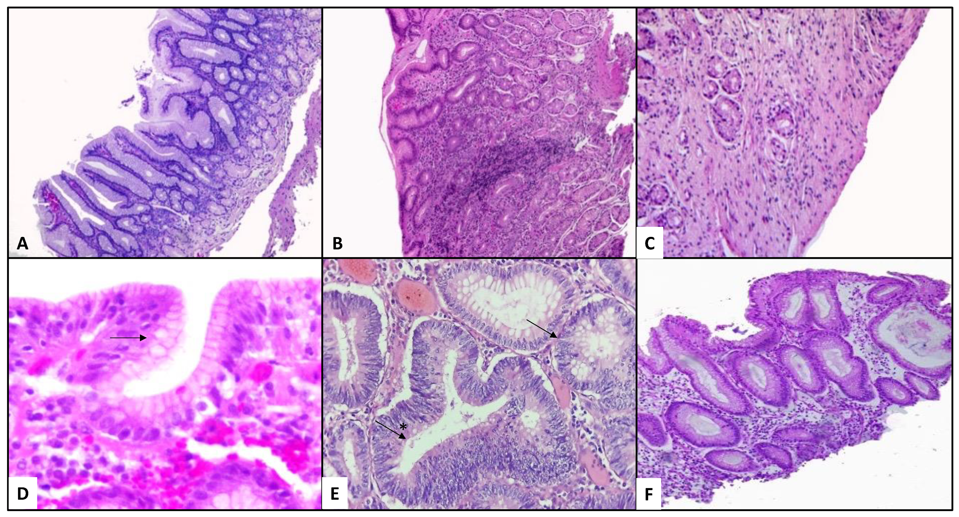
Gastritis is defined as an inflammatory condition of the gastric mucosa that shows variable symptoms in different patients, which depends on the response presented by the host) [29,30]. Most acute gastritis is usually initiated and promoted by H. pylori infection; if not eradicated, progressing into chronic active gastritis, characterized by mononuclear infiltrates and lymphoid follicles with active germinal centres, plasma cells neutrophils, eosinophils, and macrophages in the lamina propria of the gastric mucosa (Figure 3) [31].
Figure 3.
Histopathological alterations generated by Helicobacter pylori chronic infection. (A), near-normal antral mucosa is shown (H&E 10×). (B) illustrates a case of non-atrophic gastritis; increased mononuclear infiltrate with preserved antral architecture can be observed (H&E 10×). Loss of glands and increased connective tissue can be seen in (C), representing a case of atrophic gastritis (H&E 20×). (D) Goblet cells (arrow) are present in the foveolar epithelium which correspond to an initial lesion of intestinal metaplasia (H&E 40×). (E) Hyperchromatic nuclei with loss of its polarity corresponding to dysplasia in a gastric gland, are marked with a black arrow; complete distortion of a gland with cell proliferation and loss of its basal membrane are shown with a (*) marked arrow in a case of gastric adenocarcinoma (H&E 20×). (F) Mucosal distortion of the gastric antrum, with dilated and irregular glands, is observed from a patient treated with proton pump inhibitors (H&E 10×).
A study performed with pediatric patients who were diagnosed with dyspepsia, both, H. pylori-negative and positive patients showed that intestinal microbiota is modified by H. pylori infection. Relative abundances of Bacteroidaceae, Enterobacteriaceae, and Porphyromonadaceae families and genera Bacteroides, Parabacteroides, Streptococcus, and Lactococcus were observed to be significantly increased in H. pylori-positive patients when compared to the microbiota of healthy subjects. H. pylori-positive patients also showed a significantly higher abundance of Bacteroidaceae and Enterobacteriaceae, while the families Bifidobacteriaceae, Lactobacillaceae, and Lachnospiraceae showed a lower relative abundance in both H. pylori-positive and negative patients when compared to healthy subjects [32].
Aviles-Jiménez et al. observed that all patients with non-atrophic gastritis, intestinal metaplasia, and gastric cancer (n = 15) shared phyla Proteobacteria and Firmicutes with a relative abundance of 70%. In this work, gradual microbiota alterations were found, specifically decreasing relative abundances of TM7, Porphyromonas, and Neisseria, while specific genera such as Lactobacillus coleohominis, and Lachnospiraceae gradually increased in the microbiota of patients with non-atrophic gastritis to intestinal metaplasia to gastric cancer, respectively [33].
Chronic infection with H. pylori usually results in increased heartburn, disrupting nutrient availability and the innate local response, in addition to the alterations observed in gastric physiology and immunology associated with H. pylori infection, e.g., the increase in atrophy and metaplasia, particularly in patients with initiation and development of gastric cancer, which have been observed to carry an altered gastric microbiota composition [34].
3.3.4. Dysbiosis in Patients with Gastric Cancer and H. pylori Variable Status
The development of most gastric cancers involves sequential changes of the normal gastric mucosa (Figure 3A). Non-atrophic gastritis is characterized by oedema of the lamina propria, and a variable number of mononuclear cells, mainly lymphocytes, plasma cells, with or without lymph node formation; neutrophils, eosinophils, and macrophage infiltrates may also be present (Figure 3B). Atrophic gastritis (Figure 3C), in which, in addition to the changes previously described, increased distance between glands, loss of glandular units, and increased connective tissue are shown. In intestinal metaplasia (Figure 3D) the main histological change is the presence of goblet cells. The presence of dysplastic changes noted can be seen as hyperchromatic and elongation of nuclei with the loss of their normal polarity, and, finally, carcinoma [35].
Atrophic gastritis and intestinal metaplasia are well-established indicators of increased risk to develop gastric cancer [36,37,38,39,40]. The entire process is believed to be initiated and promoted by H. pylori [41], although it has been recently proposed that diverse microbial alterations interact in the diverse stages of gastric carcinogenesis; therefore, as we can see, dysbiosis in the stomach is considered to be a dynamic process intimately correlated with the progression to gastric cancer [42].
Castaño-Rodríguez et al. compared the gastric microbial composition of patients who were serologically H. pylori-positive, along with dyspepsia and gastric cancer. Identified enriched species in patients diagnosed with gastric cancer included Lactococcus lactis, Fusobacterium mortiferum, Haemophilus parahaemolyticus, H. sputorum, Staphylococcus spp., and Methylobacterium adhaesivum, while in non-nested analysis, authors identified Veillonella atypica, V. dispar, Dialister pneumosintes, and Leptotrichia buccalis. Some of these microbial communities, such as Veillonella and Leptotrichia, have been demonstrated to co-aggregate with other intestinal bacteria to produce biofilms that play a key role in other types of colorectal adenocarcinoma [43].
A study performed by Liu et al. on 276 patients with gastric cancer, assessed the diversity and composition of the gastric microbiota across three microenvironments: tumoral and peritumoral microhabitats and compared them with subjects having normal gastric mucosal morphology (healthy microenvironment). They observed that H. pylori, Streptococcus anginosus, Prevotella melaninogenica, P. copri, Cutibacterium acnes (formerly known as Propionibacterium acnes), Bacteroides fragilis, B. uniformis, Bacillus cereus, and Akkermansia muciniphila were significantly different across the three microenvironments. Interestingly, C. acnes, S. anginosus, and P. melaninogenica were abundantly present in tumoral microhabitats, while the presence of B. uniformis and P. copri decreased significantly; meanwhile, A. muciniphila and B. fragilis demonstrated similar bacterial alteration patterns when analysing the variation between peritumoral and tumoral microhabitats [44].
It has been recently hypothesized that bacterial species with nitrate-reducing metabolism contribute to gastric malignant transformation by crescent intragastric concentrations of nitrite and N-nitroso compounds. Ferreira et al. fully reconstituted the gastric metagenomes from patients with gastric carcinoma revealing that the functional composition of total microbiota from these patients increased the nitrate reductase functions, promoting the reduction of nitrate to nitrite, and nitrite reductase functions, promoting the reduction of nitrite to nitric oxide when compared to the functional composition of chronic gastritis patient’s microbiota. These findings provide evidence that microbial communities with genotoxic potential are present in patients with gastric carcinoma [42].
A case-control study performed by Gunathilake et al. which involved 556 participants, showed high C. acnes, H. pylori, and P. copri relative abundances in patients with gastric cancer when compared with the controls, whereas the relative abundance of L. lactis was significantly higher. Patients with these significantly higher relative abundances showed a higher risk of developing gastric cancer. Although H. pylori infection was observed to be the strongest single risk factor for gastric cancer, researchers also observed high C. acnes relative abundance in these patients, revealing that this bacterium increases the risk for the development of gastric cancer, highlighting a commensal skin bacterium recently identified as a gastric microbiota resident [45].
Eradication of H. pylori with multiple antibiotic therapies combined with PPIs was observed to reduce the risk of gastric cancer; an effect that may be limited to gastric cancer patients without atrophy and metaplasia [46,47,48]. In patients with malignant progression of gastric cancer, H. pylori eradication was observed to reduce symptomatology, but did not stop the progression of the neoplasia, indicating that cancer can occur even ten years after H. pylori eradication treatment [49].
3.3.5. Alterations in the Gastric Microbiota after Multiple Therapeutic Interventions
Self-medication is considered a potential contributor to human pathogen resistance to antibiotics. However, adverse consequences of such practices are difficult to evaluate due to affected bacteria by some aggressive drugs (e.g., multiple antibiotic therapies, proton pump inhibitors) are just not pathogens, but also gastrointestinal commensal bacteria, altering the native microbiota.
3.3.6. Abuse of Proton Pump Inhibitors
The use of PPIs is common in patients diagnosed with gastroesophageal reflux and functional dyspepsia. PPIs are defined as prodrugs that are activated by acid and lower pH of the local microenvironment. PPIs are known as weak bases (due to their pKa ≤ 4). These compounds are rapidly activated by the acids previously secreted by gastric parietal cells. High acidity acts as a protonation factor for activation of these weak bases to form disulphides (thiophilic drugs) by binding to cysteine residues residing in the (H+/K+ ATPase) proton pumps. Disulphides react with luminally accessed cysteines on the acid pump enzyme, blocking acid transport and altering gastric acidity to pH = 7 [50].
The microenvironmental changes generated by PPIs alter the native gastric conditions significantly and develop histopathological damage including the enlargement of fundic glands (Figure 3E,F) and modifying the commensal microbiota composition. Parsons et al. compared the microbiota profiles in different patient groups treated with PPIs: patients diagnosed with autoimmune atrophic gastritis, patients with H. pylori-induced gastritis and atrophy, and healthy controls. Patients who received PPIs showed similar bacterial profiles to those found in the stomachs of healthy subjects, despite high levels of serum gastrin concentrations. Alpha indexes showed samples from patients treated with PPIs were less diverse than normal gastric samples. Evaluation of the evenness index showed that bacterial communities from PPI-treated samples and normal gastric samples were both equal in relative abundance. Richness calculations demonstrated that normal gastric samples showed the highest diversity when compared to all the other groups, while the H. pylori-infected groups (both, H. pylori-induced atrophy and gastritis) presented with a significantly fewer number of species. In PPI-treated patients, Streptococcaceae (17%) was the most abundant family, outranking Prevotellaceae (11%), Campylobacteraceae (5%), and Leptotrichiaceae (4%). At the operational taxonomic unit (OTU) level, Cyanobacteria and Streptococcus relative abundances were significantly increased in PPI-treated patients; however, the relative abundances of genera Actinobacillus, Porphyromonas, Leptotrichiaceae, Prevotella, Treponema, Haemophilus, Fusobacterium, and Tannerella were significantly decreased in patients treated with PPIs [51].
In a second study performed on 12 patients who presented dyspepsia and were treated with a PPI, the phyla Proteobacteria, Actinobacteria, Fusobacteria, Bacteroidetes, and Firmicutes represented the five most abundant phyla in 98% of total sequence reads. Among them, Proteobacteria, Bacteroidetes, and Firmicutes were dominantly abundant; 115 genera were identified in total, including the genera Helicobacter, Prevotella, Streptococcus, Veillonella, Neisseria, Porphyromonas, Fusobacterium, Gemella, Haemophilus, and Leptotrichia, representing the ten most abundant genera (in decreasing order, respectively) [52].
3.3.7. Use of Antibiotics
Successful bacterial eradication treatments significantly affect the gastrointestinal microbiota [23]. Increased improvements in sanitization, hygienical measures, and the extensive use and abuse of antibiotics are considered the most important factors responsible for the reduction in infectious disease transmission, making it important in the induction of significant alterations in the native microbiota composition [2,53]. The consequences of antibacterial therapies are difficult to evaluate since the affected bacteria not only includes pathogens but also beneficial bacteria useful for maintaining human health [2].
Dysbiosis is the most important consequence of adverse effects of antibiotic therapies, which has favored the presence of microorganisms carrying antibiotic resistance genes, facilitating horizontal gene transfer between commensal and pathogen bacteria, as well as assisting in the recolonization of gastrointestinal mucosa by pathobionts [54]. Antibiotic properties, such as spectrum, dose and duration of administration, pharmacokinetics and pharmacodynamics, and the route of administration contribute to modifying the human microbiota [55].
3.3.8. H. pylori Eradication Treatments
H. pylori infection induces a mild-to-severe inflammatory process; however, the gastric mucosa of patients with different gastric alterations can harbor non-H. pylori bacteria, which can induce and potentialize inflammatory processes that can lead to a malignant transformation in the stomach, e.g., genera Acinetobacter and Klebsiella, associated with gastric carcinogenic processes [56,57]; genus Cutibacterium, which is a dominant genus in pilosebaceous follicles and a native member of mucosa and skin microbiota, was recently named a pathobiont related to the microbiota of immunocompromised patients and considered as a trigger of corpus-dominant lymphocytic gastritis [58,59,60]; and genus Corynebacterium, as a member of human native microbiota, is frequently isolated from the skin, mucosal membranes, and gastrointestinal tract [61,62,63].
For more than 30 years, it has been observed that multiple antibiotic therapies, which usually include metronidazole, mainly affect anaerobic and microaerophilic microorganisms. The metronidazole antimicrobial spectrum activity includes anaerobic bacteria, such as Peptococcus, Eubacterium, Clostridium difficile, C. perfringens, Veillonella, Bacteroides fragilis, Fusobacterium, and Peptostreptococcus [64]. However, when administering multiple antibiotic combination therapies with a PPI, dysbiotic states are generated in the whole gastric microbiota. In Table 1, the principal alterations to the gastric microbiota after the eradication of H. pylori infection with several therapies (i.e., standard triple therapy or quadruple therapy) in patients with distinct diseases are shown. As can be seen, not only microaerophilic or anaerobic bacteria are affected by these therapies, but also facultative and aerobic bacteria, although in different relative abundances and richness parameters. This observation may be attributed to the interindividual variability of the study subjects, which usually makes it difficult to identify the distinctive pathogenic species among patients at risk of developing gastric diseases from those who are not at risk [20,65]. Li et al. reported the effect of H. pylori eradication on the gastrointestinal microbiota of patients with duodenal ulcers. Before the eradication treatment, the Clostridium leptum and Prevotella subgroups were aberrantly different between gastric corpus and antrum. Pre-treatment peptic ulcer patients who were administered with antibiotic therapies (i.e., rabeprazole, colloidal bismuth, amoxicillin, and clarithromycin) for H. pylori eradication demonstrated that the relative abundances of the Lactobacillus group, Enterobacteria, and C. leptum subgroup were significantly increased in antrum, while for the Clostridium coccoides subgroup relative abundances were lower. After treatment, recolonization with Lactobacillus was more abundant in antrum than in the corpus. H. pylori-negative patients with antral gastritis showed higher Enterobacteria relative abundances, suggesting that H. pylori inhibited the distribution of these bacterial families [66].
Table 1.
Dominant native microbiota and alterations in gastric microbiota after Helicobacter pylori (HP) eradication.
H. pylori eradication can result in resilience, which is defined as the microbial composition recovery whether by growth, physiological or genetic adaptation, after composition alterations due to the host, or environmental factors of the gastric microbiota. Gastric resilience can occur with specific bacterial species such as Granulicatella, Streptococcus, Rothia, Leptotrichia, Acinetobacter, Faecalibacterium, Rahnella, Kaistobacter, Blautia, Caulobacter, Nocardioides, and Brevundimonas. However, recolonization and the increase in relative abundances with these bacterial species can be associated with the emergence of atrophy or intestinal metaplasia (IM) following one year of H. pylori eradication (e.g., Granulicatella, Actinomyces, Rothia, Peptostreptococcus, Abiotrophia, and Parvinomas), or the presence of persistent inflammation processes (e.g., Acinetobacter lwolffii, Streptococcus anginosus, Ralstonia, Erwinia, and Prevotella) [71].
3.3.9. Microbiota Recovery after Antibiotic Eradication Treatment
Antibiotic therapies (monotherapies or multiple antibiotic combinations) are one of the most extreme disturbances occurring in the human microbiome [72]. Therefore, increased focus is being given to evaluate how the gastrointestinal microbiome is impacted by antibiotic use, both by acute and long-term treatments [55], rendering individuals more susceptible to infections [21].
The search for new alternatives in antimicrobial therapies for pathogen eradication is much needed, with a special interest in natural product-based therapies [73], such as faecal microbiota transplantation (FMT), and supplementation of probiotics.
Defined as the transfer of stool from a healthy donor into the gastrointestinal tract of a patient to restore the composition of the microbiota [74], faecal microbiota transplantation (FMT) represents a promising paradigm for treating conditions where the microbiome and organ dysbiosis contributes to pathophysiology [75]. Although FTM is almost recognized as a safe therapeutic protocol, adverse effects can be developed due to short post-FTM follow-ups [74]. FTM can lead to the induction of several diseases, such as obesity, IBD, autism, asthma, among others. On the other hand, specific bacterial overgrowth of some species (e.g., Enterococcus faecalis, Bacteroides fragilis, and Escherichia coli) may increase the risk of developing cancer. Rare cases have been documented, where FTM-associated bacteria can cause serious problems, or even death [74], suggesting the employment of alternative therapies for optimal recolonization, such as probiotics.
3.3.10. Probiotics
Probiotic supplementation therapies are emerging therapies for H. pylori treatment [76]. Probiotics are defined as live bacteria that usually coexist in symbiosis within the human host. They are believed to improve human health when consumed or applied [73]. Lactic acid bacteria (LAB) have been usually employed as probiotics. Lactobacilli, bifidobacteria, and other LABs are isolated from fermented dairy products and the faecal microbiota. When administered, they usually interact with both the host and established microbiome through molecular effectors which are present on the bacterial cell structure or secreted as metabolic products, such as butyrate compounds, secreted proteins, such as extracellular proteins, organic acids, indole, bacteriocins, H2O2, and NO molecules [67,77]. These secreted products generate a reaction in the microbiota by cross-feeding interactions, alterations in the microenvironment, competition for nutrients and specific binding sites, and growth inhibition of other microbes by the production of bacteriocins. The effects of probiotics to mediate health benefits on the microbiota contribute to the inhibition of pathogen overgrowth states. The direct interaction between probiotics and receptors in the gastrointestinal epithelium produces important effects such as the enhancement of intestinal barrier integrity and prevention of inflammation, as well as systemic effects via host, immune, endocrine, and nervous system mediators [77].
In vitro experiments have demonstrated that several probiotics have the potential to antagonize H. pylori through metabolite production, secretion, or bacterial cells. Several studies have determined the capability of four strains of Lactobacillus for inhibiting H. pylori growth [78], urease activity suppression [79], drug-sensitivity, and drug-resistance through the secretion of lactic acid by L. pentosus LPS16 [80]; bacteriocin production and secretion can generate oxidative damage to membrane lipids, pathogenic proteins, and DNA by the formation of peroxide ions [81], such as those secreted by L. bulgaricus, that can inhibit not only antibiotic-sensitive H. pylori strains but also inhibit antibiotic-resistant strains [82]. Moreover, the effect of drug synergy, mutant prevention, biofilm, and gastrointestinal tract microbiota on H. pylori growth inhibition for maintenance of both gastric and intestinal microbiota has also been demonstrated [76].
The recent development of low-cost whole-genome sequencing has allowed the characterization of new bacteria with high potential for health benefits, with the opportunity to be developed as probiotics [77]. Although beneficial effects and mechanisms of probiotics in the gastrointestinal tract have been demonstrated [21], there is evidence that probiotics may also act in the treatment and prevention of infectious diseases. Recently, bacterial candidates have been isolated from the human gut that showcased probiotic potential (e.g., Roseburia intestinalis, Faecalibacterium prausnitzii, Eubacterium spp., Bacteroides spp., Akkermansia municiphila), thus expanding the possibilities of research on probiotics and their relationship with emerging healthcare challenges [77].
4. Conclusions
During the development of gastric diseases and their therapeutic treatments, the native microbial composition of the patient can be altered, generating dysbiotic states. The presence of Helicobacter pylori usually induces dysbiosis in the stomach, in some cases defining serious diseases, e.g., gastric cancer. Its eradication with different therapies causes significant alterations that might not always carry optimal colonization, e.g., metabolic and immunological implications, or even the initiation and development of gastric disorders caused by non-H. pylori bacteria. The use of alternative antibiotic therapies, such as probiotics, can improve, in most cases, the process of bacterial resilience.
Author Contributions
Conceptualization, U.G.-R. and N.V.-G.; investigation, P.A.-R., S.M.-E. and J.C.V.G.; methodology, U.G.-R. and V.L.J.-R.; resources, A.C.-R. and G.Z.; writing—original draft preparation, U.G.-R., P.V.-M., J.R.M.-E.; writing—review and editing, U.G.-R., N.V.-G., P.V.-M., F.S.S. and M.S.-G.; funding acquisition, N.V.-G., M.S.-G. and F.S.S.; supervision, N.V.-G. All authors have read and agreed to the published version of the manuscript.
Funding
This project was supported by Federal Resources (HIM/2017/010. SSA 1305) from SSA, Mexico.
Institutional Review Board Statement
Not applicable.
Informed Consent Statement
Not applicable.
Data Availability Statement
The data presented in this study are openly available.
Acknowledgments
We thank the Posgrado en Ciencias Quimicobiológicas; Escuela Nacional de Ciencias Biológicas; Instituto Politécnico Nacional, Mexico City, Mexico. This work was part of the MD dissertation of U.G.-R., who is a CONACyT (814136).
Conflicts of Interest
The authors declare no conflict of interest.
References
- Lederberg, J.; McCray, A.T. ‘Ome Sweet’ Omics—A Genealogical Treasury of Words. Sci. 2001, 15, 8. [Google Scholar]
- Beirão, E.M.; Padovan, A.C.B.; Furtado, J.J.D.; Colombo, A.L.; Medeiros, E.A.S. Does the Change on Gastrointestinal Tract Microbiome Affects Host? Braz. J. Infect. Dis. 2014, 18, 660–663. [Google Scholar] [CrossRef] [Scilit] [PubMed]
- Buelow, E.; Bello González, T.D.J.; Fuentes, S.; de Steenhuijsen Piters, W.A.A.; Lahti, L.; Bayjanov, J.R.; Majoor, E.A.M.; Braat, J.C.; van Mourik, M.S.M.; Oostdijk, E.A.N.; et al. Comparative Gut Microbiota and Resistome Profiling of Intensive Care Patients Receiving Selective Digestive Tract Decontamination and Healthy Subjects. Microbiome 2017, 5, 88. [Google Scholar] [CrossRef] [Scilit] [PubMed]
- Young, V.B.; Schmidt, T.M. Overview of the Gastrointestinal Microbiota. Adv. Exp. Med. Biol. 2008, 635, 29–40. [Google Scholar] [CrossRef] [Scilit] [PubMed]
- Klymiuk, I.; Bilgilier, C.; Stadlmann, A.; Thannesberger, J.; Kastner, M.-T.; Högenauer, C.; Püspök, A.; Biowski-Frotz, S.; Schrutka-Kölbl, C.; Thallinger, G.G.; et al. The Human Gastric Microbiome Is Predicated upon Infection with Helicobacter pylori. Front. Microbiol. 2017, 8, 2508. [Google Scholar] [CrossRef] [Scilit] [PubMed]
- Hillman, E.T.; Lu, H.; Yao, T.; Nakatsu, C.H. Microbial Ecology along the Gastrointestinal Tract. Microbes Environ. 2017, 32, 300–313. [Google Scholar] [CrossRef] [Scilit]
- Hung, I.F.N.; Wong, B.C.Y. Assessing the Risks and Benefits of Treating Helicobacter pylori Infection. Ther. Adv. Gastroenterol. 2009, 2, 141–147. [Google Scholar] [CrossRef] [Scilit]
- Reshetnyak, V.I.; Burmistrov, A.I.; Maev, I.V. Helicobacter pylori: Commensal, Symbiont or Pathogen? World J. Gastroenterol. 2021, 27, 545–560. [Google Scholar] [CrossRef] [Scilit] [PubMed]
- Haddaway, N.R.; McGuinness, L.A.; Pritchard, C.C. PRISMA2020: R Package and ShinyApp for Producing PRISMA 2020 Compliant Flow Diagrams (Version 0.0.2); Zenodo: Geneve, Switzerland, 2021. [Google Scholar] [CrossRef]
- Belizário, J.E.; Napolitano, M. Human Microbiomes and Their Roles in Dysbiosis, Common Diseases, and Novel Therapeutic Approaches. Front. Microbiol. 2015, 6, 1050. [Google Scholar] [CrossRef]
- Bik, E.M.; Eckburg, P.B.; Gill, S.R.; Nelson, K.E.; Purdom, E.A.; Francois, F.; Perez-Perez, G.; Blaser, M.J.; Relman, D.A. Molecular Analysis of the Bacterial Microbiota in the Human Stomach. Proc. Natl. Acad. Sci. USA 2006, 103, 732–737. [Google Scholar] [CrossRef] [Scilit] [PubMed]
- Li, X.-X.; Wong, G.L.-H.; To, K.-F.; Wong, V.W.-S.; Lai, L.H.; Chow, D.K.-L.; Lau, J.Y.-W.; Sung, J.J.-Y.; Ding, C. Bacterial Microbiota Profiling in Gastritis without Helicobacter pylori Infection or Non-Steroidal Anti-Inflammatory Drug Use. PLoS ONE 2009, 4, e7985. [Google Scholar] [CrossRef] [Scilit]
- Rajilić-Stojanović, M.; de Vos, W.M. The First 1000 Cultured Species of the Human Gastrointestinal Microbiota. FEMS Microbiol. Rev. 2014, 38, 996–1047. [Google Scholar] [CrossRef] [Scilit]
- Mailhe, M.; Ricaboni, D.; Vitton, V.; Gonzalez, J.-M.; Bachar, D.; Dubourg, G.; Cadoret, F.; Robert, C.; Delerce, J.; Levasseur, A.; et al. Repertoire of the Gut Microbiota from Stomach to Colon Using Culturomics and Next-Generation Sequencing. BMC Microbiol. 2018, 18, 157. [Google Scholar] [CrossRef] [Scilit]
- Petra, C.V.; Rus, A.; Dumitraşcu, D.L. Gastric Microbiota: Tracing the Culprit. Clujul Med. 2017, 90, 369–376. [Google Scholar] [CrossRef] [Scilit]
- Stearns, J.C.; Lynch, M.D.J.; Senadheera, D.B.; Tenenbaum, H.C.; Goldberg, M.B.; Cvitkovitch, D.G.; Croitoru, K.; Moreno-Hagelsieb, G.; Neufeld, J.D. Bacterial Biogeography of the Human Digestive Tract. Sci. Rep. 2011, 1, 170. [Google Scholar] [CrossRef] [Scilit] [PubMed]
- Dekaboruah, E.; Suryavanshi, M.V.; Chettri, D.; Verma, A.K. Human Microbiome: An Academic Update on Human Body Site Specific Surveillance and Its Possible Role. Arch. Microbiol. 2020, 202, 2147–2167. [Google Scholar] [CrossRef] [Scilit] [PubMed]
- Rajilic-Stojanovic, M.; Figueiredo, C.; Smet, A.; Hansen, R.; Kupcinskas, J.; Rokkas, T.; Andersen, L.; Machado, J.C.; Ianiro, G.; Gasbarrini, A.; et al. Systematic Review: Gastric Microbiota in Health and Disease. Aliment. Pharmacol. Ther. 2020, 51, 582–602. [Google Scholar] [CrossRef] [Scilit]
- Noto, J.M.; Peek, R.M.J. The Gastric Microbiome, Its Interaction with Helicobacter pylori, and Its Potential Role in the Progression to Stomach Cancer. PLoS Pathog. 2017, 13, e1006573. [Google Scholar] [CrossRef] [Scilit] [PubMed]
- Yang, J.-C.; Lu, C.-W.; Lin, C.-J. Treatment of Helicobacter pylori Infection: Current Status and Future Concepts. World J. Gastroenterol. 2014, 20, 5283–5293. [Google Scholar] [CrossRef] [Scilit]
- Linz, B.; Balloux, F.; Moodley, Y.; Manica, A.; Liu, H.; Roumagnac, P.; Falush, D.; Stamer, C.; Prugnolle, F.; van der Merwe, S.W.; et al. An African Origin for the Intimate Association between Humans and Helicobacter pylori. Nature 2007, 445, 915–918. [Google Scholar] [CrossRef] [Scilit] [PubMed]
- Moodley, Y.; Linz, B.; Bond, R.P.; Nieuwoudt, M.; Soodyall, H.; Schlebusch, C.M.; Bernhöft, S.; Hale, J.; Suerbaum, S.; Mugisha, L.; et al. Age of the Association between Helicobacter pylori and Man. PLoS Pathog. 2012, 8, e1002693. [Google Scholar] [CrossRef] [Scilit]
- Chey, W.D.; Leontiadis, G.I.; Howden, C.W.; Moss, S.F. ACG Clinical Guideline: Treatment of Helicobacter pylori Infection. Am. J. Gastroenterol. 2017, 112, 212–239. [Google Scholar] [CrossRef] [Scilit] [PubMed]
- Wroblewski, L.E.; Peek, R.M., Jr.; Wilson, K.T. Helicobacter pylori and Gastric Cancer: Factors That Modulate Disease Risk. Clin. Microbiol. Rev. 2010, 23, 713–739. [Google Scholar] [CrossRef] [Scilit]
- Atherton, J.C.; Blaser, M.J. Coadaptation of Helicobacter pylori and Humans: Ancient History, Modern Implications. J. Clin. Investig. 2009, 119, 2475–2487. [Google Scholar] [CrossRef] [Scilit] [PubMed]
- Maldonado-Contreras, A.; Goldfarb, K.C.; Godoy-Vitorino, F.; Karaoz, U.; Contreras, M.; Blaser, M.J.; Brodie, E.L.; Dominguez-Bello, M.G. Structure of the Human Gastric Bacterial Community in Relation to Helicobacter pylori Status. ISME J. 2011, 5, 574–579. [Google Scholar] [CrossRef] [Scilit] [PubMed]
- Eun, C.S.; Kim, B.K.; Han, D.S.; Kim, S.Y.; Kim, K.M.; Choi, B.Y.; Song, K.S.; Kim, Y.S.; Kim, J.F. Differences in Gastric Mucosal Microbiota Profiling in Patients with Chronic Gastritis, Intestinal Metaplasia, and Gastric Cancer Using Pyrosequencing Methods. Helicobacter 2014, 19, 407–416. [Google Scholar] [CrossRef] [Scilit] [PubMed]
- Sipponen, P.; Maaroos, H.-I. Chronic Gastritis. Scand. J. Gastroenterol. 2015, 50, 657–667. [Google Scholar] [CrossRef] [Scilit] [PubMed]
- Strickland, R.G. Gastritis. Front. Gastrointest. Res. 1975, 1, 12–48. [Google Scholar] [CrossRef] [Scilit]
- Dixon, M.F. Pathology of Gastritis and Peptic Ulceration. In Helicobacter Pylori: Physiology and Genetics; Mobley, H.L., Mendz, G.L., Hazell, S.L., Eds.; ASM Press: Washington, DC, USA, 2001. [Google Scholar]
- Watari, J.; Chen, N.; Amenta, P.S.; Fukui, H.; Oshima, T.; Tomita, T.; Miwa, H.; Lim, K.-J.; Das, K.M. Helicobacter pylori Associated Chronic Gastritis, Clinical Syndromes, Precancerous Lesions, and Pathogenesis of Gastric Cancer Development. World J. Gastroenterol. 2014, 20, 5461–5473. [Google Scholar] [CrossRef] [Scilit]
- Yang, L.; Zhang, J.; Xu, J.; Wei, X.; Yang, J.; Liu, Y.; Li, H.; Zhao, C.; Wang, Y.; Zhang, L.; et al. Helicobacter pylori Infection Aggravates Dysbiosis of Gut Microbiome in Children with Gastritis. Front. Cell Infect. Microbiol. 2019, 9, 375. [Google Scholar] [CrossRef] [Scilit] [PubMed]
- Aviles-Jimenez, F.; Vazquez-Jimenez, F.; Medrano-Guzman, R.; Mantilla, A.; Torres, J. Stomach Microbiota Composition Varies between Patients with Non-Atrophic Gastritis and Patients with Intestinal Type of Gastric Cancer. Sci. Rep. 2014, 4, 4202. [Google Scholar] [CrossRef] [Scilit] [PubMed]
- Cao, L.; Yu, J. Effect of Helicobacter pylori Infection on the Composition of Gastric Microbiota in the Development of Gastric Cancer. Gastrointest. Tumors 2015, 2, 14–25. [Google Scholar] [CrossRef] [Scilit] [PubMed]
- Sugano, K.; Tack, J.; Kuipers, E.J.; Graham, D.Y.; El-Omar, E.M.; Miura, S.; Haruma, K.; Asaka, M.; Uemura, N.; Malfertheiner, P. Kyoto Global Consensus Report on Helicobacter pylori Gastritis. Gut 2015, 64, 1353–1367. [Google Scholar] [CrossRef] [Scilit] [PubMed]
- Meining, A.; Stolte, M.; Hatz, R.; Lehn, N.; Miehlke, S.; Morgner, A.; Bayerdörffer, E. Differing Degree and Distribution of Gastritis in Helicobacter pylori-Associated Diseases. Virchows Arch. 1997, 431, 11–15. [Google Scholar] [CrossRef] [Scilit]
- Watabe, H.; Mitsushima, T.; Yamaji, Y.; Okamoto, M.; Wada, R.; Kokubo, T.; Doi, H.; Yoshida, H.; Kawabe, T.; Omata, M. Predicting the Development of Gastric Cancer from Combining Helicobacter pylori Antibodies and Serum Pepsinogen Status: A Prospective Endoscopic Cohort Study. Gut 2005, 54, 764–768. [Google Scholar] [CrossRef] [Scilit]
- Naylor, G.M.; Gotoda, T.; Dixon, M.; Shimoda, T.; Gatta, L.; Owen, R.; Tompkins, D.; Axon, A. Why Does Japan Have a High Incidence of Gastric Cancer? Comparison of Gastritis between UK and Japanese Patients. Gut 2006, 55, 1545–1552. [Google Scholar] [CrossRef] [Scilit]
- Rugge, M.; de Boni, M.; Pennelli, G.; de Bona, M.; Giacomelli, L.; Fassan, M.; Basso, D.; Plebani, M.; Graham, D.Y. Gastritis OLGA-Staging and Gastric Cancer Risk: A Twelve-Year Clinico-Pathological Follow-up Study. Aliment. Pharmacol. Ther. 2010, 31, 1104–1111. [Google Scholar] [CrossRef] [Scilit]
- den Hoed, C.M.; Holster, I.L.; Capelle, L.G.; de Vries, A.C.; den Hartog, B.; Ter Borg, F.; Biermann, K.; Kuipers, E.J. Follow-up of Premalignant Lesions in Patients at Risk for Progression to Gastric Cancer. Endoscopy 2013, 45, 249–256. [Google Scholar] [CrossRef] [Scilit]
- Ahn, H.J.; Lee, D.S. Helicobacter pylori in Gastric Carcinogenesis. World J. Gastrointest. Oncol. 2015, 7, 455–465. [Google Scholar] [CrossRef] [Scilit]
- Ferreira, R.M.; Pereira-Marques, J.; Pinto-Ribeiro, I.; Costa, J.L.; Carneiro, F.; Machado, J.C.; Figueiredo, C. Gastric Microbial Community Profiling Reveals a Dysbiotic Cancer-Associated Microbiota. Gut 2018, 67, 226–236. [Google Scholar] [CrossRef] [Scilit]
- Castaño-Rodríguez, N.; Goh, K.-L.; Fock, K.M.; Mitchell, H.M.; Kaakoush, N.O. Dysbiosis of the Microbiome in Gastric Carcinogenesis. Sci. Rep. 2017, 7, 15957. [Google Scholar] [CrossRef] [Scilit]
- Liu, X.; Shao, L.; Liu, X.; Ji, F.; Mei, Y.; Cheng, Y.; Liu, F.; Yan, C.; Li, L.; Ling, Z. Alterations of Gastric Mucosal Microbiota across Different Stomach Microhabitats in a Cohort of 276 Patients with Gastric Cancer. EBioMedicine 2019, 40, 336–348. [Google Scholar] [CrossRef] [Scilit]
- Gunathilake, M.N.; Lee, J.; Choi, I.J.; Kim, Y.-I.; Ahn, Y.; Park, C.; Kim, J. Association between the Relative Abundance of Gastric Microbiota and the Risk of Gastric Cancer: A Case-Control Study. Sci. Rep. 2019, 9, 13589. [Google Scholar] [CrossRef] [Scilit] [PubMed]
- Take, S.; Mizuno, M.; Ishiki, K.; Nagahara, Y.; Yoshida, T.; Yokota, K.; Oguma, K. Baseline Gastric Mucosal Atrophy Is a Risk Factor Associated with the Development of Gastric Cancer after Helicobacter pylori Eradication Therapy in Patients with Peptic Ulcer Diseases. J. Gastroenterol. 2007, 42 (Suppl. S17), 21–27. [Google Scholar] [CrossRef] [Scilit] [PubMed]
- Ford, A.C.; Forman, D.; Hunt, R.H.; Yuan, Y.; Moayyedi, P. Helicobacter pylori Eradication Therapy to Prevent Gastric Cancer in Healthy Asymptomatic Infected Individuals: Systematic Review and Meta-Analysis of Randomised Controlled Trials. BMJ 2014, 348, g3174. [Google Scholar] [CrossRef] [Scilit] [PubMed]
- Chen, L.; Xu, W.; Lee, A.; He, J.; Huang, B.; Zheng, W.; Su, T.; Lai, S.; Long, Y.; Chu, H.; et al. The Impact of Helicobacter pylori Infection, Eradication Therapy and Probiotic Supplementation on Gut Microenvironment Homeostasis: An Open-Label, Randomized Clinical Trial. EBioMedicine 2018, 35, 87–96. [Google Scholar] [CrossRef] [Scilit]
- de Vries, A.C.; Kuipers, E.J.; Rauws, E.A.J. Helicobacter pylori Eradication and Gastric Cancer: When Is the Horse out of the Barn? Am. J. Gastroenterol. 2009, 104, 1342–1345. [Google Scholar] [CrossRef] [Scilit]
- Shin, J.M.; Kim, N. Pharmacokinetics and Pharmacodynamics of the Proton Pump Inhibitors. J. Neurogastroenterol. Motil. 2013, 19, 25–35. [Google Scholar] [CrossRef] [Scilit]
- Parsons, B.N.; Ijaz, U.Z.; D’Amore, R.; Burkitt, M.D.; Eccles, R.; Lenzi, L.; Duckworth, C.A.; Moore, A.R.; Tiszlavicz, L.; Varro, A.; et al. Comparison of the Human Gastric Microbiota in Hypochlorhydric States Arising as a Result of Helicobacter pylori-Induced Atrophic Gastritis, Autoimmune Atrophic Gastritis and Proton Pump Inhibitor Use. PLoS Pathog. 2017, 13, e1006653. [Google Scholar] [CrossRef] [Scilit]
- Paroni Sterbini, F.; Palladini, A.; Masucci, L.; Cannistraci, C.V.; Pastorino, R.; Ianiro, G.; Bugli, F.; Martini, C.; Ricciardi, W.; Gasbarrini, A.; et al. Effects of Proton Pump Inhibitors on the Gastric Mucosa-Associated Microbiota in Dyspeptic Patients. Appl. Environ. Microbiol. 2016, 82, 6633–6644. [Google Scholar] [CrossRef] [Scilit]
- Davies, J.; Davies, D. Origins and Evolution of Antibiotic Resistance. Microbiol. Mol. Biol. Rev. 2010, 74, 417–433. [Google Scholar] [CrossRef] [Scilit]
- Bobo, D.; Lipatov, M.; Rodriguez-Flores, J.L.; Auton, A.; Henn, B.M. False Negatives Are a Significant Feature of Next Generation Sequencing Callsets. Biorxiv 2016, 066043. [Google Scholar] [CrossRef] [Scilit]
- Birg, A.; Ritz, N.; Lin, H.C. The Unknown Effect of Antibiotic-Induced Dysbiosis on the Gut Microbiota. In Microbiome and Metabolome in Diagnosis, Therapy, and Other Strategic Applications; Elsevier: Amsterdam, The Netherlands, 2019. [Google Scholar]
- Rathinavelu, S.; Zavros, Y.; Merchant, J.L. Acinetobacter Lwoffii Infection and Gastritis. Microbes Infect. 2003, 5, 651–657. [Google Scholar] [CrossRef] [Scilit]
- Dias-Jácome, E.; Libânio, D.; Borges-Canha, M.; Galaghar, A.; Pimentel-Nunes, P. Gastric Microbiota and Carcinogenesis: The Role of Non-Helicobacter pylori Bacteria—A Systematic Review. Rev. Esp. Enferm. Dig. 2016, 108, 530–540. [Google Scholar] [CrossRef] [Scilit]
- Delgado, S.; Suárez, A.; Mayo, B. Identification, Typing and Characterisation of Propionibacterium Strains from Healthy Mucosa of the Human Stomach. Int. J. Food Microbiol. 2011, 149, 65–72. [Google Scholar] [CrossRef] [Scilit] [PubMed]
- Montalban-Arques, A.; Wurm, P.; Trajanoski, S.; Schauer, S.; Kienesberger, S.; Halwachs, B.; Högenauer, C.; Langner, C.; Gorkiewicz, G. Propionibacterium Acnes Overabundance and Natural Killer Group 2 Member D System Activation in Corpus-Dominant Lymphocytic Gastritis. J. Pathol. 2016, 240, 425–436. [Google Scholar] [CrossRef] [Scilit] [PubMed]
- Dréno, B.; Pécastaings, S.; Corvec, S.; Veraldi, S.; Khammari, A.; Roques, C. Cutibacterium Acnes (Propionibacterium Acnes) and Acne Vulgaris: A Brief Look at the Latest Updates. J. Eur. Acad. Dermatol. Venereol. 2018, 32 (Suppl. S2), 5–14. [Google Scholar] [CrossRef] [Scilit] [PubMed]
- Mukherjee, S.; Ramesh, A. Bacteriocin-Producing Strains of Lactobacillus Plantarum Inhibit Adhesion of Staphylococcus Aureus to Extracellular Matrix: Quantitative Insight and Implications in Antibacterial Therapy. J. Med. Microbiol. 2015, 64, 1514–1526. [Google Scholar] [CrossRef] [Scilit]
- Wang, L.; Zhou, J.; Xin, Y.; Geng, C.; Tian, Z.; Yu, X.; Dong, Q. Bacterial Overgrowth and Diversification of Microbiota in Gastric Cancer. Eur. J. Gastroenterol. Hepatol. 2016, 28, 261–266. [Google Scholar] [CrossRef] [Scilit]
- Bratcher, D.F. 131—Other Corynebacteria. In Principles and Practice of Pediatric Infectious Diseases, 5th ed.; Long, S.S., Prober, C.G., Fischer, M., Eds.; Elsevier: Amsterdam, The Netherlands, 2018; pp. 778–781.e2. [Google Scholar] [CrossRef] [Scilit]
- Tally, F.P.; Sullivan, C.E. Metronidazole: In Vitro Activity, Pharmacology and Efficacy in Anaerobic Bacterial Infections. Pharmacotherapy 1981, 1, 28–38. [Google Scholar] [CrossRef] [Scilit]
- Uemura, N.; Okamoto, S.; Yamamoto, S.; Matsumura, N.; Yamaguchi, S.; Yamakido, M.; Taniyama, K.; Sasaki, N.; Schlemper, R.J. Helicobacter pylori Infection and the Development of Gastric Cancer. N. Engl. J. Med. 2001, 345, 784–789. [Google Scholar] [CrossRef] [Scilit] [PubMed]
- Sung, J.J.Y.; Coker, O.O.; Chu, E.; Szeto, C.H.; Luk, S.T.Y.; Lau, H.C.H.; Yu, J. Gastric Microbes Associated with Gastric Inflammation, Atrophy and Intestinal Metaplasia 1 Year after Helicobacter pylori Eradication. Gut 2020, 69, 1572–1580. [Google Scholar] [CrossRef] [Scilit] [PubMed]
- Adamsson, I.; Nord, C.E.; Lundquist, P.; Sjöstedt, S.; Edlund, C. Comparative Effects of Omeprazole, Amoxycillin plus Metronidazole versus Omeprazole, Clarithromycin plus Metronidazole on the Oral, Gastric and Intestinal Microflora in Helicobacter pylori-Infected Patients. J. Antimicrob. Chemother. 1999, 44, 629–640. [Google Scholar] [CrossRef] [Scilit]
- Li, L.; Zhou, X.; Xiao, S.; Ye, F.; Zhang, G. The Effect of Helicobacter pylori Eradication on the Gastrointestinal Microbiota in Patients with Duodenal Ulcer. J. Gastrointestin. Liver Dis. 2016, 25, 139–146. [Google Scholar] [CrossRef] [Scilit] [PubMed]
- Li, T.H.; Qin, Y.; Sham, P.C.; Lau, K.S.; Chu, K.-M.; Leung, W.K. Alterations in Gastric Microbiota After H. Pylori Eradication and in Different Histological Stages of Gastric Carcinogenesis. Sci. Rep. 2017, 7, 44935. [Google Scholar] [CrossRef] [Scilit] [PubMed]
- Olekhnovich, E.; Manolov, A.; Samoilov, A.; Prianichnikov, N.; Malakhova, M.; Tyakht, A.; Pavlenko, A.; Babenko, V.; Larin, A.; Kovarsky, B.; et al. Shifts in the Human Gut Microbiota Structure Caused by Quadruple Helicobacter pylori Eradication Therapy. Front. Microbiol. 2019, 10, 1902. [Google Scholar] [CrossRef] [Scilit]
- Shaw, L.P.; Bassam, H.; Barnes, C.P.; Walker, A.S.; Klein, N.; Balloux, F. Modelling Microbiome Recovery after Antibiotics Using a Stability Landscape Framework. ISME J. 2019, 13, 1845–1856. [Google Scholar] [CrossRef] [Scilit]
- Silva, D.; Sardi, J.; Pitangui, N.; MagreRoque, S.; Silva, A.; Rosalen, P. Probiotics as an Alternative Antimicrobial Therapy: Current Reality and Future Directions. J. Funct. Foods 2020, 73. [Google Scholar] [CrossRef] [Scilit]
- Cunningham, M.; Azcarate-Peril, M.A.; Barnard, A.; Benoit, V.; Grimaldi, R.; Guyonnet, D.; Holscher, H.D.; Hunter, K.; Manurung, S.; Obis, D.; et al. Shaping the Future of Probiotics and Prebiotics. Trends Microbiol. 2021, 29, 667–685. [Google Scholar] [CrossRef] [Scilit]
- Ademe, M. Benefits of Fecal Microbiota Transplantation: A Comprehensive Review. J. Infect. Dev. Ctries. 2020, 14, 1074–1080. [Google Scholar] [CrossRef] [Scilit]
- Marrs, T.; Walter, J. Pros and Cons: Is Faecal Microbiota Transplantation a Safe and Efficient Treatment Option for Gut Dysbiosis? Allergy 2021, 76, 2312–2317. [Google Scholar] [CrossRef] [Scilit]
- Ji, J.; Yang, H. Using Probiotics as Supplementation for Helicobacter pylori Antibiotic Therapy. Int. J. Mol. Sci. 2020, 21, 1136. [Google Scholar] [CrossRef] [Scilit] [PubMed]
- Liu, Q.; Yu, Z.; Tian, F.; Zhao, J.; Zhang, H.; Zhai, Q.; Chen, W. Surface Components and Metabolites of Probiotics for Regulation of Intestinal Epithelial Barrier. Microb. Cell Factories 2020, 19, 23. [Google Scholar] [CrossRef] [Scilit] [PubMed]
- Sun, L.; Zhao, H.; Liu, L.; Wu, X.; Gao, Q.; Zhao, Y. Effects of Lactobacillus on the Inhibition of Helicobacter pylori Growth. Biotechnol. Biotechnol. Equip. 2018, 32, 1533–1540. [Google Scholar] [CrossRef] [Scilit]
- Lesbros-Pantoflickova, D.; Corthésy-Theulaz, I.; Blum, A.L. Helicobacter Pylori and Probiotics. J. Nutr. 2007, 137 (Suppl. 2), 812S–818S. [Google Scholar] [CrossRef] [Scilit] [PubMed]
- Zheng, P.-X.; Fang, H.-Y.; Yang, H.-B.; Tien, N.-Y.; Wang, M.-C.; Wu, J.-J. Lactobacillus Pentosus Strain LPS16 Produces Lactic Acid, Inhibiting Multidrug-Resistant Helicobacter pylori. J. Microbiol. Immunol. Infect. 2016, 49, 168–174. [Google Scholar] [CrossRef] [Scilit] [PubMed]
- Batdorj, B.; Trinetta, V.; Dalgalarrondo, M.; Prévost, H.; Dousset, X.; Ivanova, I.; Haertlé, T.; Chobert, J.-M. Isolation, Taxonomic Identification and Hydrogen Peroxide Production by Lactobacillus Delbrueckii Subsp. Lactis T31, Isolated from Mongolian Yoghurt: Inhibitory Activity on Food-Borne Pathogens. J. Appl. Microbiol. 2007, 103, 584–593. [Google Scholar] [CrossRef] [Scilit]
- Boyanova, L.; Gergova, G.; Markovska, R.; Yordanov, D.; Mitov, I. Bacteriocin-like Inhibitory Activities of Seven Lactobacillus Delbrueckii Subsp. Bulgaricus Strains against Antibiotic Susceptible and Resistant Helicobacter pylori Strains. Lett. Appl. Microbiol. 2017, 65, 469–474. [Google Scholar] [CrossRef] [Scilit]

Publisher’s Note: MDPI stays neutral with regard to jurisdictional claims in published maps and institutional affiliations. |
© 2021 by the authors. Licensee MDPI, Basel, Switzerland. This article is an open access article distributed under the terms and conditions of the Creative Commons Attribution (CC BY) license (https://creativecommons.org/licenses/by/4.0/).