


Abstract
Light is a critical regulator of circadian physiology, yet its delivery in intensive care units (ICUs) is primarily determined by architectural features and clinical workflows rather than by biological timing. This multicentre study (34 ICUs across nine hospitals) investigated bedside light exposure (illuminance and correlated colour temperature [CCT]) in neonatal (NICU), pediatric (PICU), and adult ICUs under routine operational conditions. Measurements were performed at the patient’s eye level during morning, afternoon, and evening/night periods, with ceiling luminaires switched on and off, and stratified by window proximity. Extreme operational heterogeneity was observed, with median morning bedside illuminance spanning more than a tenfold range across centres, and most ICUs failing to reach either the biologically referenced daytime level of ≥200 lux or the EN 12464-1 bedside task reference level of 500 lux. Artificial lighting increased illuminance but did not consistently mitigate spatial inequities related to window proximity. Spectral characteristics varied markedly at the same time. The use of artificial lighting frequently altered CCT, often reducing it by more than 1500 K, thereby overriding natural daylight cues and shifting the spectral environment toward an earlier circadian phase. These findings highlight the need to treat ICU lighting as a designable, population-sensitive environmental exposure, integrating architectural layout, operational practice, and temporal intent to support both clinical care and circadian health.
1. Introduction
Life on Earth is organised around cyclic environmental events, such as the 24-h day/night cycle, temperature variations, and feeding rhythms. Several organisms have developed an internal biological timekeeping system known as the “circadian clock” to anticipate and adapt internal physiological responses to daily recurring environmental changes. Circadian rhythms are therefore central to molecular, physiological, and psychological functions, including gene expression, glucose regulation, thermoregulation, cardiovascular control, hormonal secretion, sleep, mood, and higher cognitive performance [1]. Light is the principal environmental factor regulating the circadian clock system, which activates an array of photoreceptors, each responding to a particular wavelength, and these responses are followed by interactions among various signalling pathways, ultimately culminating in effects on rhythms in gene expression [2]. The impact of light on circadian rhythms depends on its timing, duration, intensity, and spectral composition [3]. Over evolutionary time, humans adapted to the predictable progression of natural daylight, characterised by low light levels and warmer spectra in the early morning, high illuminance with higher correlated colour temperature (CCT) at midday, intermediate levels during the afternoon, and low-intensity, spectrally distinct light during the evening and night [4]. These daily light–dark patterns are essential for stable entrainment of the circadian system to the solar day [3]. Significantly, circadian regulation extends beyond sleep–wake behaviour and influences immune competence, antioxidant defence, cardiovascular regulation, endocrine function, and metabolic homeostasis [5]. Consequently, disruption of circadian organisation, commonly referred to as chronodisruption, leads to misalignment between central and peripheral clocks, altered hormonal rhythms, impaired immune and mitochondrial function, and reduced physiological resilience, ultimately increasing disease vulnerability [6,7,8]. Consistent with this biological framework, the Commission Internationale de l’Éclairage (CIE) has established a metrological system for quantifying circadian-relevant light exposure, moving beyond purely visual criteria [9,10]. Contemporary expert recommendations increasingly emphasise that “healthy” indoor lighting should be defined by its temporal and spectral characteristics across the whole 24-h cycle, rather than by static daytime illuminance alone [9,10,11]. More recently, the CIE has formalised the principle of “proper light at the proper time” as a cornerstone of integrative lighting design, explicitly linking architectural lighting decisions to biological timing [10].
Critical illness represents an extreme physiological stressor, characterised by rapid activation of neuroendocrine, inflammatory, and metabolic pathways that destabilise homeostatic regulation. This vulnerability is most pronounced during the early phase of critical illness, which has repeatedly been identified as decisive for patient outcomes. High mortality rates within the first 24–48 h after ICU admission have been consistently reported, underscoring the combined impact of acute physiological deterioration and time-critical clinical interventions [12,13]. Admission to the intensive care unit also constitutes an abrupt environmental transition. Before clinical deterioration, most patients remain entrained to relatively stable daily rhythms shaped by natural light–dark cycles and habitual routines. Upon ICU admission, this structured temporal environment is replaced by a continuously active, technologically mediated setting, in which external day–night cues are often attenuated, fragmented, or overridden by clinical workflow rather than biological need [14]. This environmental shift occurs precisely when physiological resilience is already compromised, rendering patients particularly susceptible to factors that would otherwise be buffered. Among these factors, light plays a central role. As the primary environmental zeitgeber, light governs circadian organisation via melanopsin-mediated retinal pathways and influences downstream sleep, metabolic, immune, and neuroendocrine processes. In the context of critical illness, inappropriate light exposure therefore constitutes an additional, biologically relevant stressor, with the potential to exacerbate circadian misalignment, sleep fragmentation, immune dysregulation, and metabolic instability [14].
The ICU environment and the pathophysiology of critical illness thus interact bidirectionally: illness heightens sensitivity to environmental disturbances, while disrupted environmental timing may further destabilise physiological regulation. For these reasons, reducing avoidable circadian disruption has emerged as an increasingly important goal in intensive care medicine. While architectural layout and artificial lighting systems in ICUs have traditionally been designed primarily around technical performance, safety, and visual task requirements for staff, growing evidence over the past two decades has highlighted the broader biological consequences of inappropriate light exposure, extending beyond sleep disturbance to circadian misalignment, immune dysregulation, and adverse patient outcomes, including morbidity and mortality. In response, concepts such as human-centric and integrative lighting, as well as the preservation of robust day–night contrast, have been proposed for critical care environments. However, translation of these concepts into practice remains challenging, as clear technical guidance for implementation is limited, in part due to the lack of systematic, bedside-level data describing real-world lighting conditions in operational ICUs and the architectural and operational factors that shape them. Much of the existing literature is based on single-centre studies, simulated lighting scenarios, or design-oriented recommendations that do not capture the actual light exposure patients experience during routine intensive care. Moreover, systematic multicentre bedside assessments across different age groups and under real operational conditions remain scarce.
Importantly, circadian physiology and light sensitivity are not uniform across the lifespan. Contrary to earlier assumptions, newborn infants are not circadian blank slates. During late gestation, the developing fetal circadian system is progressively entrained by maternal signals, most notably melatonin, which crosses the placenta and aligns fetal rhythms with the external day–night cycle [15,16]. Preterm birth and critical illness abruptly interrupt this biological continuity, leaving the postnatal lighting environment as the dominant external time cue at a moment of rapid physiological development [17]. At this stage, immature circadian and neuroendocrine systems may be susceptible to inappropriate or inconsistent light exposure, with potential implications for sleep organisation, autonomic stability, metabolic regulation, and longer-term developmental trajectories [18].
Paediatric intensive care patients, although older than neonates, may similarly exhibit heightened vulnerability to environmental disruption. Ongoing neurodevelopment, evolving sleep–wake regulation, and limited behavioural capacity to compensate for adverse surroundings reduce children’s ability to buffer environmental stressors. Emerging evidence from paediatric critical care settings suggests that disrupted light–dark exposure contributes to sleep fragmentation, circadian misalignment, and delirium risk, reinforcing the relevance of environmental timing cues beyond adult populations [19]. Together, these considerations underscore that intensive care lighting cannot be approached as a uniform exposure across age groups but must instead be understood within a population-sensitive biological framework.
Much of the existing literature on ICU lighting remains methodologically constrained. Several studies have reported single-centre observational data from adult or paediatric units [20,21], whereas broader surveys have been conducted within limited hospital networks or specific national settings [22,23]. Other investigations have focused exclusively on neonatal populations using incubator-based continuous sensing [24]. Intervention studies evaluating dynamic lighting implementations in PICUs address controlled system effects rather than routine operational exposure [19]. Consequently, systematic multicentre assessments of delivered bedside eye-level light exposure spanning neonatal, paediatric, and adult intensive care environments under routine clinical conditions remain scarce.
Considering this evidence gap, the present study was designed to characterise the existing lighting environments in neonatal (NICU), paediatric (PICU), and adult intensive care units under routine operational conditions. By systematically assessing bedside illuminance and spectral characteristics across clinically and biologically relevant time periods, and accounting for spatial determinants such as window proximity and use of artificial lighting, this study aims to provide an exposure-realistic baseline for current practice. Rather than evaluating idealised design specifications, the focus is on the light that patients receive at eye level during everyday care. By comparing neonatal, paediatric, and adult intensive care environments, this work further seeks to identify age-specific patterns of circadian-relevant light exposure, highlighting where current architectural and operational practices may unintentionally distort temporal light cues.
2. Materials and Methods
2.1. Study Design and Setting
This multicentre observational study examined indoor lighting conditions in intensive care units across Türkiye, aiming to characterise real-world bedside light exposure in routine clinical practice. For this study, “bedside light exposure” is defined as the luminous environment to which a patient is directly exposed while lying in bed, measured at eye level in the supine position. The analysis focused on illuminance and spectral properties relevant to circadian timing. It was designed to capture variability arising from architectural layout and operational use of lighting systems rather than idealised design conditions.
The study included nine tertiary university hospitals in Ankara, İstanbul, Adana, and Konya, encompassing 34 intensive care units and 130 individual bedside locations. Intensive care units were categorised as NICU, PICU, or adult ICU. Units were defined as functionally and spatially independent wards, even when multiple units were located within the same hospital building. All measurements were conducted during routine clinical operation without modification of existing lighting systems, window shading, or standard care workflows. This approach was chosen to ensure that recorded lighting conditions reflected the actual environmental exposure experienced by patients and staff during everyday intensive care practice.
2.2. Lighting Conditions and Measurement Protocol
Light measurements were made at the patient’s eye level at each bedside location to characterise biologically relevant light exposure rather than task-plane illumination. Measurements were grouped into three predefined observation windows corresponding to routine ICU activity periods: late morning (10:00–12:00), afternoon (14:00–17:00), and evening/night (18:00–22:00). These spot measurements were intentionally adopted as exposure snapshots rather than continuous recordings, as continuous long-term monitoring at eye level across 34 ICUs and 134 bedside locations would have been technically and logistically infeasible within a multicentre clinical setting. Measurements were conducted in February, April, July, October, and November during predefined daytime observation windows. Specific meteorological sky classifications (e.g., cloud cover category) were not systematically recorded. The study aimed to quantify delivered bedside exposure under prevailing routine conditions rather than simulate standardized climatic daylight availability. This approach therefore prioritised standardised, reproducible exposure assessments during clinically relevant time windows, capturing the light environment patients experience under routine care conditions rather than attempting to model full diurnal or seasonal daylight dynamics.
At each bedside location and within each time period, lighting conditions were assessed under two operational scenarios. First, measurements were obtained with general indoor ceiling luminaires switched off, representing a daylight-dominated ambient condition where available. Second, measurements were repeated with general indoor ceiling luminaires switched on, representing combined exposure to daylight and artificial lighting. This paired-measurement approach allowed assessment of how routine activation of ceiling lighting altered bedside light exposure compared with ambient daylight alone. In some intensive care units, however, architectural constraints limited access to natural daylight. In windowless units or in units with negligible daylight penetration, ceiling luminaires provided the primary or sole source of ambient illumination. In these settings, a daylight-only condition could not be meaningfully established, so measurements were obtained only under the luminaire-on condition. These measurements were retained to characterise real-world artificial lighting exposure in operational ICUs rather than being excluded on methodological grounds.
Local examination lights, procedure-specific luminaires, and task lights directed explicitly at the patient were excluded from the analysis. These light sources are typically used intermittently, are highly directional, and are activated in response to specific clinical tasks rather than as part of the continuous ambient lighting environment. Excluding these sources ensured that the analysis focused on background lighting conditions that patients experience for extended periods during routine care. Lighting state (ceiling luminaires on or off) was determined by direct observation at the time of measurement. When ceiling luminaires were activated, their output and any available dimming settings reflected routine clinical practice and were not standardised across sites. No modifications were made to existing lighting controls, window shading, or staff behaviour during measurements.
2.3. Measurement Locations and Bedside Classification
To account for architectural daylight availability, bedside locations were classified according to their spatial relationship to exterior windows (Figure 1). Beds located directly adjacent to or in proximity to windows, with direct or near-direct access to daylight, were classified as window-proximal. Beds situated deeper within the room, receiving limited or indirect daylight due to increased distance from windows, intervening spatial elements, or room geometry, were classified as window distant. This spatial categorisation was applied consistently across all participating units and hospitals and was independent of unit type or patient characteristics. The classification was intended to capture systematic differences in daylight penetration and attenuation within ICU rooms, enabling assessment of how architectural depth and window access shape bedside light exposure under routine operating conditions.
Figure 1.
Representative schematic example of ICU room layouts illustrating bedside classification according to spatial relationship to exterior windows and ceiling-luminaires (yellow shadings). Beds B1–B5 and B8 correspond to the window-proximal category, whereas beds B6 and B7 correspond to the window-distant category.
2.4. Lighting Parameters and Measurement Technique
At each measurement point, illuminance (lux) and correlated colour temperature (CCT, Kelvin) were recorded using calibrated handheld spectrometric light meters (UPRtek/United Power Research Technology Corp., Zhunan Township, Miaoli County, Taiwan; model MK350S Premium) that capture both photometric and spectral properties of ambient light. All measurements were conducted by a dedicated two-person technical team, with clearly assigned roles for instrument handling and documentation, ensuring standardised measurement procedures across all sites. Measurements were performed at the patient’s nominal eye position at the pillow location to approximate biologically relevant corneal exposure rather than conventional horizontal task-plane illumination. The sensor was positioned horizontally and oriented upward to capture the ambient light field dominated by ceiling luminaires and reflected daylight at the patient position. This measurement geometry reflects typical patient exposure in a supine or semi-recumbent posture and intentionally differs from standard workplace lighting assessments, which are based on horizontal work-plane illuminance.
Illuminance values are reported in lux, and spectral characteristics are expressed as correlated colour temperature (CCT) in Kelvin. All devices were operated in accordance with the manufacturer’s specifications, and calibration status was verified prior to data collection. Measurements were recorded manually at each bedside location during routine clinical procedures, without altering patient positioning or surrounding equipment, to preserve the ecological validity of the lighting-exposure measurements.
2.5. Reference Standards and Biological Framing
Illuminance values were interpreted in reference to EN 12464-1:2021 [15]. This standard defines maintained illuminance at specific task or work planes under controlled conditions and is primarily intended to ensure adequate visibility and safety for healthcare staff. Although EN 12464-1:2021 recommends a minimum maintained illuminance of 500 lx for bedside visual tasks, this specification applies to defined task-plane geometry and does not aim to characterise biologically effective light exposure at the patient’s eye. In contrast, the present study assessed ambient light exposure at the patient’s eye level using an upward-facing sensor, thereby capturing biologically relevant bedside illumination rather than task-plane lighting. Direct equivalence between these geometries is therefore not implied; comparisons with the 500-lux reference serve interpretative purposes rather than formal compliance assessment. In addition to regulatory standards, illuminance was interpreted within a biological context. A daytime level of approximately 200 lx is frequently cited in chronobiological and clinical indoor-lighting literature as representing a modest level of indoor daytime exposure relative to circadian activation ranges [9,11]. Importantly, this value does not represent a discrete physiological entrainment threshold and should not be interpreted as equivalent to melanopic stimulus metrics. Instead, it serves as a contextual indoor benchmark for interpreting delivered daytime exposure in clinical environments. Accordingly, results are presented relative to both this biological reference (200 lx) and the EN 12464-1 reference level (500 lx), distinguishing between biological contextual interpretation and task-based regulatory standards [25].
Beyond visual performance considerations, lighting conditions were evaluated within a biological and circadian framework, recognising that both light intensity and spectral composition convey time-of-day information to the human circadian system. The analysis, therefore, focused on temporal patterns of illuminance, spectral characteristics, and lighting-induced shifts relative to expected daylight progression, rather than on absolute compliance with lighting standards. This framing aligns with contemporary integrative lighting concepts that emphasise the principle of providing appropriate light at the proper time across the 24-h cycle.
2.6. Data Handling and Statistical Analysis
Lighting data were aggregated and analysed at the bedside, unit, and hospital levels. Continuous variables are reported as medians with interquartile ranges (Q1–Q3) to reflect central tendency and dispersion in non-normally distributed data. In addition, minimum–maximum ranges are reported to explicitly capture the full extent of observed variability across units. The inclusion of min–max ranges was intentional and analytically motivated. Rather than treating statistical outliers as suppressible, extreme values were considered integral to the research question, as they illustrate the magnitude of heterogeneity in real-world ICU lighting environments. Reporting full ranges enables transparent comparison of architectural and operational extremes between units, highlighting situations in which bedside light exposure deviated markedly from expected temporal or spatial patterns. This approach was adopted to support interpretation beyond statistical significance, providing actionable insight for architectural design and lighting planning by making variability and inequity in delivered light exposure visible. Given the study’s focus on real-world conditions, extreme values were retained to reflect actual patient exposure and to inform future planning and optimisation of intensive care lighting systems.
Given the skewed distribution and heterogeneity of lighting data, non-parametric statistical methods were used for all comparative analyses. Differences in illuminance and correlated colour temperature (CCT) were assessed using Kruskal–Wallis tests for comparisons across unit types (NICU, PICU, ICU), time periods (morning, afternoon, evening/night), and spatial categories (window-proximal vs. window-distant), with post hoc pairwise comparisons performed where appropriate. Comparisons between lighting conditions (ceiling luminaires switched off versus on) within the same time period were conducted using paired non-parametric tests, depending on data availability. All statistical tests were two-sided, and p-values < 0.05 were considered statistically significant. Statistical analyses were performed using IBM SPSS Statistics software (version 31; IBM Corporation, Armonk, NY, USA).
2.7. Reporting Standards
This study was reported in accordance with the Strengthening the Reporting of Observational Studies in Epidemiology (STROBE) guidelines for observational research. Adherence to these guidelines ensured transparent reporting of the study design, measurement strategy, data handling, and analytical approach.
3. Results
3.1. Overall Variability, Unit-Level Comparisons, and Bedside-Level Lighting Dynamics
Measurements were obtained across nine tertiary university hospitals, encompassing 34 intensive care units and 130 individual bedside locations. Substantial heterogeneity was observed across units in architectural configuration, including window size and number, window orientation, daylight penetration depth, glazing characteristics, and spatial layout. In addition, the use of general indoor ceiling lighting was not protocolised and varied according to staff decisions, perceived ambient conditions, and clinical workflow. These factors collectively contributed to pronounced variability in bedside light exposure under routine clinical practice. To characterise this variability, lighting measurements were first summarised at an aggregate level across unit types and time periods (Table 1).
Table 1.
Bedside illuminance (lux) and correlated colour temperature (CCT, K) were measured in Neonatal (NICU), Pediatric (PICU), and Adult (ICU) intensive care units during morning (10:00–12:00), afternoon (14:00–17:00), and evening/night (18:00–22:00) periods. Values are reported as median (Q1–Q3; min–max).
During the morning period, median bedside illuminance varied widely across centres, spanning more than an order of magnitude. Across all unit types, a substantial proportion of measurements remained below commonly referenced daytime thresholds of 200 lux. When evaluated against the EN 12464-1 reference level of 500 lux for bedside visual tasks, most measurements fell below this standard, and the median illuminance across all unit types approached 500 lux. Neonatal and paediatric units showed lower median morning illuminance than adult ICUs; however, this difference did not reach statistical significance (Kruskal–Wallis p = 0.094). Post hoc pairwise comparisons also revealed no significant differences in morning illuminance among the NICU, PICU, and ICU environments.
In contrast, spectral characteristics differed markedly between unit types at the same clock time. Morning correlated colour temperature (CCT) values exhibited wide dispersion across units and differed significantly between NICU, PICU, and ICU settings (Kruskal–Wallis H = 41.84, p < 0.001). Neonatal units showed significantly lower median morning CCT values than both paediatric and adult ICUs (adjusted p < 0.001 for both comparisons). In contrast, no significant difference was observed between paediatric and adult ICUs. These findings indicate that patients located in different intensive care environments were exposed to substantially different spectral conditions despite identical clock times.
3.2. Bedside-Level Illuminance and Spectral Characteristics Under Daylight-Only and Combined Lighting in the Morning and Afternoon
Across the morning and afternoon periods, bedside illuminance showed pronounced variability across hospitals and intensive care units (Table 2). Under daylight-only conditions, 70.1% of morning and 76.5% of afternoon measurements remained below 200 lux, indicating a limited biological daytime signal. When evaluated against the EN 12464-1 reference level of 500 lux for bedside visual tasks, the insufficiency was even more pronounced. Under daylight-only conditions, 95.9% of morning and 98.0% of afternoon measurements remained below 500 lux, demonstrating that task-plane illuminance levels recommended for demanding clinical activities were rarely achieved at eye level.
Table 2.
Morning and afternoon bedside illuminance and CCT under ceiling-luminaire OFF (daylight-only) and ON (combined daylight + electric) conditions, including the proportion of measurements below 200 lux (biological reference) and 500 lux (EN 12464-1 reference level). Illuminance and CCT are reported as median (Q1–Q3) [min–max].
Activation of general ceiling lighting resulted in a statistically significant increase in bedside illuminance in both the morning (p = 0.038) and the afternoon (p < 0.001). Despite this increase, compliance with reference thresholds remained incomplete. Under combined lighting conditions, 52.9% of morning and 50.0% of afternoon measurements remained below 200 lux, indicating persistent biological insufficiency in a substantial subset of beds. When assessed relative to the 500-lux reference level, 85.3% of morning and 86.8% of afternoon measurements under combined lighting conditions remained below 500 lux, indicating that routine artificial lighting did not consistently meet recommended bedside task illuminance levels. No significant difference in bedside illuminance was observed between the morning and afternoon periods, either under daylight-only conditions (p = 0.886) or under combined lighting conditions (p = 0.106). This absence of a consistent diurnal increase in illuminance was observed across unit types and hospitals.
Bedside-level correlated colour temperature (CCT) exhibited substantial heterogeneity across both time periods. Under daylight-only conditions, CCT values spanned wide ranges in both the morning and afternoon, reflecting variability in daylight availability and spatial configuration. Switching on ceiling luminaires led to partial clustering of CCT values around the intrinsic spectral characteristics of the luminaires; however, no statistically significant differences in CCT were observed between ceiling-OFF and ceiling-ON conditions in either the morning (p = 0.379) or the afternoon (p = 0.386). Similarly, no significant differences in CCT were detected between the morning and afternoon periods, regardless of lighting condition (ceiling ON: p = 0.756; ceiling OFF: p = 0.752). These findings indicate that, while artificial lighting increased illuminance at the bedside, it did not consistently alter spectral characteristics across daytime periods.
3.3. Window-Related Lighting Patterns and Spectral Effects of Artificial Lighting
Window proximity emerged as a dominant spatial determinant of both illuminance and spectral characteristics across intensive care environments (Table 3). Under daylight-dominated conditions with ceiling luminaires switched off, window-proximal beds consistently received higher illuminance than window-distant beds during both the morning and afternoon periods (all p < 0.001). In contrast, window-distant beds frequently remained at illuminance levels well below the commonly referenced daytime threshold of 200 lux, indicating limited penetration of natural daylight into deeper room zones. When evaluated against the 500-lux reference level, 100% of morning window-distant beds and 96–100% of afternoon window-distant beds remained below 500 lux, even when ceiling lighting was activated, highlighting persistent spatial inadequacy relative to task-based standards.
Table 3.
Window-proximal versus window-distant bedside illuminance and CCT (K, Kelvin) with ceiling luminaires OFF and ON during morning and afternoon periods; Δ values indicate the change from OFF to ON (ON−OFF) within each location. Values are reported as median (Q1–Q3; min–max).
Activation of ceiling luminaires resulted in statistically significant increases in bedside illuminance at both window-proximal and window-distant locations during the morning and afternoon. However, the magnitude of this increase differed by spatial position. Illuminance gains were substantially larger at window-proximal beds than at window-distant beds, and the activation of artificial lighting did not eliminate spatial differences in delivered light exposure. Even under combined lighting conditions, window-distant beds continued to receive markedly lower illuminance than near-window beds. Across both spatial categories, no significant increase in illuminance from morning to afternoon was observed under daylight-only conditions (all p > 0.10). This pattern was evident at both window-proximal and window-distant beds, indicating that temporal variation in daylight availability was not consistently transmitted to the bedside environment.
Spectral characteristics exhibited even greater heterogeneity. Under daylight-only conditions, CCT values spanned an extensive range at both window-proximal and window-distant beds. Across locations, daylight-dominated morning CCT values differed by more than 5000 K, indicating that patients measured at the same clock time were exposed to markedly different spectral environments depending on spatial position. This wide dispersion underscores that a uniform spectral environment could not be achieved even within the same unit under routine daylight conditions.
Activation of ceiling luminaires induced distinct but spatially asymmetric spectral shifts. At window-proximal beds, turning on ceiling lighting was associated with reductions in median CCT during both the morning and the afternoon. Although these median changes were substantial, they did not consistently reach statistical significance (morning p = 0.081; afternoon p = 0.141). In contrast, at window-distant beds, activation of ceiling luminaires was associated with increases in median CCT, with a statistically significant shift observed during the afternoon period (p = 0.018). These spatially divergent spectral responses indicate that artificial lighting altered the spectral composition of bedside light exposure differently by window proximity, with persistent heterogeneity across locations despite similar operational lighting states.
From a circadian perspective, these findings indicate that artificial lighting altered not only the quantity but also the temporal quality of spectral exposure in a spatially uneven manner. Window-proximal beds, despite higher overall light exposure, exhibited spectral shifts toward characteristics typical of earlier days. In contrast, window-distant beds remained biologically suboptimal due to persistently low illuminance and a mistimed spectral composition. Collectively, Table 3 demonstrates that artificial lighting varies heterogeneously across space and has limited capacity to provide coherent, time-appropriate circadian cues throughout intensive care environments.
3.4. Evening/Night Illuminance and Correlated Colour Temperature in Intensive Care Units
Evening and night-time measurements were analysed to characterise bedside lighting conditions during periods with negligible daylight contribution (Table 4). Under ceiling-luminaire OFF conditions, median illuminance levels were uniformly low across neonatal, paediatric, and adult intensive care units, ranging from approximately 10 to 14 lux. Despite these low illuminance levels, CCT values varied substantially between unit types.
Table 4.
Evening-period bedside illuminance and CCT by ICU type and bedside-luminaire state (ON/OFF). Values are reported as median (Q1–Q3; min–max).
In adult ICUs, median OFF-state CCT values remained relatively high (5392 K), corresponding to neutral-white spectra rather than warm evening light. In PICUs, OFF-state CCT values were slightly lower but still circadian-active (median 5251 K). In contrast, NICUs exhibited the highest OFF-state CCT values (median 6361 K), with an extensive range extending up to 14,857 K, indicating pronounced heterogeneity and exposure to highly circadian-active, daylight-like spectra in some neonatal environments despite the absence of bedside lighting.
Activation of bedside artificial lighting (luminaire ON) further amplified unit-specific differences. In adult ICUs, switching on the lighting resulted in a substantial increase in illuminance (median 181 lux) and a marked rise in CCT to a median of 8167 K (p < 0.001), closely resembling late-morning or midday daylight spectra delivered during the biological night. In PICUs, luminaire activation similarly increased illuminance (median 151 lux; p < 0.001) and was associated with a statistically significant rise in CCT (median 6290 K; p = 0.038), producing neutral-white to daylight-like spectral conditions in the evening. In NICUs, bedside lighting increased illuminance only modestly (median 89 lux; p < 0.001) and was associated with a pronounced reduction in median CCT to 3054 K (p < 0.001). However, the wide observed range (up to 7360 K) indicates that some neonatal units remained exposed to spectrally inappropriate, circadian-active lighting even when bedside luminaires were active.
Notably, these pronounced spectral differences occurred largely independently of illuminance level, demonstrating that circadian-relevant nocturnal exposure was driven primarily by spectral composition rather than light intensity alone. The persistence of elevated CCT values under both luminaire-OFF and luminaire-ON conditions highlights the influential role of indirect lighting pathways—including corridor illumination, nursing workstations, and electronic displays—in shaping the nocturnal light environment experienced by patients.
Collectively, these findings demonstrate substantial heterogeneity in artificial lighting systems across neonatal, pediatric, and adult intensive care units. During the evening/night period, patients were exposed to lighting environments that conveyed divergent biological time-of-day signals, ranging from warm, evening-like spectra to midday-like daylight characteristics at the same clock time. This absence of a coherent nocturnal lighting signal underscores the potential for circadian misalignment in intensive care settings, particularly in environments where indirect and reflected light sources predominate in the patient’s field of view.
4. Discussion
4.1. Principal Findings and Study Contribution
This multicentre field study demonstrates that bedside light exposure for critically ill patients in ICUs is not a uniform infrastructural feature, but a highly variable environmental exposure shaped by architectural configuration, spatial position, and routine operational practices. Across neonatal, paediatric, and adult ICUs, both illuminance and CCT varied markedly between units and even within the same unit at identical clock times. These findings indicate that “ICU lighting” cannot be treated as a single, standardised exposure, but rather as a locally delivered condition with substantial heterogeneity in both intensity and spectral composition [9,10].
Importantly, this study extends the existing literature by providing a large, multicentre, bedside-level characterisation of real-world lighting exposure under routine operational conditions, rather than relying on simulated environments, design specifications, or modelled daylight scenarios. Previous ICU lighting studies have consistently reported weak or fragmented light–dark cycles in critical care environments. Single-centre investigations in adult ICUs have demonstrated persistently low daytime illuminance and limited circadian contrast across 24-h periods [20,26]. Continuous monitoring in paediatric intensive care has similarly shown attenuated diurnal amplitude and substantial variability in bedside exposure [21]. Neonatal studies using incubator-integrated minute-by-minute sensors have documented reduced daytime peaks and blunted nocturnal decline, indicating disrupted temporal light patterning in NICU settings [24]. Broader surveys across multiple units or hospitals have confirmed generally low light levels and limited circadian differentiation, although often within single hospital systems or with restricted sampling intervals [22,23]. Intervention-based research evaluating dynamic lighting implementations has demonstrated potential improvements in circadian biomarkers or staff visual comfort under controlled conditions [19], yet such designs assess engineered lighting scenarios rather than routine operational exposure.
In contrast to prior studies that focused on controlled lighting interventions or single-unit assessments, the present work captures the variability inherent to everyday intensive care practice across different architectural layouts and patient populations. By combining multicentre sampling, bedside eye-level measurement, spatial stratification by window proximity, and cross-age-group comparisons within a unified analytical framework, this study complements and extends existing evidence to a broader operational context. This exposure-focused perspective advances current knowledge by shifting the emphasis from prescriptive design targets to the actual temporal light environment experienced by critically ill patients.
Three system-level patterns emerge from the results. First, daytime bedside illuminance was frequently modest. It often remained below commonly referenced indoor benchmarks for demanding care environments (e.g., around the 200-lux criterion), indicating that the effective daytime signal delivered to many patients is weaker than intended for both visual performance and circadian entrainment. When assessed against the EN 12464-1 reference level of 500 lux for bedside visual tasks, delivered eye-level exposure was markedly insufficient, with the vast majority of measurements failing to meet this standard [25]. This dual-threshold interpretation highlights a gap between architectural potential, operational practice, and both biological and regulatory lighting frameworks. Second, bedside exposure was strongly structured by spatial position, particularly window proximity, creating systematic inequities between beds within the same unit. Third, activation of artificial lighting interacted with daylight in ways that plausibly distorted temporal information: rather than consistently reinforcing natural diurnal progression, activation of fixed-spectrum ceiling luminaires sometimes flattened or reversed expected intensity and spectral trends. This pattern is consistent with integrative lighting guidance, which cautions that poorly managed electric light can degrade, rather than support, circadian timing cues when it overrides natural daylight dynamics [9,10].
Beyond documenting variability, this work contributes an exposure-oriented interpretation of ICU lighting. Delivered light emerges less as a controlled building service and more as a composite of architecture, device emissions, staff workflow, and local switching behaviour. This framing is clinically relevant because the question is not whether an ICU lighting system can achieve a design target in principle, but whether patients and staff actually experience a coherent 24-h light–dark pattern in routine care, a distinction repeatedly highlighted in circadian and critical care literature [26,27]. This distinction is critical, as biological effects are determined by delivered exposure rather than by nominal system capabilities. By comparing neonatal, paediatric, and adult intensive care environments within a unified analytical framework, this study also underscores the importance of population-sensitive approaches to ICU lighting. Differences observed between unit types suggest that age group, care model, and architectural context jointly shaped bedside light exposure, rather than lighting being a purely technical attribute of the building. Taken together, these findings position ICU lighting as a modifiable environmental factor, with direct relevance for architectural design, operational governance, and future intervention strategies aimed at improving the temporal quality of the intensive care environment.
4.2. Daytime Light Levels and Diurnal Modulation
During the daytime, reliance on daylight alone frequently resulted in weak bedside illuminance, and the anticipated strengthening from morning to afternoon was limited or absent. In several settings, afternoon illuminance was comparable to, or even lower than, morning values, indicating that natural daylight dynamics were not reliably transmitted to the patient’s eye level. This finding was consistent across unit types and hospitals, suggesting that temporal daylight availability alone was insufficient to ensure a robust daytime light signal at the bedside.
Activation of ceiling luminaires generally increased illuminance; however, this increase did not consistently translate into adequate daytime exposure across beds. Even under combined daylight and artificial-light conditions, a substantial proportion of bedside measurements remained below 200 lux even during expected peak daylight periods. In addition, more than 85% of combined-light measurements remained below 500 lux, indicating that routine electric lighting did not consistently deliver illuminance levels consistent with regulatory recommendations for bedside tasks. Importantly, artificial lighting did not restore a clear diurnal contrast between morning and afternoon periods, reinforcing the observation that operational lighting practices may flatten expected temporal light gradients.
When interpreted in relation to commonly referenced indoor lighting benchmarks for healthcare environments, these findings indicate that many daytime care episodes occur under relatively low illuminance conditions, particularly when daylight is relied upon without systematic electrical supplementation [25]. Although bedside exposure geometry differs from the controlled task planes assumed in standards, the repeated tendency toward low daytime levels highlights a gap between “adequate by design” and “adequate by delivery.”
From a circadian perspective, the absence of a robust daytime upshift is particularly relevant. Contemporary integrative lighting frameworks emphasize that circadian support depends not only on absolute light levels but also on the contrast between day and night and the temporal patterning of exposure [9,10]. The observed lack of diurnal modulation is therefore consistent with ICU environments in which lighting is governed primarily by immediate operational needs, such as perceived brightness, glare control, or workflow, rather than by temporal biological intent. Similar patterns of dim days and high variability have been reported in previous ICU and hospital field studies, suggesting that this represents a systemic feature of critical care environments rather than an isolated finding [21,26].
4.3. Spatial Variability by Window Proximity
Spatial heterogeneity, structured primarily by proximity to exterior windows, emerged as one of the most consistent determinants of bedside light exposure across intensive care environments. Beds located adjacent to windows generally receive higher daytime illuminance under daylight-dominated conditions. In contrast, beds situated deeper within the room frequently remained comparatively dim throughout the day, even within the same unit and under similar operational routines. As a result, bed allocation itself becomes a meaningful determinant of delivered light exposure, independent of clinical status.
Notably, the expected diurnal increase in daylight-derived illuminance from morning to afternoon was not reliably observed even at window-proximal beds. This finding indicates that architectural constraints such as window orientation, glazing properties, shading strategies, and spatial depth often outweigh temporal daylight availability in shaping bedside exposure. At window-distant locations, illuminance remained persistently low throughout the day, highlighting the limited penetration of useful daylight into deeper care zones.
These spatial patterns have important implications for interpreting the existing literature on daylight-related outcomes. When light exposure varies substantially within a single unit, binary indicators such as “window present” may misclassify the adequate exposure received by individual patients. Although daylight standards and guidelines emphasise balanced daylight provision, routine shading practices used to control glare or support workflow may inadvertently suppress biologically meaningful daylight signals at the bedside [25]. From an exposure-governance perspective, the key distinction is not whether a unit has windows, but whether patients receive a stable and temporally appropriate daylight pattern at eye level. The present findings therefore suggest that architectural daylight access alone is insufficient as a proxy for circadian-relevant light exposure. That spatial position within the room must be explicitly considered when evaluating ICU lighting environments.
4.4. Spectral Effects of Electric Lighting and Temporal Cue Distortion
Spectral characteristics of bedside light exposure exhibited even greater heterogeneity than illuminance, underscoring that light quantity alone does not adequately describe the temporal information conveyed to patients in intensive care environments. Under daylight-dominated conditions, CCT values spanned several thousand Kelvin within the same clock-time window, corresponding to multiple hours of biological time displacement. Such dispersion indicates that a uniform spectral environment could not be achieved even within the same unit or spatial category, consistent with integrative lighting concepts that highlight the sensitivity of circadian timing to spectral composition cues [9,10].
Crucially, the natural temporal progression of daylight spectrum, from cooler morning tones toward higher CCT values in the afternoon, was evident primarily at window-proximal beds. At window-distant locations, this spectral progression was frequently absent, suggesting that natural temporal light cues failed to penetrate deeper room zones. As a result, patients located away from windows were exposed to spectrally ambiguous daytime environments, even during periods of peak daylight availability.
Activation of ceiling lighting introduced systematic but spatially asymmetric spectral shifts. Near windows, turning on ceiling lighting was often associated with reductions in CCT, shifting the composite spectrum away from daylight-driven characteristics toward the luminaires’ intrinsic output. In contrast, at window-distant beds, the same operational action tended to increase CCT, producing a more neutral-to-cool-white spectral environment. These opposing responses indicate that artificial lighting interacted with daylight in a position-dependent manner rather than delivering a uniform spectral signal across the care space.
From a circadian perspective, these findings support the concept of artificial lighting–induced spectral time regression. By altering spectral composition during the afternoon, ceiling luminaires frequently shifted the delivered light signal toward characteristics more typical of earlier daytime phases, thereby overriding rather than reinforcing natural daylight timing cues [9,10]. Notably, these spectral shifts occurred even when illuminance appeared adequate, highlighting that intensity-based assessments alone may overlook biologically relevant distortions in temporal signalling. Consistent with integrative lighting frameworks, the present results demonstrate that electric lighting does not merely add light but reshapes its temporal meaning. When lighting systems are governed primarily by fixed spectra and operational convenience rather than by explicit temporal intent, the resulting environment may flatten or regress natural spectral progression, reducing the clarity of daytime cues delivered to patients. This mechanism provides a plausible pathway by which routine lighting practices contribute to temporal ambiguity in ICU environments, independent of overall brightness.
4.5. Night-Time Lighting Conditions
Evening and night-time measurements provided a robust proxy for the intrinsic spectral characteristics of indoor artificial lighting systems, as daylight contribution was negligible and ambient illuminance was uniformly low across unit types. Importantly, the luminaire-OFF condition did not represent complete darkness. OFF-state maxima approached ~100 lux (and exceeded 100 lux in paediatric ICUs), consistent with corridor and workstation spill light, device displays, and reflected ambient sources contributing to nocturnal exposure.
At night, bedside illuminance was typically very low when primary luminaires were off, yet spectral signatures frequently remained neutral to cool. This decoupling of light intensity and spectral composition highlights a distinctive feature of the nocturnal ICU environment: low illuminance does not necessarily imply low biological potency. Notably, spectral characteristics differed between neonatal, paediatric, and adult ICUs even under similar low-illuminance conditions, indicating that patients in different care environments were exposed to divergent nocturnal light signals at the same clock time.
During care episodes, activation of bedside luminaires produced abrupt shifts toward higher illuminance and, in some units, markedly higher correlated colour temperature. While operationally understandable to support staff visibility, repeated exposure to such lighting conditions may undermine circadian support when occurring throughout the night. These observations align with the broader literature describing ICU sleep disruption as a multifactorial, environmentally reinforced process, in which light at night acts alongside noise, care interventions, and illness-related arousal to fragment sleep and blunt circadian rhythms [26,27]. From an exposure-governance perspective, the key implication is that the night-time ICU is not uniformly “dark,” but intermittently lit with distinct spectral signatures that may be clinically consequential.
4.6. Population-Sensitive Lighting Considerations
The observed lighting patterns cannot be interpreted independently of the patient population. Neonatal and paediatric intensive care environments introduce additional layers of vulnerability related to circadian development, neurophysiological immaturity, and limited capacity to buffer environmental stressors. In neonatal units, nocturnal spectral patterns showed pronounced dispersion, and extensive ranges of CCT were observed, with cool, daylight-like values sometimes present even at very low illuminance levels. This pattern was consistently observed across neonatal units, underscoring a critical point: low illuminance does not automatically imply low circadian relevance.
While the rationale for avoiding excessive brightness in NICUs is well established, emerging evidence increasingly emphasises the importance of coherent light–dark patterning during early life for physiological organisation and developmental programming. The present results indicate that spectral composition inconsistency may persist even under dim lighting conditions, suggesting that intensity-based control alone is insufficient to ensure circadian appropriateness. At the same time, the optimal intensity, spectral composition, and temporal profile of light exposure for preterm and term infants remain incompletely defined [10]. Recent systematic reviews of cycled-light interventions in preterm infants suggest potential benefits for growth and stability, but also highlight substantial heterogeneity in intervention protocols, reporting quality, and baseline lighting conditions [17]. Observational studies further demonstrate that “control” lighting in NICUs is often low, variable, and strongly influenced by operational factors such as phototherapy, device use, and seasonality [24].
Paediatric ICUs share some of these vulnerabilities, albeit in a different developmental context. Environmental measurement studies in PICUs similarly report low daytime illuminance, marked spatial variability, and associations with sleep disruption and delirium risk, reinforcing the need to treat light as a modifiable clinical exposure rather than a background condition [21]. In the present study, paediatric units also exhibited night-time spectral characteristics that were frequently neutral to cool, indicating exposure to biologically active nocturnal light despite low illuminance. Taken together, these data argue against a one-size-fits-all approach to ICU lighting. Instead, lighting strategies should be explicitly population-sensitive, specifying target ranges and temporal patterns that balance developmental needs, clinical visibility, and circadian support across neonatal, paediatric, and adult care settings.
4.7. Flicker and Colour Quality Considerations
Although this study focused on illuminance and CCT, a comprehensive appraisal of ICU lighting quality must also consider temporal light modulation (flicker) and colour rendering. This is not merely a technical detail. The European ecodesign framework for light sources explicitly regulates flicker and stroboscopic effects through defined performance metrics, reflecting a regulatory consensus that temporal artefacts are a material aspect of lighting quality with potential perceptual and biological implications [25].
Complementing this regulatory perspective, IEEE guidance highlights that flicker sensitivity varies with modulation frequency, waveform, and individual vulnerability, and recommends risk-informed limits, particularly in environments where neurological sensitivity or task safety is critical [28]. In the context of intensive care, where patients may be neurologically compromised, sleep–wake regulation is fragile, and staff performance is safety-critical, temporal lighting artefacts represent an additional, largely unmeasured exposure dimension. The pronounced variability in illuminance and spectral characteristics documented in the present results suggests that similar heterogeneity is likely to exist in temporal lighting behaviour, even when nominally identical luminaires are used.
Embedding these parameters into ICU lighting procurement and commissioning would therefore strengthen the conceptual shift advanced by this study: treating lighting as a clinically relevant environmental exposure rather than solely as a visual amenity or technical specification. Significantly, flicker, spectrum, and intensity interact; the present findings illustrate that optimising one dimension in isolation—such as increasing illuminance without regard to spectral or temporal characteristics—may fail to improve, or may even degrade, the biological quality of the lighting environment. Design and operational strategies should therefore adopt an integrated, multi-dimensional approach to lighting quality, aligned with both visual performance and circadian support.
4.8. Design and Operational Implications
The observed bedside patterns indicate that routine switching practices and fixed-spectrum ceiling systems do not reliably deliver a coherent day–night signal across beds. Instead, exposure inequity and temporal ambiguity were consistently observed, emerging from the combined effects of architecture, workflow, and device emissions. As demonstrated in the results, patients in different beds within the same unit may receive markedly different light signals at the same clock time. These findings point to several concrete design and operational implications.
First, ICU lighting should be specified, commissioned, and evaluated as a 24-h system with explicit temporal intent, consistent with integrative lighting guidance [9,10]. The absence of robust diurnal modulation and the persistence of low daytime bedside illuminance observed across units underscore the need for lighting concepts that prioritise delivered exposure rather than nominal system capability. Second, functional zoning is essential. Separating patient-rest lighting from staff task lighting can substantially reduce the need to illuminate the entire room during nocturnal care. In practice, staff visual performance and colour discrimination requirements during procedures can be met by dedicated, localised task or procedure luminaires that deliver high illuminance, good colour rendering, and a neutral-to-cool white appearance (often around 4000–5000 K) when clinically required. This approach directly addresses the results, which show that ceiling luminaire activation frequently altered spectral characteristics at the bedside without eliminating spatial inequities. This strategy aligns with healthcare lighting guidance that supports higher-CCT, high-Colour Rendering Index (CRI) lighting for treatment tasks, while recommending warmer, lower-intensity lighting for patient rest environments.
At the same time, ambient ceiling lighting should be governed with explicit circadian intent: prioritising a robust daytime signal, preferably daylight-led, and minimising melanopic stimulation during the evening and night. Given that artificial lighting is frequently associated with spectral shifts that alter or attenuate temporal cues, ambient lighting should reinforce, rather than override, the natural progression of daylight. Operationally, this implies that high-output task lighting is time-limited and need-driven, whereas ambient lighting remains at the lowest level compatible with safety and care outside active interventions.
Third, daylight should be treated as a valuable but variable resource. Architectural strategies that support adequate daylight access while controlling glare, through glazing design and shading systems that preserve daylight gradients where possible, and through thoughtful bed orientation, may reduce spatial inequities and improve exposure consistency between window-proximal and window-distant beds. This is particularly relevant given the pronounced differences in illuminance and spectral characteristics across windows.
Finally, implementation success depends on usability and staff acceptance. Field evaluations of circadian lighting systems in hospitals consistently show that perceived ease of use and workflow compatibility determine whether intended lighting profiles are maintained in practice [29]. Recent work in paediatric ICU settings suggests that dynamic lighting can be implemented and may influence comfort and physiological markers, but also highlights that intervention fidelity depends more on operational integration than on technology alone [19]. In this sense, the present findings reinforce that engineering, governance, and everyday clinical practice are inseparable determinants of the effective exposure to light delivered to patients.
4.9. Strengths, Limitations, and Future Research
This study captured lighting conditions under routine operational practice across multiple hospitals and ICU populations, providing an exposure-realistic perspective rather than an idealised snapshot of commissioning. While continuous circadian light monitoring would provide additional temporal resolution, such approaches are currently challenging to implement in large-scale, multicentre ICU studies without interfering with clinical workflows. Single-centre investigations have successfully implemented long-term, fixed-sensor installations combined with outdoor irradiance monitoring to characterise seasonal and directional variability [30]. However, such designs require permanent mounting infrastructure, sustained technical oversight, and architectural homogeneity within one ICU environment. Strengths include the multicentre design, bedside-level measurements, and the simultaneous consideration of illuminance, spectral characteristics, and spatial drivers such as window proximity, which enabled the identification of pronounced variability both between and within units.
Several limitations warrant consideration. Measurements were obtained within defined observation windows rather than through continuous 24-h logging, limiting resolution of short-term variability arising from episodic care, transient switching behaviour, and device-related light emissions. In contrast to single-centre studies employing permanent sensor installation and parallel outdoor monitoring, the present multicentre field design prioritised standardised, non-invasive spot measurements to preserve routine clinical care, infection-control procedures, and device access across heterogeneous ICU environments. Although data collection spanned multiple months (February, April, July, October, and November), individual ICUs were not repeatedly assessed across seasons, limiting the ability to disentangle site-specific and seasonal effects of daylight.
While meteorological variability and seasonal changes in daylight undoubtedly influence indoor illuminance near façades, the consistency of findings at window-distant bedside locations across nine hospitals and 34 ICUs suggests that the observed insufficiency is primarily spatially and operationally structured rather than meteorologically driven. Notably, no window-distant measurement reached the EN 12464-1 reference level of 500 lux for general tasks under either morning or afternoon conditions. This structural gradient in delivered exposure is therefore unlikely to be reversed solely by short-term weather variation. Accordingly, although precise sky-condition documentation would refine temporal modelling, the core spatial findings of the present study may remain robust across heterogeneous climatic contexts.
Bedside measurements were taken at the nominal patient eye position with an upward-oriented sensor to approximate corneal exposure in a supine or semi-recumbent posture; as a result, light entering laterally from windows within the visual field may be underrepresented relative to vertical eye-level metrics. Although melanopic metrics such as mel-EDI provide a more direct quantification of circadian-effective light, their application was beyond the scope of this multicentre field study due to practical, logistical, and standardisation constraints. Photopic illuminance and CCT were therefore used as exposure descriptors, consistent with current ICU lighting standards and most of the existing clinical literature. Throughout this study, “biological” or “circadian-relevant” exposure refers to relative temporal and spectral patterning inferred from illuminance and CCT, rather than to a quantified melanopic stimulus. Finally, translating bedside exposure into formal compliance statements must be conducted cautiously, as lighting standards typically assume maintained illuminance on defined task planes under controlled conditions. In contrast, the present results demonstrate that delivered bedside exposure is inherently geometric, position-, and behaviour-dependent.
Future research should therefore combine continuous vertical eye-level logging with circadian-relevant spectral metrics, including melanopic quantities. Such approaches would be particularly valuable for capturing the intermittent nocturnal lighting patterns and spectral shifts identified in the present Results. Integrating these measurements with operational data (e.g., switching behaviour, shading use, device emissions) and clinically meaningful outcomes would support a transition from descriptive assessment toward evidence-based lighting governance. This integration is essential to determine whether the exposure heterogeneity documented here translates into measurable effects on sleep, circadian stability, recovery, or developmental trajectories across age groups. Such work would enable ICU lighting to move from an incidental by-product of workflow to a deliberately optimised clinical exposure across neonatal, paediatric, and adult care settings [10,27].
5. Conclusions
This multi-centre field study demonstrates that bedside light exposure for critically ill patients in ICUs is delivered as a highly variable environmental exposure rather than as a coherently governed 24-h system. Across adult, pediatric, and neonatal ICUs, daytime light exposure at the patient’s eye level was frequently modest, indicating that many patients may receive a weak or inconsistent daytime signal under routine clinical conditions. Importantly, this attenuation was observed even during periods of expected peak daylight availability, underscoring a gap between architectural potential and delivered exposure. Conversely, evening and night-time environments were not uniformly biologically dark: low illuminance often coexisted with comparatively high CCT, reflecting the persistence of circadian-relevant spectral input at times when a clear nocturnal cue would be expected.
Spatial and operational factors were key drivers of this variability. Proximity to windows created materially different light environments between beds within the same unit, resulting in exposure inequities that were not reliably mitigated by routine activation of ceiling lighting. In addition, the interaction between daylight and fixed-spectrum electric lighting frequently distorted the natural temporal progression of light, consistent with artificial lighting–induced spectral time regression. Consequently, biologically meaningful timing information was often decoupled from clock time, such that patients in different beds or under different switching practices received divergent time-of-day signals at the same hour.
These findings support a conceptual shift in how ICU lighting is understood and managed. Critically ill patients typically enter intensive care from natural, rhythmically structured environments, making the ICU not only a medical intervention but also an abrupt environmental transition. In the context of severe physiological stress, avoidable environmental stressors, particularly those related to circadian disruption, should be actively minimised. Lighting should therefore be regarded not as a static building service or purely visual aid, but as a modifiable, population-sensitive clinical exposure relevant to circadian organisation, sleep–wake stability, and physiological resilience.
A layered lighting strategy is warranted: one that delivers a robust and equitable daytime signal, preferably daylight-led; minimises circadian activation during the evening and night; and decouples staff task lighting from patient rest environments through local, time-limited procedure luminaires. Achieving this balance requires close collaboration between hospital planners, architects, lighting designers, and clinicians, ensuring that circadian principles and patient vulnerability are considered alongside safety, workflow, and technical constraints.
Future research should combine continuous eye-level measurements with circadian-relevant spectral metrics and link exposure profiles to patient- and staff-centred outcomes. Such integration will be essential to move ICU lighting from an incidental by-product of workflow toward an intentionally designed and governable component of supportive critical care across the lifespan.
Author Contributions
Conceptualization, S.B.; methodology, S.K. and S.B.; software, S.K. and S.B.; validation, G.I.T., S.K. and S.B.; formal analysis, S.B.; investigation, G.I.T., S.K. and S.B.; resources, F.I.G., D.Y. and M.C.; data curation, S.B.; writing—original draft preparation, G.I.T.; writing—review and editing, S.B.; visualization, G.I.T. and S.B.; supervision, F.I.G., D.Y., M.C. and S.B.; project administration, S.B. All authors have read and agreed to the published version of the manuscript.
Funding
This publication was supported by the Open Access Publication Fund of the University of Bonn.
Data Availability Statement
The data presented in this study are available on request from the corresponding author due to privacy reasons.
Acknowledgments
We thank Doğan Coşkun and LAMP 83 Lighting San. ve Tic. A.Ş for technical support during the study. During the preparation of this manuscript, the authors used ChatGPT-5, Copilot, and Grammarly to support language editing, improve clarity, and enhance text organisation. The authors developed the scientific content, interpreted the data, and drew the conclusions. After using these tools, the authors critically reviewed, edited, and validated the manuscript and took full responsibility for the integrity and accuracy of the published work.
Conflicts of Interest
Author Seyma Kilic was employed by the companies LAMP83 and Well-Lighting. The remaining authors declare that the research was conducted in the absence of any commercial or financial relationships that could be construed as a potential conflict of interest. The LAMP83 had no role in the design of the study, the collection, analysis, or interpretation of data, the writing of the manuscript, or the decision to publish the results.
Abbreviations
The following abbreviations are used in this manuscript:
| CCT | Correlated Colour Temperature |
| CRI | Colour Rendering Index |
| CIE | Commission Internationale de l’Éclairage |
| DIN | Deutsches Institut für Normung |
| EN | European Norm |
| ICU | Intensive Care Unit |
| NICU | Neonatal Intensive Care Unit |
| PICU | Pediatric Intensive Care Unit |
| LED | Light-Emitting Diode |
References
- Plano, S.A.; Casiraghi, L.P.; García Moro, P.; Paladino, N.; Golombek, D.A.; Chiesa, J.J. Circadian and Metabolic Effects of Light: Implications in Weight Homeostasis and Health. Front. Neurol. 2017, 8, 558. [Google Scholar] [CrossRef] [Scilit] [PubMed]
- Cao, M.; Xu, T.; Yin, D. Understanding light pollution: Recent advances on its health threats and regulations. J. Environ. Sci. 2023, 127, 589–602. [Google Scholar] [CrossRef] [Scilit] [PubMed]
- Blume, C.; Garbazza, C.; Spitschan, M. Effects of light on human circadian rhythms, sleep and mood. Somnologie 2019, 23, 147–156. [Google Scholar] [CrossRef] [Scilit] [PubMed]
- Ghosh, A.; Kundu, P.K.; Sarkar, G. Internet of human centric lighting: A brief overview on Indian aspects. Sci. Cult. 2020, 86, 350–356. [Google Scholar] [CrossRef] [Scilit]
- Erren, T.C.; Reiter, R.J. Melatonin: A universal time messenger. Neuro Endocrinol. Lett. 2015, 36, 187–192. [Google Scholar]
- Bass, J.; Lazar, M.A. Circadian Time Signatures of Fitness and Disease. American Association for the Advancement of Science. 25 November 2016. Available online: https://www.science.org/doi/full/10.1126/science.aah4965 (accessed on 7 January 2026).
- Olejniczak, I.; Oster, H.; Ray, D.W. Glucocorticoid circadian rhythms in immune function. Semin. Immunopathol. 2022, 44, 153–163. [Google Scholar] [CrossRef] [Scilit]
- Fishbein, A.B.; Knutson, K.L.; Zee, P.C. Circadian disruption and human health. J. Clin. Investig. 2021, 131, e148286. [Google Scholar] [CrossRef] [Scilit]
- CIE S 026/E:2018; CIE System for Metrology of Optical Radiation for ipRGC-Influenced Responses to Light. Commission Internationale de l’Éclairage: Vienna, Austria, 2018. Available online: https://cie.co.at/publications/cie-system-metrology-optical-radiation-iprgc-influenced-responses-light (accessed on 17 December 2025).
- CIE PS 001:2024; CIE Position Statement on Integrative Lighting—Recommending Proper Light at the Proper Time, 3rd Edition. Commission Internationale de l’Éclairage: Vienna, Austria, 2024. Available online: https://cie.co.at/publications/cie-position-statement-integrative-lighting-recommending-proper-light-proper-time-3rd (accessed on 20 December 2025).
- Brown, T.M.; Brainard, G.C.; Cajochen, C.; Czeisler, C.A.; Hanifin, J.P.; Lockley, S.W.; Lucas, R.J.; Münch, M.; O’Hagan, J.B.; Peirson, S.N.; et al. Recommendations for daytime, evening, and nighttime indoor light exposure to best support physiology, sleep, and wakefulness in healthy adults. PLoS Biol. 2022, 20, e3001571. [Google Scholar] [CrossRef] [Scilit]
- Andersen, S.K.; Montgomery, C.L.; Bagshaw, S.M. Early mortality in critical illness—A descriptive analysis of patients who died within 24 hours of ICU admission. J. Crit. Care 2020, 60, 279–284. [Google Scholar] [CrossRef] [Scilit]
- Nouvel, A.; Leprovost, P.; Larrat, C.; Valette, X.; Vinatier, I.; Delbove, A.; Schnell, D.; Renault, A.; Cailliez, P.; Jonas, M.; et al. Life-Supporting Treatment Limitations in Patients Who Die Within 48 Hours After ICU Admission: A French, Multicenter, Observational, Exploratory Study. Crit. Care Explor. 2025, 7, e1300. [Google Scholar] [CrossRef] [Scilit]
- Felten, M.; Dame, C.; Lachmann, G.; Spies, C.; Rubarth, K.; Balzer, F.; Kramer, A.; Witzenrath, M. Circadian rhythm disruption in critically ill patients. Acta Physiol. 2023, 238, e13962. [Google Scholar] [CrossRef] [Scilit] [PubMed]
- Bagci, S.; Sabir, H.; Müller, A.; Reiter, R.J. Effects of altered photoperiod due to COVID-19 lockdown on pregnant women and their fetuses. Chronobiol. Int. 2020, 37, 961–973. [Google Scholar] [CrossRef] [Scilit] [PubMed]
- Reiter, R.J.; Tan, D.X.; Korkmaz, A.; Rosales-Corral, S.A. Melatonin and stable circadian rhythms optimize maternal, placental and fetal physiology. Hum. Reprod. Update 2014, 20, 293–307. [Google Scholar] [CrossRef] [Scilit]
- Morag, I.; Xiao, Y.-T.; Bruschettini, M. Cycled light in the intensive care unit for preterm and low birth weight infants. Cochrane Database Syst. Rev. 2024, 12, CD006982. [Google Scholar] [CrossRef] [Scilit]
- van Gilst, D.; Puchkina, A.V.; Roelants, J.A.; Kervezee, L.; Dudink, J.; Reiss, I.K.M.; van der Horst, G.T.J.; Vermeulen, M.J.; Chaves, I. Effects of the neonatal intensive care environment on circadian health and development of preterm infants. Front. Physiol. 2023, 14, 1243162. [Google Scholar] [CrossRef] [Scilit]
- Aguilar-Carrasco, M.T.; Fernández-Agüera, J.; Álvarez-López, A.I.; Álvarez-Ríos, A.I.; Domínguez-Amarillo, S. Dynamic lighting in a Pediatric ICU: Effects on staff visual comfort and circadian biomarkers. Build. Environ. 2026, 287, 113839. [Google Scholar] [CrossRef] [Scilit]
- Danielson, S.J.; Rappaport, C.A.; Loher, M.K.; Gehlbach, B.K. Looking for light in the din: An examination of the circadian-disrupting properties of a medical intensive care unit. Intensive Crit. Care Nurs. 2018, 46, 57–63. [Google Scholar] [CrossRef] [Scilit] [PubMed]
- Greenfield, K.D.; Karam, O.; Iqbal O’Meara, A.M. Brighter Days May Be Ahead: Continuous Measurement of Pediatric Intensive Care Unit Light and Sound. Front. Pediatr. 2020, 8, 590715. [Google Scholar] [CrossRef] [Scilit] [PubMed]
- Hu, R.-F.; Hegadoren, K.M.; Wang, X.-Y.; Jiang, X.-Y. An investigation of light and sound levels on intensive care units in China. Aust. Crit. Care 2016, 29, 62–67. [Google Scholar] [CrossRef] [Scilit]
- Leone, M.J.; Dashti, H.S.; Coughlin, B.; Tesh, R.A.; Quadri, S.A.; Bucklin, A.A.; Adra, N.; Krishnamurthy, P.V.; Ye, E.M.; Hemmige, A.; et al. Sound and light levels in intensive care units in a large urban hospital in the United States. Chronobiol. Int. 2023, 40, 759–768. [Google Scholar] [CrossRef] [Scilit]
- van der Linden, I.A.; Hazelhoff, E.M.; de Groot, E.R.; Vijlbrief, D.C.; Schlangen, L.J.M.; de Kort, Y.A.W.; Vermeulen, M.J.; van Gilst, D.; Dudink, J.; Kervezee, L. Characterizing light-dark cycles in the Neonatal Intensive Care Unit: A retrospective observational study. Front. Physiol. 2023, 14, 1217660. [Google Scholar] [CrossRef] [Scilit]
- EN 12464-1:2021; Light and Lighting—Lighting of Work Places—Part 1: Indoor Work Places. European Committee for Standardization: Brussels, Belgium, 2021. [CrossRef] [Scilit]
- Durrington, H.J.; Clark, R.; Greer, R.; Martial, F.P.; Blaikley, J.; Dark, P.; Lucas, R.J.; Ray, D.W. ‘In a dark place, we find ourselves’: Light intensity in critical care units. Intensive Care Med. Exp. 2017, 5, 9. [Google Scholar] [CrossRef] [Scilit]
- Knauert, M.P.; Ayas, N.T.; Bosma, K.J.; Drouot, X.; Heavner, M.S.; Owens, R.L.; Watson, P.L.; Wilcox, M.E.; Anderson, B.J.; Cordoza, M.L.; et al. Causes, Consequences, and Treatments of Sleep and Circadian Disruption in the ICU: An Official American Thoracic Society Research Statement. Am. J. Respir. Crit. Care Med. 2023, 207, e49–e68. [Google Scholar] [CrossRef] [Scilit] [PubMed]
- IEEE. IEEE Recommended Practices for Modulating Current in High-Brightness LEDs for Mitigating Health Risks to Viewers. 2015. Available online: https://standards.ieee.org/ieee/1789/4479/ (accessed on 27 December 2025).
- Schledermann, K.M.; Bjørner, T.; West, A.S.; Hansen, T.S. Evaluation of staff’s perception of a circadian lighting system implemented in a hospital. Build. Environ. 2023, 242, 110488. [Google Scholar] [CrossRef] [Scilit]
- Chilakapati, N.; Timple, L.; Pizzi, S.; Fine, J.M.; Makhija, H.; Bruce, L.K.; Owens, R.L.; Alden, D.; Malhotra, A.; Martin, J.L.; et al. Light levels in a modern intensive care unit: Impact of time of year, window directionality, and outdoor light levels. Chronobiol. Int. 2025, 42, 351–359. [Google Scholar] [CrossRef] [Scilit] [PubMed]
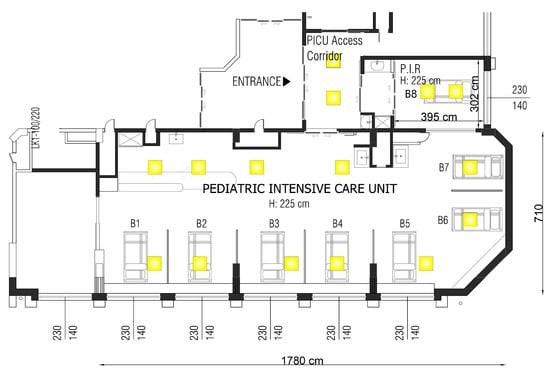
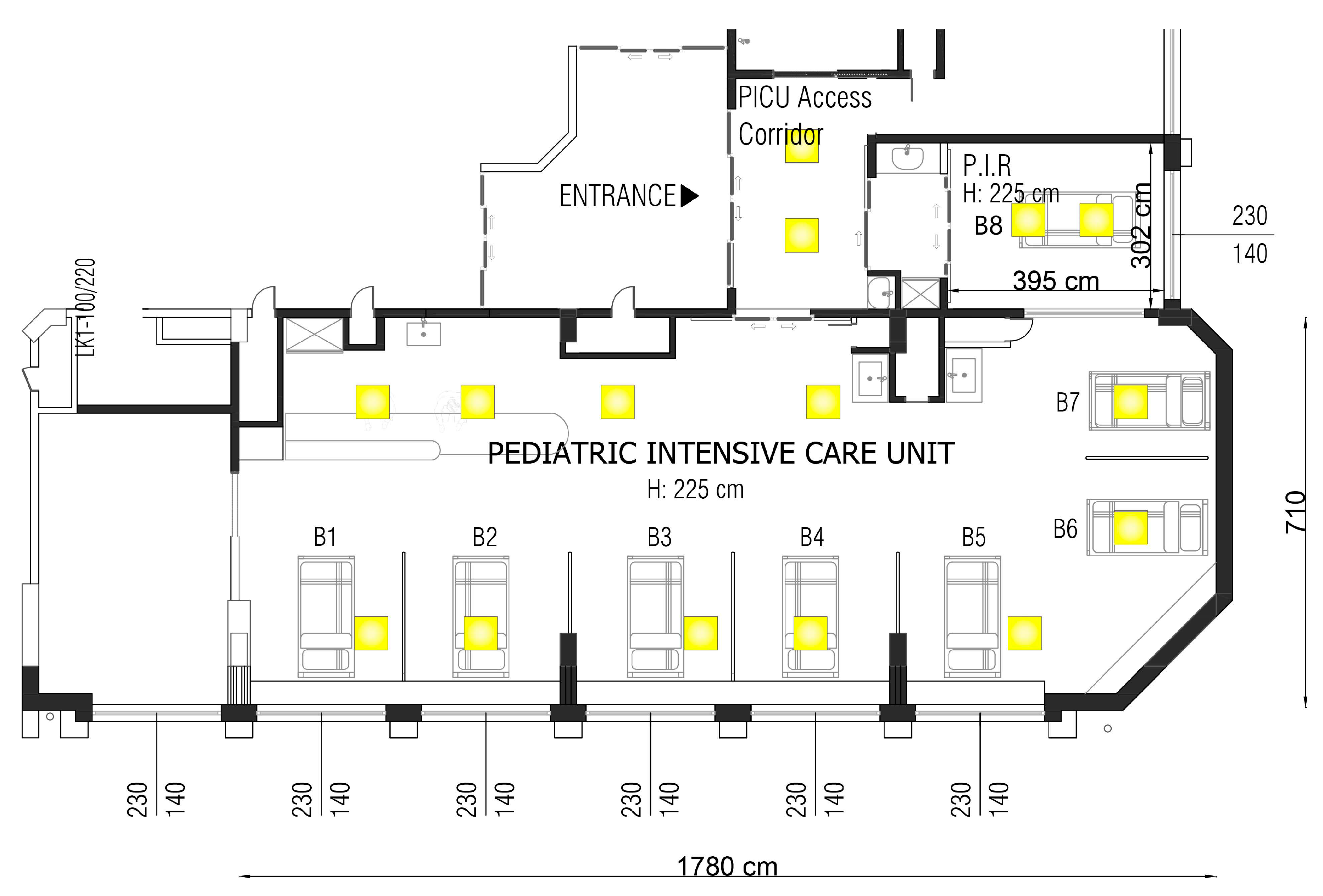

Disclaimer/Publisher’s Note: The statements, opinions and data contained in all publications are solely those of the individual author(s) and contributor(s) and not of MDPI and/or the editor(s). MDPI and/or the editor(s) disclaim responsibility for any injury to people or property resulting from any ideas, methods, instructions or products referred to in the content. |
© 2026 by the authors. Licensee MDPI, Basel, Switzerland. This article is an open access article distributed under the terms and conditions of the Creative Commons Attribution (CC BY) license.