


Abstract
This study investigated the acute cardiac and hemodynamic responses to high-intensity resistance training (HIRT), low-intensity resistance training (LIRT), and low-intensity resistance training with blood flow restriction (LIRT+BFR) during sessions composed of sequential exercises performed to muscular failure. Fourteen resistance-trained men (age: 21.8 ± 1.7 years; height: 178.3 ± 8.3 cm; body mass: 76.0 ± 8.1 kg) completed a repeated-measures crossover design in which each participant performed all three experimental conditions in a fixed, non-randomized order: HIRT (four sets at 70% 1RM), LIRT (four sets at 30% 1RM), and LIRT+BFR (four sets at 30% 1RM with a fixed cuff pressure of 80 mmHg), all executed in repetition-to-failure mode. Heart rate (HR), rate–pressure product (RPP), systolic blood pressure (SBP), mean arterial pressure (MAP), and diastolic blood pressure (DBP) were measured at rest, immediately after each session, and 30 min post-exercise. At the end of the sessions, HIRT, LIRT, and LIRT+BFR all showed significant increases in HR, RPP, SBP, MAP, and DBP compared with resting values (p < 0.01), with no differences between protocols. Delta values (pre–post changes) were similar across protocols. Post-exercise, HR and RPP remained elevated compared with baseline in all protocols (p < 0.01), with no differences between them. SBP and MAP were reduced when compared with rest at 30 min post-exercise in HIRT, LIRT, and LIRT+BFR (p < 0.05), with no differences among protocols. For DBP, only LI showed a reduction compared with the rest (p < 0.01); however, LIRT+BFR values were significantly higher than HIRT (p < 0.01) and LIRT (p < 0.01). No statistically significant between-protocol differences were observed for the immediate post-exercise cardiac and hemodynamic responses under the conditions investigated.
1. Introduction
High-intensity resistance training (HIRT; ≥60% of 1RM) has been widely recommended to promote muscle hypertrophy in healthy adults, and such sessions are typically comprising a multi-exercise sequence that engages distinct muscle groups across both upper and lower body segments [1]. However, during HI exercise, activation of the mechanoreflex occurs as a result of muscle tension and deformation induced by active muscle contractions. Simultaneously, the compression of blood vessels during contractions leads to the accumulation of metabolites (e.g., lactate, adenosine, H+ ions), which stimulates the metaboreflex [2]. Together, these mechanisms enhance sympathetic stimulation, resulting in increases in heart rate (HR), systolic blood pressure (SBP), mean arterial pressure (MAP), diastolic blood pressure (DBP), and rate–pressure product (RPP) [3,4]. In addition, HIRT sessions have been shown to promote reductions in arterial blood pressure after exercise, a phenomenon referred to as post-exercise hypotension (PEH), which is attributed to the release of vasodilatory substances and decreased sympathetic tone [5,6].
It has been shown that HIRT promotes greater muscle hypertrophy compared with low-intensity resistance training (LIRT) [7]. However, the achievement of muscular failure appears to influence this response, as when sets are performed to failure, no differences in muscle hypertrophy have been observed between HIRT and LIRT [8,9]. A possible explanation is that resistance exercise performed to failure elicits similar neuromuscular recruitment between HIRT and LIRT [9]. Furthermore, reaching muscular failure during resistance exercise seems to intensify hemodynamic responses via enhanced sympathetic activation, likely due to the increased neuromuscular recruitment [4,10].
The literature on cardiac and hemodynamic responses to HIRT and LIRT remains controversial, with some studies reporting greater responses during LIRT, while others indicate no differences between intensities [11,12]. For instance, in knee extension sessions performed to failure, SBP was higher in LIRT (3 sets at 40% 1RM) compared with HIRT (3 seta at 80% 1RM) [11]. In contrast, between HIRT (4 sets at 80% 1RM) and LIRT (4 sets at 30% 1RM), no differences were reported in HR, SBP, MAP nor DBP immediately post-exercise [12]. Similarly, when protocols with predetermined sets and repetitions were used, HIRT (3 sets and 8 reps at 80% 1RM) and LIRT (3 sets and 15 reps at 20% 1RM) showed comparable post-exercise values for HR and RPP in both upper- and lower-limb exercises [13].
The blood flow restriction (BFR) technique consists of applying a cuff to partially restrict blood flow in a limb or muscle. When combined with low loads ranging from 20% to 40% 1RM (referred to as LIRT+BFR), training can be performed using either predetermined repetition schemes (e.g., 4 sets of 30, 15, 15, and 15 repetitions) or executed until muscular failure (i.e., repetition-to-failure mode) [14]. This method has been shown to be an effective alternative for promoting muscle hypertrophy [14]. In practice, cuff pressure should be individualized and based on the percentage of arterial occlusion pressure (40–80% of AOP) but often prescribed based on absolute pressures or a percentage of SBP [14,15]. The application of the cuff leads to greater metabolite accumulation, which intensifies metaboreflex activation and consequently increases sympathetic excitation and hemodynamic responses [15,16]. Because LIRT+BFR modifies the internal physiological demand of exercise while allowing substantially lower external loads, its relevance extends beyond conventional strength-training settings.
Beyond its application in healthy populations, LIRT+BFR has gained considerable clinical relevance because it allows meaningful gains in muscular force and mass despite applying markedly reduced lifting loads [17,18]. This characteristic makes LIRT+BFR particularly useful for individuals who are unable to tolerate high mechanical stress, such as older adults, patients undergoing rehabilitation after musculoskeletal injury or surgery, and individuals with chronic diseases [19,20]. Therefore, understanding the cardiovascular and hemodynamic responses associated with LIRT+BFR is important not only for athletic populations but also for clinical exercise prescription, where cardiovascular safety is a major concern [5].
Research has investigated and compared the acute cardiac and hemodynamic responses of HIRT, LIRT, and LIRT+BFR in healthy men, but most involved only a single resistance exercise. Using protocols with predetermined repetitions, HIRT (3 seta and 8 reps at 80% 1RM) produced higher values compared with LIRT (3 sets and 15 reps at 20% 1RM) or LIRT+BFR (3 sets and 15 reps at 20% 1RM) [21]. Conversely, when sets were performed to muscular failure in HIRT and LIRT protocols, hemodynamic responses were greater than those observed in LIRT+BFR (4 sets and 15 reps at 30% 1RM) [22]. Regarding post-exercise hypotension (PEH), sessions composed of four sequential exercises demonstrated greater reductions in HIRT [23] or in both HIRT and LIRT+BFR [24].
To our knowledge, previous studies have primarily investigated isolated exercises or protocols with predetermined repetitions. Evidence regarding acute cardiovascular responses during resistance training sessions composed of multiple upper- and lower-body exercises performed to muscular failure is lacking. Furthermore, direct comparisons among HI, LI, and LIBFR under these ecologically valid training conditions remain unaddressed, hence constraining the confirmation of whether the interchangeable usage of protocols for similar resistance exercise training purposes should be recommended.
Based on this lack of evidence, the purpose of the present study was to examine whether HI, LI, and BFR sessions composed of sequential exercises performed to muscular failure would elicit distinct acute cardiac and hemodynamic responses, as well as whether the PEH responses differed between training sessions.
It was hypothesized that, despite utilizing a full-body sequential exercise design closer to real-world training practice, the high internal effort driven by muscular failure across all protocols would elicit distinct acute hemodynamic stress profiles. Specifically, HIRT was expected to induce greater immediate cardiovascular strain, whereas LIRT and LIRT+BFR would demonstrate unique recovery kinetics (such as distinct post-exercise hypotensive patterns or peripheral vascular dynamics), thereby providing a preliminary physiological benchmark for multi-exercise protocols without assuming physiological equivalence across loading conditions.
2. Materials and Methods
2.1. Participants
Fourteen resistance-trained males with a minimum of six months of continuous training experience (age: 21.8 ± 1.7 years; height: 178.3 ± 8.3 cm; body mass: 76.0 ± 8.1 kg) were recruited for this study. Sample size estimation was performed a priori using G*Power software (version 3.1.9.7; Universität Düsseldorf, Düsseldorf, Germany). Due to the lack of prior literature investigating acute hemodynamic kinetics during multi-exercise resistance training sessions performed with BFR to muscular failure, the calculation was anchored on the Rating of Perceived Exertion (RPE) reported by Macedo et al. [25] for the difference between high-load and low-load plus BFR training protocols (8.1 ± 0.6 vs. 6.2 ± 1.1, r2 = −0.46). Importantly, RPE was not treated as a surrogate for the cardiovascular outcomes investigated in the present study. Rather, it was selected as an established indicator of internal training load arising from the external demands imposed by resistance exercise [26]. In this context, external variables of training load such as relative load (%1RM), number of repetitions, sets, time under tension, and rest intervals generate an integrated internal response that can be quantified perceptually through RPE. Therefore, RPE provided an empirically grounded estimate of the magnitude of the internal response to a closely related resistance-training stimulus when effect-size data from an identical hemodynamic protocol were unavailable. Cardiac and hemodynamic variables remained the primary physiological outcomes of the present investigation and were analyzed independently of RPE. The a priori sample-size calculation was performed assuming a very large effect size (d = 1.2875, verified with Hedges’ g correction for small cohorts). Statistical parameters were set for a two-tailed matched-pairs design using a significance level of α = 0.05 (Z1−α/2 = 1.960, corresponding to a 95% confidence level) and a statistical power of 1 − β = 0.95 (Z1−β = 1.645, corresponding to 95% power), and an allocation ratio (N2/N1) of 1.0. The analysis indicated a minimum required sample size of 10 participants. To establish a safe statistical buffer against potential dropouts, the cohort was increased by 40%, resulting in a final sample of 14 participants.
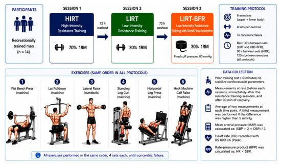
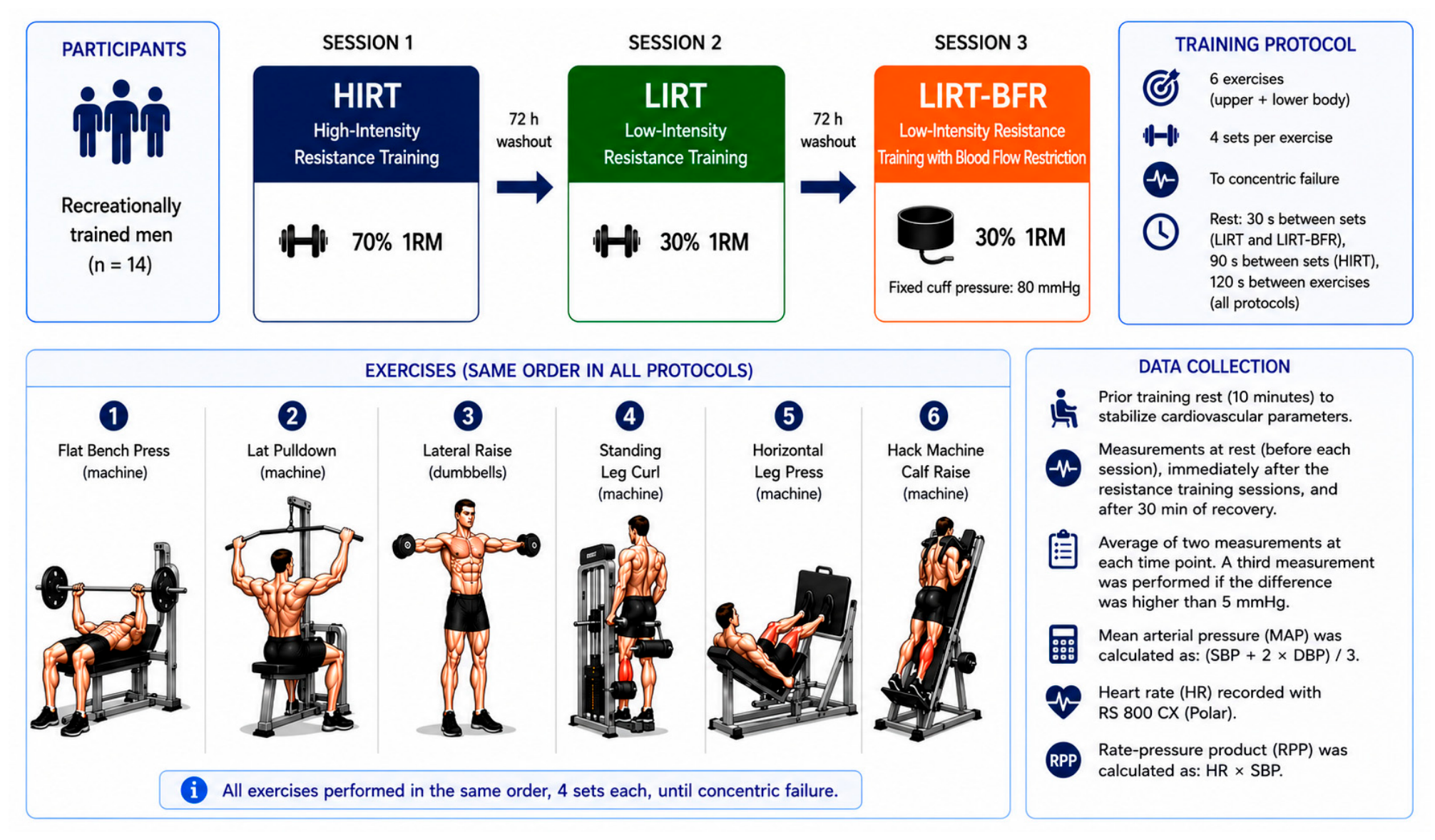
Participants were required to be 18–30 years old, to have engaged in resistance training for at least six months, and to have no history of ischemia, diabetes, arrhythmias, hypertension, or obesity (body mass index ≥ 30 kg/m2). Participants were instructed to maintain their habitual dietary intake, avoid alcohol and strenuous exercise for 24 h, arrive adequately hydrated, and perform all sessions at the same time of day (±1 h); additionally, to minimize confounding factors, participants were instructed to refrain from consuming caffeinated beverages for 24 h before each experimental session. Prior to enrollment, all volunteers received a comprehensive explanation of the study design (Figure 1) and potential risks, after which written consent was formally recorded. Protocol authorization was obtained from the Human Research Ethics Committee of UNIFAL (CAEE 76049423.6.0000.5142) in compliance with the guidelines set forth in the Declaration of Helsinki.
Figure 1.
Diagrammatic illustration depicting the study framework, exercise conditions (HIRT, LIRT, and LIRT+BFR), prescribed movement order, and parameter collection timeline. The figure was produced via ChatGPT’s image generation feature (OpenAI, GPT-5.5 version, 2026) and modified by the research team for structural clarity.
2.2. One-Repetition Maximum Test
The one-repetition maximum (1RM) test was assessed for all six exercises incorporated into the training sessions, which was employed to determine maximum strength values across all exercises scheduled for the experimental conditions. Test procedures followed previous recommendations [27]. According to this study, a standardized 15-min warm-up was performed prior to the testing sessions, consisting of static stretching and low-intensity aerobic exercise (cycling or running at a light workload). The 1RM assessment followed the criteria: (i) a specific warm-up sequence was executed with light loads and submaximal repetitions to avoid early concentric fatigue; (ii) the initial 1RM attempt was estimated based on reference strength scores for upper and lower limbs, customized to the participant’s age, biological sex, and body mass; (iii) participants were allowed three to five discrete attempts to achieve their maximum load, interspersed with a 3-min passive recovery period. The lifted load was adjusted (increased or decreased by 1.1 to 4.5 kg) between attempts based on the perceived difficulty and technical execution of the preceding trial. The maximum weight successfully moved through a full range of motion in a single repetition was recorded as the absolute 1RM value.
2.3. Experimental Training Sessions
All participants were recreationally resistance-trained and familiar with the conventional resistance-exercise movement patterns included in the protocol. The experimental design consisted of three full-body resistance training protocols targeting major upper- and lower-limb muscle groups, this fixed sequence was adopted to maintain methodological consistency with closely related protocols previously used by our group and in the literature [25,28,29]. Each participant completed all three conditions separated by a 72-h washout period, and performed in a non-randomized and non-counterbalanced order: high-intensity traditional training (HIRT), low-intensity traditional training (LIRT), and low-intensity training with blood flow restriction (LIRT+BFR). The experimental conditions were completed in a fixed, non-randomized order (HIRT → LIRT → LIRT+BFR) separated by 72 h. This specific sequence was selected primarily to maintain consistency with established protocols in closely related literature, thereby facilitating direct methodological and physiological comparability [12,29]. Additionally, this fixed order provided important physiological controls: first, because all participants were resistance-trained and fully accustomed to traditional high-load resistance training, executing the HIRT protocol first minimized the risk of unaccustomed muscle damage or atypical delayed-onset muscle soreness that could confound subsequent sessions; second, placing the LIRT+BFR condition last prevented potential residual physiological carryover—such as prolonged ischemic, metabolic, or vascular tone alterations associated with cuff occlusion and reperfusion [15,17]—from interfering with the non-restricted baseline responses of the HIRT and LIRT sessions.
The configuration of training volume, load intensities, and inter-set recovery periods was structured as follows: (i) HIRT protocol: performed at 70% of 1RM, encompassing 4 sets per exercise with 90 s of rest between sets; (ii) LIRT protocol: performed at 30% of 1RM, encompassing 4 sets per exercise with 30 s of rest between sets; and (iii) LIRT+BFR protocol: performed at 30% of 1RM combined with continuous vascular occlusion, encompassing 4 sets per exercise with 30 s of rest between sets. For all protocols, a standardized 120-s rest interval was enforced between different exercises [14,30]. Every set across all conditions was executed until volitional muscular failure, which was defined as the absolute inability to complete the concentric phase of a repetition while maintaining proper technical form. Furthermore, execution cadence was strictly controlled using a metronome set to a 3-s repetition duration (1 s for the concentric phase and 2 s for the eccentric phase) [1,30].
2.4. Protocol Blood Flow Restriction
Limb occlusion was implemented utilizing dedicated restriction cuffs sized appropriately for the dimensions of the upper and lower limbs. For upper-limb exercises, a cuff measuring 80 cm × 7 cm (Scientific-arm-WCS, Cardiomed®, Curitiba, Brazil) was used, whereas for lower-limb exercises, cuffs measuring 84 cm × 12.5 cm (Scientific-leg-WCS, Cardiomed®, Curitiba, Brazil) were employed; it appears that in BFR practice, the wide cuff (7–9 cm) is most commonly used for the upper body, and the wide cuff (10–12 cm) for the lower body. For upper-limb exercises, the cuffs were placed around the most proximal region of the arms, whereas for lower-limb exercises, they were positioned proximally on the thighs [30]. The cuff pressure applied during BFR exercises was standardized at 80 mmHg [31,32], maintained throughout each exercise set, and deflated only during the rest intervals between exercises. Based on these reference mean values, the fixed pressure of 80mmHg corresponds approximately to 61–63% of mean upper-limb AOP and 44–51% of mean lower-limb AOP, placing it withing the lower-to-middle portion of the commonly recommended 40–80% AOP range. However, because individual AOP was not measured, these reference percentages should not be interpreted as the actual relative occlusion applied to each participant.
Although BFR pressure is ideally prescribed individually as a percentage of arterial occlusion pressure (AOP; 40–80%), this procedure requires Doppler to determine AOP [14]. The use of Doppler, however, demands technical expertise from the evaluator and could introduce additional variability [33], which may limit its feasibility in practical training settings. Another important factor is that AOP values vary with body position (e.g., standing, supine, or seated) [34], which further complicates the application of individualized BFR during whole-body resistance training sessions that typically involve exercises performed in multiple positions. Thus, while our use of a standardized 80 mmHg pressure does not normalize relative occlusion across participants, this value aligns with the range of absolute pressures evaluated in prior research, where physiological responses have been documented at pressures as low as 50 mmHg [14,25]. Within the context of multi-joint, full-body sessions—where postural changes continuously alter individual AOP—a fixed 80 mmHg pressure represents a pragmatic methodological approach, though individual variations in relative occlusion degree cannot be ruled out.
2.5. Hemodynamic Measurements
Blood pressure (BP) was measured using the auscultatory method and recorded manually by a trained and experienced evaluator, employing a sphygmomanometer (cuff width: 7 cm; length: 52 cm) and a stethoscope [35,36]. Prior to the training sessions, participants remained seated for 10 min to stabilize cardiovascular parameters. Measurements were taken at rest before each session, immediately after completion of the session, and after 30 min of recovery. Two consecutive blood pressure measurements were obtained at each time point and averaged for analysis. If measurements differed by more than 5 mmHg, an additional measurement was performed and the closest two values were averaged. All measurements followed the recommendations of the American Heart Association guidelines [37].
Mean arterial pressure (MAP) was calculated using the standard equation: (SBP + 2 × DBP)/3 [25]. Heart rate (HR) was recorded using a Polar RS800CX heart rate monitor with a chest strap (Polar Electro, Kempele, Finland) [12]. The rate–pressure product (RPP) was calculated as HR (beats·min−1) × SBP (mmHg) [13]. Additionally, delta values (Δ) for cardiovascular and hemodynamic parameters were calculated immediately post-exercise (Δpost–rest) and after 30 min of recovery (Δ30-min post-exercise).
2.6. Statistical Analysis
Data distribution and sphericity were evaluated through Shapiro–Wilk and Mauchly’s tests. The Greenhouse–Geisser adjustment was implemented upon detection of sphericity violations. Fully repeated-measures two-way analysis of variance (ANOVA: Repeated Measures, within-subjects factors: Protocol [HIRT vs. LIRT vs. LIRT+BFR] × Time [pre-exercise, post-exercise, and 30-min recovery]) with Bonferroni-adjusted post-hoc tests was used to analyze absolute cardiovascular and hemodynamic responses (HR, RPP, SBP, MAP, and DBP). Similarly, between-protocol comparisons for delta values (Δ) were conducted using a one-way repeated-measures ANOVA (within-subjects factor: Protocol) to account for data dependency across conditions. Effect size for ANOVA was calculated using partial eta squared (η2p) and interpreted as: 0.01 [small], 0.06 [medium], and 0.14 [large]. Data are presented as mean ± standard deviation. For between-protocol delta comparisons, mean differences and corresponding 95% confidence intervals (95% CIs) were reported to indicate effect magnitude and precision. All analyses were performed using SPSS software (version 28.0, IBM Corp., Armonk, NY, USA). Statistical significance was set at α = 0.05.
3. Results
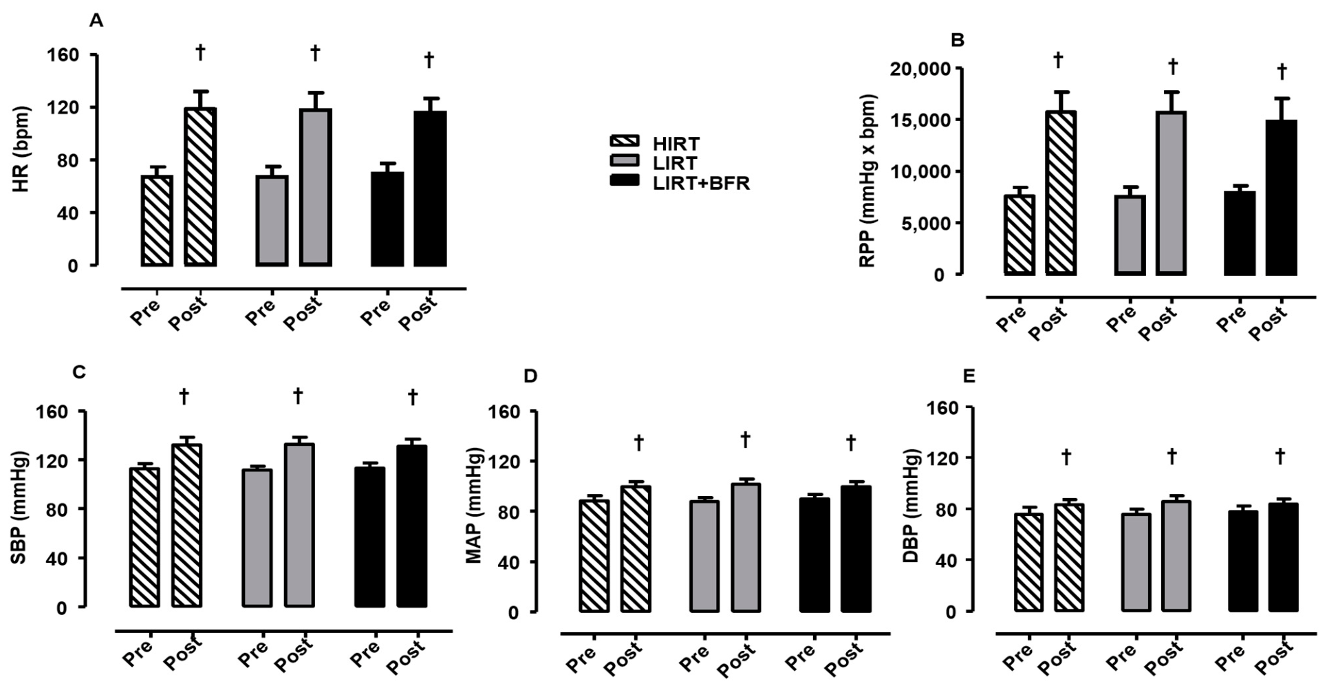
Figure 2 illustrates cardiac and hemodynamic measurements pre- and post-sessions for the HIRT, LIRT, and LIRT+BFR protocols: HR (panel A), RPP (panel B), SBP (panel C), MAP (panel D), and DBP (panel E). HR increased significantly from pre- to post-session across all protocols (F[1,39] = 825.3 p ˂ 0.01, η2p = 0.955 [large], OP = 1.00; HIRT: p < 0.01; LIRT: p < 0.01; LIBFR: p < 0.01), with no significant differences observed between protocols (F[2,39] = 0.019, p = 0.98) regardless of time point (p > 0.05 for all comparisons; Figure 2, Panel A).
Figure 2.
Pre vs. post-exercise values (i.e., pre vs. immediately after the session) for heart rate (HR, Panel (A)), rate–pressure product (RPP, Panel (B)), systolic blood pressure (SBP, Panel (C)), mean arterial pressure (MAP, Panel (D)), and diastolic blood pressure (DBP, Panel (E)) during high-intensity resistance training (HIRT, N = 14), low-intensity resistance training (LIRT, N = 14), and low-intensity training with blood flow restriction (LIRT+BFR, N = 14). † Significant difference compared to pre-exercise (p < 0.05).
Similarly, RPP showed a significant effect of time (F[1,39] = 673.3; p ˂ 0.01, η2p = 0.945 [large], OP = 1.00) with increases from pre- to post-session (HIRT: p < 0.01; LIRT: p < 0.01; LIRT+BFR: p < 0.001), but no differences were found between protocols (F[2,39] = 0.133; p > 0.05 for all comparisons; Figure 2, Panel B).
The patterns for SBP, MAP, and DBP (Figure 2, Panels C, D, and E) were similar to cardiovascular parameters comparing pre- and post-session across all protocols: SBP (F[1,39] = 331.8, p ˂ 0.01, η2p = 0.895 [large], OP = 1.00), MAP (F[1,39] = 192.6, p ˂ 0.01, η2p = 0.832 [large], OP = 1.00), and DBP (F[1,39] = 69.91, p ˂ 0.01, η2p = 0.642 [large], OP = 1.00). No significant differences were observed between protocols at either time point for SBP (F[2,39] = 0.028, p = 0.97), MAP (F[2,39] = 0.452, p = 0.64), or DBP (F[2,39] = 0.884, p = 0.42).
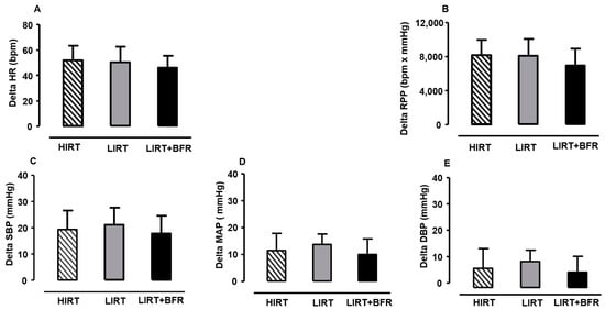
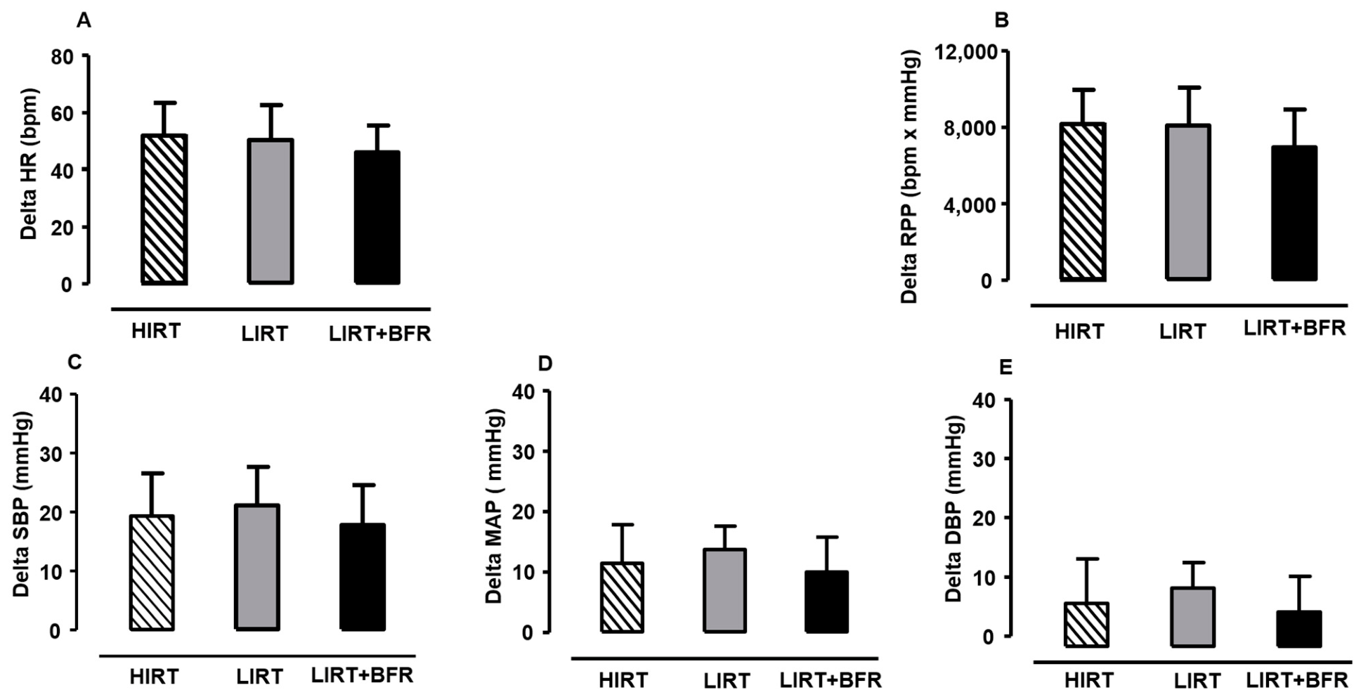
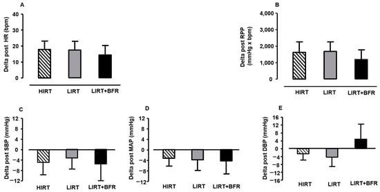
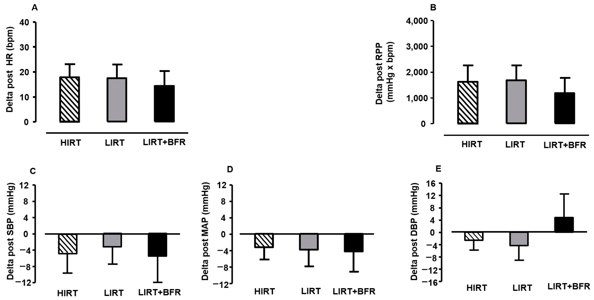
Figure 3 shows delta values (post–pre) for HR (Panel A), RPP (Panel B), SBP (Panel C), MAP (Panel D), and DBP (Panel E) for HIRT, LIRT, and LIRT+BFR. No significant differences in delta values were observed between protocols for HR (F[2,26] = 2.777, p = 0.08, η2 = 0.176 [large], OP = 0.50), RPP (F[2,26] = 1.771, p = 0.08, η2 = 0.083 [medium], OP = 0.53), SBP (F[2,26] = 0.824, p = 0.45, η2 = 0.060 [medium], OP = 0.35), MAP (F[2,26] = 1.703, p = 0.20, η2 = 0.116 [medium], OP = 0.32), or DBP (F[2,26] = 1.532, p = 0.23, η2 = 0.105 [medium], OP = 0.30). The corresponding between-protocol mean differences and 95% CI for post-pre delta values are presented in Supplementary Table S1. The confidence intervals are provided to complement the omnibus hypothesis and effect-size estimates and to characterize the precision of the observed between-protocol differences.
Figure 3.
Delta values (post–pre-exercise) for heart rate (HR, Panel (A)), rate–pressure product (RPP, Panel (B)), systolic blood pressure (SBP, Panel (C)), mean arterial pressure (MAP, Panel (D)), and diastolic blood pressure (DBP, Panel (E)) during high-intensity resistance training (HIRT, N = 14), low-intensity resistance training (LIRT, N = 14), and low-intensity training with blood flow restriction (LIRT+BFR, N = 14).
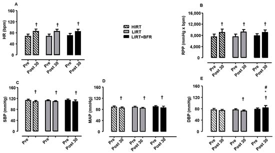
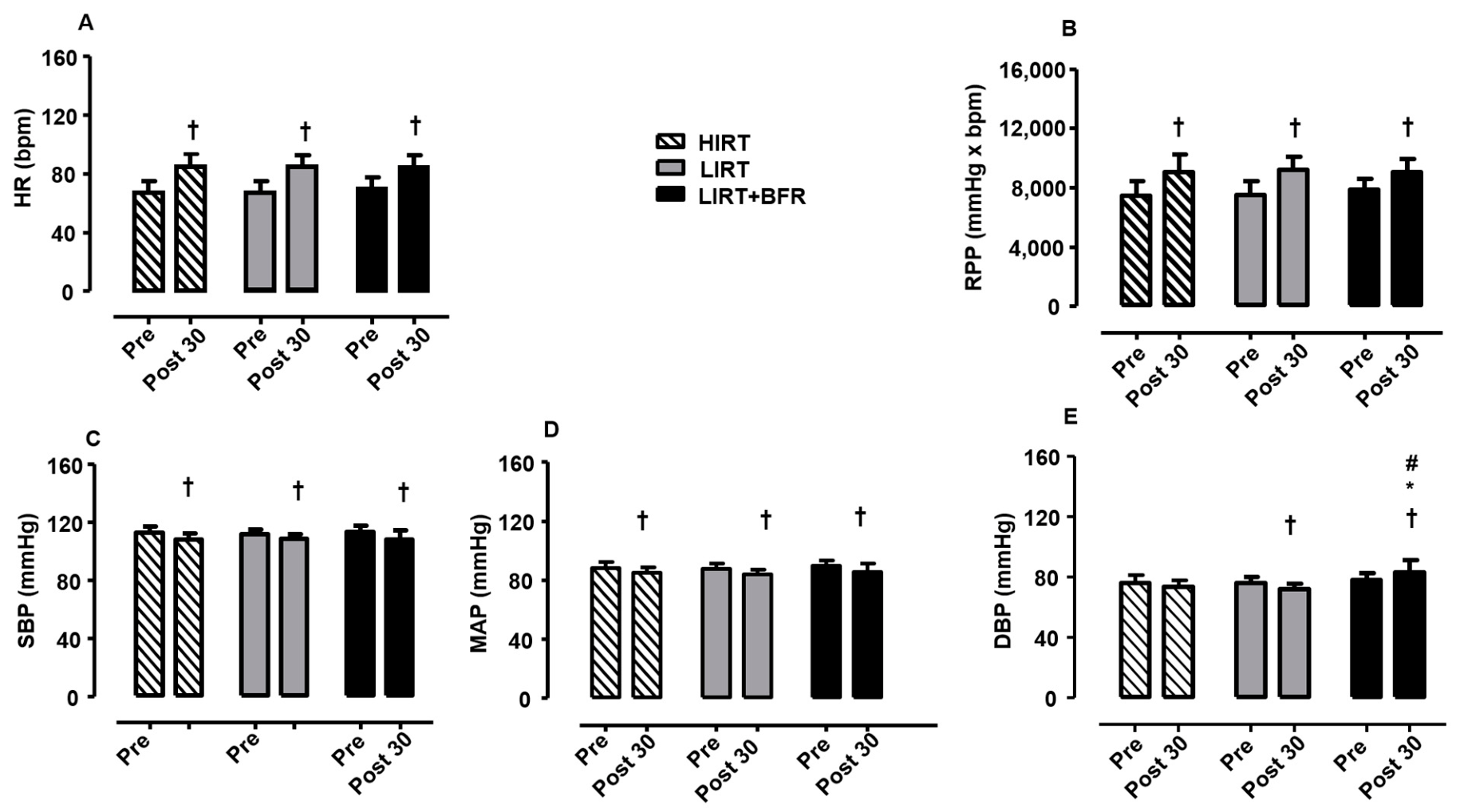
Figure 4 depicts the cardiac and hemodynamic responses 30 min after HIRT, LIRT, and LIRT+BFR sessions: HR (Panel A), RPP (Panel B), SBP (Panel C), MAP (Panel D), and DBP (Panel E). HR remained elevated compared to pre-exercise values (F[1,39] = 373.2, p ˂ 0.001, η2p = 0.905 [medium], OP = 1.00) across all protocols (HIRT: p < 0.01; LIRT: p < 0.01; LIRT+BFR: p < 0.01), with no differences between time points (F[2,39] = 0.054, p = 0.947, η2p = 0.003 [trivial]). RPP was also higher at 30 min post compared to pre-exercise (F[1,39] = 201. 8, p ˂ 0.01, η2p = 0.838 [large], OP = 1.00) across all protocols (HIRT: p < 0.01; LIRT: p < 0.01; LIRT+BFR: p < 0.01) with no differences between time points (pre: p > 0.05, post 30 min: p > 0.05).
Figure 4.
Pre-exercise (rest) vs. 30-min post-exercise values for heart rate (HR, Panel (A)), rate–pressure product (RPP, Panel (B)), systolic blood pressure (SBP, Panel (C)), mean arterial pressure (MAP, Panel (D)), and diastolic blood pressure (DBP, Panel (E)) during high-intensity resistance training (HIRT, N = 14), low-intensity resistance training (LIRT, N = 14), and low-intensity training with blood flow restriction (LIRT+BFR, N = 14). † Significant difference compared to pre-exercise, p < 0.05. * Significant difference compared to HIRT, p < 0.05. # Significant difference compared to LIRT, (p < 0.05).
Regarding hemodynamic parameters, a reduction in the values of SBP (F[1,39] = 29.284, p ˂ 0.01, η2p = 0.429 [large], OP = 0.99; HIRT: p < 0.01, LIRT: p < 0.05, LIRT+BFR: p < 0.01; Figure 4, Panel C) and MAP (F[1,39] = 35.343, p ˂ 0.01, η2p = 0.475 [large], OP = 0.99; HIRT: p < 0.01; LIRT: p < 0.01; LIRT+BFR: p < 0.01; Figure 4, Panel D) decreased 30 min post-exercise compared to pre-exercise values. No significant differences between HIRT, LIRT and LIRT+BFR were observed for either parameter at pre- or 30-min post-exercise (SBP: F[1,39] = 0.061, p = 0.94, η2p = 0.003 [trivial], OP = 0.09; MAP: F[1,39] = 0.920, p = 0.41, η2p = 0.045 [trivial], OP = 0.86).
In contrast to SBP and MAP, DBP showed a significant reduction (F[1,39] = 0.914, p = 0.439, η2p = 0.015 [small], OP = 0.32) only for LIRT (p < 0.01), whereas LIRT+BFR values were higher at 30 min post-exercise compared to pre-exercise (p < 0.01). Between-protocol comparisons at 30 min post-exercise revealed that DBP was significantly higher in LIRT+BFR compared to both HIRT (p < 0.01) and LI (p < 0.01) (F[2,39] = 9.144, p = 0.01, η2p = 0.319 [large], OP = 1.00).
Figure 5 shows delta values (30 min post-exercise minus pre-exercise) for HR (Panel A), RPP (Panel B), SBP (Panel C), MAP (Panel D), and DBP (Panel E) across HIRT, LIRT, and LIRT+BFR. No significant differences in delta values were observed for HR (F[2,39] = 1.605, p = 0.21, η2p = 0.076 [medium], OP = 0.32), RPP (F[2,39] = 2.886, p = 0.068, η2p = 0.129 [medium], OP = 0.53), SBP (F[2,39] = 0.797, p = 0.46, η2p = 0.039 [small], OP = 0.18), MAP (F[2,39] = 1.644, p = 0.21, η2p = 0.078 [medium], OP = 0.33), or DBP (F[2,39] = 1.493, p = 0.24, η2p = 0.071 [medium], OP = 0.30). Between-protocol mean differences and corresponding 95% CI for the 30-min post-pre delta values are also provided in Supplementary Table S1.
Figure 5.
Delta values (30 min post–pre exercise) for heart rate (HR, Panel (A)), rate–pressure product (RPP, Panel (B)), systolic blood pressure (SBP, Panel (C)), mean arterial pressure (MAP, Panel (D)), and diastolic blood pressure (DBP, Panel (E)) during high-intensity resistance training (HIRT, N = 14), low-intensity resistance training (LIRT, N = 14), and low-intensity training with blood flow restriction (LIRT+BFR, N = 14).
4. Discussion
The present study investigated acute cardiac and hemodynamic responses across HIRT, LIRT, and LIRT+BFR resistance training protocols in which consecutive upper- and lower-body exercises were executed to volitional fatigue. Our primary findings demonstrate that at the immediate cessation of the sessions, no statistically significant between-protocol differences were observed in the magnitude of the immediate cardiac and hemodynamic responses. Furthermore, during the 30-min post-exercise recovery period, while HR and RPP remained elevated above baseline under all conditions, the LIRT protocol induced the most pronounced post-exercise hypotension (PEH), as evidenced by significant reductions in SBP, MAP, and DBP. Crucially, these non-significant acute differences must be interpreted with appropriate caution; as previously highlighted, a lack of statistical significance does not mathematically guarantee complete physiological equivalence. Given that several effect sizes fell within the small-to-medium range, subtle biological variations in cardiovascular strain between conditions cannot be entirely ruled out. When contrasting these outcomes with the existing literature, previous investigations testing single-exercise models (either performed to failure or under predetermined repetitions) universally reported acute cardiovascular elevations relative to resting states [25,29,30].
Current results extend these findings to a more ecologically valid multi-exercise session, confirming that cumulative volume to failure maintains this elevated cardiovascular profile regardless of the load configuration. Rather than merely reiterating established theoretical models, this uniform hemodynamic peak across HIRT, LIRT, and LIRT+BFR can be interpreted through the unique physiological demands of reaching complete muscular failure. Performing sets to volitional failure under lower external loads (LIRT and LIRT+BFR) inherently prolongs the time under tension and accelerates motor unit recruitment. This sustained muscular contraction compounds local vascular compression and metabolic accumulation. Consequently, the intensified stimulation of intramuscular mechanoreceptors and chemoreceptors triggers a compensatory increase in sympathetic drive—likely amplifying the exercise pressor reflex via metaboreflex pathways [16,38]—which ultimately matches the peak cardiac strain typically induced by the higher mechanical tension of traditional high-intensity training.
In single-exercise protocols with predetermined repetitions, LIRT typically produced lower values than HIRT or LIRT+BFR at the end of the third set [25]. Conversely, when exercises were performed to failure (HIRT and LIRT), the delta values of HR were similar across protocols, whereas hemodynamic deltas for LIRT+BFR (predetermined repetitions) were lower than HIRT and LIRT [31]. In the present study, end-of-session values and deltas for HR, RPP, SBP, MAP, and DBP were similar across HIRT, LIRT, and LIRT+BFR in multi-exercise (upper- and lower-body) sessions performed to failure, contrasting with findings from single-exercise protocols. This suggests that factors such as load, number of sets, and muscular failure influence hemodynamic responses [39]. The higher number of exercises, sets, recruited muscle mass, and time under tension likely amplified contraction magnitude, vascular compression, and metabolite accumulation [4]. Particularly in LIRT+BFR, the restriction of venous return may have enhanced local ischemia and metabolite retention, potentially increasing peripheral vascular resistance and sympathetic drive [38]. However, despite these protocol-specific variations in exercise parameters, the overall hemodynamic and cardiovascular responses observed immediately post-session remained broadly comparable across HIRT, LIRT, and LIRT+BFR.
HR and DP remained elevated above baseline 30 min after all training sessions, with no statistically significant differences among HIRT, LIRT, and LIRT+BRF, as also reflected by the recovery delta values. This pattern is consistent with previous evidence showing sustained HR elevations during the early recovery period after resistance exercise, although protocol-dependent differences have also been reported [23]. Resistance exercise acutely increases sympathetic activation and reduces parasympathetic modulation, and incomplete vagal reactivation may contribute to the delayed normalization of HR during post-exercise recovery [4,12,40]. In the present study, the persistence of elevated RPP 30 min after exercise may indicate that cardiac workload had not yet returned to baseline within the observed recovery window. Because RPP integrates HR and SBP responses and is commonly used as an indirect indicator of myocardial workload, its sustained elevation provides additional information regarding the time course of cardiovascular recovery following these resistance-training sessions. Importantly, these findings were obtained exclusively in healthy, trained young men and should not be interpreted as evidence for exercise prescription, safety, or recovery requirements in individuals with cardiovascular disease, hypertension, or impaired autonomic regulation. Rather, the sustained RPP response observed in the present sample identifies a physiological feature that warrants direct investigation in these populations, in whom the magnitude, duration, and clinical significance of post-exercise cardiovascular recovery may differ.
Prior research had shown that performing four consecutive lower-body exercises did not induce PEH of SBP 30 min after the session for HIRT, LIRT, or LIRT+BFR protocols [29]. In contrast, sessions involving two exercises targeting both upper- and lower-body muscles elicited PEH of SBP 30 min post-exercise for HIRT, LIRT, and LIRT+BFR, while for DBP decreased only in HIRT and LIRT+BFR [24]. The PEH effect observed 30 min after the session, following exercises targeting multiple muscle groups with sets performed to muscular failure, was evident for SBP and MAP in the HIRT, LIRT, and LIRT+BFR protocols. In contrast, post-exercise reductions in DBP were observed following the LIRT protocol, whereas this hypotensive response was attenuated in LIRT+BFR compared with both HIRT and LIRT due to higher post-exercise DBP values observed after training.
The present results aligned with those reported by Neto et al. [24], which demonstrated reductions in SBP relative to resting values 30 min after HIRT, LIRT, and LIRT+BFR sessions, confirming that exercises targeting multiple muscle groups can influence PEH in SBP regardless of whether sets are performed to muscular failure. Resistance exercises involving both upper- and lower-body muscles appear to promote greater PEH compared with single-segment exercises, likely due to the greater muscle mass involved and consequent increased release of vasodilatory substances, such as prostaglandins [6,41,42]. Furthermore, local vasodilatory mechanisms, combined with transient reductions in sympathetic vasoconstrictor activity after exercise, may contribute to the observed post-exercise reductions in SBP and MAP [5]. These responses may be clinically relevant, particularly considering that even modest acute reductions in blood pressure may contribute to chronic cardiovascular benefits when resistance exercise is performed regularly.
Interestingly, Neto et al. [24] reported that DBP decreased 30 min post-exercise only in HIRT and LIRT+BFR, but not in LI. Contrarily, the present results showed that reductions in DBP after 30 min occurred only in LIRT. Two factors may explain these discrepancies: the performance of sets to muscular failure and the inclusion of multi-joint exercises, both of which directly influence PEH [5]. The observation that only the LIRT protocol induced DBP reductions suggests that sessions with lower relative loads (%1RM) may exert a greater influence on PEH of DBP [41]. Conversely, the higher post-session DBP values observed in the LIBFR protocol may be attributed to increased peripheral vascular resistance associated with blood flow restriction. Additionally, the maintenance of cuff-induced vascular compression during exercise may have delayed vascular recovery and attenuated the magnitude of post-exercise vasodilation in LIRT+BFR. This mechanism may partially explain why DBP remained comparatively higher in this condition despite similar immediate post-exercise cardiovascular responses. However, this mechanism was not directly measured; therefore, this interpretation remains speculative, and the isolated DBP response should be considered hypothesis-generating until replicated.
From a practical standpoint, the present findings suggest that low-load resistance training performed to muscular failure may elicit acute cardiovascular response similar to those observed for LIRT+BFR protocol, as well comparable to that reported in the literature for training with BFR [42], although equivalence between the protocols cannot be established. Consequently, while these outcomes cannot be directly generalized to clinical cohorts, they offer a relevant physiological benchmark that aligns with the safety guidelines and therapeutic frameworks discussed in previous comprehensive literature [43,44]. Although subtle hemodynamic discrepancies between protocols might have remained undetected due to the relatively small sample size, the present data did not indicate a greater immediate hemodynamic response during LIRT+BFR compared with the other protocols in this sample. While the selected acute cardiovascular responses were comparable across conditions in this cohort, these findings are restricted to the measured hemodynamic parameters and do not imply broader physiological equivalence between protocols. This baseline supports future investigations into the clinical utility of low-intensity strategies for individuals restricted from high mechanical lifting due to elevated cardiovascular risk. Therefore, current findings may be potentially relevant for older adults and clinical populations, although direct confirmation in these groups is still required. In addition, the novelty of the present study lies in the examination of acute cardiac and hemodynamic responses during whole-body resistance training sessions composed of multiple exercises performed to muscular failure, a training protocol that more closely resembles real-world resistance training practice than previously investigated single-exercise protocols.
Limitations
The limitations of this study are as follows:
(i) The experimental conditions were performed in a fixed, non-randomized and non-counterbalanced order. All participants were resistance-trained and familiar with the exercises, which likely minimized motor-learning effects, and sessions were separated by 72 h under standardized pre-session conditions to reduce residual carryover. Nevertheless, an order-dependent physiological effect cannot be completely excluded. Future investigations should employ randomized and counterbalanced designs to confirm these findings.
(ii) The cuff pressure for LIBFR was standardized at 80 mmHg for all participants. Although current guidelines recommend individualizing pressure within a relative range (40–80% of individual arterial occlusion pressure, AOP), absolute AOP values in the literature exhibit substantial inter-individual and anatomical variability, typically spanning from 60 to 220 mmHg depending on limb circumference, cuff width, and posture [5]. Because individual AOP was not directly measured, the primary limitation of this fixed-pressure approach is that a standardized 80 mmHg does not guarantee an equivalent relative occlusion stimulus across all participants or anatomical limbs. The cuff pressure for LIBFR was standardized at 80 mmHg for all participants.
(iii) The absence of additional HIRT, LIRT, and LIRT+BFR groups performing sets not carried to muscular failure limited the ability to determine whether muscular failure itself was a critical factor influencing the cardiac and hemodynamic responses under the present training sessions.
(iv) The recovery period was monitored only up to 30 min post-exercise. Extending the observation window would have allowed confirmation of whether the PEH effects observed persisted or were attenuated over a longer time course.
(v) The sample size (N = 14), while aligned with similar acute protocols, was largely determined by pragmatic considerations and restricted to recreationally trained young men. Consequently, this relatively small cohort limits immediate generalizability and may have been insufficient to detect subtle, small between-condition physiological variances; thus, minor protocol-specific effects cannot be fully ruled out.
(vi) The DBP difference at 30 min represented an isolated between-protocol finding among several cardiovascular outcomes. Although statistically significant, it should be interpreted cautiously and considered hypothesis-generating until replicated.
Future studies involving larger, diverse samples, including women, older adults, and clinical populations, are warranted to enhance external validity and confirm these exploratory physiological trends.
5. Conclusions
In healthy, recreationally resistance-trained young men, whole-body resistance training sessions performed to volitional muscular failure elicited marked acute increases in cardiac and hemodynamic responses across HIRT, LI, and LIRT+BFR conditions. No statistically significant between-protocol differences were detected for the immediate responses of HR, RPP, SBP, MAP or DBP, although these findings should not be interpreted as evidence of physiological equivalence between conditions. During the 30-min recovery period, LIRT showed a broader post-exercise hypotensive response, with reductions in SBP, MAP, and DBP, whereas LIRT+BFR showed higher DBP values than HIRT and LIRT at 30 min. The latter finding should be interpreted cautiously and considered hypothesis-generating until independently replicated. Overall, the present results provide a physiological benchmark for understanding the acute cardiovascular responses to multi-exercise resistance-training sessions performed to muscular failure under different loading conditions. These findings directly apply to healthy young men and should be confirmed in larger and more diverse samples, including women, older adults, and clinical populations. In summary, this study offers initial physiological evidence to inform future research exploring multi-exercise protocols in contexts where high mechanical loads are poorly tolerated or contraindicated, rather than establishing direct clinical guidelines.
Supplementary Materials
The following supporting information can be downloaded at: https://www.mdpi.com/article/10.3390/sports14110490/s1, Table S1: Between-protocol mean differences and 95% confidence intervals for changes in cardiac and hemodynamic outcomes immediately and 30 min after resistance-training sessions.
Author Contributions
A.G.M., T.A.F.A. and D.M.P.F.: conceptualization; F.A.d.O.T. and A.G.M.: methodology; F.A.d.O.T., G.d.S.Z. and A.G.M.: data collection; D.A.M., J.M.G., M.C.E. and F.A.d.O.T.: formal analysis; D.M.d.O., G.d.S.Z., F.A.d.O.T., V.H.-B., J.M.G., M.C.E., R.O., J.P.B. and D.M.P.F.: writing—original draft preparation; A.G.M., V.H.-B., M.C.E., R.O., J.P.B. and D.M.P.F.: writing—review and editing. All authors have read and agreed to the published version of the manuscript.
Funding
Financial support for this study was provided by the São Paulo Research Foundation (FAPESP, grants 2017/23717-9 and 2018/16706-3), Polytechnic University of Setúbal, and the Foundation for Science and Technology (FCT) via the project registered under DOI https://doi.org/10.54499/UID/06185/2025 awarded to the SPRINT-Sport Physical Activity and Health Research & Innovation Center. Partial funding was granted by the Research Vicerectory of Universidad Nacional and the Optimisation of Training and Sports Performance Research Group (GOERD) at the Faculty of Sports Sciences, University of Extremadura. Additional financial assistance was received through the Research Group Support Grant (GR24133), co-financed at 85% by the European Union’s European Regional Development Fund (ERDF) alongside the Regional Government of Extremadura (Department of Education, Science, and Vocational Training), with the Spanish Ministry of Finance serving as the Managing Authority. Also, this work was carried out within the framework of the Research Network on Physical Activity and Health in Disability and Neurodevelopmental Disorders (DISANET), funded by the Spanish Sports Council (Consejo Superior de Deportes, CSD) through the Research Networks in Sport Sciences Programme (2026).
Institutional Review Board Statement
This study was conducted in accordance with the Declaration of Helsinki and was approved by the UNIFAL ethics committee under number CAEE 76049423.6.0000.5142, approval date on 4 January 2024.
Data Availability Statement
The data that support the findings of this study are available from correspondence author (dalton.pessoa-filho@unesp.br) upon reasonable request.
Acknowledgments
Author A.G.M. acknowledges financial support provided by the Brazilian Federal Agency for Support and Evaluation of Graduate Education (CAPES; Finance Code 001). Author T.A.F.A. was supported by CAPES through the CAPES-PrInt Program (grant process 88887.310463/2018-00; Mobility 88887.580265/2020-00). Authors D.A.M., G.S.Z. and D.M.P.F. express gratitude to the National Council for Scientific and Technological Development (CNPq) for their research fellowships (grants 150328/2024-9; 150075/2025-1; and 310784/2026-3, respectively). R.O. and J.P.B. are affiliated with the Research Center in Sport Sciences, Health Sciences and Human Development, funded by Portuguese national funds via FCT—Foundation for Science and Technology under projects UID/04045/2025 (https://doi.org/10.54499/UID/04045/2025), UID/PRR/04045/2025 (https://doi.org/10.54499/UID/PRR/04045/2025), and UID/PRR2/04045/2025 (https://doi.org/10.54499/UID/PRR2/04045/2025). Furthermore, ChatGPT (OpenAI, GPT-5.5 version, 2026) was utilized during manuscript drafting solely to generate the experimental design schematic diagram; all generated content was thoroughly verified, refined, and endorsed by the authors, who retain sole responsibility for this work.
Conflicts of Interest
The authors declare no conflicts of interest.
Abbreviations
The following abbreviations are used in this manuscript:
| RE | Resistance exercise |
| BFR | Blood flow restriction |
| HIRT | High-intensity resistance training |
| LIRT | Low-intensity resistance training |
| LIRT+BFR | Low-intensity training with blood flow restriction |
| 1RM | One repetition maximum |
| HR | Heart rate |
| SBP | Systolic blood pressure |
| MAP | Mean arterial pressure |
| DBP | Diastolic blood pressure |
| RPP | Rate–pressure product |
| AOP | Arterial Occlusion Pressure |
References
- Ratamess, A.; Evetoch, T.K.; Housh, T.J.; Kibler, W.B.; Kraemer, W.J. Progression Models in Resistance Training for Healthy Adults. Med. Sci. Sports Exerc. 2009, 41, 687–708. [Google Scholar] [CrossRef] [Scilit] [PubMed]
- Katayama, K.; Saito, M. Muscle Sympathetic Nerve Activity during Exercise. J. Physiol. Sci. 2019, 69, 589–598. [Google Scholar] [CrossRef] [Scilit] [PubMed]
- Paluch, A.E.; Boyer, W.R.; Franklin, B.A.; Laddu, D.; Lobelo, F.; Lee, D.; McDermott, M.M.; Swift, D.L.; Webel, A.R.; Lane, A.; et al. Resistance Exercise Training in Individuals with and Without Cardiovascular Disease: 2023 Update: A Scientific Statement from the American Heart Association. Circulation 2024, 149, e217–e231. [Google Scholar] [CrossRef] [Scilit] [PubMed]
- MacDougall, J.D.; Tuxen, D.; Sale, D.G.; Moroz, J.R.; Sutton, J.R. Arterial Blood Pressure Response to Heavy Resistance Exercise. J. Appl. Physiol. 1985, 58, 785–790. [Google Scholar] [CrossRef] [Scilit] [PubMed]
- Macedo, A.; Massini, D.; Almeida, T.; Reis, L.M.D.; Galdino, G.; Santos, A.T.S.; Da Silva Júnior, O.; Júnior, R.V.; Pessôa Filho, D.M. Effects of Resistance Exercise with and without Blood Flow Restriction on Acute Hemodynamic Responses: A Systematic Review and Meta-Analysis. Life 2024, 14, 826. [Google Scholar] [CrossRef] [Scilit] [PubMed]
- de Freitas Brito, A.; Brasileiro-Santos, M.d.S.; Coutinho de Oliveira, C.V.; Sarmento da Nóbrega, T.K.; Lúcia de Moraes Forjaz, C.; da Cruz Santos, A. High-Intensity Resistance Exercise Promotes Postexercise Hypotension Greater than Moderate Intensity and Affects Cardiac Autonomic Responses in Women Who Are Hypertensive. J. Strength Cond. Res. 2015, 29, 3486–3493. [Google Scholar] [CrossRef] [Scilit] [PubMed]
- Holm, L.; Reitelseder, S.; Pedersen, T.G.; Doessing, S.; Petersen, S.G.; Flyvbjerg, A.; Andersen, J.L.; Aagaard, P.; Kjaer, M. Changes in Muscle Size and MHC Composition in Response to Resistance Exercise with Heavy and Light Loading Intensity. J. Appl. Physiol. 2008, 105, 1454–1461. [Google Scholar] [CrossRef] [Scilit] [PubMed]
- Mitchell, C.J.; Churchward-Venne, T.A.; West, D.W.D.; Burd, N.A.; Breen, L.; Baker, S.K.; Phillips, S.M. Resistance Exercise Load Does Not Determine Training-Mediated Hypertrophic Gains in Young Men. J. Appl. Physiol. 2012, 113, 71–77. [Google Scholar] [CrossRef] [Scilit] [PubMed]
- Burd, N.A.; Mitchell, C.J.; Churchward-Venne, T.A.; Phillips, S.M. Bigger Weights May Not Beget Bigger Muscles: Evidence from Acute Muscle Protein Synthetic Responses after Resistance Exercise. Appl. Physiol. Nutr. Metab. 2012, 37, 551–554. [Google Scholar] [CrossRef] [Scilit] [PubMed]
- O’Connor, P.; Sforzo, G.A.; Frye, P. Effect of Breathing Instruction on Blood Pressure Responses During Isometric Exercise. Phys. Ther. 1989, 69, 757–761. [Google Scholar] [CrossRef] [Scilit] [PubMed]
- de Souza Nery, S.; Gomides, R.S.; da Silva, G.V.; de Moraes Forjaz, C.L.; Mion, D.; Tinucci, T. Intra-Arterial Blood Pressure Response in Hypertensive Subjects during Low- and High-Intensity Resistance Exercise. Clinics 2010, 65, 271–277. [Google Scholar] [CrossRef] [Scilit] [PubMed]
- Sardeli, A.V.; do Carmo Santos, L.; Ferreira, M.L.V.; Gaspari, A.F.; Rodrigues, B.; Cavaglieri, C.R.; Chacon-Mikahil, M.P.T. Cardiovascular Responses to Different Resistance Exercise Protocols in Elderly. Int. J. Sports Med. 2017, 38, 928–936. [Google Scholar] [CrossRef] [Scilit] [PubMed]
- Neto, G.R.; Sousa, M.S.C.; Costa e Silva, G.V.; Gil, A.L.S.; Salles, B.F.; Novaes, J.S. Acute Resistance Exercise with Blood Flow Restriction Effects on Heart Rate, Double Product, Oxygen Saturation and Perceived Exertion. Clin. Physiol. Funct. Imaging 2016, 36, 53–59. [Google Scholar] [CrossRef] [Scilit] [PubMed]
- Patterson, S.D.; Hughes, L.; Warmington, S.; Burr, J.; Scott, B.R.; Owens, J.; Abe, T.; Nielsen, J.L.; Libardi, C.A.; Laurentino, G.; et al. Blood Flow Restriction Exercise Position Stand: Considerations of Methodology, Application, and Safety. Front. Physiol. 2019, 10, 533. [Google Scholar] [CrossRef] [Scilit] [PubMed]
- Jessee, M.B.; Mattocks, K.T.; Buckner, S.L.; Dankel, S.J.; Mouser, J.G.; Abe, T.; Loenneke, J.P. Mechanisms of blood flow restriction: The New Testament. Tech. Orthop. 2018, 33, 72–79. [Google Scholar] [CrossRef] [Scilit]
- Mannozzi, J.; Al-Hassan, M.-H.; Kaur, J.; Lessanework, B.; Alvarez, A.; Massoud, L.; Aoun, K.; Spranger, M.; O’Leary, D.S. Blood Flow Restriction Training Activates the Muscle Metaboreflex during Low-Intensity Sustained Exercise. J. Appl. Physiol. 2023, 135, 260–270. [Google Scholar] [CrossRef] [Scilit] [PubMed]
- Wernbom, M.; Augustsson, J.; Raastad, T. Ischemic strength training: A low-load alternative to heavy resistance exercise? Scand. J. Med. Sci. Sports 2008, 18, 401–416. [Google Scholar] [CrossRef] [Scilit] [PubMed]
- Loenneke, J.P.; Kim, D.; Fahs, C.A.; Thiebaud, R.S.; Abe, T.; Larson, R.D.; Bemben, D.A.; Bemben, M.G. The influence of exercise load with and without different levels of blood flow restriction on acute changes in muscle thickness and lactate. Clin. Physiol. Funct. Imaging 2017, 37, 734–740. [Google Scholar] [CrossRef] [Scilit] [PubMed]
- Centner, C.; Wiegel, P.; Gollhofer, A.; König, D. Effects of Blood Flow Restriction Training on Muscular Strength and Hypertrophy in Older Individuals: A Systematic Review and Meta-Analysis. Sports Med. 2019, 49, 95–108. [Google Scholar] [CrossRef] [Scilit] [PubMed]
- Hughes, L.; Paton, B.; Rosenblatt, B.; Gissane, C.; Patterson, S.D. Blood Flow Restriction Training in Clinical Musculoskeletal Rehabilitation: A Systematic Review and Meta-Analysis. Br. J. Sports Med. 2017, 51, 1003–1011. [Google Scholar] [CrossRef] [Scilit] [PubMed]
- Poton, R.; Polito, M.D. Hemodynamic Response to Resistance Exercise with and without Blood Flow Restriction in Healthy Subjects. Clin. Physiol. Funct. Imaging 2016, 36, 231–236. [Google Scholar] [CrossRef] [Scilit] [PubMed]
- Libardi, C.A.; Catai, A.M.; Miquelini, M.; Borghi-Silva, A.; Minatel, V.; Alvarez, I.F.; Milan-Mattos, J.C.; Roschel, H.; Tricoli, V.; Ugrinowitsch, C. Hemodynamic Responses to Blood Flow Restriction and Resistance Exercise to Muscular Failure. Int. J. Sports Med. 2017, 38, 134–140. [Google Scholar] [CrossRef] [Scilit] [PubMed]
- Rossow, L.M.; Fahs, C.A.; Sherk, V.D.; Seo, D.; Bemben, D.A.; Bemben, M.G. The Effect of Acute Blood-Flow-Restricted Resistance Exercise on Postexercise Blood Pressure. Clin. Physiol. Funct. Imaging 2011, 31, 429–434. [Google Scholar] [CrossRef] [Scilit] [PubMed]
- Neto, G.R.; Sousa, M.S.C.; Costa, P.B.; Salles, B.F.; Novaes, G.S.; Novaes, J.S. Hypotensive Effects of Resistance Exercises with Blood Flow Restriction. J. Strength Cond. Res. 2015, 4, 1527–1533. [Google Scholar] [CrossRef] [Scilit] [PubMed]
- Macedo, A.G.; Massini, D.A.; Almeida, T.A.F.; Santos, A.T.S.; Galdino, G.; de Oliveira, D.M.; Pessôa Filho, D.M. Perceptual and Metabolic Responses During Resistance Training Sessions: Comparing Low-Load Plus Blood Flow Restriction with High-Load Plans. Sports 2025, 13, 148. [Google Scholar] [CrossRef] [Scilit] [PubMed]
- Suminski, R.R.; Robertson, R.J.; Arslanian, S.; Kang, J.; Utter, A.C.; DaSilva, S.G.; Goss, F.L.; Metz, K.F. Perception of effort during resistance exercise. J. Strength. Cond. Res. 1997, 11, 261–265. [Google Scholar] [CrossRef] [Scilit]
- Guimarães, B.R.; Pimenta, L.D.; Massini, D.A.; dos Santos, D.; Siqueira, L.O.C.; Simionato, A.R.; dos Santos, L.G.A.; Neiva, C.M.; Pessôa Filho, D.M. Muscle strength and regional lean body mass influence on mineral bone health in young male adults. PLoS ONE 2018, 13, e0191769. [Google Scholar] [CrossRef] [Scilit] [PubMed]
- Lopes, C.R.; Crisp, A.H.; Schoenfeld, B.; Ramos, M.; Germano, M.D.; Verlengia, R.; da Mota, G.R.; Marchetti, P.H.; Aoki, M.S. Effect of Rest Interval Length between Sets on Total Load Lifted and Blood Lactate Response during Total-Body Resistance Exercise Session. Asian J. Sports Med. 2018, 9, e57500. [Google Scholar] [CrossRef] [Scilit]
- Steiger, P.H.d.S.; Almeida, T.A.F.; Massini, D.A.; Zanini, G.d.S.; de Oliveira, D.M.; Hernández-Beltrán, V.; Gamonales, J.M.; Espada, M.C.; Pessôa Filho, D.M.; Macedo, A.G. Acute Autonomic and Perceptual Responses to Resistance Training Performed with and Without Blood Flow Restriction. J. Funct. Morphol. Kinesiol. 2026, 11, 235. [Google Scholar] [CrossRef] [Scilit] [PubMed]
- Lorenz, D.S.; Bailey, L.; Wilk, K.E.; Mangine, R.E.; Head, P.; Grindstaff, T.L.; Morrison, S. Blood flow restriction training. J. Athl. Train. 2021, 56, 937–944. [Google Scholar] [CrossRef] [Scilit] [PubMed]
- Credeur, D.P.; Hollis, B.C.; Welsch, M.A. Effects of Handgrip Training with Venous Restriction on Brachial Artery Vasodilation. Med. Sci. Sports Exerc. 2010, 42, 1296–1302. [Google Scholar] [CrossRef] [Scilit] [PubMed]
- Hunt, J.E.A.; Walton, L.A.; Ferguson, R.A. Brachial Artery Modifications to Blood Flow-Restricted Handgrip Training and Detraining. J. Appl. Physiol. 2011, 112, 956–961. [Google Scholar] [CrossRef] [Scilit] [PubMed]
- Ray, S.A.; Srodon, P.D.; Taylor, R.S.; Dormandy, J.A. Reliability of Ankle: Brachial Pressure Index Measurement by Junior Doctors. Br. J. Surg. 1994, 81, 188–190. [Google Scholar] [CrossRef] [Scilit] [PubMed]
- Sieljacks, P.; Knudsen, L.; Wernbom, M.; Vissing, K. Body position influences arterial occlusion pressure: Implications for the standardization of pressure during blood flow restricted exercise. Eur. J. Appl. Physiol. 2018, 118, 303–312. [Google Scholar] [CrossRef] [Scilit] [PubMed]
- Brandner, C.R.; Kidgell, D.J.; Warmington, S.A. Unilateral Bicep Curl Hemodynamics: Low-Pressure Continuous vs High-Pressure Intermittent Blood Flow Restriction. Scand. J. Med. Sci. Sports 2015, 25, 770–777. [Google Scholar] [CrossRef] [Scilit] [PubMed]
- Vilaça-Alves, J.; Neto, G.R.; Morgado, N.M.; Saavedra, F.; Lemos, R.; Moreira, T.R.; Novaes, J.S.; Rosa, C.; Reis, V.M. Acute Effect of Resistance Exercises Performed by the Upper and Lower Limbs with Blood Flow Restriction on Hemodynamic Responses. J. Exerc. Physiol. Online 2016, 19, 100–109. [Google Scholar]
- Pickering, T.G.; Hall, J.E.; Appel, L.J.; Falkner, B.E.; Graves, J.; Hill, M.N.; Jones, D.W.; Kurtz, T.; Sheps, S.G.; Roccella, E.J. Recommendations for Blood Pressure Measurement in Humans and Experimental Animals. Circulation 2005, 111, 697–716. [Google Scholar] [CrossRef] [Scilit] [PubMed]
- Spranger, M.D.; Krishnan, A.C.; Levy, P.D.; O’Leary, D.S.; Smith, S.A. Blood Flow Restriction Training and the Exercise Pressor Reflex: A Call for Concern. Am. J. Physiol.-Heart Circ. Physiol. 2015, 309, H1440–H1452. [Google Scholar] [CrossRef] [Scilit] [PubMed]
- Mayo, J.J.; Kravitz, L.E.N. A Review of the Acute Cardiovascular Responses to Resistance Exercise of Healthy Young and Older Adults. J. Strength Cond. Res. 1999, 13, 90–96. [Google Scholar] [CrossRef] [Scilit]
- Kim, Y.J.; Chung, I.M.; Park, C.H.; Lee, J.Y. Blood Flow Restriction Training as a Non-Pharmacologic Therapy with Exercise-Induced Hypertension. J. Clin. Med. 2025, 14, 4466. [Google Scholar] [CrossRef] [Scilit] [PubMed]
- Focht, B.C.; Koltyn, K.F. Influence of Resistance Exercise of Different Intensities on State Anxiety and Blood Pressure. Med. Sci. Sports Exerc. 1999, 31, 456–463. [Google Scholar] [CrossRef] [Scilit] [PubMed]
- Alberti, G.; Cavaggioni, L.; Silvaggi, N.; Caumo, A.; Garufi, M. Resistance Training with Blood Flow Restriction Using the Modulation of the Muscle’s Contraction Velocity. Strength Cond. J. 2013, 35, 42–47. [Google Scholar] [CrossRef] [Scilit]
- Pope, Z.K.; Wilardosn, J.M.; Shoefeld, B.J. Exercise and blood flow restriction. J. Strength Cond. Res. 2013, 27, 2914–2926. [Google Scholar] [CrossRef] [Scilit] [PubMed]
- Weakley, J.; Schoenfeld, B.J.; Ljungberg, J.; Halson, S.L.; Phillips, S.M. Physiological responses and adaptations to lower load resistance training: Implications for health and performance. Sports Med.—Open 2023, 9, 28. [Google Scholar] [CrossRef] [Scilit] [PubMed]
Disclaimer/Publisher’s Note: The statements, opinions and data contained in all publications are solely those of the individual author(s) and contributor(s) and not of MDPI and/or the editor(s). MDPI and/or the editor(s) disclaim responsibility for any injury to people or property resulting from any ideas, methods, instructions or products referred to in the content. |
© 2026 by the authors. Licensee MDPI, Basel, Switzerland. This article is an open access article distributed under the terms and conditions of the Creative Commons Attribution (CC BY) license.