


Abstract
N6-methyladenosine (m6A), the most prevalent internal modification in eukaryotic RNA, has emerged as a key epitranscriptomic regulator in the central nervous system, coordinating gene expression programs that govern neural development, synaptic plasticity, and neuronal adaptation throughout the lifespan. This review synthesizes emerging evidence that m6A dysregulation represents a shared epitranscriptomic mechanism linking neurodevelopmental and neurodegenerative disorders, highlighting a mechanistic convergence that bridges these traditionally distinct disease categories. Dysregulation of m6A-mediated control of neurogenesis, synaptic maturation, and dopaminergic development has been implicated in neurodevelopmental disorders, including autism spectrum disorder, attention-deficit/hyperactivity disorder, and intellectual disability. In neurodegenerative diseases such as Alzheimer’s disease, Parkinson’s disease, and Huntington’s disease, m6A dysregulation mediates multiple interconnected pathogenic processes, including neuroinflammation, mitochondrial dysfunction, tau hyperphosphorylation and aggregation, α-synuclein dysregulation, and impaired neuronal repair. Environmental stressors and metabolic perturbations modulate m6A modification patterns, suggesting that gene–environment interactions contribute to disease susceptibility through epitranscriptomic mechanisms. m6A modification functions as a dynamic molecular switch governing neural plasticity and circuit function across early development, adulthood, and aging, and its dysregulation predisposes individuals to neurological disease manifestation throughout the lifespan. Advances in cell-type-specific and circuit-resolved m6A profiling will deepen our understanding of m6A dysregulation and strengthen its translational potential as both a diagnostic biomarker and a therapeutic target in neurological diseases. Furthermore, we evaluate pharmacological and molecular strategies targeting m6A regulatory machinery, including the writers METTL3/METTL14, the erasers FTO/ALKBH5, and reader proteins, as promising therapeutic approaches to restore aberrant neuronal circuit function in neurodevelopmental and neurodegenerative disorders.
1. Introduction
The regulation of gene expression in the nervous system extends beyond DNA sequence and chromatin architecture to include post-transcriptional modifications of RNA, collectively referred to as the epitranscriptome. Among more than 150 identified RNA modifications, N6-methyladenosine (m6A) is the most abundant internal modification of eukaryotic messenger RNA and decorates thousands of transcripts across diverse cell types [1,2]. The advent of transcriptome-wide m6A profiling technologies has revealed RNA methylation as a dynamic and information-rich regulatory layer, reshaping traditional models of gene regulation in complex tissues such as the brain. m6A modification is regulated by conserved and reversible machinery comprising methyltransferases (‘writers’), demethylases (‘erasers’), and RNA-binding proteins (‘readers’) that collectively influence RNA splicing, stability, localization, and translation efficiency [1,2]. Yang et al. demonstrated that m6A modification affects nearly every stage of mRNA metabolism, establishing it as a versatile post-transcriptional regulatory signal capable of tuning gene expression in response to cellular context [2].
The reversibility of m6A distinguishes it from static genetic or chromatin-based modifications and enables temporal flexibility that is particularly well-suited to the dynamic regulatory demands of neural systems. Neurons require precise spatiotemporal control of RNA metabolism to support synaptogenesis, dendritic spine remodeling, axonal growth, and activity-dependent plasticity. Accumulating evidence indicates that m6A-modified transcripts are enriched in neuronal genes and preferentially localized to dendrites, axons, and synapses, where local translation supports circuit refinement and plasticity [3]. Within this framework, m6A functions as a molecular switch, linking neuronal activity to selective control of transcript fate.
The functional importance of this epitranscriptomic regulation has been demonstrated in vivo. A study showed that genetic deletion of the m6A reader YTHDF1 (YTH domain-containing family protein 1) in mice leads to impaired hippocampus-dependent learning, reduced synaptic transmission, and defective long-term potentiation [4]. These deficits were reversible by hippocampal re-expression of YTHDF1, while acute disruption of YTHDF1 or the m6A writer METTL3 (N6-adenosine methyltransferase 3) in the adult hippocampus recapitulated the synaptic and behavioral phenotypes. Mechanistically, m6A recognition by YTHDF1 promotes activity-dependent translation of synaptic plasticity-related transcripts, establishing m6A as a direct molecular driver of learning-related protein synthesis.
Despite growing evidence of shared molecular mechanisms across brain disorders, neurodevelopmental and neurodegenerative diseases have largely been studied in isolation, limiting opportunities to identify common therapeutic targets. Studying neurodevelopmental and neurodegenerative disorders in isolation risks overlooking two important possibilities: that early-life disruptions in RNA metabolism may confer lasting vulnerability to neurodegeneration, and that age-related epitranscriptomic alterations may reactivate maladaptive developmental programs later in life. Recent evidence implicates m6A dysregulation as a convergent mechanism across the lifespan. Castro-Hernández et al. reported a conserved reduction in m6A RNA modifications during aging across multiple neurodegeneration models, with preferential loss of m6A on synaptic transcripts critical for neuronal communication and plasticity [5]. Notably, these changes were observed across species and disease models, suggesting that age-related decline in m6A-dependent synaptic regulation may represent a conserved molecular signature linking normal aging to neurodegenerative pathology.
Taken together, these findings position m6A as a regulatory axis spanning early neurodevelopment, adult neural plasticity, and aging-related neurodegeneration. In this review, we adopt a lifespan perspective to examine how m6A RNA methylation influences brain health and contributes to neurological disease across early development, adulthood, and aging. We synthesize current evidence on the roles of m6A methylation in neurodevelopmental and neurodegenerative diseases, integrating findings across neuronal and glial cell types, environmental exposures, and epigenetic crosstalk. By bridging these traditionally distinct research paradigms, we aim to highlight m6A as a central epitranscriptomic regulator of neural plasticity, vulnerability, and resilience (Figure 1).
Figure 1.
Role of m6A RNA methylation in neurodevelopmental and neurodegenerative disorders. m6A RNA methylation is dynamically regulated by methyltransferases (“writers”), demethylases (“erasers”), and m6A-binding proteins (“readers”). These regulators control multiple aspects of RNA metabolism, including mRNA stability, splicing, export, translation, and decay, thereby influencing neural progenitor proliferation, neuronal differentiation, migration, and synaptic maturation. Dysregulation of m6A signaling disrupts neuronal circuit formation and contributes to the pathogenesis of neurodevelopmental and neurodegenerative disorders.
2. m6A RNA Methylation Machinery and Regulation of Lifespan
The m6A modification system comprises three functional components: writers, erasers, and reader proteins. Writers install m6A marks through the catalytic activity of the METTL3-METTL14 methyltransferase complex, in which METTL3 is the catalytic subunit, METTL14 contributes to RNA substrate recognition and complex stability, and Wilms tumor 1-associated protein (WTAP) facilitates complex assembly and localization. Erasers remove m6A through demethylase activity; the two characterized erasers are fat mass and obesity-associated protein (FTO) and α-ketoglutarate-dependent dioxygenase alkB homolog 5 (ALKBH5). Readers recognize m6A-modified RNA and recruit downstream effectors that determine mRNA fate. Key readers include the YTH-domain proteins YTHDF1, YTHDF2, and YTHDF3, which are predominantly localized in the cytoplasm, and YTHDC1 and YTHDC2, which are primarily localized in the nucleus and cytoplasm, respectively; these proteins directly recognize m6A through their YTH domains. In addition, the IGF2 mRNA-binding proteins IGF2BP1, IGF2BP2, and IGF2BP3, which are predominantly cytoplasmic, recognize m6A-containing RNAs through their RNA-binding domains, whereas heterogeneous nuclear ribonucleoproteins (HNRNPA2B1, HNRNPC, and HNRNPG), which are primarily nuclear, recognize RNA features associated with m6A, including m6A-induced changes in RNA structure. Collectively, this reversible system dynamically regulates mRNA metabolism including stability, translation efficiency, and subcellular localization [6].
m6A writer, eraser, and reader activity is dynamically regulated across neurodevelopmental stages, controlling critical neurogenic processes. METTL14 and FTO direct neuronal proliferation and differentiation; their genetic ablation produces distinct developmental phenotypes. Embryonic Mettl14-knockout mice exhibit reduced neurogenesis and delayed differentiation of cortical neuron subtypes [7], whereas postnatal Mettl3- and Alkbh5-knockout mice show disrupted glial maturation and impaired dendritic arborization [8]. Similarly, YTHDF1 knockdown in human glioblastoma cells suppresses cell migration and proliferation [9], highlighting m6A-reader-dependent control of developmental and pathogenic processes.
Across the brain’s lifespan, m6A methylation demonstrates ever-changing dynamics. In the embryonic brain, increased m6A activity impacts neural stem cell proliferation and differentiation. m6A writers and erasers regulate oncogenic signaling cascades; their upregulation promotes aberrant proliferation of neural progenitors and differentiated neurons, contributing to gliomagenesis and neuronal malignancy [10]. During the postnatal stage, m6A methylation serves to regulate cerebellar development through additional neuronal cell proliferation and differentiation, as well as improve function in RNA splicing, transport, and translation [8]. m6A machinery is essential for brain development, regulating mRNA stability, translation efficiency, and localization to control neurogenesis, neuronal differentiation, and synaptic maturation [11]. As m6A levels increase in adult brains compared to embryonic brains, it is speculated that m6A plays a role in regulating more complex brain functions [12,13]. In the adult brain, the function of m6A shifts towards regulation of mature neurons. The m6A machinery is responsible for the transportation and translation of mRNA, which is crucial for the maintenance of synapses and neuronal plasticity [14]. In an aging brain, dysfunction in m6A activity contributes to neurodegeneration and cognitive decline. Mettl3 deletion impairs memory consolidation in mice, underscoring the critical role of m6A methylation in long-term memory formation [15]. A link has also been established between m6A modifications and Alzheimer’s disease. When comparing the makeup of RNA-seq data in individuals with Alzheimer’s disease to normal individuals, there is a decrease in expression of synapse-related genes. m6A methylation levels are dysregulated in individuals with Alzheimer’s disease compared to healthy controls. Specifically, m6A modification regulates microtubule-associated protein tau (MAPT) pre-mRNA splicing and stability, controlling tau protein expression; dysregulation of this m6A-dependent control can lead to pathological tau accumulation and aggregation, hallmarks of Alzheimer’s disease [16].
A comprehensive literature search of PubMed, Web of Science, and Scopus (2016–2026) identified peer-reviewed articles examining m6A methylation in neurodevelopmental and neurodegenerative diseases. Search terms included combinations of: (‘m6A methylation’ OR ‘N6-methyladenosine’) AND (‘neurodevelopmental disorder*’ OR ‘autism spectrum disorder’ OR ‘attention-deficit/hyperactivity disorder’ OR ‘intellectual disability’ OR ‘Rett syndrome’ OR ‘developmental delay’) AND (‘neurodegenerative disease*’ OR ‘Alzheimer’s disease’ OR ‘Parkinson’s disease’ OR ‘Huntington’s disease’). Inclusion criteria were: (1) peer-reviewed original research or comprehensive reviews, (2) studies in human subjects, animal models, or cell culture systems, (3) direct mechanistic examination of m6A modification in neurodevelopmental or neurodegenerative pathology, and (4) English-language publications. Studies were excluded if they: (1) presented review commentary without novel data, (2) focused exclusively on non-neural tissues, or (3) were opinion pieces or editorials. Title and abstract screening was performed independently, and full texts of potentially relevant articles were retrieved and evaluated against inclusion criteria.
3. m6A Methylation and Neurodevelopmental Disorders
m6A RNA methylation is critical for neurodevelopment, modulating neural stem cell proliferation, cortical neurogenesis, synaptogenesis, and circuit maturation. Dysregulation of these processes has been linked to neurodevelopmental disorders such as autism spectrum disorder (ASD), attention-deficit/hyperactivity disorder (ADHD), and intellectual disability (Table 1). For example, the m6A “writers” METTL3 and METTL14 control the timing of neurogenesis by regulating the balance between proliferation and differentiation of neural progenitor cells; conditional loss of these enzymes in mouse models produces abnormal cortical layering and impaired neuronal differentiation, highlighting m6A’s role in cortical development [17]. Similarly, the m6A “reader” YTHDF2 promotes the degradation of methylated transcripts to refine gene expression programs during cortical neurodevelopment, indicating that precise control of transcript stability is required for proper developmental timing [18].
Table 1.
Association of m6A RNA methylation and neurodevelopmental disorders.
3.1. m6A RNA Methylation in Autism Spectrum Disorder (ASD)
Accumulating evidence implicates m6A RNA methylation as a key epitranscriptomic regulator of processes frequently disrupted in autism spectrum disorder (ASD), including progenitor proliferation, cortical lamination, axon guidance, synaptogenesis, and dendritic spine maturation. m6A “writers,” “readers,” and “erasers” modulate transcript stability, localization, and translation to control the timing and fidelity of neurodevelopmental gene programs; perturbation of this regulation can produce circuit- and behavior-level deficits relevant to ASD.
Functional studies in animal models further illustrate these principles. Conditional loss of core m6A writer components, such as METTL3 or METTL14, or readers such as YTHDF2, results in abnormal cortical layering, impaired neuronal differentiation, reduced axonal branching, and disorganized neural circuits; these phenotypes mirror aspects of ASD-associated neuropathology [13,19,21,42]. Likewise, fragile X mental retardation protein (FMRP), an RNA-binding protein that interacts with m6A-modified transcripts, facilitates the nuclear export of transcripts, including microtubule-associated protein 1B (MAP1B) and β-III-tubulin precursor mRNAs, that are required for neuronal differentiation. Loss of FMRP function leads to nuclear retention of these transcripts, impaired progenitor cell-cycle progression, and abnormal dendritic spine morphology, features associated with Fragile X syndrome and related neurodevelopmental phenotypes [22,23,24].
m6A also directly influences synaptic function. YTHDF1 promotes translation of synaptic proteins (for example, Robo3.1) by recruiting translation initiation machinery and stabilizing a closed-loop mRNA configuration [20,21,25,43,44]. Ythdf1 knockout mice exhibit reduced hippocampal synaptic transmission, impaired learning, repetitive behaviors, and striatal dysfunction; these deficits are rescued by Ythdf1 re-expression [4,21,26]. These findings align with work showing that dysregulated translation initiation (e.g., elevated eIF4E activity) produces ASD-like phenotypes that can be mitigated by pharmacologic modulation of translation initiation complexes [45,46,47].
Transcriptome-wide and human genetic studies further support the relevance of m6A regulation to ASD. m6A-modified transcripts, including those encoding key neurodevelopmental transcription factors such as PAX6, SOX1, SOX2, EMX2, and NEUROG2 (Neurogenin 2), are enriched in neurodevelopmental pathways and ASD-associated gene sets in human and mouse forebrain tissues [17,48]. Human genetic studies have also identified components of the m6A regulatory machinery, including YTHDC2 in East Asian cohorts, as candidate ASD-associated genes, suggesting a potential link between genetic variation in m6A-related pathways and ASD risk [27]. Together, these molecular, cellular, transcriptomic, and genetic findings suggest that dysregulation of m6A-mediated RNA processing may alter neurodevelopmental timing and synaptic function and thereby contribute to ASD-related phenotypes.
3.2. m6A RNA Methylation in Attention-Deficit/Hyperactivity Disorder (ADHD)
Emerging evidence implicates m6A RNA methylation, particularly FTO-mediated demethylation, in the pathophysiology of attention-deficit/hyperactivity disorder (ADHD). Genetic association studies and animal models have linked FTO variants to attention deficits, impulsivity, and broader neurodevelopmental syndromes characterized by behavioral dysregulation. For example, common FTO polymorphisms such as rs9939609 have been associated with increased ADHD risk in several cohorts [28,29], and targeted manipulation of Fto in rodents alters locomotor activity, reward sensitivity, and attentional performance [30].
Mechanistically, FTO regulates m6A marks on mRNAs that control neuronal differentiation, synaptic formation, and circuit refinement during development. In the midbrain and hippocampus, i.e., the regions important for attention, working memory, and executive control, FTO activity modulates the stability and translational efficiency of transcripts encoding synaptic proteins, ion channels, and components of dopamine signaling pathways [30,31]. Key FTO target mRNAs include Drd2 (dopamine receptor D2), Slc6a3 (dopamine transporter, DAT), Snap25 (synaptosomal-associated protein 25), and Th (tyrosine hydroxylase). FTO-mediated m6A removal on Drd2 mRNA increases its stability and translational efficiency, elevating D2 receptor surface expression on medium spiny neurons; conversely, targeted deletion of Fto in D2-expressing medium spiny neurons increases m6A occupancy on Drd2 mRNA, reduces D2 receptor levels, and measurably alters locomotor reactivity, attentional set-shifting, and reward sensitivity in behavioral paradigms directly relevant to ADHD [30]. FTO similarly demethylates m6A on Snap25 mRNA; reduced SNAP-25 protein following FTO perturbation impairs presynaptic vesicle fusion and dopamine release kinetics, contributing to the hypodopaminergic state that underlies attentional and impulse-control deficits [31]. Furthermore, hypermethylation of Th mRNA in FTO-deficient dopaminergic neurons reduces tyrosine hydroxylase expression, dampening dopamine biosynthesis and reinforcing the dopaminergic deficit. Together, these transcript-specific FTO-m6A interactions provide a direct molecular mechanism through which genetic variation in FTO shapes dopaminergic circuit function and ADHD symptomatology.
At the cellular level, altered m6A dynamics can affect dendritic spine maturation and synaptic plasticity, leading to deficits in information processing and cognitive flexibility. At the systems level, such changes may disrupt frontostriatal and frontoparietal networks that subserve attention and inhibitory control [5,32,33]. In rodent studies, Fto knockdown or overexpression leads to measurable changes in synaptic transmission and behavioral paradigms relevant to ADHD [34], supporting a causal link between m6A regulation and attentional phenotypes.
Importantly, m6A-mediated regulation is context- and cell type-specific: the consequences of FTO perturbation differ between excitatory and inhibitory neurons and across developmental windows [49]. This specificity may help explain heterogeneity in ADHD presentation and treatment response. Although the detailed molecular cascades connecting FTO variants to human ADHD remain to be fully delineated, converging genetic, molecular, and functional data support a model in which dysregulated m6A methylation contributes to the development and maintenance of attention and impulse-control deficits. Further studies integrating human genetics, cell-type-resolved epitranscriptomics, and circuit-level functional assays are needed to clarify the therapeutic potential of targeting m6A pathways in ADHD.
3.3. m6A RNA Methylation in Other Neurodevelopmental Disorders
Beyond ASD and ADHD, m6A RNA methylation has been implicated in several other neurodevelopmental disorders, including intellectual disability, neurodevelopmental delay, and syndromic conditions such as Rett syndrome and certain forms of epileptic encephalopathy.
m6A RNA methylation has emerged as a key contributor to intellectual disability by regulating gene expression programs essential for neurodevelopment. Mutations or dysregulated expression of m6A writers (e.g., METTL5), erasers (ALKBH5, FTO), and readers (YTH family) have been identified in cohorts with intellectual disability and produce defects in neural progenitor proliferation, neuronal differentiation, and synaptic maturation in cellular and animal models [50]. Notably, pathogenic variants in METTL5, which encodes an 18S rRNA m6A methyltransferase, cause autosomal recessive intellectual disability and microcephaly [36,51], highlighting the importance of m6A-mediated RNA regulation in brain development. Collectively, these findings indicate that m6A dysregulation can generate heterogeneous cognitive phenotypes, positioning the m6A pathway as a critical mechanistic axis and a promising therapeutic target in intellectual disabilities.
m6A RNA methylation regulates post-transcriptional programs essential for early brain maturation, and perturbations in this pathway impair neurodevelopmental trajectories. Loss-of-function or altered expression of m6A writers (METTL3, METTL14), erasers (ALKBH5, FTO), or readers (YTH proteins) disrupt neural progenitor proliferation, neuronal differentiation, and migration in both in vitro and in vivo models [18,37,52,53]. For example, embryos from YTHDF2-deficient mothers exhibit developmental delay in both mouse and zebrafish models, demonstrating the maternal contribution of m6A reader function to neurodevelopment [18,37]. Mechanistically, m6A dysregulation alters the stability, splicing, and localized translation of specific transcripts encoding cytoskeletal proteins, synaptic factors, and metabolic enzymes that are indispensable for normal brain maturation [18]. For cytoskeletal transcripts, impaired m6A regulation of MAP1B, TUBB3, and DCLK1 mRNAs reduces microtubule-associated protein availability at developing growth cones, compromising axon guidance, neuronal migration, and dendritic branching. For synaptic transcripts, dysregulated m6A modification of DLG4/PSD-95, SHANK3, SYNGAP1, and BDNF mRNAs impairs activity-dependent local translation at nascent synapses, reducing post-synaptic scaffold assembly, disrupting glutamatergic receptor clustering, and attenuating BDNF-mediated synaptic strengthening. For metabolic transcripts, aberrant m6A methylation of GLUL, GLS, and LDHA mRNAs compromises glutamate biosynthesis and glycolytic energy production, limiting the metabolic capacity required to sustain synaptogenesis and circuit refinement. Together, these transcript-specific alterations impair dendritic growth, reduce synaptogenesis, and disrupt neural circuit assembly. By coordinately controlling the stability and translational efficiency of these developmentally critical transcripts, m6A modification represents a key mechanistic hub in neurodevelopmental processes and a compelling target for investigating the molecular etiology of developmental delay and developing novel therapeutic strategies.
Rett syndrome, a neurodevelopmental disorder most often caused by mutations in MECP2, shows links to m6A RNA methylation through convergent effects on neuronal maturation and synaptic function [38]. Dysregulation of m6A writers, erasers, or readers alters the stability, splicing, and translation of MECP2-regulated transcripts, including those governing dendritic arborization, synaptic scaffolding, and activity-dependent plasticity. Because m6A regulation is highly cell-type and developmental-stage specific, dysregulated m6A signaling may modulate the severity and progression of Rett phenotypes and represents a complementary epitranscriptomic axis for mechanistic study and potential therapeutic exploration.
m6A RNA methylation has been implicated in epilepsy through its regulation of transcripts that control neuronal excitability, synaptic development, and network stability. In cellular and animal models, disruption of m6A machinery is associated with abnormal neuronal firing patterns, impaired synaptic maturation, and seizure-like phenotypes, linking epitranscriptomic dysregulation to the pathophysiology of severe, early-onset epileptic disorders [39,40,41,54]. These findings suggest m6A pathways may modulate seizure susceptibility and represent a potential target for mechanistic studies and therapeutic development.
3.4. Summary of m6A RNA Methylation and Neurodevelopmental Disorders
Collectively, these data indicate that aberrant m6A-mediated post-transcriptional control is a convergent mechanism that can perturb neurodevelopmental programs and lead to cognitive, motor, and epileptic phenotypes, making the m6A pathway a promising focus for mechanistic investigation and potential therapeutic targeting.
4. m6A Methylation and Neurodegenerative Diseases
m6A methylation plays a critical role in regulating neuronal homeostasis and is increasingly implicated in neurodegenerative disease pathogenesis. Dysregulation of m6A methylation machinery, including alterations in writer, eraser, and reader protein function, has been linked to multiple pathological hallmarks of neurodegeneration: disrupted proteostasis, impaired autophagy, mitochondrial dysfunction, and aberrant RNA metabolism [55,56,57,58]. These molecular disturbances are observed across major neurodegenerative diseases, including Alzheimer’s disease (AD), Parkinson’s disease (PD), and Huntington’s disease (HD). Mounting evidence suggests that m6A dysregulation directly contributes to the accumulation of pathogenic proteins, defective neuroinflammatory responses, and accelerated neuronal degeneration, positioning m6A modification as a promising and therapeutically actionable target in these devastating conditions (Table 2).
Table 2.
Association of m6A RNA methylation and neurodegenerative diseases.
4.1. m6A RNA Methylation in Alzheimer’s Disease (AD)
Accumulating evidence indicates that dysregulation of m6A methylation is a central pathological mechanism in Alzheimer’s disease (AD). Multiple studies report significant alterations in m6A writers (METTL3, METTL14), erasers (FTO), and reader proteins in AD brain tissue, with profound functional consequences for disease-relevant signaling pathways [26,59,80]. Reduced m6A levels have been documented in hippocampal and cortical pyramidal neurons from both AD patients and transgenic AD models, correlating with decreased METTL3 expression in memory-critical regions including the hippocampus and middle temporal gyrus [60,61]. This m6A hypomethylation directly contributes to impaired synaptic integrity and defective memory consolidation. Supporting this mechanism, METTL3 knockdown in mice induces a spectrum of AD-relevant pathological features: memory deficits, reduced synaptic density, tau hyperphosphorylation, and increased susceptibility to amyloid-β (Aβ)-induced synaptic damage. Notably, these detrimental phenotypes are rescued by METTL3 overexpression, providing compelling evidence for the neuroprotective role of m6A methylation in maintaining cognitive function and neuronal homeostasis [61].
Mechanistically, m6A dysregulation contributes to multiple pathological hallmarks of AD. FTO influences mTOR signaling and tau phosphorylation, while METTL3 directly localizes to both Aβ plaques and neurofibrillary tangles [60,62,81]. Aβ stimulation suppresses METTL3 expression in neurons, leading to oxidative stress, synaptic protein loss (e.g., PSD95), and activation of apoptotic pathways; conversely, FTO overexpression enhances tau phosphorylation via mTOR activation, whereas METTL3 promotes autophagic clearance of phosphorylated tau [61]. The m6A reader protein HNRNPA2B1 further exacerbates neurotoxicity by translocating from the nucleus to the cytoplasm during tau oligomerization, facilitating interaction between m6A-tagged transcripts and tau aggregates [63]. METTL3 loss also triggers aberrant cell-cycle reentry in post-mitotic neurons, driving oxidative stress, neuroinflammation, and apoptosis, i.e., key contributors to AD pathology [61].
Spatial and temporal aspects of m6A dysregulation add another layer of complexity. In the 5×FAD mouse model, m6A methylation is locally reduced near Aβ plaques despite stable global brain m6A levels, with early reductions in METTL3 expression followed by WTAP loss at later stages [64]. Notably, dysregulation of m6A methylation manifests as early as three months of age in these models, substantially preceding the onset of cognitive symptoms. This temporal relationship suggests that localized m6A dysregulation functions as an upstream, initiating molecular event in the Aβ-driven pathogenic cascade [64].
Beyond neuronal dysfunction, m6A methylation plays a critical cell-type-specific role in neuroimmune signaling. While METTL3 expression is diminished in neurons, it is upregulated in activated microglia, where it promotes neuroinflammatory signaling via the TRAF6/NF-κB pathway [20]. MeRIP-seq and RNA-seq analyses revealed a redistribution of m6A peaks on transcripts enriched in immune, inflammatory, and senescence-associated pathways [65,66]. Notably, m6A levels positively correlate with expression of disease-associated microglia (DAM) genes (Trem2, Cst7, Cd74), suggesting that m6A modifications regulate microglial activation states [35,82]. Elevated ALKBH5 expression in microglia from AD mouse brains, particularly in regions proximal to Aβ plaques, indicates that m6A dysregulation may exacerbate neuroinflammation and cognitive decline by modulating transcriptional programs within microglial populations [67,83].
Transcriptomic and functional studies further illuminate the molecular landscape of m6A dysregulation in AD. Integrated transcriptomic analyses identified 42 m6A-related differentially expressed genes in postmortem Alzheimer’s disease brain tissue [68]. These genes were enriched for pathways implicated in mitochondrial dysfunction, retinoic acid signaling, and DNA damage repair. Key hub genes included SOX2, KLF4, CD44, YAP1, and AQP1, which are predominantly expressed in hippocampal and white matter regions selectively vulnerable to AD pathology. Regional colocalization suggests that altered m6A methylation patterns coordinate dysregulation of neuroplasticity, apoptotic signaling, and inflammatory responses in susceptible brain compartments. Additionally, m6A methylation is essential for maintaining neurovascular unit (NVU) integrity and promoting angiogenesis. Dysregulation of m6A writers (METTL3) and erasers (FTO) impairs the expression of critical vascular growth factors, including VEGF and HDGF, leading to compromised blood–brain barrier function, reduced neurogenesis, and impaired glymphatic clearance [69,84].
Collectively, these findings position m6A methylation as a multifaceted pathogenic driver in AD, offering promising therapeutic targets for intervention across neuronal, glial, and vascular compartments.
4.2. m6A Methylation in Parkinson’s Disease (PD)
m6A RNA methylation is increasingly recognized as a critical regulator of dopaminergic neuron survival and function in Parkinson’s disease (PD), a neurodegenerative disorder characterized by selective loss of dopaminergic neurons in the substantia nigra and pathological accumulation of α-synuclein in Lewy bodies. m6A modifications control key aspects of RNA metabolism, including transcript stability, splicing, nuclear export, and translation, and are particularly abundant in the central nervous system, where they maintain neuronal homeostasis and support regenerative capacity [84,85]. Converging evidence demonstrates dysregulation of m6A regulatory machinery in PD. Conditional deletion of the m6A writer METTL14 in the substantia nigra reduces tyrosine hydroxylase expression and impairs dopaminergic signaling [70,71,86], while loss of the m6A eraser FTO triggers axonal degeneration and compromises dopaminergic neuron viability [72]. Additionally, downregulation of the m6A reader HNRNPC attenuates dopaminergic neuron proliferation [73,87], collectively highlighting m6A’s essential role in dopaminergic neuron integrity.
Mechanistic studies reveal that global m6A levels are reduced in PD cellular and animal models, accompanied by upregulation of demethylases (FTO, ALKBH5) and downregulation of writers (METTL3, METTL14) and readers (YTHDF2, IGF2BP2) [72]. Elevated FTO activity destabilizes neuroprotective transcripts including GRIN1 and ATM, enhancing excitotoxic vulnerability, while reciprocal loss of writer and reader function impairs protective regulatory mechanisms and promotes dysregulation of pathological transcripts such as SNCA (coding for α-synuclein) [74,75,88,89].
Recent machine learning analysis identified 11 m6A-related genes with differential expression in PD patients, four of which (YTHDC2, HNRNPC, LRPPRC, and IGFBP3) emerged as robust diagnostic candidates [76]. YTHDC2 enhances translation efficiency and mRNA stability; HNRNPC mediates m6A-dependent mRNA splicing; LRPPRC regulates mitochondrial function and neurodegeneration; and IGFBP3 modulates neuronal apoptosis and immune signaling. Unsupervised clustering of m6A gene expression signatures delineated two PD subtypes with distinct immune infiltration patterns [76], suggesting that m6A dysregulation contributes to both cell-autonomous neuronal injury and immune-mediated neurodegeneration.
Overall, m6A methylation is dysregulated in PD, with reduced global m6A levels and impaired expression of protective genes that maintain dopaminergic neuron survival. Thus, m6A-related genes are potential diagnostic biomarkers and therapeutic targets for PD.
4.3. m6A Methylation in Huntington’s Disease (HD)
Huntington’s disease (HD) is a progressive neurodegenerative disorder characterized by motor dysfunction, cognitive decline, and selective striatal neurodegeneration. Emerging evidence implicates dysregulation of m6A RNA methylation in HD pathogenesis, particularly through effects on synaptic plasticity, alternative splicing, and neuronal gene expression.
Studies in the Hdh+/Q111 HD mouse model revealed altered m6A methylation patterns in the hippocampus, with both pre-symptomatic and symptomatic animals showing hypermethylation of HD-related genes including Htt, Grin2b, and Ntrk2, which are critical for synaptic transmission and plasticity [77,78]. In contrast to wild-type controls, which exhibit dynamic m6A remodeling during cognitive training, HD mice display a markedly attenuated m6A response characterized by impaired demethylation of learning-associated transcripts. This defect correlates with dysregulated expression of the m6A demethylase FTO and the writer METTL14, which fail to respond to training stimuli appropriately in HD mice. Notably, targeted FTO knockdown in hippocampal CA1 restored m6A levels and improved memory performance, establishing a causal link between m6A dysregulation and cognitive impairment [77,78].
Complementary studies in the R6/2 HD mouse model demonstrated region- and gene-specific m6A dysregulation [79]. Reduced METTL3 expression in the striatum and cortex led to widespread decreases in nuclear m6A deposition. Single-nucleotide m6A eCLIP-seq revealed that genes critical for striatal identity and synaptic function, normally enriched for m6A methylation, showed markedly reduced m6A tagging in disease tissue. Importantly, this m6A deficit impaired TDP-43 binding, an RNA-binding protein that recognizes m6A-marked transcripts to regulate alternative splicing. Decreased m6A at TDP-43 motifs resulted in exon skipping in long-intron transcripts enriched for neuronal development genes [79]. These findings, corroborated in mouse and human HD tissue, indicate that m6A-TDP-43 cooperation maintains post-transcriptional integrity in neurons; dysregulation of this axis contributes to selective vulnerability of striatal and cortical neurons in HD.
Together, m6A methylation is dysregulated in Huntington’s disease through impaired remodeling in response to cognitive stimuli, leading to hippocampal dysfunction and cognitive decline. Additionally, reduced m6A writer expression in the striatum and cortex disrupts the m6A-TDP-43 axis, impairing alternative splicing of genes critical for striatal neuron identity and contributing to selective neurodegeneration.
4.4. Summary of m6A RNA Methylation and Neurodegenerative Diseases
Collectively, dysregulation of m6A methylation has emerged as a critical factor in neurodegenerative disease pathogenesis across Alzheimer’s disease (AD), Parkinson’s disease (PD), and Huntington’s disease (HD). In AD, altered m6A patterns dysregulate genes involved in mitochondrial function, DNA repair, and neuroinflammation, with hub genes including SOX2, KLF4, and CD44. In PD, reduced global m6A levels, accompanied by upregulation of demethylases (FTO, ALKBH5) and downregulation of writers and readers, impair dopamine synthesis and promote α-synuclein accumulation. In HD, m6A dysregulation disrupts coordinated pathways in the hippocampus and striatum. Hippocampal m6A dysregulation impairs learning and memory consolidation, while striatal m6A-dependent TDP-43 function is compromised, resulting in aberrant splicing of neuronally enriched transcripts and synaptic dysfunction. These region-specific yet mechanistically linked alterations position m6A regulatory enzymes as tractable therapeutic targets and potential diagnostic biomarkers for HD and related neurodegenerative diseases.
5. m6A Regulation in Neural Cell Types: From Neurodevelopment to Neurodegeneration
m6A RNA methylation regulates critical functions across distinct neural cell types that collectively maintain brain homeostasis from development through aging. The coordinated dysregulation across multiple brain cell types (neurons, astrocytes, microglia, and oligodendrocytes) amplifies pathogenic cascades. There is also evidence that cell-type-selective restoration of m6A function may represent a promising therapeutic strategy for neurodevelopmental and neurodegenerative diseases.
5.1. m6A Regulation in Neuronal Circuit Assembly: Dendritic Spines, Axon Guidance, and Synaptic Function
Neurons require precise spatiotemporal control of RNA metabolism to support dendritic spine morphogenesis, axon guidance, and synaptic plasticity. m6A RNA methylation has emerged as a central regulator of these processes through its control of transcript stability, subcellular localization, and translation efficiency of synaptic and cytoskeletal genes. Transcriptome-wide profiling reveals that m6A-modified mRNAs are significantly enriched for genes encoding neuronal differentiation factors, axon-guidance molecules, and synaptic signaling proteins [3,90], indicating that epitranscriptomic regulation is tightly coupled to neuronal morphology and circuit connectivity.
m6A-dependent local translation in dendrites and synapses is particularly critical for activity-dependent spine remodeling and synaptic strengthening. The m6A reader protein YTHDF1 promotes translation of m6A-marked synaptic transcripts in response to neuronal activity, while disruption of m6A writers or readers impairs synaptic transmission and long-term potentiation [4]. Beyond synaptic plasticity, m6A regulates developmental neuronal wiring through modulation of cytoskeletal regulators and axon-guidance transcripts [7,91].
Collectively, these findings establish neuronal m6A signaling as a critical interface between transcriptional programs and activity-dependent local translation, enabling neurons to dynamically remodel their structure and function in response to developmental and experiential signals.
5.2. Astrocytic m6A Regulation of Neurotransmitter Homeostasis and Metabolic Support
Astrocytes serve critical functions in regulating neurotransmitter recycling, metabolic coupling, and synaptic homeostasis, acting as essential intermediaries in neuron-glia communication. Although astrocyte-specific m6A studies remain limited compared to neuronal investigations, emerging evidence indicates that epitranscriptomic mechanisms regulate astrocytic gene expression programs involved in energy metabolism, gliotransmission, and synaptic support [92,93,94]. Recent transcriptomic analyses demonstrate that m6A regulatory machinery is dynamically expressed in astrocytes and controls glial gene networks linked to metabolic and synaptic functions [95], suggesting that m6A-dependent regulation contributes to astrocytic functional plasticity.
Given astrocytes’ essential role in maintaining extracellular glutamate balance and preventing excitotoxicity, dysregulation of m6A-dependent RNA processing may have significant consequences for synaptic stability. In neurodevelopmental contexts, impaired astrocytic support can exacerbate excitatory-inhibitory imbalance, while in aging and neurodegenerative disease, astrocytic m6A dysfunction contributes to reduced neuroprotection and increased neuronal vulnerability [5].
Together, these observations establish astrocytic m6A signaling as a potentially critical but underexplored regulator of neuron-glia interactions across the lifespan, with implications for understanding astrocyte-dependent pathology in neurodegenerative disease.
5.3. Microglial m6A Signaling in Synaptic Pruning and Neuroinflammation
Microglia perform essential functions in synaptic pruning during neurodevelopment and in immune surveillance and inflammatory responses in the mature brain. Although direct, microglia-specific investigations of m6A regulation in developmental synaptic pruning remain limited, emerging evidence indicates that epitranscriptomic mechanisms influence microglial activation states and inflammatory gene expression. m6A writers, readers, and erasers regulate the stability and translation of immune-related transcripts, controlling cytokine production and inflammatory signaling pathways [96,97,98,99]. Moreover, Zhang et al. demonstrated that the m6A machinery modulates microglial inflammatory responses in models of ischemic brain injury, showing that altered m6A signaling influences pro-inflammatory gene expression and microglial activation [100]. These findings establish m6A as a functional regulator of microglial inflammatory states and suggest that similar mechanisms may operate during developmental synaptic refinement.
During development, precisely regulated microglial synaptic pruning is critical for normal circuit maturation; aberrant pruning has been implicated in neurodevelopmental disorders including autism spectrum disorder and schizophrenia. In aging and neurodegenerative disease, sustained microglial activation contributes to chronic neuroinflammation and synaptic loss. Age-associated reductions in m6A levels and altered regulation of synaptic transcripts have been observed across neurodegenerative contexts [101], suggesting that disruption of epitranscriptomic homeostasis may indirectly influence microglial-synaptic interactions.
Together, these findings support a model in which m6A signaling contributes to microglial functional plasticity across the lifespan, while highlighting the need for future cell-type-specific studies addressing developmental pruning mechanisms.
5.4. Oligodendrocyte m6A Regulation in Myelination and Remyelination
Oligodendrocyte maturation and myelin production depend on dynamic, precisely coordinated transcriptional programs controlling the expression of myelin-associated genes, lipid biosynthetic enzymes, and cytoskeletal regulators essential for myelin sheath biogenesis, maintenance, and repair. m6A RNA methylation has emerged as a critical regulator of oligodendrocyte lineage progression and myelination. Using oligodendrocyte-lineage-specific Mettl3/Mettl14 conditional knockouts, Xu et al. demonstrated that loss of m6A methylation impairs oligodendrocyte differentiation and myelin formation, resulting in hypomyelination with reduced expression of myelin structural proteins (MBP, PLP1) and compromised white matter integrity [102]. m6A signaling mechanistically modulates the stability and processing of transcripts involved in lipid biosynthesis, cytoskeletal dynamics, and myelin protein assembly—pathways fundamental to efficient myelin formation. Loss of proper m6A regulation alters the expression and splicing of oligodendrocyte-specific transcripts, disrupting the temporal coordination required for developmental myelination [102].
These findings have direct implications for neurodevelopmental disorders characterized by white matter abnormalities, including autism spectrum disorder and intellectual disability, in which altered myelination and oligodendrocyte dysfunction are increasingly implicated. In the aging and diseased brain, m6A-dependent regulation of oligodendrocyte function influences remyelination capacity, which declines with age and is impaired in neurodegenerative and demyelinating conditions.
Together, these observations establish dysregulated oligodendrocyte m6A signaling as a shared molecular mechanism linking developmental myelination deficits with impaired remyelination capacity in neurodegeneration.
6. m6A in Gene–Environment Interactions: From Neurodevelopment to Neurodegeneration
Environmental exposures shape brain development and aging by modulating gene regulatory programs that integrate external signals with cellular state. m6A RNA methylation is dynamic, reversible, and responsive to metabolic and stress-related cues, positioning m6A machinery as a critical interface between environmental perturbations and post-transcriptional gene regulation in the nervous system.
6.1. Prenatal Exposures and m6A Disruption in Neurodevelopmental Disorders
Prenatal and perinatal hypoxia are well-established environmental risk factors for altered neurodevelopment by disrupting transcriptional and metabolic programs in neural progenitors. The m6A demethylases FTO and ALKBH5 are Fe(II)/α-ketoglutarate-dependent dioxygenases, rendering m6A regulation intrinsically sensitive to oxygen availability and cellular redox state [103,104]. Experimental studies in neural stem and progenitor cell models demonstrate that hypoxia and hypoxia-reoxygenation cycles induce m6A methylation remodeling alongside gene expression changes [105,106], supporting the concept that oxygen-related stress directly alters epitranscriptomic regulation during neurodevelopment. These findings suggest that prenatal hypoxic stress influences neurodevelopmental trajectories, at least in part, through disruption of m6A-dependent RNA regulation.
Maternal immune activation (MIA) is also a robust prenatal risk factor for neurodevelopmental disorders including autism spectrum disorder and schizophrenia, with documented effects on long-term gene regulatory states in the offspring’s brain [107,108]. While direct, cell-type-resolved demonstrations of m6A remodeling in the fetal brain following MIA remain limited, m6A pathways regulate innate immune and inflammatory gene expression across cell types [109,110]. m6A machinery responds to extracellular signaling cues in inflammatory contexts, raising the possibility that prenatal immune challenges could shape m6A-dependent regulation of neurodevelopmentally critical transcripts [108].
Together, these observations motivate future studies to test whether prenatal immune signaling induces persistent epitranscriptomic alterations relevant to the risk of neurodevelopmental disorders.
6.2. Aging and Environmental Stressors in Neurodegenerative Vulnerability
Oxidative stress is a central feature of brain aging and neurodegenerative disease, influencing RNA metabolism, protein homeostasis, and synaptic maintenance. Cellular stress states, including oxidative and metabolic stress, alter the expression or activity of m6A writers, erasers, and readers, thereby reshaping m6A landscapes [111,112]. Aging and neurodegeneration are associated with conserved reductions in global m6A RNA modifications, with preferential loss of synaptic transcripts involved in neuronal communication and plasticity [5]. These findings support a model in which age-related environmental and metabolic stressors converge on m6A-dependent regulation of synaptic gene expression, contributing to progressive synaptic vulnerability in neurodegenerative disease.
Environmental toxicants and chronic stress exposures increase neurodegenerative risk through mechanisms involving oxidative damage, inflammation, and impaired RNA processing. Although brain-specific toxicant-m6A studies remain relatively limited, m6A is characterized as a stress-responsive regulatory layer that can be perturbed by environmental insults affecting RNA stability and translation [5,56]. Altered m6A homeostasis may represent one molecular pathway through which environmental exposures exacerbate age-related declines in neuronal resilience and accelerate neurodegenerative processes.
6.3. m6A-Chromatin Interactions in Neural Epigenetic Regulation
m6A RNA methylation operates within a broader epigenetic regulatory landscape that includes DNA methylation and histone modifications. A key mechanistic link is the coupling between histone H3 lysine 36 trimethylation (H3K36me3) and m6A deposition. H3K36me3 promotes the co-transcriptional recruitment of the METTL3/METTL14 writer complex to actively transcribed regions [113], establishing a direct molecular connection between chromatin state and RNA methylation. Conversely, m6A-dependent RNA processing can influence transcriptional output, highlighting bidirectional crosstalk between RNA methylation and chromatin-based epigenetic regulation [114,115]. This interplay may be particularly relevant in the nervous system, where environmental exposures during development and aging can induce persistent changes in chromatin states that may be propagated or modulated through epitranscriptomic mechanisms. Together, these findings suggest that m6A may serve as an important integrator of environmental signals within multilayered epigenetic networks across the lifespan.
7. Discussion
This review synthesizes emerging literature on m6A RNA methylation across the lifespan as a central mechanism regulating brain development, plasticity, and dysregulation. Neurodevelopment and neurodegeneration have traditionally been studied separately, yet converging evidence indicates they share common epitranscriptomic bases [26,116]. By modifying RNA structure and dynamics, m6A fine-tunes gene expression in neurons and glia, enabling adaptive responses to diverse cellular contexts including development, stress, and environmental signals [117,118]. Dysregulation of synaptic and structural maintenance occurs when genetic variation, chronic inflammation, metabolic dysfunction, or exposure to addictive substances disrupts m6A homeostasis, leading to neurodevelopmental and neurodegenerative disorders.
7.1. Shared m6A Dysregulation in Neurodevelopmental and Neurodegenerative Disorders
m6A dysregulation in neurodevelopmental disorders manifests during early neural progenitor proliferation, synaptic determination, and dopaminergic signaling [53,119,120], contributing to core features of autism spectrum disorder, ADHD, and intellectual disability. Notably, the same m6A regulatory machinery (METTL3, METTL14, FTO, YTHDF proteins) is dysregulated in neurodegenerative disorders [64,119,121]. However, the consequences differ: aberrant m6A in neurodegeneration leads to tau hyperphosphorylation, α-synuclein dysregulation, mitochondrial dysfunction, autophagy impairment, neuroinflammation, and synaptic dysfunction [57,122]. Furthermore, chronic exposure to opiates, alcohol, and psychostimulants extensively reprograms the m6A epitranscriptome, amplifying maladaptive neuroplasticity through dysregulation of stress-sensitive transcripts, dopamine receptor pathways, and BDNF-antisense transcripts [123,124,125]. These findings highlight the potential role of m6A as an integrator of genetic risk factors, environmental influences, and stress exposures, contributing to shared epitranscriptomic alterations associated with neurodevelopmental and neurodegenerative disorders.
7.2. Translating m6A Biology into Clinical Practice
m6A regulatory proteins may serve as early biomarkers of disease progression. Region-specific changes in METTL3 or FTO expression precede cognitive decline in some Alzheimer’s models [60,126,127]. m6A-based biomarkers could enable risk stratification and personalized treatment planning in neurodevelopmental disorders. Therapeutically, m6A stabilization represents a promising strategy through multiple approaches: enzymatic modulation of epitranscriptomic machinery, redox and metabolic support for m6A restoration, or targeting m6A-sensitive non-coding RNAs [128,129].
Restoring m6A writer activity represents a compelling therapeutic strategy where m6A hypomethylation drives pathology. METTL3 overexpression rescued amyloid-β-induced synaptic damage, reduced tau hyperphosphorylation, and improved spatial memory in Alzheimer’s disease mouse models [61], while METTL14 restoration in substantia nigra neurons mitigated dopaminergic neuron loss and motor deficits in Parkinson’s disease models [70,71]. AAV vector-mediated delivery of METTL3 or METTL14 to defined neural populations, guided by cell-type-specific promoters, represents a precision strategy for restoring transcriptome-wide m6A in disorders where writer activity is specifically deficient.
FTO inhibition has emerged as a pharmacologically tractable approach for restoring m6A homeostasis where aberrant demethylase overactivity erases protective m6A marks. Several FTO inhibitors, including rhein, meclofenamic acid (MA2), and FB23-2, have been characterized biochemically and shown efficacy in preclinical models. FTO-targeted siRNA delivery via mesenchymal stem cell-derived exosomes restored m6A-dependent ATM mRNA stability and reduced dopaminergic neuron death in Parkinson’s disease models [75], demonstrating that biologically derived nanocarriers can achieve effective CNS delivery of m6A-targeting agents. ALKBH5 inhibition in microglia attenuated neuroinflammation in brain injury models [67], suggesting additional therapeutic potential in neuroinflammatory disease components. The contrasting outcomes of FTO inhibition across Huntington’s disease brain regions [78] and Parkinson’s disease dopaminergic neurons [75] underscore the necessity of region- and cell-type-targeted eraser inhibition strategies rather than global demethylase suppression. However, evidence for FTO inhibition as a therapeutic strategy in Huntington’s disease demonstrates that m6A modulation warrants careful, disease-specific evaluation [79].
m6A reader proteins offer additional therapeutic entry points. Re-expression of YTHDF1 in Ythdf1-knockout mice restored hippocampal long-term potentiation and rescued learning and behavioral deficits [4,26], establishing proof-of-concept for reader augmentation therapy. Disrupting pathological HNRNPA2B1-tau-m6A interactions may attenuate tau propagation in Alzheimer’s disease [63], while enhancing IGF2BP-mediated stabilization of neuroprotective m6A-marked transcripts may promote neuronal resilience under neurodegenerative stress conditions. Small molecules targeting YTH domain binding pockets, informed by structural biology, represent an emerging frontier in epitranscriptomic drug discovery.
7.3. Key Limitations of Current m6A Research
Several key limitations constrain current knowledge. First, many mechanistic studies employ rodent models or in vitro systems with limited translational relevance to human disease. Human postmortem studies, while valuable, typically have limited temporal resolution and sample scope. Second, the lack of standardized approaches for capturing m6A changes across development, aging, and disease hampers cross-study comparison and causal inference. Third, m6A’s extensive crosstalk with DNA methylation, histone modifications, and non-coding RNA systems complicates mechanistic isolation and interpretation. Fourth, expression changes in writers, erasers, and readers by bulk-tissue analysis may reflect altered cell composition (neuronal loss, gliosis) rather than regulated methylation changes. Future studies must address these limitations through improved experimental design and standardization.
7.4. Future Directions
Several critical research areas require development. Longitudinal studies tracking m6A biomarkers in at-risk populations (children at genetic risk, aging adults) before symptom onset would illuminate m6A’s role in disease trajectories. Integration of m6A profiling with single-cell and spatial epitranscriptomic technologies would enable cell-type-specific and brain-region-specific characterization of m6A biology. Finally, integrating m6A with microbiome, metabolic, and environmental variables could elucidate gene–environment interactions influencing brain health across the lifespan.
8. Conclusions
This review demonstrates that m6A RNA methylation plays a critical role in both neurodevelopmental and neurodegenerative disorders. Converging evidence indicates that m6A functions as a dynamic regulator of neural plasticity across the lifespan, enabling adaptive responses during development and maintenance of synaptic function during aging. Dysregulation of m6A regulatory machinery, including writers (METTL3/METTL14), erasers (FTO/ALKBH5), and reader proteins, contributes to aberrant neural circuit function in both neurodevelopmental and neurodegenerative disease contexts. Importantly, restoring m6A homeostasis through targeted modulation of these pathways represents a promising therapeutic strategy. These findings establish m6A dysregulation as a converging mechanism in neurological disease and position m6A regulators as both diagnostic biomarkers and therapeutic targets for neurodevelopmental and neurodegenerative disorders.
Author Contributions
Conceptualization, H.Z. and A.L.N.; writing—original draft preparation, A.L.N., N.G.A., L.N., U.T.N. and H.Z.; writing—review and editing, A.L.N., X.Q. and H.Z.; visualization, A.L.N., X.Q. and H.Z. All authors have read and agreed to the published version of the manuscript.
Funding
This work was supported by the National Institute on Alcohol Abuse and Alcoholism (grant number R01AA029758) and the National Institute of Child Health and Human Development (grant number R01HD113143).
Institutional Review Board Statement
Not applicable.
Data Availability Statement
No new data were created or analyzed in this study. Data sharing is not applicable to this article.
Acknowledgments
The figure was generated using Canva (https://www.canva.com, accessed on 4 August 2026), a freely accessible online design tool. During manuscript preparation, the authors utilized professional English language editing services to ensure linguistic accuracy, clarity, and readability. This editing service addressed grammar, syntax, and overall presentation without altering the scientific content or conclusions. All data interpretations and scientific claims remain the sole responsibility of the authors.
Conflicts of Interest
The authors declare no conflicts of interest.
Abbreviations
The following abbreviations are used in this manuscript:
| ATM | ATM serine/threonine kinase |
| AD | Alzheimer’s disease |
| ADHD | Attention-deficit/hyperactivity disorder |
| ALKBH5 | α-ketoglutarate-dependent dioxygenase alkB homolog 5 |
| ASD | Autism spectrum disorder |
| D2 MSNs | Dopamine D2 receptor-expressing medium spiny neurons |
| DAM | Disease-associated microglia |
| FTO | Fat mass and obesity-associated protein |
| FXS | Fragile X syndrome |
| GRIN1 | Glutamate ionotropic receptor NMDA type subunit 1 |
| GRIN2B | Glutamate ionotropic receptor NMDA type subunit 2B |
| GWAS | Genome-wide association study |
| H3K36me3 | Histone H3 lysine 36 trimethylation |
| HD | Huntington’s disease |
| HNRNPA2B1 | Heterogeneous nuclear ribonucleoprotein A2/B1 |
| HNRNPC | Heterogeneous nuclear ribonucleoprotein C |
| ID | Intellectual disability |
| IGFBP3 | Insulin-like growth factor-binding protein 3 |
| m6A eCLIP-seq | N6-methyladenosine enhanced crosslinking and immunoprecipitation followed by sequencing |
| m6A | N6-methyladenosine |
| MAPT | Microtubule-associated protein tau |
| MBP | Myelin basic protein |
| MeCP2 | Methyl-CpG-binding protein 2 |
| MeRIP-seq | Methylated RNA immunoprecipitation sequencing |
| METTL14 | Methyltransferase 14 |
| METTL3 | Methyltransferase 3 |
| METTL5 | Methyltransferase 5 |
| MIA | Maternal immune activation |
| NTRK2 | Neurotrophic receptor tyrosine kinase 2 |
| NVU | Neurovascular unit |
| PD | Parkinson’s disease |
| PLP1 | Proteolipid Protein 1 |
| RNA-seq | RNA sequencing |
| RPE | Retinal pigment epithelium |
| TDP-43 | TAR DNA-binding protein 43 |
| vmPFC | Ventromedial prefrontal cortex |
| WTAP | Wilms tumor-1-associated protein |
| YTHDC1 | YTH N6-Methyladenosine RNA Binding Protein C1 |
| YTHDF1 | YTH N6-methyladenosine RNA binding protein F1 |
| YTHDF2 | YTH N6-Methyladenosine RNA Binding Protein F2 |
| YTHDF3 | YTH N6-Methyladenosine RNA Binding Protein F3 |
References
- Meyer, K.D.; Jaffrey, S.R. Rethinking m6A Readers, Writers, and Erasers. Annu. Rev. Cell Dev. Biol. 2017, 33, 319–342. [Google Scholar] [CrossRef] [Scilit] [PubMed]
- Yang, Y.; Hsu, P.J.; Chen, Y.S.; Yang, Y.G. Dynamic transcriptomic m6A decoration: Writers, erasers, readers and functions in RNA metabolism. Cell Res. 2018, 28, 616–624. [Google Scholar] [CrossRef] [Scilit] [PubMed]
- Widagdo, J.; Anggono, V. The m6A-epitranscriptomic signature in neurobiology: From neurodevelopment to brain plasticity. J. Neurochem. 2018, 147, 137–152. [Google Scholar] [CrossRef] [Scilit] [PubMed]
- Shi, H.; Zhang, X.; Weng, Y.L.; Lu, Z.; Liu, Y.; Lu, Z.; Li, J.; Hao, P.; Zhang, Y.; Zhang, F.; et al. m6A facilitates hippocampus-dependent learning and memory through YTHDF1. Nature 2018, 563, 249–253. [Google Scholar] [CrossRef] [Scilit] [PubMed]
- Castro-Hernandez, R.; Berulava, T.; Metelova, M.; Epple, R.; Pena Centeno, T.; Richter, J.; Kaurani, L.; Pradhan, R.; Sakib, M.S.; Burkhardt, S.; et al. Conserved reduction of m6A RNA modifications during aging and neurodegeneration is linked to changes in synaptic transcripts. Proc. Natl. Acad. Sci. USA 2023, 120, e2204933120. [Google Scholar] [CrossRef] [Scilit] [PubMed]
- Shi, R.; Ying, S.; Li, Y.; Zhu, L.; Wang, X.; Jin, H. Linking the YTH domain to cancer: The importance of YTH family proteins in epigenetics. Cell Death Dis. 2021, 12, 346. [Google Scholar] [CrossRef] [Scilit] [PubMed]
- Wang, Y.; Li, Y.; Yue, M.; Wang, J.; Kumar, S.; Wechsler-Reya, R.J.; Zhang, Z.; Ogawa, Y.; Kellis, M.; Duester, G.; et al. N(6)-methyladenosine RNA modification regulates embryonic neural stem cell self-renewal through histone modifications. Nat. Neurosci. 2018, 21, 195–206. [Google Scholar] [CrossRef] [Scilit] [PubMed]
- Ma, C.; Chang, M.; Lv, H.; Zhang, Z.W.; Zhang, W.; He, X.; Wu, G.; Zhao, S.; Zhang, Y.; Wang, D.; et al. RNA m6A methylation participates in regulation of postnatal development of the mouse cerebellum. Genome Biol. 2018, 19, 68. [Google Scholar] [CrossRef] [Scilit] [PubMed]
- Yarmishyn, A.A.; Yang, Y.P.; Lu, K.H.; Chen, Y.C.; Chien, Y.; Chou, S.J.; Tsai, P.H.; Ma, H.I.; Chien, C.S.; Chen, M.T.; et al. Musashi-1 promotes cancer stem cell properties of glioblastoma cells via upregulation of YTHDF1. Cancer Cell Int. 2020, 20, 597. [Google Scholar] [CrossRef] [Scilit] [PubMed]
- Biswas, S.; Rao, C.M. Epigenetic tools (The Writers, The Readers and The Erasers) and their implications in cancer therapy. Eur. J. Pharmacol. 2018, 837, 8–24. [Google Scholar] [CrossRef] [Scilit] [PubMed]
- Tegowski, M.; Meyer, K.D. Studying m6A in the brain: A perspective on current methods, challenges, and future directions. Front. Mol. Neurosci. 2024, 17, 1393973. [Google Scholar] [CrossRef] [Scilit] [PubMed]
- Meyer, K.D.; Saletore, Y.; Zumbo, P.; Elemento, O.; Mason, C.E.; Jaffrey, S.R. Comprehensive analysis of mRNA methylation reveals enrichment in 3′ UTRs and near stop codons. Cell 2012, 149, 1635–1646. [Google Scholar] [CrossRef] [Scilit] [PubMed]
- Yen, Y.P.; Chen, J.A. The m6A epitranscriptome on neural development and degeneration. J. Biomed. Sci. 2021, 28, 40. [Google Scholar] [CrossRef] [Scilit] [PubMed]
- Doyle, M.; Kiebler, M.A. Mechanisms of dendritic mRNA transport and its role in synaptic tagging. EMBO J. 2011, 30, 3540–3552. [Google Scholar] [CrossRef] [Scilit] [PubMed]
- Zhang, Z.; Wang, M.; Xie, D.; Huang, Z.; Zhang, L.; Yang, Y.; Ma, D.; Li, W.; Zhou, Q.; Yang, Y.G.; et al. METTL3-mediated N(6)-methyladenosine mRNA modification enhances long-term memory consolidation. Cell Res. 2018, 28, 1050–1061. [Google Scholar] [CrossRef] [Scilit] [PubMed]
- Hu, B.; Shi, Y.; Xiong, F.; Chen, Y.T.; Zhu, X.; Carrillo, E.; Wen, X.; Drolet, N.; Rajpurohit, C.S.; Xu, X.; et al. Rewired m6A of promoter antisense RNAs in Alzheimer’s disease regulates neuronal genes in 3D nucleome. Nat. Commun. 2025, 16, 5251. [Google Scholar] [CrossRef] [Scilit] [PubMed]
- Yoon, K.J.; Ringeling, F.R.; Vissers, C.; Jacob, F.; Pokrass, M.; Jimenez-Cyrus, D.; Su, Y.; Kim, N.S.; Zhu, Y.; Zheng, L.; et al. Temporal Control of Mammalian Cortical Neurogenesis by m6A Methylation. Cell 2017, 171, 877–889.e17. [Google Scholar] [CrossRef] [Scilit] [PubMed]
- Li, M.; Zhao, X.; Wang, W.; Shi, H.; Pan, Q.; Lu, Z.; Perez, S.P.; Suganthan, R.; He, C.; Bjoras, M.; et al. Ythdf2-mediated m6A mRNA clearance modulates neural development in mice. Genome Biol. 2018, 19, 69. [Google Scholar] [CrossRef] [Scilit] [PubMed]
- Du, K.; Zhang, Z.; Zeng, Z.; Tang, J.; Lee, T.; Sun, T. Distinct roles of Fto and Mettl3 in controlling development of the cerebral cortex through transcriptional and translational regulations. Cell Death Dis. 2021, 12, 700. [Google Scholar] [CrossRef] [Scilit] [PubMed]
- Wen, L.; Sun, W.; Xia, D.; Wang, Y.; Li, J.; Yang, S. The m6A methyltransferase METTL3 promotes LPS-induced microglia inflammation through TRAF6/NF-kappaB pathway. Neuroreport 2022, 33, 243–251. [Google Scholar] [CrossRef] [Scilit] [PubMed]
- Wen, X.; Liu, X.; Wang, J.; Huang, L.; Liu, G.; Wang, H.; Ou, P. Research progress on the relationship between m6A methylation of YTHDF1 gene in the striatum and stereotyped behavior. Chin. Med. J. 2022, 135, 1120–1122. [Google Scholar] [CrossRef] [Scilit] [PubMed]
- Edens, B.M.; Vissers, C.; Su, J.; Arumugam, S.; Xu, Z.; Shi, H.; Miller, N.; Rojas Ringeling, F.; Ming, G.L.; He, C.; et al. FMRP Modulates Neural Differentiation through m6A-Dependent mRNA Nuclear Export. Cell Rep. 2019, 28, 845–854.e5. [Google Scholar] [CrossRef] [Scilit] [PubMed]
- Ledoux, N.; Gauthier-Naud, W.; Lavoie, O.; Watters, V.; Hussein, S.; Adjibade, P.; Mazroui, R. The nuclear isoforms of the Fragile X mental retardation RNA-binding protein associate with genomic DNA bridges. Mol. Biol. Cell 2023, 34, ar36. [Google Scholar] [CrossRef] [Scilit] [PubMed]
- Zhang, F.; Kang, Y.; Wang, M.; Li, Y.; Xu, T.; Yang, W.; Song, H.; Wu, H.; Shu, Q.; Jin, P. Fragile X mental retardation protein modulates the stability of its m6A-marked messenger RNA targets. Hum. Mol. Genet. 2018, 27, 3936–3950. [Google Scholar] [CrossRef] [Scilit] [PubMed]
- Zhuang, M.; Li, X.; Zhu, J.; Zhang, J.; Niu, F.; Liang, F.; Chen, M.; Li, D.; Han, P.; Ji, S.J. The m6A reader YTHDF1 regulates axon guidance through translational control of Robo3.1 expression. Nucleic Acids Res. 2019, 47, 4765–4777. [Google Scholar] [CrossRef] [Scilit] [PubMed]
- Liu, L.; Zhao, Y.J.; Zhang, F. RNA methylation modifications in neurodegenerative diseases: Focus on their enzyme system. J. Adv. Res. 2025, 77, 535–549. [Google Scholar] [CrossRef] [Scilit] [PubMed]
- Liu, X.; Shimada, T.; Otowa, T.; Wu, Y.Y.; Kawamura, Y.; Tochigi, M.; Iwata, Y.; Umekage, T.; Toyota, T.; Maekawa, M.; et al. Genome-wide Association Study of Autism Spectrum Disorder in the East Asian Populations. Autism Res. 2016, 9, 340–349. [Google Scholar] [CrossRef] [Scilit] [PubMed]
- Choudhry, Z.; Sengupta, S.M.; Grizenko, N.; Thakur, G.A.; Fortier, M.E.; Schmitz, N.; Joober, R. Association between obesity-related gene FTO and ADHD. Obesity 2013, 21, E738–E744. [Google Scholar] [CrossRef] [Scilit] [PubMed]
- Velders, F.P.; De Wit, J.E.; Jansen, P.W.; Jaddoe, V.W.; Hofman, A.; Verhulst, F.C.; Tiemeier, H. FTO at rs9939609, food responsiveness, emotional control and symptoms of ADHD in preschool children. PLoS ONE 2012, 7, e49131. [Google Scholar] [CrossRef] [Scilit] [PubMed]
- Ruud, J.; Alber, J.; Tokarska, A.; Engstrom Ruud, L.; Nolte, H.; Biglari, N.; Lippert, R.; Lautenschlager, A.; Cieslak, P.E.; Szumiec, L.; et al. The Fat Mass and Obesity-Associated Protein (FTO) Regulates Locomotor Responses to Novelty via D2R Medium Spiny Neurons. Cell Rep. 2019, 27, 3182–3198.e9. [Google Scholar] [CrossRef] [Scilit] [PubMed]
- Hess, M.E.; Hess, S.; Meyer, K.D.; Verhagen, L.A.; Koch, L.; Bronneke, H.S.; Dietrich, M.O.; Jordan, S.D.; Saletore, Y.; Elemento, O.; et al. The fat mass and obesity associated gene (Fto) regulates activity of the dopaminergic midbrain circuitry. Nat. Neurosci. 2013, 16, 1042–1048. [Google Scholar] [CrossRef] [Scilit] [PubMed]
- Mitsuhashi, H.; Lin, R.; Chawla, A.; Mechawar, N.; Nagy, C.; Turecki, G. Altered m6A RNA methylation profiles in depression implicate the dysregulation of discrete cellular functions in males and females. iScience 2024, 27, 111316. [Google Scholar] [CrossRef] [Scilit] [PubMed]
- Roy, B.; Dwivedi, Y. M(6)A RNA epitranscriptome dynamics linked to major depressive disorder and suicide risk. Neuropsychopharmacology 2025, 50, 1524–1535. [Google Scholar] [CrossRef] [Scilit] [PubMed]
- Deng, J.; Liao, Y.; Chen, J.; Chen, A.; Wu, S.; Huang, Y.; Qian, H.; Gao, F.; Wu, G.; Chen, Y.; et al. N6-methyladenosine demethylase FTO regulates synaptic and cognitive impairment by destabilizing PTEN mRNA in hypoxic-ischemic neonatal rats. Cell Death Dis. 2023, 14, 820. [Google Scholar] [CrossRef] [Scilit] [PubMed]
- Qu, X.; Lin, L.; Li, Y.; Chen, Y.; Chen, Y. N6-methyladenosine RNA modification regulates microglial phagocytosis in the APP/PS1 mouse model of Alzheimer’s disease. Genes. Immun. 2025, 26, 438–448. [Google Scholar] [CrossRef] [Scilit] [PubMed]
- Tao, W.; Ying, Y.; Sun, J.; Wu, Y.; Jiang, X.; Zhang, J.; Zhou, J. Heterozygous loss-of-function variant in METTL5 is associated with intellectual disability. Hum. Mol. Genet. 2026, 35, ddag018. [Google Scholar] [CrossRef] [Scilit] [PubMed]
- Zhao, B.S.; Wang, X.; Beadell, A.V.; Lu, Z.; Shi, H.; Kuuspalu, A.; Ho, R.K.; He, C. m6A-dependent maternal mRNA clearance facilitates zebrafish maternal-to-zygotic transition. Nature 2017, 542, 475–478. [Google Scholar] [CrossRef] [Scilit] [PubMed]
- Zhao, X.; Zhang, Y.; Wu, F.; Li, X.; Guo, S.; Li, X. MeCP2-Induced Alternations of Transcript Levels and m6A Methylation in Human Retinal Pigment Epithelium Cells. ACS Omega 2023, 8, 47964–47973. [Google Scholar] [CrossRef] [Scilit] [PubMed]
- Zhang, K.; Yang, Z.; Yang, Z.; Du, L.; Zhou, Y.; Fu, S.; Wang, X.; Li, X.; Liu, D.; He, X. The m6A reader YTHDC2 promotes the pathophysiology of temporal lobe epilepsy by modulating SLC7A11-dependent glutamate dysregulation in astrocytes. Theranostics 2024, 14, 5551–5570. [Google Scholar] [CrossRef] [Scilit] [PubMed]
- Liu, X.; Sun, Q.; Cao, Z.; Liu, W.; Zhang, H.; Xue, Z.; Zhao, J.; Feng, Y.; Zhao, F.; Wang, J.; et al. Identification of RNA N6-methyladenosine regulation in epilepsy: Significance of the cell death mode, glycometabolism, and drug reactivity. Front. Genet. 2022, 13, 1042543. [Google Scholar] [CrossRef] [Scilit] [PubMed]
- Xiong, Z.; Zhang, J.; Deng, Q.; Wang, M.; Li, T. Vagus Nerve Stimulation Inhibits DNA and RNA Methylation in a Rat Model of Pilocarpine-Induced Temporal Lobe Epilepsy. CNS Neurosci. Ther. 2025, 31, e70484. [Google Scholar] [CrossRef] [Scilit] [PubMed]
- Li, J.; Yang, X.; Qi, Z.; Sang, Y.; Liu, Y.; Xu, B.; Liu, W.; Xu, Z.; Deng, Y. The role of mRNA m6A methylation in the nervous system. Cell Biosci. 2019, 9, 66. [Google Scholar] [CrossRef] [Scilit] [PubMed]
- Chen, Z.; Zhong, X.; Xia, M.; Zhong, J. The roles and mechanisms of the m6A reader protein YTHDF1 in tumor biology and human diseases. Mol. Ther. Nucleic Acids 2021, 26, 1270–1279. [Google Scholar] [CrossRef] [Scilit] [PubMed]
- Ren, W.; Yuan, Y.; Li, Y.; Mutti, L.; Peng, J.; Jiang, X. The function and clinical implication of YTHDF1 in the human system development and cancer. Biomark. Res. 2023, 11, 5. [Google Scholar] [CrossRef] [Scilit] [PubMed]
- Santini, E.; Huynh, T.N.; MacAskill, A.F.; Carter, A.G.; Pierre, P.; Ruggero, D.; Kaphzan, H.; Klann, E. Exaggerated translation causes synaptic and behavioural aberrations associated with autism. Nature 2013, 493, 411–415. [Google Scholar] [CrossRef] [Scilit] [PubMed]
- Gkogkas, C.G.; Khoutorsky, A.; Ran, I.; Rampakakis, E.; Nevarko, T.; Weatherill, D.B.; Vasuta, C.; Yee, S.; Truitt, M.; Dallaire, P.; et al. Autism-related deficits via dysregulated eIF4E-dependent translational control. Nature 2013, 493, 371–377. [Google Scholar] [CrossRef] [Scilit] [PubMed]
- Hooshmandi, M.; Wong, C.; Khoutorsky, A. Dysregulation of translational control signaling in autism spectrum disorders. Cell Signal 2020, 75, 109746. [Google Scholar] [CrossRef] [Scilit] [PubMed]
- Yu, J.; She, Y.; Ji, S.J. m6A Modification in Mammalian Nervous System Development, Functions, Disorders, and Injuries. Front. Cell Dev. Biol. 2021, 9, 679662. [Google Scholar] [CrossRef] [Scilit] [PubMed]
- Zhu, C.; Zhang, X.; Huang, B.; Wang, X.; Wang, H. FTO-m6A demethylation in cerebral I/R injury: Dual regulation of cell death and neuroinflammation towards therapeutic translation: A review. Int. J. Biol. Macromol. 2025, 328, 147739. [Google Scholar] [CrossRef] [Scilit] [PubMed]
- Knight, H.M.; Demirbugen Oz, M.; PerezGrovas-Saltijeral, A. Dysregulation of RNA modification systems in clinical populations with neurocognitive disorders. Neural Regen. Res. 2024, 19, 1256–1261. [Google Scholar] [CrossRef] [Scilit] [PubMed]
- Richard, E.M.; Polla, D.L.; Assir, M.Z.; Contreras, M.; Shahzad, M.; Khan, A.A.; Razzaq, A.; Akram, J.; Tarar, M.N.; Blanpied, T.A.; et al. Bi-allelic Variants in METTL5 Cause Autosomal-Recessive Intellectual Disability and Microcephaly. Am. J. Hum. Genet. 2019, 105, 869–878. [Google Scholar] [CrossRef] [PubMed]
- Park, C.W.; Lee, S.M.; Yoon, K.J. Epitranscriptomic regulation of transcriptome plasticity in development and diseases of the brain. BMB Rep. 2020, 53, 551–564. [Google Scholar] [CrossRef] [Scilit] [PubMed]
- Xia, W.; Liu, Y.; Lu, J.; Cheung, H.H.; Meng, Q.; Huang, B. RNA methylation in neurodevelopment and related diseases. Acta Biochim. Biophys. Sin. 2024, 56, 1723–1732. [Google Scholar] [CrossRef] [Scilit] [PubMed]
- Maqbool, M.; Khan, Y.; Arab, M.M.; Alshammari, S.O.; Hussain, M.S.; Almufarriji, F.M. m6A methylation: A new frontier in epilepsy research and therapeutics. EXCLI J. 2025, 24, 578–611. [Google Scholar]
- Yao, B.; Wan, L.; Deng, J.; Chen, Z.; Zhao, L.; Wang, W.; Han, Z. m6A-mediated silencing of RNF41 by METTL3/YTHDC1 disrupts autophagy to drive intervertebral disc degeneration. Cell Biol. Toxicol. 2025, 41, 147. [Google Scholar] [CrossRef] [Scilit] [PubMed]
- Wu, J.; Frazier, K.; Zhang, J.; Gan, Z.; Wang, T.; Zhong, X. Emerging role of m6A RNA methylation in nutritional physiology and metabolism. Obes. Rev. 2020, 21, e12942. [Google Scholar] [CrossRef] [Scilit] [PubMed]
- Wang, S.; Feng, Z.; Wu, H.; Wang, S.; Qin, S.; Wang, X.; Zhou, F.; Zheng, K.; Huang, X.; Liu, X. The m6A Modification in Neurodegenerative Disease: A Cellular Perspective. Cells 2025, 14, 1820. [Google Scholar] [CrossRef] [Scilit] [PubMed]
- Kahl, M.; Xu, Z.; Arumugam, S.; Edens, B.; Fischietti, M.; Zhu, A.C.; Platanias, L.C.; He, C.; Zhuang, X.; Ma, Y.C. m6A RNA methylation regulates mitochondrial function. Hum. Mol. Genet. 2024, 33, 969–980. [Google Scholar] [CrossRef] [Scilit] [PubMed]
- Shafik, A.M.; Zhang, F.; Guo, Z.; Dai, Q.; Pajdzik, K.; Li, Y.; Kang, Y.; Yao, B.; Wu, H.; He, C.; et al. N6-methyladenosine dynamics in neurodevelopment and aging, and its potential role in Alzheimer’s disease. Genome Biol. 2021, 22, 17. [Google Scholar] [CrossRef] [Scilit] [PubMed]
- Huang, H.; Camats-Perna, J.; Medeiros, R.; Anggono, V.; Widagdo, J. Altered Expression of the m6A Methyltransferase METTL3 in Alzheimer’s Disease. eNeuro 2020, 7, ENEURO.0125-20.2020. [Google Scholar] [CrossRef] [Scilit] [PubMed]
- Zhao, F.; Xu, Y.; Gao, S.; Qin, L.; Austria, Q.; Siedlak, S.L.; Pajdzik, K.; Dai, Q.; He, C.; Wang, W.; et al. METTL3-dependent RNA m6A dysregulation contributes to neurodegeneration in Alzheimer’s disease through aberrant cell cycle events. Mol. Neurodegener. 2021, 16, 70. [Google Scholar] [CrossRef] [Scilit] [PubMed]
- Li, H.; Ren, Y.; Mao, K.; Hua, F.; Yang, Y.; Wei, N.; Yue, C.; Li, D.; Zhang, H. FTO is involved in Alzheimer’s disease by targeting TSC1-mTOR-Tau signaling. Biochem. Biophys. Res. Commun. 2018, 498, 234–239. [Google Scholar] [CrossRef] [Scilit] [PubMed]
- Jiang, L.; Lin, W.; Zhang, C.; Ash, P.E.A.; Verma, M.; Kwan, J.; van Vliet, E.; Yang, Z.; Cruz, A.L.; Boudeau, S.; et al. Interaction of tau with HNRNPA2B1 and N(6)-methyladenosine RNA mediates the progression of tauopathy. Mol. Cell 2021, 81, 4209–4227.e2. [Google Scholar] [CrossRef] [Scilit] [PubMed]
- Gao, S.; Wang, Y.; Li, X.; Liang, Y.; Jin, Z.; Yang, B.; Yuan, T.F.; Tian, H.; Peng, B.; Rao, Y. Dynamics of N6-methyladenosine modification during Alzheimer’s disease development. Heliyon 2024, 10, e26911. [Google Scholar] [CrossRef] [Scilit] [PubMed]
- Abdelmohsen, K.; Banskota, N.; Rossi, M.; Pal, A.; Baranzini, M.; Prampolini, C.; Arends, E.M.; De, S.; Gorospe, M. Characterization of the m6A Epitranscriptome in Fibroblast Senescence. Mol. Cell. Biol. 2026, 46, 692–710. [Google Scholar] [CrossRef] [Scilit] [PubMed]
- Peng, Q.; Qiao, J.; Li, W.; You, Q.; Hu, S.; Liu, Y.; Liu, W.; Hu, K.; Sun, B. Global m6A methylation and gene expression patterns in human microglial HMC3 cells infected with HIV-1. Heliyon 2023, 9, e21307. [Google Scholar] [CrossRef] [Scilit] [PubMed]
- Cai, L.; Liang, Y.; Li, X.; Fu, R.; Niu, X.; Jin, Y.; Li, Y.; Zhang, Y.; Ouyang, P.; Wang, C.; et al. ALKBH5 demethylates the m6A modification of SOCS3 in microglia/macrophages and alleviates neuroinflammation after brain injury. Proc. Natl. Acad. Sci. USA 2025, 122, e2504697122. [Google Scholar] [CrossRef] [Scilit] [PubMed]
- Ni, P.; Pan, K.; Zhao, B. Influence of N6-methyladenosine (m6A) modification on cell phenotype in Alzheimer’s disease. PLoS ONE 2023, 18, e0289068. [Google Scholar] [CrossRef] [Scilit] [PubMed]
- Wang, Q.; Chen, C.; Ding, Q.; Zhao, Y.; Wang, Z.; Chen, J.; Jiang, Z.; Zhang, Y.; Xu, G.; Zhang, J.; et al. METTL3-mediated m6A modification of HDGF mRNA promotes gastric cancer progression and has prognostic significance. Gut 2020, 69, 1193–1205. [Google Scholar] [CrossRef] [Scilit] [PubMed]
- Teng, Y.; Liu, Z.; Chen, X.; Liu, Y.; Geng, F.; Le, W.; Jiang, H.; Yang, L. Conditional deficiency of m6A methyltransferase Mettl14 in substantia nigra alters dopaminergic neuron function. J. Cell Mol. Med. 2021, 25, 8567–8572. [Google Scholar] [CrossRef] [Scilit] [PubMed]
- Teng, Y.; Liu, Z.; Wei, F.; Tang, Q.; Li, M.; Chen, X.; Yi, J.; He, S.; Xu, J.; Hang, Y.; et al. Loss of METTL14 in dopaminergic neurons disrupts ER homeostasis via m6A-dependent regulation of Atp2a3 mRNA: Implications for Parkinson’s Disease. NPJ Park. Dis. 2026, 12, 108. [Google Scholar] [CrossRef] [Scilit] [PubMed]
- Chen, X.; Yu, C.; Guo, M.; Zheng, X.; Ali, S.; Huang, H.; Zhang, L.; Wang, S.; Huang, Y.; Qie, S.; et al. Down-Regulation of m6A mRNA Methylation Is Involved in Dopaminergic Neuronal Death. ACS Chem. Neurosci. 2019, 10, 2355–2363. [Google Scholar] [CrossRef] [Scilit] [PubMed]
- Quan, W.; Li, J.; Liu, L.; Zhang, Q.; Qin, Y.; Pei, X.; Chen, J. Influence of N6-Methyladenosine Modification Gene HNRNPC on Cell Phenotype in Parkinson’s Disease. Park. Dis. 2021, 2021, 9919129. [Google Scholar] [CrossRef] [Scilit] [PubMed]
- Wen, Y.; Fu, Z.; Li, J.; Liu, M.; Wang, X.; Chen, J.; Chen, Y.; Wang, H.; Wen, S.; Zhang, K.; et al. Targeting m6A mRNA demethylase FTO alleviates manganese-induced cognitive memory deficits in mice. J. Hazard. Mater. 2024, 476, 134969. [Google Scholar] [CrossRef] [Scilit] [PubMed]
- Geng, Y.; Long, X.; Zhang, Y.; Wang, Y.; You, G.; Guo, W.; Zhuang, G.; Zhang, Y.; Cheng, X.; Yuan, Z.; et al. FTO-targeted siRNA delivery by MSC-derived exosomes synergistically alleviates dopaminergic neuronal death in Parkinson’s disease via m6A-dependent regulation of ATM mRNA. J. Transl. Med. 2023, 21, 652. [Google Scholar] [CrossRef] [Scilit] [PubMed]
- Yan, J.; Wang, Z.; Li, Y.; Li, R.; Xiang, K. m6A-related genes and their role in Parkinson’s disease: Insights from machine learning and consensus clustering. Medicine 2024, 103, e40484. [Google Scholar] [CrossRef] [Scilit] [PubMed]
- Pupak, A.; Singh, A.; Sancho-Balsells, A.; Alcala-Vida, R.; Espina, M.; Giralt, A.; Marti, E.; Orom, U.A.V.; Gines, S.; Brito, V. Altered m6A RNA methylation contributes to hippocampal memory deficits in Huntington’s disease mice. Cell. Mol. Life Sci. CMLS 2022, 79, 416. [Google Scholar] [CrossRef] [Scilit] [PubMed]
- Pupak, A.; Rodriguez-Navarro, I.; Sathasivam, K.; Singh, A.; Essmann, A.; Del Toro, D.; Gines, S.; Mouro Pinto, R.; Bates, G.P.; Vang Orom, U.A.; et al. m6A modification of mutant huntingtin RNA promotes the biogenesis of pathogenic huntingtin transcripts. EMBO Rep. 2024, 25, 5026–5052. [Google Scholar] [CrossRef] [Scilit] [PubMed]
- Nguyen, T.B.; Miramontes, R.; Chillon-Marinas, C.; Maimon, R.; Vazquez-Sanchez, S.; Lau, A.L.; McClure, N.R.; Wu, Z.; Wang, K.Q.; England, W.E.; et al. Aberrant splicing in Huntington’s disease accompanies disrupted TDP-43 activity and altered m6A RNA modification. Nat. Neurosci. 2025, 28, 280–292. [Google Scholar] [CrossRef] [Scilit] [PubMed]
- Liu, Z.; Xia, Q.; Zhao, X.; Zheng, F.; Xiao, J.; Ge, F.; Wang, D.; Gao, X. The Landscape of m6A Regulators in Multiple Brain Regions of Alzheimer’s Disease. Mol. Neurobiol. 2023, 60, 5184–5198. [Google Scholar] [CrossRef] [Scilit] [PubMed]
- Mueed, Z.; Tandon, P.; Maurya, S.K.; Deval, R.; Kamal, M.A.; Poddar, N.K. Tau and mTOR: The Hotspots for Multifarious Diseases in Alzheimer’s Development. Front. Neurosci. 2018, 12, 1017. [Google Scholar] [CrossRef] [Scilit] [PubMed]
- Cheng, Y.H.; Ho, M.S. Disease-associated microglia in neurodegenerative diseases: Friend or foe? PLoS Biol. 2025, 23, e3003426. [Google Scholar] [CrossRef] [Scilit] [PubMed]
- Lu, N.; Jin, Z.; Liu, N.; Zhu, C.; Wei, H.; Xu, Q. Microglial glycolytic reprogramming in alzheimer’s disease: Association with impaired phagocytic function and altered vascular proximity. J. Neuroinflammation 2025, 22, 223. [Google Scholar] [CrossRef] [Scilit] [PubMed]
- Fan, Y.; Lv, X.; Chen, Z.; Peng, Y.; Zhang, M. m6A methylation: Critical roles in aging and neurological diseases. Front. Mol. Neurosci. 2023, 16, 1102147. [Google Scholar] [CrossRef] [Scilit] [PubMed]
- Zhou, J.; Han, Y.; Hou, R. Potential role of N6-methyladenosine modification in the development of Parkinson’s disease. Front. Cell Dev. Biol. 2023, 11, 1321995. [Google Scholar] [CrossRef] [Scilit] [PubMed]
- Hyung, H.; Jang, S.; Kim, S.Y.; Bae, J.E.; Park, J.Y.; Lim, S.G.; Ko, J.; Jang, S.; Kim, J.B.; Chae, H.Y.; et al. Down-regulation of HSPA9 reduces tyrosine hydroxylase-positive neurons in mouse substantia nigra and induces Parkinson’s disease-like motor impairments. Anim. Cells Syst. 2025, 29, 615–627. [Google Scholar] [CrossRef] [Scilit] [PubMed]
- Jiang, L.; Li, X.; Wang, S.; Yuan, Z.; Cheng, J. The role and regulatory mechanism of m6A methylation in the nervous system. Front. Genet. 2022, 13, 962774. [Google Scholar] [CrossRef] [Scilit] [PubMed]
- Benak, D.; Sevcikova, A.; Holzerova, K.; Hlavackova, M. FTO in health and disease. Front. Cell Dev. Biol. 2024, 12, 1500394. [Google Scholar] [CrossRef] [Scilit] [PubMed]
- Wang, Y.; Zhao, T.; Yuan, C.; Chen, X. The Role of N6-Methyladenosine (m6A) RNA Modification in the Pathogenesis of Parkinson’s Disease. Biomolecules 2025, 15, 617. [Google Scholar] [CrossRef] [Scilit] [PubMed]
- Merkurjev, D.; Hong, W.T.; Iida, K.; Oomoto, I.; Goldie, B.J.; Yamaguti, H.; Ohara, T.; Kawaguchi, S.Y.; Hirano, T.; Martin, K.C.; et al. Synaptic N(6)-methyladenosine (m6A) epitranscriptome reveals functional partitioning of localized transcripts. Nat. Neurosci. 2018, 21, 1004–1014. [Google Scholar] [CrossRef] [Scilit] [PubMed]
- Chen, L.; He, C.; Ou, Z.; Zhao, C. TNF-alpha drives bladder cancer metastasis via METTL3-mediated m6A modification to promote CLASP2/IQGAP1-dependent cytoskeleton remodeling. Biochim. Biophys. Acta Mol. Basis Dis. 2025, 1871, 167811. [Google Scholar] [CrossRef] [Scilit] [PubMed]
- Wen, Y.L.; Guo, F.; Gu, T.T.; Zeng, Y.P.; Cao, X. Transcriptomic Regulation by Astrocytic m6A Methylation in the mPFC. Genes. Cells 2025, 30, e70003. [Google Scholar] [CrossRef] [Scilit] [PubMed]
- Deitmer, J.W.; Theparambil, S.M.; Ruminot, I.; Noor, S.I.; Becker, H.M. Energy Dynamics in the Brain: Contributions of Astrocytes to Metabolism and pH Homeostasis. Front. Neurosci. 2019, 13, 1301. [Google Scholar] [CrossRef] [Scilit] [PubMed]
- Harada, K.; Kamiya, T.; Tsuboi, T. Gliotransmitter Release from Astrocytes: Functional, Developmental, and Pathological Implications in the Brain. Front. Neurosci. 2015, 9, 499. [Google Scholar] [CrossRef] [Scilit] [PubMed]
- Wang, J.; Sha, Y.; Sun, T. m6A Modifications Play Crucial Roles in Glial Cell Development and Brain Tumorigenesis. Front. Oncol. 2021, 11, 611660. [Google Scholar] [CrossRef] [Scilit] [PubMed]
- Chokkalla, A.K.; Mehta, S.L.; Vemuganti, R. Epitranscriptomic regulation by m6A RNA methylation in brain development and diseases. J. Cereb. Blood Flow. Metab. 2020, 40, 2331–2349. [Google Scholar] [CrossRef] [Scilit] [PubMed]
- Li, Q.; Wen, S.; Ye, W.; Zhao, S.; Liu, X. The potential roles of m6A modification in regulating the inflammatory response in microglia. J. Neuroinflammation 2021, 18, 149. [Google Scholar] [CrossRef] [Scilit] [PubMed]
- Ding, L.; Wu, H.; Wang, Y.; Li, Y.; Liang, Z.; Xia, X.; Zheng, J.C. m6A Reader Igf2bp1 Regulates the Inflammatory Responses of Microglia by Stabilizing Gbp11 and Cp mRNAs. Front. Immunol. 2022, 13, 872252. [Google Scholar] [CrossRef] [Scilit] [PubMed]
- Malovic, E.; Ealy, A.; Miller, C.; Jang, A.; Hsu, P.J.; Sarkar, S.; Rokad, D.; Goeser, C.; Hartman, A.K.; Zhu, A.; et al. Epitranscriptomic reader YTHDF2 regulates SEK1(MAP2K4)-JNK-cJUN inflammatory signaling in astrocytes during neurotoxic stress. iScience 2024, 27, 110619. [Google Scholar] [CrossRef] [Scilit] [PubMed]
- Zhang, F.; Ran, Y.; Tahir, M.; Li, Z.; Wang, J.; Chen, X. Regulation of N6-methyladenosine (m6A) RNA methylation in microglia-mediated inflammation and ischemic stroke. Front. Cell Neurosci. 2022, 16, 955222. [Google Scholar] [CrossRef] [Scilit] [PubMed]
- Zhou, X.; Yu, P.; Shen, X. m6A RNA methylation in neural plasticity, brain aging, and neurodegenerative vulnerability. Mol. Brain 2026, 19, 34. [Google Scholar] [CrossRef] [Scilit] [PubMed]
- Xu, H.; Dzhashiashvili, Y.; Shah, A.; Kunjamma, R.B.; Weng, Y.L.; Elbaz, B.; Fei, Q.; Jones, J.S.; Li, Y.I.; Zhuang, X.; et al. m6A mRNA Methylation Is Essential for Oligodendrocyte Maturation and CNS Myelination. Neuron 2020, 105, 293–309.e5. [Google Scholar] [CrossRef] [Scilit] [PubMed]
- Kaur, S.; Maheswaran, P.; Kaur, S.; Lai, Y.; Salah, E.; Zhang, D.; Shishodia, S.; Schofield, C.J.; Aik, W.S. Evidence human FTO catalyses hydroxylation of N6-methyladenosine without direct formation of a demethylated product contrasting with ALKBH5/2/3 and bacterial AlkB. Nucleic Acids Res. 2025, 53, gkaf813. [Google Scholar] [CrossRef] [Scilit] [PubMed]
- Liu, X.; Li, P.; Huang, Y.; Li, H.; Liu, X.; Du, Y.; Lin, X.; Chen, D.; Liu, H.; Zhou, Y. M(6)A demethylase ALKBH5 regulates FOXO1 mRNA stability and chemoresistance in triple-negative breast cancer. Redox Biol. 2024, 69, 102993. [Google Scholar] [CrossRef] [Scilit] [PubMed]
- Zhang, S.; Cui, K.; Li, Y.; Fan, Y.; Wang, D.; Yao, X.; Fang, B. The m6A methylation and expression profiles of mouse neural stem cells after hypoxia/reoxygenation. Stem Cell Res. Ther. 2024, 15, 43. [Google Scholar] [CrossRef] [Scilit] [PubMed]
- Wang, Y.J.; Yang, B.; Lai, Q.; Shi, J.F.; Peng, J.Y.; Zhang, Y.; Hu, K.S.; Li, Y.Q.; Peng, J.W.; Yang, Z.Z.; et al. Reprogramming of m6A epitranscriptome is crucial for shaping of transcriptome and proteome in response to hypoxia. RNA Biol. 2021, 18, 131–143. [Google Scholar] [CrossRef] [Scilit] [PubMed]
- Knuesel, I.; Chicha, L.; Britschgi, M.; Schobel, S.A.; Bodmer, M.; Hellings, J.A.; Toovey, S.; Prinssen, E.P. Maternal immune activation and abnormal brain development across CNS disorders. Nat. Rev. Neurol. 2014, 10, 643–660. [Google Scholar] [CrossRef] [Scilit] [PubMed]
- Yong, Q.; Zhao, C.; Xia, L.; Zhu, T.; Xia, K. Maternal Immune Activation and Neurodevelopmental Disorders: Integrating Molecular, Cellular and Systems Mechanisms. Neuropsychiatr. Dis. Treat. 2025, 21, 2575–2594. [Google Scholar] [CrossRef] [Scilit] [PubMed]
- Gao, Y.; Vasic, R.; Song, Y.; Teng, R.; Liu, C.; Gbyli, R.; Biancon, G.; Nelakanti, R.; Lobben, K.; Kudo, E.; et al. m6A Modification Prevents Formation of Endogenous Double-Stranded RNAs and Deleterious Innate Immune Responses during Hematopoietic Development. Immunity 2020, 52, 1007–1021.e8. [Google Scholar] [CrossRef] [Scilit] [PubMed]
- Teng, Y.; Yi, J.; Chen, J.; Yang, L. N6-Methyladenosine (m6A) Modification in Natural Immune Cell-Mediated Inflammatory Diseases. J. Innate Immun. 2023, 15, 804–821. [Google Scholar] [CrossRef] [Scilit] [PubMed]
- Anders, M.; Chelysheva, I.; Goebel, I.; Trenkner, T.; Zhou, J.; Mao, Y.; Verzini, S.; Qian, S.B.; Ignatova, Z. Dynamic m6A methylation facilitates mRNA triaging to stress granules. Life Sci. Alliance 2018, 1, e201800113. [Google Scholar] [CrossRef] [Scilit] [PubMed]
- Yang, B.; Chen, Q. Cross-Talk between Oxidative Stress and m6A RNA Methylation in Cancer. Oxid. Med. Cell Longev. 2021, 2021, 6545728. [Google Scholar] [CrossRef] [Scilit] [PubMed]
- Huang, H.; Weng, H.; Zhou, K.; Wu, T.; Zhao, B.S.; Sun, M.; Chen, Z.; Deng, X.; Xiao, G.; Auer, F.; et al. Histone H3 trimethylation at lysine 36 guides m6A RNA modification co-transcriptionally. Nature 2019, 567, 414–419. [Google Scholar] [CrossRef] [Scilit] [PubMed]
- Pilala, K.M.; Panoutsopoulou, K.; Papadimitriou, M.A.; Soureas, K.; Scorilas, A.; Avgeris, M. Exploring the methyl-verse: Dynamic interplay of epigenome and m6A epitranscriptome. Mol. Ther. J. Am. Soc. Gene Ther. 2025, 33, 447–464. [Google Scholar] [CrossRef] [Scilit] [PubMed]
- Kan, R.L.; Chen, J.; Sallam, T. Crosstalk between epitranscriptomic and epigenetic mechanisms in gene regulation. Trends Genet. TIG 2022, 38, 182–193. [Google Scholar] [CrossRef] [Scilit] [PubMed]
- Shafik, A.M.; Allen, E.G.; Jin, P. Epitranscriptomic dynamics in brain development and disease. Mol. Psychiatry 2022, 27, 3633–3646. [Google Scholar] [CrossRef] [Scilit] [PubMed]
- You, S.; Su, X.; Ying, J.; Li, S.; Qu, Y.; Mu, D. Research Progress on the Role of RNA m6A Modification in Glial Cells in the Regulation of Neurological Diseases. Biomolecules 2022, 12, 1158. [Google Scholar] [CrossRef] [Scilit] [PubMed]
- Qu, W.; Li, Q.; Wang, M.; Zhao, X.; Wu, J.; Liu, D.; Hong, S.; Yang, Y.; Shu, Q.; Li, X. m6A Modification Involves in Enriched Environment-Induced Neurogenesis and Cognition Enhancement. Front. Cell Dev. Biol. 2022, 10, 903179. [Google Scholar] [CrossRef] [Scilit] [PubMed]
- Shu, L.; Huang, X.; Cheng, X.; Li, X. Emerging Roles of N6-Methyladenosine Modification in Neurodevelopment and Neurodegeneration. Cells 2021, 10, 2694. [Google Scholar] [CrossRef] [Scilit] [PubMed]
- Sokpor, G.; Xie, Y.; Nguyen, H.P.; Tuoc, T. Emerging Role of m6A Methylome in Brain Development: Implications for Neurological Disorders and Potential Treatment. Front. Cell Dev. Biol. 2021, 9, 656849. [Google Scholar] [CrossRef] [Scilit] [PubMed]
- Dermentzaki, G.; Furlan, M.; Tanaka, I.; Leonardi, T.; Rinchetti, P.; Passos, P.M.S.; Bastos, A.; Ayala, Y.M.; Hanna, J.H.; Przedborski, S.; et al. Depletion of Mettl3 in cholinergic neurons causes adult-onset neuromuscular degeneration. Cell Rep. 2024, 43, 113999. [Google Scholar] [CrossRef] [Scilit] [PubMed]
- Miller, C.; Ealy, A.; Gregory, A.; Janarthanam, C.; Albers, W.; Richardson, G.; Jin, H.; Zenitsky, G.; Anantharam, V.; Kanthasamy, A.; et al. Pathological alpha-synuclein dysregulates epitranscriptomic writer METTL3 to drive neuroinflammation in microglia. Cell Rep. 2025, 44, 115618. [Google Scholar] [CrossRef] [Scilit] [PubMed]
- Dabrowski, K.R.; Daws, S.E. Morphine-Driven m6A Epitranscriptomic Neuroadaptations in Primary Cortical Cultures. Mol. Neurobiol. 2024, 61, 10684–10704. [Google Scholar] [CrossRef] [Scilit] [PubMed]
- Nielsen, D.A.; Utrankar, A.; Reyes, J.A.; Simons, D.D.; Kosten, T.R. Epigenetics of drug abuse: Predisposition or response. Pharmacogenomics 2012, 13, 1149–1160. [Google Scholar] [CrossRef] [Scilit] [PubMed]
- Cruise, T.M.; Kotlo, K.; Malovic, E.; Pandey, S.C. Advances in DNA, histone, and RNA methylation mechanisms in the pathophysiology of alcohol use disorder. Adv. Drug Alcohol. Res. 2023, 3, 10871. [Google Scholar] [CrossRef] [Scilit] [PubMed]
- Han, M.; Liu, Z.; Xu, Y.; Liu, X.; Wang, D.; Li, F.; Wang, Y.; Bi, J. Abnormality of m6A mRNA Methylation Is Involved in Alzheimer’s Disease. Front. Neurosci. 2020, 14, 98. [Google Scholar] [CrossRef] [Scilit] [PubMed]
- Jiang, L.; Tucker, A.; Sepehri, C.; Patel, D.; Wang, Q.; Yuan, S.; Sherman, E.; Chen, Y.; Beh, J.; Downey, A.; et al. Inhibition of N6-Methyladenosine Accumulation by Targeting METTL3 Mitigates Tau Pathology and Cognitive Decline in Alzheimer’s Disease. Res. Sq. 2025, rs.3.rs-8379573. [Google Scholar] [CrossRef] [Scilit] [PubMed]
- He, P.C.; He, C. m6A RNA methylation: From mechanisms to therapeutic potential. EMBO J. 2021, 40, e105977. [Google Scholar] [CrossRef] [Scilit] [PubMed]
- Roy, B.; Ochi, S.; Dwivedi, Y. M6A RNA Methylation-Based Epitranscriptomic Modifications in Plasticity-Related Genes via miR-124-C/EBPalpha-FTO-Transcriptional Axis in the Hippocampus of Learned Helplessness Rats. Int. J. Neuropsychopharmacol./Off. Sci. J. Coll. Int. Neuropsychopharmacol. (CINP) 2022, 25, 1037–1049. [Google Scholar] [CrossRef] [Scilit] [PubMed]
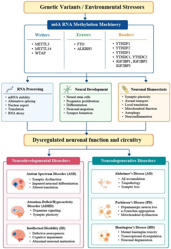
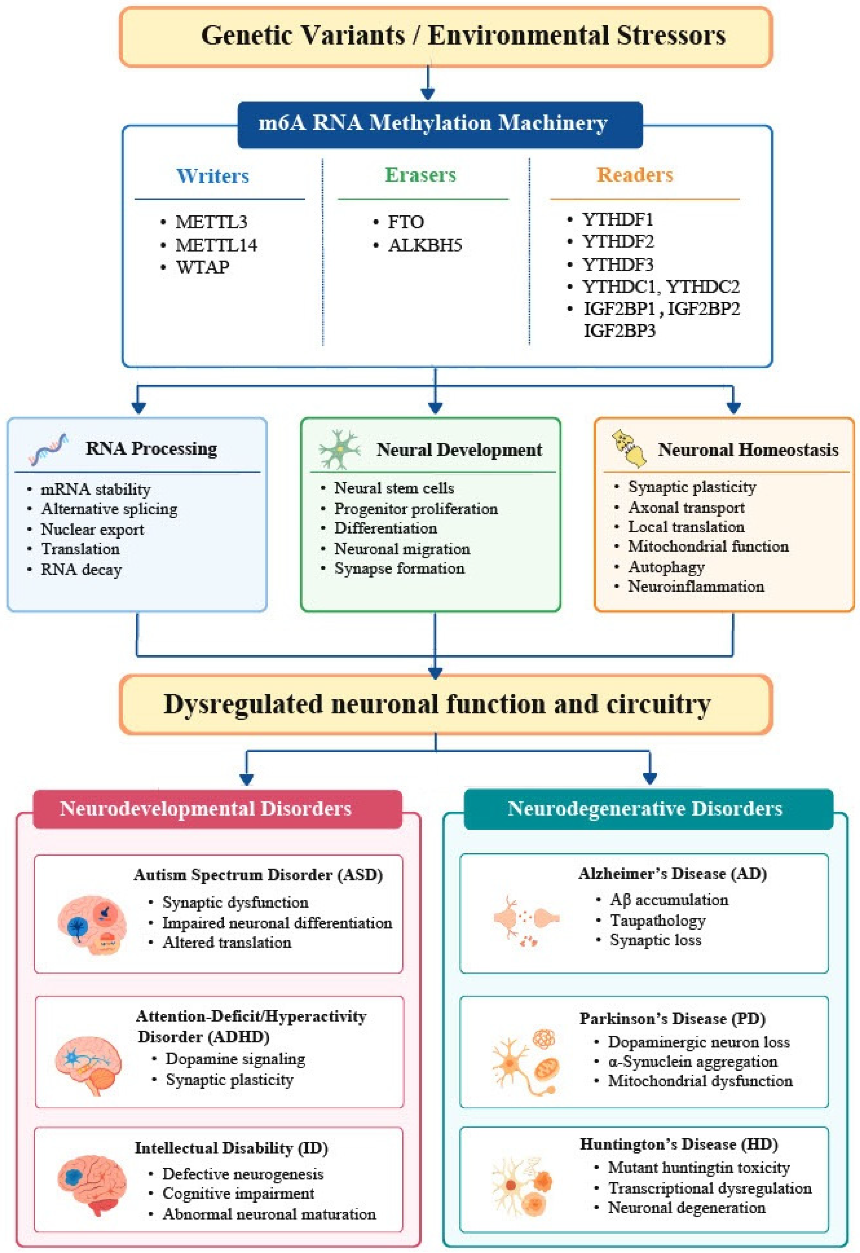

Disclaimer/Publisher’s Note: The statements, opinions and data contained in all publications are solely those of the individual author(s) and contributor(s) and not of MDPI and/or the editor(s). MDPI and/or the editor(s) disclaim responsibility for any injury to people or property resulting from any ideas, methods, instructions or products referred to in the content. |
© 2026 by the authors. Licensee MDPI, Basel, Switzerland. This article is an open access article distributed under the terms and conditions of the Creative Commons Attribution (CC BY) license.