


Simple Summary
Fopius arisanus is a parasitoid wasp widely used to control destructive Bactrocera pests. Quantitative real-time PCR (qRT-PCR) is widely used to quantify gene expression. However, accurate measurement in such studies relies on the careful selection of reference genes. In this study, we assessed ten candidate genes across different tissues of male and female F. arisanus to identify the most reliable ones. We found that the optimal reference genes varied between the female and male tissue panels, and that using unstable genes produced misleading results. This work provides a useful basis for future molecular studies in this species.
Abstract
Fopius arisanus (Hymenoptera: Braconidae) is an important parasitoid species in tephritid fruit fly biological control and host–parasitoid interaction research. Quantitative real-time PCR (qRT-PCR) is widely used to analyze gene expression, which requires stable reference genes for accurate normalization. However, no systematic evaluation of suitable reference genes for qRT-PCR analysis across F. arisanus tissues has been conducted. We evaluated the expression stability of ten candidate reference genes (Actin, α-tubulin, β-tubulin, RPL37a, EF1-α, EF1-δ, RPL4, RPS18, RPL13, and GAPDH) across five adult tissues from females and males. The stability of these genes was analyzed using the ΔCt method, geNorm, NormFinder, BestKeeper, and RefFinder, and the optimal number of reference genes was determined by geNorm pairwise variation analysis. The results revealed that the optimal reference genes differed between the female and male tissue panels: Actin and RPL13 were the most stable combination for female tissues, whereas RPL13 and RPS18 were recommended for male tissues. Notably, β-tubulin and GAPDH showed the poorest stability in female tissues, whereas α-tubulin and β-tubulin were the least stable in male tissues. The suitability of the stable and unstable reference genes was further validated by normalizing the expression of the target genes vitellogenin (Vg) and dual-specificity phosphatase CDC14A (CDC14A). Our findings represent a systematic evaluation of reference genes for F. arisanus and establish a reliable normalization strategy for future gene expression studies in this parasitoid wasp.
1. Introduction
Quantitative real-time PCR (qRT-PCR) is one of the most widely used methods for analyzing gene expression because of its high sensitivity and specificity [1]. It has been extensively applied in insect studies to examine gene expression patterns across developmental stages, tissues, sexes, and various experimental conditions [2,3]. However, the reliability of qRT-PCR results depends largely on the selection of appropriate reference genes for normalization. Internal reference genes are expected to show stable expression across different biological samples and are commonly used to normalize qRT-PCR data. Traditional housekeeping genes, such as Actin, tubulin, glyceraldehyde-3-phosphate dehydrogenase (GAPDH), elongation factor-1 alpha (EF1-α), and ribosomal protein genes, have been widely used as internal controls [2,4,5]. Nevertheless, increasing evidence has shown that the expression of these genes is not always constant, and their stability may vary among species, tissues, developmental stages, sexes, and experimental treatments [5,6]. The use of unstable reference genes can lead to inaccurate normalization and may distort the apparent expression patterns of target genes [6,7]. Therefore, systematic validation of candidate reference genes is essential for reliable qRT-PCR-based gene expression analysis.
Fopius arisanus (Hymenoptera: Braconidae) is an egg-pupal endoparasitoid of tephritid fruit flies and an important biological control agent against economically important Bactrocera pests [8]. It has been successfully established in Hawaii and French Polynesia, where it contributed to the suppression of Bactrocera dorsalis and other fruit fly populations, and has been widely studied in classical biological control, host suitability, and tri-trophic interactions [9,10,11]. Recent genomic and transcriptomic advances in F. arisanus have yielded valuable molecular resources, including a whole-genome assembly [12], transcriptomic datasets across developmental stages [13], and endogenous nudivirus (FaENV) genes associated with virus-like particle production in the female reproductive system [14]. These advances provide candidate genes for understanding the molecular basis of development, reproduction, and host–parasitoid interactions in F. arisanus. However, to translate these resources into functional understanding, accurate gene expression quantification is essential. To date, reference genes for qRT-PCR normalization in F. arisanus have not been systematically validated across different tissues, representing a critical gap for reliable gene expression analysis in F. arisanus.
In the present study, we evaluated the expression stability of ten candidate reference genes, including Actin, alpha-tubulin (α-tubulin), beta-tubulin (β-tubulin), ribosomal protein L37a (RPL37a), EF1-α, elongation factor 1-delta (EF1-δ), ribosomal protein L4 (RPL4), ribosomal protein S18 (RPS18), ribosomal protein L13 (RPL13), and GAPDH in F. arisanus across different male and female tissues. Reference gene stability was evaluated by ΔCt, geNorm, NormFinder, BestKeeper, and RefFinder, and the optimal gene number was determined by geNorm pairwise variation (Vn/Vn + 1) analysis. Finally, selected stable and unstable reference genes were used to normalize the expression of target genes to validate the impact of reference gene selection on qRT-PCR results. This study provides a systematic evaluation of reference genes for F. arisanus and establishes a reliable normalization strategy for future gene expression studies in this important biological control parasitoid.
2. Materials and Methods
2.1. Insect Rearing and Sample Preparation
Fopius arisanus was maintained in an insectary at 24 ± 2 °C, 55 ± 10% relative humidity, and a photoperiod of 14 h:10 h (L:D). Adult wasps were provided with a 20% (w/v) honey solution [15]. Seven-day-old male and female adults were dissected in sterile phosphate-buffered saline (PBS) under a stereomicroscope (SMZ161; Motic, Xiamen, China) for tissue collection. Individual wasps were dissected sequentially in ice-cold sterile PBS, and tissues were collected from individual wasps one by one before being pooled to generate each biological replicate. To minimize potential cross-contamination between tissue types, the dissection forceps were thoroughly cleaned with 75% ethanol before dissecting each new tissue type. For females, the head, fat body, gut, ovaries, and venom gland were collected; for males, the head, fat body, gut, testes, and Hagen’s gland (a male-specific gland on the internal surface of the 9th abdominal tergum) were collected. Tissues shared by both sexes (head, fat body, and gut) were collected separately for females and males without pooling across sexes. The dissected tissues were immediately transferred into RNase-free microcentrifuge tubes, frozen in liquid nitrogen, and stored at −80 °C until total RNA extraction. Each biological replicate consisted of an independent pool of tissues from 20 individual wasps; each tissue type had three independent biological replicates (60 wasps in total per tissue type).
2.2. RNA Extraction and cDNA Synthesis
Total RNA was extracted from each sample using FreeZol Reagent (R711; Vazyme, Nanjing, China) according to the manufacturer’s instructions. RNA purity and concentration were quantified via spectrophotometer (Eva 3200, Monad, Suzhou, China), and samples with A260/A280 ratios between 1.8 and 2.0 were used for subsequent analyses. Genomic DNA was removed and first-strand cDNA was synthesized from 50 ng of total RNA using HiScript III RT SuperMix for qPCR (+gDNA wiper) (R323-01; Vazyme, China). The final volume of each reverse-transcription reaction was 20 μL according to the manufacturer’s protocol. After synthesis, the cDNA was diluted 1:7 (v/v) with nuclease-free water and stored at −20 °C until use.
2.3. Primer Design and Standard Curve Construction
Fragments of ten candidate reference genes (Actin, α-tubulin, β-tubulin, RPL37a, EF1-α, EF1-δ, RPL4, RPS18, RPL13, and GAPDH) and target genes vitellogenin (Vg) and dual-specificity phosphatase CDC14A (CDC14A) were amplified from cDNA. The purified PCR products were separately cloned into the pClone007 Blunt Vector (CV16; Vazyme, China). The recombinant vectors were then transformed into DH5α Escherichia coli competent cells (Tsingke, Beijing, China). Positive recombinant plasmids were extracted using a plasmid extraction kit (DP103, TIANGEN, Beijing, China). For each gene, plasmids were serially diluted 10-fold to generate six to eight dilution points for standard curve construction. Cq values were plotted against the logarithm of plasmid copy numbers, and the correlation coefficient (R2) was calculated from the linear regression equation. The amplification efficiency was calculated using the equation Efficiency (%) = [10^(−1/slope) − 1] × 100 [16,17]. Primer sequences, product lengths, amplification efficiencies, R2 values, and slopes of the candidate reference and target genes are listed in Table 1.
Table 1.
Primer information for candidate reference genes and target genes used for qRT-PCR in Fopius arisanus.
2.4. Quantitative Real-Time PCR (qRT-PCR) Analysis
qRT-PCR was performed on a LightCycler 480 II Real-Time PCR System (Roche, Basel, Switzerland) using ChamQ SYBR Color qPCR Master Mix (Q411-02, Vazyme, China). Each 10 μL reaction contained 5 μL of 2× master mix, 0.2 μL of each primer (10 μM), 1 μL of cDNA template, and 3.6 μL of nuclease-free water. The cycling program consisted of an initial denaturation at 95 °C for 30 s, followed by 40 cycles of 95 °C for 10 s and 60 °C for 30 s. Three independent biological replicates were analyzed, each with three technical replicates.
2.5. Determination of Reference Gene Expression Stability
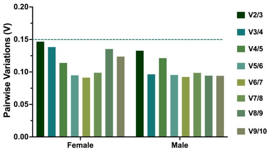
The expression stability of the ten candidate reference genes was evaluated using the comparative ΔCt method [18], BestKeeper [19], NormFinder, and geNorm [6]. The comprehensive ranking of candidate reference genes was obtained using RefFinder (https://blooge.cn/RefFinder/, v2020.10.20). The optimal number of reference genes required for normalization was determined by geNorm based on pairwise variation (Vn/Vn + 1). A threshold value of 0.15 was applied, and when Vn/Vn + 1 was below 0.15, n reference genes were considered sufficient for reliable normalization.
2.6. Validation of Reference Genes
To further evaluate the suitability of the selected reference genes, Vg and CDC14A were used as target genes to assess the performance of the stable reference genes RPL13 and RPS18, individually and in combination, and the unstable reference gene GAPDH. The expression levels of the target genes were analyzed across different tissues of male and female adults. Relative mRNA expression levels were calculated using the 2−ΔΔCt method. Statistical analysis was performed using one-way analysis of variance (ANOVA) followed by Tukey’s multiple comparisons test in GraphPad Prism 8.0.
3. Results
3.1. Primer Specificity and Amplification Efficiency
Prior to evaluating the expression stability of candidate reference genes, the specificity and amplification efficiency of the ten candidate reference gene primer pairs were assessed. Agarose gel electrophoresis showed that each candidate reference gene primer pair amplified a single band of the expected size, ranging from 89 to 193 bp (Figure S1). Standard curves were generated using 10-fold serial dilutions of recombinant plasmids to determine the amplification efficiency and linear correlation of each primer pair. The amplification efficiencies of the ten candidate reference gene primer pairs ranged from 90.5% to 106.9%, and the correlation coefficients ranged from 0.9908 to 1.0000 (Table 1; Figure S2). The slopes of the standard curves ranged from −3.573 to −3.167, indicating good amplification performance. These results demonstrated that all ten candidate reference gene primer pairs showed high specificity and acceptable amplification efficiency, and were therefore suitable for subsequent reference gene stability analysis.
3.2. Expression Profiles of Candidate Reference Genes
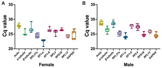
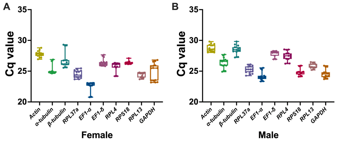
The Cq values of the ten candidate reference genes were analyzed to evaluate their expression levels and variation across different tissues of female and male F. arisanus. The Cq distribution showed that the candidate reference genes exhibited different expression levels among tissues (Figure 1). Among these candidates, EF1-α and Actin exhibited the highest and lowest expression levels, respectively, across both sexes. Based on the Cq distribution, RPL13 showed relatively small variation across tissues within each sex, approximately 0.9 cycles in the female tissue panel and approximately 0.8 cycles in the male tissue panel, whereas GAPDH, α-tubulin, and β-tubulin exhibited pronounced tissue-dependent fluctuations, with variation (range) exceeding 2 cycles in the female tissue panel and approximately 2–3 cycles in the male tissue panel (Figure 1). These results indicated that the expression levels of candidate reference genes varied among tissues, highlighting the need for systematic stability evaluation before target gene normalization.
Figure 1.
Expression profiles of ten candidate reference genes in different tissues of female (A) and male (B) F. arisanus. Boxes indicate the interquartile range, horizontal lines represent medians, and whiskers show minimum and maximum values.
3.3. Stability Analysis of Candidate Reference Genes in Female and Male Tissues
To systematically evaluate the stability of the ten candidate reference genes in different tissues of female and male F. arisanus, four commonly used algorithms, including the comparative ΔCt method, NormFinder, BestKeeper, and geNorm, were applied. Because different algorithms may generate different rankings, RefFinder was further used to integrate the results and provide a comprehensive ranking of candidate reference genes.
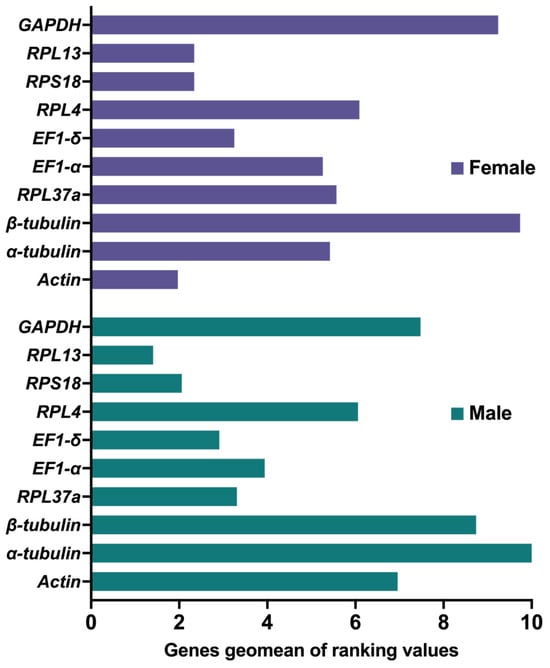
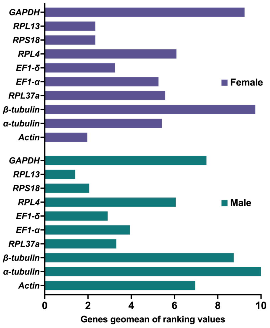
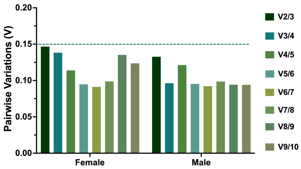
For female tissues, the comparative ΔCt method and NormFinder both identified Actin as the most stable candidate reference gene, followed by EF1-δ and RPL13 (Table 2). BestKeeper and geNorm identified RPL13 and RPS18 as the two most stable genes, which yielded the lowest SD and M-values, respectively (Table 2). Although the rankings differed slightly among the four algorithms, Actin, RPL13, and RPS18 consistently showed relatively high stability in female tissues. By contrast, β-tubulin and GAPDH were ranked among the least stable genes by most algorithms, occupying the last or second-last positions in all four analyses (Table 2), indicating that they were not suitable for normalization in female tissue samples. According to the comprehensive ranking generated by RefFinder, the overall stability of the candidate reference genes in female tissues was as follows: Actin > RPL13 > RPS18 > EF1-δ > EF1-α > α-tubulin > RPL37a > RPL4 > GAPDH > β-tubulin (Figure 2). Furthermore, geNorm pairwise variation analysis showed that the V2/3 value was 0.1466, which was below the recommended threshold of 0.15 (Figure 3). This result indicated that two reference genes were sufficient for accurate normalization in female tissues. Therefore, Actin and RPL13 were recommended as the optimal reference gene combination for female tissue samples.
Table 2.
Stability rankings of candidate reference genes in female and male tissues of F. arisanus using different algorithms.
Figure 2.
Comprehensive ranking of candidate reference genes in female and male F. arisanus based on RefFinder analysis. The geometric mean value was used to indicate the overall expression stability of each candidate reference gene. Lower geometric mean values indicate higher expression stability.
Figure 3.
Determination of the optimal number of reference genes for normalization in F. arisanus. Pairwise variation (Vn/Vn + 1) values were calculated using geNorm. The dashed line indicates the threshold value of 0.15; values below this threshold suggest that n reference genes are sufficient for reliable normalization.
For male tissues, RPL13 showed consistently high stability, ranking first in the comparative ΔCt method, NormFinder, and BestKeeper analyses. EF1-δ and RPS18 also showed relatively high stability, and RPS18 was ranked the most stable gene by geNorm. By contrast, α-tubulin and β-tubulin exhibited the poorest stability performance, occupying the bottom two ranks across all four algorithms. Thus, they are not recommended as reference genes for normalization in male tissues. According to RefFinder comprehensive analysis, the overall stability ranking of the candidate reference genes in male tissues was as follows: RPL13 > RPS18 > EF1-δ > RPL37a > EF1-α > RPL4 > Actin > GAPDH > β-tubulin > α-tubulin (Figure 2). GeNorm pairwise-variation analysis indicated that two reference genes were adequate for male-tissue samples. Thus, RPL13 and RPS18 were identified as the optimal reference gene combination for male tissue samples.
3.4. Validation of Candidate Reference Genes
To validate the selected reference genes, the expression levels of dual-specificity phosphatase CDC14A (CDC14A) and Vitellogenin (Vg) were analyzed in female and male tissue panels. The primer specificities for CDC14A and Vg were verified by agarose gel electrophoresis, yielding single amplification products of the expected fragment sizes (141 bp and 191 bp, respectively) (Figure S3). Standard-curve analysis showed that the amplification efficiencies of CDC14A and Vg were 91.1% and 103.4%, respectively, comparable to those of the validated reference genes (90.5–106.9%), thus satisfying the assumption underlying the 2−ΔΔCt relative quantification method (Table 1; Figure S4). The target genes were normalized using the stable reference genes RPS18, RPL13, and RPL13/RPS18, as well as the unstable reference gene GAPDH. For consistency in the validation analyses, RPL13/RPS18 was used for normalization in both the female and male tissue panels.
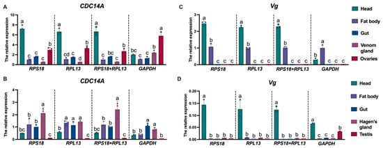
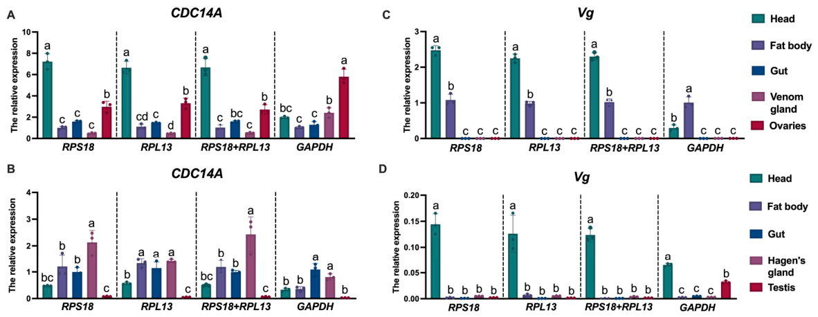
For CDC14A, normalization with RPS18, RPL13, or RPL13/RPS18 produced similar expression patterns. In female tissues, CDC14A showed the highest expression in the head, followed by the ovaries (Figure 4A). In male tissues, CDC14A was relatively highly expressed in the Hagen’s gland and showed the lowest expression in the testis. However, when GAPDH was used for normalization, the expression pattern was altered, particularly in female tissues, where CDC14A was no longer most highly expressed in the head but instead showed the highest expression in the ovaries. A similar discrepancy was also observed for Vg expression. For Vg, normalization with the stable reference genes showed that Vg was mainly expressed in the female head and fat body, while its expression in the gut, venom gland, and ovary was extremely low. In male tissues, Vg expression was generally low, with relatively higher expression detected in the head. In contrast, normalization with GAPDH changed the apparent expression pattern.
Figure 4.
Validation of selected reference genes using target gene expression analysis in female and male F. arisanus. Relative expression levels of Vg and CDC14A were normalized using the stable reference genes RPS18, RPL13, and RPL13/RPS18, as well as the unstable reference gene GAPDH. (A,C): expression patterns of CDC14A and Vg in female tissues; (B,D): expression patterns of CDC14A and Vg in male tissues. Data are shown as mean ± SE from three biological replicates. Different letters indicate significant differences among tissues based on one-way ANOVA followed by Tukey’s multiple comparison test (p < 0.05).
Overall, these results confirmed that RPL13 and RPS18 are reliable reference genes for expression analysis across female and male tissues in F. arisanus. The use of GAPDH led to altered expression profiles, indicating that unstable reference genes may cause biased interpretation of target gene expression.
4. Discussion
The present study showed that candidate reference gene stability differed markedly between female and male tissues of F. arisanus. This variation between tissue panels is consistent with previous reports in other insect orders, including Hemiptera, Lepidoptera, Coleoptera, and Diptera, in which optimal reference genes differed among species, tissues, developmental stages, and experimental treatments [2,5,20,21,22]. These findings reinforce that commonly used housekeeping genes cannot be assumed to be universally stable, and highlight the need for empirical validation before reference genes are adopted for qRT-PCR normalization.
Among the tested candidate genes, GAPDH was not recommended as a suitable reference gene for either female or male tissues of F. arisanus. Although GAPDH was identified as a suitable reference gene for tissue samples in some insects, such as Bombyx mori [23] and Spodoptera exigua [24], it showed poor stability in larval tissues of Chlorops oryzae [25]. The poor stability of GAPDH in F. arisanus may be related to its biological functions, as GAPDH encodes a key glycolytic enzyme involved in energy metabolism, development, and hormone-regulated processes [26]. In addition, GAPDH homologs have been detected in venom gland or venom-associated transcriptomes or proteomic studies of several parasitoid wasps, including Leptopilina, Ganaspis, and Psyttalia species [27,28,29,30]. In the present study, GAPDH showed pronounced tissue-dependent variation, particularly in female tissues, suggesting that its expression may be influenced by tissue function or physiological state in F. arisanus. Although the presence of GAPDH in parasitoid venom-associated studies raises the possibility that it may be associated with venom gland physiology [29,30], further evidence is required to determine whether GAPDH has a venom-related role in F. arisanus. Regardless of its specific function, its marked tissue-dependent variation makes GAPDH unsuitable as a reference gene for cross-tissue normalization. Similarly, α-tubulin and β-tubulin also showed poor stability, suggesting that cytoskeleton-related housekeeping genes are not necessarily suitable internal controls for tissue-specific expression analysis in F. arisanus.
In contrast, ribosomal protein genes are frequently identified as relatively stable reference genes in insects [31]. For example, RPL13 and RPS18 have been recommended in Aphidius gifuensis for developmental, tissue, or sex-related comparisons [32], and both genes also ranked among the stable candidates in Lipaphis erysimi [33]. Consistent with these studies, RPL13 exhibited high stability in both female and male tissues of F. arisanus, ranking as the most stable gene in males and among the top two in females. RPS18 also showed high stability, particularly in male tissues where it ranked second. Their relatively stable expression may be associated with their roles in ribosome structure and basal protein synthesis [34,35]. Notably, Actin was identified as the most stable gene in female tissues and was therefore included in the optimal combination with RPL13. However, its stability ranking dropped substantially in male tissues, further highlighting the tissue panel-specific nature of reference gene performance. Nevertheless, reference genes should still be empirically validated according to the target species, tissue types, and experimental conditions [36,37].
The target-gene validation further demonstrated the practical impact of reference gene selection on expression analysis. Based on the stability analyses, Actin/RPL13 was identified as the optimal combination for the female tissue panel, whereas RPL13/RPS18 was identified as the optimal combination for the male tissue panel. The use of the same reference-gene combination can reduce potential normalization bias caused by different internal control systems between tissues. Normalization with the stable reference genes produced generally consistent expression patterns, whereas the unstable reference gene GAPDH altered the apparent expression profiles of both target genes, confirming that inappropriate reference genes can lead to biased interpretation of gene expression [3,38,39]. CDC14A encodes a dual-specificity protein phosphatase and may be associated with cell-cycle regulation or tissue-specific physiological activity in F. arisanus [40]. For Vg, relatively high expression was detected in the female head and fat body. Vg is traditionally considered to be mainly synthesized in the fat body and transported to the ovary for yolk deposition and oocyte maturation [41]. However, the relatively high expression of Vg in the head may reflect a tissue-specific role beyond classical yolk protein synthesis, though this interpretation requires further validation [42,43]. Overall, these results demonstrate that the selection of reference genes can directly affect the biological interpretation of target-gene expression and highlight the importance of validated normalization systems for future functional studies in F. arisanus.
5. Conclusions
This study evaluated ten candidate reference genes in female and male tissues of Fopius arisanus. The optimal reference genes differed between the female and male tissues: Actin/RPL13 was recommended for female tissues, whereas RPL13/RPS18 was recommended for male tissues. GAPDH and tubulin showed poor stability and are not suitable for tissue-specific normalization in the respective tissue panels. Validation with CDC14A and Vg further demonstrated that reference-gene selection can substantially affect qRT-PCR expression profiles in F. arisanus.
Supplementary Materials
The following supporting information can be downloaded at https://www.mdpi.com/article/10.3390/insects17090927/s1, Figure S1: Specificity of PCR amplification for the ten candidate reference genes in F. arisanus. PCR products were separated by agarose gel electrophoresis. M, DNA marker; lanes 1–10 represent Actin, α-tubulin, β-tubulin, RPL37a, EF1-α, EF1-δ, RPL4, RPS18, RPL13, and GAPDH, respectively; Figure S2: Quantitative real-time PCR amplification curves of ten candidate reference genes in F. arisanus. A–J represent Actin, α-tubulin, β-tubulin, RPL37a, EF1-α, EF1-δ, RPL4, RPS18, RPL13, and GAPDH, respectively; Figure S3: Specificity of PCR amplification for the target genes Vg and CDC14A in F. arisanus. Amplicons were separated by agarose gel electrophoresis. M: DNA marker; lane 1: Vg; lane 2: CDC14A; Figure S4: Standard curves for the target genes CDC14A (A) and Vg (B) in F. arisanus. Cq values were plotted against the logarithm of plasmid copy numbers. The amplification efficiencies were 91.1% for CDC14A and 103.4% for Vg.
Author Contributions
Conceptualization, Y.C.; methodology, Y.C. and M.C.; software, Y.C. and M.C.; validation, X.Y., Y.C. and M.C.; formal analysis, C., Y.C. and M.C.; investigation, C., X.L. and M.C.; resources, X.L. and K.R.; data curation, X.L. and K.R.; writing—original draft preparation, Y.C. and M.C.; writing—review and editing, Y.C., X.Y. and M.C. All authors have read and agreed to the published version of the manuscript.
Funding
This research was funded by the National Natural Science Foundation of China (32502473 to Y.C. and 32472631 to X.Y.) and the Zhejiang Provincial Natural Science Foundation of China (LQN25C140004 to Y.C.).
Data Availability Statement
Data are contained within this article or the Supplementary Materials.
Acknowledgments
We are grateful to the developers of RefFinder for providing this web-based tool for evaluating the expression stability of candidate reference genes. The comprehensive ranking of candidate reference genes was generated using this online platform (https://blooge.cn/RefFinder/, v2020.10.20).
Conflicts of Interest
The authors declare no conflicts of interest.
References
- Wong, M.L.; Medrano, J.F. Real-time PCR for mRNA quantitation. Biotechniques 2005, 39, 75–85. [Google Scholar] [CrossRef] [Scilit] [PubMed]
- Wang, L.; Liu, Q.; Guo, P.; Gao, Z.; Chen, D.; Zhang, T.; Ning, J. Evaluation of reference genes for quantitative real-time PCR analysis in the bean bug, Riptortus pedestris (Hemiptera: Alydidae). Insects 2023, 14, 960. [Google Scholar] [CrossRef] [Scilit] [PubMed]
- Wang, L.; Yang, C.; Liu, Q.; Zhang, X.; Mei, X.; Zhang, T.; Ning, J. Validation and evaluation of reference genes for quantitative real-time PCR analysis in Mythimna loreyi (Lepidoptera: Noctuidae). Insects 2024, 15, 185. [Google Scholar] [CrossRef] [Scilit] [PubMed]
- Kozera, B.; Rapacz, M. Reference genes in real-time PCR. J. Appl. Genet. 2013, 54, 391–406. [Google Scholar] [CrossRef] [Scilit] [PubMed]
- Lü, J.; Yang, C.; Zhang, Y.; Pan, H. Selection of reference genes for the normalization of RT-qPCR data in gene expression studies in insects: A systematic review. Front. Physiol. 2018, 9, 1560. [Google Scholar] [CrossRef] [Scilit] [PubMed]
- Vandesompele, J.; De Preter, K.; Pattyn, F.; Poppe, B.; Van Roy, N.; De Paepe, A.; Speleman, F. Accurate normalization of real-time quantitative RT-PCR data by geometric averaging of multiple internal control genes. Genome Biol. 2002, 3, research0034. [Google Scholar] [CrossRef] [Scilit] [PubMed]
- Khan, M.M.; Guo, C.-F.; Peng, J.; Fan, Z.-Y.; Hafeez, M.; Ali, D.; Wang, K.; Almarzoug, M.H.A.; Qiu, B.-L. Screening and validation of reference genes using in RT-qPCR for gene expression studies in Paederus fuscipes, a medically and agriculturally important insect. J. King Saud Univ. Sci. 2022, 34, 101654. [Google Scholar] [CrossRef] [Scilit]
- Rousse, P.; Harris, E.J.; Quilici, S. Fopius arisanus, an egg-pupal parasitoid of Tephritidae. Overview. Biocontrol News Inf. 2005, 26, 59N–69N. [Google Scholar]
- Vargas, R.I.; Leblanc, L.; Harris, E.J.; Manoukis, N.C. Regional suppression of Bactrocera fruit flies (Diptera: Tephritidae) in the pacific through biological control and prospects for future introductions into other areas of the world. Insects 2012, 3, 727–742. [Google Scholar] [CrossRef] [Scilit] [PubMed]
- Moquet, L.; Jobart, B.; Fontaine, R.; Delatte, H. Tri-trophic interactions among Fopius arisanus, Tephritid species and host plants suggest apparent competition. Ecol. Evol. 2023, 13, e9742. [Google Scholar] [CrossRef] [Scilit] [PubMed]
- Vargas, R.I.; Leblanc, L.; Putoa, R.; Eitam, A. Impact of introduction of Bactrocera dorsalis (Diptera: Tephritidae) and classical biological control releases of Fopius arisanus (Hymenoptera: Braconidae) on economically important fruit flies in French Polynesia. J. Econ. Entomol. 2007, 100, 670–679. [Google Scholar] [CrossRef] [Scilit]
- Geib, S.M.; Liang, G.H.; Murphy, T.D.; Sim, S.B. Whole-Genome sequencing of the braconid parasitoid wasp Fopius arisanus, an important biocontrol agent of pest tephritid fruit flies. G3 Genes Genomes Genet. 2017, 7, 2407–2411. [Google Scholar] [CrossRef] [Scilit] [PubMed]
- Calla, B.; Sim, S.B.; Hall, B.; DeRego, T.; Liang, G.H.; Geib, S.M. Transcriptome of the egg parasitoid Fopius arisanus: An important biocontrol tool for Tephritid fruit fly suppression. GigaScience 2015, 4, 36. [Google Scholar] [CrossRef] [Scilit] [PubMed]
- Burke, G.R.; Simmonds, T.J.; Sharanowski, B.J.; Geib, S.M. Rapid viral symbiogenesis via changes in parasitoid wasp genome architecture. Mol. Biol. Evol. 2018, 35, 2463–2474. [Google Scholar] [CrossRef] [Scilit] [PubMed]
- Manoukis, N.; Geib, S.; Seo, D.; McKenney, M.; Vargas, R.; Jang, E. An optimized protocol for rearing Fopius arisanus, a parasitoid of Tephritid fruit flies. J. Vis. Exp. 2011, 2, 2901. [Google Scholar] [CrossRef] [Scilit] [PubMed]
- Bustin, S.A.; Benes, V.; Garson, J.A.; Hellemans, J.; Huggett, J.; Kubista, M.; Mueller, R.; Nolan, T.; Pfaffl, M.W.; Shipley, G.L.; et al. The MIQE guidelines: Minimum information for publication of quantitative real-time PCR experiments. Clin. Chem. 2009, 55, 611–622. [Google Scholar] [CrossRef] [Scilit] [PubMed]
- Pfaffl, M.W. A new mathematical model for relative quantification in real-time RT-PCR. Nucleic Acids Res. 2001, 29, e45. [Google Scholar] [CrossRef] [Scilit] [PubMed]
- Silver, N.; Best, S.; Jiang, J.; Thein, S.L. Selection of housekeeping genes for gene expression studies in human reticulocytes using real-time PCR. BMC Mol. Biol. 2006, 7, 33. [Google Scholar] [CrossRef] [Scilit] [PubMed]
- Pfaffl, M.W.; Tichopad, A.; Prgomet, C.; Neuvians, T.P. Determination of stable housekeeping genes, differentially regulated target genes and sample integrity: BestKeeper–Excel-based tool using pair-wise correlations. Biotechnol. Lett. 2004, 26, 509–515. [Google Scholar] [CrossRef] [Scilit] [PubMed]
- Wang, Z.; Meng, Q.; Zhu, X.; Sun, S.; Liu, A.; Gao, S.; Gou, Y. Identification and evaluation of reference genes for normalization of gene expression in developmental stages, sexes, and tissues of Diaphania caesalis (Lepidoptera, Pyralidae). J. Insect Sci. 2020, 20, 6. [Google Scholar] [CrossRef] [Scilit] [PubMed]
- Sellamuthu, G.; Amin, S.; Bílý, J.; Synek, J.; Modlinger, R.; Sen, M.K.; Chakraborty, A.; Roy, A. Reference gene selection for normalizing gene expression in Ips sexdentatus (Coleoptera: Curculionidae: Scolytinae) under different experimental conditions. Front. Physiol. 2021, 12, 752768. [Google Scholar] [CrossRef] [Scilit] [PubMed]
- Dzaki, N.; Azzam, G. Assessment of Aedes albopictus reference genes for quantitative PCR at different stages of development. PLoS ONE 2018, 13, e0194664. [Google Scholar] [CrossRef] [Scilit] [PubMed]
- Teng, X.; Zhang, Z.; He, G.; Yang, L.; Li, F. Validation of reference genes for quantitative expression analysis by real-time RT-PCR in four lepidopteran insects. J. Insect Sci. 2012, 12, 60. [Google Scholar] [CrossRef] [Scilit] [PubMed]
- Zhu, X.; Yuan, M.; Shakeel, M.; Zhang, Y.; Wang, S.; Wang, X.; Zhan, S.; Kang, T.; Li, J. Selection and evaluation of reference genes for expression analysis using qRT-PCR in the beet armyworm Spodoptera exigua (Hübner) (Lepidoptera: Noctuidae). PLoS ONE 2014, 9, e84730. [Google Scholar] [CrossRef] [Scilit] [PubMed]
- Tian, P.; Qiu, L.; Zhou, A.; Chen, G.; He, H.; Ding, W.; Li, Y. Evaluation of appropriate reference genes for investigating gene expression in Chlorops oryzae (Diptera: Chloropidae). J. Econ. Entomol. 2019, 112, 2207–2214. [Google Scholar] [CrossRef] [Scilit] [PubMed]
- Zhao, W.; Zhang, B.; Geng, Z.; Chang, Y.; Wei, J.; An, S. The uncommon function and mechanism of the common enzyme glyceraldehyde-3-phosphate dehydrogenase in the metamorphosis of Helicoverpa armigera. Front. Bioeng. Biotechnol. 2022, 10, 1042867. [Google Scholar] [CrossRef] [Scilit] [PubMed]
- Chou, J.; Li, M.Z.; Wey, B.; Mumtaz, M.; Ramroop, J.R.; Singh, S.; Govind, S. Venomous Cargo: Diverse toxin-related proteins are associated with extracellular vesicles in parasitoid wasp venom. Pathogens 2025, 14, 255. [Google Scholar] [CrossRef] [Scilit] [PubMed]
- Goecks, J.; Mortimer, N.T.; Mobley, J.A.; Bowersock, G.J.; Taylor, J.; Schlenke, T.A. Integrative approach reveals composition of endoparasitoid wasp venoms. PLoS ONE 2013, 8, e64125. [Google Scholar] [CrossRef] [Scilit] [PubMed]
- Anderson, T.; Long, L.; Santisteban, M.; Sterne-Marr, R.; Mortimer, N.T. Expression, purification, and enzymology of GAPDH from the venom of the parasitoid wasp Ganaspis hookeri. J. Biol. Chem. 2024, 300, 106437. [Google Scholar] [CrossRef] [Scilit]
- Mathé-Hubert, H.; Colinet, D.; Deleury, E.; Belghazi, M.; Ravallec, M.; Poulain, J.; Dossat, C.; Poirié, M.; Gatti, J.-L. Comparative venomics of Psyttalia lounsburyi and P. concolor, two olive fruit fly parasitoids: A hypothetical role for a GH1 β-glucosidase. Sci. Rep. 2016, 6, 35873. [Google Scholar] [CrossRef] [Scilit] [PubMed]
- Li, M.; Li, X.; Wang, C.; Li, Q.; Zhu, S.; Zhang, Y.; Li, X.; Yang, F.; Zhu, X. Selection and validation of reference genes for qRT-PCR analysis of Rhopalosiphum padi (Hemiptera: Aphididae). Front. Physiol. 2021, 12, 663338. [Google Scholar] [CrossRef] [Scilit] [PubMed]
- Gao, X.-K.; Zhang, S.; Luo, J.-Y.; Wang, C.-Y.; Lü, L.-M.; Zhang, L.-J.; Zhu, X.-Z.; Wang, L.; Cui, J.-J. Identification and validation of reference genes for gene expression analysis in Aphidius gifuensis (Hymenoptera: Aphidiidae). PLoS ONE 2017, 12, e0188477. [Google Scholar] [CrossRef] [Scilit] [PubMed]
- Koramutla, M.K.; Aminedi, R.; Bhattacharya, R. Comprehensive evaluation of candidate reference genes for qRT-PCR studies of gene expression in mustard aphid, Lipaphis erysimi (Kalt). Sci. Rep. 2016, 6, 25883. [Google Scholar] [CrossRef] [Scilit] [PubMed]
- Melnikov, S.; Ben-Shem, A.; Garreau de Loubresse, N.; Jenner, L.; Yusupova, G.; Yusupov, M. One core, two shells: Bacterial and eukaryotic ribosomes. Nat. Struct. Mol. Biol. 2012, 19, 560–567. [Google Scholar] [CrossRef] [Scilit] [PubMed]
- Bhavsar, R.B.; Makley, L.N.; Tsonis, P.A. The other lives of ribosomal proteins. Hum. Genom. 2010, 4, 327–344. [Google Scholar] [CrossRef] [Scilit] [PubMed]
- Wang, Z.; Meng, Q.; Zhu, X.; Sun, S.; Gao, S.; Gou, Y.; Liu, A. Evaluation and validation of reference genes for quantitative real-time PCR in Helopeltis theivora Waterhouse (Hemiptera: Miridae). Sci. Rep. 2019, 9, 13291. [Google Scholar] [CrossRef] [Scilit] [PubMed]
- Hou, Q.; Yuan, L.; Jin, H.; Yan, H.; Li, F.; Wu, S. Identification and validation of reference genes for normalization of gene expression analysis using qRT-PCR in Megalurothrips usitatus (Thysanoptera: Thripidae). Front. Physiol. 2023, 14, 1161680. [Google Scholar] [CrossRef] [Scilit] [PubMed]
- Dheda, K.; Huggett, J.F.; Bustin, S.A.; Johnson, M.A.; Rook, G.; Zumla, A. Validation of housekeeping genes for normalizing RNA expression in real-time PCR. Biotechniques 2004, 37, 112–114, 116, 118–119. [Google Scholar] [CrossRef] [Scilit] [PubMed]
- Pinheiro, D.H.; Siegfried, B.D. Selection of reference genes for normalization of RT-qPCR data in gene expression studies in Anthonomus eugenii Cano (Coleoptera: Curculionidae). Sci. Rep. 2020, 10, 5070. [Google Scholar] [CrossRef] [Scilit] [PubMed]
- Manzano-López, J.; Monje-Casas, F. The multiple roles of the Cdc14 phosphatase in cell cycle control. Int. J. Mol. Sci. 2020, 21, 709. [Google Scholar] [CrossRef] [Scilit] [PubMed]
- Wu, Z.; Yang, L.; He, Q.; Zhou, S. Regulatory mechanisms of vitellogenesis in insects. Front. Cell Dev. Biol. 2021, 8, 593613. [Google Scholar] [CrossRef] [Scilit] [PubMed]
- Roy-Zokan, E.M.; Cunningham, C.B.; Hebb, L.E.; McKinney, E.C.; Moore, A.J. Vitellogenin and vitellogenin receptor gene expression is associated with male and female parenting in a subsocial insect. Proc. R. Soc. B Biol. Sci. 2015, 282, 20150787. [Google Scholar] [CrossRef] [Scilit] [PubMed]
- Dittmer, J.; Alafndi, A.; Gabrieli, P. Fat body–specific vitellogenin expression regulates host-seeking behaviour in the mosquito Aedes albopictus. PLoS Biol. 2019, 17, e3000238. [Google Scholar] [CrossRef] [Scilit] [PubMed]

Disclaimer/Publisher’s Note: The statements, opinions and data contained in all publications are solely those of the individual author(s) and contributor(s) and not of MDPI and/or the editor(s). MDPI and/or the editor(s) disclaim responsibility for any injury to people or property resulting from any ideas, methods, instructions or products referred to in the content. |
© 2026 by the authors. Licensee MDPI, Basel, Switzerland. This article is an open access article distributed under the terms and conditions of the Creative Commons Attribution (CC BY) license.