


Simple Summary
Mythimna separata exhibits density-dependent prophylaxis (DDP), but mechanisms beyond immune regulation remain underexplored. Here we show that two cuticular protein genes, MsCP1 and MsCP2, contribute to DDP in this pest. Both genes were significantly upregulated in the cuticle of gregarious (high-density) larvae compared to solitary (low-density) individuals. Star polycation (SPc) nanocarrier-mediated knockdown of MsCP1 and MsCP2 in gregarious larvae reduced their expression to levels comparable to solitary controls, and led to abnormal molting, reduced cuticle thickness, and altered ultrastructure. Moreover, silenced gregarious larvae showed increased mortality following Beauveria bassiana infection, similar to solitary larvae. These results indicate that MsCP1 and MsCP2 enhance fungal resistance in crowded M. separata by modulating cuticle properties, expanding our understanding of DDP and informing biological control strategies.
Abstract
Mythimna separata, an important insect pest of grain crops, exhibits density-dependent prophylaxis (DDP). Most studies have focused on DDP from the perspective of immune system regulation. In this report, we show that two cuticular protein genes, MsCP1 and MsCP2, are involved in DDP of M. separata. MsCP1 and MsCP2 were highly expressed in 4th and 5th instar larvae of the M. separata gregarious high-density phase as compared to solitary, low-density individuals. The expression levels of MsCP1 and MsCP2 were significantly higher in the cuticle of gregarious larvae than in solitary individuals. When MsCP1 and MsCP2 were knocked down by star polycation (SPc) nanocarrier-mediated RNA interference (RNAi), the expression levels of the two genes were significantly downregulated in gregarious larvae as compared to dsGFP/SPc-treated gregarious larvae but were not significantly different from dsGFP/SPc-treated solitary larvae. Furthermore, silencing of MsCP1 and MsCP2 caused abnormal molting, alterations in ultrastructure, and reduced cuticle thickness in gregarious larvae. The mortality rates in dsMsCP1/SPc- and dsMsCP2/SPc-treated gregarious larvae were significantly higher after infection with the entomopathogenic fungus Beauveria bassiana as compared to the dsGFP/SPc-treated gregarious larva but were not significantly different from mortality rates in dsGFP/SPc-treated solitary larvae. Taken together, these findings suggest that upregulation of MsCP1 and MsCP2 are associated with resistance of gregarious M. separata to B. bassiana. These results broaden our understanding of insect DDP and are of great significance in the biological control of M. separata.
1. Introduction
Insects represent one of the most successful animal groups in nature, largely because of their highly efficient immune systems that respond to invading pathogens. In addition to innate immunity, insects exhibit plasticity in their immune systems, which allows them to adjust immune capacity according to environmental conditions and physiological states [1,2]. It has been established that changes in population density can alter the immune response of insects [3], and it is generally assumed that high population densities weaken insect immunity due to lack of resources and density-dependent physiological stress [4,5]. Interestingly, some insect taxa exhibit enhanced disease resistance under high population densities, which is termed “density-dependent prophylaxis (DDP)” [6]. The first report of DDP was described in Pseudaletia separata [7]; thereafter, this phenomenon was reported in Tenebrio molitor [8], Schistocerca gregaria [9], Bombus terrestris [10], Locusta migratoria [11], Anticarsia gemmatalis [12], Plutella xylostella [13], Ceratina okinawana [14] and Spodoptera litura [15]. These studies suggest that DDP occurs in a variety of insect species, especially in those that exhibit phase polymorphism.
The underlying mechanisms of DDP have been elucidated in some insect taxa. For example, immune-related parameters such as phenoloxidase activity, lysozyme activity and total hemocyte counts were significantly improved in some species under high-density conditions [9,16,17,18]. Importantly, the induction of DDP can be pathogen-specific in insects. For example, Mythimna separata larvae showed the greatest resistance to the bacterial pathogen, Bacillus thuringiensis, at a density of 10 larvae/vial, whereas the highest level of resistance to the fungus Beauveria bassiana was observed at 30 larvae/vial [18]. This suggests that insects may deploy distinct immune signaling pathways for bacterial and fungal pathogens.
Significant progress has been made in understanding the molecular mechanisms underlying insect DDP in the past decade. Early transcriptomic analyses using RNA-seq revealed that genes encoding protease inhibitors, antioxidants and pattern recognition proteins were upregulated in gregarious locusts as compared to solitary individuals [11]. In recent years, RNA interference (RNAi) technology has emerged as a transformative tool for gene function characterization and pest management [19]. For example, Wang et al. demonstrated that RNA interference (RNAi)-mediated silencing of gnbp3, which encodes a pattern recognition protein, significantly improved the susceptibility of gregarious locusts to Metarhizium anisopliae [11]. The Toll-Spätzle signaling pathway, a core component of insect innate immune systems, plays a critical role in defense against bacterial, fungal, and other pathogenic infections. In line with this, Jiang et al. reported that the expression of MsToll-1 and MsSpätzle-4 in M. separata were significantly upregulated with increasing larval density. Moreover, RNAi-mediated knockdown of MsToll-1 or MsSpätzle-4 resulted in significant downregulation of multiple immune-related genes in gregarious M. separata larvae; more importantly, the survival rate of RNAi-treated gregarious larvae decreased after infection with B. thuringiensis [20]. Octopamine, a crucial stress hormone in insects, is involved in various physiological processes, including metabolism, locomotion, and immunity. The octopamine content in the hemolymph of M. separata larvae reared under high-density conditions was significantly increased as compared to low-density larvae [18]. Exogenous injection of octopamine significantly enhanced larval phenoloxidase activity and hemocyte counts, whereas injection of its antagonist epinastine suppressed the upregulation of these immune parameters [18]. Furthermore, silencing the gene encoding the octopamine synthesis enzyme, tyramine β-hydroxylase (Tβh), in larvae reared in high-density conditions resulted in a 25.1% reduction in octopamine content and a 55.7% decrease in survival of gregarious larvae following exposure to B. bassiana [21]. These results suggest that insects perceive signals associated with changes in population density via an unknown mechanism, which subsequently activates the octopamine-mediated neuroendocrine system and Toll–Spätzle immune pathway, ultimately leading to enhanced resistance to pathogens. However, whether other genes or pathways participate in this process is largely unknown.
The insect cuticle is a multilayered structure that functions as an exoskeleton and also provides a physical barrier against adverse environmental factors [22]. The major components of the insect cuticle are cuticular proteins (CPs), chitin, lipids and water [23]. It is well-established that cuticular proteins play an important role in insect growth and development [24,25,26,27]. Interestingly, recent studies have shown that CPs are involved in insect resistance to pesticides [28]. Long-term insecticide exposure improves cuticle thickness and upregulates the expression of genes encoding CPs, thereby decreasing the ability of insecticides to traverse the cuticular barrier [29,30,31,32,33]. Similarly, entomopathogenic fungi adhere to and grow on the insect exoskeleton, ultimately penetrating the cuticle and moving into hemolymph. It is not clear whether cuticular proteins participate in insect DDP.
The oriental armyworm, Mythimna separata, is an important pest of grain crops in Asia and exhibits DDP [34]. Our recent studies demonstrated that gregarious, high-density larvae of M. separata have thicker cuticles and greater expression of cuticular proteins than solitary, low-density larvae [35], leading us to hypothesize that cuticular proteins are critical for DDP. To test this hypothesis, a reliable gene function analysis tool is required. RNAi has been widely used for studying gene functions in insects; however, its efficiency is often low in lepidopteran species due to the existence of dsRNA-degrading nucleases in the hemolymph and midgut [36]. To overcome these barriers, various nanomaterial-based dsRNA delivery systems have been developed, including chitosan nanoparticles, liposomes, carbon nanotubes, layered double hydroxides, and star polycation (SPc) [37,38,39]. Among these, SPc-mediated dsRNA delivery has been successfully applied in multiple insect species, significantly improving RNAi efficiency compared with naked dsRNA [40,41,42,43]. In the present study, we first identified two genes encoding cuticular proteins (designated MsCP1 and MsCP2) from the M. separata transcriptome database [34] and analyzed their transcription in different developmental stages and tissues of solitary and gregarious larvae. Subsequently, we employed SPc-mediated RNAi targeting MsCP1 and MsCP2 to evaluate their impact on cuticle morphology and resistance to B. bassiana. Our study provides insight into the role of M. separata cuticular proteins in DDP and further broadens our understanding of this phenomenon, thus revealing potential novel management strategies for this insect pest.
2. Materials and Methods
2.1. Insect Colonies and Fungal Strain
The long-term laboratory population of M. separata used in this study was originally collected as larvae on maize in Qianxi City, China, in 2014. The larval colony containing both solitary and gregarious M. separata was established as described previously [44] and reared on a corn-based diet at 25 ± 1 °C with a 14:10 h light/dark photoperiod. Emerging adults of both phases were transferred to nylon cages and provided with a 10% honey/water diet and plastic ropes for oviposition.
The star polycation (SPc) nanocarrier used to delive dsRNA was provided by professor Yan at China Agricultural University and stored at 4 °C. The Beauveria bassiana strain used in this study was obtained from Shanxi Luhai Pesticide Technology Co., Ltd. (Shanxi, China) and stored at −80 °C until needed. Prior to experiment, the B. bassiana strain was inoculated onto potato dextrose agar (PDA) medium (potato 200 g, glucose 20 g, agar 20 g, peptone 5 g, distilled water 1000 mL) and cultured in an artificial climate chamber at 26 °C ± 1 °C, relative humidity 80% ± 5%, and a photoperiod of 12 h light: 12 h dark for 10 days. Based on the preliminary experiment, the B. bassiana spore was diluted with DEPC-treated water to a concentration of 1.0 × 10⁷ CFU/mL.
2.2. Bioinformatic Analysis of MsCP1 and MsCP2
The cDNA sequences of MsCP1 and MsCP2 were obtained from our recently sequenced M. separata transcriptome database [43]. Nucleotide and deduced amino acid sequences of MsCP1 and MsCP2 were identified with ORF finder (https://www.ncbi.nlm.nih.gov/orffinder/), and SMART (http://smart.embl.de/) was used to predict conserved domains. Deduced CP amino acid sequences from other insects were compared with MsCPs using DNAMAN (https://www.lynnon.com/), and a phylogenetic tree was constructed using MEGA v. 7.0 and the neighbor-joining algorithm with 2000 repetitions.
2.3. Expression Patterns of MsCP Genes in Gregarious and Solitary M. separata
Samples were collected from 4th to 6th instar larvae because the crowding effect becomes apparent only after the 3rd instar stage. The tissues including heads, cuticles, hemolymph, fat bodies, guts and feet were collected from the 5th instar larvae of both phases. Total RNA of each sample was extracted using Eastep® Super Total RNA Isolation Kit following the manufacturer’s protocol (Promega, Madison, WI, USA), and genomic DNA was removed with DNase I. After RNA quality was checked and concentrations were determined, the iScript cDNA Synthesis Kit (Bio-Rad, Hercules, CA, USA) was used to synthesize first-strand cDNA from total RNA. Quantitative real-time PCR (qRT-PCR) was performed using a CFX96 Real-time PCR System (Bio-Rad, Hercules, CA, USA) as follows: 95 °C for 2 min, followed by 35 cycles of 95 °C for 5 s, and 30 s at 60 °C. Melting curves for each primer pair were determined, and β-Actin and Tubulin were used as reference genes for developmental stages and tissues, respectively [45]. The 2−ΔΔCt method was utilized to determine expression levels of the two MsCPs. Experiments consisted of three independent biological replications, with each replicate containing 3–5 larvae. Primers used in the experiment are listed in Table S1.
2.4. RNAi of MsCP1 and MsCP2
The double-stranded RNAs (dsRNAs) for MsCP1 and MsCP2 were amplified using gene-specific primers with integrated T7 promoters (Table S1). dsRNA synthesis was performed using the Transcript Aid T7 High Yield Transcription Kit (Thermo Scientific, Waltham, MA, USA). The synthesized dsRNA was purified and dissolved in ddH2O at 5 µg/µL. Owing to the low efficacy of RNAi in lepidopteran species, a star polycation (SPC) nanocarrier was used to deliver the dsRNA according to the previously described method with minor modifications [40]. Briefly, the dsRNA samples and SPc were diluted in DEPC-treated water to concentration of 5 μg/μL and 10 μg/μL, respectively. Then, two volumes of the dsRNA and one volume of SPc solution were mixed thoroughly and incubated at 25 °C for 30 min to allow for the formation of dsRNA/SPc complexes. Finally, this solution was diluted with DEPC-treated water to a final concentration of 2.5 μg/μL. On the first day of the 4th instar larval stage, individuals from gregarious (G) and solitary (S) phases were used in RNAi experiments. Each larvae was injected with 1 μL dsRNA/SPc (2.5 µg/μL). Treatments included the following: (1) solitary larvae injected with dsGFP/SPc (S-dsGFP/SPc); (2) gregarious larvae injected with dsMsCP1/SPc (G-dsMsCP1/SPc); (3) gregarious larvae injected with dsMsCP2/SPc (G-dsMsCP2/SPc); and (4) gregarious larvae injected with dsGFP/SPc (G-dsGFP/SPc). At 48 h after injection, samples were collected for RNA extraction, and qRT-PCR was conducted to determine the interference efficiency of MsCP1 and MsCP2 expression as described above. Phenotypes for each treatment were observed and photographed with a digital camera (DS-Fi3, Nikon, Tokyo, Japan). Each treatment observed 20 larvae.
2.5. Scanning Electron Microscopy
To evaluate the potential effects of MsCPs knockdown on cuticle development, the ultrastructure of larval cuticles from the above four treatments were analyzed by scanning electron microscopy (SEM). Cuticles on the dorsal side of solitary and gregarious larvae were dissected at 72 h after RNAi. Dissected samples were washed with PBS (pH 7.4), fixed for 2 h at room temperature, and stored at 4 °C until needed. Fixed samples were then washed three times in 0.1 M PBS for 15 min. Samples were transferred to 1% OsO4 for 2 h and incubated at room temperature in darkness, followed by three washes with 0.1 M PBS as described above. Washed cuticles were then incubated for 15 min in a graded ethanol series (30, 50, 70, 80, 90, 95 and 100%) and then dried in an EM CPD300 critical point dryer (Leica, Solms, Germany). Samples were then coated with gold for 30 s using a K550X sputter coater (Electron Microscopy Sciences, Hatfield, PA, USA). Finally, a TM-1000 SEM (Hitachi, Tokyo, Japan) was used to record images at 3 kV. Since we did not observe any differences in cuticles of the same phase [35], the middle regions of cuticles were compared. Five individuals from each phase were used for SEM analysis.
2.6. Transmission Electron Microscopy
Cuticle thickness in solitary and gregarious larvae treated with dsRNAs was evaluated by transmission electron microscopy (TEM). Cuticles from the four treatments (Section 2.4) were prepared for TEM as described previously [29], and a JEM-1210 transmission electron microscope (HT7800, Hitachi, Tokyo, Japan) was used to observe dissected cuticles at 80 kV. Digital images for each sample were photographed and used to measure thickness with Image J2 (https://imagej.net/ij/). Since no obvious differences were apparent among different cuticular sections in our previous study [34], five cuticular regions were randomly selected in gregarious (n = 4) and solitary larvae (n = 4) and analyzed for thickness.
2.7. Resistance to B. bassiana
To evaluate the effects of MsCP1 and MsCP2 knockdown on disease resistance, surviving larvae were collected at 24 h after injection and exposed to a B. bassiana suspension containing 1.0 × 10⁷ CFU/mL. Larvae were exposed to the B. bassiana solution for 5 s, air-dried at room temperature and transferred to their initial rearing conditions. Survival rates were recorded daily until larvae either died or pupated. This experiment included three independent biological replications, with each replicate containing 20 larvae.
2.8. Statistical Analyses
Significant differences among treatments were determined by one-way ANOVA, followed by Tukey’s honest significant difference (HSD) test. Student’s t-test was used to assess differences between the gregarious and solitary larval phases, and significance at p < 0.05 (*), p ≤ 0.01 (**), and p ≤ 0.001 (***) was determined. Datapoints represent as means ± standard error (SE). All statistical analyses were carried out using DPS v. 17.0 software [46].
3. Results
3.1. Bioinformatic Analysis of MsCP1 and MsCP2
Two cuticular protein genes were identified in the M. separata transcriptome and designated MsCP1 and MsCP2; these two genes encoded 396 and 336 bp ORFs with deduced protein sequences of 131 and 111 amino acids, respectively (Figure S1). Sequence analysis showed that both CPs contained a chitin-binding domain in the RR1 subfamily. The two MsCPs and CPs from other insect species were analyzed for phylogenetic relatedness. As shown in Figure S2, the phylogenetic tree was clearly divided into two branches, including Lepidoptera species and other species. Within Lepidoptera branch, MsCP1 and MsCP2 clustered with CP in Mythimna loreyi on a branch adjacent to CP in Colics croceus.
3.2. Expression of MsCP1 and MsCP2 in Gregarious and Solitary M. separata
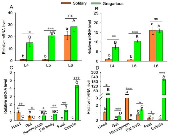
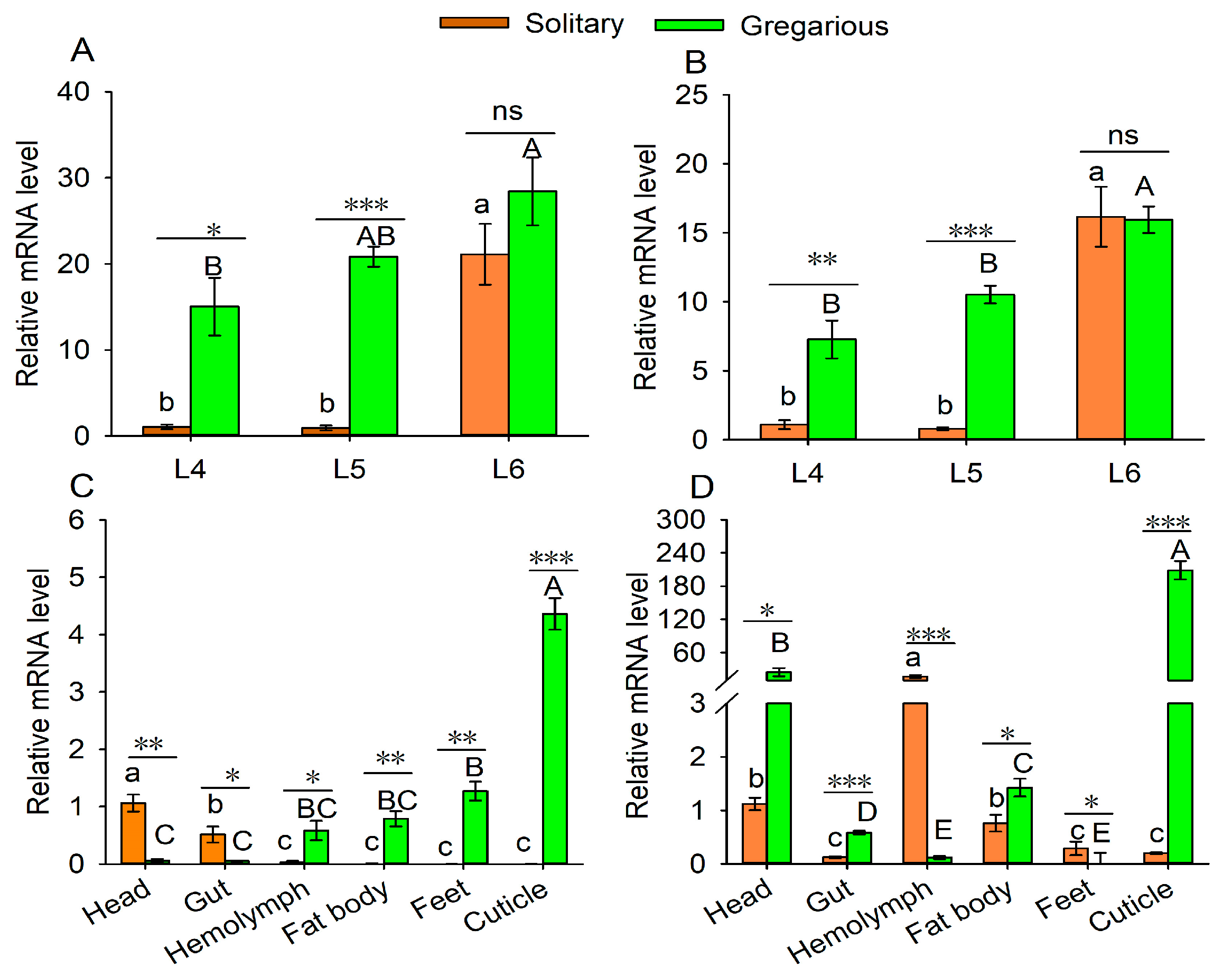
The developmental and tissue-specific expression patterns of the two MsCPs were analyzed by qRT-PCR. As shown in Figure 1, the two CP genes were differently expressed in 4–6th instar larvae and were up-regulated as development proceeded (MsCP1: Fsolitary = 31.96, p = 0.0006; Fgregarious = 4.83, p = 0.0362; MsCP2: Fsolitary = 47.642, p = 0.0002; Fgregarious = 17.921, p = 0.0029). When subjected to t-test analysis, the expression levels of MsCP1 and MsCP2 were higher in gregarious 4th and 5th instar larvae as compared to solitary larvae, but there was no significant difference between the two phases when analyzed for expression in 6th instar larvae (Figure 1A,B).
Figure 1.
Expression levels of MsCP1 and MsCP2 in gregarious and solitary M. separata. Panels (A,B) show expression levels of MsCP1 and MsCP2 across developmental stages of gregarious and solitary M. separata, respectively. Panels (C,D) illustrate tissue-specific expression patterns of MsCP1 and MsCP2 in 5th instar larvae of gregarious and solitary M. separata. Abbreviations: L4–L6, the fourth- to sixth- instar larvae. Data represent means ± SE (n = 3). Different lowercase or capital letters indicate significant differences among stages or tissues at p ≤ 0.05 based on one-way ANOVA with Tukey’s honest significant difference test. Asterisks (*), (**) and (***) indicate significant differences among gregarious and solitary larvae at p ≤ 0.05, p ≤ 0.01 and p ≤ 0.001, respectively. The notation “ns” indicates no significant difference between the two phases at p ≤ 0.05.
Tissue distribution analysis showed that the two CP genes were differentially expressed in the various tissue types (MsCP1: Fsolitary = 27.66, p < 0.001; Fgregarious = 105.27, p < 0.001; MsCP2: Fsolitary = 31.56, p < 0.001; Fgregarious = 122.40, p < 0.001). For example, MsCP1 expression was higher in the heads and midguts of solitary individuals as compared to insects in the gregarious phase; however, expression levels in the hemolymph, fat bodies, foot, and cuticle were higher in gregarious larvae than in solitary individuals (Figure 1C,D). MsCP2 expression was lower in the hemolymph and feet of gregarious vs. solitary larvae but was more highly expressed in heads, midguts, fat bodies, and cuticles of gregarious larvae as compared to solitary individuals (Figure 1C,D).
3.3. Knockdown of MsCP1 and MsCP2 Expression by RNAi
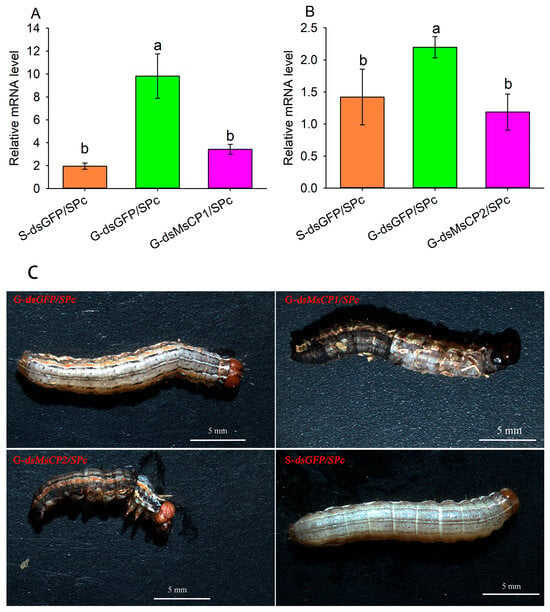
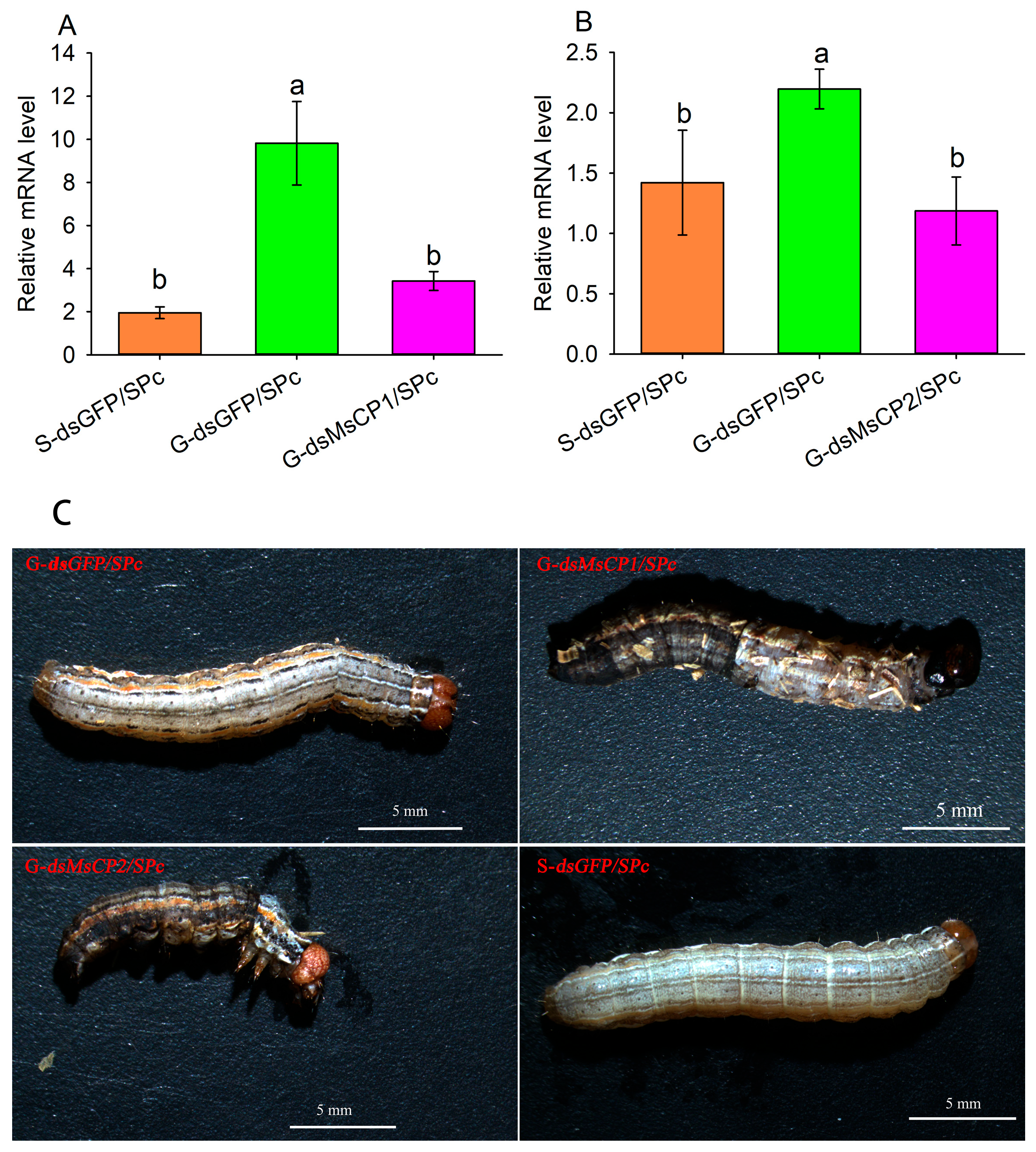
To explore the function of MsCP1 and MsCP2, we silenced the two genes by injecting dsCP/SPc complexes into 4th instar larvae of gregarious and solitary M. separata. The expression levels of MsCP1 and MsCP2 in gregarious larvae treated with dsMsCP1/SPc or dsMsCP2/SPc, respectively, were 65.12% and 46.00% lower compared than the control (G-dsGFP/SPc) and were not significantly different from expression in solitary larvae treated with dsGFP (S-dsGFP/SPc, Figure 2A,B). Knockdown of MsCP1 and MsCP2 expression resulted in abnormal molting in gregarious larvae as compared to solitary and gregarious larvae treated with dsGFP/SPc (Figure 2C).
Figure 2.
Silencing efficiency of MsCPs and larval phenotypes after SPc-mediated RNAi. Panels (A,B) show expression levels of MsCP1 and MsCP2 at 48 h after injection with dsRNA/SPc, respectively. Panels (C) show phenotypes of solitary and gregarious larvae injected dsRNA/SPc. Abbreviations: S-dsGFP/SPc, solitary larvae injected with dsGFP/SPc; G-dsGFP/SPc, gregarious larvae injected with dsGFP/SPc; G-dsMsCP1/SPc and G-dsMsCP2/SPc, gregarious larvae injected with dsMsCP1/SPc and dsMsCP2/SPc, respectively. β-actin was used as a reference gene. Data points represent means ± SE from three independent biological replications and three technical replications. Different lowercase letters indicate significant differences among treatments at p ≤ 0.05 based on one-way ANOVA with Tukey’s honest significant difference test.
3.4. SEM Analysis of Cuticles After Silencing MsCPs Expression
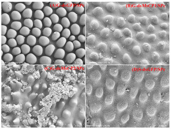
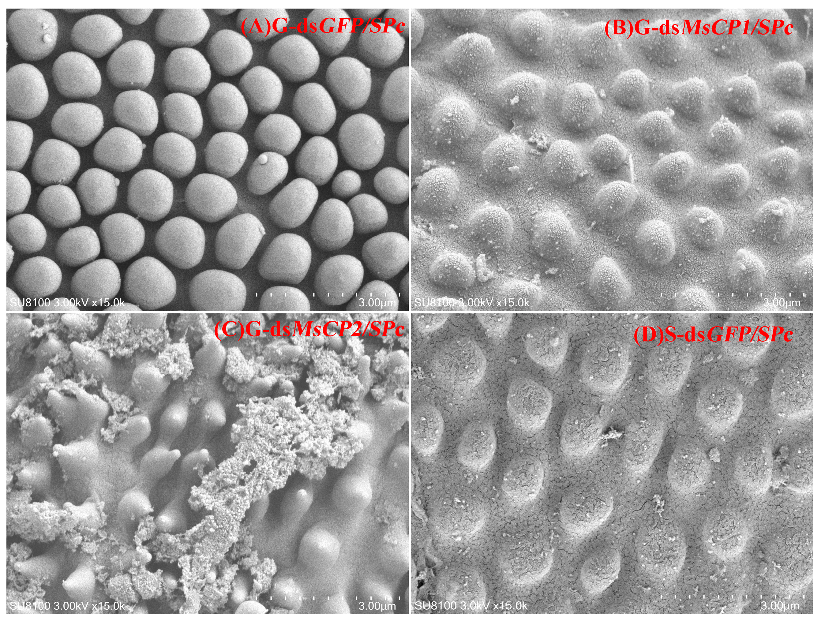
To further examine the roles of MsCP1 and MsCP2, SEM was used to observe cuticle ultrastructure in both gregarious and solitary larvae injected with dsMsCP1/SPc, dsMsCP2/SPc or dsGFP/SPc (Figure 3). Results showed that silencing MsCP1 or MsCP2 in gregarious larvae resulted in blurry cuticular surfaces (Figure 3B, dsMsCP1/SPc) or upward-facing polygonal particles (Figure 3C, dsMsCP2/SPc) when compared to dsGFP/SPc-treated gregarious larvae (Figure 3A, G-dsGFP/SPc). Furthermore, the distance between particles in dsMsCP1/SPc and dsMsCP2/SPc-treated gregarious individuals were larger, which was similar to results observed in dsGFP/SPc-treated solitary larvae (Figure 3D, S-dsGFP/SPc).
Figure 3.
SPc-mediated RNAi of MsCP1 and MsCP2 changes cuticle ultrastructure of gregarious larvae. Panels (A,D) represent cuticle ultrastructure in gregarious (G) and solitary (S) larvae injected with dsGFP/SPc, respectively. Panels (B,C) show cuticle ultrastructure in gregarious larvae injected with dsMsCP1/SPc or dsMsCP2/SPc, respectively. The image was generated with a TM-1000 scanning electron microscope at a 3-kV acceleration voltage.
3.5. TEM Analysis of Cuticle Thickness After MsCPs Knockdown
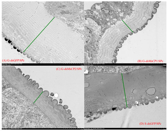
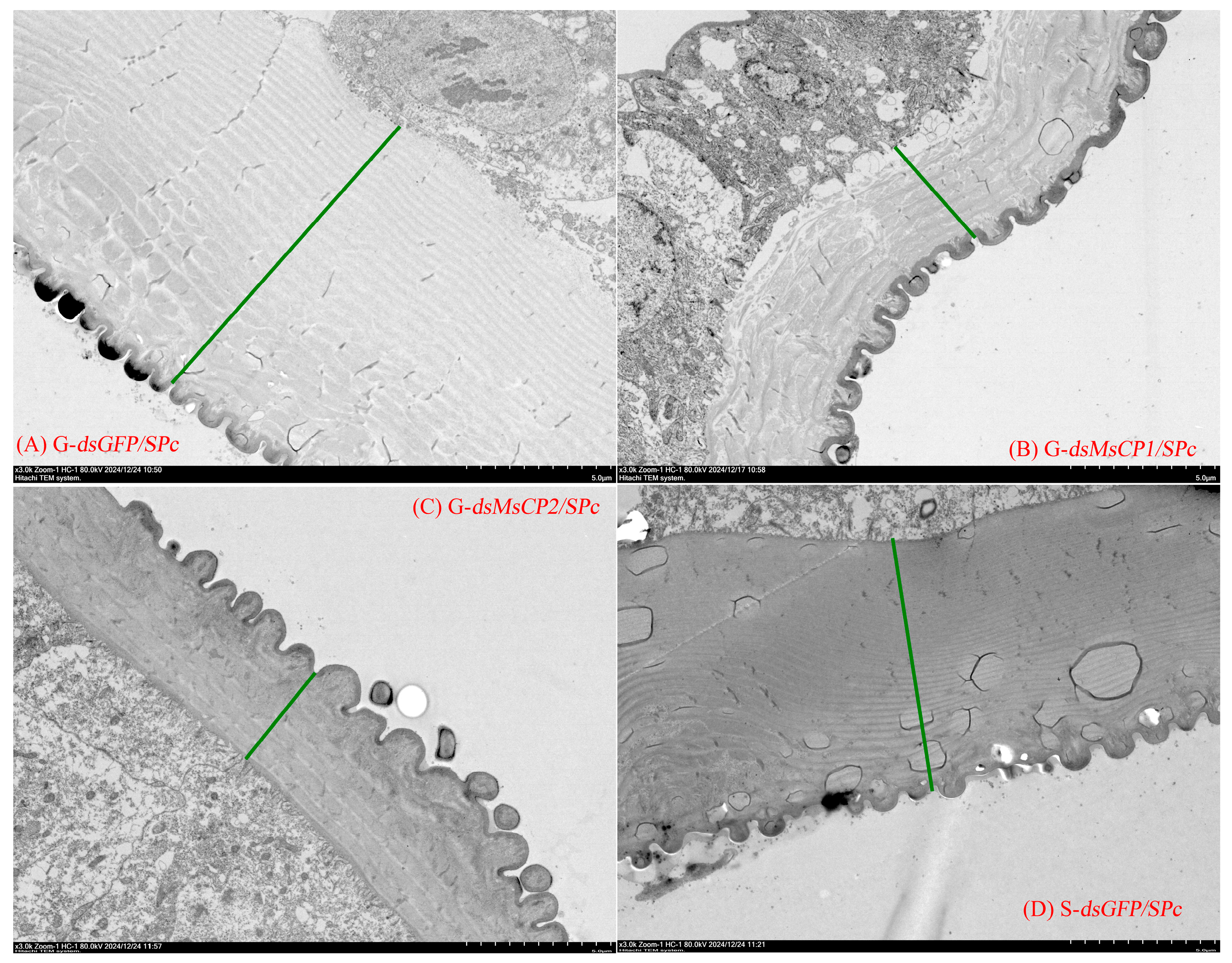
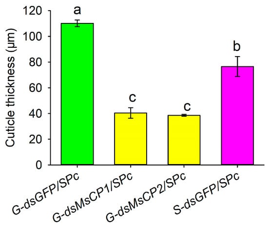
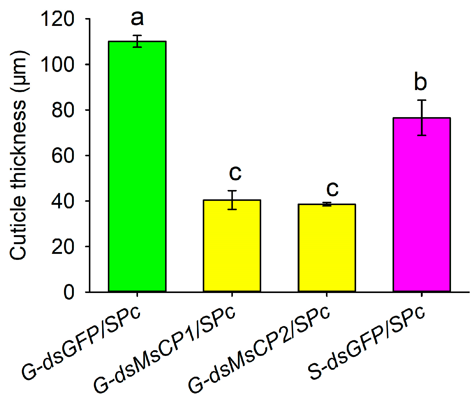
Longitudinal sections of cuticles were relatively smooth in dsGFP/SPc-treated larvae, whereas the cuticular surface became more uneven and exhibited fissures in dsMsCP1/SPc- and dsMsCP1/SPc-treated larvae (Figure 4). Furthermore, cuticle thickness in dsMsCP1/SPc- or dsMsCP2/SPc-treated gregarious larvae was significantly lower (dsMsCP1/SPc: 40.42 µm, dsMsCP2/SPc: 38.54 µm) as compared to dsGFP/SPc-treated gregarious larvae (110.12 µm) and was also less than thickness in dsGFP/SPc-treated solitary larvae (72.37 µm) (Figure 4 and Figure 5).
Figure 4.
Transmission electron microscopy demonstrated that knockdown of MsCP1 and MsCP2 via SPc-mediated RNAi modifies cuticular morphology and thickness in gregarious larvae of M. separata. Panels (A,D) represent TEM images of longitudinal sections of cuticles from gregarious (G) and solitary (S) larvae injected with dsGFP/SPc, respectively. Panels (B,C) show TEM images of longitudinal cuticular sections in gregarious larvae injected with dsMsCP1/SPc and dsMsCP2/SPc, respectively. Images were obtained with a JEM-1210 transmission electron microscope at 80 kV. Green lines illustrate cuticle thickness in gregarious and solitary larvae.
Figure 5.
SPc-mediated RNAi of MsCP1 and MsCP2 decreases cuticle thickness of gregarious larvae. Abbreviations: S-dsGFP/SPc, solitary larvae injected with dsGFP/SPc; G-dsGFP/SPc, gregarious larvae injected with dsGFP/SPc; G-dsMsCP1/SPc and G-dsMsCP2/SPc, gregarious larvae injected with dsMsCP1/SPc and dsMsCP2/SPc, respectively. Data represent means ± SE from four independent biological replications. Different lower letters indicate significant differences among treatments at p ≤ 0.05 as calculated by one-way ANOVA with Tukey’s honest significant difference test.
3.6. Assays for Resistance to B. bassiana
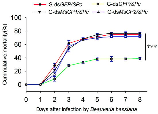
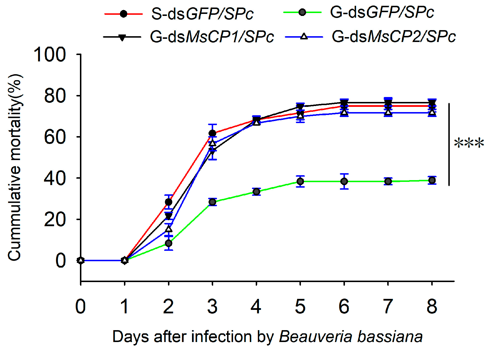
As shown in Figure 6, the mortality of both dsGFP/SPc-treated solitary and gregarious larvae gradually increased after exposure to B. bassiana. The cumulative mortality of dsGFP/SPc-treated gregarious larvae (38.33%) was significantly lower than dsGFP/SPc-treated solitary larvae (75.00%) at the 8th d after infection with B. bassiana. However, the cumulative mortality rates in dsMsCP1/SPc- or dsMsCP2/SPc-treated gregarious larvae (dsMsCP1/SPc: 76.67%; dsMsCP1/SPc: 71.66%) increased after B. bassiana infection as compared to the dsGFP/SPc-treated gregarious control (G-dsGFP/SPc).
Figure 6.
SPc-mediated RNAi of MsCP1 and MsCP2 increases the mortality of gregarious M. separata larvae infected with B. bassiana. Abbreviations: S-dsGFP/SPc, solitary larvae injected with dsGFP/SPc; G-dsGFP/SPc, gregarious larvae injected with dsGFP/SPc; G-dsMsCP1/SPc and G-dsMsCP2/SPc, gregarious larvae injected with dsMsCP1/SPc and dsMsCP2/SPc, respectively. Asterisks (***) indicate significant differences between the two phases of M. separata at p ≤ 0.001 as calculated by Student’s t-test.
4. Discussion
A major factor contributing to substantial yield losses in crops infected with M. separata is the formation of the gregarious phase in response to high population density. Notably, gregarious M. separata larvae exhibit improved immunity and decreased susceptibility to fungal biopesticides, thereby facilitating outbreaks in field crops [47]. In a prior study, we showed that gregarious M. separata larvae are more resistant to B. bassiana than their solitary counterparts [34]. Nevertheless, the mechanistic basis of DDP in M. separata remains unclear. Several studies have suggested that neurohormones and the Toll–Spätzle immune pathway provide a more effective defense against pathogens in M. separata [18,20,21]. In the current study, we identified two cuticular protein genes, MsCP1 and MsCP2, which improved the resistance of gregarious M. separata to B. bassiana by modifying cuticle ultrastructure and thickness. To our knowledge, this is the first report providing insight into the role of cuticular proteins in insect DDP.
To explore the biological function of cuticular proteins in DDP, we first examined the expression of MsCP1 and MsCP2 in the two phases of M. separata. Expression of these genes was higher in 4th and 5th instars of gregarious M. separata larvae than in solitary individuals. This result was similar to those reported in previous studies, where an obvious upregulation of stress-related genes was observed in both gregarious M. separata and locusts [44,48,49]. The upregulation of the two MsCPs is presumably due to the crowding that occurs in the 4th and 5th larval stages of M. separata. In addition, the elevated expression of these cuticular protein genes suggests that M. separata possibly reinforces its cuticle as a defensive strategy.
In this study, SPc-mediated RNAi targeting MsCP1 and MsCP2 were performed in 4th instar larvae of gregarious M. separata. Injection with dsMsCP1/SPc and dsMsCP2/SPc altered the morphology of gregarious larvae and resulted in a smaller body size and abnormal molting, suggesting a role for MsCPs in cuticle development. The altered larval morphology induced by dsMsCP1/SPc and dsMsCP2/SPc treatment were further investigated by SEM. Consistent with previous findings [35], dsGFP/SPc-treated gregarious larvae exhibited more polygonal particles on the cuticular surface and showed higher resistance to B. bassiana than dsGFP/SPc-treated solitary larvae. Similarly, Grizanova et al. reported that melanic G. mellonella larvae had a greater number of melanized spots on the cuticle surface as compared to non-melanic morphs, and melanic larvae had a higher level of resistance to entomopathogenic fungi than non-melanic counterparts [50]. The dense polygonal particles on the cuticular surface of gregarious larvae may physically reduce the contact area available for conidial attachment or alter surface free energy and wettability, thereby decreasing conidial adhesion efficiency. This speculation was supported by previous studies showing fewer conidial adhered to and germinated on the cuticle of insects exhibiting resistance to fungal pathogens [50,51,52]. In other words, the enhanced resistance of gregarious larvae may primarily depend on a physical “barrier effect” of the cuticular surface architecture rather than solely on immune activation. As noted in the current study, SPc-mediated RNAi targeting MsCP1 in gregarious larvae resulted in blurred cuticular edges, whereas SPc-mediated RNAi targeting MsCP2 resulted in upward-facing polygonal particles. Both alterations likely disrupt the original smooth or compact surface topology, providing more favorable attachment sites for conidial. Consistent with this, mortality rates of dsMsCP1/SPc- and dsMsCP2/SPc-treated gregarious larvae exposed to B. bassiana were significantly higher than those of dsGFP/SPc-treated gregarious individuals. These results support the mechanistic inference that specific cuticular ultrastructure regulates antifungal resistance by influencing conidial attachment efficiency. Our study was similar to a previous study in Tribolium castaneum [53], in which combined knockdown of adult-specific cuticular protein genes weakened antifungal phenotypes, improved germination of conidial attached on the adult body surface, and increased the susceptibility of host to two fungi biopesticides. Taken together, we propose that MsCP1 and MsCP2 contribute to the construction of a specific cuticular surface ultrastructure that indirectly modulates conidial attachment and germination in gerearious larvae. Although the infection process of conidial on the cuticle was not directly observed, the above reasoning suggests that changes in cuticular ultrastructure at least partially explain the enhanced resistance of gregarious larvae to B. bassiana. Given that the cuticle is primarily composed of structural cuticular proteins, chitin, and lipids [23], with the latter two components also serving important protective functions [54,55], we further speculate that silence of the two MsCPs may alter the arrangement or cross-linking status of cuticular components, thereby affecting the physicochemical properties of cuticle constituents [55] and ultimately weakening antifungal defense of gregarious larvae. Nevertheless, further studies are needed to validate this speculation.
Another notable phenotype was that dsGFP/SPc-treated gregarious larvae exhibited thicker cuticles than dsGFP/SPc-treated solitary larvae. SPc-mediated silence of MsCP1 and MsCP2 decreased cuticle thickness in gregarious larvae and resulted in high mortality following exposure to B. bassiana. These data suggest that the thicker cuticle of gregarious larvae acts as a physical barrier against fungal hyphal penetration. Mechanistically, increased cuticle thickness primarily extends the distance that fungal hyphae must traverse and increases the total amount of cuticular matrix that must be degraded. Because entomopathogenic fungi must secrete proteases and chitinases to degrade the cuticle before reaching the hemolymph, a thicker cuticle requires more time and greater enzymatic investment for successful penetration. The phenomenon of cuticle thickening mediating resistance to chemical insecticides has been well documented in several insect species, often involving in overexpression of CP genes [28]. For example, when the CP-encoding gene SlMsCP26 was silenced, cuticle thickness decreased, which caused increased susceptibility to indoxacarb in resistant strains of Spodoptera litura [32]. In mosquitos, knockdown of multiple CP genes resulted in thinner legs and resulted in higher mortality after insecticide exposure [29,30,31,32]. RNAi-mediated silencing of the CP-encoding gene, BgCPLCP1, resulted in a thinner cuticle and higher mortality to the insecticide β-cypermethrin in cockroaches [56]. Although these studies focused on chemical insecticides, the physical barrier logic is comparable to fungal penetration: chemical insecticides enter primarily through cuticular penetration, while fungi must physically breach the cuticle. Therefore, we propose that gregarious larvae, through MsCPs-mediated cuticle thickening, increase the time and energy costs required for fungal hyphae to penetrate the cuticle, thereby delaying or reducing the probability of successful fungal entry into the hemolymph. Given that insect mortality depends on fungal ingress into the hemolymph [57], this thickness-dependent “penetration retardation mechanism” reasonably explains the differential susceptibility to B. bassiana between gregarious and solitary larvae. Furthermore, this retardation mechanism may provide a broader time window for the activation of humoral immune responses (e.g., the phenoloxidase system and antimicrobial peptide synthesis), thereby creating a synergistic effect between physical barrier and immune defense and resulting in a stronger resistance phenotype.
It has been demonstrated that efficient knockdown of MsCP1 and MsCP2 can be achieved by injecting dsRNA/SPc complexes. Although injection enables precise delivery of dsRNA doses and rapid systemic distribution, this method is labor-intensive and impractical for large-scale pest management. From a practical standpoint, oral delivery (i.e., feeding dsRNA/SPc to larvae) would be far more feasible for potential field applications. Notably, a recent study has shown that chitosan-complexed dsRNA can be effectively delivered via feeding to Bombyx mori larvae, resulting in significant gene silencing and mortality [58]. This pioneering work suggests that oral administration of nanoparticle-formulated dsRNA is indeed a viable strategy for controlling lepidopteran insect pests. Therefore, further studies employing oral delivery of dsMsCPs/SPc to disrupt DDP represent a promising novel management strategy against M. separata.
5. Conclusions
In this study, we identified two CP-encoding genes that were highly expressed in gregarious larvae of M. separata. RNAi-mediated knockdown experiments provided strong evidence that upregulation of the two MsCPs-mediated changes in cuticle ultrastructure and thickness are associated with resistance to B. bassiana in gregarious M. separata. These novel findings broaden our understanding of insect DDP and are highly relevant in terms of successful biological control of M. separata.
Supplementary Materials
The following supporting information can be downloaded at: https://www.mdpi.com/article/10.3390/insects17040418/s1.
Author Contributions
H.L. and W.L.; methodology, H.L.; investigation, W.L., J.L. and L.L.; data curation, W.L., J.L. and C.D.; writing—original draft preparation, W.L., L.L. and J.L.; writing—review and editing, W.L. and H.L.; project administration, Y.H., Y.Q. and H.L.; funding acquisition, H.L. All authors have read and agreed to the published version of the manuscript.
Funding
This work was supported by the National Science Foundation of China (32160641), the Science and Technology Program of Guizhou Province ([2020]IZ022, 2023055), the post-foundation for the National Science Foundation of China (202206), the Opening Foundation of the Guizhou Academy of Agricultural Sciences (202403), Construction foundation of Guizhou Key Laboratory of Agricultural Biosecurity (QKHZSYS 2025024) and the innovation capacity construction of biological breeding for specialty crops in Karst mountainous areas (Qianke Hefuqi [2022] 014, [2024] 003-1, [2024] 003-2).
Data Availability Statement
The original contributions presented in this study are included in the article/supplementary material. Further inquiries can be directed to the corresponding authors.
Acknowledgments
Many thanks are due to Carol Bender for editing the manuscript.
Conflicts of Interest
The authors declare no conflicts of interest.
References
- Vilcinskas, A. Evolutionary plasticity of insect immunity. J. Insect Physiol. 2013, 59, 123–129. [Google Scholar] [CrossRef] [Scilit]
- Silva, F.W.S.; Elliot, S.L. Temperature and population density: Interactional effects of environmental factors on phenotypic plasticity, immune defenses, and disease resistance in an insect pest. Ecol. Evol. 2016, 6, 3672–3683. [Google Scholar] [CrossRef] [Scilit]
- Rolff, J.; Siva-Jothy, M.T. Invertebrate ecological immunology. Science 2003, 301, 472–475. [Google Scholar] [CrossRef] [Scilit]
- Miller, G.A.; Pell, J.K.; Simpson, S.J. Crowded locusts produce hatchlings vulnerable to fungal attack. Biol. Lett. 2009, 5, 845–848. [Google Scholar] [CrossRef] [Scilit]
- Nath, D.P.; Kumar, B.A.; Guru, P.N. Increasing adult density compromises survival following bacterial infections in Drosophila melanogaster. J. Insect Physiol. 2022, 141, 104415. [Google Scholar] [CrossRef] [Scilit] [PubMed]
- Wilson, K.; Reeson, A. Density-dependent prophylaxis: Evidence from Lepidoptera-baculovirus interactions. Ecol. Entomol. 1998, 23, 100. [Google Scholar] [CrossRef] [Scilit]
- Kunimi, Y.; Yamada, E. Relationship between larval phase and susceptibility of the armyworm, Pseudaletia separata Walker(Lepidoptera: Noctuidae) to a nuclear polyhedrosis virus and a granulosis virus. Appl. Entomol. Zool. 1990, 25, 289–297. [Google Scholar] [CrossRef] [Scilit]
- Barnes, A.I.; Siva-Jothy, M.T. Density-dependent prophylaxis in the mealworm beetle Tenebrio molitor L. (Coleoptera: Tenebrionidae): Cuticular melanization is an indicator of investment in immunity. Proc. Biol. Sci. 2000, 267, 177–182. [Google Scholar] [CrossRef] [Scilit] [PubMed]
- Wilson, K.; Thomas, M.B.; Blanford, S.; Doggett, M.; Simpson, S.J.; Moore, S.L. Coping with crowds: Density-dependent disease resistance in desert locusts. Proc. Natl. Acad. Sci. USA 2002, 99, 5471–5475. [Google Scholar] [CrossRef] [Scilit] [PubMed]
- Ruiz-González, M.X.; Moret, Y.; Brown, M.J. Rapid induction of immune density-dependent prophylaxis in adult social insects. Biol. Lett. 2009, 5, 781–783. [Google Scholar] [CrossRef] [Scilit]
- Wang, Y.; Yang, P.; Cui, F.; Kang, L. Altered immunity in crowded locust reduced fungal (Metarhizium anisopliae) pathogenesis. PLoS Pathog. 2013, 9, e1003102. [Google Scholar] [CrossRef] [Scilit]
- Silva, F.W.S.; Serrao, J.; Elliot, S.L. Density-dependent prophylaxis in primary anti-parasite barriers in the velvetbean caterpillar. Ecol. Entomol. 2016, 41, 451–458. [Google Scholar] [CrossRef] [Scilit]
- Kong, H.; Liu, Z.; Yang, P.; Yuan, L.; Jing, W.; Dong, C.; Zheng, M.; Tian, Z.; Hou, Q.; Zhu, S. Effects of larval density on Plutella xylostella resistance to granulosis virus. Insects 2020, 11, 857. [Google Scholar] [CrossRef] [Scilit]
- Nguyen, T.T.H.; Asano, T.; Cronin, A.L. Group size rather than social status influences personal immune efficacy in a socially polymorphic bee. Biol. Lett. 2023, 19, 20230149. [Google Scholar] [CrossRef] [Scilit]
- Wen, L.; Jin, T.; Luo, X.; Yuan, H.; Li, J.; Xu, C.; Jin, F.; Zhang, J.; Yu, X. The effect of population density on the phenotype, metabolic and immunological adaptations in the cuticle of Spodoptera litura larvae. Pest Manag. Sci. 2025, 81, 2379–2393. [Google Scholar] [CrossRef] [Scilit] [PubMed]
- Reeson, A.F.; Wilson, K.; Gunn, A.; Hails, R.S.; Goulson, D. Baculovirus resistance in the noctuid Spodoptera exempta is phenotypically plastic and responds to population density. Proc. Biol. Sci. 1998, 265, 1787–1791. [Google Scholar] [CrossRef] [Scilit]
- Wilson, K.; Cotter, S.C.; Reeson, A.F.; Pell, J.K. Melanism and disease resistance in insects. Ecol. Lett. 2001, 4, 637–649. [Google Scholar] [CrossRef] [Scilit]
- Kong, H.; Dong, C.; Tian, Z.; Mao, N.; Wang, C.; Cheng, Y.; Zhang, L.; Jiang, X.; Luo, L. Altered immunity in crowded Mythimna separata is mediated by octopamine and dopamine. Sci. Rep. 2018, 8, 3215. [Google Scholar] [CrossRef] [Scilit]
- Li, Z.; Liu, Y.; Liang, Y.; Pan, T.; Liu, J. RNA interference-based pesticides: Mechanism, application, and commercialization in sustainable pest management. Pestic. Biochem. Physiol. 2026, 219, 107034. [Google Scholar] [CrossRef] [Scilit]
- Jiang, S.; Bao, J.; Chen, Y.; Liu, Z.; Liu, R.; Cheng, Y.; Zhang, L.; Jiang, X.; Kong, H. Immunological regulation by Toll-1 and Spätzle-4 in larval density-dependent prophylaxis of the oriental armyworm, Mythimna separata. Int. J. Biol. Macromol. 2025, 264, 130778. [Google Scholar] [CrossRef] [Scilit]
- Kong, H.; Dong, C.; Jing, W.; Tian, Z.; Zheng, M.; Wang, C.; Hou, Q.; Cheng, Y.; Zhang, L.; Jiang, X.; et al. The role of tyramine β-hydroxylase in density dependent immunity of oriental armyworm (Mythmina separata) Larva. Int. J. Mol. Sci. 2019, 20, 1553. [Google Scholar] [CrossRef] [Scilit]
- Liu, W.; Gray, S.; Huo, Y.; Li, L.; Wei, T.; Wang, X. Proteomic analysis of interaction between a plant virus and its vector insect reveals new functions of hemipteran cuticular protein. Mol. Cell. Proteom. 2015, 14, 2229–2242. [Google Scholar] [CrossRef] [Scilit]
- Moussian, B. The arthropod cuticle. In Arthropod Biology and Volution; Minelli, A., Boxshall, G., Fusco, G., Eds.; Springer: Berlin/Heidelberg, Germany, 2013; pp. 171–196. [Google Scholar]
- Soares, M.P.; Elias-Neto, M.; Simões, Z.L.; Bitondi, M.M. A cuticle protein gene in the honeybee: Expression during development and in relation to the ecdysteroid titer. Insect Biochem. Mol. Biol. 2007, 37, 1272–1282. [Google Scholar] [CrossRef] [Scilit] [PubMed]
- Charles, J.P. The regulation of expression of insect cuticle protein genes. Insect Biochem. Mol. Biol. 2010, 40, 205–213. [Google Scholar] [CrossRef] [Scilit]
- Xie, J.; Peng, G.; Wang, M.; Zhong, Q.; Song, X.; Bi, J.; Tang, J.; Feng, F.; Gao, H.; Li, B. RR-1 cuticular protein TcCPR69 is required for growth and metamorphosis in Tribolium castaneum. Insect Sci. 2022, 29, 1612–1628. [Google Scholar] [CrossRef] [Scilit]
- Ali, M.S.; Takaki, K. Transcriptional regulation of cuticular genes during insect metamorphosis. Front. Biosci. 2020, 25, 106–117. [Google Scholar] [CrossRef] [Scilit] [PubMed]
- Balabanidou, V.; Grigorakim, L.; Vontas, J. Insect cuticle: A critical determinant of insecticide resistance. Curr. Opin. Insect Sci. 2018, 27, 68–74. [Google Scholar] [CrossRef] [Scilit]
- Huang, Y.; Guo, Q.; Sun, X.; Zhang, C.; Xu, N.; Xu, Y.; Zhou, D.; Sun, Y.; Ma, L.; Zhu, C.; et al. Culex pipiens Pallens cuticular protein CPLCG5 participates in pyrethroid resistance by forming a rigid matrix. Parasites Vectors 2018, 11, 6. [Google Scholar] [CrossRef] [Scilit] [PubMed]
- Xu, Y.; Xu, J.; Zhou, Y.; Li, X.; Meng, Y.; Ma, L.; Zhou, D.; Shen, B.; Sun, Y.; Zhu, C. CPR63 promotes pyrethroid resistance by increasing cuticle thickness in Culex pipiens pallens. Parasites Vectors 2022, 15, 54. [Google Scholar] [CrossRef] [Scilit]
- Xu, Y.; Yang, X.; Sun, X.; Li, X.; Liu, Z.; Yin, Q.; Ma, L.; Zhou, D.; Sun, Y.; Shen, B.; et al. Transcription factor FTZ-F1 regulates mosquito cuticular protein CPLCG5 conferring resistance to pyrethroids in Culex pipiens pallens. Parasites Vectors 2020, 13, 514. [Google Scholar] [CrossRef] [Scilit]
- Li, L.; Gu, L.; Tu, L.; Deng, S.; Hu, J.; Zhang, Z.; Li, J.; Zhang, M.; Cao, J.; Tang, J.; et al. A leg cuticle protein enhances the resistance of Anopheles sinensis mosquitoes to deltamethrin. Int. J. Mol. Sci. 2025, 26, 2182. [Google Scholar] [CrossRef] [Scilit] [PubMed]
- Wang, B.; Yi, M.; Wang, M.; Wang, H.; Tang, Z.; Zhao, H.; Wei, P.; Liao, X.; Xue, W.; Pan, L.; et al. Cuticle thickening mediates insecticide penetration resistance in Spodoptera litura. J. Adv. Res. 2025, 78, 163–178. [Google Scholar] [CrossRef] [Scilit]
- Li, H.; Dai, C.; Zhu, Y.; Hu, Y. Larvae crowding increases development rate, improves disease resistance, and induces expression of antioxidant enzymes and heat shock proteins in Mythimna separata (Lepidoptera: Noctuidae). J. Econ. Entomol. 2021, 114, 1808–1816. [Google Scholar] [CrossRef] [Scilit]
- Li, L.; Li, W.; Liao, J.; Fu, J.; Dai, C.; Hu, Y.; Li, H. Ultrastructure and transcriptome analysis of the larval integument in solitary and gregarious phases of Mythimna separata. Insects 2025, 16, 190. [Google Scholar] [CrossRef] [Scilit]
- Nagamine, K.; Kayukawa, T.; Watanabe, K.; Tanaka, Y. Diversity of RNAi efficiency-related nuclease in lepidopteran insects. Insect Biochem. Mol. Biol. 2026, 189, 104501. [Google Scholar] [CrossRef] [Scilit]
- Yan, S.; Ren, B.Y.; Shen, J. Nanoparticle-mediated double-stranded RNA delivery system: A promising approach for sustainable pest management. Insect Sci. 2021, 28, 21–34. [Google Scholar] [CrossRef] [Scilit] [PubMed]
- Silver, K.; Cooper, A.M.; Zhu, K. Strategies for enhancing the efficiency of RNA interference in insects. Pest Manag. Sci. 2021, 77, 2645–2658. [Google Scholar] [CrossRef] [Scilit] [PubMed]
- Liu, J.; He, Q.; Lin, X.; Smagghe, G. Recent progress in nanoparticle-mediated RNA interference in insects: Unveiling new frontiers in pest control. J. Insect Physiol. 2025, 167, 104884. [Google Scholar] [CrossRef] [Scilit]
- Zhang, Y.; Ma, Z.; Zhou, H.; Chao, Z.; Yan, S.; Shen, J. Nanocarrier-delivered dsRNA suppresses wing development of green peach aphids. Insect Sci. 2022, 29, 669–682. [Google Scholar] [CrossRef] [Scilit]
- Wei, Z.; Zhao, P.; Ning, X.; Xie, Y.; Li, Z.; Liu, X. Nanomaterial-encapsulated dsRNA-targeting chitin pathway─a potential efficient and eco-Friendly strategy against cotton aphid, Aphis gossypii (Hemiptera: Aphididae). J. Agric. Food Chem. 2024, 72, 20905–20917. [Google Scholar] [CrossRef] [Scilit]
- Lv, D.; Kassen, K.; Men, C.; Yang, X.; Pan, D.; Wang, X.; Wang, N.; Wang, P.; Yuan, X.; Li, Y. Trypsin-encoding gene function of efficient star polycation nanomaterial-mediated dsRNA feeding delivery system of Grapholita molesta. Pest Manag. Sci. 2024, 80, 5718–5727. [Google Scholar] [CrossRef] [Scilit] [PubMed]
- Kong, X.; Tan, S.; Guan, M.; Lin, X.; Shen, J.; Shi, W.; Wang, D. Nanocarrier-mediated transdermal delivery of Lmidgf4 dsRNA expedites biological control of locusts by Beauveria bassiana. J. Nanobiotechnol. 2025, 23, 272. [Google Scholar] [CrossRef] [Scilit]
- Li, H.; Dai, C.; He, Y.; Hu, Y. Characterization and expression of genes encoding superoxide dismutase in the oriental armyworm, Mythimna separata (Lepidoptera: Noctuidae). J. Econ. Entomol. 2019, 112, 2381–2388. [Google Scholar] [CrossRef] [Scilit]
- Li, H.; Dai, C.; Zhang, C.; He, Y.; Ran, H.; Chen, S. Screening potential reference genes for quantitative real-time PCR analysis in the oriental armyworm, Mythimna separata. PLoS ONE 2018, 13, e0195096. [Google Scholar] [CrossRef] [Scilit]
- Tang, Q.; Zhang, C. Data Processing System (DPS) software with experimental design, statistical analysis and data mining developed for use in entomological research. Insect Sci. 2013, 20, 254–260. [Google Scholar] [CrossRef] [Scilit]
- Jiang, X.; Zhang, L.; Cheng, Y.; Luo, L. Novel features, occurrence trends and economic impact of the oriental armyworm, Mythimna separata (Walker) in China. Chin. J. Appl. Entomol. 2014, 51, 1444–1449. [Google Scholar]
- Wang, H.; Wang, X.; Zhou, C.; Huang, L.; Zhang, S.; Guo, W.; Kang, L. cDNA cloning of heat shock proteins and their expression in the two phases of the migratory locust. Insect Mol. Biol. 2007, 16, 207–219. [Google Scholar] [CrossRef] [Scilit] [PubMed]
- Li, H.; Dai, C.; Hu, Y. Characterization and expression analysis of genes encoding three small heat shock proteins in the oriental armyworm, Mythimna separata (Walker). PLoS ONE 2020, 15, e0235912. [Google Scholar] [CrossRef] [Scilit]
- Grizanova, E.V.; Coates, C.J.; Dubovskiy, I.M.; Butt, T.M. Metarhizium brunneum infection dynamics differ at the cuticle interface of susceptible and tolerant morphs of Galleria mellonella. Virulence 2019, 10, 999–1012. [Google Scholar] [CrossRef] [Scilit]
- Ment, D.; Churchill, A.C.; Gindin, G.; Belausov, E.; Glazer, I.; Rehner, S.A.; Rot, A.; Donzelli, B.G.; Samish, M. Resistant ticks inhibit Metarhizium infection prior to haemocoel invasion by reducing fungal viability on the cuticle surface. Environ. Microbiol. 2012, 14, 1570–1583. [Google Scholar] [CrossRef] [Scilit] [PubMed]
- Ment, D.; Gindin, G.; Samish, M.; Glazer, I. Comparative response of Metarhizium brunneum to the cuticles of susceptible and resistant hosts. Arch. Insect Biochem. Physiol. 2020, 105, e21756. [Google Scholar] [CrossRef] [Scilit] [PubMed]
- Sirasoonthorn, P.; Kamiya, K.; Miura, K. Antifungal roles of adult-specific cuticular protein genes of the red flour beetle, Tribolium castaneum. J. Invertebr. Pathol. 2021, 186, 107674. [Google Scholar] [CrossRef] [Scilit]
- Hayakawa, Y.; Kato, D.; Kamiya, K.; Minakuchi, C.; Miura, K. Chitin synthase 1 gene is crucial to antifungal host defense of the model beetle, Tribolium castaneum. J. Invertebr. Pathol. 2017, 143, 26–34. [Google Scholar] [CrossRef] [Scilit] [PubMed]
- Gołębiowski, M.; Cerkowniak, M.; Urbanek, A.; Dawgul, M.; Kamysz, W.; Boguś, M.I.; Sosnowska, D.; Stepnowski, P. Antimicrobial activity of untypical lipid compounds in the cuticular and internal lipids of four fly species. J. Appl. Microbiol. 2014, 116, 269–287. [Google Scholar] [CrossRef] [Scilit]
- Cai, T.; Wang, X.; Liu, B.; Zhao, H.; Liu, C.; Zhang, X.; Zhang, Y.; Gao, H.; Schal, C.; Zhang, F. A cuticular protein, BgCPLCP1, contributes to insecticide resistance by thickening the cockroach endocuticle. Int. J. Biol. Macromol. 2024, 254, 127642. [Google Scholar] [CrossRef] [Scilit]
- Butt, T.M.; Goettel, M.S. Bioassays of entomogenous fungi. In Bioassays of Entomopathogenic Microbes and Nematodes; Navon, A., Ascher, K.R.S., Eds.; CABI Publishing: Wallingford, UK, 2000; pp. 141–195. [Google Scholar]
- Liu, J.; Yang, Y.; Yang, Q.; Lin, X.; Liu, Y.; Li, Z.; Swevers, L. Successful oral RNA interference efficiency in the silkworm Bombyx mori through nanoparticle-shielded dsRNA delivery. J. Insect Physiol. 2025, 161, 104749. [Google Scholar] [CrossRef] [Scilit] [PubMed]
Disclaimer/Publisher’s Note: The statements, opinions and data contained in all publications are solely those of the individual author(s) and contributor(s) and not of MDPI and/or the editor(s). MDPI and/or the editor(s) disclaim responsibility for any injury to people or property resulting from any ideas, methods, instructions or products referred to in the content. |
© 2026 by the authors. Licensee MDPI, Basel, Switzerland. This article is an open access article distributed under the terms and conditions of the Creative Commons Attribution (CC BY) license.