


Abstract
Background: Ocular-surface abnormalities are common in patients with moderate-to-severe atopic dermatitis and may predispose to dupilumab-associated ocular surface disease (DAOSD). We investigated early ocular-surface changes during dupilumab treatment and whether baseline-guided hyaluronic acid (HA) eye-drop use was associated with more favorable ocular outcomes. Methods: In this prospective single-center cohort study, patients with moderate-to-severe atopic dermatitis underwent ophthalmologic assessment before dupilumab initiation and after 4 months. HA 0.15% eye drops were prescribed to patients with at least one basal or reflex Schirmer value ≤15 mm and ocular symptoms. Ocular outcomes included tear break-up time (TBUT), basal and reflex Schirmer tests, meibography, and the Ocular Surface Disease Index (OSDI). Follow-up values were compared between HA users and non-users using baseline-adjusted models with false-discovery-rate correction. Results: Thirty-nine patients were included; 18 received HA eye drops and 21 did not. Twenty-seven patients (69.2%) had at least one baseline Schirmer value ≤15 mm. At 4 months, reflex Schirmer values were higher in HA users than in non-users after baseline adjustment (adjusted mean difference, 5.60 mm; 95% CI, 1.64–9.57; p = 0.007; q = 0.035). In the low-Schirmer subgroup, HA use was associated with higher reflex Schirmer (8.50 mm; 95% CI, 4.84–12.17; q < 0.001) and basal Schirmer values (5.07 mm; 95% CI, 1.41–8.73; q = 0.022). Among untreated patients with low baseline Schirmer values, reflex tear production decreased in eight of nine patients, despite OSDI scores remaining within the conventional normal range. Conclusions: HA supplementation at dupilumab initiation was associated with more favorable changes in tear production, particularly among patients with reduced baseline Schirmer measurements. Objective deterioration in tear production despite persistently low OSDI scores highlights the limitations of symptom-based assessment alone. These findings support baseline ophthalmologic evaluation and combined objective and symptom monitoring during dupilumab treatment. Larger controlled studies are needed to determine whether early, targeted HA eye-drop use can prevent clinically relevant dupilumab-associated ocular surface disease.
1. Introduction
Atopic dermatitis (AD) is a chronic inflammatory skin disease characterized by pruritus, recurrent eczematous lesions, and a substantial impact on quality of life [1,2].
The diagnosis is made clinically and is based on historical features, morphology and distribution of skin lesions, and associated clinical signs such as atypical vascular responses (facial pallor, white dermographism), keratosis pilaris, pityriasis alba, ocular/periorbital changes, perifollicular accentuation, lichenification and others. Pruritus is a hallmark feature of the condition, accounting for a significant portion of the disease burden experienced by patients and their families [3].
Dupilumab, a monoclonal antibody that inhibits interleukin (IL)-4 and IL-13 signaling through blockade of the IL-4 receptor α, has demonstrated marked efficacy in moderate-to-severe AD, improving clinical signs, pruritus, and patient-reported quality of life [4]. However, conjunctivitis occurred more frequently in dupilumab-treated patients than in placebo recipients in AD trials and was associated with greater baseline AD severity and a previous history of conjunctivitis [4,5]. Prospective ophthalmologic studies have subsequently shown that the ocular spectrum extends beyond conjunctivitis to include dry-eye disease, blepharitis, and superficial punctate keratitis, supporting the broader concept of dupilumab-associated ocular surface disease (DAOSD) [6].
Importantly, ocular-surface abnormalities may already be present before dupilumab initiation and may remain underrecognized when assessment relies predominantly on symptoms. Prospective studies have documented a high burden of pre-existing ocular surface disease in patients with moderate-to-severe AD, with objective abnormalities not always accompanied by corresponding symptoms [7,8,9].
Baseline ocular impairment may also identify patients at greater risk of DAOSD during treatment: dry-eye disease at baseline was independently associated with dupilumab-induced blepharoconjunctivitis in a prospective multicenter study [10]. In addition, reductions in tear break-up time (TBUT) and Schirmer tests, procedures measuring tear-film stability and tear production, have been reported during dupilumab therapy [11], and low baseline TBUT has been identified as a predictor of subsequent ocular adverse events [12]. These findings support the concept that DAOSD may result, at least in part, from the interaction between pre-existing ocular-surface vulnerability and treatment-related changes.
The optimal preventive strategy for DAOSD, however, remains uncertain. Current expert consensus considers the evidence insufficient to support routine prophylactic lubricant eye-drop use in patients without pre-existing ocular disease, while emphasizing appropriate management of patients with established ocular-surface abnormalities [13]. Hyaluronic acid (HA) is widely used in dry-eye disease and has a favorable safety profile, with evidence of improvement in ocular-surface signs and symptoms [14]. In dupilumab-treated patients, individualized ophthalmologic management including artificial tears has been associated with a lower observed incidence of conjunctivitis in a nonrandomized study [15]. Moreover, an open-label prospective study reported a lower incidence of dupilumab-associated conjunctivitis with prophylactic diquafosol, an aqueous secretagogue that stimulates P2Y2 receptors in the cornea and conjunctiva, promoting water secretion and enhancing mucin release from conjunctival goblet cells [16].
However, whether a baseline-guided strategy using HA is associated with preservation of normal quantitative ocular-surface parameters remains unclear [13].
We conducted a prospective cohort study to evaluate changes in ocular-surface parameters from baseline to 4 months post-dupilumab initiation between HA users (prescribed based on baseline clinical features and symptoms) and non-users. We aimed to examine whether HA use was associated with changes in ocular-surface parameters during dupilumab treatment.
2. Materials and Methods
2.1. Study Design and Setting
This was a single-center prospective cohort study involving patients with moderate-to-severe AD who initiated dupilumab at Policlinico di Bari, Italy. Ocular surface status was assessed at baseline, before dupilumab initiation (T0), and after 4 months of treatment (T1). Consecutive patients with moderate-to-severe AD initiating dupilumab during the study period were screened for eligibility and enrolled according to the predefined inclusion and exclusion criteria.
The study assessed early changes in ocular-surface parameters comparing patients prescribed HA eye drops at dupilumab initiation with non-users. HA use was nonrandomized and guided by predefined baseline ocular findings and symptoms. The resulting comparisons describe associations and do not establish causal effects of HA.
Participants initiated dupilumab between February 2021 and February 2022.
2.2. Inclusion and Exclusion Criteria
Patients were eligible for inclusion if they were aged 12 years or older, had dermatologist-confirmed moderate-to-severe AD, and were clinically eligible to initiate dupilumab because of inadequate disease control, intolerance, or contraindications to conventional systemic treatment. AD was diagnosed clinically by a dermatologist based on pruritus, typical eczematous lesions with age-appropriate morphology and distribution, and a chronic or relapsing course [3]. Severe disease was defined by an EASI score > 21. Exclusion criteria were ocular conditions requiring treatment that could interfere with ocular-surface assessment and concomitant systemic or topical ophthalmic treatments expected to materially affect ocular-surface measurements.
2.3. Ethics Approval
The study protocol (“Valutazione oculistica nel paziente affetto da dermatite atopica da moderata a grave eleggibile a trattamento con dupilumab: fattori di rischio e patogenesi”; study no. 6738) was approved by the Independent Ethics Committee of the Azienda Ospedaliero-Universitaria “Consorziale Policlinico”, Bari, Italy, on 24 February 2021.
2.4. Dupilumab Regimen and Hyaluronic Acid Eye Drops Allocation
All participants received dupilumab according to the standard regimen for AD, consisting of a 600 mg loading dose subcutaneously followed by 300 mg every 2 weeks.
HA eye drops were not prescribed universally. At baseline, their use was determined by a clinical rule requiring both:
- At least one basal or reflex Schirmer measurement of 15 mm or less in either eye;
- At least one ocular symptom elicited during direct ophthalmologic interview (ocular itching, burning, and/or dryness sensation).
The lubricant formulation, dosing frequency, and treatment duration were the following: 0.15% hyaluronic acid eye drops, one drop in each eye four times daily for 4 months. The use of HA-containing lubricants is supported by studies in dry-eye disease [14,17]. Continuation was verified, and all 18 HA patients maintained treatment without interruption throughout follow-up.
Patients in the non-HA group were instructed not to initiate HA or other topical ocular treatments expected to affect ocular-surface measurements during follow-up unless clinically indicated; any rescue treatment was to be recorded. No patient in the non-HA group required rescue therapy or initiated HA during the 4-month follow-up.
2.5. Ophthalmologic Assessment
A standardized ophthalmologic assessment was performed at baseline, before dupilumab initiation (T0), and after 4 months of treatment (T1). At baseline, previous ophthalmologic history, history of allergic conjunctivitis, periocular eczema, and current ocular symptoms were recorded separately. Current ocular symptoms included itching, burning, and/or dryness and were elicited during the baseline ophthalmologic interview.
At each visit, ocular-surface evaluation included BUT, reflex Schirmer testing, basal Schirmer testing, infrared meibography, and the 12-item Ocular Surface Disease Index (OSDI).
BUT is a standard test for tear-film stability performed by using standard fluorescein strips or minimal fluorescein solutions. Shorter BUT values indicate reduced tear-film stability. Values below 10 s are commonly considered abnormal, with values below 5 s indicating more marked tear-film instability [18].
The Schirmer test measures tear production to diagnose dry eye syndrome using 5 × 30 mm filter paper strips placed for 5 min between the palpebral conjunctiva and the eye’s bulbar conjunctiva. Without anesthesia, it measures combined basal and reflex (triggered by eye irritation from the filter paper) tearing, whereas topical anesthesia isolates basal secretion by preventing reflex tearing. Results are recorded as the length of the moistened paper. A score greater than 15 mm in 5 min is considered normal. A score of less than 15 mm in 5 min indicates the presence of dry eye [19].
Infrared meibography uses infrared wavelength light to visualize meibomian glands in vivo. These glands secrete the lipid meibum into the pre-corneal tear film to prevent tear evaporation and desiccation of the ocular surface. Meibomian gland dysfunction, which involves terminal duct obstruction, inflammation, keratinization, and eventual gland loss, is the main cause of evaporative dry-eye disease [20]. Meibography grading quantifies meibomian gland loss or atrophy using infrared imaging. Clinicians typically employ a 0–4 scale to evaluate the extent of glandular dropout in the upper and lower eyelids. Grade 0 indicates normal glands while grade 4 means advanced glandular atrophy [21].
The OSDI consists of a 12-item questionnaire designed to assess the visual disability caused by dry eye syndrome. The OSDI score can range from 0 to 100, with higher scores indicating greater disability; scores below 13 were considered within the conventional normal range [22].
BUT, Schirmer and meibography measurements were recorded separately for the right and left eyes.
A low-Schirmer subgroup was defined by the presence of at least one baseline basal or reflex Schirmer measurement of 15 mm or less in either eye regardless of the presence of symptoms.
2.6. Dermatologic Assessment
At T0 and T1, AD severity was assessed using the Eczema Area and Severity Index (EASI) and the Scoring Atopic Dermatitis (SCORAD) index. Patient-reported disease burden was evaluated using the Dermatology Life Quality Index (DLQI) and a numerical rating scale (NRS) for pruritus. Higher scores indicated greater disease severity or patient-reported burden [3]. The SCORAD sleep component (0–10) was also evaluated separately at T0 and T1. Baseline atopic comorbidities, non-allergic comorbidities, and previous AD treatments were also recorded.
2.7. Study Outcomes
The primary outcome was to evaluate 4-month changes from baseline in BUT, basal and reflex Schirmer test values, meibography grades, OSDI scores, and dry eye clinical symptoms in patients receiving HA eye drops compared to controls.
Additional study outcomes were to compare the dermatologic response to dupilumab between groups and to investigate the relationship between baseline dermatologic severity and baseline ocular-surface parameters.
2.8. Statistical Analysis
The study cohort comprised participants with documented lubricant eye drop allocation and complete T0 and T1 ocular assessments. For bilateral ocular measurements—BUT, reflex Schirmer, basal Schirmer, and meibography—the arithmetic mean of the right- and left-eye values was calculated for each participant at each ophthalmologic visit.
Continuous variables were summarized as mean and standard deviation, whereas categorical variables were reported as absolute values and percentages. Baseline differences between the HA and non-HA groups were analyzed using Welch’s t test for continuous variables and Fisher’s exact test for categorical variables. Welch’s t test does not require equality of variances between groups. Independence of observations followed from the study design, and continuous-variable distributions were assessed for marked departures from normality and extreme outliers before parametric comparisons; no major violations affecting the applicability of the test were identified.
For each ocular outcome, the primary between-group comparison was performed using linear analysis of covariance, with the T1 value as the dependent variable, HA group as the group indicator, and the corresponding T0 value as a covariate. Statistical inference, including 95% confidence intervals and p values, was based on heteroskedasticity-consistent type 3 (HC3) standard errors. Results were reported as adjusted mean differences between the hyaluronic acid and no-lubricant groups. The Benjamini–Hochberg procedure was applied across the five ocular outcomes to control the false discovery rate.
A subgroup analysis was conducted among participants with at least one baseline basal or reflex Schirmer measurement of 15 mm or less in either eye. Between-group comparisons within this subgroup used the same baseline-adjusted models applied in the complete cohort, with false-discovery-rate correction across the five ocular outcomes. Among low-Schirmer participants who did not receive lubricant, T0-to-T1 changes were additionally examined using paired Wilcoxon signed-rank tests. OSDI values below 13 were considered within the conventional normal range. These subgroup analyses were considered exploratory.
Dermatologic response was described using changes in EASI, SCORAD, DLQI, and pruritus NRS from T0 to T1. Between-group comparisons were based on linear models of the T1 value adjusted for the corresponding baseline value, with HC3 robust standard errors and Benjamini–Hochberg correction across the four outcomes.
All statistical tests were two-sided. p Values below 0.05 were considered nominally significant; for analyses subject to multiplicity correction, q values below 0.05 were considered significant after false-discovery-rate control. Analyses were performed using R version 4.6.1 (R Foundation for Statistical Computing, Vienna, Austria).
3. Results
3.1. Study Cohort and Baseline Characteristics
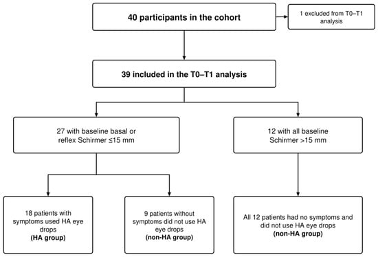
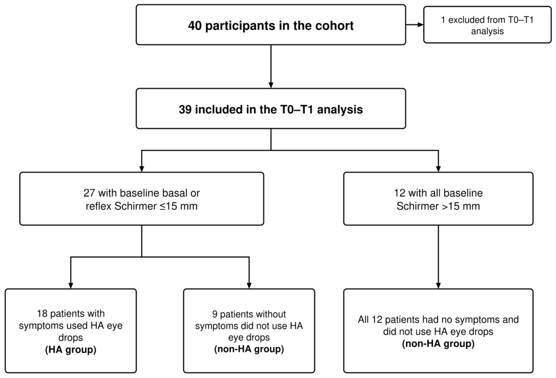
Of the 40 participants in the source cohort, 39 completed T0 and T1 dermatological and ocular assessments and were included in the analysis. One participant was lost to follow-up after ceasing to attend the scheduled follow-up visits.
Twenty-seven participants were assigned to the low-Schirmer subgroup (69.2% of the total) according to a baseline basal or reflex Schirmer test result of ≤15 mm. Among them, 18 symptomatic patients (46.1% of the global cohort) received HA eye drops (HA group), whereas the remaining nine asymptomatic patients received no HA treatment (non-HA group). An additional 12 participants with baseline Schirmer values above 15 mm and without symptoms were also assigned to the non-HA group.
Overall, 18 participants received HA eye drops and 21 patients did not (Figure 1).
Figure 1.
Study flow from cohort enrollment to the T0–T1 analysis, with allocation according to baseline Schirmer values, ocular symptoms, and HA eye-drop use. HA, hyaluronic acid; T0, baseline; T1, 4-month follow-up.
Baseline demographic characteristics were similar between groups, including age, and sex (Table 1). The mean age of the overall study cohort at dupilumab initiation was 40.95 ± 19.79 years. Baseline TBUT and meibography grades were also comparable. In contrast, participants of the HA group had lower reflex Schirmer values (10.31 vs. 18.14 mm), lower basal Schirmer values (12.92 vs. 19.81 mm), and higher OSDI scores (11.03 vs. 2.31) compared to the non-HA group. SCORAD was also higher in the HA group (73.63 vs. 65.03), whereas EASI was similar between groups. Atopic and non-allergic comorbidities were present in both groups. Allergic conjunctivitis was reported in 12/18 (66.7%) HA patients and 9/21 (42.9%) non-HA patients, allergic rhinitis in 16/18 (88.9%) and 13/21 (61.9%), allergic asthma in 7/18 (38.9%) and 6/21 (28.6%), and non-allergic comorbidities in 11/18 (61.1%) and 11/21 (52.4%), respectively. Previous ophthalmologic history was reported in 11/18 (61.1%) HA patients and 11/21 (52.4%) non-HA patients, while periocular eczema was present in 3/18 (16.7%) and 4/21 (19.0%), respectively. Previous AD treatment exposure was also broadly shared between groups: antihistamine, phototherapy, and topical calcineurin-inhibitor use were similar, while all patients had previously received topical corticosteroids. Previous systemic corticosteroid treatment was more frequent in the HA group (13/18, 72.2% vs. 10/21, 47.6%), whereas cyclosporine use was more frequent in the non-HA group (10/18, 55.6% vs. 15/21, 71.4%).
Table 1.
Baseline demographic, dermatologic, and ocular characteristics in non-HA and HA groups.
No other clinically relevant adverse events related to dupilumab were recorded during the 4-month follow-up.
3.2. Ocular Outcomes at 4 Months
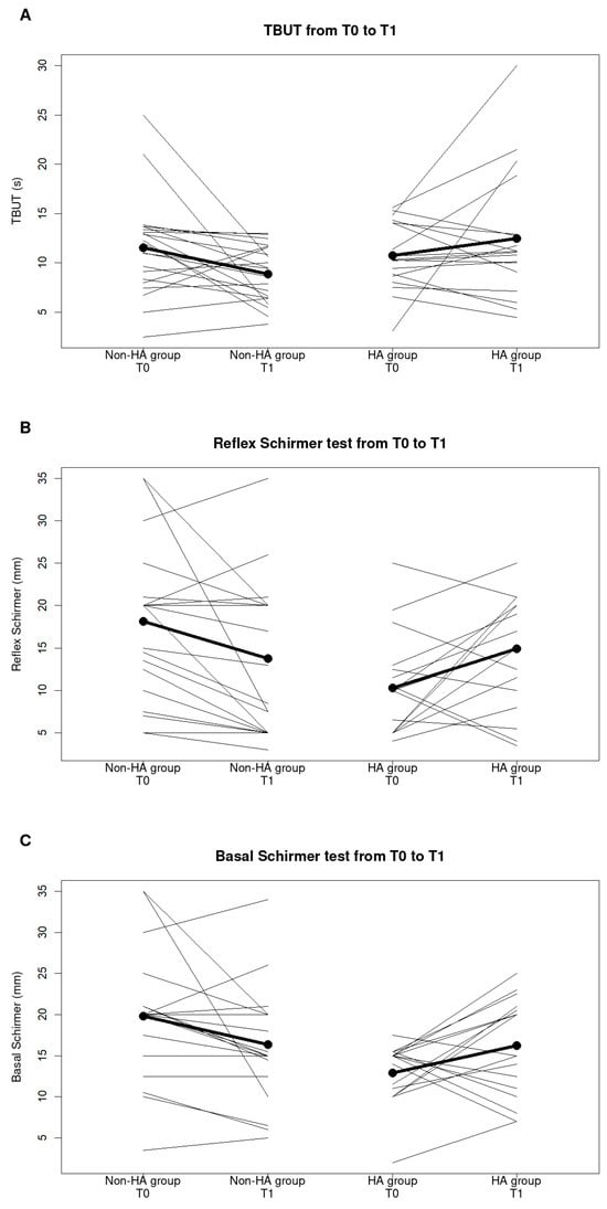
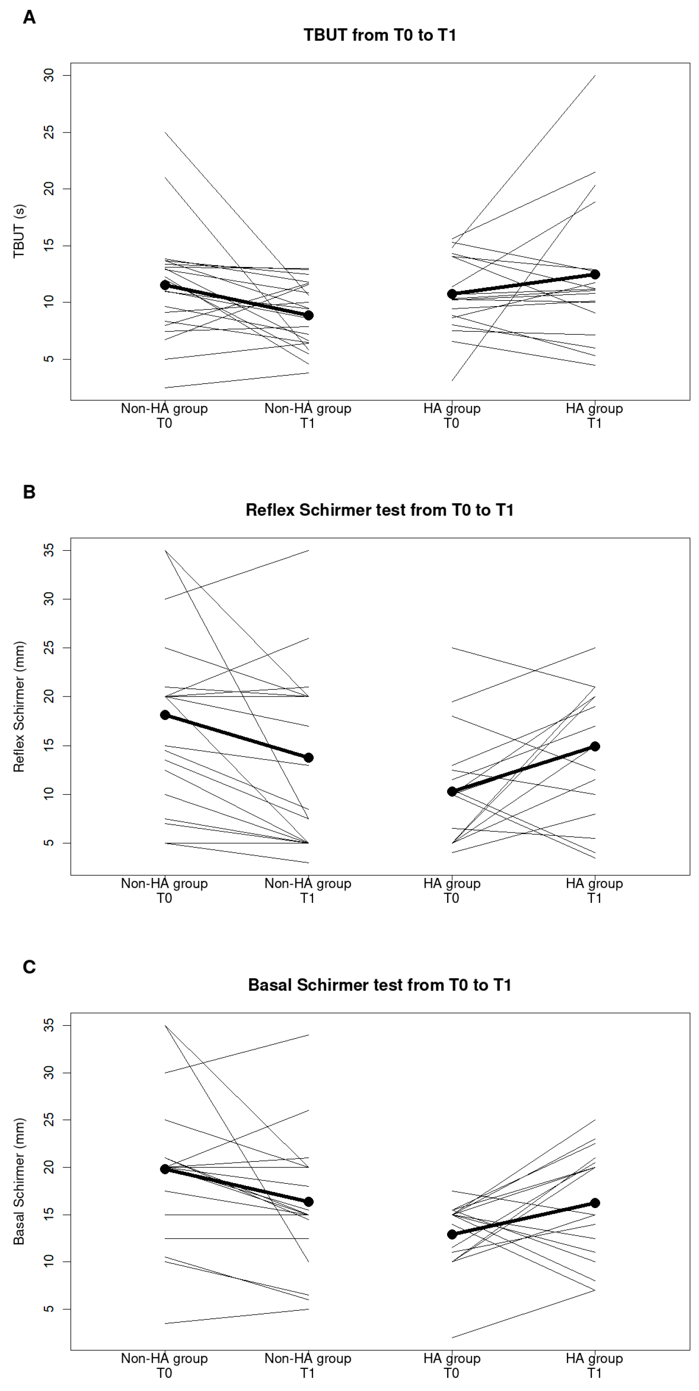
Over the 4-month follow-up, mean TBUT decreased by 2.65 s in the non-HA group and increased by 1.73 s in the HA group. In the baseline-adjusted analysis, TBUT was 3.81 s higher in the HA group at T1 (95% CI, 0.39 to 7.24; p = 0.030), although this association did not remain significant after false-discovery-rate correction (q = 0.076).
Mean reflex Schirmer decreased by 4.40 mm without HA eye drops and increased by 4.64 mm with HA eye drops. The adjusted between-group difference was 5.60 mm (95% CI, 1.64 to 9.57; p = 0.007; q = 0.035). Basal Schirmer also showed more favorable changes over time in the HA group, but the adjusted difference was inconclusive (3.12 mm; 95% CI, −1.00 to 7.24; p = 0.133; q = 0.222).
No adjusted between-group differences were observed for meibography (0.03; 95% CI, −0.18 to 0.24; p = 0.796; q = 0.796) or OSDI (−1.08 points; 95% CI, −3.51 to 1.35; p = 0.375; q = 0.469) (Table 2; Figure 2).
Table 2.
Ocular outcomes at 4 months according to HA eye-drop use.
Figure 2.
Individual ocular-surface changes from baseline (T0) to 4 months (T1) in the complete cohort according to HA use. (A) Tear break-up time (TBUT), (B) reflex Schirmer test, and (C) basal Schirmer test. Thin lines show within-patient changes, whereas thick black lines with filled circles represent group means. Abbreviations: HA, hyaluronic acid; TBUT, tear break-up time.
3.3. Low-Schirmer Subgroup
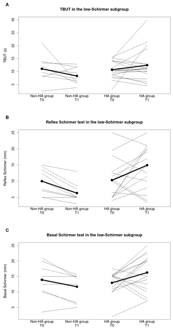
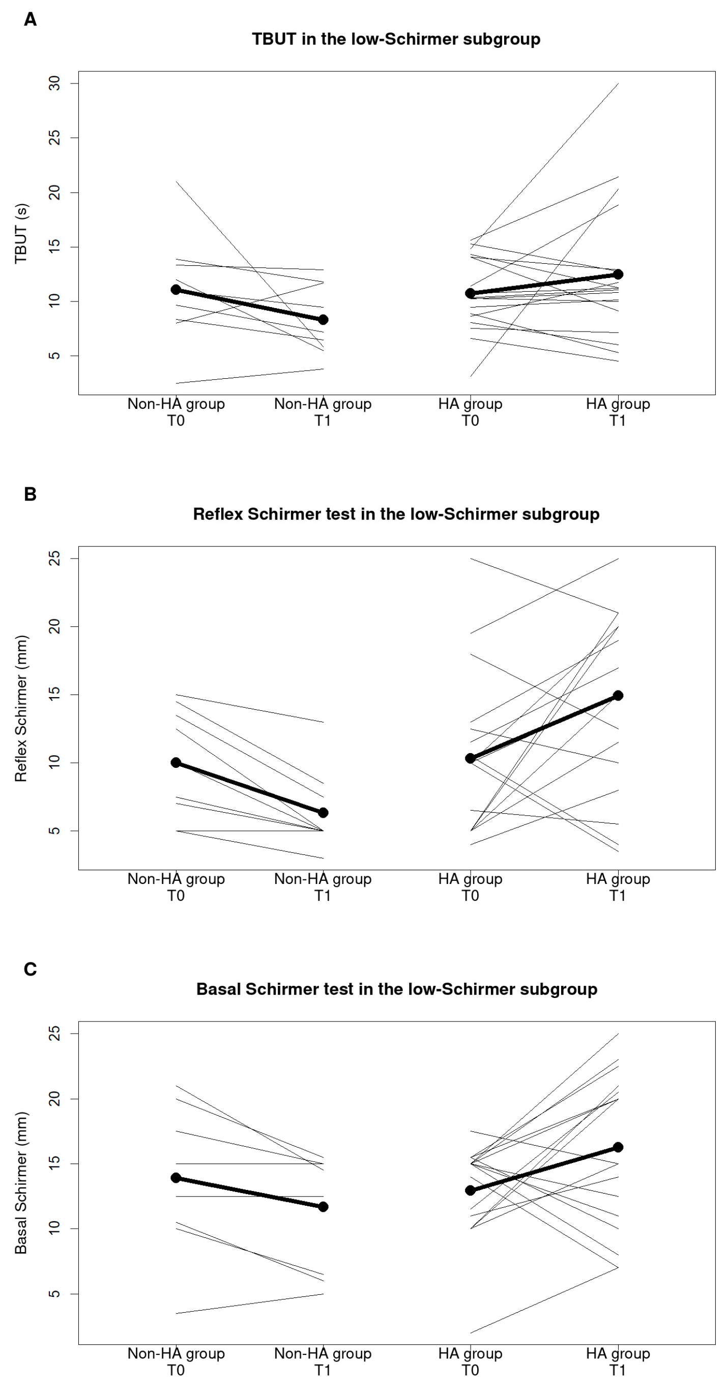
The low-Schirmer subgroup included 27 participants, of whom 18 were HA users and nine were non-HA users. Baseline objective ocular measurements were similar between groups, including TBUT (10.74 vs. 11.07 s), reflex Schirmer (10.31 vs. 10.00 mm), and basal Schirmer (12.92 vs. 13.89 mm). Baseline OSDI was higher in the HA group (11.03 vs. 1.92).
After adjustment for baseline values, TBUT at T1 was estimated to be 4.31 s higher in the HA group than in the non-HA group; however, the confidence interval included zero and the association was not significant after multiplicity correction (95% CI, −0.04 to 8.66; p = 0.052; q = 0.086).
However, reflex Schirmer at T1 was 8.50 mm higher in the HA group (95% CI, 4.84 to 12.17; p < 0.001; q < 0.001), and basal Schirmer was 5.07 mm higher (95% CI, 1.41 to 8.73; p = 0.009; q = 0.022). No clear between-group differences were observed for meibography or OSDI (Table 3; Figure 3).
Table 3.
Baseline-adjusted ocular outcomes at 4 months in the low-Schirmer subgroup.
Figure 3.
Individual ocular-surface changes from baseline (T0) to 4 months (T1) in the low-Schirmer subgroup according to HA use. (A) Tear break-up time (TBUT), (B) reflex Schirmer test, and (C) basal Schirmer test. Thin lines show within-patient changes, whereas thick black lines with filled circles represent group means. HA, hyaluronic acid; TBUT, tear break-up time.
3.4. Low-Schirmer Participants Without HA Eye Drops
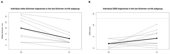
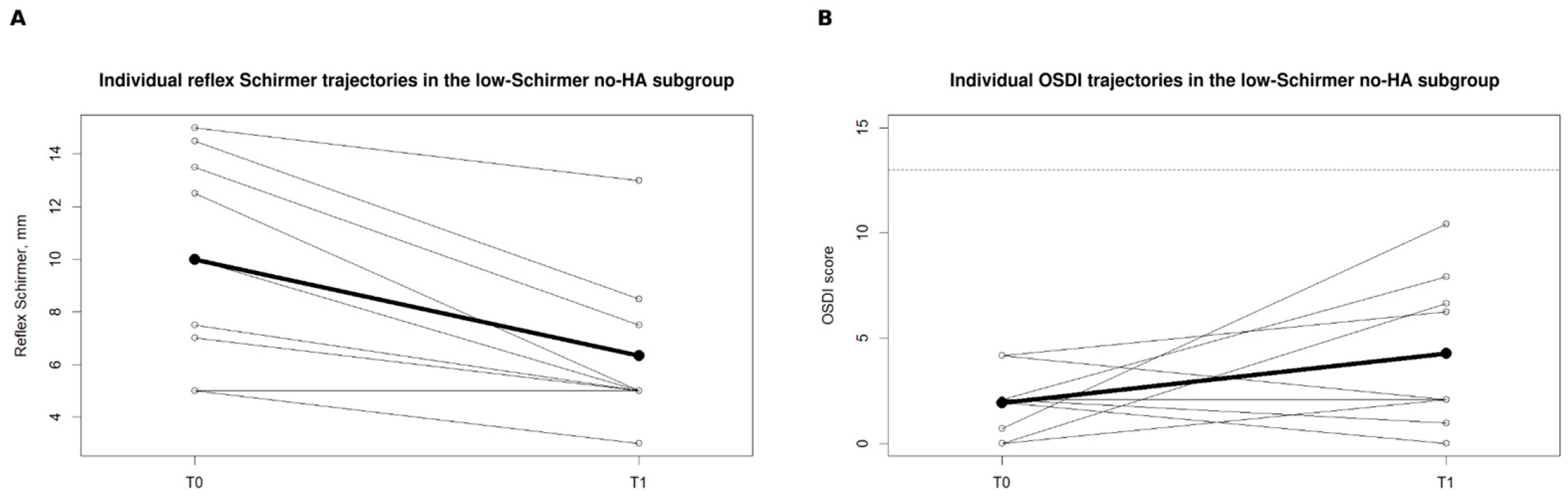
Among the nine low-Schirmer participants of the non-HA group, mean reflex Schirmer decreased from 10.00 to 6.33 mm over 4 months. Eight of nine participants showed a reduction, corresponding to a mean change of −3.67 mm (paired Wilcoxon p = 0.014). TBUT decreased from 11.07 to 8.29 s in seven participants, although the within-group change was not statistically significant (mean change, −2.78 s; p = 0.129). Basal Schirmer decreased from 13.89 to 11.67 mm (mean change, −2.22 mm; p = 0.058).
Mean OSDI increased from 1.92 to 4.27 points (p = 0.183), but all nine participants had OSDI scores below 13 at both visits (Figure 4).
Figure 4.
Individual ocular-surface changes from baseline (T0) to 4 months (T1) in the low-Schirmer non-HA subgroup. (A) Reflex Schirmer test and (B) Ocular Surface Disease Index (OSDI). Thin lines show within-patient changes, whereas thick black lines with filled circles represent group means. The dashed line indicates the conventional OSDI threshold of 13. Abbreviations: HA, hyaluronic acid; OSDI, Ocular Surface Disease Index.
3.5. Eye-Level Sensitivity Analysis
The eye-level generalized estimating equation analysis confirmed the main findings obtained from the patient-level models. The estimated difference in T0-to-T1 change between groups was 4.38 s for TBUT (95% CI, 1.00 to 7.77; p = 0.011), 9.04 mm for reflex Schirmer (95% CI, 4.30 to 13.79; p < 0.001), and 6.76 mm for basal Schirmer (95% CI, 2.76 to 10.76; p = 0.001). No clear difference was observed for meibography (0.12; 95% CI, −0.24 to 0.47; p = 0.525).
3.6. Dermatologic Response
Dermatologic scores decreased markedly in both groups over the 4-month follow-up. Mean EASI decreased by 25.00 points in the non-HA group and by 24.89 points in the HA group. Mean SCORAD decreased by 53.10 and 62.30 points, respectively, whereas DLQI and pruritus NRS also improved substantially in both groups. After adjustment for the corresponding baseline values, no between-group differences were observed for EASI (adjusted difference, −1.79; 95% CI, −4.65 to 1.07; p = 0.212), SCORAD (−3.43; 95% CI, −11.82 to 4.95; p = 0.412), DLQI (0.43; 95% CI, −1.39 to 2.24; p = 0.637), or pruritus NRS (0.69; 95% CI, −1.14 to 2.51; p = 0.451). None of these comparisons was significant after false-discovery-rate correction. SCORAD sleep scores also decreased in both groups. In the non-HA group, the mean sleep score decreased from 6.7 ± 2.4 at T0 to 1.2 ± 1.4 at T1 (mean change, −5.5). In the HA group, it decreased from 7.6 ± 2.2 at T0 to 1.0 ± 1.2 at T1 (mean change, −6.6).
3.7. Association Between Dermatologic Severity and Ocular-Surface Parameters
At baseline, SCORAD was inversely correlated with reflex Schirmer (Spearman ρ = −0.483; p = 0.002; q = 0.009). No significant associations were observed between SCORAD and TBUT, basal Schirmer, meibography, or OSDI after false-discovery-rate correction. In the multivariable analysis adjusted for age, positive ophthalmologic history, and periocular eczema, each one-point increase in SCORAD was associated with a 0.25 mm lower reflex Schirmer value (adjusted coefficient, −0.250; 95% CI, −0.464 to −0.036; p = 0.023).
4. Discussion
In this prospective cohort, low Schirmer values, which can be regarded as subclinical markers of dry-eye disease, were very common, involving 69.2% of the recruited patients with AD. Interestingly, according to the literature [7,8,9], less than half of the subjects in the cohort reported ocular symptoms.
Ocular-surface parameters during the first 4 months of dupilumab treatment differed according to the use of HA eye drops. After baseline adjustment and correction for multiple comparisons, reflex Schirmer values were higher at follow-up in participants receiving HA. The findings were particularly notable in the low-Schirmer subgroup, in which subjects that were HA users and non-users reported similar baseline TBUT and reflex/basal Schirmer measurements. Within this subgroup, both reflex and basal Schirmer values were higher at follow-up among HA users.
The results are therefore consistent with an association between targeted early ocular-surface support (through the use of HA eye drops) and more favorable changes in tear production in patients with pre-existing objective impairment, but they should not be interpreted as evidence of treatment efficacy. This interpretation is clinically plausible given the evidence that ocular surface disease is frequently present before dupilumab initiation and that baseline dry-eye disease or impaired tear-film function may identify patients at increased risk of subsequent ocular adverse events [7,8,9,10,11,12].
An additional clinically relevant finding was the discrepancy between objective tear production and patient-reported symptoms. Among low-Schirmer participants who did not receive HA, reflex Schirmer decreased in eight of nine patients, while OSDI remained below the conventional abnormality threshold in all patients at both visits. Previous studies in AD have similarly shown that ocular-surface abnormalities may be present despite limited or absent symptoms [7,8,9]. This observation is also consistent with the broader dry-eye literature: in a recent analysis of 535 participants from the DRy Eye Assessment and Management (DREAM) study, discordance between symptoms and clinical signs was common [23]. Thus, symptom questionnaires alone may not adequately identify early changes in tear production during dupilumab treatment, particularly in patients with objective abnormalities at baseline. Nevertheless, OSDI and Schirmer testing assess different dimensions of ocular surface disease, and the clinical relevance of an isolated reduction in tear production in minimally symptomatic patients remains uncertain. These findings therefore support combining symptom assessment with objective ocular-surface evaluation in selected at-risk patients.
AD-related ocular disease and DAOSD have overlapping manifestations, including itching, conjunctival inflammation, blepharitis and tear-film disturbance [6,9]. Pre-existing or recurrent allergic/atopic inflammation may persist during treatment, while a new or worsening ocular disorder after dupilumab initiation raises the possibility of a treatment-associated component. Because of this clinical overlap, temporal association alone may be insufficient to establish causality.
The inverse association between baseline SCORAD and reflex Schirmer further suggests that ocular-surface vulnerability may partly reflect the underlying severity of AD rather than being exclusively treatment-induced. Greater baseline AD severity has previously been associated with a higher risk of conjunctivitis during dupilumab treatment [5], and ocular surface disease is highly prevalent before treatment initiation [8,9]. Baseline sleep scores were somewhat higher in the HA group than in the non-HA group (7.6 vs. 6.7), suggesting that sleep disturbance may have contributed in part to the higher baseline SCORAD observed in this group. Mechanistic observations also support an interaction between pre-existing susceptibility and treatment-related changes in ocular-surface homeostasis: conjunctival goblet-cell dysfunction has been described during dupilumab treatment [24], while recent transcriptomic data suggest that patients who subsequently develop ocular adverse events may already display a distinct conjunctival molecular profile before dupilumab exposure [25]. Our findings also extend the limited literature on preventive ocular management during dupilumab treatment. Previous studies have mainly evaluated clinically diagnosed conjunctivitis: individualized ophthalmologic management including artificial tears was associated with a lower observed incidence of conjunctivitis in a nonrandomized study [15], and prophylactic diquafosol was associated with a lower incidence in an open-label prospective study [16]. Current expert consensus, however, considers the evidence insufficient to recommend routine prophylactic lubricant eye-drop use in patients without pre-existing ocular disease [13]. Our study addresses a different question because HA eye drops were prescribed selectively rather than universally. Given the established use and favorable safety profile of HA-containing artificial tears in dry-eye disease [14], our observations support a risk-stratified approach, even if it does not establish that HA prevents DAOSD. Before dupilumab initiation, ocular history and current symptoms should be reviewed; ophthalmology assessment is particularly relevant for significant pre-existing or active disease. Persistent symptoms, pain, photophobia or visual change warrant appropriate specialist assessment. Routine comprehensive ophthalmologic testing for every asymptomatic patient is not established by this small cohort [13].
Ocular events have also been reported in other dermatologic populations treated with dupilumab. Conjunctivitis was reported in the PRIME/PRIME2 trials in prurigo nodularis [26] and in a multicenter cohort of patients with bullous pemphigoid treated with dupilumab [27], whereas in chronic spontaneous urticaria conjunctivitis was not increased with dupilumab compared with placebo in the LIBERTY-CSU CUPID trials [28]. Thus, ocular events are not exclusive to AD, although their occurrence may vary across indications.
Management should follow the ocular phenotype and severity. Alternatives or additions to HA include other preservative-free lubricants and lid care, with antihistamine/mast-cell-stabilizing drops when allergy contributes. Persistent inflammation may require ophthalmologist-supervised topical corticosteroids, ciclosporin or selected tacrolimus treatment [13,29]. Diquafosol remains supported by limited preventive evidence [16]. Uncontrolled ocular disease may require joint dermatologic–ophthalmologic consideration of systemic treatment modification. This cohort provides no comparative evidence for choosing among these approaches.
This study has several strengths, including prospective ophthalmologic assessment before and after dupilumab initiation, combined evaluation of objective and patient-reported ocular outcomes, baseline-adjusted analyses with correction for multiple comparisons, and sensitivity analyses accounting for within-patient correlation between eyes. However, important limitations must be acknowledged. Lubricant allocation was not randomized and was determined by baseline ocular findings and symptoms, creating confounding by indication; although baseline adjustment and restriction to the low-Schirmer subgroup reduced some of the imbalance, residual confounding cannot be excluded. Several baseline demographic and dermatologic characteristics were similar between groups; however, differences in SCORAD, allergic conjunctivitis, allergic rhinitis, and some previous systemic treatments may also have contributed to residual confounding. Previous ocular treatments and concomitant medications during follow-up were not systematically recorded and could not be reliably compared between groups. Demodex infestation, which may contribute to blepharitis and ocular-surface symptoms [30], was not systematically assessed in this cohort and therefore represents a potential unmeasured confounder. The sample size was limited, particularly among untreated low-Schirmer participants, and the single-center design and 4-month follow-up restrict precision and generalizability. Baseline overlap was also limited for basal Schirmer in the complete cohort, making that estimate less robust. Accordingly, the observed differences should be interpreted as associations. Overall, the findings suggest that baseline ophthalmologic assessment may identify patients with reduced tear production that is not fully reflected by symptoms and who may warrant closer objective monitoring during dupilumab treatment. Larger multicenter studies with controlled treatment allocation and longer follow-up are needed to determine whether targeted early ocular-surface support can prevent clinically relevant DAOSD.
5. Conclusions
In this prospective cohort, HA supplementation at dupilumab initiation was associated with more favorable changes in tear production, particularly among patients with reduced baseline Schirmer measurements, while the nonrandomized design of the study does not consent to establish a protective effect of HA. Objective deterioration in tear production was observed in several untreated low-Schirmer patients despite persistently low OSDI scores, highlighting the potential limitation of symptom-based assessment alone. These findings highlight the potential value of baseline ophthalmologic evaluation and combined objective and symptom monitoring during dupilumab treatment. Larger controlled studies are needed to determine whether targeted early HA eye-drop use can prevent clinically relevant DAOSD.
Author Contributions
Conceptualization, G.D., G.C. and C.F.; methodology, G.D., G.C. and C.F.; validation, F.A., A.R.M. and W.A.R.; formal analysis, G.D., G.C. and C.F.; investigation, C.M., A.D.M., C.B., R.S., R.A.F. and G.P.; data curation, G.D., G.P., C.M., A.D.M., R.A.F. and C.B.; writing—original draft preparation, G.D. and G.C.; writing—review and editing, G.D., G.C. and C.F.; supervision, C.F., D.B., G.A., M.G.L.T. and P.R. All authors have read and agreed to the published version of the manuscript.
Funding
This research received no external funding.
Institutional Review Board Statement
The study was conducted in accordance with the Declaration of Helsinki, and approved by the Institutional Review Board of Azienda Ospedaliero-Universitaria “Consorziale Policlinico”, Bari, Italy on 24 February 2021 (study no. 6738).
Informed Consent Statement
Informed consent was obtained from all subjects involved in the study.
Data Availability Statement
The original contributions presented in this study are included in the article. Further inquiries can be directed to the corresponding author.
Conflicts of Interest
The authors declare no conflicts of interest.
Abbreviations
The following abbreviations are used in this manuscript:
| AD | atopic dermatitis |
| BUT | break-up time |
| CI | confidence interval |
| DAOSD | dupilumab-associated ocular surface disease |
| DLQI | Dermatology Life Quality Index |
| DREAM | Dry Eye Assessment and Management |
| EASI | Eczema Area and Severity Index |
| HA | hyaluronic acid |
| HC3 | heteroskedasticity-consistent type 3 |
| IL | interleukin |
| NRS | numerical rating scale |
| OSDI | Ocular Surface Disease Index |
| P2Y2 | purinergic receptor P2Y2 |
| SCORAD | SCORing Atopic Dermatitis |
| SMD | standardized mean difference |
| T0 | baseline |
| T1 | 4-month follow-up |
| TBUT | tear break-up time |
References
- Weidinger, S.; Beck, L.A.; Bieber, T.; Kabashima, K.; Irvine, A.D. Atopic dermatitis. Nat. Rev. Dis. Primers 2018, 4, 1. [Google Scholar] [CrossRef] [Scilit] [PubMed]
- Ziehfreund, S.; Saak, M.; Schaal, A.; Mazilu, R.; Mahé, E.; Hajj, C.; Johansson, E.K.; Lysell, J.; Sigurdardottir, G.; Legat, F.J.; et al. Addiction and chronic skin diseases: A Pan-European study on prevalence, associations and patient impact. J. Eur. Acad. Dermatol. Venereol. 2026, 40, 1392–1405. [Google Scholar] [CrossRef] [Scilit] [PubMed]
- Eichenfield, L.F.; Tom, W.L.; Chamlin, S.L.; Feldman, S.R.; Hanifin, J.M.; Simpson, E.L.; Berger, T.G.; Bergman, J.N.; Cohen, D.E.; Cooper, K.D.; et al. Guidelines of care for the management of atopic dermatitis: Section 1. Diagnosis and assessment of atopic dermatitis. J. Am. Acad. Dermatol. 2014, 70, 338–351. [Google Scholar] [CrossRef] [Scilit] [PubMed]
- Simpson, E.L.; Bieber, T.; Guttman-Yassky, E.; Beck, L.A.; Blauvelt, A.; Cork, M.J.; Silverberg, J.I.; Deleuran, M.; Kataoka, Y.; Lacour, J.P.; et al. Two phase 3 trials of dupilumab versus placebo in atopic dermatitis. N. Engl. J. Med. 2016, 375, 2335–2348. [Google Scholar] [CrossRef] [Scilit] [PubMed]
- Ferreira, S.; Torres, T. Conjunctivitis in patients with atopic dermatitis treated with dupilumab. Drugs Context 2020, 9, 2020-2-3. [Google Scholar] [CrossRef] [Scilit] [PubMed]
- Touhouche, A.T.; Cassagne, M.; Bérard, E.; Giordano-Labadie, F.; Didier, A.; Fournié, P.; Paul, C.; Tauber, M. Incidence and risk factors for dupilumab associated ocular adverse events: A real-life prospective study. J. Eur. Acad. Dermatol. Venereol. 2021, 35, 172–179. [Google Scholar] [CrossRef] [Scilit] [PubMed]
- Bortoluzzi, P.; Ferrucci, S.; Galimberti, D.; Garavelli, F.; Pozzo Giuffrida, F.; Pizzati, A.; Marzano, A.V.; Mapelli, C. New insights on ocular surface disease in patients with atopic dermatitis treated with dupilumab. Br. J. Dermatol. 2022, 186, 186–187. [Google Scholar] [CrossRef] [Scilit] [PubMed]
- Achten, R.; Thijs, J.; van der Wal, M.; van Luijk, C.; Bakker, D.; Knol, E.; van Luin, M.; El Amrani, M.; Delemarre, E.; Elfiky, A.M.I.; et al. Ocular surface disease in moderate-to-severe atopic dermatitis patients and the effect of biological therapy. Clin. Exp. Allergy 2024, 54, 241–252. [Google Scholar] [CrossRef] [Scilit] [PubMed]
- Achten, R.E.; Bakker, D.S.; van Luijk, C.M.; van der Wal, M.; de Graaf, M.; van Wijk, F.; Zuithoff, N.P.A.; van der Rijst, L.P.; Boesjes, C.M.; Thijs, J.L.; et al. Ocular surface disease is common in moderate-to-severe atopic dermatitis patients. Clin. Exp. Allergy 2022, 52, 801–805. [Google Scholar] [CrossRef] [Scilit] [PubMed]
- Costedoat, I.; Wallaert, M.; Gaultier, A.; Vasseur, R.; Vanhaecke, C.; Viguier, M.; Cordelette, C.; Denoyer, A.; Ferrier Le Bouëdec, M.C.; Coutu, A.; et al. Multicenter prospective observational study of dupilumab-induced ocular events in atopic dermatitis patients. J. Eur. Acad. Dermatol. Venereol. 2023, 37, 1056–1063. [Google Scholar] [CrossRef] [Scilit] [PubMed]
- Marolo, P.; Ribero, S.; Caselgrandi, P.; Ghilardi, A.; de Sanctis, U.; Parisi, G.; Fallico, M.; Borrelli, E.; Ortoncelli, M.; Gelato, F.; et al. Ocular surface disease in patients with atopic dermatitis treated with dupilumab: A prospective case-control study. Cornea 2024, 43, 221–227. [Google Scholar] [CrossRef] [Scilit] [PubMed]
- Chiricozzi, A.; Di Nardo, L.; Gori, N.; Antonelli, F.; Pinto, L.; Cuffaro, G.; Piro, G.; Savino, G.; Tortora, G.; Peris, K. Dupilumab-associated ocular adverse events are predicted by low tear break-up time and correlate with high IL-33 tear concentrations in patients with atopic dermatitis. Exp. Dermatol. 2023, 32, 1531–1537. [Google Scholar] [CrossRef] [Scilit] [PubMed]
- Ardern-Jones, M.R.; Brown, S.J.; Flohr, C.; Hossain, P.; Irvine, A.D.; Johnston, G.A.; Lane, M.; Langan, S.M.; Laws, P.; O’Driscoll, D.; et al. An expert consensus on managing dupilumab-related ocular surface disorders in people with atopic dermatitis 2024. Br. J. Dermatol. 2024, 191, 865–885. [Google Scholar] [CrossRef] [Scilit] [PubMed]
- Hynnekleiv, L.; Magno, M.; Vernhardsdottir, R.R.; Moschowits, E.; Tønseth, K.A.; Dartt, D.A.; Vehof, J.; Utheim, T.P. Hyaluronic acid in the treatment of dry eye disease. Acta Ophthalmol. 2022, 100, 844–860. [Google Scholar] [CrossRef] [Scilit] [PubMed]
- Maudinet, A.; Law-Koune, S.; Duretz, C.; Lasek, A.; Modiano, P.; Tran, T.H.C. Ocular surface diseases induced by dupilumab in severe atopic dermatitis. Ophthalmol. Ther. 2019, 8, 485–490. [Google Scholar] [CrossRef] [Scilit] [PubMed]
- Shin, K.; Lee, J.S.; Jeon, H.; Lee, G.W.; Shin, J.O.; Roh, D.; Kim, Y.; Won, S.H.; Lee, J.; Kim, H.S.; et al. Prophylactic use of diquafosol eye drops for dupilumab-associated conjunctivitis in patients with severe atopic dermatitis: An open-label prospective study. Allergy 2024, 79, 746–748. [Google Scholar] [CrossRef] [Scilit] [PubMed]
- Park, Y.; Song, J.S.; Choi, C.Y.; Yoon, K.C.; Lee, H.K.; Kim, H.S. A randomized multicenter study comparing 0.1%, 0.15%, and 0.3% sodium hyaluronate with 0.05% cyclosporine in the treatment of dry eye. J. Ocul. Pharmacol. Ther. 2017, 33, 66–72. [Google Scholar] [CrossRef] [Scilit] [PubMed]
- Vidas Pauk, S.; Petriček, I.; Jukić, T.; Popović-Suić, S.; Tomić, M.; Kalauz, M.; Jandroković, S.; Masnec, S. Noninvasive tear film break-up time assessment using handheld lipid layer examination instrument. Acta Clin. Croat. 2019, 58, 63–71. [Google Scholar] [CrossRef] [Scilit] [PubMed]
- Brott, N.R.; Zeppieri, M.; Ronquillo, Y. Schirmer test. In StatPearls [Internet]; updated 24 February 2024; StatPearls Publishing: Treasure Island, FL, USA, 2026. Available online: https://www.ncbi.nlm.nih.gov/books/NBK559159/ (accessed on 18 August 2026).
- Pondelis, N.; Dieckmann, G.M.; Jamali, A.; Kataguiri, P.; Senchyna, M.; Hamrah, P. Infrared meibography allows detection of dimensional changes in meibomian glands following intranasal neurostimulation. Ocul. Surf. 2020, 18, 511–516. [Google Scholar] [CrossRef] [Scilit] [PubMed]
- Daniel, E.; Maguire, M.G.; Pistilli, M.; Bunya, V.Y.; Massaro-Giordano, G.M.; Smith, E.; Kadakia, P.A.; Asbell, P.A.; Dry Eye Assessment and Management (DREAM) Study Research Group. Grading and baseline characteristics of meibomian glands in meibography images and their clinical associations in the Dry Eye Assessment and Management (DREAM) study. Ocul. Surf. 2019, 17, 491–501. [Google Scholar] [CrossRef] [Scilit] [PubMed]
- Aljarousha, M.; Alghamdi, W.M.; Attaallah, S.; Alhoot, M.A. Ocular Surface Disease Index questionnaire in different languages. Med. Hypothesis Discov. Innov. Ophthalmol. 2025, 13, 190–200. [Google Scholar] [CrossRef] [Scilit] [PubMed]
- Matar, K.; Yu, Y.; Augello, P.; Ying, G.S.; Asbell, P.A.; Sayegh, R.R.; DREAM Research Group. Defining discordance between signs and symptoms in dry eye disease: Insights from the DRy Eye Assessment and Management (DREAM) study. Am. J. Ophthalmol. 2026, 286, 41–49. [Google Scholar] [CrossRef] [Scilit] [PubMed]
- Barnett, B.P.; Afshari, N.A. Dupilumab-associated mucin deficiency (DAMD). Transl. Vis. Sci. Technol. 2020, 9, 29. [Google Scholar] [CrossRef] [Scilit] [PubMed]
- Cassagne, M.; Galiacy, S.; Kychygina, A.; Chapotot, E.; Wallaert, M.; Vabres, B.; Tauber, M.; Barbarot, S.; Paul, C.; Fournié, P.; et al. Superficial conjunctival cells from dupilumab-treated patients with atopic dermatitis with ocular adverse events display a transcriptomic psoriasis signature. J. Investig. Dermatol. 2025, 145, 1050–1059.e6. [Google Scholar] [CrossRef] [Scilit] [PubMed]
- Yosipovitch, G.; Mollanazar, N.; Ständer, S.; Kwatra, S.G.; Kim, B.S.; Laws, E.; Mannent, L.P.; Amin, N.; Akinlade, B.; Staudinger, H.W.; et al. Dupilumab in patients with prurigo nodularis: Two randomized, double-blind, placebo-controlled phase 3 trials. Nat. Med. 2023, 29, 1180–1190. [Google Scholar] [CrossRef] [Scilit] [PubMed]
- Zhao, L.; Wang, Q.; Liang, G.; Zhou, Y.; Yiu, N.; Yang, B.; Zhang, G.; Li, W.; Feng, S.; Shang, P.; et al. Evaluation of dupilumab in patients with bullous pemphigoid. JAMA Dermatol. 2023, 159, 953–960. [Google Scholar] [CrossRef] [Scilit] [PubMed]
- Maurer, M.; Casale, T.B.; Saini, S.S.; Ben-Shoshan, M.; Giménez-Arnau, A.M.; Bernstein, J.A.; Yagami, A.; Stjepanovic, A.; Radin, A.; Staudinger, H.W.; et al. Dupilumab in patients with chronic spontaneous urticaria (LIBERTY-CSU CUPID): Two randomized, double-blind, placebo-controlled, phase 3 trials. J. Allergy Clin. Immunol. 2024, 154, 184–194. [Google Scholar] [CrossRef] [Scilit] [PubMed]
- Popiela, M.Z.; Barbara, R.; Turnbull, A.M.J.; Corden, E.; Martinez-Falero, B.S.; O’Driscoll, D.; Ardern-Jones, M.R.; Hossain, P.N. Dupilumab-associated ocular surface disease: Presentation, management and long-term sequelae. Eye 2021, 35, 3277–3284. [Google Scholar] [CrossRef] [Scilit] [PubMed]
- Yeu, E.; Wirta, D.L.; Karpecki, P.; Baba, S.N.; Holdbrook, M.; Saturn I Study Group. Lotilaner ophthalmic solution, 0.25%, for the treatment of Demodex blepharitis: Results of a prospective, randomized, vehicle-controlled, double-masked, pivotal trial (Saturn-1). Cornea 2023, 42, 435–443. [Google Scholar] [CrossRef] [Scilit] [PubMed]

Disclaimer/Publisher’s Note: The statements, opinions and data contained in all publications are solely those of the individual author(s) and contributor(s) and not of MDPI and/or the editor(s). MDPI and/or the editor(s) disclaim responsibility for any injury to people or property resulting from any ideas, methods, instructions or products referred to in the content. |
© 2026 by the authors. Licensee MDPI, Basel, Switzerland. This article is an open access article distributed under the terms and conditions of the Creative Commons Attribution (CC BY) license.