Article

Identification of Leukemia Subtypes from Microscopic Images Using Convolutional Neural Network


Department of Computer Engineering, Dokuz Eylul University, 35160 Izmir, Turkey
*
Author to whom correspondence should be addressed.
Diagnostics 2019, 9(3), 104; https://doi.org/10.3390/diagnostics9030104

Submission received: 2 June 2019 / Revised: 22 August 2019 / Accepted: 23 August 2019 / Published: 25 August 2019
(This article belongs to the Section Medical Imaging and Theranostics)

Abstract

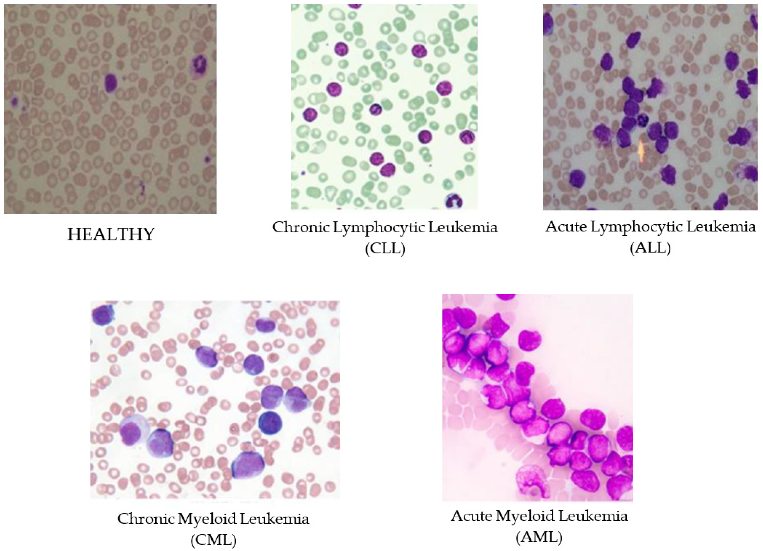
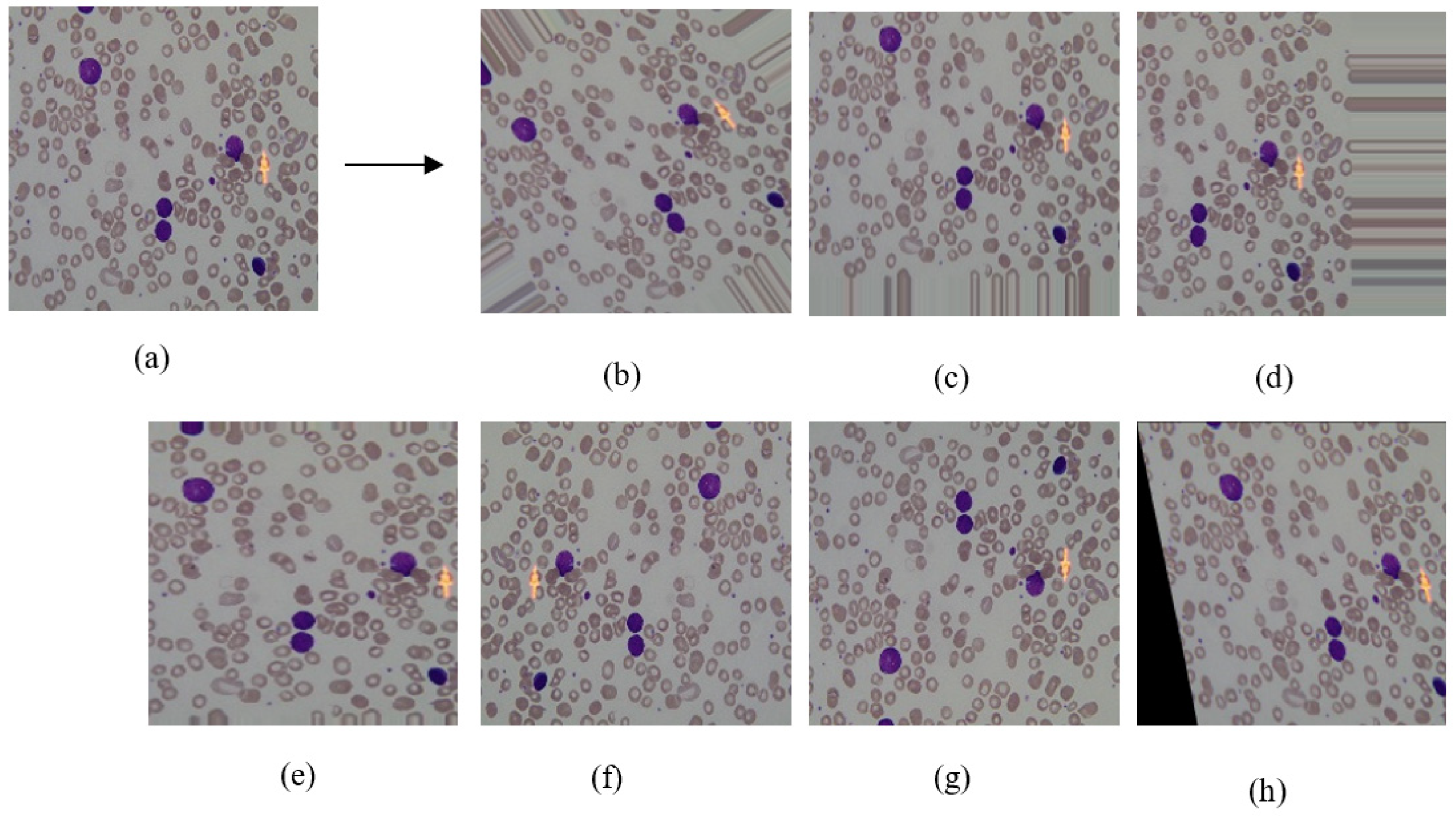
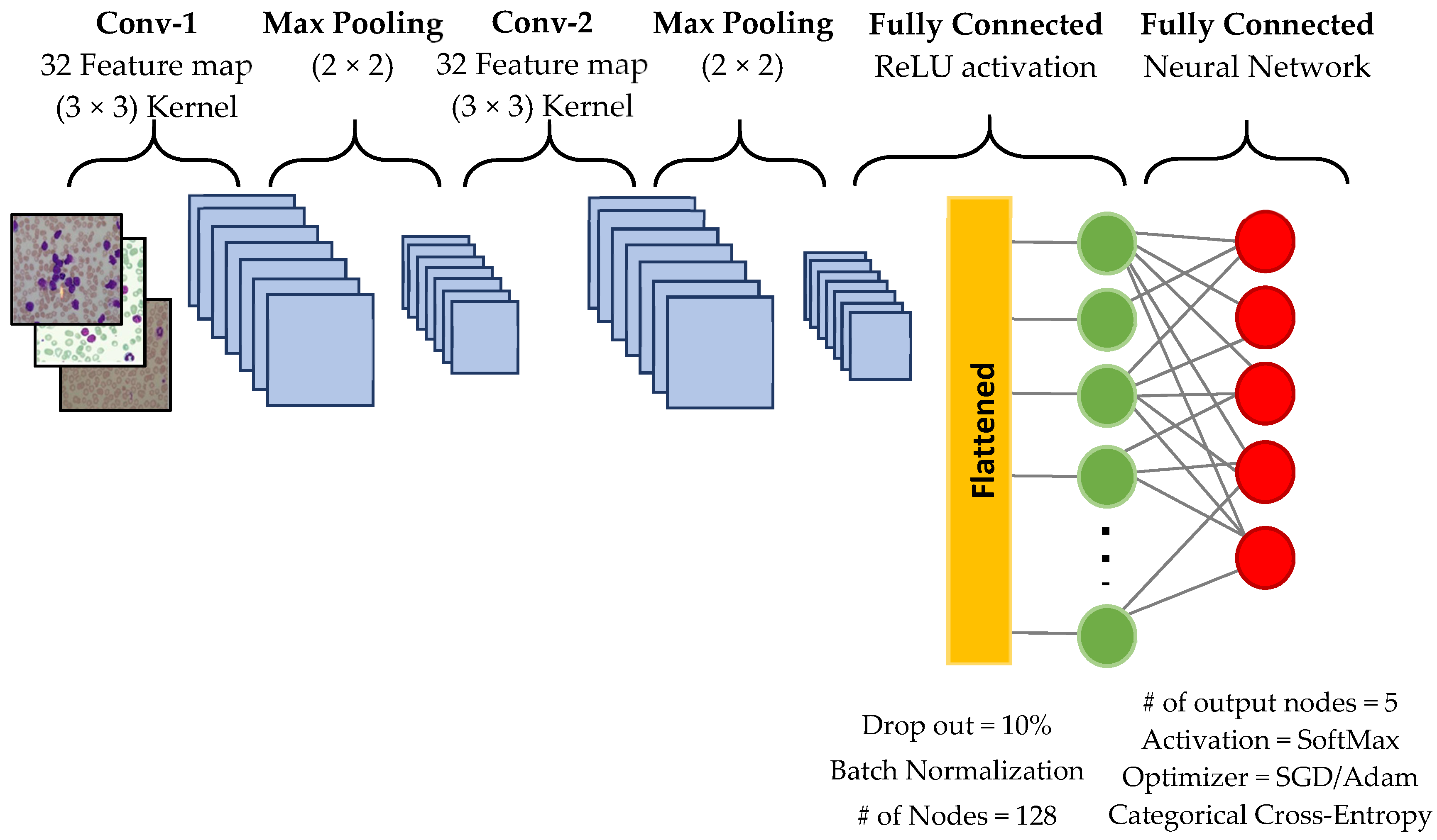
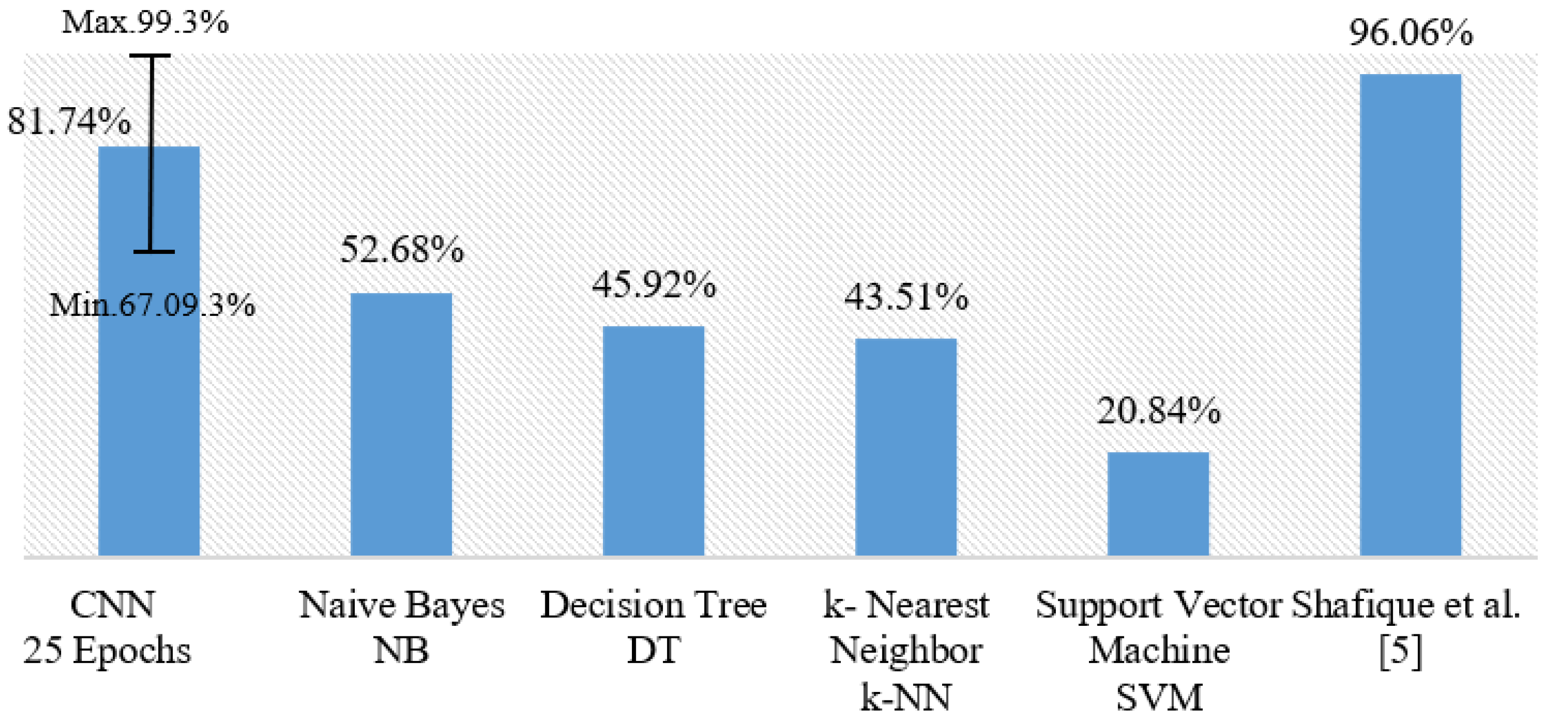
Leukemia is a fatal cancer and has two main types: Acute and chronic. Each type has two more subtypes: Lymphoid and myeloid. Hence, in total, there are four subtypes of leukemia. This study proposes a new approach for diagnosis of all subtypes of leukemia from microscopic blood cell images using convolutional neural networks (CNN), which requires a large training data set. Therefore, we also investigated the effects of data augmentation for an increasing number of training samples synthetically. We used two publicly available leukemia data sources: ALL-IDB and ASH Image Bank. Next, we applied seven different image transformation techniques as data augmentation. We designed a CNN architecture capable of recognizing all subtypes of leukemia. Besides, we also explored other well-known machine learning algorithms such as naive Bayes, support vector machine, k-nearest neighbor, and decision tree. To evaluate our approach, we set up a set of experiments and used 5-fold cross-validation. The results we obtained from experiments showed that our CNN model performance has 88.25% and 81.74% accuracy, in leukemia versus healthy and multi-class classification of all subtypes, respectively. Finally, we also showed that the CNN model has a better performance than other well-known machine learning algorithms.
Keywords: leukemia diagnosis; recognizing leukemia subtypes; multi-class classification; microscopic blood cells images; data augmentation; deep learning; convolutional neural network leukemia diagnosis; recognizing leukemia subtypes; multi-class classification; microscopic blood cells images; data augmentation; deep learning; convolutional neural network