Morphological Variability of Sphenoid Sinus Pneumatization and Its Impact on Adjacent Neurovascular Structures
 ,
,  ,
,  , ,
, , 
Abstract
1. Introduction
2. Materials and Methods
2.1. Sample Demographics
2.2. Radiological Protocol
2.3. Methodology
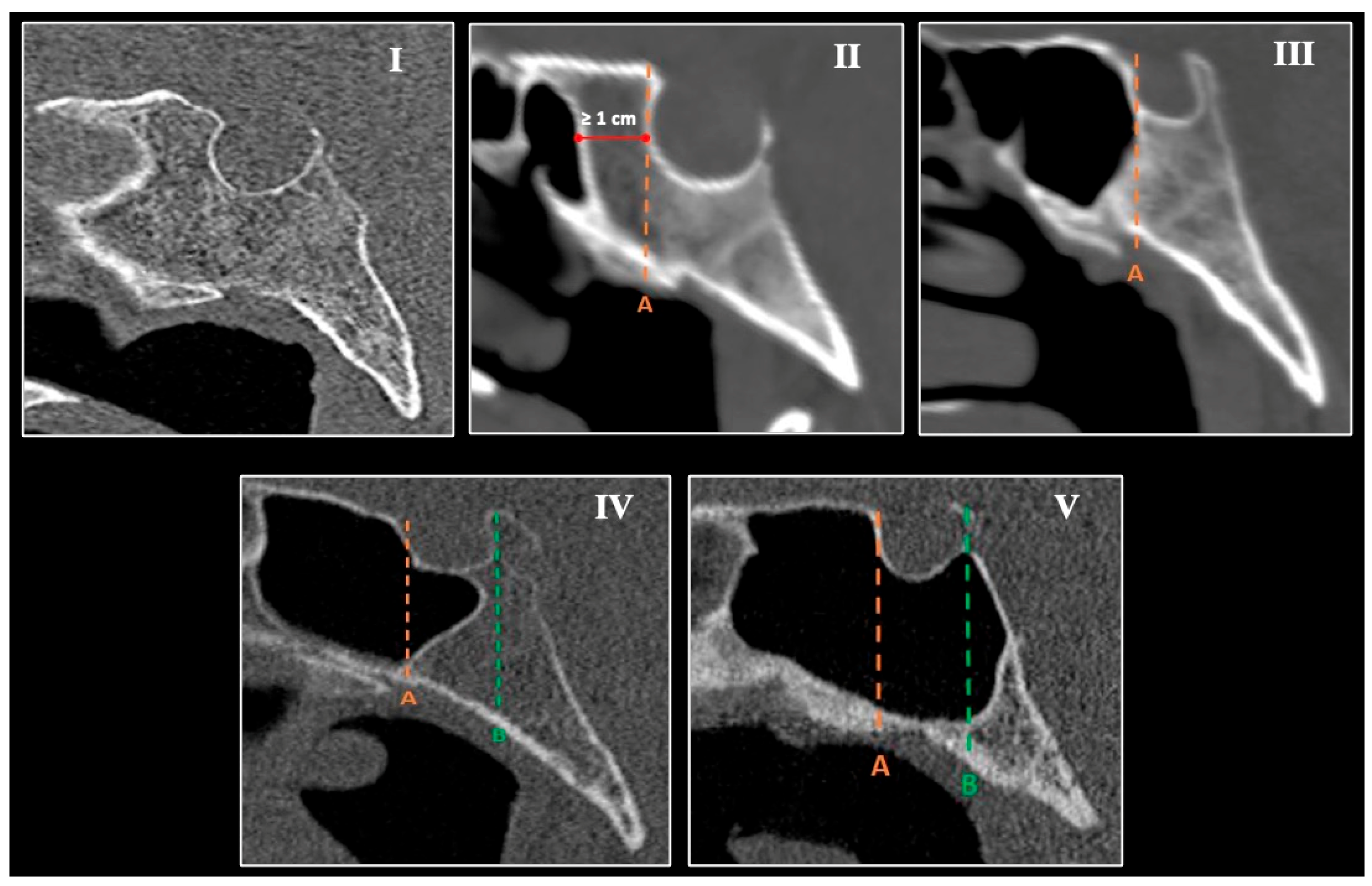
- Agenesis—absence of the SS;
- Conchal—slight pneumatization; sinus does not reach the anterior sellar wall and terminates ≥10 mm anterior to it;
- Presellar—pneumatization limited to the vertical plane of the anterior sellar wall;
- Sellar incomplete—pneumatization reaches the vertical plane of the posterior clinoid process, involving the anterior wall and sellar floor;
- Sellar complete—pneumatization extends beyond the sella toward the basilar occiput.
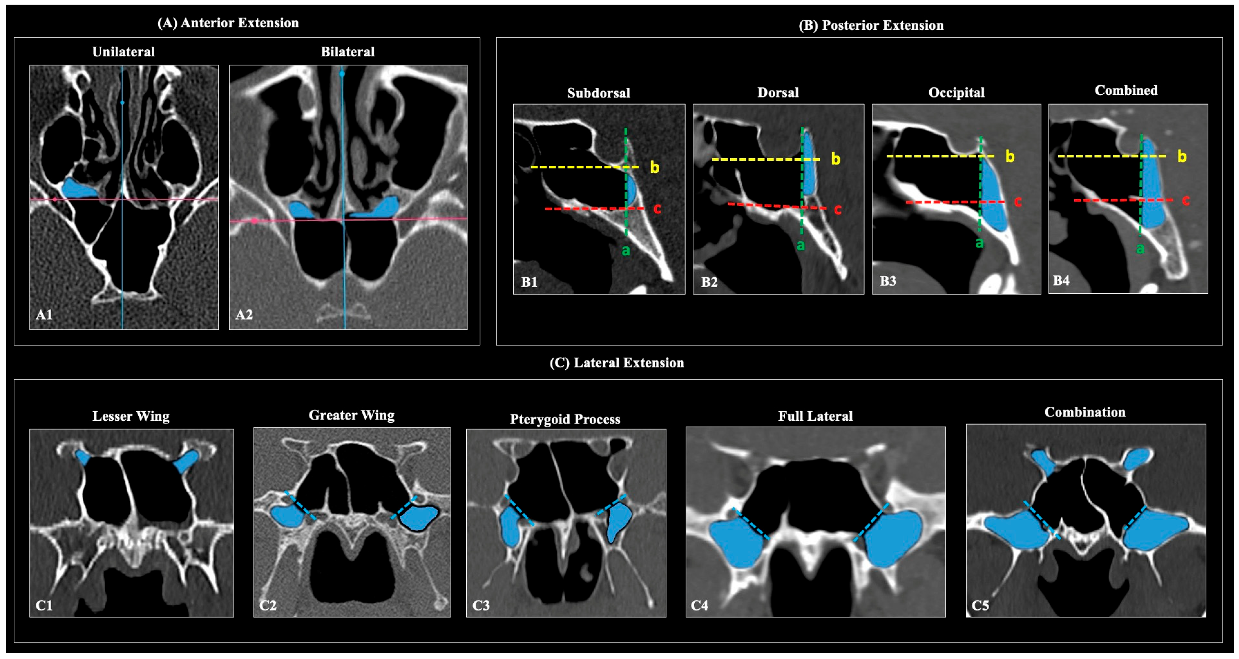
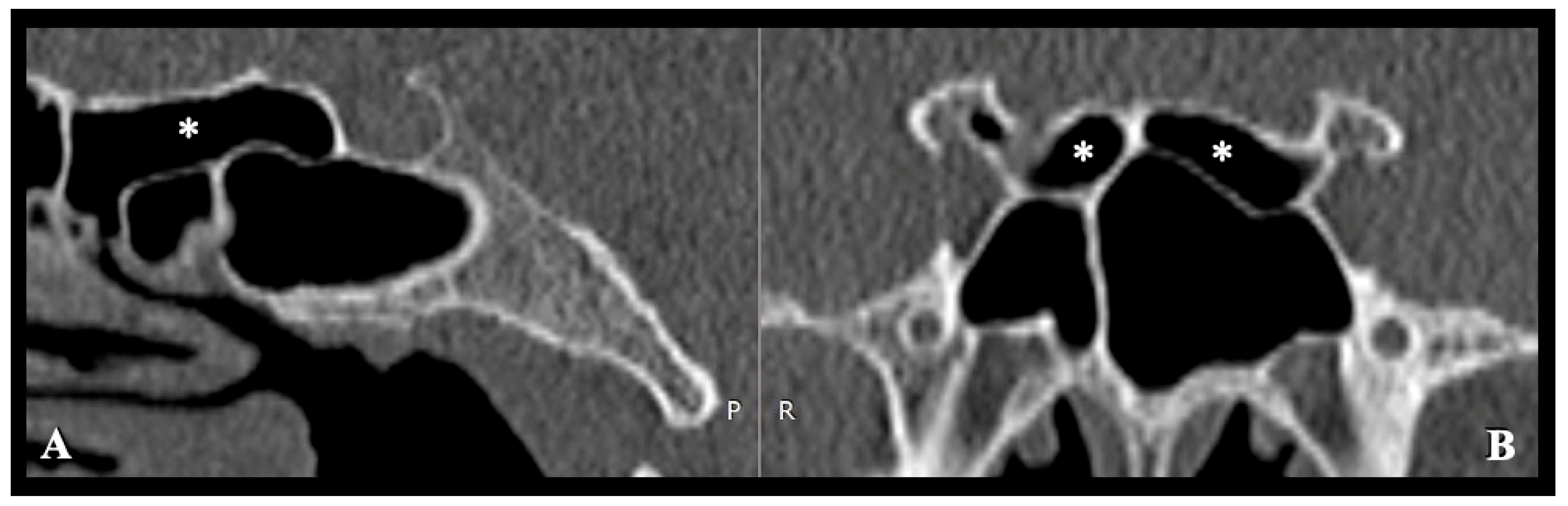
- Anterior extension (axial): pneumatization extending anteriorly beyond a line drawn along the sinus side of the sphenoid crest.
- Posterior (clival) extension (sagittal): subtypes—subdorsal, dorsal, occipital, and combined (dorsal + occipital)—defined relative to the posterior sellar wall, sellar floor line, and the horizontal plane through the superior margins of the paired Vidian canals.
- Lateral extension (coronal): presence of pneumatization lateral to the SS categorized as LW, GW, PP, full-lateral (GW + PP), and combinations thereof.
2.4. Statistical Analysis
3. Results
3.1. Sphenoid Sinus Pneumatization—Main Types
3.2. Sphenoid Sinus Pneumatization—Extension Patterns
3.3. Adjacent Neurovascular Structures
3.4. Association Between Sphenoid Sinus Pneumatization Types and Adjacent Neurovascular Structures
4. Discussion
4.1. Anatomical Considerations of Sphenoid Sinus Pneumatization (SSP)
4.2. Anatomical Considerations of Neurovascular Relationships
4.3. Surgical Significance of Morphological Variations
4.4. Strengths and Limitations
5. Conclusions
Author Contributions
Funding
Institutional Review Board Statement
Informed Consent Statement
Data Availability Statement
Conflicts of Interest
References
- Cope, V.Z. The Internal Structure of the Sphenoidal Sinus. J. Anat. 1917, 51, 127–136. [Google Scholar]
- Goksel, S.; Unsal, G.; Cakir Karabas, H.; Erturk, A.F.; Ozcan, I.; Orhan, K. Association of Accessory Sphenoidal Septum with Protrusions of Sphenoid Sinus. Eur. Arch. Oto-Rhino-Laryngol. 2023, 280, 2323–2329. [Google Scholar] [CrossRef] [Scilit] [PubMed]
- Idowu, O.E.; Balogun, B.O.; Okoli, C.A. Dimensions, Septation, and Pattern of Pneumatization of the Sphenoidal Sinus. Folia Morphol. 2009, 68, 228–232. [Google Scholar]
- Tursun, S.; Bayar Muluk, N.; Inal, M.; Göncüoğlu, A.; Şencan, Z. The Relationship between Sphenoid Sinus, Carotid Canal, and Optical Canal in Paranasal Sinus Computed Tomography in Children. J. Neurol. Surg. B Skull Base 2023, 84, 513–520. [Google Scholar] [CrossRef] [Scilit]
- Kinnman, J. Surgical Aspects of the Anatomy of the Sphenoidal Sinuses and the Sella Turcica. J. Anat. 1977, 124, 541–553. [Google Scholar]
- Tan, H.K.K.; Ong, Y.K. Sphenoid Sinus: An Anatomic and Endoscopic Study in Asian Cadavers. Clin. Anat. 2007, 20, 745–750. [Google Scholar] [CrossRef] [Scilit]
- Meloni, F.; Mini, R.; Rovasio, S.; Stomeo, F.; Teatini, G.P. Anatomic Variations of Surgical Importance in Ethmoid Labyrinth and Sphenoid Sinus. A Study of Radiological Anatomy. Surg. Radiol. Anat. 1992, 14, 65–70. [Google Scholar] [CrossRef] [Scilit]
- Fujii, K.; Chambers, S.M.; Rhoton, A.L. Neurovascular Relationships of the Sphenoid Sinus. J. Neurosurg. 1979, 50, 31–39. [Google Scholar] [CrossRef] [Scilit]
- Papadopoulos-Manolarakis, P.; Triantafyllou, G.; Georgalas, C.; Strantzalis, G.; Piagkou, M. Morphological Variability of the Sphenoid Sinus Septations. J. Neurol. Surg. B Skull Base 2025. [Google Scholar] [CrossRef] [Scilit]
- Asal, N.; Bayar Muluk, N.; Inal, M.; Şahan, M.H.; Doğan, A.; Arıkan, O.K. Carotid Canal and Optic Canal at Sphenoid Sinus. Neurosurg. Rev. 2019, 42, 519–529. [Google Scholar] [CrossRef] [Scilit] [PubMed]
- Triantafyllou, G.; Piagkou, M. The Developing Human Sphenoid Bone: Linking Embryological Development to Adult Morphology. Biology 2025, 14, 1090. [Google Scholar] [CrossRef] [Scilit]
- Hammer, G.; Radberg, C. The Sphenoidal Sinus. An Anatomical and Roentgenologic Study with Reference to Transsphenoid Hypophysectomy. Acta Radiol. 1961, 56, 401–422. [Google Scholar] [CrossRef] [Scilit]
- Wang, J.; Bidari, S.; Inoue, K.; Yang, H.; Rhoton, A. Extensions of the Sphenoid Sinus. Neurosurgery 2010, 66, 797–816. [Google Scholar] [CrossRef] [Scilit]
- Hiremath, S.B.; Gautam, A.B.; Sheeja, K.; Benjamin, G. Assessment of Variations in Sphenoid Sinus Pneumatization in Indian Population: A Multidetector Computed Tomography Study. Indian J. Radiol. Imaging 2018, 28, 273–279. [Google Scholar] [CrossRef] [Scilit]
- Vaezi, A.; Cardenas, E.; Pinheiro-Neto, C.; Paluzzi, A.; Branstetter, B.F.; Gardner, P.A.; Snyderman, C.H.; Fernandez-Miranda, J.C. Classification of Sphenoid Sinus Pneumatization: Relevance for Endoscopic Skull Base Surgery. Laryngoscope 2015, 125, 577–581. [Google Scholar] [CrossRef] [Scilit]
- Renn, W.H.; Rhoton, A.L. Microsurgical Anatomy of the Sellar Region. J. Neurosurg. 1975, 43, 288–298. [Google Scholar] [CrossRef] [Scilit] [PubMed]
- Luzzi, S.; Giotta Lucifero, A.; Rabski, J.; Kadri, P.A.S.; Al-Mefty, O. The Party Wall: Redefining the Indications of Transcranial Approaches for Giant Pituitary Adenomas in Endoscopic Era. Cancers 2023, 15, 2235. [Google Scholar] [CrossRef] [Scilit]
- Anusha, B.; Baharudin, A.; Philip, R.; Harvinder, S.; Shaffie, B.M. Anatomical Variations of the Sphenoid Sinus and Its Adjacent Structures: A Review of Existing Literature. Surg. Radiol. Anat. 2014, 36, 419–427. [Google Scholar] [CrossRef] [Scilit] [PubMed]
- Dal Secchi, M.; Dolci, R.; Teixeira, R.; Lazarini, P. An Analysis of Anatomic Variations of the Sphenoid Sinus and Its Relationship to the Internal Carotid Artery. Int. Arch. Otorhinolaryngol. 2018, 22, 161–166. [Google Scholar] [CrossRef] [Scilit] [PubMed]
- Anusha, B.; Baharudin, A.; Philip, R.; Harvinder, S.; Shaffie, B.M.; Ramiza, R.R. Anatomical Variants of Surgically Important Landmarks in the Sphenoid Sinus: A Radiologic Study in Southeast Asian Patients. Surg. Radiol. Anat. 2015, 37, 1183–1190. [Google Scholar] [CrossRef] [Scilit]
- Lazaridis, N.; Natsis, K.; Koebke, J.; Themelis, C. Nasal, Sellar, and Sphenoid Sinus Measurements in Relation to Pituitary Surgery. Clin. Anat. 2010, 23, 629–636. [Google Scholar] [CrossRef] [Scilit]
- Orhan, M.; Govsa, F.; Saylam, C. A Quite Rare Condition: Absence of Sphenoidal Sinuses. Surg. Radiol. Anat. 2010, 32, 551–553. [Google Scholar] [CrossRef] [Scilit]
- Kiciński, P.; Podgórski, M.; Grzelak, P.; Małachowska, B.; Polguj, M. Authors’ Classification of Sphenoid Sinus Pneumatizations into the Sphenoid Bone Processes. J. Clin. Med. 2025, 14, 5811. [Google Scholar] [CrossRef] [Scilit] [PubMed]
- Cho, J.H.; Kim, J.K.; Lee, J.-G.; Yoon, J.-H. Sphenoid Sinus Pneumatization and Its Relation to Bulging of Surrounding Neurovascular Structures. Ann. Otol. Rhinol. Laryngol. 2010, 119, 646–650. [Google Scholar] [CrossRef] [Scilit]
- Štoković, N.; Trkulja, V.; Dumić-Čule, I.; Čuković-Bagić, I.; Lauc, T.; Vukičević, S.; Grgurević, L. Sphenoid Sinus Types, Dimensions and Relationship with Surrounding Structures. Ann. Anat. Anat. Anz. 2016, 203, 69–76. [Google Scholar] [CrossRef] [Scilit]
- Refaat, R.; Basha, M.A.A. The Impact of Sphenoid Sinus Pneumatization Type on the Protrusion and Dehiscence of the Adjacent Neurovascular Structures: A Prospective MDCT Imaging Study. Acad. Radiol. 2020, 27, e132–e139. [Google Scholar] [CrossRef] [Scilit]
- Mohebbi, A.; Rajaeih, S.; Safdarian, M.; Omidian, P. The Sphenoid Sinus, Foramen Rotundum and Vidian Canal: A Radiological Study of Anatomical Relationships. Braz. J. Otorhinolaryngol. 2017, 83, 381–387. [Google Scholar] [CrossRef] [Scilit]
- Tawfik, A.; El-fattah, A.M.A.; Nour, A.I.; Tawfik, A.M. Neurovascular Surgical Keys Related to Sphenoid Window: Radiologic Study of Egyptian’s Sphenoid. World Neurosurg. 2018, 116, e840–e849. [Google Scholar] [CrossRef] [Scilit]
- Farrell, C.J.; Nyquist, G.G.; Farag, A.A.; Rosen, M.R.; Evans, J.J. Principles of Pituitary Surgery. Otolaryngol. Clin. N. Am. 2016, 49, 95–106. [Google Scholar] [CrossRef] [Scilit] [PubMed]
- ELKammash, T.H.; Enaba, M.M.; Awadalla, A.M. Variability in Sphenoid Sinus Pneumatization and Its Impact upon Reduction of Complications Following Sellar Region Surgeries. Egypt. J. Radiol. Nucl. Med. 2014, 45, 705–714. [Google Scholar] [CrossRef] [Scilit]
- Dessi, P.; Moulin, G.; Castro, F.; Chagnaud, C.; Cannoni, M. Protrusion of the Optic Nerve into the Ethmoid and Sphenoid Sinus: Prospective Study of 150 CT Studies. Neuroradiology 1994, 36, 515–516. [Google Scholar] [CrossRef] [Scilit] [PubMed]



| SS Main Types | Total (n = 253) | Females (n = 124) | Males (n = 129) | p-Value | Age Distribution | p-Value |
|---|---|---|---|---|---|---|
| Sellar Complete | 160 (63.2%) | 81 (65.3%) | 79 (61.2%) | 0.288 | 50.46 (14.6) | 0.634 |
| Sellar Incomplete | 70 (27.7%) | 33 (26.6%) | 37 (28.7%) | 53.63 (15.8) | ||
| Presellar | 22 (8.7%) | 9 (7.3%) | 13 (10.1%) | 54.14 (14.8) | ||
| Agenesis † | 1 (0.4%) | 1 (0.8%) | 0 (0%) | 70.00 (-) |
| SSP Extension Patterns | Total (n = 506) | Left (n = 253) | Right (n = 253) | p-Value | Females (n = 248) | Males (n = 258) | p-Value |
|---|---|---|---|---|---|---|---|
| Anterior | 102 (20.2%) | 52 (20.6%) | 50 (19.8%) | 0.825 | 42 (16.9%) | 60 (23.3%) | 0.076 |
| Lateral | 236 (46.6%) | 121 (47.8%) | 115 (45.5%) | 0.366 | 117 (47.2%) | 119 (46.1%) | 0.184 |
| LW | 35 (6.9%) | 13 (5.1%) | 22 (8.7%) | 11 (4.4%) | 24 (9.3%) | ||
| GW | 56 (11.1%) | 34 (13.4%) | 22 (8.7%) | 31 (12.5%) | 25 (9.7%) | ||
| PP | 23 (4.5%) | 10 (4%) | 13 (5.1%) | 14 (5.6%) | 9 (3.5%) | ||
| FL | 58 (11.5%) | 29 (11.5%) | 29 (11.5%) | 33 (13.3%) | 25 (9.7%) | ||
| LW + GW | 11 (2.2%) | 5 (2%) | 6 (2.4%) | 6 (2.4%) | 5 (1.9%) | ||
| LW + PP | 5 (1%) | 4 (1.6%) | 1 (0.4%) | 3 (1.2%) | 2 (0.8%) | ||
| LW + FL | 48 (9.5%) | 26 (10.3%) | 22 (8.7%) | 19 (7.7%) | 29 (11.2%) | ||
| Posterior | 161 (63.6%) | - | - | - | 81 (65.3%) | 80 (62%) | <0.001 * |
| Dorsal | 16 (6.2%) | 3 (2.4%) | 13 (10.1%) | ||||
| Subdorsal | 136 (53.8%) | 73 (58.9%) | 63 (48.8%) | ||||
| Occipital | 7 (2.8%) | 5 (4%) | 2 (1.6%) | ||||
| Combination | 2 (0.8%) | 0 (0%) | 2 (1.6%) |
| Parameters | Total (n = 506) | Left (n = 253) | Right (n = 253) | p-Value | Females (n = 248) | Males (n = 258) | p-Value |
|---|---|---|---|---|---|---|---|
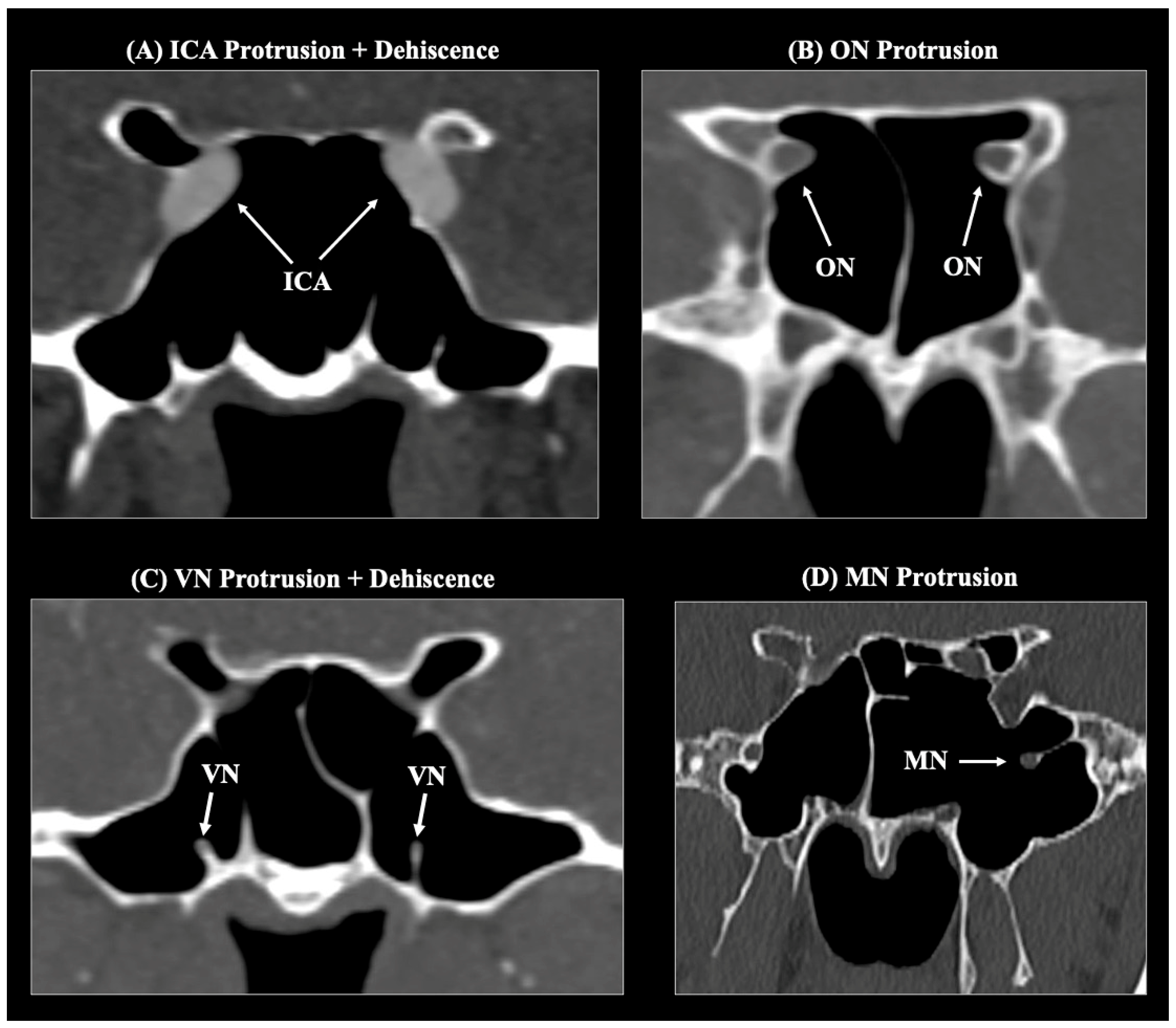
| ICA Pt | 103 (20.4%) | 52 (20.6%) | 51 (20.2%) | 0.728 | 42 (16.9%) | 61 (23.6%) | 0.139 |
| ICA Pt + Dh | 14 (2.8%) | 8 (3.2%) | 6 (2.4%) | 9 (3.6%) | 5 (1.9%) | ||
| ON Pt | 83 (16.4%) | 40 (15.8%) | 43 (17%) | 0.335 | 31 (12.5%) | 52 (20.2%) | 0.051 |
| ON Pt + Dh | 15 (3%) | 5 (2%) | 10 (4%) | 8 (3.2%) | 7 (2.7%) | ||
| VN Pt | 78 (15.4%) | 40 (15.8%) | 38 (15%) | 0.242 | 44 (16.9%) | 34 (13.2%) | 0.232 |
| VN Pt + Dh | 64 (12.6%) | 38 (15%) | 26 (10.3%) | 27 (10.9%) | 37 (14.3%) | ||
| MN Pt | 28 (5.5%) | 17 (6.7%) | 11 (4.3%) | 0.320 | 14 (5.6%) | 14 (5.4%) | 0.278 |
| MN Pt + Dh | 11 (2.2%) | 7 (2.8%) | 4 (1.6%) | 8 (3.2%) | 3 (1.2%) |
| Pneumatization Types | ICA | ON | VN | MN | p-Value | |||||
|---|---|---|---|---|---|---|---|---|---|---|
| Typical (n = 389) | Pt/Pt + Dh (n = 117) | Typical (n = 408) | Pt/Pt + Dh (n = 98) | Typical (n = 364) | Pt/Pt + Dh (n = 142) | Typical (n = 467) | Pt/Pt + Dh (n = 39) | |||
| Main | Aplasia | 2 (100%) | 0 (0%) | 2 (100%) | 0 (0%) | 2 (100%) | 0 (0%) | 2 (100%) | 0 (0%) | <0.001 * |
| Conchal | ND | ND | ND | ND | ND | ND | ND | ND | ||
| Presellar | 41 (93.2%) | 3 (6.8%) | 41 (93.2%) | 3 (6.8%) | 44 (100%) | 0 (0%) | 44 (100%) | 0 (0%) | ||
| SI | 137 (99.3%) | 1 (0.7%) | 129 (93.5%) | 9 (6.5%) | 127 (92%) | 11 (8%) | 136 (98.6%) | 2 (1.4%) | ||
| SC | 209 (64.9%) | 113 (35.1%) | 236 (73.3%) | 86 (26.7%) | 191 (59.4%) | 141 (40.6%) | 285 (88.5%) | 37 (11.5%) | ||
| Posterior Extension | Subdorsal | 192 (70.6%) | 80 (29.4%) | 208 (76.5%) | 64 (23.5%) | 171 (62.9%) | 101 (37.1%) | 246 (90.4%) | 26 (9.6%) | <0.001 * |
| Dorsal | 6 (18.8%) | 26 (81.2%) | 18 (56.3%) | 14 (43.7%) | 11 (34.4%) | 21 (65.6%) | 23 (71.9%) | 9 (28.1%) | ||
| Occipital | 9 (64.3%) | 5 (35.7%) | 9 (64.3%) | 5 (35.7%) | 7 (50%) | 7 (50%) | 12 (85.7%) | 2 (14.3%) | ||
| Combo | 2 (50%) | 2 (50%) | 1 (25%) | 3 (75%) | 2 (50%) | 2 (50%) | 4 (100%) | 0 (0%) | ||
| Lateral Extension | FL | 49 (46.2%) | 57 (53.8%) | 58 (54.7%) | 48 (45.3%) | 16 (15.1%) | 90 (79.9%) | 75 (70.8%) | 31 (29.2%) | <0.001 * |
| GW | 50 (74.6%) | 17 (25.4%) | 56 (83.6%) | 11 (16.4%) | 35 (52.2%) | 32 (47.8%) | 59 (88.1%) | 8 (11.9%) | ||
| PP | 18 (64.3%) | 10 (35.7%) | 23 (100%) | 0 (0%) | 12 (42.9%) | 16 (56.1%) | 28 (100%) | 0 (0%) | ||
| LW | 43 (43.4%) | 56 (56.6%) | 4 (4%) | 95 (96%) | 44 (44.4%) | 45 (55.6%) | 81 (81.8%) | 18 (18.2%) | ||
| Anterior Extension | Yes | 87 (85.3%) | 15 (14.7%) | 91 (89.2%) | 11 (10.8%) | 73 (71.6%) | 29 (28.4%) | 94 (92.2%) | 8 (7.8%) | >0.05 |
Disclaimer/Publisher’s Note: The statements, opinions and data contained in all publications are solely those of the individual author(s) and contributor(s) and not of MDPI and/or the editor(s). MDPI and/or the editor(s) disclaim responsibility for any injury to people or property resulting from any ideas, methods, instructions or products referred to in the content. |
© 2026 by the authors. Licensee MDPI, Basel, Switzerland. This article is an open access article distributed under the terms and conditions of the Creative Commons Attribution (CC BY) license.
Share and Cite
Papadopoulos-Manolarakis, P.; Triantafyllou, G.; Georgalas, C.; Paschopoulos, I.; Stranjalis, G.; Piagkou, M. Morphological Variability of Sphenoid Sinus Pneumatization and Its Impact on Adjacent Neurovascular Structures. Diagnostics 2026, 16, 809. https://doi.org/10.3390/diagnostics16050809
Papadopoulos-Manolarakis P, Triantafyllou G, Georgalas C, Paschopoulos I, Stranjalis G, Piagkou M. Morphological Variability of Sphenoid Sinus Pneumatization and Its Impact on Adjacent Neurovascular Structures. Diagnostics. 2026; 16(5):809. https://doi.org/10.3390/diagnostics16050809
Chicago/Turabian StylePapadopoulos-Manolarakis, Panagiotis, George Triantafyllou, Christos Georgalas, Ioannis Paschopoulos, George Stranjalis, and Maria Piagkou. 2026. "Morphological Variability of Sphenoid Sinus Pneumatization and Its Impact on Adjacent Neurovascular Structures" Diagnostics 16, no. 5: 809. https://doi.org/10.3390/diagnostics16050809
APA StylePapadopoulos-Manolarakis, P., Triantafyllou, G., Georgalas, C., Paschopoulos, I., Stranjalis, G., & Piagkou, M. (2026). Morphological Variability of Sphenoid Sinus Pneumatization and Its Impact on Adjacent Neurovascular Structures. Diagnostics, 16(5), 809. https://doi.org/10.3390/diagnostics16050809