CT Radiomic Features of the Crystalline Lens and Association with Age, Hypertension and Cerebral White Matter Lesions

 , , , , , ,
  and
and
Abstract
1. Introduction
2. Methods
2.1. Patient Selection and Study Design
2.2. CT Acquisition Protocol
2.3. Assessment of White Matter Lesions
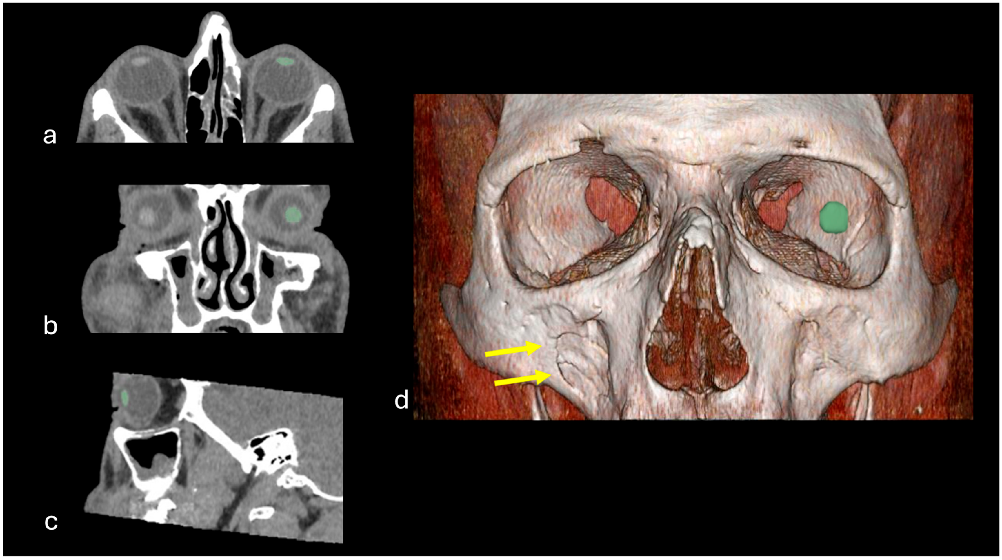
2.4. Semiautomatic Segmentation of the Lens and Feature Extraction
2.5. Statistical Analysis
3. Results
3.1. Patient Characteristics
3.2. Age-Related Changes in Radiomic Features of the Eye Lens Texture
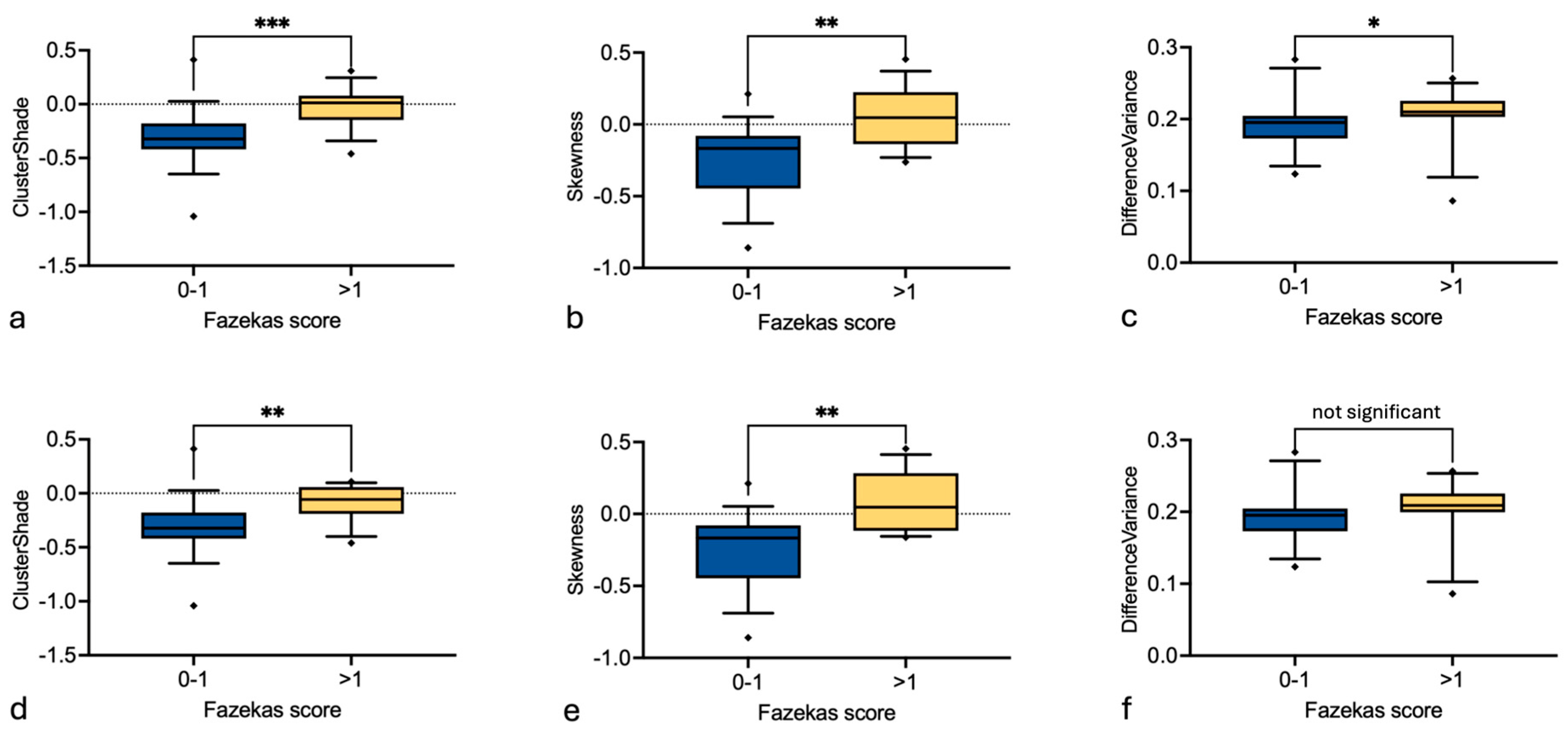
3.3. Effect of Fazekas and Hypertension on the Eye Lens Texture
4. Discussion
5. Conclusions
Author Contributions
Funding
Institutional Review Board Statement
Informed Consent Statement
Data Availability Statement
Conflicts of Interest
Abbreviations
| CT | Computed tomography |
| WML | White matter lesions |
References
- Cellina, M.; Cè, M.; Marziali, S.; Irmici, G.; Gibelli, D.; Oliva, G.; Carrafiello, G. Computed tomography in traumatic orbital emergencies: A pictorial essay-imaging findings, tips, and report flowchart. Insights Imaging 2022, 13, 4. [Google Scholar] [CrossRef] [Scilit]
- Sachs, J.R.; Nahmias, J.A.; Hiatt, K.D.; Bomar, J.G.; West, T.G.; Bunch, P.M.; Benayoun, M.D.; Lack, C.; Thompson, A.C. Study of the Natural Crystalline Lens Characteristics Using Dual-Energy Computed Tomography. Diagnostics 2022, 12, 2857. [Google Scholar] [CrossRef] [Scilit]
- Bioletto, F.; Prencipe, N.; Berton, A.M.; Aversa, L.S.; Cuboni, D.; Varaldo, E.; Gasco, V.; Ghigo, E.; Grottoli, S. Radiomic Analysis in Pituitary Tumors: Current Knowledge and Future Perspectives. J. Clin. Med. 2024, 13, 336. [Google Scholar] [CrossRef] [Scilit]
- Park, J.E.; Kickingereder, P.; Kim, H.S. Radiomics and Deep Learning from Research to Clinical Workflow: Neuro-Oncologic Imaging. Korean J. Radiol. 2020, 21, 1126–1137. [Google Scholar] [CrossRef] [Scilit]
- Abdel Razek, A.A.K.; Alksas, A.; Shehata, M.; AbdelKhalek, A.; Abdel Baky, K.; El-Baz, A.; Helmy, E. Clinical applications of artificial intelligence and radiomics in neuro-oncology imaging. Insights Imaging 2021, 12, 152. [Google Scholar] [CrossRef] [Scilit]
- Nemet, A.Y.; Vinker, S.; Levartovsky, S.; Kaiserman, I. Is cataract associated with cardiovascular morbidity? Eye 2010, 24, 1352–1358. [Google Scholar] [CrossRef] [Scilit]
- Hu, W.S.; Lin, C.L.; Huang, C.Y.; Chang, S.S.; Chang, K.C. Cataract increases the risk of peripheral artery occlusive disease: A nationwide population-based cohort study with propensity score. Int. J. Cardiol. 2016, 220, 768–771. [Google Scholar] [CrossRef] [Scilit]
- Zaliuniene, D.; Jasinskas, V.; Jurkevicius, R.; Gustiene, O.; Zaliunas, R. Endothelial function, intima-media thickness, and ankle-brachial index in patients with cataract and age-related macular degeneration. Eur. J. Ophthalmol. 2008, 18, 384–391. [Google Scholar] [CrossRef] [Scilit]
- Kynast, J.; Lampe, L.; Luck, T.; Frisch, S.; Arelin, K.; Hoffmann, K.T.; Loeffler, M.; Riedel-Heller, S.G.; Villringer, A.; Schroeter, M.L. White matter hyperintensities associated with small vessel disease impair social cognition beside attention and memory. J. Cereb. Blood Flow Metab. 2018, 38, 996–1009. [Google Scholar] [CrossRef] [Scilit]
- Andere, A.; Jindal, G.; Molino, J.; Collins, S.; Merck, D.; Burton, T.; Stretz, C.; Yaghi, S.; Sacchetti, D.C.; Jamal, S.E.; et al. Volumetric White Matter Hyperintensity Ranges Correspond to Fazekas Scores on Brain MRI. J. Stroke Cerebrovasc. Dis. 2022, 31, 106333. [Google Scholar] [CrossRef] [Scilit]
- Schuerch, K.; Frech, H.; Zinkernagel, M. Conjunctival Microangiopathy in Diabetes Mellitus Assessed with Optical Coherence Tomography Angiography. Transl. Vis. Sci. Technol. 2020, 9, 10. [Google Scholar] [CrossRef] [Scilit]
- Madonna, R.; Balistreri, C.R.; Geng, Y.J.; de Caterina, R. Diabetic microangiopathy: Pathogenetic insights and novel therapeutic approaches. Vascul. Pharmacol. 2017, 90, 1–7. [Google Scholar] [CrossRef] [Scilit]
- Kao, Y.H.; Chou, M.C.; Chen, C.H.; Yang, Y.H. White Matter Changes in Patients with Alzheimer’s Disease and Associated Factors. J. Clin. Med. 2019, 8, 167. [Google Scholar] [CrossRef] [Scilit]
- Wahlund, L.O.; Westman, E.; van Westen, D.; Wallin, A.; Shams, S.; Cavallin, L.; Larsson, E.M. Imaging biomarkers of dementia: Recommended visual rating scales with teaching cases. Insights Imaging 2017, 8, 79–90. [Google Scholar] [CrossRef] [Scilit]
- Rizzo, S.; Botta, F.; Raimondi, S.; Origgi, D.; Fanciullo, C.; Morganti, A.G.; Bellomi, M. Radiomics: The facts and the challenges of image analysis. Eur. Radiol. Exp. 2018, 2, 36. [Google Scholar] [CrossRef] [Scilit]
- Kursa, M.B.; Rudnicki, W.R. Feature Selection with the Boruta Package. J. Stat. Softw. 2010, 36, 1–13. [Google Scholar] [CrossRef] [Scilit]
- Lambin, P.; Leijenaar, R.T.H.; Deist, T.M.; Peerlings, J.; de Jong, E.E.C.; van Timmeren, J.; Sanduleanu, S.; Larue, R.; Even, A.; Jochems, A.; et al. Radiomics: The bridge between medical imaging and personalized medicine. Nat. Rev. Clin. Oncol. 2017, 14, 749–762. [Google Scholar] [CrossRef] [Scilit]
- Zhang, H.; Zhang, H.; Jiang, M.; Li, J.; Li, J.; Zhou, H.; Song, X.; Fan, X. Radiomics in ophthalmology: A systematic review. Eur. Radiol. 2025, 35, 542–557. [Google Scholar] [CrossRef] [Scilit]
- Ilginis, T.; Clarke, J.; Patel, P.J. Ophthalmic imaging. Br. Med. Bull. 2014, 111, 77–88. [Google Scholar] [CrossRef] [Scilit]
- Rust, G.; Ye, J.; Baltrus, P.; Daniels, E.; Adesunloye, B.; Fryer, G.E. Practical barriers to timely primary care access: Impact on adult use of emergency department services. Arch. Intern. Med. 2008, 168, 1705–1710. [Google Scholar] [CrossRef] [Scilit]
- Taslakian, B.; Hourani, R. Don’t forget to report “simple” finding on CT: The hypodense eye lens. Eur. J. Pediatr. 2013, 172, 131–132. [Google Scholar] [CrossRef] [Scilit]
- Sharma, N.; Aggarwal, L.M. Automated medical image segmentation techniques. J. Med. Phys. 2010, 35, 3–14. [Google Scholar] [CrossRef] [Scilit]
- Han, S.; Lee, J.K.; Lee, D.; Lee, J. Automated eyeball volume measurement based on CT images using neural network-based segmentation and simple estimation. Sci. Rep. 2024, 14, 15094. [Google Scholar] [CrossRef] [Scilit]
- Flaiban, E.; Orhan, K.; Gonçalves, B.C.; Lopes, S.; Costa, A.L.F. Radiomics in Action: Multimodal Synergies for Imaging Biomarkers. Bioengineering 2025, 12, 1139. [Google Scholar] [CrossRef] [Scilit]
- Wolffsohn, J.S.; Davies, L.N. Presbyopia: Effectiveness of correction strategies. Prog. Retin. Eye Res. 2019, 68, 124–143. [Google Scholar] [CrossRef] [Scilit]
- Dubbelman, M.; van der Heijde, G.L.; Weeber, H.A.; Vrensen, G.F.J.M. Changes in the internal structure of the human crystalline lens with age and accommodation. Vision Res. 2003, 43, 2363–2375. [Google Scholar] [CrossRef] [Scilit]
- Michael, R.; Bron, A.J. The ageing lens and cataract: A model of normal and pathological ageing. Philos. Trans. R. Soc. Lond. B Biol. Sci. 2011, 366, 1278–1292. [Google Scholar] [CrossRef] [Scilit] [PubMed]
- Lim, J.C.; Grey, A.C.; Zahraei, A.; Donaldson, P.J. Age-dependent changes in glutathione metabolism pathways in the lens: New insights into therapeutic strategies to prevent cataract formation-A review. Clin. Exp. Ophthalmol. 2020, 48, 1031–1042. [Google Scholar] [CrossRef] [Scilit]
- Yoshikawa, T.; Obayashi, K.; Miyata, K.; Nishi, T.; Ueda, T.; Kurumatani, N.; Saeki, K.; Ogata, N. Cataracts and Subclinical Carotid Atherosclerosis in Older Adults—A Cross-Sectional Study of the HEIJO-KYO Cohort. Circ. J. 2019, 83, 2044–2048. [Google Scholar] [CrossRef] [Scilit]
- Alber, J.; Alladi, S.; Bae, H.-J.; Barton, D.A.; Beckett, L.A.; Bell, J.M.; Berman, S.E.; Biessels, G.J.; Black, S.E.; Bos, I.; et al. White matter hyperintensities in vascular contributions to cognitive impairment and dementia (VCID): Knowledge gaps and opportunities. Alzheimers Dement 2019, 5, 107–117. [Google Scholar] [CrossRef] [Scilit]
- Cai, Y.; Liu, Y.; Qiao, H.; Chen, H.; Li, R.; Zhang, J.; Zhao, X. Carotid artery atherosclerosis and white matter lesions in the elderly: A community-based MR imaging study. Eur. Radiol. 2025, 35, 5561–5568. [Google Scholar] [CrossRef] [Scilit]
- Grinberg, L.T.; Thal, D.R. Vascular pathology in the aged human brain. Acta Neuropathol. 2010, 119, 277–290. [Google Scholar] [CrossRef] [Scilit]
- Debette, S.; Markus, H.S. The clinical importance of white matter hyperintensities on brain magnetic resonance imaging: Systematic review and meta-analysis. BMJ 2010, 341, c3666. [Google Scholar] [CrossRef] [Scilit]
- Kang, H.-J.; Stewart, R.; Park, M.-S.; Bae, K.-Y.; Kim, S.-W.; Kim, J.-M.; Shin, I.-S.; Cho, K.-H.; Yoon, J.-S. White matter hyperintensities and functional outcomes at 2 weeks and 1 year after stroke. Cerebrovasc. Dis. 2013, 35, 138–145. [Google Scholar] [CrossRef] [Scilit]
- Atchaneeyasakul, K.; Leslie-Mazwi, T.; Donahue, K.; Giese, A.-K.; Rost, N.S. White Matter Hyperintensity Volume and Outcome of Mechanical Thrombectomy With Stentriever in Acute Ischemic Stroke. Stroke 2017, 48, 2892–2894. [Google Scholar] [CrossRef] [Scilit]
- Elia, S.; Pompeo, E.; Santone, A.; Rigoli, R.; Chiocchi, M.; Patirelis, A.; Mercaldo, F.; Mancuso, L.; Brunese, L. Radiomics and Artificial Intelligence Can Predict Malignancy of Solitary Pulmonary Nodules in the Elderly. Diagnostics 2023, 13, 384. [Google Scholar] [CrossRef] [Scilit]
- Kwapong, W.R.; Gao, Y.; Yan, Y.; Zhang, Y.; Zhang, M.; Wu, B. Assessment of the outer retina and choroid in white matter lesions participants using swept-source optical coherence tomography. Brain Behav. 2021, 11, e2240. [Google Scholar] [CrossRef] [Scilit]
- Song, Y.; Lai, Z.; Ding, K.; Sun, Y.; Zeng, L. Peripapillary vessels density is closely related to cerebral white matter hyperintensities: An OCTA study. PLoS ONE 2024, 19, 312534. [Google Scholar] [CrossRef] [Scilit]
- Kiel, J.W.; Hollingsworth, M.; Rao, R.; Chen, M.; Reitsamer, H.A. Ciliary blood flow and aqueous humor production. Prog. Retin. Eye Res. 2011, 30, 1–17. [Google Scholar] [CrossRef] [Scilit]
- Reitsamer, H.A.; Kiel, J.W. Relationship between ciliary blood flow and aqueous production in rabbits. Investig. Ophthalmol. Vis. Sci. 2003, 44, 3967–3971. [Google Scholar] [CrossRef] [Scilit]
- Goel, M.; Picciani, R.G.; Lee, R.K.; Bhattacharya, S.K. Aqueous humor dynamics: A review. Open Ophthalmol. J. 2010, 4, 52–59. [Google Scholar] [CrossRef] [Scilit]
- Ebuchi, Y.; Nagaoka, T.; Fukamachi, D.; Kojima, K.; Akutsu, N.; Murata, N.; Saito, Y.; Kitano, D.; Yokota, H.; Yamagami, S.; et al. Comprehensive assessment of systemic arteriosclerosis in relation to the ocular resistive index in acute coronary syndrome patients. Sci. Rep. 2022, 12, 2321. [Google Scholar] [CrossRef] [Scilit]
- Dziedziak, J.; Zaleska-Żmijewska, A.; Szaflik, J.P.; Cudnoch-Jędrzejewska, A. Impact of Arterial Hypertension on the Eye: A Review of the Pathogenesis, Diagnostic Methods, and Treatment of Hypertensive Retinopathy. Med. Sci. Monit. 2022, 28, e935135. [Google Scholar] [CrossRef] [Scilit]




| Characteristics | Control Cohort | Fazekas Cohort | Hypertension Cohort | All Subjects |
|---|---|---|---|---|
| Number | 82 | 17 | 17 | 112 |
| Age (years) mean ± SD | 41.2 ± 17.9 | 71.5 ± 13.0 | 59.6 ± 16.1 | 48 ± 20 |
| Female n (%) | 31 (37.8%) | 7 (41.2%) | 7 (41.2%) | 43 (38.4%) |
| Indication for CT: | ||||
| Trauma | 78 (95.1%) | 15 (88.2%) | 16 (94.1%) | 105 (93.8%) |
| Foreign body | 2 (2.4%) | 0 (0.0%) | 0 (0.0%) | 2 (1.8%) |
| Surgery planning | 1 (1.2%) | 0 (0.0%) | 0 (0.0%) | 1 (0.9%) |
| Inflammatory focus | 0 (0.0%) | 1 (5.9%) | 0 (0.0%) | 1 (0.9%) |
| Postoperative control | 1 (1.2%) | 0 (0.0%) | 1 (5.9%) | 2 (1.8%) |
| Retrobulbar mass | 0 (0.0%) | 1 (5.9%) | 0 (0.0%) | 1 (0.9%) |
Disclaimer/Publisher’s Note: The statements, opinions and data contained in all publications are solely those of the individual author(s) and contributor(s) and not of MDPI and/or the editor(s). MDPI and/or the editor(s) disclaim responsibility for any injury to people or property resulting from any ideas, methods, instructions or products referred to in the content. |
© 2026 by the authors. Licensee MDPI, Basel, Switzerland. This article is an open access article distributed under the terms and conditions of the Creative Commons Attribution (CC BY) license.
Share and Cite
Strübing, A.; Akl, E.; Lappe, C.; Polei, S.; Stachs, O.; Lindner, T.; Manzke, M.; Langner, S.; Meinel, F.G.; Weber, M.-A.; et al. CT Radiomic Features of the Crystalline Lens and Association with Age, Hypertension and Cerebral White Matter Lesions. Diagnostics 2026, 16, 763. https://doi.org/10.3390/diagnostics16050763
Strübing A, Akl E, Lappe C, Polei S, Stachs O, Lindner T, Manzke M, Langner S, Meinel FG, Weber M-A, et al. CT Radiomic Features of the Crystalline Lens and Association with Age, Hypertension and Cerebral White Matter Lesions. Diagnostics. 2026; 16(5):763. https://doi.org/10.3390/diagnostics16050763
Chicago/Turabian StyleStrübing, Anne, Estelle Akl, Chris Lappe, Stefan Polei, Oliver Stachs, Tobias Lindner, Mathias Manzke, Sönke Langner, Felix G. Meinel, Marc-André Weber, and et al. 2026. "CT Radiomic Features of the Crystalline Lens and Association with Age, Hypertension and Cerebral White Matter Lesions" Diagnostics 16, no. 5: 763. https://doi.org/10.3390/diagnostics16050763
APA StyleStrübing, A., Akl, E., Lappe, C., Polei, S., Stachs, O., Lindner, T., Manzke, M., Langner, S., Meinel, F. G., Weber, M.-A., Niendorf, T., & Beller, E. (2026). CT Radiomic Features of the Crystalline Lens and Association with Age, Hypertension and Cerebral White Matter Lesions. Diagnostics, 16(5), 763. https://doi.org/10.3390/diagnostics16050763