


Abstract
Background/Objectives: Background parenchymal enhancement (BPE) on breast magnetic resonance imaging reflects hormonal and vascular activity of fibroglandular tissue and is studied as a prognostic marker for breast cancer. This paper serves as a review that evaluates quantitative methods for BPE measurements for predicting treatment outcomes. Methods: PubMed was searched for papers on evaluating BPE with outcomes to compare, such as pathologic complete response, recurrence-free survival, disease-free survival, and overall survival, from 2015 to 2025. In total, eleven papers using quantitative methods to measure BPE were selected. Results: Quantitative results showed that BPE reduction during neoadjuvant chemotherapy and high pre-treatment/baseline BPE are linked to improved treatment response and reduced risk of recurrence. Conclusions: Quantitative assessment methods yield objective and reproducible prognostic information. Incorporating quantitative BPE measurements alongside tumor-focused imaging features may further improve predictive accuracy in clinical settings.
1. Introduction
Breast cancer is the most common type of cancer in women. Thus, it is important that accurate and accessible tools are available for assessing neoplasm aggressiveness to then individualize treatment. Traditionally, clinicopathologic parameters such as TNM classification, tumor histology, and receptor types have been used to evaluate breast cancer behavior, appropriate treatment, prognosis, and recurrence. In recent times, imaging biomarkers have been increasingly of interest for prognosis [1,2], risk stratification [3], treatment planning [4], evaluation of treatment response [5,6], recurrence [7], and distant metastasis [8] since they are non-invasive and widely available.
Background parenchymal enhancement (BPE) is an imaging feature seen on contrast-enhanced breast magnetic resonance imaging (MRI) that has been assessed in the literature as a potential biomarker for breast cancer. Specifically, BPE is a measure of the degree of contrast uptake in fibroglandular tissue (FGT), reflecting vascularity, and is influenced by age, breast density, and hormones. One recent systematic review suggested that a decrease in BPE during and after neoadjuvant chemotherapy (NAC) was associated with pathologic complete response (pCR), but the role of BPE as a prognostic factor was less clear [9]. A separate review also reported a decrease in BPE in women undergoing NAC [10].
In clinical radiology practice, BPE is typically a subjective imaging feature of contrast-enhanced breast MRI interpreted by the radiologist as minimal, mild, moderate, and marked using the breast imaging-reporting assessment and data system (BI-RADS). While the BI-RADS classification is useful for clinical practice, qualitative interpretations are prone to inter- and intra-observer variability, which may affect the predictive and prognostic strength of BPE as an imaging biomarker [11].
Quantitative measures have been used to better objectively evaluate BPE, although employment across institutions and systems has not been standardized. Quantitative methods of measuring BPE include semi-automated volumetric approaches to fully automated machine learning algorithms. Quantitative BPE has the potential to limit subjectivity and variability and improve reproducibility and consistency, which can not only better imaging precision but also further research in BPE as a biomarker [11]. In fact, one retrospective case–control study that investigated the prediction of breast cancer risk using a semi-automated segmentation algorithm out-performed the radiologist-assigned BPE value [12].
Prior review articles have explored the relationship between BPE and breast cancer characteristics and outcome [9,10,13]. However, given ongoing debate regarding the role of BPE as a predictive and prognostic biomarker, we propose that some of the inconsistent findings in the literature may stem from the use of subjective, non-quantitative BPE evaluations. To address this, our review focuses exclusively on studies employing quantitative BPE measurements to ensure methodological consistency and improved objectivity. In this review, we aim to critically appraise the previous literature on quantitative measures of BPE and their prognostic significance, specifically looking at the outcomes recurrence-free survival (RFS), disease-free survival (DFS), overall survival (OS), and pCR in breast cancer patients.
2. Materials and Methods
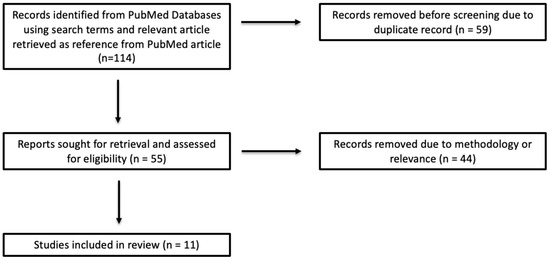
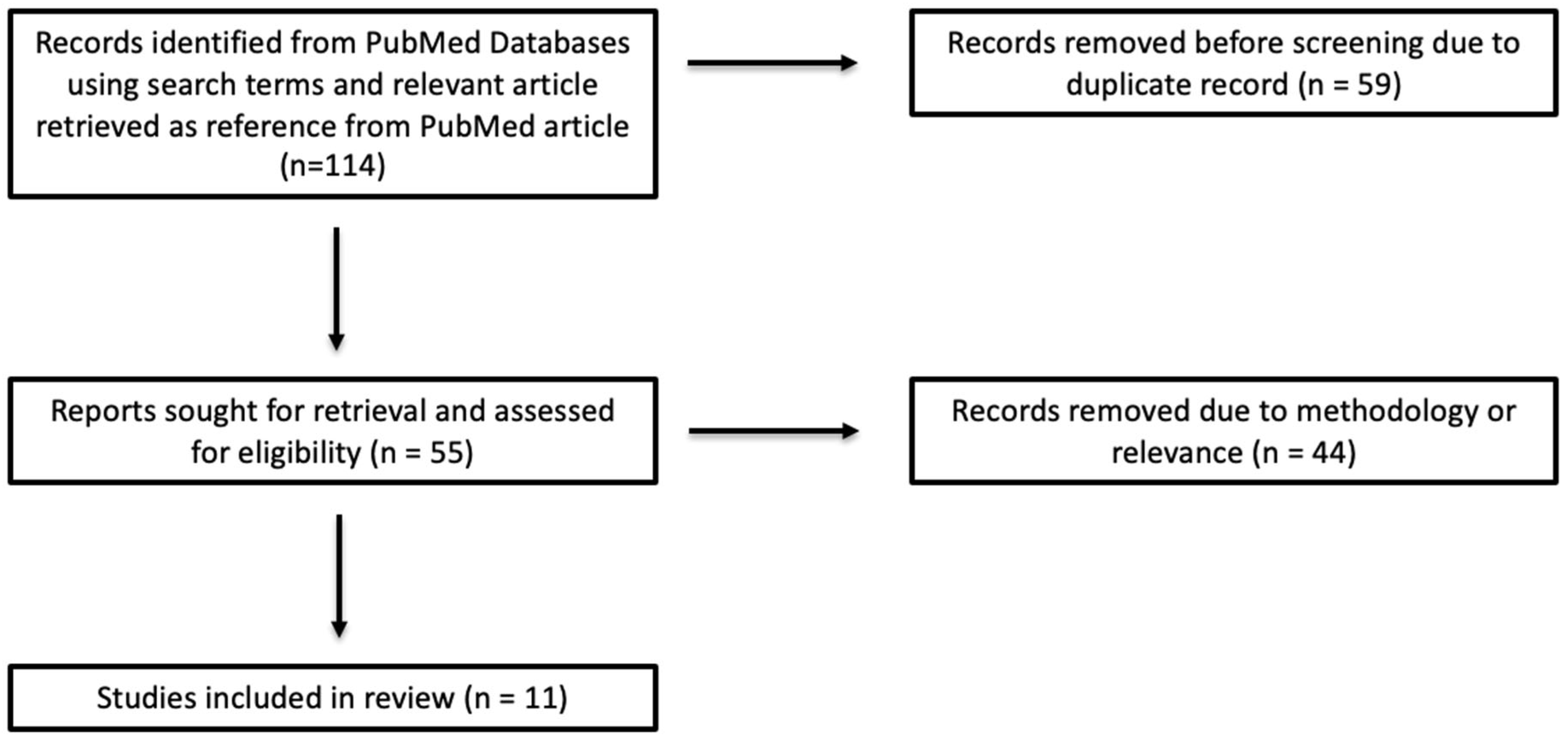
This review did not require institutional review board approval. The literature review was performed on PubMed from 2015 to 2025. Using specified keywords, 113 total articles were identified, 59 of which were duplicates. In addition, one paper, by van der Valen et al. [14], was also selected from the reference section of a 2021 review article by Rella et al. [9] due to its relevance despite not being identified in the initial article search, to make a total of 114 articles, with 53 articles remaining once 59 duplicate articles were removed.
Three independent reviewers screened all identified titles and abstracts for eligibility. Full-text articles were reviewed when eligibility was unclear. Inclusion criteria were studies that evaluated BPE quantitatively in invasive breast cancer patients and reported an outcome measure such as pCR, OS, DFS, or RFS. Exclusion criteria included review articles, studies not measuring BPE quantitatively, studies without the target clinical outcome(s) studied, research published outside the time range, and duplicate studies.
Ten search terms were used to identify studies for this review, five used by one investigator and five used by a different investigator. The first search term was “(“background parenchymal enhancement” OR “BPE”) AND “breast cancer” AND “pathologic complete response”,” which yielded 13 articles, some of which were not included due to systematic review format, non-independent analysis of BPE, qualitative measures of BPE, and utilization of parenchymal kinetics instead of BPE. A similar second search term “(background parenchymal enhancement [title]) AND (breast cancer) AND (pathologic complete response),” yielded 9 articles, all of which were duplicates except one.
The third keyword was “(“background parenchymal enhancement” OR “BPE”) AND “breast cancer” AND “overall survival”,” which yielded 4 papers, 3 of which were excluded due to their qualitative measure of BPE. A similar fourth search term was used, “(background parenchymal enhancement [title]) and (breast cancer) and (overall survival),” which yielded 10 papers, many of which were duplicates or included investigation into qualitative BPE measurements.
The fifth key term, “(“background parenchymal enhancement” OR “BPE”) AND “breast cancer” AND “recurrence free survival”,” yielded 7 articles, which had duplicates and papers to undergo further review. The sixth search term was similarly “(background parenchymal enhancement [title]) and (breast cancer) and (recurrence free survival),” which revealed all repeated papers.
The seventh search term, “(“background parenchymal enhancement” OR “BPE”) AND “breast cancer” AND “disease free survival”,” provided 11 papers, all of which were duplicates or qualitative BPE studies. The eighth search term was “(background parenchymal enhancement [title] AND (breast cancer) AND (disease free survival),” which yielded 7 duplicate articles.
The ninth search term, “(“background parenchymal enhancement” OR “BPE”) AND “breast cancer” AND “prognosis”,” yielded 31 papers, with several duplicates and articles lacking relevance or pertinent methodology. The tenth and final search term, “(background parenchymal enhancement [title]) and (breast cancer) and (prognosis),” resulted in 21 articles, all of which were duplicates or lacked relevance.
Data extraction was performed using a standardized template, which included author, year, study population, MRI acquisition protocol, BPE assessment method (quantitative), prognostic outcomes studied, and key findings. Ten articles were included in the review based on our inclusion and exclusion criteria, which is shown in our article selection flowchart (Figure 1).
Figure 1.
Article selection flowchart.
3. Results/Discussion
Most qualitative BPE studies find that visually graded BI-RADS BPE, whether at baseline or as a change over time, does not independently predict survival (RFS, DFS, or OS) once standard clinical covariates are included [15,16,17]. Overall, when assessed qualitatively, BI-RADS BPE is a plausible indicator of parenchymal activity and treatment-related change but is not a reliable independent prognostic biomarker in unselected populations by itself. The clearest exception is the chemo-treated triple-negative breast cancer (TNBC) subgroup, in which higher contralateral BPE associates with improved survival endpoints [18]. Outside this setting, qualitative BPE remains non-prognostic. These patterns all together, with the variability inherent to qualitative reads, support a shift toward quantitative/semi-automated methods and integration with tumor-centric features to stabilize the signal and improve outcome modeling [15,16,17]. Therefore, we will focus our review on quantitative BPE methods relating to prognosis (Table 1).
Quantitative approaches to BPE consistently show that during NAC, the magnitude/direction of change carries much more information than any single baseline measurement. In particular, larger reductions in quantitative BPE over the course of treatment tend to track with better therapeutic response. This association is noted in some studies in HR-positive groups, where endocrine-responsive background tissue is common [14,19,20]. Other investigators found reduction in BPE associated with pCR in HR-negative patients, particularly early on during NAC [21,22,23]. Combining BPE with other features, such as sphericity, longest diameter, and functional tumor volume (FTV), has the potential to yield an even higher area under the receiver operating characteristic curve (AUC) [24].
Van der Velden et al. evaluated three-phase post-contrast pre-treatment MRI exams of 322 patients with ER-positive/HER2-negative invasive ductal breast cancer and found that high BPE on pre-treatment MRI in the disease-free contralateral breast is an independent biomarker for disease-free invasive cancer survival and overall survival in this cohort [14]. This study used automatic segmentation. Their BPE calculation was unique in that it was derived by calculating enhancement of the late phase (last of three post-contrast phases) at each voxel location as the relative increase in signal intensity between the first post-contrast scan and the third post-contrast scan, sorting lowest to highest of these late enhancement values, and evaluating the top 10% (values exceeding the 90th percentile), then calculating the mean of these top 10%. This highlights the broader relevance of parenchymal enhancement beyond treatment response prediction and illustrates how a standardized, quantitative approach and methodology can improve reproducibility and strengthens the clinical utility of this metric.
However, there is also asymmetry between pre- and post-treatment prognostic values. Pre-treatment quantitative BPE overall is not predictive of outcome in mixed cohorts. This reinforces the limited use of BPE for long-term risk stratification. In contrast, persistently high BPE after therapy has been linked to higher recurrence risk [25]. Moliere et al. evaluated 102 patients with biopsy-proven invasive breast cancer for post NAC response and demonstrated that quantitative BPE post-NAC, but not pre-NAC BPE, significantly predicted recurrence and correlated with DFS, independently of pCR [25]. Pathological complete response did not reach statistical significance, which was felt to be related to the small number of events during the follow-up period. Quantitative post-NAC BPE was significantly lower relative to pre-NAC BPE. Their quantitative method was more sophisticated and refined than the other studies, as they utilized a percent threshold where anything below that threshold was not included as BPE. They calculated VBPE as the total volume of the enhancing voxels over the fibroglandular region that had an enhancement ratio of greater than or equal to 20% (relative difference of 20% or greater). Their findings suggest that parenchyma following NAC carries prognostic information that is most likely not captured by just baseline values.
Timing of BPE measurement relative to therapy can significantly change performance, with several studies finding that early to mid-treatment timepoints often yield the most useful signal for response prediction [21,22,23,24]. Overall, longitudinal BPE can be a treatment response signal and potential prognostic marker post-NAC. Supporting these findings, the study by You et al. showed a continual reduction in BPE regardless of menopausal status, as well as a reduction in tumor size, throughout NAC treatment, with the reduction in BPE after second NAC demonstrating the highest AUC (0.726) for predicting pCR, especially in HR-negative patients [21]. You et al. examined change in BPE from pre-treatment BPE and second, fourth, and sixth NAC timepoints. These investigators utilized an automated three-step segmentation pipeline, demonstrating how standardizing BPE quantification can improve reproducibility and treatment response assessments.
Chen et al. examined change in BPE between baseline and two follow-up MRI exams and its relationship to pCR [22]. They found that pre-treatment BPE was higher in the pCR group, which on sub-group analysis was only seen in patients under age 55. They found that the change in BPE was significantly decreased on first follow-up MRI in the pCR group, which on subgroup analysis was only seen in the patients under age 55 and on receptor type stratification was only seen in the ER-negative cohort. In their study, the pCR rate was higher in ER-negative than ER-positive patients and higher in HER2-postive than HER2-negative patients, as would be expected. They also used a semi-automated segmentation method, further demonstrating how standardization of quantitative methodology can yield meaningful links between early BPE and NAC response.
Segmentation methods also matter. Nguyen et al. observed timepoint-specific differences in predictive performance of BPE with AUC that was optimized with the automated half-stack segmentation protocol versus automated full segmentation and versus automated segmentation of the central five slices [23]. Full-stack segmentation consisted of all axial slices containing FGT voxels, half-stack consisted of the central 50% of the latter, and the center five consisted of the central five slices. While %ΔBPE02 (later interval) showed potential predictive value in HR+/HER2− patients, the strongest associations appeared at earlier timepoints in HR−/HER2+ tumors treated with taxane-based NAC, suggesting that the timing at which BPE change is measured may significantly influence its use as an imaging biomarker. Statistically significant and highest AUC (0.87) for predicting pCR from change in BPE was noted in HR−/HER2+ at an early timepoint (T1) with half-stack segmentation, showing that segmentation methods matter. They suggest that BPE may serve as a good imaging biomarker in this cohort for early detection of pCR during NAC. A major limitation of their results is the small size of their HR−/HER2+ cohort (27 patients), of which 22 attained pCR.
Onishi et al. used a fully automated segmentation method, derived a voxel-by-voxel percent enhancement map, and averaged the percent enhancement of all voxels in the masked volume to determine BPE of the unaffected breast [20]. Pre-treatment (T0), early treatment (T1), inter-regimen (T2), and pre-surgical (T3) timepoints of BPE were evaluated. This study showed that insufficient suppression of background parenchymal enhancement (BPE) is linked to a poorer response to NAC in hormone receptor-positive patients, both after 12 weeks of treatment (inter-regimen point, T2) and at the pre-surgery timepoint (T3). Notably, the association observed at T2 suggests that early identification of patients with persistent BPE could help predict suboptimal response, enabling timely, personalized adjustments to their treatment plan.
Arasu et al. utilized manual whole breast segmentation, followed by deriving a mask classifying FGT and using fuzzy c-means clustering to remove non-breast elements [19]. Per voxel basis calculation of BPE was then performed with an average value of all voxels used to determine a final BPE estimate. They prospectively studied BPE at pre-treatment (T0), early treatment (T1), inter-regimen (T2), and pre-surgery (T3) in HR+/HER2− and HR−/HER2− patients. They studied 45 HR−/HER2− patients and 43 HR+/HER2− patients. They found that the change in BPE from baseline to pre-surgery was statistically significant in their cohort of 43 women with HR+/HER2− breast cancer undergoing taxane- and anthracycline-based regimens, with the highest AUC of 0.77 for predicting pCR for change in BPE between pre-surgery and pre-treatment/baseline timepoints. BPE of the contralateral unaffected breast demonstrated similar diagnostic accuracy compared to FTV for HR+/HER2− patients under univariate analysis. While BPE demonstrated potential to be an independent marker of response, their study found limited additive effect of BPE to FTV in predicting pCR, possibly related to the small sample size. Their findings demonstrate that the reaction of normal FGT to neoadjuvant therapy as reflected by BPE may serve as a biomarker of treatment response. The heightened sensitivity of changes in BPE for predicting pCR in HR+ tumors aligns with the known influence of estrogen on BPE. The progressive increase in both the magnitude and predictive strength of BPE at later timepoints in HR+ cancers suggests a consistent trend, making it less likely that these findings are due to random variation.
Li et al. utilized a fully automated segmentation method and demonstrated the following AUC for BPE in predicting pCR for various receptor types: 0.69 combined group of all receptor types, 0.66 for HR+/HER2−, 0.76 for HR+/HER2+, 0.75 for HR−/HER2+, and 0.62 for HR−/HER2− [24]. When combining all four features they examined (functional tumor volume, sphericity, longest diameter, and BPE), the AUC for the same receptor groups, respectively, were 0.81, 0.83, 0.88, 0.83, and 0.82. One hundred repeated five-fold cross validation was applied to ensure classification accuracy. Their study demonstrated that multifeature analysis was superior to any single feature in predicting pCR, which illustrates that the addition of BPE to prediction models enhances accuracy. Their study included 384 patients (162 HR+/HER2−, 60 HR+/HER2+, 30 HR−/HER2+, and 132 HR−/HER2−). The larger population size of their study compared to the study by Arasu et al. may explain why they were able to show that multifeature analysis that includes BPE is superior to univariable analysis. Across multiple analyses, quantitative BPE alone adds little value beyond tumor morphology, enhancement kinetics, and volumetric response measures. But when BPE is combined with FTV and biologic subtype, discrimination for pCR improves significantly. This indicates that background and tumor signals are complementary [19,24]. Thus, this pattern adds support for a combined modeling that uses background tissue change with other features, like volume and subtype, in NAC.
The only studies that failed to demonstrate correlation of change in BPE with pCR or RFS/DFS was conducted by Rella et al. and Shin et al., respectively [26,27]. The study by Rella et al. relied on initial manual segmentation of the fibroglandular tissue. Rella et al. also evaluated OS and DFS in a later study (2022) where they reported no significant associations between baseline BPE, final BPE, and change in BPE to outcomes [28]. Shin et al. performed quantitative BPE using three manual regions of interest (ROIs) placed for each study with average values for BPE calculated, in addition to a quantitative BPE assessment using fully automated segmentation of the FGT (to eliminate subjectivity) with later enhancement calculated for each voxel of FGT in the contralateral breast, taking the mean of the top 10 percent for analysis in ER+/HER2− node negative breast cancer patients, which showed no association with RFS or DFS [27]. They also used qualitative BPE assessment from two radiologists independently, also showing no association with RFS or DFS [27].
Overall, these findings highlight that quantitative BPE can behave differently across subtypes and treatment contexts, while also underscoring the sensitivity of BPE metrics to methodological factors such as study design and cohort characteristics. While full-breast, multi-timepoint analyses are ideal in theory, protocols that prioritize a standardized, reproducible automated segmentation methodology and a prespecified early or mid-NAC are most likely the best balance between feasibility and accuracy. Accordingly, contralateral quantitative BPE may be better regarded as a contextual imaging feature, with associations to prognostic variables that are sensitive to methodological factors, and that may enhance risk stratification within select subgroups rather than serve as a universal prognostic biomarker.
The literature reflects a clear shift in BPE quantification from single timepoint assessments to longitudinal approaches during NAC, giving importance to changes in BPE over time. Although pre- and post-treatment BPE comparisons have demonstrated utility as functional biomarkers of tumor response, they are limited in scope and fail to relay the tumor and microenvironmental changes that occur throughout NAC. Furthermore, analysis based on pre-treatment and post-treatment BPE values alone precludes the ability to assess early response to NAC, limiting opportunities for timely treatment adaptation. To address these limitations, contemporary studies increasingly employ longitudinal BPE analysis across multiple NAC timepoints, with promising results. This approach allows monitoring of temporal BPE dynamics, enabling earlier and more accurate prediction of treatment response and supporting more personalized treatment strategies.
The literature increasingly supports the adoption of fully automated approaches for quantifying BPE across multiple timepoints, as these methods demonstrate greater reliability than qualitative assessments. The results from the study by Rella et al. [26] illustrate how the limitations in accuracy and reproducibility associated with manual or semi-automated preliminary segmentation of the ROI can lead to results that diverge from the multitude of studies that employ automated methods. Accordingly, fully automated segmentation strategies are recommended to improve measurement robustness and consistency. A limitation to this strategy of purely automated segmentation of FGT lies in the lack of a standardized segmentation framework, which continues to introduce and propagate variability and potential error. These challenges underscore the need to transition toward deep learning-based methodologies. Such approaches offer the potential for fully automated, reproducible, and more precise segmentation, thereby improving BPE quantification and mitigating limitations inherent to existing methods. Consistent with this, comparative studies of manual and algorithmic whole-breast and FGT segmentation have demonstrated superior performance of deep learning architectures, including U-Net-based models, over traditional techniques [29]. Beyond improved reproducibility, these methods enable more accurate BPE assessment and support the development of more robust predictive models.
Table 1.
Quantitative BPE papers.
4. Conclusions
The quantitative assessment of BPE is a more objective and reproducible approach than the qualitative methods, which often are subjective and are prone to inter-reader variability. Quantitative BPE eliminates subjectivity and reduces observer bias and variability between studies by providing a numerical measurement approach. However, the reliability of quantitative measurements is dependent on strict methodology. This includes standardization of segmentation techniques, correct timing of imaging, and overall consistent acquisition parameters for data. Without these strict criteria for methodology, the results of quantitative measurements may vary widely, which limits generalizability and interpretability. Virtually all of the quantitative studies that utilized automated segmentation found some association between BPE and prognosis/pCR. Further refinements, such as utilization of a percent threshold for inclusion in BPE calculation as employed by Moliere et al., may strengthen these analyses.
The literature increasingly supports fully automated approaches for quantifying BPE across multiple timepoints, as these methods are more reliable and reproducible than qualitative or semi-automated assessments. Manual intervention in ROI segmentation can introduce variability and reduce measurement accuracy, underscoring the need for automated solutions.
Given the critical role of accurate FGT segmentation and the absence of a standardized methodology, deep learning-based approaches offer a promising alternative. This also provides an opportunity to integrate BPE data with broader clinical and imaging data. Such a combination allows for even greater analysis than with BPE data alone. In a clinical setting, these advancements could improve prediction and treatment monitoring in oncology. Integration of this imaging biomarker with molecular or histologic tumor features, patient risk factors, genetics (e.g., BRCA status), hormone receptor status, and other relevant variables may prove to be very informative. Inclusion of quantitative BPE in machine learning prediction models may allow more accurate prediction of outcomes and may help in guiding treatment. A current limitation is that many of the quantitative BPE studies are retrospective and based on modest sample sizes, which limits generalizability. There is a clear need for larger, prospective, multi-institutional cohorts employing harmonized imaging acquisition parameters, standardized definitions of quantitative BPE metrics, and consistent post-processing methods. Large-scale validation using standardized protocols will be essential before quantitative BPE can be reliably integrated into routine clinical practice.
Author Contributions
Conceptualization, T.M. and T.Q.D.; methodology, T.M., J.W., E.H., Y.Z. and T.Q.D.; software, Y.Z.; validation, J.W., E.H. and T.M.; formal analysis, J.W.; investigation, J.W., E.H. and T.M.; resources, T.M., Y.Z. and T.Q.D.; writing—original draft preparation, J.W., E.H., Y.Z., T.Q.D. and T.M.; writing—review and editing, J.W., E.H., Y.Z., T.Q.D. and T.M.; visualization, J.W., T.M. and E.H.; supervision, T.M. and T.Q.D.; project administration, T.M. and T.Q.D. All authors have read and agreed to the published version of the manuscript.
Funding
This research received no external funding.
Institutional Review Board Statement
Not applicable.
Informed Consent Statement
Not applicable.
Data Availability Statement
Data is available on PubMed. See “Section 2” for our process.
Conflicts of Interest
The authors declare no conflicts of interest.
Abbreviations
The following abbreviations are used in this manuscript:
| BPE | Background parenchymal enhancement |
| MRI | Magnetic resonance imaging |
| pCR | Pathologic complete response |
| RFS | Recurrence-free survival |
| DFS | Disease-free survival |
| OS | Overall survival |
| NAC | Neoadjuvant chemotherapy |
| BI-RADS | Breast imaging-reporting assessment and data system |
| HR | Hormone receptor |
| HER2 | Human epidermal growth factor receptor |
| FTV | Functional tumor volume |
| FGT | Fibroglandular tissue |
| AUC | Area under the receiver operating characteristic curve |
| ROI | Region of interest |
| BRCA | Breast cancer gene |
| TNBC | Triple-negative breast cancer |
| DCE-MRI | Dynamic contrast-enhanced MRI |
| IDFS | Invasive DFS |
References
- Kim, N.; Adam, R.; Maldjian, T.; Duong, T.Q. Radiomics Analysis of Breast MRI to Predict Oncotype Dx Recurrence Score: Systematic Review. Diagnostics 2025, 15, 1054. [Google Scholar] [CrossRef] [Scilit]
- Khan, N.; Adam, R.; Huang, P.; Maldjian, T.; Duong, T.Q. Deep Learning Prediction of Pathologic Complete Response in Breast Cancer Using MRI and Other Clinical Data: A Systematic Review. Tomography 2022, 8, 2784–2795. [Google Scholar] [CrossRef] [Scilit]
- Hadidchi, R.; Agrawal, A.; Liu, M.Z.; Maldijan, T.; Zhu, Y.; Nguyen, H.Q.; Lu, J.; Makower, D.; Fineberg, S.; Duong, T.Q. A deep learning framework to stratify Nottingham histologic grade 2 breast tumors based on dynamic contrast-enhanced MRI. Eur. Radiol. 2025. Epub ahead of printing. [Google Scholar] [CrossRef] [Scilit]
- Hussain, L.; Huang, P.; Nguyen, T.; Lone, K.J.; Ali, A.; Khan, M.S.; Li, H.; Suh, D.Y.; Duong, T.Q. Machine learning classification of texture features of MRI breast tumor and peri-tumor of combined pre- and early treatment predicts pathologic complete response. Biomed. Eng. Online 2021, 20, 63. [Google Scholar] [CrossRef] [Scilit]
- Nishizawa, T.; Maldjian, T.; Jiao, Z.; Duong, T.Q. Attention-based multimodal deep learning for interpretable and generalizable prediction of pathological complete response in breast cancer. J. Transl. Med. 2025, 23, 774. [Google Scholar] [CrossRef] [Scilit] [PubMed]
- Dell’Aquila, K.; Vadlamani, A.; Maldjian, T.; Fineberg, S.; Eligulashvili, A.; Chung, J.; Adam, R.; Hodges, L.; Hou, W.; Makower, D.; et al. Machine learning prediction of pathological complete response and overall survival of breast cancer patients in an underserved inner-city population. Breast Cancer Res. 2024, 26, 7. [Google Scholar] [CrossRef] [Scilit]
- Dammu, H.; Ren, T.; Duong, T.Q. Deep learning prediction of pathological complete response, residual cancer burden, and progression-free survival in breast cancer patients. PLoS ONE 2023, 18, e0280148. [Google Scholar] [CrossRef] [Scilit] [PubMed]
- Karagiannis, G.S.; Bianchi, A.; Sanchez, L.R.; Ambadipudi, K.; Cui, M.-H.; Anampa, J.M.; Asiry, S.; Wang, Y.; Harney, A.S.; Pastoriza, J.M.; et al. Assessment of MRI to estimate metastatic dissemination risk and prometastatic effects of chemotherapy. npj Breast Cancer 2022, 8, 101. [Google Scholar] [CrossRef] [Scilit]
- Rella, R.; Contegiacomo, A.; Bufi, E.; Mercogliano, S.; Belli, P.; Manfredi, R. Background parenchymal enhancement and breast cancer: A review of the emerging evidences about its potential use as imaging biomarker. Br. J. Radiol. 2021, 94, 20200630. [Google Scholar] [CrossRef] [Scilit]
- Liao, G.J.; Bancroft, L.C.H.; Strigel, R.M.; Chitalia, R.D.; Kontos, D.; Moy, L.; Partridge, S.C.; Rahbar, H. Background parenchymal enhancement on breast MRI: A comprehensive review. J. Magn. Reson. Imaging 2020, 51, 43–61. [Google Scholar] [CrossRef] [Scilit] [PubMed]
- Duong, K.S.; Rubner, R.; Siegel, A.; Adam, R.; Ha, R.; Maldjian, T. Machine Learning Assessment of Background Parenchymal Enhancement in Breast Cancer and Clinical Applications: A Literature Review. Cancers 2024, 16, 3681. [Google Scholar] [CrossRef] [Scilit]
- Niell, B.L.; Abdalah, M.; Stringfield, O.; Raghunand, N.; Ataya, D.; Gillies, R.; Balagurunathan, Y. Quantitative Measures of Background Parenchymal Enhancement Predict Breast Cancer Risk. AJR Am. J. Roentgenol. 2021, 217, 64–75. [Google Scholar] [CrossRef] [Scilit]
- Li, X.; Yan, F. Predictive value of background parenchymal enhancement on breast magnetic resonance imaging for pathological tumor response to neoadjuvant chemotherapy in breast cancers: A systematic review. Cancer Imaging 2024, 24, 35. [Google Scholar] [CrossRef] [Scilit]
- van der Velden, B.H.M.; Sutton, E.J.; Carbonaro, L.A.; Pijnappel, R.M.; Morris, E.A.; Gilhuijs, K.G. Contralateral parenchymal enhancement on dynamic contrast-enhanced MRI reproduces as a biomarker of survival in ER-positive/HER2-negative breast cancer patients. Eur. Radiol. 2018, 28, 4705–4716. [Google Scholar] [CrossRef] [Scilit]
- Gullo, R.L.; Daimiel, I.; Saccarelli, C.R.; Bitencourt, A.; Sevilimedu, V.; Martinez, D.F.; Jochelson, M.S.; Morris, E.A.; Reiner, J.S.; Pinker, K. MRI background parenchymal enhancement, fibroglandular tissue, and mammographic breast density in patients with invasive lobular breast cancer on adjuvant endocrine hormonal treatment: Associations with survival. Breast Cancer Res. 2020, 22, 93. [Google Scholar] [CrossRef] [Scilit]
- Kim, J.Y.; Lee, J.W.; Lee, N.K.; Kim, S.; Nam, K.J.; Lee, K.; Choo, K.S. Are background breast parenchymal features on preoperative breast MRI associated with disease-free survival in patients with invasive breast cancer? Radiol. Med. 2024, 129, 1790–1801. [Google Scholar] [CrossRef] [Scilit]
- Lee, Y.J.; Youn, I.K.; Kim, S.H.; Kang, B.J.; Park, W.-C.; Lee, A. Triple-negative breast cancer: Pretreatment magnetic resonance imaging features and clinicopathological factors associated with recurrence. Magn. Reson. Imaging 2020, 66, 36–41. [Google Scholar] [CrossRef] [Scilit]
- Xu, C.; Yu, J.; Wu, F.; Li, X.; Hu, D.; Chen, G.; Wu, G. High-background parenchymal enhancement in the contralateral breast is an imaging biomarker for favorable prognosis in patients with triple-negative breast cancer treated with chemotherapy. Am. J. Transl. Res. 2021, 13, 4422–4436. [Google Scholar] [PubMed]
- Arasu, V.A.; Kim, P.; Li, W.; Strand, F.; McHargue, C.; Harnish, R.; Newitt, D.C.; Jones, E.F.; Glymour, M.M.; Kornak, J.; et al. Predictive Value of Breast MRI Background Parenchymal Enhancement for Neoadjuvant Treatment Response among HER2- Patients. J. Breast Imaging 2020, 2, 352–360. [Google Scholar] [CrossRef] [Scilit] [PubMed]
- Onishi, N.; Li, W.; Newitt, D.C.; Harnish, R.J.; Strand, F.; Nguyen, A.A.-T.; Arasu, V.A.; Gibbs, J.; Jones, E.F.; Wilmes, L.J.; et al. Breast MRI during Neoadjuvant Chemotherapy: Lack of Background Parenchymal Enhancement Suppression and Inferior Treatment Response. Radiology 2021, 301, 295–308. [Google Scholar] [CrossRef] [Scilit] [PubMed]
- You, C.; Peng, W.; Zhi, W.; He, M.; Liu, G.; Xie, L.; Jiang, L.; Hu, X.; Shen, X.; Gu, Y. Association Between Background Parenchymal Enhancement and Pathologic Complete Remission Throughout the Neoadjuvant Chemotherapy in Breast Cancer Patients. Transl. Oncol. 2017, 10, 786–792. [Google Scholar] [CrossRef] [Scilit]
- Chen, J.H.; Yu, H.J.; Hsu, C.; Mehta, R.S.; Carpenter, P.M.; Su, M.Y. Background Parenchymal Enhancement of the Contralateral Normal Breast: Association with Tumor Response in Breast Cancer Patients Receiving Neoadjuvant Chemotherapy. Transl. Oncol. 2015, 8, 204–209. [Google Scholar] [CrossRef] [Scilit]
- Nguyen, A.A.; Arasu, V.A.; Strand, F.; Li, W.; Onishi, N.; Gibbs, J.; Jones, E.F.; Joe, B.N.; Esserman, L.J.; Newitt, D.C.; et al. Comparison of Segmentation Methods in Assessing Background Parenchymal Enhancement as a Biomarker for Response to Neoadjuvant Therapy. Tomography 2020, 6, 101–110. [Google Scholar] [CrossRef] [Scilit] [PubMed]
- Li, W.; Newitt, D.C.; Gibbs, J.; Wilmes, L.J.; Jones, E.F.; Arasu, V.A.; Strand, F.; Onishi, N.; Nguyen, A.A.-T.; Kornak, J.; et al. Predicting breast cancer response to neoadjuvant treatment using multi-feature MRI: Results from the I-SPY 2 TRIAL. npj Breast Cancer 2020, 6, 63. [Google Scholar] [CrossRef] [Scilit]
- Moliere, S.; Oddou, I.; Noblet, V.; Veillon, F.; Mathelin, C. Quantitative background parenchymal enhancement to predict recurrence after neoadjuvant chemotherapy for breast cancer. Sci. Rep. 2019, 9, 19185. [Google Scholar] [CrossRef] [Scilit] [PubMed]
- Rella, R.; Bufi, E.; Belli, P.; Petta, F.; Serra, T.; Masiello, V.; Scrofani, A.; Barone, R.; Orlandi, A.; Valentini, V.; et al. Association between background parenchymal enhancement and tumor response in patients with breast cancer receiving neoadjuvant chemotherapy. Diagn. Interv. Imaging 2020, 101, 649–655. [Google Scholar] [CrossRef] [Scilit] [PubMed]
- Shin, G.W.; Zhang, Y.; Kim, M.J.; Su, M.; Kim, E.; Moon, H.J.; Yoon, J.H.; Park, V.Y. Role of dynamic contrast-enhanced MRI in evaluating the association between contralateral parenchymal enhancement and survival outcome in ER-positive, HER2-negative, node-negative invasive breast cancer. J. Magn. Reson. Imaging 2018, 48, 1678–1689. [Google Scholar] [CrossRef] [Scilit]
- Rella, R.; Bufi, E.; Belli, P.; Scrofani, A.R.; Petta, F.; Borghetti, A.; Marazzi, F.; Valentini, V.; Manfredi, R. Association between contralateral background parenchymal enhancement on MRI and outcome in patients with unilateral invasive breast cancer receiving neoadjuvant chemotherapy. Diagn. Interv. Imaging 2022, 103, 486–494. [Google Scholar] [CrossRef] [Scilit]
- Dalmış, M.U.; Litjens, G.; Holland, K.; Setio, A.; Mann, R.; Karssemeijer, N.; Gubern-Mérida, A. Using deep learning to segment breast and fibroglandular tissue in MRI volumes. Med. Phys. 2017, 44, 533–546. [Google Scholar] [CrossRef] [Scilit]
Disclaimer/Publisher’s Note: The statements, opinions and data contained in all publications are solely those of the individual author(s) and contributor(s) and not of MDPI and/or the editor(s). MDPI and/or the editor(s) disclaim responsibility for any injury to people or property resulting from any ideas, methods, instructions or products referred to in the content. |
© 2026 by the authors. Licensee MDPI, Basel, Switzerland. This article is an open access article distributed under the terms and conditions of the Creative Commons Attribution (CC BY) license.