


Abstract
Background/Objectives: Maternal sepsis contributes substantially to maternal mortality, with recurrent delays in recognition and considerable opportunities for prevention. More than twenty screening tools have been evaluated in obstetric care; their reported performance varies widely, a variation often read as evidence of superiority. This critical narrative review examines how thresholds, pregnancy physiology, clinical periods, target and reference conditions, and intended clinical tasks influence the interpretation of published performance estimates, rather than seeking a universally superior instrument. Methods: Primary studies, guidance, and methodological literature identified by a structured non-systematic search were synthesised narratively. Results: In a period-stratified analysis identified in this review, one pregnancy-adjusted rule detected 57.8% of administratively coded sepsis cases before 20 weeks and 82.8% at 28–36 weeks, at false-positive rates of 6.3% and 6.7% in matched comparison series; physiology may explain this difference, but case mix was not accounted for. Among women with chorioamnionitis or endometritis, five rules (systemic inflammatory response syndrome criteria, two pregnancy-adjusted screens, and two maternal early warning tools) discriminated poorly. Conclusions: The evidence does not establish a universally superior obstetric sepsis screening instrument, nor equivalence between instruments: each estimate describes an instrument under specified conditions. Published estimates should be interpreted for the intended task (screening, diagnostic confirmation or prognostication), and validation studies should report the target condition, reference condition, and clinical period with every estimate.
1. Introduction
Infection remains among the leading causes of maternal death. World Health Organization (WHO) systematic analyses attributed 10.7% of maternal deaths in 2003–2009 to sepsis (uncertainty interval 5.9–18.6%) [1] and 7% (80% uncertainty interval 5–9) in 2009–2020 to pregnancy-related infection, with most such deaths occurring after delivery [2]. In the WHO Global Maternal Sepsis Study (GLOSS), 10.9 women per 1000 live births (95% confidence interval [CI]: 9.8–12.0) presented with an infection-related severe maternal outcome [3]. Delay is common: a statewide case series identified delay in initial appropriate antibiotic administration in 11 of 15 maternal sepsis deaths with retrievable records [4], and the United Kingdom confidential enquiry for 2021–23 recorded 16 direct maternal deaths from pregnancy-related sepsis, 0.80 per 100,000 maternities [5].
Recognition is difficult because the physiological changes of pregnancy, labour, and the early puerperium overlap with the abnormalities used to define a systemic response to infection: healthy women in later pregnancy and in labour commonly meet systemic inflammatory response syndrome (SIRS) criteria for every parameter except temperature (87 studies, 8834 women) [6]. This overlap has driven a proliferation of instruments, 28 of which a secondary analysis of GLOSS identified in obstetric use or under investigation [7], but not a consensus: the Society for Maternal-Fetal Medicine recommends local detection protocols while “avoiding the use of a single screening tool alone” (Grading of Recommendations Assessment, Development and Evaluation [GRADE] 1B) [8], the Alliance for Innovation on Maternal Health consensus bundle states that the optimal tools are not known [9], and the 2025 National Institute for Health and Care Excellence (NICE) guideline recommends considering an early warning score without naming one [10].
Existing syntheses (a scoping review [11], a 2025 narrative review [12], an integrative review [13], and a methodological review that found 19 of 20 maternal early-warning-score studies at high risk of bias [14]) have mapped this literature without resolving it, and a 2026 meta-analysis of sepsis screening tools in low- and middle-income countries included no obstetric population [15]. Comparative accuracy estimates are difficult to interpret because the studies that produced them applied instruments at different thresholds, in different clinical periods, against different reference conditions, and for different clinical tasks, so that apparently conflicting results may answer different questions. We identified no synthesis that treats the interaction of these factors as the analytic object.
This critical narrative review addresses that gap. Its subject is the methodological interpretation of published screening performance. Our objective is to examine how the reported diagnostic performance of obstetric sepsis screening instruments should be interpreted when physiological thresholds vary across pregnancy, labour, and the postpartum period (referred to here as clinical periods), when studies validate these instruments against imperfect and heterogeneous reference conditions, and when target conditions and intended clinical tasks differ between studies. We do not attempt to identify a universally superior instrument, but argue instead that the question of which tool performs best is insufficiently specified until the threshold, the clinical period, the intended task, and the comparator are stated.
The review is written for clinicians and guideline developers who must judge whether a published estimate applies to their own service, and for investigators who design or appraise validation studies; it is not intended as a general clinical overview of maternal sepsis. After the approach (Section 2), Section 3 and Section 4 set out the target condition, the instruments, and the threshold problem; Section 5 the comparative evidence; Section 6 its interpretation by reference condition and clinical task; and Section 7, Section 8 and Section 9 present the implications, limitations, and conclusions.
2. Scope, Literature Identification, and Interpretative Approach
2.1. Design and Scope
This is a critical narrative review. It does not follow a systematic review methodology, does not claim exhaustive identification of eligible studies, and does not pool quantitative estimates. Its approach is interpretative: instead of ranking instruments, it reads reported estimates in relation to the attributes of the studies that produced them, namely the threshold applied; the clinical period and care setting; the target condition and the reference condition against which cases were classified; the prevalence in the population studied; whether the threshold was selected in the same dataset; the number of outcome events; and the clinical task for which the instrument was intended. These attributes structure the sections that follow and are revisited as practice points in Section 7.4.
The scope is confined to bedside screening instruments and organ-dysfunction frameworks; microbiological aetiology, antimicrobial treatment, foetal surveillance, and biomarker-based diagnosis appear only where they enter a published screening rule or reference-condition definition.
2.2. Literature Identification and Selection
Literature was identified through a structured but non-systematic search of PubMed/MEDLINE, supplemented by Europe PMC, the websites of WHO and of obstetric and guideline organisations, and backward and forward citation tracing, with Google Scholar used only for supplementary discovery and citation tracing; a targeted update of the recent primary literature and current guidance followed. Search terms combined maternal, obstetric, or puerperal sepsis with the screening, early warning, and organ-dysfunction instruments discussed and with terms for diagnostic or prognostic accuracy, thresholds, and reference standards. Literature published after 2000 was prioritised, with earlier work retained where it remains the primary source for a definition, derivation, or methodological principle. Estimates were preferentially obtained from primary publications; values reproduced from secondary sources are explicitly identified in the Supplementary Materials. Guidance was checked for currency, and the Scale for the Assessment of Narrative Review Articles (SANRA) [16] was used as an unscored quality checklist.
Selection of studies for detailed discussion was a matter of judgement, giving greater weight to studies that compared several instruments in the same women, stated the threshold and the reference condition, reported sensitivity with specificity or a false-positive rate, preferably with confidence intervals, and rested on larger numbers of outcome events; other studies were retained where they covered an instrument, clinical period, population, or reference condition not otherwise represented (Table S1). The two companion analyses from a United States hospital consortium receive the most attention because they are the only studies identified in this review that compared several screening rules in the same patients separately for delivery, antepartum, and postpartum admissions, with interval estimates and several hundred cases per analysis. They were conducted by the same investigators, with shared data-extraction methods, case definitions, and operational rules, and therefore do not constitute independent replications. Conflicting evidence, including the multi-country GLOSS analysis [7], the Sepsis in Obstetrics Score validation series, infection-enriched cohorts, and studies of generic deterioration systems, was reported rather than reconciled.
3. Target Conditions and Screening Instruments
3.1. Target and Reference Conditions
Any statement about diagnostic performance presupposes a target condition. Three terms are used distinctly throughout this review. The target condition is the clinical state an instrument is intended to detect. The reference condition is the operational rule by which a study decided who had that state, whether a coded diagnosis, chart adjudication, explicit organ-dysfunction criteria, or a future event. A reference standard is a reference condition assumed to classify without error, which, as Section 6 sets out, does not exist for sepsis. In obstetric sepsis the target condition is defined differently by the frameworks in routine use, and the differences are not merely semantic.
3.2. Definitions of Maternal Sepsis
The Third International Consensus Definitions (Sepsis-3) define sepsis as “life-threatening organ dysfunction caused by a dysregulated host response to infection”, operationalised as an acute increase of at least 2 points in the Sequential Organ Failure Assessment (SOFA) score in the setting of suspected infection [17]; the accompanying quick SOFA (qSOFA) was derived in general adult patients against in-hospital mortality [18], an outcome that is rare in obstetric populations (Section 1). The obstetrically modified SOFA (omSOFA) and qSOFA (omqSOFA) of the Society of Obstetric Medicine of Australia and New Zealand (SOMANZ) adjust thresholds for pregnancy by consensus [19], and the 2023 SOMANZ position statement states that it “has abandoned the use of the [qSOFA] score to diagnose sepsis due to its poor performance in clinical practice”, retaining an omSOFA increase of 2 points or more as the organ-dysfunction criterion [19,20]. The WHO definition leaves organ dysfunction to be operationalised [21]; GLOSS used the SOFA-based WHO maternal near-miss criteria, so its target condition, an infection-related severe maternal outcome of near-miss or death, was considerably more severe than sepsis as applied at the bedside [3,7].
Programmatic frameworks differ again. In the two-step process of the California Maternal Quality Care Collaborative (CMQCC), a first-step screen is positive when at least two of four criteria (temperature, heart rate above 110 beats per minute, respiratory rate above 24 breaths per minute, and leucocyte count; Table S2) are met in suspected infection, and sepsis is confirmed only by an end-organ injury criterion [22,23]. The consortium analyses evaluated the first-step criteria of the 2020 toolkit; the second edition, published in September 2025, restricts the pregnancy-adjusted criteria to the interval from 20 weeks of gestation to 72 h after delivery and does not recommend lactate in the second stage of labour or the first hour after delivery [24]. The United Kingdom Obstetric Surveillance System (UKOSS) case definition required each criterion on two occasions, at least four hours apart for the vital signs [25]. In the United States, hospitals report adherence to the Severe Sepsis and Septic Shock Management Bundle (SEP-1) to the Centers for Medicare & Medicaid Services, the federal agency that administers the public health-insurance programmes; this quality-reporting measure retains the earlier severe-sepsis construct, so a patient may meet its definition without meeting Sepsis-3 [8,12].
Microbiological confirmation is neither necessary nor sufficient: bacteraemia may occur without organ dysfunction, organ dysfunction attributable to infection often occurs with negative cultures, and a positive culture may reflect contamination [26]. The Society for Maternal-Fetal Medicine advises considering sepsis in pregnant or postpartum patients with otherwise unexplained end-organ damage and suspected or confirmed infection, regardless of fever (GRADE 1C) [8]. Every estimate of sensitivity or specificity is therefore an estimate against a chosen operational construct (Section 6).
3.3. Classes of Screening Instruments
Table S2 summarises the intended purpose, derivation, and principal limitations of each instrument and gives its published criteria. SIRS criteria, qSOFA, and SOFA were derived in general adult populations, none in an obstetric cohort. Pregnancy-adjusted infection screens (the CMQCC first step, the UKOSS criteria reused as a single-measurement, any-two rule [25,27], and obstetrically modified SIRS variants [28]) retain the structure of SIRS with higher thresholds and are not intended to establish a diagnosis. Organ-dysfunction scores (omSOFA, omqSOFA [19], and a pregnancy-adapted qSOFA, qSOFA-P, whose cut-points were selected and evaluated in the same 104 pregnant patients [29]) sit closer to diagnostic confirmation.
The Sepsis in Obstetrics Score (SOS), a weighted composite scored from 0 to 28 with a threshold of ≥6, was derived against intensive care admission within 48 h and is a risk-stratification instrument [30]. Generic deterioration systems, namely the Modified Early Obstetric Warning System (MEOWS) [31], the Maternal Early Warning Criteria (MEWC) [32,33], the Maternal Early Warning Trigger (MEWT) [34], the shock index [35], and general adult scores [36], are indifferent to aetiology and detect mainly haemorrhage and hypertensive disease [31]. Instruments in different groups therefore do not compete for the same task, and most were consensus-derived: 6 of 10 development studies of modified obstetric early warning scores used consensus alone, and no data-driven model had been independently validated [14].
4. Pregnancy Physiology and Competing Thresholds
Screening criteria apply fixed thresholds to continuous variables whose distribution pregnancy shifts, differently for different variables and at different times.
4.1. Vital Signs
The vital signs move unequally. In a prospective cohort of 1041 low-risk women, median heart rate rose from 82 beats/min at 12 weeks to 91 (3rd–97th centile 68–115) at 34.1 weeks, so a SIRS criterion of >90 beats/min sits close to the healthy third-trimester median and one of >110 beats/min close to the upper centile [37]. Median respiratory rate (15 breaths/min) did not change materially with gestation [37]; frequent breaching of the SIRS threshold in meta-analytic data [6] may partly reflect inconsistent recording [38]. Median systolic pressure rose only from 114 to 121 mm Hg between 12 and 40 weeks [37], so the qSOFA threshold of ≤100 mm Hg is met by a minority of healthy women, which is one reason qSOFA behaves as a specific rather than a sensitive instrument. After delivery, heart rate peaks on the day of birth and returns towards non-pregnant values over two weeks [39].
4.2. Laboratory Variables, Temperature, and Lactate
Laboratory variables shift in the direction that makes infection-oriented criteria less discriminating. The pregnancy reference interval for leucocyte count is about one third above the non-pregnant range (5.7–15.0 × 10⁹/L) and widens to 8.4–23.2 × 10⁹/L on the first postpartum day before returning to the antenatal average by about day 7 [40], and antenatal corticosteroids raise the count further [22]. Platelet count falls to a mean of 217 × 109/L at delivery, when 9.9% of uncomplicated pregnancies are below 150 × 109/L [41] and so score one SOFA coagulation point before any infection, whereas creatinine falls (trimester-specific 2.5th–97.5th centiles: 34–70 µmol/L) [42], so that a value within the non-pregnant range may represent a substantial acute rise while scoring zero renally. Neither implies pathological SOFA scores in healthy pregnancy, but both support pregnancy-aware interpretation of organ-dysfunction criteria.
Temperature overlaps least with normal pregnancy physiology, although the mean plus two standard deviations reaches 38.1 °C, marginally above the SIRS threshold of >38 °C [6]. It is nonetheless confounded in labour: in a secondary analysis of a randomised trial of 715 women at term, epidural analgesia was independently associated with maternal fever ≥38 °C (odds ratio: 4.0, 95% CI: 2.0–7.7) [43]. Heart rate, one of the variables most displaced by pregnancy, still discriminated infection numerically better than the composite scores tested in one ward cohort, with an interval overlapping that of an electronic risk score [36]. A rule requiring any two abnormal components therefore behaves differently according to which two are recorded, and retrospective studies could not capture the subjective components of MEOWS, MEWC, and MEWT [27,36].
Lactate is confounded by muscular work. In 1279 women at term with good perinatal outcomes and a very low likelihood of sepsis, lactate on the day of delivery or in the following 24 h had a median of 1.8 mmol/L; 40.8% had at least 2 mmol/L, but 95.3% were below 4 mmol/L [44]. Such a reference distribution does not establish sensitivity or specificity for sepsis, but it shows that a 2 mmol/L threshold has limited specificity during labour, which is why the CMQCC end-organ criterion applies it only outside labour [22].
Two properties should be kept apart. Limited specificity of a fixed threshold during labour means that a modest elevation, taken alone, does not establish sepsis; it does not mean that the measurement lacks clinical or prognostic significance. Outside labour, among 159 pregnant or postpartum women with possible sepsis in whom lactate was measured, each 1 mmol/L increase carried an adjusted odds ratio of 2.34 (95% CI: 1.33–4.12) for intensive care or telemetry admission [45]. In a labouring woman with suspected infection, a raised value should therefore be interpreted with the clinical probability of infection, haemodynamic findings, other evidence of organ dysfunction, and its change on repeated measurement, consistent with current guidance [8]; physiological elevation during labour is no reason to disregard a concerning value or delay assessment. In the Sepsis-3 septic-shock criterion, by contrast, lactate above 2 mmol/L is conjunctive with a vasopressor requirement [17].
4.3. Inherited and Data-Driven Thresholds
Most thresholds are inherited rather than derived: the SIRS cut-points from a consensus intended to define a trial population, those of qSOFA from a general adult cohort with mortality as the outcome [18], the omSOFA and omqSOFA adjustments from expert consensus [19], the CMQCC heart-rate and respiratory-rate cut-points from about two standard deviations above the pregnant mean [22], and the UKOSS values from a surveillance case definition [25]. The few data-driven obstetric thresholds, those of qSOFA-P and of several small single-centre studies, were chosen and evaluated in the same dataset [29,30], which inflates apparent accuracy, the more so in small samples: where sensitivity and specificity are truly 84%, a study of 40 patients yields roughly 90% and one of 200 patients roughly 86% [46]. Figure 1 summarises the composite effect: adjusting thresholds upward for pregnancy changes where a rule sits on the distribution of a shifted variable, unevenly by variable and clinical period.
Figure 1.
Pregnancy physiology and the threshold collision (conceptual schematic). The upper matrix summarises the direction of physiological and laboratory change across five clinical periods relative to the non-pregnant reference range; arrow density is qualitative and indicates the relative magnitude of the shift. For leucocytes in the later postpartum period, ↑→≈ denotes progressive normalisation: counts return approximately to antenatal levels by day 7 and to non-pregnant levels by day 21 [40]. The reference values listed beneath the matrix are derived from published studies on heart rate, respiratory rate and systolic pressure [37], leucocyte count [40], serum creatinine [42], platelet count [41], lactate during labour [44] and epidural-associated fever [43]. The lower panel traces the consequences for screening: overlap between normal adaptation and the variables used in sepsis screening criteria [6] motivates pregnancy-adjusted thresholds [22]. In the comparative data these were associated with a lower false-positive rate in matched comparison series at delivery admission [27] and with lower sensitivity than unmodified criteria before 20 weeks of gestation and beyond 72 hours postpartum [47]; physiological adaptation is one explanation for this pattern, but differences in case mix, infection source and care setting between clinical periods were not accounted for (Section 5.1). Abbreviations: CI, confidence interval; CMQCC, California Maternal Quality Care Collaborative; SIRS, systemic inflammatory response syndrome.
5. Comparative Screening Performance Across Clinical Periods
The two consortium analyses applied five rules (unmodified SIRS, the CMQCC and UKOSS screens, the MEWC and MEWT) to the same patients against an administrative sepsis definition in a 1:4 matched case–control design matched on gestational age at delivery for delivery admissions and at admission for antepartum admissions [27,47]. Population-representative predictive values therefore cannot be estimated directly from these matched samples without additional assumptions and external prevalence information, and false-positive rates describe matched comparison series (Section 5.3); Table S1 gives the estimates with their study-level detail.
5.1. Delivery Admission and Clinical Periods
At delivery (647 sepsis cases, 2588 comparison patients, chorioamnionitis, and endometritis excluded), the five rules separated principally on false-positive rate: the CMQCC and UKOSS screens combined sensitivities above 90% with false-positive rates below 10%, whereas unmodified SIRS and the MEWC reached 96.9% sensitivity at false-positive rates of 21.3% and 38.3%, and MEWT was the least sensitive [27]. Replacing SIRS with the CMQCC screen thus reduced the false-positive rate in the matched comparison series to 6.9% for a loss of 6.3 percentage points of sensitivity, an ordering that held against coded end-organ injury.
The companion analysis stratified 525 antepartum and 728 postpartum-readmission sepsis cases by clinical period, each stratum with its own matched comparison series: antepartum admissions by gestational age (before 20 weeks, 20 0/7–27 6/7 weeks, 28 0/7–36 6/7 weeks, and 37 weeks or more) and postpartum readmissions by time since delivery (less than or more than 72 h). Each ordering below holds only within that cohort, threshold, reference condition, and stratum. In the four strata with at least 166 sepsis cases, the CMQCC screen had false-positive rates of 6.3% to 8.6% in the matched comparison series, while its sensitivity ranged from 57.8% (95% CI: 49.9–65.4) before 20 weeks to 82.8% (95% CI: 76.3–88.2) between 28 0/7 and 36 6/7 weeks (Table S1), a difference of about 25 percentage points; the term stratum (13 cases; sensitivity interval: 64.0–99.8%) is too imprecise for a stratum-specific estimate.
The strata may also have differed in case mix: across the whole antepartum cohort, pyelonephritis accounted for 61.0% of cases, whereas endometritis (25.7%), post-procedural infection (24.9%), and pyelonephritis (23.6%) predominated among postpartum readmissions, and antepartum cases had longer stays and, except for diabetes and hypertension, more comorbidity than their comparison group; performance was not adjusted for these differences [47].
Rules compared within one period operate at different false-positive rates and must be read as pairs of values. Before 20 weeks, unmodified SIRS detected 77.7% (95% CI: 70.6–83.8) of coded cases at a false-positive rate of 20.9% (17.9–24.2), against 57.8% at 6.3% (4.6–8.5) for the CMQCC screen; the sensitivity intervals do not overlap, although no formal test was reported and non-overlap is a conservative criterion. The lower false-positive rate thus cost roughly 20 percentage points of sensitivity, a less favourable exchange than at delivery. Beyond 72 h postpartum, CMQCC sensitivity fell again, to 68.8% (65.2–72.2) at a false-positive rate of 6.8% (5.8–7.7), against 87.5% (84.8–89.9) at 21.3% (19.8–22.9) for unmodified SIRS [47]. In these two periods, where SIRS also discriminated slightly better, the C-statistics of the two rules differ by only 0.02, so the pattern may reflect the operating position of a fixed rule more than its measured discrimination.
These observations support a limited, testable hypothesis: at an approximately constant false-positive rate in matched comparison series, a fixed rule detects different proportions of coded cases in different clinical periods. One explanation is physiological, with maternal haemodynamic adaptation being incomplete before 20 weeks and substantially, though not completely, reversed within the first week after delivery [37,39]. The mechanism is part of that hypothesis. The observed differences are compatible with a physiological shift relative to a fixed threshold, but may also arise from differences between strata in patient selection, infection source, severity at presentation, comorbidity and obstetric complications, clinical management and care setting, monitoring intensity, or coding practice; testing it requires prospective data stratified by clinical period that record these variables. Because coded diagnoses followed clinical assessment in hospitals that may have used screening protocols, the absolute sensitivities are also probably optimistic (Section 6.1). Guidance has nonetheless adopted the 20-week and 72 h boundaries, in the 2025 CMQCC edition (Section 3) and in the consensus bundle, which indicates that a non-pregnancy-adjusted tool may be reasonable before 20 weeks of gestation or more than three days postpartum [9].
5.2. Infection-Enriched Populations and Clinical Collision Zones
Performance changes qualitatively in infection-enriched populations. In an unmatched cohort of 14 591 patients with chorioamnionitis or endometritis, 1049 with sepsis, the five rules gave sensitivities of 78.5–99.4% at false-positive rates of 45.7–92.3%, with C-statistics of 0.53–0.67 [27]. Among 913 women with chorioamnionitis, 63% (95% CI: 59.8–66.2) met SIRS criteria while 5 developed sepsis, a positive predictive value of 0.9% [48], and six modified obstetric early warning systems gave positive predictive values of 15% or below in the 364 with complete observations [49]. Among 545 women with clinical chorioamnionitis and 11 sepsis cases, obstetrically modified SIRS was positive in 60.0% with a sensitivity of 100% (71.5–100), and omqSOFA in 1.5% with a sensitivity of 18.0% (2.3–51.8) [28]. Of 541 women with suspected intraamniotic infection, 336 (62%) screened positive by the first-step CMQCC criteria, yet composite infectious morbidity did not differ significantly between screen-positive and screen-negative women (17.6% versus 11.7%; odds ratio: 1.6, 95% CI: 0.96–2.68), a composite that mixes concurrent morbidity with later readmission and is better read as prognostic [50].
Such populations are collision zones, in which obstetric conditions satisfy screening criteria without sepsis or coexist with infection. Intra-amniotic infection, whose terminology and diagnostic criteria have been revised [51,52,53], is defined by criteria that overlap with screening criteria, and postpartum endometritis, which accounted for 233 of 428 cases of puerperal fever in one tertiary series [54], presents the same difficulty after delivery. In an unselected immediate postpartum cohort of 638 women, 72.1% of recorded vital signs met SIRS criteria while 1.3% of women were reported as having sepsis [55]. Postpartum haemorrhage and hypertensive disorders trigger generic early warning systems and are independently associated with progression from sepsis to severe sepsis (adjusted odds ratios were 4.18 for postpartum haemorrhage and 3.72 for pre-eclampsia in 1,622,474 live births) [56]. Haemolysis, elevated liver enzymes and low platelets (HELLP) syndrome, as defined by the Tennessee criteria, requires a platelet count of 100 × 109/L or below and so scores SOFA coagulation points without infection; we did not identify a primary study quantifying how often pre-eclampsia or HELLP without infection produces an abnormal SOFA or omSOFA score.
5.3. False-Positive Rates, Repeated Screening, and Alert Burden
A screening rule is recomputed whenever observations are recorded, so the probability of triggering at least once rises with the number of observations, the length of stay, and the frequency of monitoring, none of which is a property of the instrument. Four quantities should therefore be kept apart: the false-positive rate in a matched comparison series, the screen-positive proportion of a defined cohort, the cumulative proportion of an unselected population triggering at least once during an admission, and the operational alert burden. Two published figures approach the third quantity: 70% of a delivery population triggered the MEWC at least once [57], and in the original MEOWS validation, 200 of 676 consecutive obstetric admissions (30%) triggered MEOWS at least once during their stay, and 86 (13%) met a composite morbidity reference, of which suspected infection accounted for 20%; against that composite, the positive predictive value of a trigger was 39% (95% CI: 32–46) [31].
The consortium analyses also assigned screening status per admission [27,47], so their lower false-positive rates do not reflect a different unit of analysis. Who was counted and how the rules were operationalised are more plausible explanations: the comparison series excluded women with haemorrhage, endometritis and, at delivery, chorioamnionitis, whereas the 70% and 30% figures include women with the morbidity sought; the rules could only be approximated from stored observations [27]; and septic cases had a median of 27 to 36 recorded sets of vital signs, against 9 to 14 for comparison patients, and so more opportunities to meet a rule [27,47]. A false-positive rate from a matched case–control series therefore orders instruments within that series; it does not estimate how often an instrument would alert in routine care, and neither alert rates nor predictive values should be derived from it.
Rules requiring sustained abnormality, such as the CMQCC criteria and the MEWT, are designed to suppress isolated observations [22,34]; the consortium analyses approximated this by counting heart rate, blood pressure, and oxygen saturation only when confirmed within 20 min, but accepted single values of respiratory rate and temperature and omitted criteria that could not be extracted [27]. Few studies report both accuracy against a sepsis-related reference condition and the proportion of women triggering for the same rule and population, the pairing an implementing service needs; an electronic MEWT implementation, with a trigger rate of 2.3% and a sensitivity of 69% for septic morbidity, is one [58].
6. Reference Conditions and Clinical Tasks
6.1. The Reference-Standard Problem
Sepsis has no single confirmatory test [17,21]: infection is inferred from clinical features, imaging, biomarkers, and cultures, none decisive, and organ dysfunction from variables whose pregnancy-specific reference intervals differ from those used to construct SOFA [19,41,42]. Even prospective, protocol-driven classification leaves much unresolved: in a Welsh cohort of 154 pregnant women with clinically suspected infection, 156 episodes were classified as infection confirmed (n = 58), unknown (n = 81), or unlikely (n = 17) using microbiological, histological, and clinical data, with microbiology reviewed by a consultant microbiologist who was a member of the study team [59].
Figure 2 shows how the choice of comparator propagates to the reported accuracy metrics, and Table 1 sets out the reference conditions of the principal obstetric validation studies, which fall into at least seven families: coded sepsis [27,47]; chart-adjudicated sepsis [60]; explicitly defined organ dysfunction [61]; intensive care admission within a fixed window [30,62]; composite severe maternal outcome or near-miss [7]; microbiological or multi-modal adjudication [59,63]; and composite deterioration [36]. Each is defensible for its own purpose, and none measures the same quantity as the others.
Figure 2.
The reference-standard problem in obstetric sepsis screening (conceptual schematic; not a study-selection diagram). An index screening instrument classifies a woman with suspected infection as screen positive or screen negative. That classification is then compared against one of at least seven distinct reference conditions used in the obstetric literature, each of which classifies the same women differently: administrative or ICD-coded sepsis [27,47]; chart-adjudicated sepsis with organ failure [60]; explicitly defined organ dysfunction [61]; intensive care admission within a fixed window [30,62]; composite severe maternal outcome or near-miss [7]; microbiological or multi-modal adjudication [59,63]; and composite deterioration [36]. The worked example shows the Sepsis in Obstetrics Score evaluated against four different comparators, at a fixed threshold of 6 in three studies [30,61,62] and as a continuous score modified for intensive care use in the fourth [64], with the resulting estimates. The dark blue arrows indicate the initial screening classification and the concluding interpretation; grey arrows connect screening classifications to reference conditions and individual comparators to their reported performance estimates; the orange arrow indicates the transition from the chosen reference condition to disease classification; and the purple arrow links differences in disease classification to differences in apparent performance estimates. Arrow colours distinguish conceptual relationships and have no quantitative meaning. Abbreviations: AUROC, area under the receiver operating characteristic curve; ICD, International Classification of Diseases; MODS, Multiple Organ Dysfunction Score; PPV, positive predictive value; SOFA, Sequential Organ Failure Assessment.
The SOS shows the resulting discordance (Table S1): at one threshold its sensitivity was 88.9% against intensive care admission in a derivation cohort with nine outcome events [30], 64% on prospective internal validation [62], and 68.9% against organ dysfunction at a 58% prevalence of severe sepsis [61], and a version modified for intensive care use discriminated mortality less well than generic organ-dysfunction scores, although only the difference from the Multiple Organ Dysfunction Score was statistically significant [64]. Population and prevalence differed as well as the comparator, and we did not identify an obstetric study that held the population fixed while varying the reference condition.
Changing the operational definition of the target condition can materially alter measured accuracy, in a direction that depends on which patients are reclassified. Several mechanisms are often conflated: incorporation bias, when the index test contributes to the reference diagnosis [65]; work-up dependence, when a positive screen prompts the assessment that later generates a coded diagnosis; partial verification, when only some screened women undergo reference assessment, as is likely where cultures, lactate, and imaging are obtained mainly in women who already appear unwell (not assessed here study by study); differential verification, when screen-positive and screen-negative women are classified by different standards [66]; an imperfect microbiological reference standard [63]; spectrum effects [67]; composite reference conditions with components of unequal importance [68]; and in-sample threshold selection (Section 4). The 2026 revision of the Quality Assessment of Diagnostic Accuracy Studies tool (QUADAS-3) accordingly renames the “reference standard” domain “target condition” and replaces “flow and timing” with an “analysis” domain covering missing data, the unit of analysis, and the calculation of accuracy estimates [69].
Table 1.
Families of reference conditions in obstetric sepsis validation studies and their principal interpretative implications.
Mortality [35,64] and other composite outcomes, including all-cause morbidity [31,57] and a composite of sepsis, transfer to intensive care, or death [48], were also used as comparators. Operational definitions, estimates, and study-level limitations are given in Table S1. Estimates obtained against different families answer different questions and should not be ranked against one another (Section 6.2).
Prevalence enters in two ways. Predictive values are mathematically determined by it; sensitivity and specificity are not, yet their estimates vary with prevalence because it accompanies differences in spectrum, setting, and verification: across 6909 diagnostic accuracy studies from 552 meta-analyses, sensitivity rose and specificity fell across prevalence quartiles [70], and within individual reviews the variation reached 40 percentage points [71]. By our own calculation, hypothetical operating characteristics of 90% sensitivity and a 7% false-positive rate would give a positive predictive value of about 1.3% at a delivery-admission prevalence of coded sepsis of about 1 per 1000 [72] and about 95% at the 58% prevalence of severe sepsis in one emergency department cohort [61]; a matched case–control false-positive rate should not be substituted for such values (Section 5.3).
6.2. Reading Estimates Obtained Against Different Reference Conditions
Apparently conflicting estimates become interpretable once each is mapped to the question its comparator asks (Table 1). Agreement with administrative codes ranks instruments within the system of care and coding that generated the diagnoses and is susceptible to work-up dependence [27,47,66]; clinician-adjudicated sepsis is the closest available approximation to the target condition but depends on the adjudication rules, the information available and whether adjudicators knew the index-test result [60,65]; infection-associated organ dysfunction matches the Sepsis-3 and WHO constructs but depends on the thresholds and observation window applied [17,21,61]; and microbiological confirmation identifies infection rather than sepsis [63]. Intensive care admission is a prognostic and partly organisational outcome [30,62], severe maternal outcome a prognostic composite [7,68], and mortality a rare endpoint against which most instruments were not designed [64]. For early recognition, estimates against a concurrent clinical or organ-dysfunction reference condition are the most relevant, whereas estimates against intensive care admission or death indicate who is likely to deteriorate rather than who has sepsis. An instrument can therefore discriminate coded sepsis well and predict intensive care admission poorly without contradiction.
6.3. Screening, Diagnosis, and Prognostication
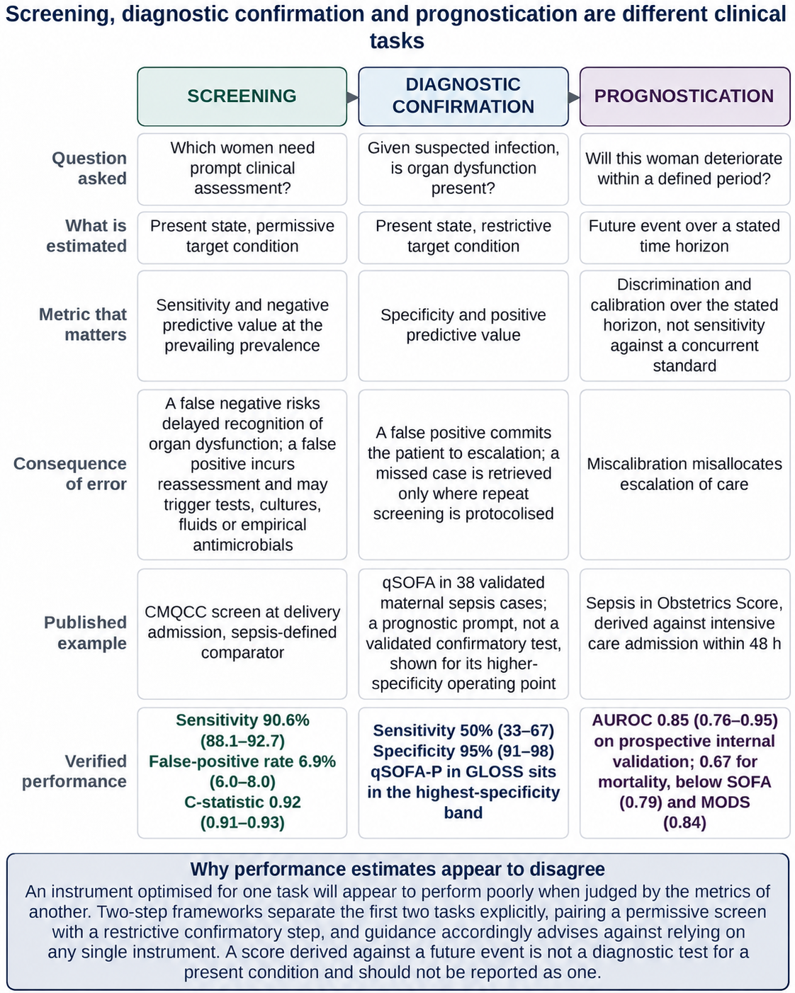
These instruments perform three tasks with different optimal operating points, and much of the apparent disagreement between studies becomes interpretable once the task is specified (Figure 3).
A screening instrument identifies women who need prompt clinical assessment. A false-negative screen risks delayed recognition of evolving organ dysfunction, and a false-positive screen incurs reassessment and, depending on the pathway, investigations, intravenous fluids, or empirical antimicrobial treatment [73]. Screening therefore tolerates a low positive predictive value and demands a high negative predictive value: the MEOWS trigger (Section 5.3) would not support a diagnostic decision, yet reasonably prompts examination of the patient. What limits such a screen is alert burden, and the authors of the MEWC audit concluded that improving specificity might be necessary before widespread implementation [57].
Diagnostic support asks whether a woman with suspected infection has organ dysfunction compatible with maternal sepsis; specificity matters here, because the answer changes management, and the two-step CMQCC structure encodes that separation [22,23], although the accuracy of the sequence as a whole has not been established (Section 7.2). The qSOFA family operates at high specificity and low sensitivity, with specificities at or above 83% and sensitivities at or below 50% against chart-adjudicated sepsis [60] and against severe maternal outcome in GLOSS [7], a profile suited to risk stratification rather than first-stage screening and consistent with the prognostic purpose of qSOFA [18]; the higher sensitivity reported for qSOFA-P in its derivation study rests on cut-points selected in the sample that tested them [29]. Conversely, maternity SIRS achieved 89.2% sensitivity at 27.4% specificity in GLOSS, a defensible screening profile and an indefensible diagnostic one, and no score in that analysis had sufficient accuracy to be used alone [7].
Prognostication concerns whether a woman will deteriorate and is judged by calibration and discrimination over a defined horizon [74]; unlike a diagnostic model, which estimates the probability that a condition is present at the moment of assessment, a prognostic model estimates the probability of an event within a defined future period [75]. Several obstetric instruments are prognostic in construction or validation but diagnostic in application: the SOS was built against intensive care admission [30], MEOWS was validated against composite morbidity [31], the shock index was evaluated against death and other adverse outcomes in women already diagnosed with sepsis [35,76], and GLOSS used severe maternal outcome [7]. Reporting these as sensitivities for sepsis invites the reader to treat a prediction as a diagnosis.
Figure 3.
Screening, diagnostic confirmation and prognostication are related but distinct clinical tasks with different optimal operating points. Each column states the question asked, the quantity estimated, the metric that determines adequacy, the consequence of error, and a published obstetric example with verified performance data against a stated reference condition. In the screening column, the CMQCC performance estimates are derived from delivery-admission data [27], and the false-positive rate is that of a matched comparison series and not an alert rate in routine care. The diagnostic confirmation column illustrates the higher-specificity operating point of qSOFA [60] and qSOFA-P in GLOSS [7]. The prognostication column presents the derivation [30], prospective internal validation [62] and mortality discrimination [64] of the Sepsis in Obstetrics Score, the latter using a version modified for intensive care. An instrument optimised for one task will appear to perform poorly when judged by the metrics of another, which can account for much of the apparent disagreement between validation studies. Two-step frameworks distinguish screening from diagnostic confirmation [22,23], and current guidance advises against reliance on a single screening instrument [8]. Prognostic models should be distinguished from diagnostic models [75]. Abbreviations: AUROC, area under the receiver operating characteristic curve; CMQCC, California Maternal Quality Care Collaborative; GLOSS, Global Maternal Sepsis Study; MODS, Multiple Organ Dysfunction Score; qSOFA, quick Sequential Organ Failure Assessment; qSOFA-P, quick Sequential Organ Failure Assessment adapted for pregnancy; SOFA, Sequential Organ Failure Assessment. Values in parentheses are 95% confidence intervals; predictive values depend on the prevalence of the target condition.
The 2026 Surviving Sepsis Campaign guidelines recommend the National Early Warning Score (NEWS), NEWS2, the Modified Early Warning Score (MEWS), or SIRS over qSOFA as a single tool to screen acutely ill hospitalised patients for sepsis (strong recommendation, moderate certainty evidence) [77]. That direction agrees with the obstetric evidence on qSOFA, but these instruments are calibrated to non-pregnant physiology, unmodified SIRS has a high false-positive rate in later pregnancy and labour, which pregnancy-adjusted criteria substantially reduced in matched comparison series [6,27], and the obstetric ordering of rules changes with clinical period [47]; a maternity service adopting the recommendation is extrapolating, and pregnancy-specific guidance continues to decline to recommend a specific instrument [9,10].
7. Implications for Validation Research and Clinical Practice
New scores are unlikely to resolve the current uncertainty unless validation studies are designed so that their estimates can be interpreted and compared; we therefore propose none.
7.1. Design and Reporting of Validation Studies
Specify the target condition, the reference condition, and the intended task, using the language of QUADAS-3, which treats the target condition as a domain in its own right [69]. When the reference condition is based on administrative codes, the code set should be reported; when it is based on adjudication, the adjudication rules and the handling of cases that remain unresolved should be described [78]. Metrics should follow the task: sensitivity and false-negative burden for screening, specificity and false-positive burden for diagnostic confirmation, and discrimination with calibration for prognostication, with predictive values interpreted at the prevalence of the population studied [74,75].
Use a prespecified, independently adjudicated clinical reference condition. For studies of early recognition, the comparator should be a concurrent clinical reference condition rather than a future event: suspected or confirmed infection with new organ dysfunction attributable to it, defined in advance with pregnancy-appropriate organ-dysfunction criteria [19,20,21]. A defensible design would recruit consecutive screened women in a defined setting and period and classify all of them, not only those who screen positive [66]; use at least two adjudicators independent of the treating team, a fixed classification window and blinding to the index test where feasible [78,79]; record which components of the reference condition overlap with the index test [65]; and assign women whose infection status remains uncertain to an explicit indeterminate category, reported in the participant flow and counted as positive, as negative, and as excluded in prespecified sensitivity analyses [59,78]. Parallel classification of the same cohort against the other reference conditions would show how much of the variation between published estimates is due to the comparator. This is a methodological recommendation, not a validated reference standard: adjudication remains a judgement, and its reproducibility should be reported as inter-rater agreement.
Report false-positive rates and alert burden separately (Section 5.3), with intervals, and avoid in-sample cut-point selection. Because several estimates rest on fewer than 15 outcome events [30,62] and in-sample optimism is greatest in small samples [46], multicentre collaboration is a methodological requirement, and repeated measures and missing data need explicit handling [7,14,64].
Evaluate clinical utility and the added value of laboratory markers. A screening rule earns its place if acting on it does more good than harm at the threshold probability at which a clinician would act, which decision-curve analysis expresses as net benefit and which, for a risk score, depends on calibration rather than on discrimination alone [74]; we identified no obstetric sepsis screening study reporting it. Whether a pregnancy-calibrated laboratory marker adds to a pregnancy-adjusted vital-sign rule is also open; a cohort that evaluated temperature, leucocytes, neutrophils, C-reactive protein, and lactate could not resolve the target condition in over half of its episodes [59]. Reporting standards for diagnostic accuracy and prediction-model studies [78,80] and methods for estimating accuracy without a gold standard [79,81] address several of these points.
7.2. From Screening Accuracy to Clinical Benefit
Evidence about a screening pathway accumulates at five levels, and evidence at one level does not establish the next. The first is the accuracy of the initial criteria: the comparative CMQCC estimates cited here come from the consortium analyses and are for the first-step criteria of the 2020 toolkit, approximated retrospectively from electronic records [22,27,47]. The second is the accuracy of the complete screening–confirmation sequence that guidance recommends. We identified no formal peer-reviewed study that independently validated this; in 2023 it was described as undergoing validation [8], and preliminary performance estimates have been reported from clinical implementation datasets. The figures the 2020 toolkit itself reports for the complete sequence, a sensitivity of about 97% and a specificity of about 99%, derive from 14,752 patients screened across two health systems and are described by its authors as extracted from clinical practice data sets rather than from formal research [22].
The third is process outcomes, such as earlier treatment or escalation, evidence for which comes from non-randomised implementations that introduced a trigger and a management pathway together [34,58]. The fourth is patient outcomes: two randomised trials of early warning systems in low-resource maternity settings showed no demonstrated effect on sepsis (odds ratio: 0.21, 95% CI: 0.02–1.80; one trial) or intensive care admission (0.78, 0.53–1.15) [82], and the period-stratified analysis could not, by its retrospective design, show whether screening improves outcomes [47]. The fifth is the unintended consequences of false-positive alerts, including investigation, empirical antimicrobial treatment, and alarm fatigue, which the accuracy studies reviewed here did not quantify; clinicians describe broad-spectrum prescribing when sepsis can be neither confirmed nor excluded [73].
High sensitivity at the first step is therefore necessary but not sufficient, and it is not evidence that a pathway shortens time to treatment or reduces severe morbidity or death. Before a particular pathway is recommended for wider use, it would need prospective evaluation as a complete sequence in consecutive women, reporting accuracy together with process and patient outcomes and with the frequency and consequences of false-positive alerts, preferably in cluster-randomised or stepped-wedge implementation designs.
7.3. Applicability Across Care Settings
Four questions should be kept apart when an instrument is considered for a new setting: whether it discriminates (diagnostic accuracy); whether it can be applied reliably with the observations, laboratory tests, electronic systems, and staff available (feasibility); whether acting on it improves care (clinical utility); and whether the service can deliver the response it triggers (implementation effectiveness). Accuracy estimates answer only the first, and most come from United States hospitals with electronic records [27,47], although 12 of the 67 hospitals contributing antepartum cases and 14 of the 71 contributing postpartum cases had no labour and delivery unit [47]. GLOSS was a one-week multi-country cohort without confidence intervals for sensitivity or specificity [7]; cohorts from India and South Africa were small and used different reference conditions [35,61,83]; a cohort of 855 women in Sierra Leone evaluated the shock index only as a predictor of adverse outcomes [76]; and the two randomised trials evaluated early warning systems rather than sepsis-specific screens [82].
Requirements also differ. Vital-sign rules such as MEOWS, the MEWC, MEWT, the qSOFA family, and the shock index need no laboratory, although several depend on inconsistently recorded respiratory rate and mental status [38]; the SIRS, UKOSS, and CMQCC first-step criteria include a leucocyte count, and most CMQCC second-step criteria and the SOS require laboratory measurement, including lactate [22,30]. Because a rule recomputed at each observation triggers more often when observations are frequent (Section 5.3), estimates from inpatient units with frequent electronic observations cannot be assumed to hold in an emergency department, on a ward with intermittent observations, or where critical care is distant. We identified no evaluation of these instruments across such settings with comparable methods. The evidence therefore does not support setting-specific recommendations, and a service would need local evaluation of accuracy, alert burden, and response capacity before adopting a particular rule.
7.4. Practice Points
Whether a published estimate applies to a given service depends on the target condition (Section 3), on whether the reference condition captured a present state or a future event (Section 6), on the clinical period and care setting (Section 5.1 and Section 7.3), on prevalence (Section 6.1), on whether the threshold was selected in the dataset that tested it (Section 4), and on the number of outcome events.
Before 20 weeks of gestation and beyond three days postpartum, unmodified criteria were more sensitive than pregnancy-adjusted criteria in the period-stratified comparison identified in this review, at a substantially higher false-positive rate in matched comparison series [47]; whether this reflects physiology or case mix is unknown, and the choice of rule depends on the alert burden a service can absorb and on what follows a positive screen. In a labouring woman with suspected infection, a raised lactate should prompt assessment of haemodynamic status, organ function, and repeat measurement rather than be attributed to labour (Section 4); and in chorioamnionitis or endometritis a first-stage signal should be interpreted with direct assessment of organ dysfunction and trajectory (Section 5.2). A permissive screen followed by a restrictive confirmatory step is one plausible strategy [8,23], but its accuracy as a complete sequence and its effect on outcomes have not been established (Section 7.2).
8. Limitations of This Narrative Review
This is a narrative review (SANRA checklist in Table S3): the search was structured but not systematic, so relevant studies may have been missed; no formal screening, duplicate selection, risk-of-bias scoring, or quantitative synthesis was undertaken, and studies were selected by judgement (Section 2). Statements that no study of a particular kind was identified are therefore conditional on the search described in Section 2 and are not demonstrations of absence.
The underlying literature imposes further constraints. Study designs are heterogeneous, many single-centre estimates rest on very few outcome events, and values unavailable from the source material consulted are identified accordingly in the Supplementary Tables. For several instruments, including omqSOFA, we identified no dedicated derivation-and-validation programme independent of the original consensus framework, although omqSOFA has been evaluated within a larger comparative dataset [7], and some criteria could be confirmed only from secondary reproductions (Table S2). The central comparative argument rests largely on two companion analyses from a single consortium, matched case–control in design and referenced to administrative codes, so the ordering of instruments it supports may not hold under independent replication; no comparative study reviewed here evaluated the 2025 CMQCC edition.
9. Conclusions
This review examined how thresholds, pregnancy physiology, clinical periods, target and reference conditions, and intended clinical tasks influence the interpretation of published estimates of obstetric sepsis screening performance. The answer is that an estimate can be interpreted only in relation to all of them and that, read in this way, the evidence does not establish a universally superior obstetric sepsis screening instrument. This is an absence of evidence for superiority, not evidence of equivalence: within a given clinical period and comparator, instruments differed, as their false-positive rates in the matched comparison series at delivery admission show. Instruments differ in what they were built to detect, in the physiological state for which their thresholds were calibrated, and in the comparator against which they were validated, and those differences are large enough to reverse an apparent ordering.
The established findings are descriptive: within the studies identified, reported performance varied with threshold, clinical period, comparator, and population. The unresolved questions are explanatory and practical: how far the differences between clinical periods reflect pregnancy physiology rather than case mix, infection source or care setting, whether they persist outside a single consortium, what cumulative alert rate each rule generates in routine care, and whether acting on any screen improves maternal outcomes.
Threshold placement moves the balance between detection and false-positive classification, and the clinical period in which a rule is applied was associated with a further shift: in the period-stratified comparison identified in this review, one pregnancy-adjusted screen detected about 25 percentage points more coded cases at 28–36 weeks than before 20 weeks, at an almost constant false-positive rate in matched comparison series. This is compatible with a period-dependent shift in the operating position of a fixed threshold, but case mix, infection source, and care setting may also have differed, and a finding from one consortium against administrative codes is a working hypothesis, not a basis for clinical policy. Differences between studies that vary in comparator and population are also substantial, so sensitivity and specificity should be read as conditional on study design and target condition.
Separating screening, diagnostic confirmation, and prognostication makes much of the apparent disagreement between studies interpretable. First-step accuracy does not show that a screening pathway improves maternal outcomes, which requires prospective evaluation of the complete pathway, including the consequences of false-positive alerts, and the limited specificity of lactate during labour does not make a raised value in a woman with suspected infection uninformative. Validation studies should state the target condition, the reference condition, the clinical period, and the intended task, and report the false-positive rate, the per-woman cumulative alert rate, and confidence intervals alongside sensitivity.
Supplementary Materials
The following are available online at https://www.mdpi.com/article/10.3390/diagnostics16223883/s1, Table S1: Characteristics, reference conditions and diagnostic or prognostic performance of studies evaluating obstetric sepsis screening and severity instruments; Table S2: Screening instruments, definitions and published criteria; Table S3: SANRA quality checklist.
Author Contributions
Conceptualisation, I.P.S.C., R.A.M., and A.C.; methodology, I.P.S.C., E.T.C., and R.G.U.; validation, R.P., D.R.M., and I.S.S.; formal analysis, R.A.M., E.T.C., and C.M.B.; investigation, I.P.S.C., R.A.M., E.T.C., and C.M.B.; resources, D.R.M., D.S., and A.C.; data curation, E.T.C., R.G.U., and I.S.S.; writing—original draft preparation, I.P.S.C., R.A.M., and E.T.C.; writing—review and editing, R.P., D.R.M., C.M.B., R.G.U., I.S.S., D.S., and A.C.; visualisation, R.A.M. and E.T.C.; supervision, D.R.M., D.S., and A.C.; project administration, R.A.M. and A.C. All authors have read and agreed to the published version of the manuscript.
Funding
This research received no external funding.
Institutional Review Board Statement
Not applicable.
Informed Consent Statement
Not applicable.
Data Availability Statement
No new data were created or analysed in this study. Data sharing is not applicable to this article.
Conflicts of Interest
The authors declare no conflicts of interest.
Abbreviations
The following abbreviations are used in this manuscript:
| Abbreviation | Definition |
| AUROC | Area under the receiver operating characteristic curve |
| CI | Confidence interval |
| CMQCC | California Maternal Quality Care Collaborative |
| C-statistic | Concordance statistic |
| GLOSS | Global Maternal Sepsis Study |
| GRADE | Grading of Recommendations Assessment, Development and Evaluation |
| HELLP | Haemolysis, elevated liver enzymes and low platelets |
| MEOWS | Modified Early Obstetric Warning System |
| MEWC | Maternal Early Warning Criteria |
| MEWS | Modified Early Warning Score |
| MEWT | Maternal Early Warning Trigger |
| MODS | Multiple Organ Dysfunction Score |
| NEWS | National Early Warning Score |
| NEWS2 | National Early Warning Score, version 2 |
| NICE | National Institute for Health and Care Excellence |
| omqSOFA | Obstetrically modified quick Sequential Organ Failure Assessment |
| omSOFA | Obstetrically modified Sequential Organ Failure Assessment |
| PPV | Positive predictive value |
| qSOFA | Quick Sequential Organ Failure Assessment |
| qSOFA-P | Pregnancy-adapted quick Sequential Organ Failure Assessment |
| QUADAS-3 | Quality Assessment of Diagnostic Accuracy Studies, version 3 |
| SANRA | Scale for the Assessment of Narrative Review Articles |
| SEP-1 | Severe Sepsis and Septic Shock Management Bundle |
| Sepsis-3 | Third International Consensus Definitions for Sepsis and Septic Shock |
| SIRS | Systemic inflammatory response syndrome |
| SOFA | Sequential Organ Failure Assessment |
| SOMANZ | Society of Obstetric Medicine of Australia and New Zealand |
| SOS | Sepsis in Obstetrics Score |
| UKOSS | United Kingdom Obstetric Surveillance System |
| WHO | World Health Organization |
References
- Say, L.; Chou, D.; Gemmill, A.; Tunçalp, Ö.; Moller, A.-B.; Daniels, J.; Gülmezoglu, A.M.; Temmerman, M.; Alkema, L. Global causes of maternal death: A WHO systematic analysis. Lancet Glob. Health 2014, 2, e323–e333. [Google Scholar] [CrossRef] [Scilit] [PubMed]
- Cresswell, J.A.; Alexander, M.; Chong, M.Y.C.; Link, H.M.; Pejchinovska, M.; Gazeley, U.; Ahmed, S.M.A.; Chou, D.; Moller, A.-B.; Simpson, D.; et al. Global and regional causes of maternal deaths 2009–20: A WHO systematic analysis. Lancet Glob. Health 2025, 13, e626–e634. [Google Scholar] [CrossRef] [Scilit] [PubMed]
- Bonet, M.; Brizuela, V.; Abalos, E.; Cuesta, C.; Baguiya, A.; Chamillard, M.; Fawole, B.; Knight, M.; Kouanda, S.; Lumbiganon, P.; et al. Frequency and management of maternal infection in health facilities in 52 countries (GLOSS): A 1-week inception cohort study. Lancet Glob. Health 2020, 8, e661–e671. [Google Scholar] [CrossRef] [Scilit] [PubMed]
- Bauer, M.E.; Lorenz, R.P.; Bauer, S.T.; Rao, K.; Anderson, F.W.J. Maternal deaths due to sepsis in the state of Michigan, 1999–2006. Obstet. Gynecol. 2015, 126, 747–752. [Google Scholar] [CrossRef] [Scilit] [PubMed]
- Felker, A.; Patel, R.; Kotnis, R.; Kenyon, S.; Knight, M. (Eds.) Saving Lives, Improving Mothers’ Care: Lessons Learned to Inform Maternity Care from the UK and Ireland Confidential Enquiries into Maternal Deaths and Morbidity 2021–23; National Perinatal Epidemiology Unit, University of Oxford: Oxford, UK, 2025. [Google Scholar] [CrossRef]
- Bauer, M.E.; Bauer, S.T.; Rajala, B.; MacEachern, M.P.; Polley, L.S.; Childers, D.; Aronoff, D.M. Maternal physiologic parameters in relationship to systemic inflammatory response syndrome criteria: A systematic review and meta-analysis. Obstet. Gynecol. 2014, 124, 535–541. [Google Scholar] [CrossRef] [Scilit] [PubMed]
- Chimwaza, Y.; Hunt, A.; Oliveira-Ciabati, L.; Bonnett, L.; Abalos, E.; Cuesta, C.; Souza, J.P.; Bonet, M.; Brizuela, V.; Lissauer, D.; et al. Early warning systems for identifying severe maternal outcomes: Findings from the WHO global maternal sepsis study. eClinicalMedicine 2025, 79, 102981. [Google Scholar] [CrossRef] [Scilit] [PubMed]
- Society for Maternal-Fetal Medicine; Shields, A.D.; Plante, L.A.; Pacheco, L.D.; Louis, J.M. Society for Maternal-Fetal Medicine Consult Series #67: Maternal sepsis. Am. J. Obstet. Gynecol. 2023, 229, B2–B19. [Google Scholar] [CrossRef] [Scilit] [PubMed]
- Bauer, M.E.; Albright, C.; Prabhu, M.; Heine, R.P.; Lennox, C.; Allen, C.; Burke, C.; Chavez, A.; Hughes, B.L.; Kendig, S.; et al. Alliance for Innovation on Maternal Health: Consensus bundle on sepsis in obstetric care. Obstet. Gynecol. 2023, 142, 481–492. [Google Scholar] [CrossRef] [Scilit] [PubMed]
- National Institute for Health and Care Excellence. Suspected Sepsis in Pregnant or Recently Pregnant People: Recognition, Diagnosis and Early Management; NICE Guideline NG255; NICE: London, UK, 2025; ISBN 978-1-4731-7294-4. [Google Scholar]
- Nakamura, E.; Naruse, K.; Miyake, R.; Hara, T.; Furuta, M.; Tanaka, H.; Matsunaga, S.; Sakurai, A. Early warning systems to predict severe complications during preterm and postpartum: A scoping review. J. Obstet. Gynaecol. Res. 2025, 51, e70079. [Google Scholar] [CrossRef] [Scilit] [PubMed]
- Yahya, F.B.; Van Abel, A.L.; Brakke, B.D.; Palraj, R.; Szymanski, L.M.; Chapman, M.L.; Butler Tobah, Y.S.; Rajapakse, N.S.; Dinnes, L.M. Maternal sepsis review and update. Mayo Clin. Proc. 2025, 100, 1212–1230. [Google Scholar] [CrossRef] [Scilit] [PubMed]
- Kumar, A.; Abbenbroek, B.; Delaney, A.; Hammond, N.; Grattan, S.; Finfer, S. Sepsis triggers and tools to support early identification in healthcare settings: An integrative review. Aust. Crit. Care 2023, 36, 1117–1128. [Google Scholar] [CrossRef] [Scilit] [PubMed]
- Chester-Jones, M.; Kirtley, S.; Er, F.; Jogarah, V.; Qiao, Y.; Tunn, R.; Vides, N.; Watkinson, P.J.; Knight, M.; Gerry, S.; et al. Maternal early warning scores shown to be methodologically weak and at high risk of bias. J. Clin. Epidemiol. 2025, 184, 111833. [Google Scholar] [CrossRef] [Scilit] [PubMed]
- Tong, J.; Deng, P. Sepsis screening tools in resource-limited settings: A systematic review and meta-analysis of diagnostic accuracy in low- and middle-income countries. Front. Public Health 2026, 14, 1782420. [Google Scholar] [CrossRef] [Scilit] [PubMed]
- Baethge, C.; Goldbeck-Wood, S.; Mertens, S. SANRA—A scale for the quality assessment of narrative review articles. Res. Integr. Peer Rev. 2019, 4, 5. [Google Scholar] [CrossRef] [Scilit] [PubMed]
- Singer, M.; Deutschman, C.S.; Seymour, C.W.; Shankar-Hari, M.; Annane, D.; Bauer, M.; Bellomo, R.; Bernard, G.R.; Chiche, J.-D.; Coopersmith, C.M.; et al. The Third International Consensus Definitions for Sepsis and Septic Shock (Sepsis-3). JAMA 2016, 315, 801–810. [Google Scholar] [CrossRef] [Scilit] [PubMed]
- Seymour, C.W.; Liu, V.X.; Iwashyna, T.J.; Brunkhorst, F.M.; Rea, T.D.; Scherag, A.; Rubenfeld, G.; Kahn, J.M.; Shankar-Hari, M.; Singer, M.; et al. Assessment of clinical criteria for sepsis: For the Third International Consensus Definitions for Sepsis and Septic Shock (Sepsis-3). JAMA 2016, 315, 762–774. [Google Scholar] [CrossRef] [Scilit] [PubMed]
- Bowyer, L.; Robinson, H.L.; Barrett, H.; Crozier, T.M.; Giles, M.; Idel, I.; Lowe, S.; Lust, K.; Marnoch, C.A.; Morton, M.R.; et al. SOMANZ guidelines for the investigation and management sepsis in pregnancy. Aust. N. Z. J. Obstet. Gynaecol. 2017, 57, 540–551. [Google Scholar] [CrossRef] [Scilit] [PubMed]
- Bowyer, L.; Cutts, B.A.; Barrett, H.L.; Bein, K.; Crozier, T.M.; Gehlert, J.; Giles, M.L.; Hocking, J.; Lowe, S.; Lust, K.; et al. SOMANZ position statement for the investigation and management of sepsis in pregnancy 2023. Aust. N. Z. J. Obstet. Gynaecol. 2025, 65, 37–46. [Google Scholar] [CrossRef] [Scilit] [PubMed]
- Bonet, M.; Nogueira Pileggi, V.; Rijken, M.J.; Coomarasamy, A.; Lissauer, D.; Souza, J.P.; Gülmezoglu, A.M. Towards a consensus definition of maternal sepsis: Results of a systematic review and expert consultation. Reprod. Health 2017, 14, 67. [Google Scholar] [CrossRef] [Scilit] [PubMed]
- Gibbs, R.; Bauer, M.; Olvera, L.; Sakowski, C.; Cape, V.; Main, E. Improving Diagnosis and Treatment of Maternal Sepsis: A Quality Improvement Toolkit; California Maternal Quality Care Collaborative: Stanford, CA, USA, 2020. [Google Scholar]
- Main, E.K.; Nath, R.; Bauer, M.E. CMQCC obstetric sepsis toolkit update: A patient-centered approach to quality improvement. Semin. Perinatol. 2024, 48, 151976. [Google Scholar] [CrossRef] [Scilit] [PubMed]
- California Maternal Quality Care Collaborative. Improving Diagnosis and Treatment of Obstetric Sepsis, V2.0 Toolkit; Sepsis Task Force Advisory Group V2.0; California Maternal Quality Care Collaborative: Stanford, CA, USA, 2025; Available online: https://www.cmqcc.org/resource/improving-diagnosis-and-treatment-obstetric-sepsis-v20-toolkit-2025 (accessed on 10 November 2026).
- Acosta, C.D.; Kurinczuk, J.J.; Lucas, D.N.; Tuffnell, D.J.; Sellers, S.; Knight, M. Severe maternal sepsis in the UK, 2011–2012: A national case-control study. PLoS Med. 2014, 11, e1001672. [Google Scholar] [CrossRef] [Scilit] [PubMed]
- Cosovanu, E.T.; Cosovanu, E.O.; Ionescu, S.G.; Asaftei, A.; Damian, C.; Asaftei-Titianu, G.S.; Donos, M.A.; Petroaie, A.D.; Ioniuc, I.K.; Petris, O.R.; et al. Diagnosing neonatal sepsis: A comprehensive review of conventional culture, molecular methods, and host-response biomarkers. Acta Microbiol. Hell. 2026, 71, 28. [Google Scholar] [CrossRef] [Scilit]
- Main, E.K.; Fuller, M.; Kovacheva, V.P.; Elkhateb, R.; Azar, K.; Caldwell, M.; Chiem, V.; Foster, M.; Gibbs, R.; Hughes, B.L.; et al. Performance characteristics of sepsis screening tools during delivery admissions. Obstet. Gynecol. 2024, 143, 326–335. [Google Scholar] [CrossRef] [Scilit] [PubMed]
- Yahya, F.B.; Yousufuddin, M.; Gaston, H.J.; Fagbongbe, E.; Rangel Latuche, L.J. Validating the performance of 3 sepsis screening tools in patients with clinical chorioamnionitis. AJOG Glob. Rep. 2023, 3, 100271. [Google Scholar] [CrossRef] [Scilit] [PubMed]
- Cagino, S.G.; Burke, A.A.; Letner, D.R.; Leizer, J.M.; Zelig, C.M. Quick Sequential Organ Failure Assessment: Modifications for identifying maternal morbidity and mortality in obstetrical patients. Am. J. Perinatol. 2022, 39, 1–7. [Google Scholar] [CrossRef] [Scilit] [PubMed]
- Albright, C.M.; Ali, T.N.; Lopes, V.; Rouse, D.J.; Anderson, B.L. The Sepsis in Obstetrics Score: A model to identify risk of morbidity from sepsis in pregnancy. Am. J. Obstet. Gynecol. 2014, 211, 39.e1–39.e8. [Google Scholar] [CrossRef] [Scilit] [PubMed]
- Singh, S.; McGlennan, A.; England, A.; Simons, R. A validation study of the CEMACH recommended modified early obstetric warning system (MEOWS). Anaesthesia 2012, 67, 12–18. [Google Scholar] [CrossRef] [Scilit] [PubMed]
- Mhyre, J.M.; D’Oria, R.; Hameed, A.B.; Lappen, J.R.; Holley, S.L.; Hunter, S.K.; Jones, R.L.; King, J.C.; D’Alton, M.E. The maternal early warning criteria: A proposal from the National Partnership for Maternal Safety. Obstet. Gynecol. 2014, 124, 782–786. [Google Scholar] [CrossRef] [Scilit] [PubMed]
- Friedman, A.M.; Campbell, M.L.; Kline, C.R.; Wiesner, S.; D’Alton, M.E.; Shields, L.E. Implementing obstetric early warning systems. AJP Rep. 2018, 8, e79–e84. [Google Scholar] [CrossRef] [Scilit] [PubMed]
- Shields, L.E.; Wiesner, S.; Klein, C.; Pelletreau, B.; Hedriana, H.L. Use of Maternal Early Warning Trigger tool reduces maternal morbidity. Am. J. Obstet. Gynecol. 2016, 214, 527.e1–527.e6. [Google Scholar] [CrossRef] [Scilit] [PubMed]
- Nathan, H.L.; Seed, P.T.; Hezelgrave, N.L.; De Greeff, A.; Lawley, E.; Anthony, J.; Steyn, W.; Hall, D.R.; Chappell, L.C.; Shennan, A.H. Shock index thresholds to predict adverse outcomes in maternal hemorrhage and sepsis: A prospective cohort study. Acta Obstet. Gynecol. Scand. 2019, 98, 1178–1186. [Google Scholar] [CrossRef] [Scilit] [PubMed]
- Arnolds, D.E.; Carey, K.A.; Braginsky, L.; Holt, R.; Edelson, D.P.; Scavone, B.M.; Churpek, M. Comparison of early warning scores for predicting clinical deterioration and infection in obstetric patients. BMC Pregnancy Childbirth 2022, 22, 295. [Google Scholar] [CrossRef] [Scilit] [PubMed]
- Green, L.J.; Mackillop, L.H.; Salvi, D.; Pullon, R.; Loerup, L.; Tarassenko, L.; Mossop, J.; Edwards, C.; Gerry, S.; Birks, J.; et al. Gestation-specific vital sign reference ranges in pregnancy. Obstet. Gynecol. 2020, 135, 653–664. [Google Scholar] [CrossRef] [Scilit] [PubMed]
- Sela, H.Y.; Seri, V.; Zimmerman, F.S.; Cortegiani, A.; Levin, P.D.; Smueloff, A.; Einav, S. A retrospective study of the proportion of women at high and low risk of intrauterine infection meeting sepsis criteria. Microorganisms 2022, 10, 82. [Google Scholar] [CrossRef] [Scilit] [PubMed]
- Green, L.J.; Pullon, R.; Mackillop, L.H.; Gerry, S.; Birks, J.; Salvi, D.; Davidson, S.; Loerup, L.; Tarassenko, L.; Mossop, J.; et al. Postpartum-specific vital sign reference ranges. Obstet. Gynecol. 2021, 137, 295–304. [Google Scholar] [CrossRef] [Scilit] [PubMed]
- Dockree, S.; Shine, B.; Pavord, S.; Impey, L.; Vatish, M. White blood cells in pregnancy: Reference intervals for before and after delivery. eBioMedicine 2021, 74, 103715. [Google Scholar] [CrossRef] [Scilit] [PubMed]
- Reese, J.A.; Peck, J.D.; Deschamps, D.R.; McIntosh, J.J.; Knudtson, E.J.; Terrell, D.R.; Vesely, S.K.; George, J.N. Platelet counts during pregnancy. N. Engl. J. Med. 2018, 379, 32–43. [Google Scholar] [CrossRef] [Scilit] [PubMed]
- Groenendijk, W.; Bogdanet, D.; Dervan, L.; Finn, O.; Islam, M.N.; Doheny, H.; Griffin, T.P.; Blake, L.; Lyons, M.; Kilcooley, M.; et al. Reference intervals for clinical biochemistry and haematology tests during normal pregnancy. Ann. Clin. Biochem. 2022, 59, 433–446. [Google Scholar] [CrossRef] [Scilit] [PubMed]
- Philip, J.; Alexander, J.M.; Sharma, S.K.; Leveno, K.J.; McIntire, D.D.; Wiley, J. Epidural analgesia during labor and maternal fever. Anesthesiology 1999, 90, 1271–1275. [Google Scholar] [CrossRef] [Scilit] [PubMed]
- Dockree, S.; O’Sullivan, J.; Shine, B.; James, T.; Vatish, M. How should we interpret lactate in labour? A reference study. BJOG 2022, 129, 2150–2156. [Google Scholar] [CrossRef] [Scilit] [PubMed]
- Albright, C.M.; Ali, T.N.; Lopes, V.; Rouse, D.J.; Anderson, B.L. Lactic acid measurement to identify risk of morbidity from sepsis in pregnancy. Am. J. Perinatol. 2015, 32, 481–486. [Google Scholar] [CrossRef] [Scilit] [PubMed]
- Leeflang, M.M.G.; Moons, K.G.M.; Reitsma, J.B.; Zwinderman, A.H. Bias in sensitivity and specificity caused by data-driven selection of optimal cutoff values: Mechanisms, magnitude, and solutions. Clin. Chem. 2008, 54, 729–737. [Google Scholar] [CrossRef] [Scilit] [PubMed]
- Bauer, M.E.; Fuller, M.; Kovacheva, V.; Elkhateb, R.; Azar, K.; Caldwell, M.; Chiem, V.; Foster, M.; Gibbs, R.; Hughes, B.L.; et al. Performance characteristics of sepsis screening tools during antepartum and postpartum admissions. Obstet. Gynecol. 2024, 143, 336–345. [Google Scholar] [CrossRef] [Scilit] [PubMed]
- Lappen, J.R.; Keene, M.; Lore, M.; Grobman, W.A.; Gossett, D.R. Existing models fail to predict sepsis in an obstetric population with intrauterine infection. Am. J. Obstet. Gynecol. 2010, 203, 573.e1–573.e5. [Google Scholar] [CrossRef] [Scilit] [PubMed]
- Edwards, S.E.; Grobman, W.A.; Lappen, J.R.; Winter, C.; Fox, R.; Lenguerrand, E.; Draycott, T. Modified obstetric early warning scoring systems (MOEWS): Validating the diagnostic performance for severe sepsis in women with chorioamnionitis. Am. J. Obstet. Gynecol. 2015, 212, 536.e1–536.e8. [Google Scholar] [CrossRef] [Scilit] [PubMed]
- Ruymann, K.; Agarwal, E.; Ogle, M.; Al-Kouatly, H.B.; Boelig, R.C. Association of pregnancy-adjusted sepsis screening criteria with risk of maternal infectious morbidity in the setting of intrapartum intraamniotic infection. Am. J. Perinatol. 2025, 42, 1793–1797. [Google Scholar] [CrossRef] [Scilit] [PubMed]
- Higgins, R.D.; Saade, G.; Polin, R.A.; Grobman, W.A.; Buhimschi, I.A.; Watterberg, K.; Silver, R.M.; Raju, T.N.K. Evaluation and management of women and newborns with a maternal diagnosis of chorioamnionitis: Summary of a workshop. Obstet. Gynecol. 2016, 127, 426–436. [Google Scholar] [CrossRef] [Scilit] [PubMed]
- American College of Obstetricians and Gynecologists’ Committee on Obstetric Practice. Committee Opinion No. 712: Intrapartum management of intraamniotic infection. Obstet. Gynecol. 2017, 130, e95–e101. [Google Scholar] [CrossRef] [Scilit] [PubMed]
- American College of Obstetricians and Gynecologists. ACOG Clinical Practice Update: Update on criteria for suspected diagnosis of intraamniotic infection. Obstet. Gynecol. 2024, 144, e17–e19. [Google Scholar] [CrossRef] [Scilit] [PubMed]
- San-Juan, R.; Sanz-Prieto, A.; Contreras-Mora, J.; Fojo-Suárez, I.; Caso-Laviana, J.M.; Fernández-Ruiz, M.; López-Medrano, F.; Rodríguez-Goncer, I.; Fayos, M.; Brañas, P.; et al. Comprehensive analysis of current epidemiology, clinical features and prognostic factors of puerperal endometritis: A retrospective cohort analysis. Eur. J. Obstet. Gynecol. Reprod. Biol. X 2023, 18, 100199. [Google Scholar] [CrossRef] [Scilit] [PubMed]
- Shafik, S.; Mallick, S.; Fogel, J.; Tetrokalashvili, M.; Hsu, C.-D. The utility of systemic inflammatory response syndrome (SIRS) for diagnosing sepsis in the immediate postpartum period. J. Infect. Public Health 2019, 12, 799–802. [Google Scholar] [CrossRef] [Scilit] [PubMed]
- Acosta, C.D.; Knight, M.; Lee, H.C.; Kurinczuk, J.J.; Gould, J.B.; Lyndon, A. The continuum of maternal sepsis severity: Incidence and risk factors in a population-based cohort study. PLoS ONE 2013, 8, e67175. [Google Scholar] [CrossRef] [Scilit] [PubMed]
- Arnolds, D.E.; Smith, A.; Banayan, J.M.; Holt, R.; Scavone, B.M. National Partnership for Maternal Safety recommended maternal early warning criteria are associated with maternal morbidity. Anesth. Analg. 2019, 129, 1621–1626. [Google Scholar] [CrossRef] [Scilit] [PubMed]
- Blumenthal, E.A.; Hooshvar, N.; Tancioco, V.; Newman, R.; Senderoff, D.; McNulty, J. Implementation and evaluation of an electronic Maternal Early Warning Trigger tool to reduce maternal morbidity. Am. J. Perinatol. 2021, 38, 869–879. [Google Scholar] [CrossRef] [Scilit] [PubMed]
- Culling, T.; Bertorelli, C.; Strang, A.; Oram, S.; Faggian, F.; Sharma, S.; Ridgeway, A.; Zaher, S.; Labeta, M.; Jones, S.A.; et al. Maternal physiological parameters and routine laboratory tests to screen for maternal sepsis: An observational cohort study. Int. J. Obstet. Anesth. 2025, 63, 104683. [Google Scholar] [CrossRef] [Scilit] [PubMed]
- Bauer, M.E.; Housey, M.; Bauer, S.T.; Behrmann, S.; Chau, A.; Clancy, C.; Clark, E.A.S.; Einav, S.; Langen, E.; Leffert, L.; et al. Risk factors, etiologies, and screening tools for sepsis in pregnant women: A multicenter case-control study. Anesth. Analg. 2019, 129, 1613–1620. [Google Scholar] [CrossRef] [Scilit] [PubMed]
- Agarwal, R.; Yadav, R.K.; Mohta, M.; Sikka, M.; Radhakrishnan, G. Sepsis in Obstetrics Score (SOS) utility and validation for triaging patients with obstetric sepsis in the emergency department: Evidence from a low income health care setting. Obstet. Med. 2019, 12, 90–96. [Google Scholar] [CrossRef] [Scilit] [PubMed]
- Albright, C.M.; Has, P.; Rouse, D.J.; Hughes, B.L. Internal validation of the Sepsis in Obstetrics Score to identify risk of morbidity from sepsis in pregnancy. Obstet. Gynecol. 2017, 130, 747–755. [Google Scholar] [CrossRef] [Scilit] [PubMed]
- Abutheraa, N.; Grant, J.; Mullen, A.B. Sepsis scoring systems and use of the Sepsis six care bundle in maternity hospitals. BMC Pregnancy Childbirth 2021, 21, 524. [Google Scholar] [CrossRef] [Scilit] [PubMed]
- Aarvold, A.B.R.; Ryan, H.M.; Magee, L.A.; von Dadelszen, P.; Fjell, C.; Walley, K.R. Multiple Organ Dysfunction Score is superior to the obstetric-specific Sepsis in Obstetrics Score in predicting mortality in septic obstetric patients. Crit. Care Med. 2017, 45, e49–e57. [Google Scholar] [CrossRef] [Scilit] [PubMed]
- Whiting, P.; Rutjes, A.W.S.; Reitsma, J.B.; Bossuyt, P.M.M.; Kleijnen, J. The development of QUADAS: A tool for the quality assessment of studies of diagnostic accuracy included in systematic reviews. BMC Med. Res. Methodol. 2003, 3, 25. [Google Scholar] [CrossRef] [Scilit] [PubMed]
- de Groot, J.A.H.; Bossuyt, P.M.M.; Reitsma, J.B.; Rutjes, A.W.S.; Dendukuri, N.; Janssen, K.J.M.; Moons, K.G.M. Verification problems in diagnostic accuracy studies: Consequences and solutions. BMJ 2011, 343, d4770. [Google Scholar] [CrossRef] [Scilit] [PubMed]
- Ransohoff, D.F.; Feinstein, A.R. Problems of spectrum and bias in evaluating the efficacy of diagnostic tests. N. Engl. J. Med. 1978, 299, 926–930. [Google Scholar] [CrossRef] [Scilit] [PubMed]
- Cordoba, G.; Schwartz, L.; Woloshin, S.; Bae, H.; Gøtzsche, P.C. Definition, reporting, and interpretation of composite outcomes in clinical trials: Systematic review. BMJ 2010, 341, c3920. [Google Scholar] [CrossRef] [Scilit] [PubMed]
- Whiting, P.F.; Tomlinson, E.; Rutjes, A.W.S.; Davenport, C.F.; Yang, B.; Westwood, M.E.; Takwoingi, Y.; Reitsma, J.B.; Hyde, C.; Bossuyt, P.M.M.; et al. QUADAS-3: A revised tool for the quality assessment of diagnostic test accuracy studies. Ann. Intern. Med. 2026, 179, 548–555. [Google Scholar] [CrossRef] [Scilit] [PubMed]
- Murad, M.H.; Lin, L.; Chu, H.; Hasan, B.; Alsibai, R.A.; Abbas, A.S.; Mustafa, R.A.; Wang, Z. The association of sensitivity and specificity with disease prevalence: Analysis of 6909 studies of diagnostic test accuracy. CMAJ 2023, 195, E925–E931. [Google Scholar] [CrossRef] [Scilit] [PubMed]
- Leeflang, M.M.G.; Rutjes, A.W.S.; Reitsma, J.B.; Hooft, L.; Bossuyt, P.M.M. Variation of a test’s sensitivity and specificity with disease prevalence. CMAJ 2013, 185, E537–E544. [Google Scholar] [CrossRef] [Scilit] [PubMed]
- Liu, L.Y.; Wen, T.; Reddy, U.M.; Mourad, M.; Goffman, D.; Nathan, L.; Sheen, J.-J.; D’Alton, M.E.; Friedman, A.M. Risk factors, trends, and outcomes associated with postpartum sepsis readmissions. Obstet. Gynecol. 2024, 143, 346–354. [Google Scholar] [CrossRef] [Scilit] [PubMed]
- Kingdon, C.; Greenfield, B.; Cunningham, C.; Freeman, D.; Ming, D.; Lissauer, D.; Holmes, A.; Merriel, A. Clinicians’ decision-making about broad-spectrum antibiotic prescribing for suspected maternal sepsis during childbirth in the UK: A qualitative study. BMJ Open 2026, 16, e110559. [Google Scholar] [CrossRef] [Scilit] [PubMed]
- Van Calster, B.; McLernon, D.J.; van Smeden, M.; Wynants, L.; Steyerberg, E.W. Calibration: The Achilles heel of predictive analytics. BMC Med. 2019, 17, 230. [Google Scholar] [CrossRef] [Scilit] [PubMed]
- Moons, K.G.M.; Altman, D.G.; Reitsma, J.B.; Ioannidis, J.P.A.; Macaskill, P.; Steyerberg, E.W.; Vickers, A.J.; Ransohoff, D.F.; Collins, G.S. Transparent Reporting of a multivariable prediction model for Individual Prognosis or Diagnosis (TRIPOD): Explanation and elaboration. Ann. Intern. Med. 2015, 162, W1–W73. [Google Scholar] [CrossRef] [Scilit] [PubMed]
- Kuhrt, K.; Janneh, F.; Cole, R.; Ridout, A.; Fernandez-Turienzo, C.; Seed, P.T.; Bramham, K.; Shennan, A.H. Evaluating shock index for prediction of adverse maternal outcomes related to postpartum haemorrhage and maternal sepsis in Sierra Leone: A prospective observational cohort study. eClinicalMedicine 2025, 89, 103578. [Google Scholar] [CrossRef] [Scilit] [PubMed]
- Prescott, H.C.; Antonelli, M.; Alhazzani, W.; Møller, M.H.; Alshamsi, F.; Azevedo, L.C.P.; Belley-Cote, E.; De Waele, J.; Derde, L.; Dionne, J.C.; et al. Surviving Sepsis Campaign: International guidelines for management of sepsis and septic shock 2026. Intensive Care Med. 2026, 52, 863–936, co-published in Crit. Care Med. 2026, 54, 725–812. https://doi.org/10.1097/CCM.0000000000007075. [Google Scholar] [CrossRef] [Scilit] [PubMed]
- Bossuyt, P.M.; Reitsma, J.B.; Bruns, D.E.; Gatsonis, C.A.; Glasziou, P.P.; Irwig, L.; Lijmer, J.G.; Moher, D.; Rennie, D.; de Vet, H.C.; et al. STARD 2015: An updated list of essential items for reporting diagnostic accuracy studies. BMJ 2015, 351, h5527. [Google Scholar] [CrossRef] [Scilit] [PubMed]
- Reitsma, J.B.; Rutjes, A.W.S.; Khan, K.S.; Coomarasamy, A.; Bossuyt, P.M. A review of solutions for diagnostic accuracy studies with an imperfect or missing reference standard. J. Clin. Epidemiol. 2009, 62, 797–806. [Google Scholar] [CrossRef] [Scilit] [PubMed]
- Collins, G.S.; Moons, K.G.M.; Dhiman, P.; Riley, R.D.; Beam, A.L.; Van Calster, B.; Ghassemi, M.; Liu, X.; Reitsma, J.B.; van Smeden, M.; et al. TRIPOD+AI statement: Updated guidance for reporting clinical prediction models that use regression or machine learning methods. BMJ 2024, 385, e078378. [Google Scholar] [CrossRef] [Scilit] [PubMed]
- Okkaoglu, Y.; Welton, N.J.; Ades, A.E.; Jones, H.E. Latent class log-linear models for estimating diagnostic test accuracy without a gold standard: A simulation study. Stat. Med. 2026, 45, e70660. [Google Scholar] [CrossRef] [Scilit] [PubMed]
- Smith, V.; Kenny, L.C.; Sandall, J.; Devane, D.; Noonan, M. Physiological track-and-trigger/early warning systems for use in maternity care. Cochrane Database Syst. Rev. 2021, 9, CD013276. [Google Scholar] [CrossRef] [Scilit] [PubMed]
- Agarwal, R.; Goyal, P.; Mohta, M.; Kar, R. Comparison of Sequential Organ Failure Assessment (SOFA) and Sepsis in Obstetrics Score (SOS) in women with pregnancy-associated sepsis with respect to critical care admission and mortality: A prospective observational study. J. Obstet. Gynaecol. India 2021, 71, 45–51. [Google Scholar] [CrossRef] [Scilit] [PubMed]
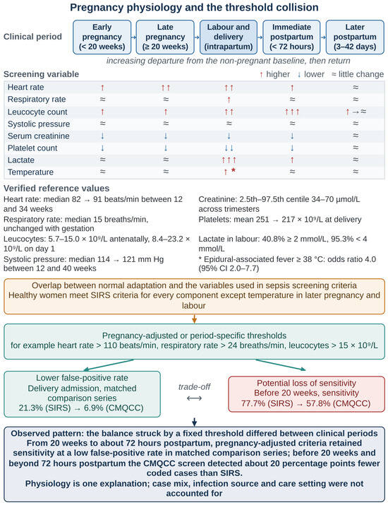
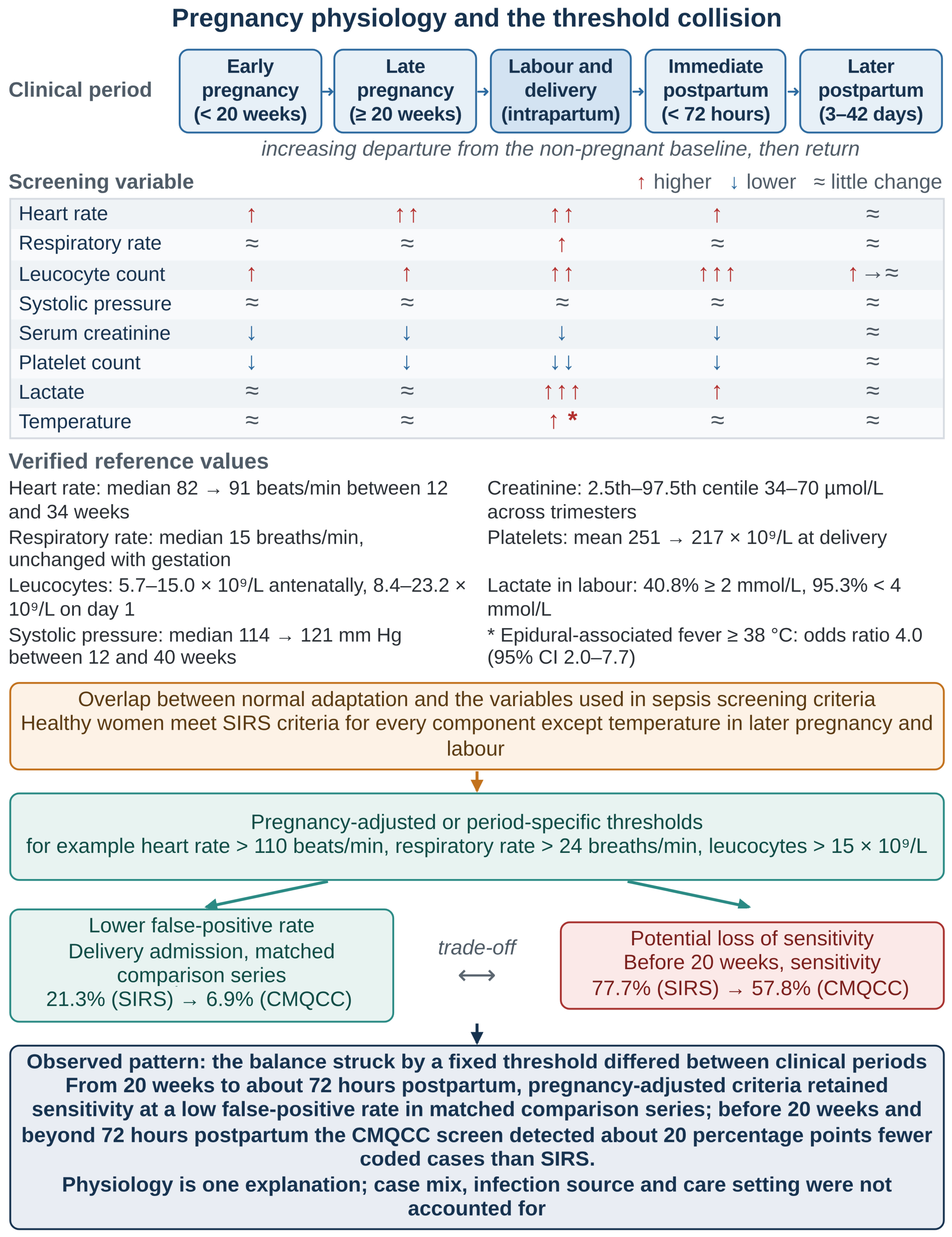
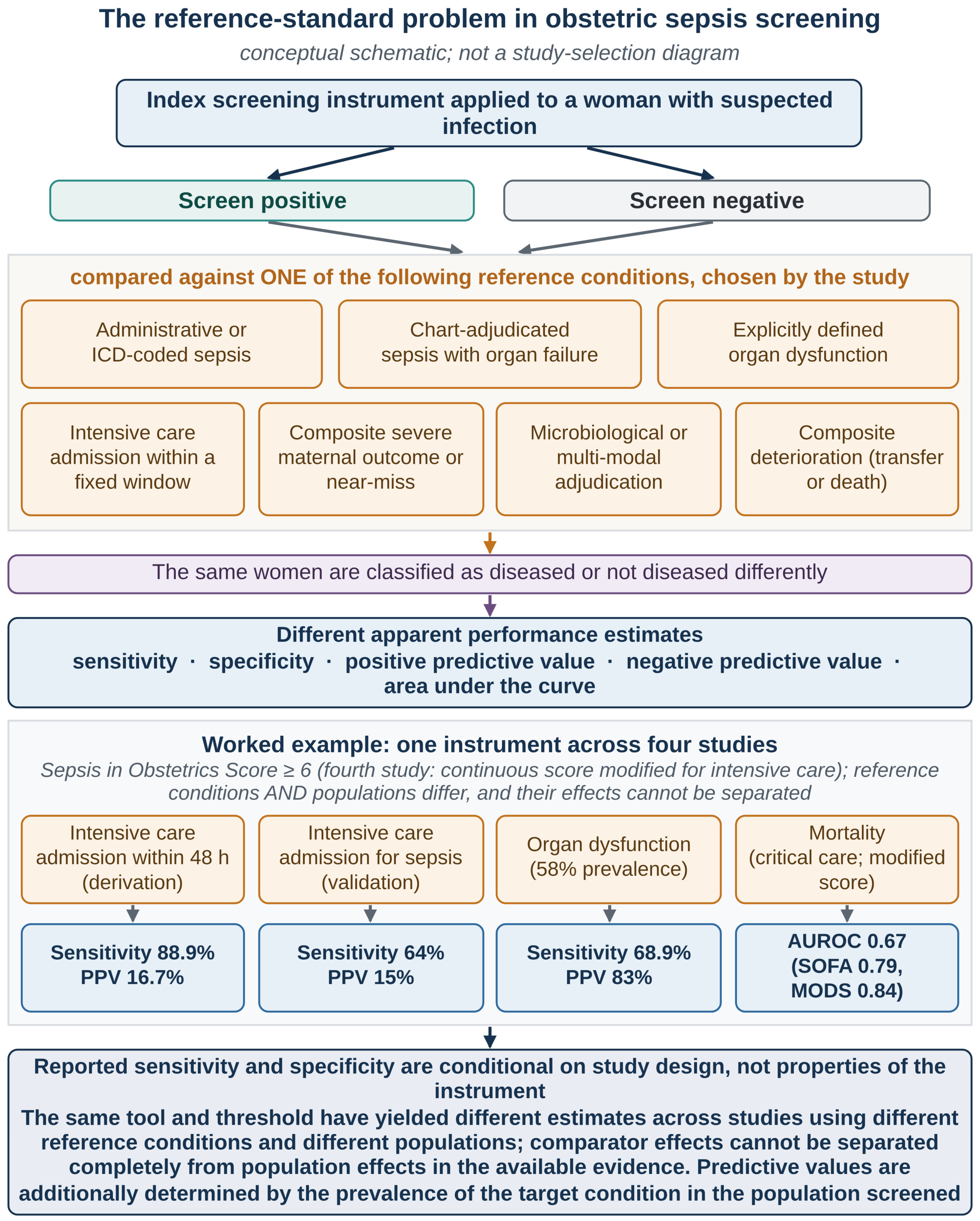
Disclaimer/Publisher’s Note: The statements, opinions and data contained in all publications are solely those of the individual author(s) and contributor(s) and not of MDPI and/or the editor(s). MDPI and/or the editor(s) disclaim responsibility for any injury to people or property resulting from any ideas, methods, instructions or products referred to in the content. |
© 2026 by the authors. Licensee MDPI, Basel, Switzerland. This article is an open access article distributed under the terms and conditions of the Creative Commons Attribution (CC BY) license.