


Abstract
Background: Leiomyomatosis peritonealis disseminata (LPD) is a rare benign condition characterized by multiple smooth-muscle nodules throughout the peritoneal cavity. Increasing evidence suggests that uncontained morcellation during laparoscopic myomectomy represents a clinically relevant mechanism of iatrogenic dissemination. Because LPD frequently mimics advanced intra-abdominal malignancy, preoperative diagnosis remains challenging, particularly in emergency settings. Case Presentation: We report the case of a 46-year-old nulligravid woman presenting with progressive abdominal pain, abnormal uterine bleeding, and acute abdominal deterioration thirteen years after laparoscopic myomectomy performed with uncontained power morcellation. Ultrasound and contrast-enhanced computed tomography demonstrated a large, complex abdominopelvic mass with multiple associated lesions but failed to determine its precise origin or establish a definitive diagnosis. Emergency exploratory laparotomy revealed torsion of a giant parasitic leiomyoma together with multiple disseminated peritoneal leiomyomatous implants involving the broad ligament, paracolic fat, and paraintestinal peritoneum. Complete surgical excision, including hysterectomy with bilateral salpingo-oophorectomy and removal of all visible implants, was performed successfully. Histopathological examination confirmed benign LPD without evidence of malignant transformation. Postoperative recovery was uneventful, and pelvic magnetic resonance imaging performed one month after surgery showed no residual disease at that time. Conclusions: This case illustrates an delayed presentation 13 years after laparoscopic myomectomy of LPD manifesting as an acute abdomen caused by torsion of a giant parasitic leiomyoma. It highlights the diagnostic limitations of preoperative imaging and the need to consider LPD in women presenting with abdominal masses years after laparoscopic myomectomy. Although the timing and distribution of the lesions are compatible with dissemination following uncontained morcellation, causality cannot be established from this case, and the possible contribution of the coexisting FH-deficient leiomyoma remains uncertain. Long-term postoperative surveillance and meticulous tissue containment techniques during minimally invasive gynecologic surgery remain essential strategies to reduce delayed complications.
1. Introduction
Uterine leiomyomas are the most common benign tumors of the female reproductive tract, affecting up to 70% of women of reproductive age [1]. Leiomyomatosis peritonealis disseminata (LPD) is a rare form of extrauterine leiomyomatous disease characterized by multiple smooth-muscle nodules throughout the peritoneal cavity [2,3]. Although generally benign [4,5], LPD may mimic peritoneal carcinomatosis, ovarian malignancy, or metastatic disease, and definitive diagnosis often requires histopathological examination [6]. In this report, “parasitic leiomyoma” refers to the dominant extrauterine mass, whereas LPD refers to the multiple peritoneal nodules.
The pathogenesis of LPD remains incompletely understood. Proposed mechanisms include hormonal stimulation [7] and iatrogenic dissemination following minimally invasive gynecologic surgery [8]. Following morcellation, tissue fragments may implant on peritoneal surfaces and develop into independent nodules [9,10]. The interval between surgery and diagnosis varies from months to more than a decade [11]; patients may remain asymptomatic, and diagnosis may be incidental [12]. Symptomatic disease can cause pain, abdominal distension, or symptoms related to mass effect, while acute presentations due to tumor torsion are rarely reported [13,14].
We report LPD diagnosed 13 years after laparoscopic myomectomy with uncontained morcellation. Because the same interval has previously been reported by Huang et al. [6], prolonged latency is not the novel contribution of this case. Rather, the case documents an acute abdomen caused by torsion of a giant parasitic leiomyoma in a patient with multiple peritoneal implants. The coexisting FH-deficient leiomyoma adds diagnostic and etiologic uncertainty, but its relationship to the peritoneal implants cannot be determined from this case.
2. Materials and Methods
Literature Search Strategy
A focused narrative search of PubMed was conducted on 4 November 2026. The following searches were performed: (“leiomyomatosis peritonealis disseminata” OR “disseminated peritoneal leiomyomatosis”); (“parasitic leiomyoma” AND morcellation); and (“peritoneal leiomyomatosis” AND torsion). Titles and abstracts were screened for relevance, and the full texts of potentially eligible publications were reviewed. Eligible publications were case reports, case series, or reviews addressing peritoneal leiomyomatous disease, particularly its association with morcellation, time to diagnosis, clinical presentation, torsion, histopathology, or management. Publications not addressing these conditions or outcomes were excluded. The selected literature was summarized narratively; no meta-analysis was performed.
3. Case Report
A 46-year-old nulligravid woman was referred to the gynecologic emergency department because of persistent vaginal bleeding associated with pelvic and lower abdominal pain. According to the patient, the symptoms had first appeared approximately three weeks before admission and initially varied in intensity. During the last 24 h, however, the pain became progressively more severe, prompting emergency evaluation.
The patient also reported progressive abdominal enlargement and pelvic pressure over the preceding months. She had not attended gynecological follow-up since her laparoscopic myomectomy in 2013.
At presentation, the patient was hemodynamically stable but appeared pale and in significant pain. Abdominal examination revealed marked distension and a large, tender abdominopelvic mass, predominantly involving the lower abdomen.
Gynecological examination demonstrated a pedunculated hemorrhagic lesion protruding through the vaginal canal, accompanied by moderate active bleeding. The uterus appeared enlarged, whereas valuation of the adnexa was limited by the presence of the voluminous pelvic mass.
Initial laboratory investigations showed moderate anemia and evidence of systemic inflammation. Hemoglobin was 8.9 g/dL, hematocrit 27.3%, leukocyte count 14.74 × 10⁹/L, neutrophil count 13.23 × 10⁹/L, and C-reactive protein 66.3 mg/L.
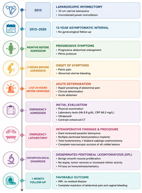
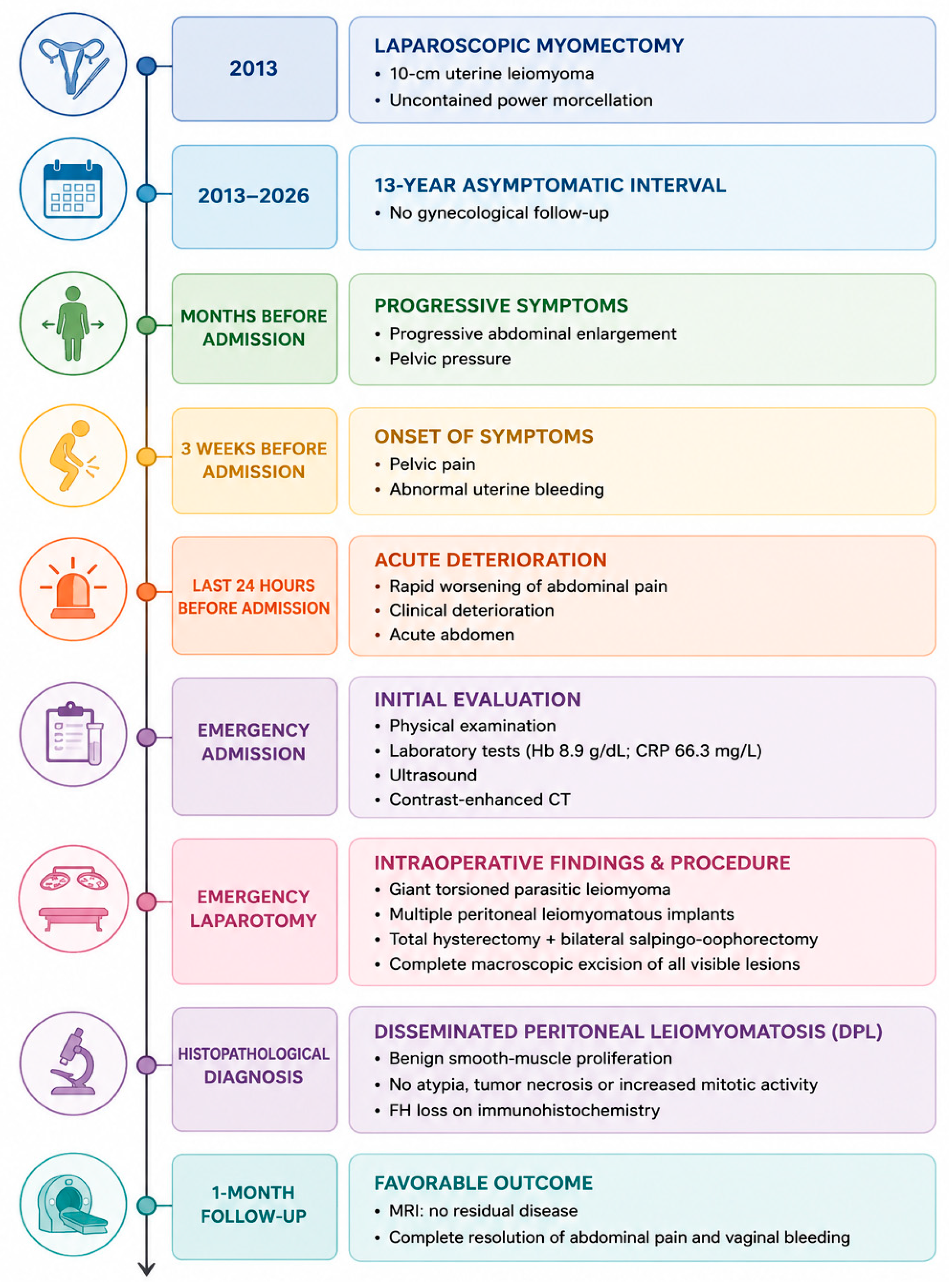
Her relevant medical history included laparoscopic myomectomy in 2013 for a symptomatic uterine leiomyoma measuring approximately 10 cm. Review of the available operative records confirmed that tissue extraction had been performed by uncontained morcellation without a retrieval bag. Her mother had a history of uterine leiomyomas requiring hysterectomy. The chronological sequence of the patient’s clinical course is summarized in Figure 1.
Figure 1.
Timeline summarizing the key clinical events, diagnostic investigations, surgical management, histopathological findings, and postoperative outcome of the present case of Leiomyomatosis peritonealis disseminated, diagnosed 13 years after laparoscopic myomectomy with uncontained power morcellation.
Although initially stable, the clinical picture changed rapidly during hospitalization. Abdominal pain intensified over the following hours, becoming diffuse and increasingly difficult to control with analgesic treatment. Progressive abdominal distension was observed, and repeat examination revealed marked tenderness together with signs of peritoneal irritation. At that stage, the clinical findings were highly suggestive of an evolving surgical abdomen.
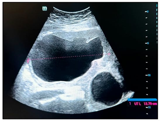
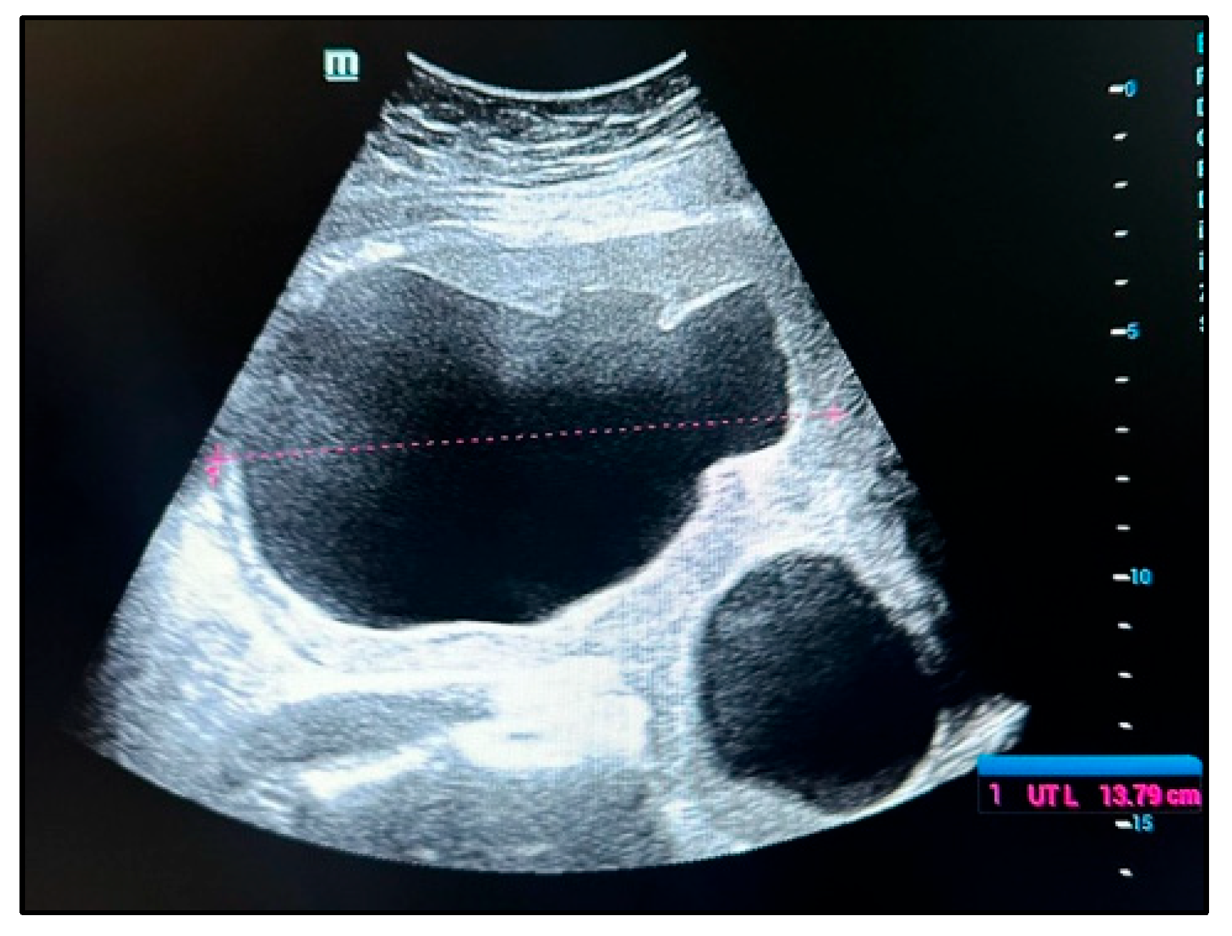
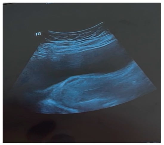
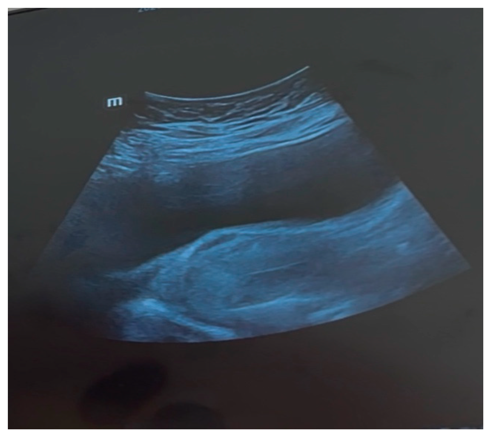
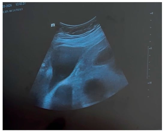
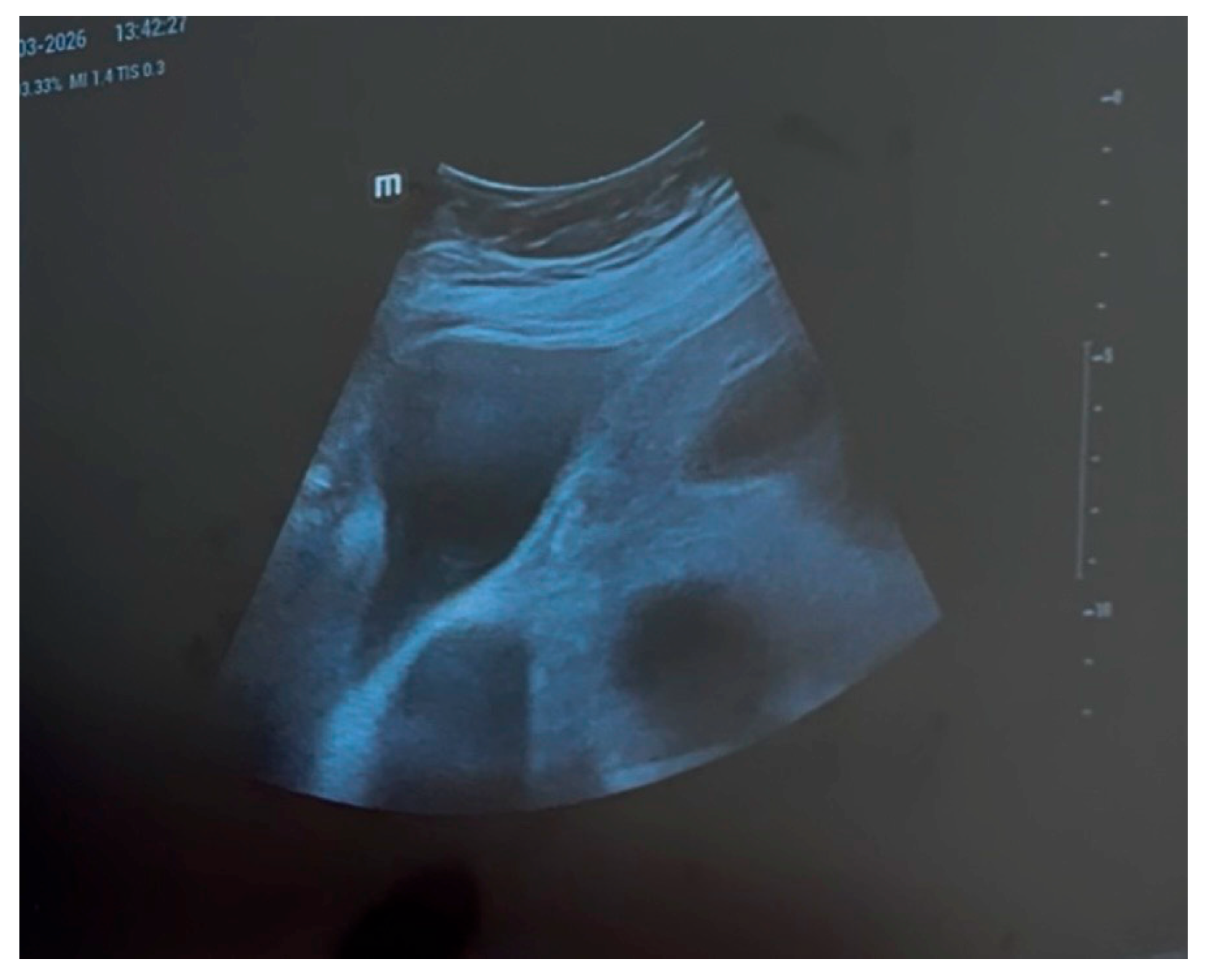
Transabdominal and transvaginal ultrasound revealed a large bilobed confluent pelvic mass measuring approximately 18 cm in maximum diameter. The lesion appeared predominantly hypoechoic, with a thick capsule, peripheral vascularization, and a central vascular pedicle, with a compressive mass effect on neighboring organs. Because of its considerable size and the displacement of adjacent pelvic structures, the precise site of origin could not be confidently established. The uterus appeared mildly enlarged and fibromatous. A simple cystic lesion measuring approximately 6 cm was identified within the left adnexa, while a small amount of free fluid was present in the pouch of Douglas (Figure 2, Figure 3 and Figure 4)
Figure 2.
Transabdominal ultrasound demonstrating a large cystic component bilobed abdominopelvic mass occupying the lower abdomen.
Figure 3.
Ultrasound image showing the urinary bladder and the uterus with inhomogeneous echogenicity of myometrium and linear endometrium.
Figure 4.
Additional ultrasonographic view of the complex pelvic mass and associated left adnexal cyst.
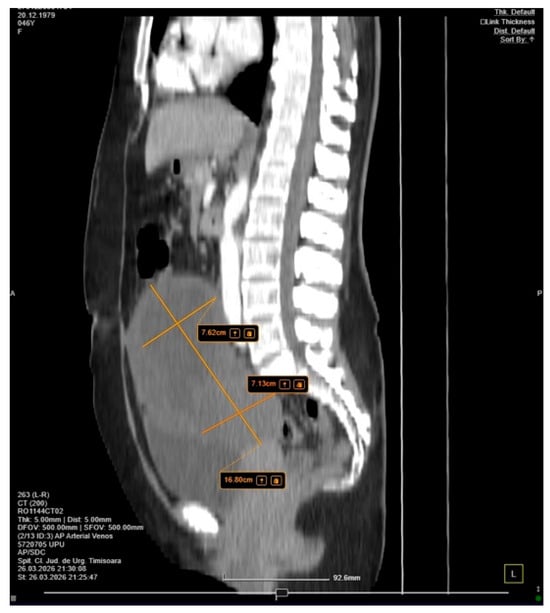
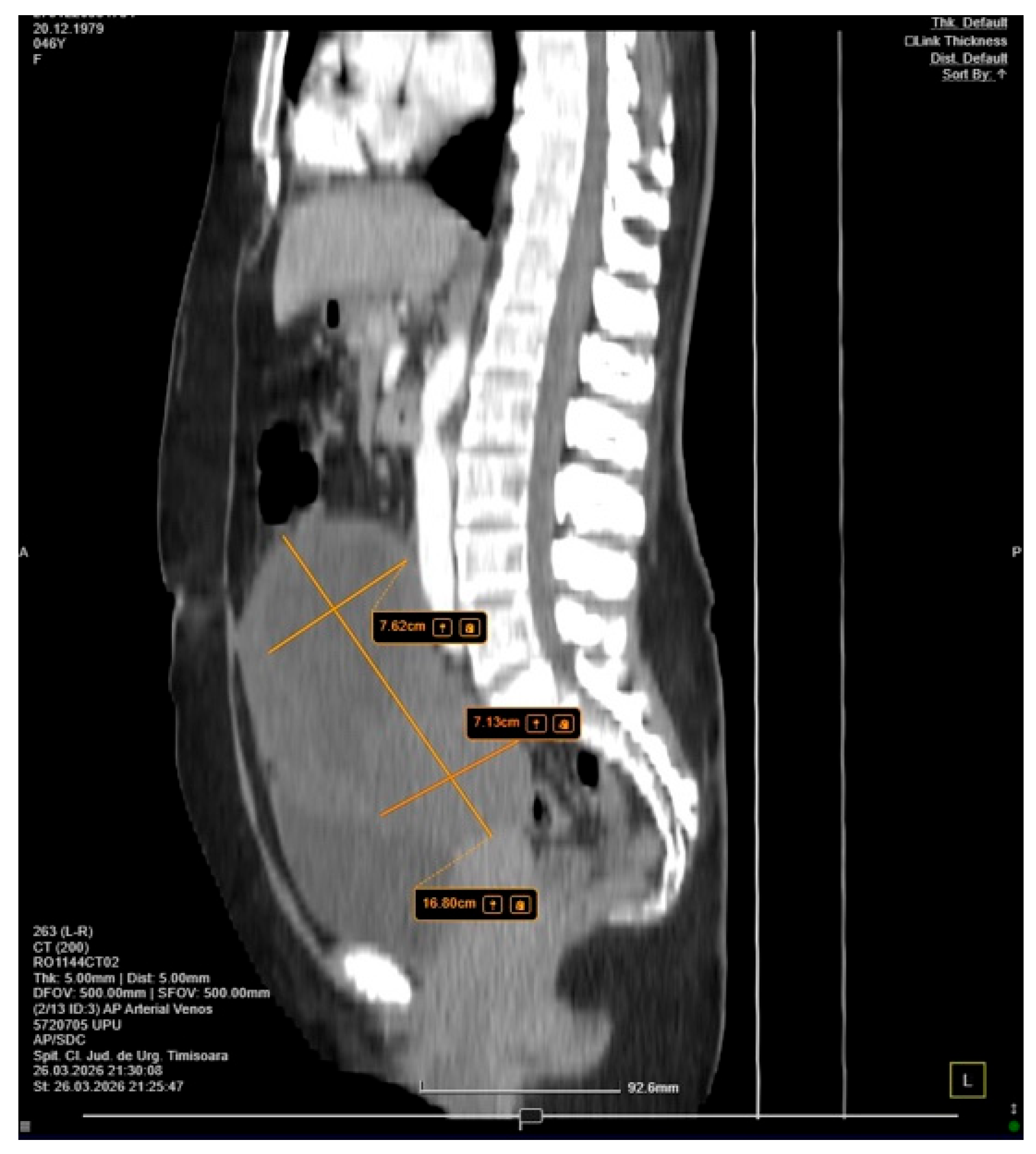
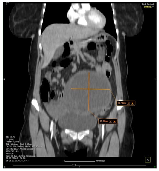
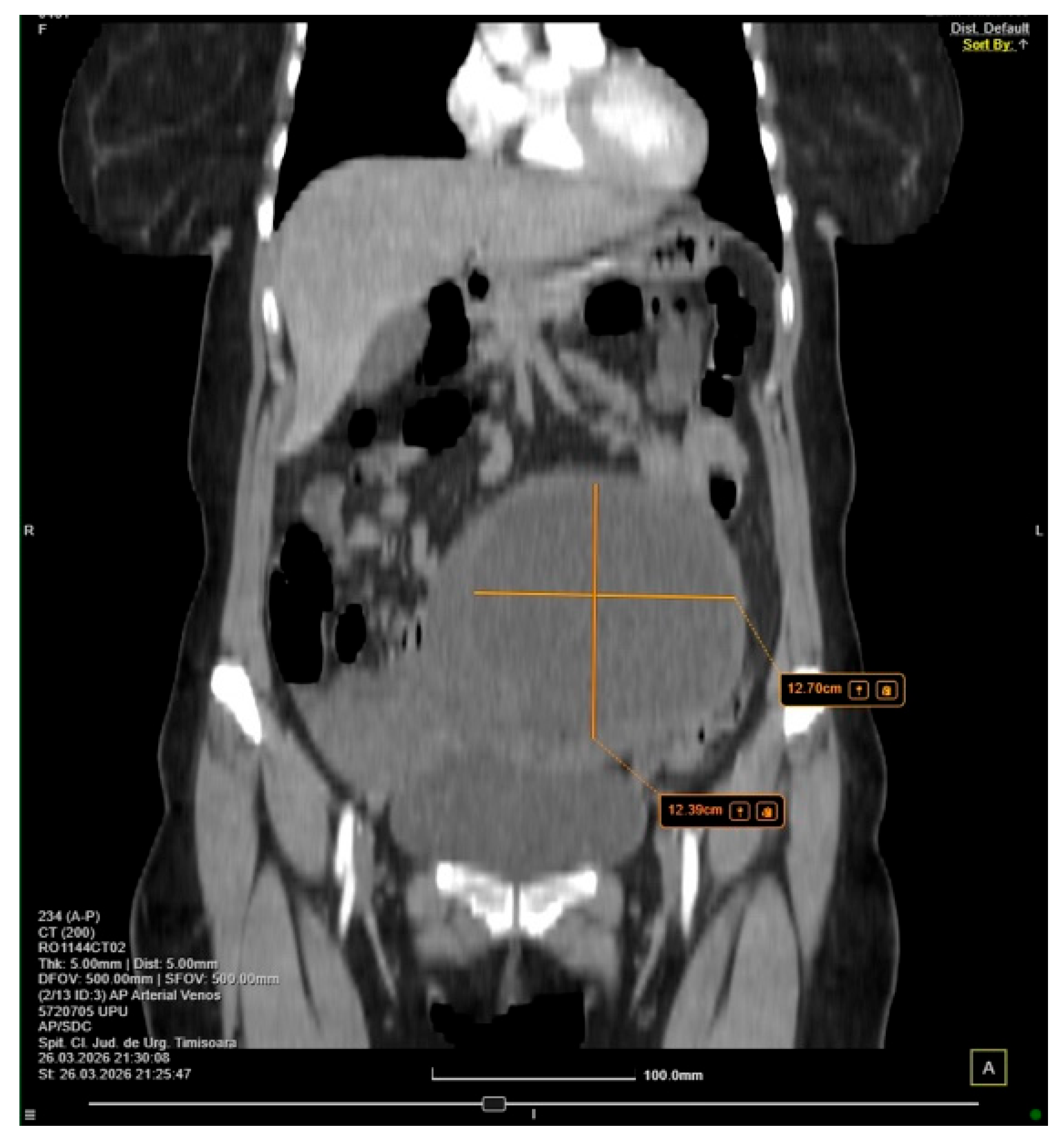
To further characterize the lesion, contrast-enhanced computed tomography of the abdomen and pelvis was performed. Imaging demonstrated a complex abdominopelvic mass measuring approximately 17 × 14 × 11 cm, containing both cystic and soft-tissue components with heterogeneous contrast enhancement. The lesion occupied much of the pelvic cavity and exerted a marked mass effect on surrounding structures. Additional findings included a simple left adnexal cyst and a small amount of intraperitoneal fluid. No evidence of active hemorrhage, pneumoperitoneum, or distant metastatic disease was identified (Figure 5 and Figure 6).
Figure 5.
Sagittal contrast-enhanced computed tomography image demonstrating the large abdominopelvic mass extending from the pelvis into the lower abdomen.
Figure 6.
Coronal contrast-enhanced computed tomography image showing the voluminous complex lesion with marked mass effect on adjacent pelvic structures.
The differential diagnosis included ovarian neoplasm, degenerating pedunculated leiomyoma, broad ligament tumor, peritoneal sarcoma, and disseminated peritoneal malignancy. Although pelvic magnetic resonance imaging was recommended for further characterization, the patient’s ongoing clinical deterioration and signs of peritoneal irritation prompted emergency exploratory laparotomy.
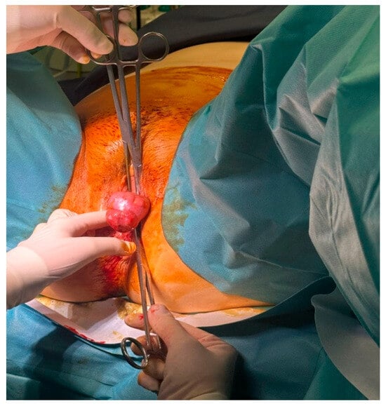
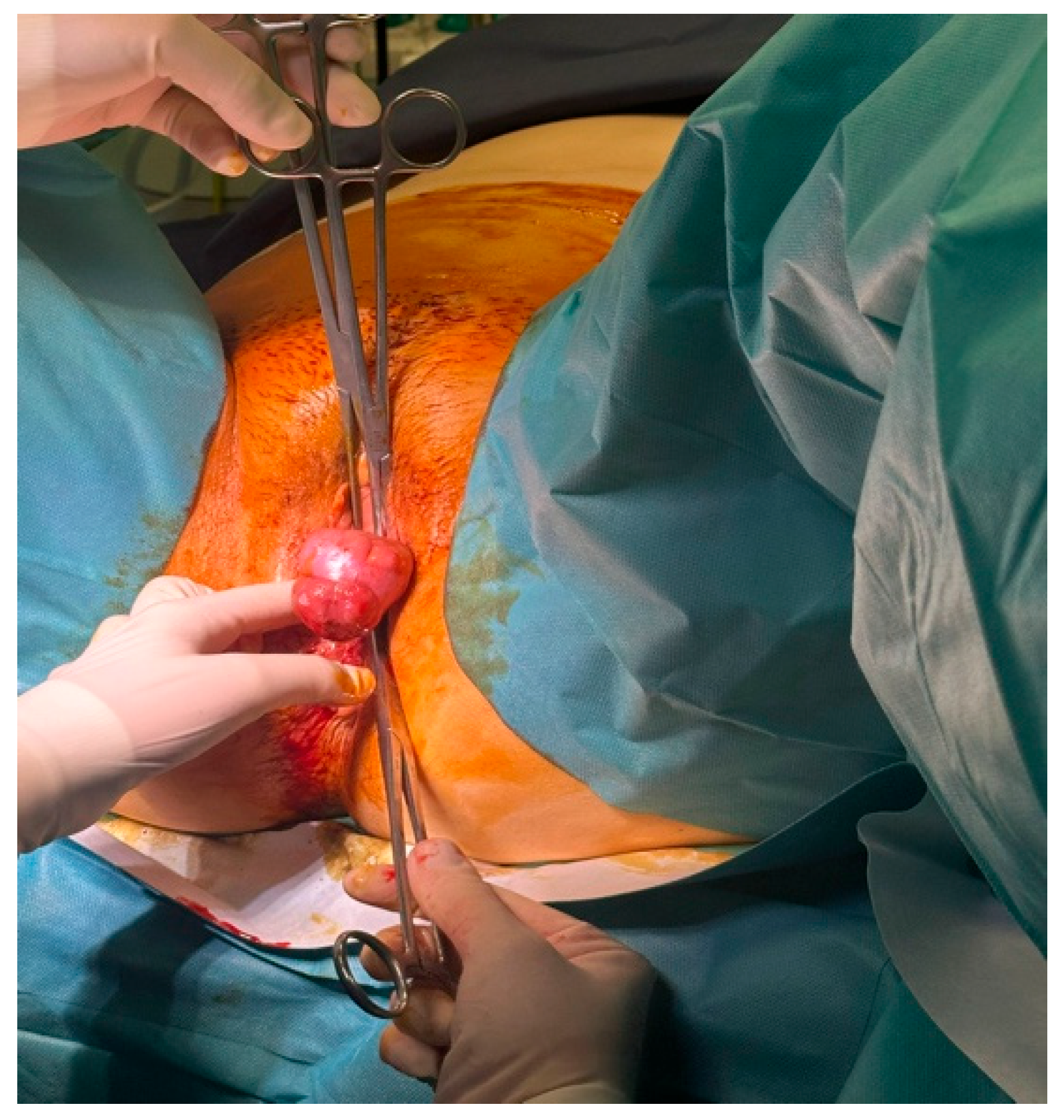
At the beginning of surgery, a hemorrhagic pedunculated leiomyomatous mass protruding through the cervical canal and vaginal introitus was identified and excised (Figure 7). Following median pubo-umbilical laparotomy, a small amount of serous ascitic fluid was encountered and collected for cytological analysis.
Figure 7.
Hemorrhagic pedunculated leiomyoma prolapsing through the cervical canal and vaginal introitus at the beginning of the surgical procedure.
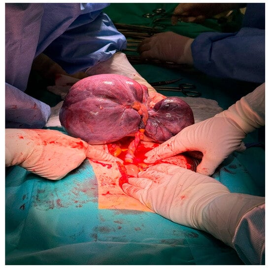
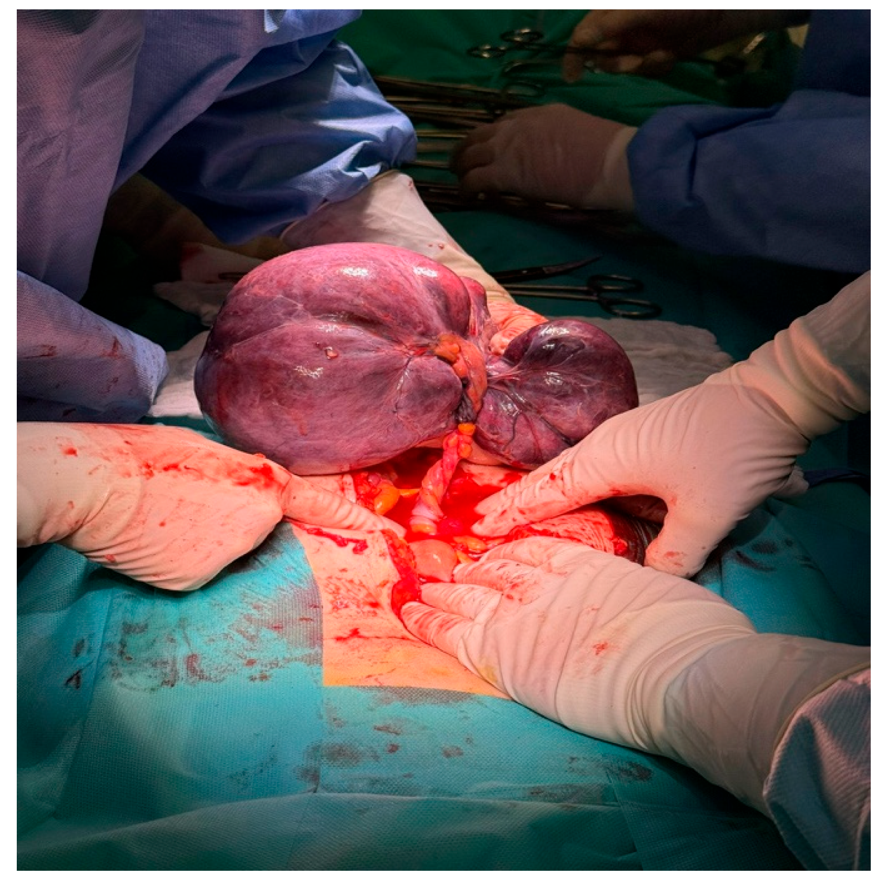
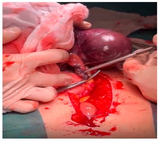
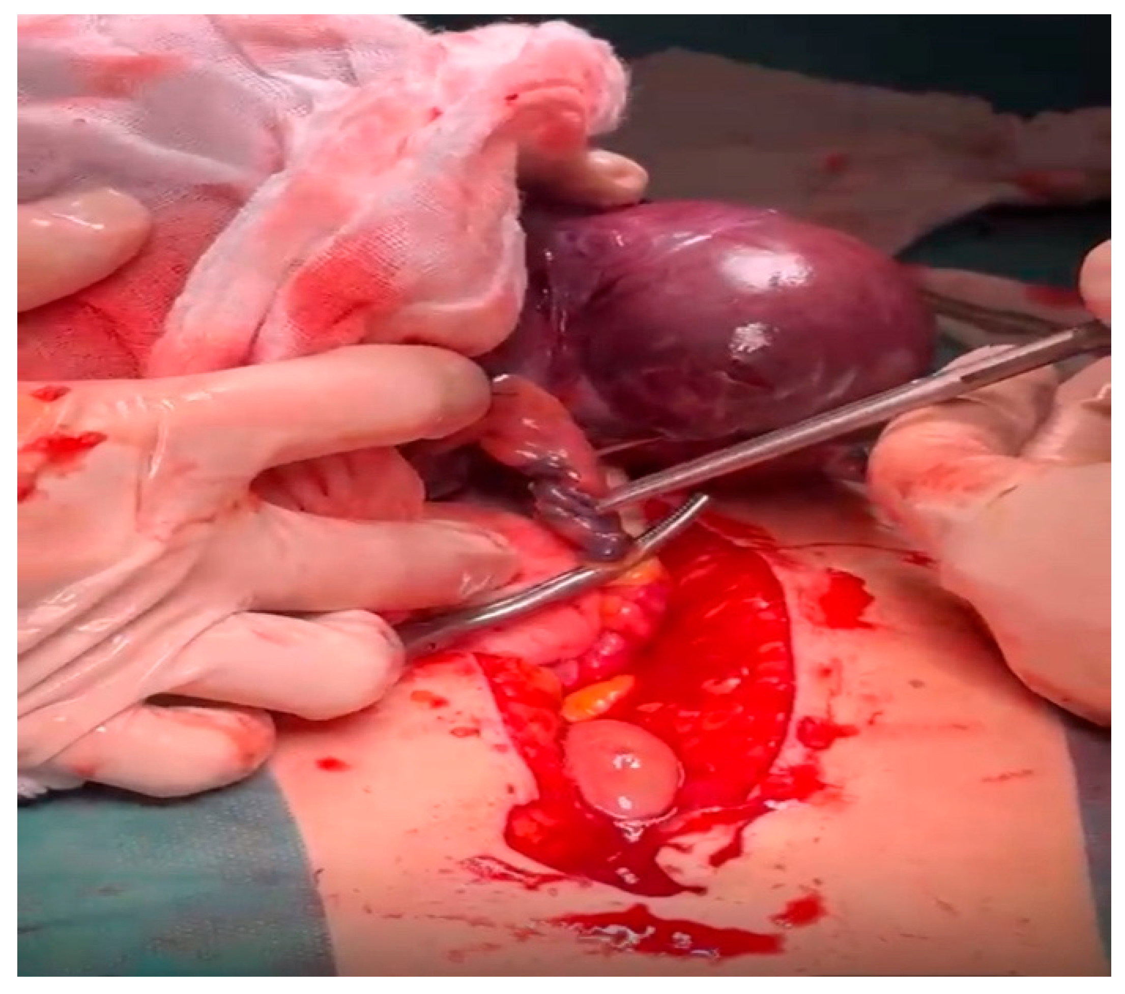
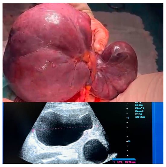
Exploration of the abdominal cavity revealed a large bilobed tumor measuring approximately 17 × 14 cm. The mass was dark purple in appearance, markedly congested, and attached by two vascular pedicles originating from the paracolic fat. Intraoperative findings were consistent with torsion and vascular compromise, explaining the patient’s abrupt onset of severe abdominal pain and acute clinical deterioration (Figure 8 and Figure 9).
Figure 8.
Intraoperative appearance of the giant bilobed parasitic leiomyoma following abdominal exploration.
Figure 9.
Torsioned vascular pedicle of the parasitic leiomyoma demonstrating marked congestion and vascular compromise.
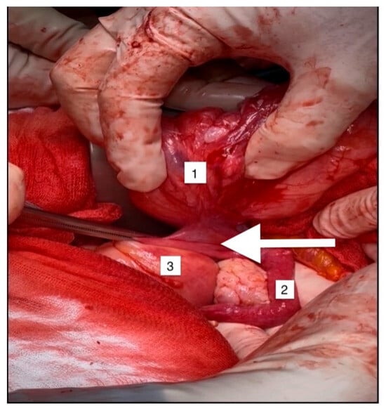
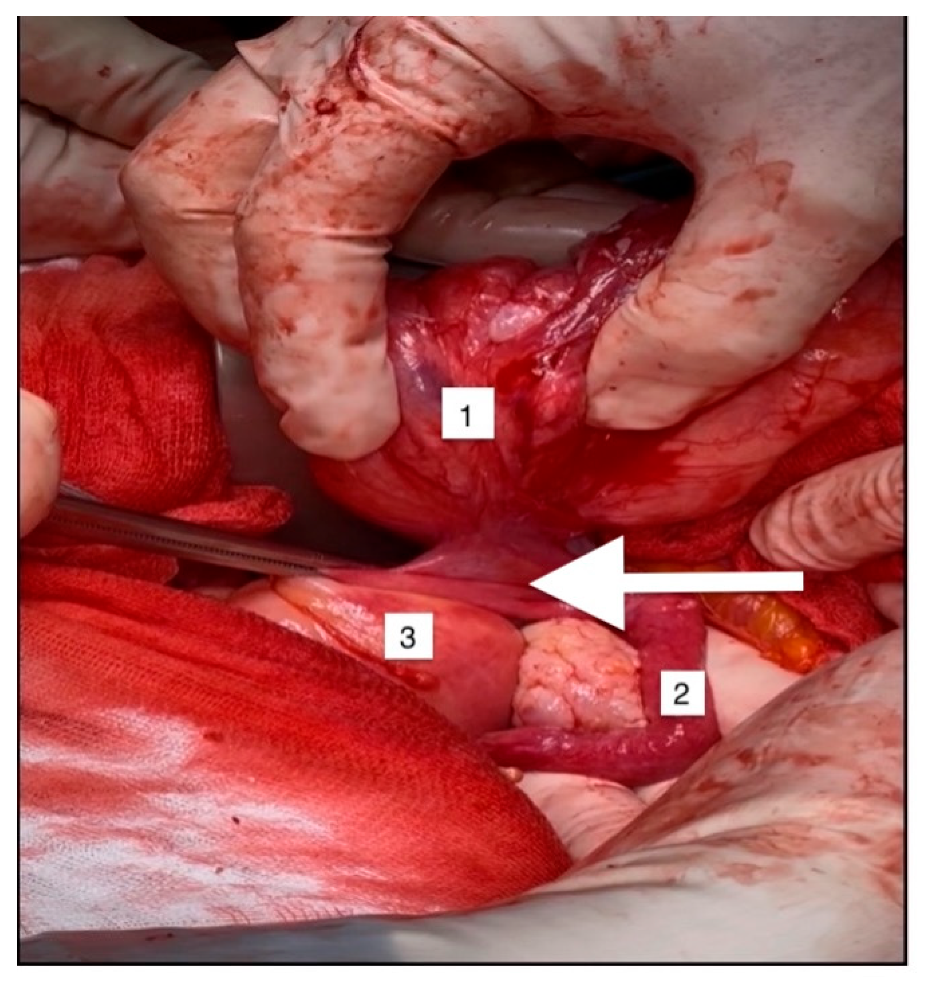
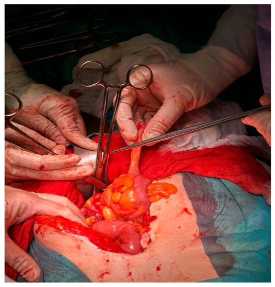
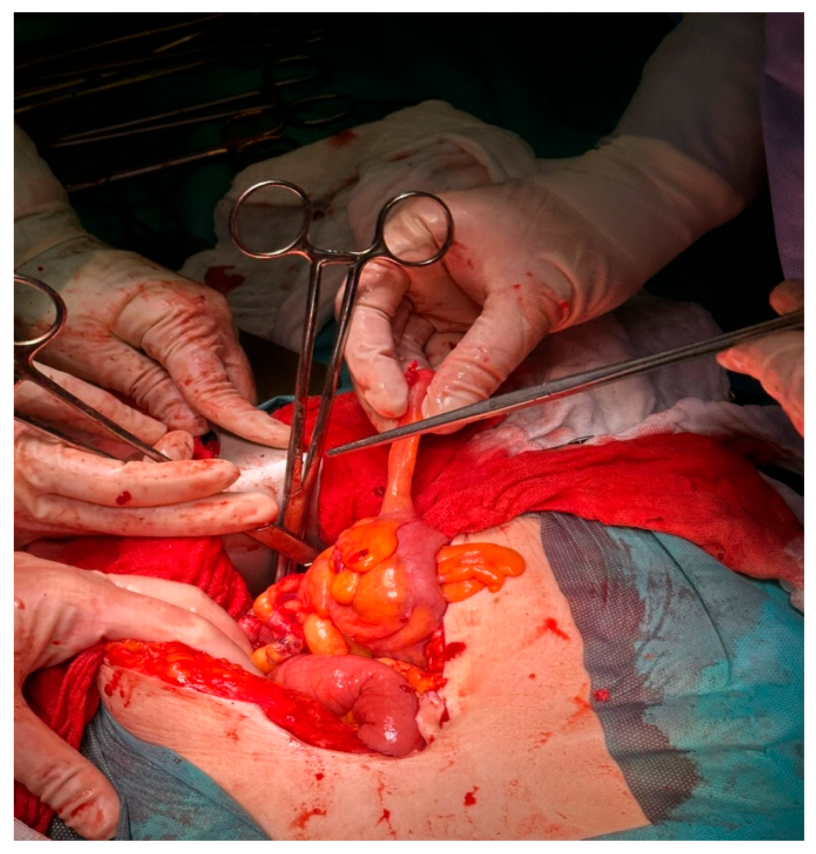
Further inspection demonstrated several additional smooth-muscle nodules scattered throughout the peritoneal cavity. A well-circumscribed lesion measuring approximately 4–5 cm was identified within the anterior leaf of the broad ligament (Figure 10, Figure 11 and Figure 12), while smaller nodules were present along the paracolic and paraintestinal peritoneal surfaces, including lesions arising from the colic fat (Figure 13). The uterus displayed a fibromatous appearance, and the left ovary was replaced by a cystic lesion. No macroscopic abnormalities were observed in the liver, omentum, or remaining abdominal organs.
Figure 10.
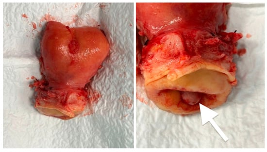
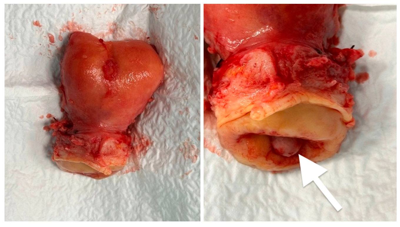
Surgical specimen following total hysterectomy. The uterus showed a fibromatous appearance without evidence of malignant transformation. Polypoid mass in the cervix after removing prolapsed vaginal fibroid (arrow).
Figure 11.
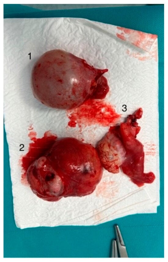
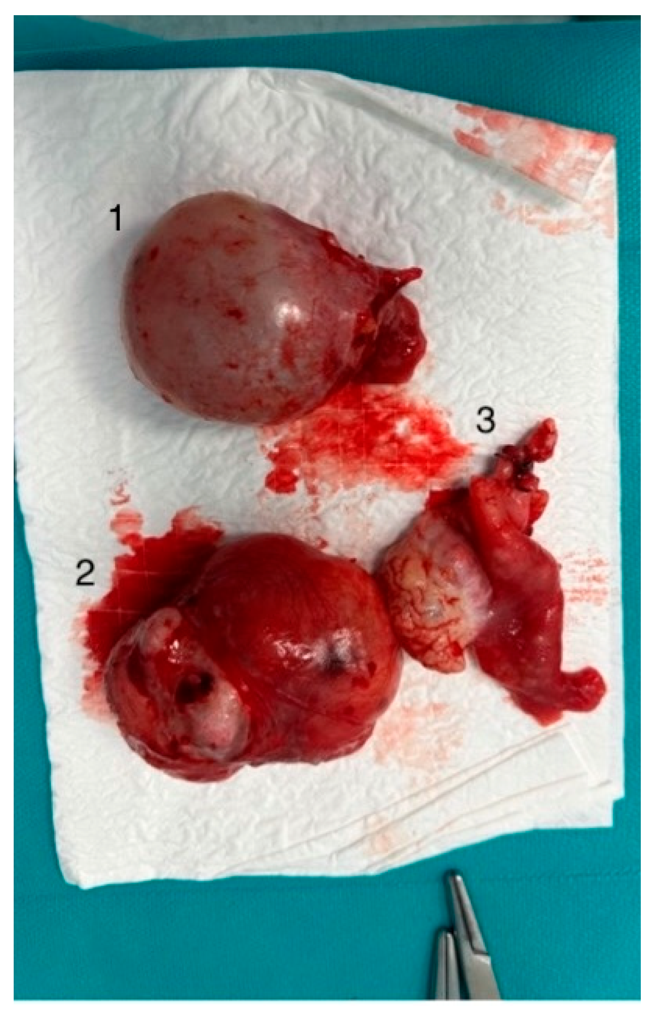
Gross appearance of the resected specimens. (1) Left adnexa containing a cystic ovarian lesion subsequently diagnosed as serous cystadenoma; (2) leiomyomatous implant arising from the anterior leaf of the broad ligament; (3) right adnexa removed during definitive surgical treatment.
Figure 12.
Intraoperative view demonstrating a leiomyomatous implant (arrow) arising from the anterior leaf of the broad ligament (1), located adjacent to the right adnexa (2) and the uterus (3). The lesion appeared macroscopically consistent with a benign smooth-muscle tumor and was completely excised during surgical exploration.
Figure 13.
Leiomyomatous implant arising from the paracolic fat identified during surgical exploration.
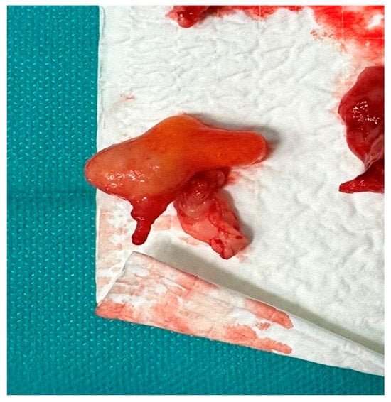
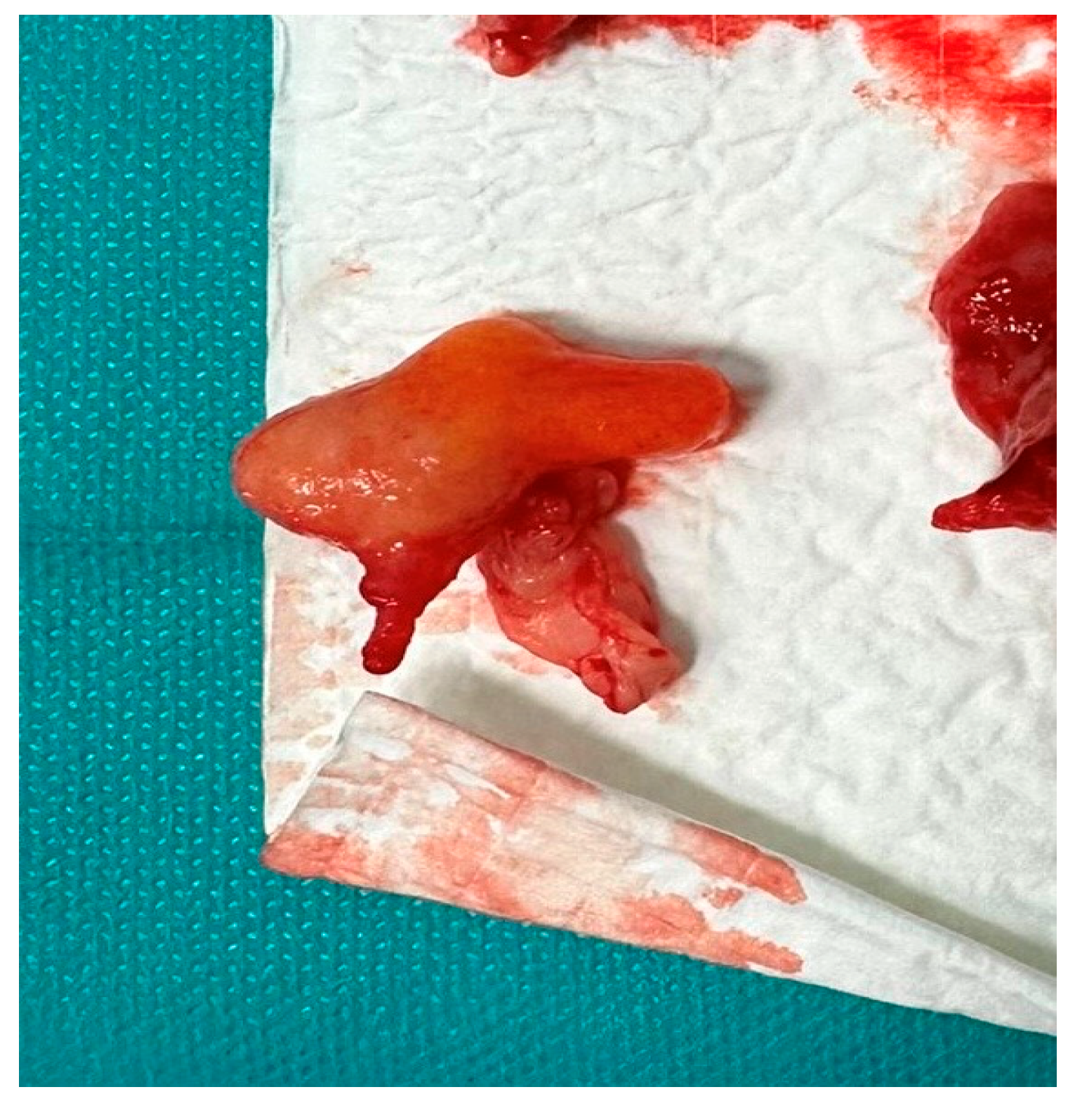
Considering the extent of the disease, the patient’s age, absence of reproductive plans, and the intraoperative findings, complete surgical treatment was performed. The procedure consisted of vaginal tumorectomy, excision of the torsioned abdominal mass, removal of all visible peritoneal nodules, total hysterectomy with bilateral salpingo-oophorectomy, and excision of the paracolic and paraintestinal lesions. Peritoneal implants adherent to the bowel were carefully dissected and removed with the assistance of a general surgeon. Complete macroscopic resection was achieved (Figure 10, Figure 11, Figure 12, Figure 13 and Figure 14).
Figure 14.
Resected paracolic leiomyomatous nodule following complete excision.
After surgery, the patient was monitored in the intensive care unit. Her hemoglobin decreased to 6.9 g/dL, and one unit of packed red blood cells was transfused. Her subsequent recovery was uneventful, and she was discharged in good clinical condition on postoperative day seven.
Histopathological examination demonstrated multiple benign smooth-muscle proliferations involving several anatomically distinct sites. The vaginal lesion corresponded to a conventional leiomyomatous nodule with degenerative changes and diffuse desmin positivity. Examination of the uterus revealed chronic papillary endocervicitis, an endocervical fibroglandular polyp, and small intramural leiomyomatous nodules. The right adnexa showed chronic nonspecific salpingitis associated with a follicular cyst and a hemorrhagic corpus luteum cyst, while the left ovary contained a serous cystadenoma lined by flattened epithelium.
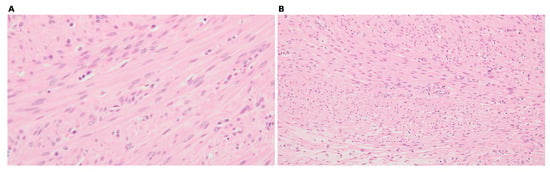
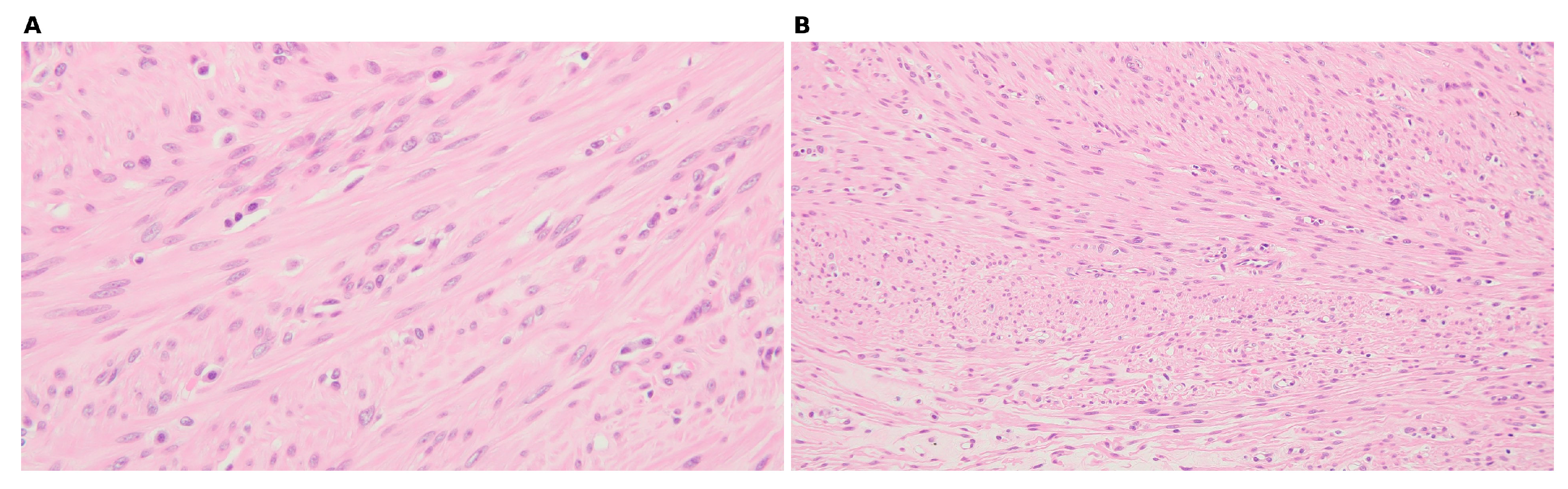
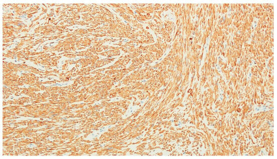
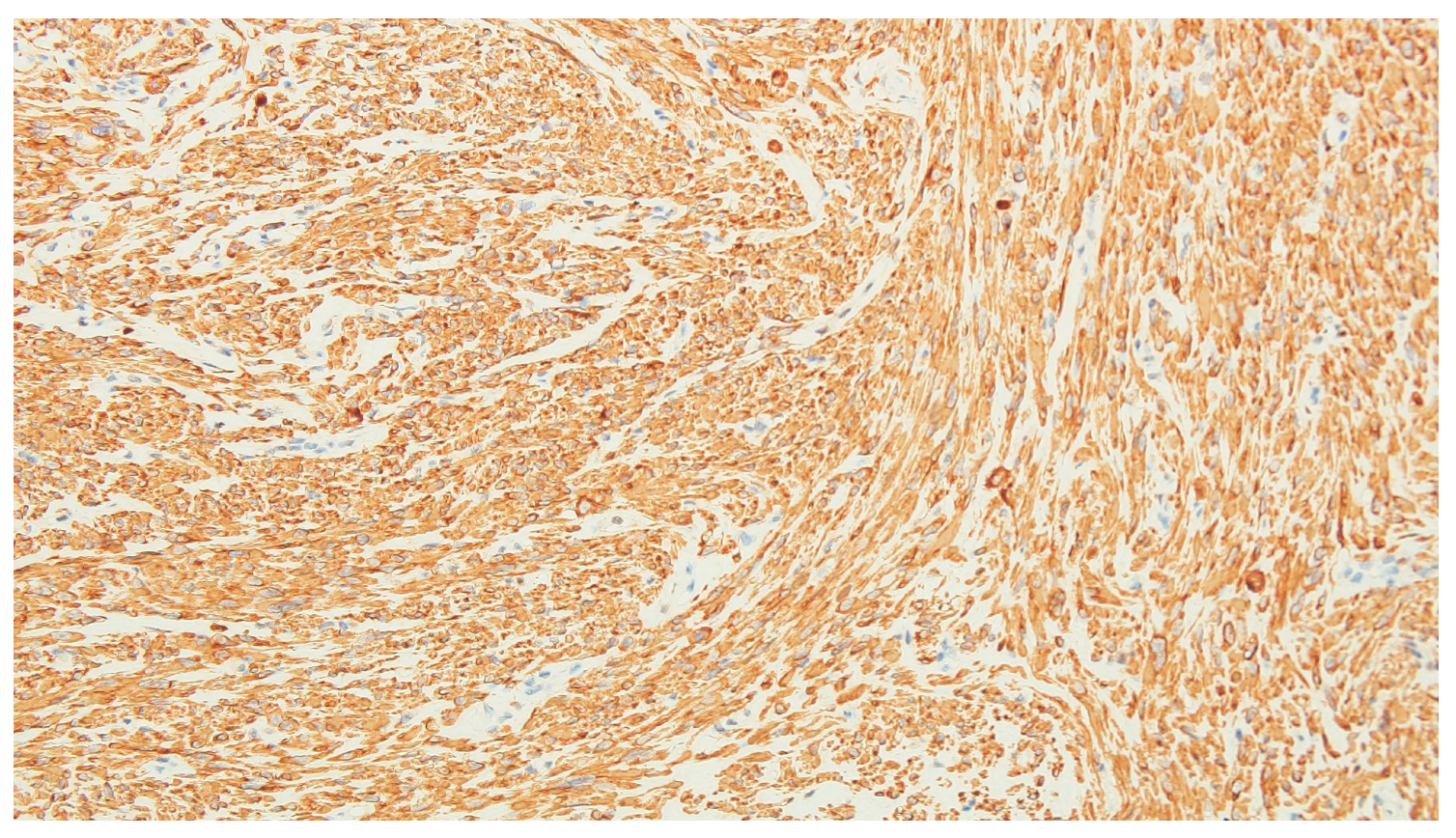
Particular attention was paid to the extrauterine lesions. Both the broad ligament tumor and the paracolic nodules displayed the morphological features of conventional leiomyomas, without cytological atypia, coagulative tumor necrosis, or increased mitotic activity. Immunohistochemical analysis confirmed smooth-muscle differentiation through diffuse desmin expression. Multifocal cystic degeneration was observed in the broad ligament lesion, whereas the paracolic implant showed areas of hyalinization. The overall histopathological appearance was entirely benign. Representative histopathological examination of the extrauterine leiomyomatous implants demonstrated benign smooth-muscle proliferation, without significant cytological atypia, coagulative tumor necrosis, or increased mitotic activity, supporting the diagnosis of disseminated peritoneal leiomyomatosis (Figure 15). Immunohistochemical examination demonstrated diffuse positivity for desmin in the neoplastic cells, confirming smooth-muscle differentiation and further supporting the leiomyomatous nature of the extrauterine lesions (Figure 16).
Figure 15.
Representative histopathological findings of a leiomyomatous lesion from the reported case. Hematoxylin and eosin (H&E) staining demonstrating a benign proliferation of spindle-shaped smooth-muscle cells arranged in intersecting fascicles, without significant cytological atypia or coagulative tumor necrosis. (A) Original magnification ×400; (B) original magnification ×200.
Figure 16.
Immunohistochemical staining of a leiomyomatous lesion from the reported case, demonstrating diffuse cytoplasmic positivity for desmin in the spindle-shaped tumor cells, supporting smooth-muscle differentiation. Anti-desmin antibody, DAB chromogen, hematoxylin counterstaining, original magnification ×400.
The large complex mass identified on ultrasound and computed tomography corresponded intraoperatively to the congested, torsed bilobed tumor attached by vascular pedicles to the paracolic fat. Histopathological examination identified the dominant parasitic leiomyoma as FH-deficient, whereas the separate broad ligament and paracolic implants showed conventional benign leiomyomatous morphology. The presence of these multiple peritoneal leiomyomatous nodules supported the diagnosis of LPD. Although the history of uncontained morcellation and the distribution of the lesions are compatible with peritoneal implantation, the origin of the individual nodules cannot be established from this case alone.
Immunohistochemical analysis of the main giant parasitic leiomyoma demonstrated loss of fumarate hydratase (FH) expression, supporting its classification as an FH-deficient leiomyoma. The broad ligament and paracolic implants were separately described as conventional leiomyomas; FH immunohistochemical findings were not reported for these lesions. Given the patient’s family history of uterine leiomyomas, genetic counseling was recommended because of the possibility of Hereditary Leiomyomatosis and Renal Cell Cancer (HLRCC) syndrome. Although postoperative renal imaging showed no abnormalities, the patient declined further genetic testing. Therefore, the hereditary or sporadic nature of the FH deficiency could not be determined. No histological images or immunohistochemical results from the 2013 myomectomy were available to the authors; therefore, a direct histopathological and immunohistochemical comparison with the current lesions could not be provided.
At one-month follow-up, pelvic magnetic resonance imaging showed no residual nodules or suspicious abdominopelvic lesions. The patient remained asymptomatic, with complete resolution of abdominal pain and vaginal bleeding.
For clarity, the principal clinical findings, diagnostic investigations, treatment, and outcome are summarized in Table 1.
Table 1.
Summary of the patient’s clinical presentation, diagnostic work-up, treatment, and outcome.
4. Discussion
This case illustrates the diagnostic complexity of LPD and its potential to present as an acute surgical emergency. The main distinguishing feature was the torsion of a giant parasitic leiomyoma. Although the diagnosis was made 13 years after laparoscopic myomectomy, the same interval has previously been reported [6].
4.1. Disease Latency and Possible Iatrogenic Dissemination
The interval between laparoscopic myomectomy and LPD diagnosis varies. Published reports describe a mean interval of approximately 6 years, with intervals ranging from 1 to 12 years; the 13-year interval in this case is therefore prolonged but not unprecedented [15,16]. The literature also describes varied anatomical distributions and clinical manifestations, with abdominal pain frequently reported [17,18,19]. The patient’s history of uncontained morcellation and the distribution of her lesions are compatible with peritoneal implantation. However, this temporal and anatomical association cannot establish causality in a single case. The relationship between the FH-deficient dominant leiomyoma and the separate peritoneal implants also remains uncertain.
The reported incidence of parasitic leiomyomas after laparoscopic morcellation is low [20]. Contained morcellation or intact specimen extraction may help reduce tissue dissemination when feasible [21]. These strategies should be considered alongside appropriate preoperative patient selection.
4.2. Clinical Presentation and Radiological–Pathological Correlation
LPD may be asymptomatic or present with abdominal or pelvic pain, pressure, distension, or symptoms related to mass effect, and it can mimic peritoneal malignancy [8]. Abdominopelvic pain was reported in 63.9% of patients in one large series [22]. In this case, ultrasound and computed tomography showed a large complex mass with solid and cystic components but did not establish its organ of origin. The differential diagnosis included ovarian neoplasm, a degenerating pedunculated leiomyoma, a broad ligament tumor, peritoneal sarcoma, and disseminated peritoneal malignancy. Imaging differentials also include peritoneal metastases, leiomyosarcoma, endometriosis, gastrointestinal tumors, intestinal tuberculosis, and peritoneal sarcomatosis [8,23].
At surgery, the dominant mass was congested and torsed, with vascular pedicles attached to the paracolic fat (Figure 17). Histopathology identified the dominant parasitic leiomyoma as FH-deficient; the separate broad ligament and paracolic implants showed conventional benign leiomyomatous morphology, without cytological atypia, coagulative tumor necrosis, or increased mitotic activity. Together, the distribution and histological findings supported LPD and helped distinguish the lesions from leiomyosarcoma. Rare malignant transformation has nevertheless been reported [24]. The prolapsed cervical leiomyoma accounted for the abnormal bleeding and anemia that prompted admission.
Figure 17.
Radiological–intraoperative correlation of the giant parasitic leiomyoma. Intraoperative appearance of the torsed, congested bilobed mass (top) and corresponding preoperative ultrasonographic image (bottom), illustrating the correlation between imaging findings and the surgically identified lesion.
4.3. FH Deficiency and Patient-Specific Consideration
FH loss in the dominant leiomyoma warrants consideration of hereditary leiomyomatosis and renal cell cancer (HLRCC), although FH-deficient leiomyomas may also arise sporadically [25]. HLRCC is associated with cutaneous and uterine leiomyomas and an increased risk of aggressive renal cell carcinoma [26,27]. Genetic counseling, consideration of germline FH testing, and appropriate renal surveillance are therefore relevant [28]. The patient declined germline testing, so the hereditary or sporadic nature of the FH deficiency remains unresolved.
The patient was nulligravid. Studies concerning pregnancy, parity, and uterine leiomyomas cannot be extrapolated to the pathogenesis of LPD; no etiological role for nulligravidity can be inferred in this case [29,30,31,32].
4.4. Follow-Up, Clinical Implications, and Limitations
LPD may recur after prolonged intervals and after apparently complete excision [8,33]. As standardized surveillance recommendations are lacking [34,35], follow-up should be individualized. The one-month MRI in this case represents an early postoperative assessment and cannot establish durable disease control or exclude subsequent recurrence. Contained morcellation or intact specimen extraction should be considered when feasible, with careful patient selection and avoidance of morcellation when malignancy is suspected [36,37].
This report provides a clinicopathological description of an acute LPD presentation caused by torsion of a giant parasitic leiomyoma. Its limitations include the single-case design, the inability to establish causality or determine the origin of the individual peritoneal implants, and the short follow-up period. Histological images and immunohistochemical results from the 2013 myomectomy were unavailable to the authors, preventing direct comparison with the current lesions. Longer-term follow-up and further studies are needed to better define disease recurrence and surveillance.
5. Conclusions
Leiomyomatosis peritonealis disseminata should be considered in the differential diagnosis of abdominopelvic masses in patients with a history of uterine morcellation, even many years after surgery. In this case, torsion of a giant parasitic leiomyoma caused an acute abdomen, and the diagnosis depended on integrating the clinical history with imaging, operative, and histopathological findings.
The association between previous uncontained morcellation and the peritoneal lesions is compatible with implantation but cannot be established as causal from a single case. The relationship between the coexisting FH-deficient leiomyoma and the implants also remains uncertain. Because no standardized postoperative surveillance protocol exists, follow-up should be individualized; this case does not establish a specific clinical or radiological schedule.
Author Contributions
Conceptualization, M.T., A.M.B. and S.-A.A.-A.; methodology, M.T., A.M.B., A.A.-A. and Z.L.P.; validation, M.T., A.M.B., S.-A.A.-A. and A.G.; formal analysis, A.A.-A. and S.-A.A.-A.; investigation, M.T., A.M.B., A.A.-A. and S.B.; resources, Z.L.P., S.B. and A.G.; data curation, M.T., A.M.B., A.A.-A. and S.-A.A.-A.; writing—original draft preparation, M.T., A.M.B. and A.A.-A.; writing—review and editing, S.-A.A.-A., Z.L.P., A.G., S.B., M.T. and A.M.B.; visualization, M.T., A.M.B. and A.A.-A.; supervision, S.-A.A.-A. and A.G. All authors have read and agreed to the published version of the manuscript.
Funding
We would like to acknowledge Victor Babes, University of Medicine and Pharmacy, Timisoara, for covering the costs of publishing this research paper.
Institutional Review Board Statement
The study was conducted in accordance with the Declaration of Helsinki and approved by the Local Research Ethics Committee of the “Pius Brînzeu” County Emergency Clinical Hospital, Timișoara, Romania (Approval No. 622/18 May 2026). The study was conducted retrospectively using anonymized data extracted from the hospital database, in compliance with applicable data protection regulations.
Informed Consent Statement
Written informed consent has been obtained from the patient to publish this paper.
Data Availability Statement
All relevant data supporting the findings of this study are included within the article. Additional patient-level data are not publicly available due to patient privacy and ethical restrictions. However, further information may be made available by the corresponding author upon reasonable request, provided that such disclosure complies with ethical requirements and does not compromise patient confidentiality.
Acknowledgments
During the preparation of this manuscript, the authors used Grammarly v1.140.1.0 for proofreading and language editing. The scientific content of the manuscript was entirely written by the authors. The authors reviewed and edited the output where applicable and take full responsibility for the content of this publication.
Conflicts of Interest
The authors declare no conflicts of interest.
References
- Munro, M.G.; Tchaikovski, S.N.; Murji, A. The Epidemiology and Pathogenesis of Uterine Fibroids. Int. J. Gynaecol. Obstet. 2025, 171, 1029–1045. [Google Scholar] [CrossRef] [Scilit] [PubMed]
- Yang, L.; Lv, C. Leiomyomas beyond the Uterus Presenting with Multiple Growth Patterns in a Single Patient. BMC Womens Health 2025, 25, 525. [Google Scholar] [CrossRef] [Scilit] [PubMed]
- Liu, X.; Hu, Y.; Chen, L.; Zhou, Q. Disseminated Peritoneal Leiomyomatosis: A Case Report and Review of the Literature. J. Int. Med. Res. 2021, 49, 3000605211033194. [Google Scholar] [CrossRef] [Scilit] [PubMed]
- Lee, M.; Morey, G.; Lopez, H.; Bass, R.; Ferra, S. Disseminated Peritoneal Leiomyomatosis Mimicking Carcinomatosis: A Case Report. Int. J. Surg. Case Rep. 2024, 120, 109908. [Google Scholar] [CrossRef] [Scilit] [PubMed]
- Chirayath, M.; Titus, T.T.; Sebastian, L.M.; Cicilet, S. A Rare Case of Disseminated Peritoneal Leiomyomatosis: A Case Report. Indian J. Radiol. Imaging 2024, 34, 765–769. [Google Scholar] [CrossRef] [Scilit] [PubMed]
- Huang, S.F.; Wen, C.Y.; Liao, C.I.; Lin, J.C.; Tsai, C.C. Leiomyomatosis Peritonealis Disseminata Mimicking Peritoneal Carcinomatosis 13 Years after Laparoscopic Uterine Myomectomy: A Case Report. Int. J. Surg. Case Rep. 2021, 81, 105745. [Google Scholar] [CrossRef] [Scilit] [PubMed]
- Yuri, T.; Kinoshita, Y.; Yuki, M.; Yoshizawa, K.; Emoto, Y.; Tsubura, A. Leiomyomatosis Peritonealis Disseminata Positive for Progesterone Receptor. Am. J. Case Rep. 2015, 16, 300–304. [Google Scholar] [CrossRef] [Scilit] [PubMed]
- Bucuri, C.E.; Ciortea, R.; Malutan, A.M.; Oprea, V.; Toma, M.; Roman, M.P.; Ormindean, C.M.; Nati, I.; Suciu, V.; Simon-Dudea, M.; et al. Disseminated Peritoneal Leiomyomatosis—A Challenging Diagnosis Mimicking Malignancy: Scoping Review of the Last 14 Years. Biomedicines 2024, 12, 1749. [Google Scholar] [CrossRef] [Scilit] [PubMed]
- Yadati, R.; Katamreddy, B.R.; Noori, M.; Ahmed, H.; Reddy, P. Port Site Leiomyoma and Leiomyomatosis Peritonealis Disseminata Following Uncontained Uterine Power Morcellation: A Case Report. J. Obstet. Gynaecol. India 2023, 73, 183–188. [Google Scholar] [CrossRef] [Scilit] [PubMed]
- Lu, B.; Xu, J.; Pan, Z. Iatrogenic Parasitic Leiomyoma and Leiomyomatosis Peritonealis Disseminata Following Uterine Morcellation. J. Obstet. Gynaecol. Res. 2016, 42, 990–999. [Google Scholar] [CrossRef] [Scilit] [PubMed]
- Gebreslase, M.N.; Reta, B.K.; Girmay, H.G.; Zeray, M.A.; Gebremariam, A.S. Leiomyomatosis Peritonealis Disseminata Presenting as Acute Abdomen: A Rare Case Report. Int. J. Surg. Case Rep. 2025, 134, 111711. [Google Scholar] [CrossRef] [Scilit] [PubMed]
- Watanabe, K.; Koyanagi, T.; Takahashi, Y.; Taneichi, A.; Takei, Y.; Fujiwara, H. Disseminated Peritoneal Leiomyomatosis Incidentally Diagnosed during Surgery for Torsion of a Subserosal Uterine Fibroid: A Case Report. J. Med. Case Rep. 2026, 20, 118. [Google Scholar] [CrossRef] [Scilit] [PubMed]
- Ye, Z.; Chen, L. Leiomyomatosis Peritonealis Disseminata with Low-Grade Malignant Change: A Case Report. Medicine 2022, 101, e30528. [Google Scholar] [CrossRef] [Scilit] [PubMed]
- Li, J.; Zhuge, Y.Y.; Lin, K.Q. Torsed Retroperitoneal Leiomyomas: A Case Report and Review of Literature. World J. Clin. Cases 2024, 12, 4762–4769. [Google Scholar] [CrossRef] [Scilit] [PubMed]
- Chen, X.; Liu, H.; Shi, H.; Fan, Q.; Sun, D.; Lang, J. Leiomyomatosis Peritonealis Disseminata Following Laparoscopic Surgery with Uncontained Morcellation: 13 Cases from One Institution. Front. Surg. 2021, 8, 788749. [Google Scholar] [CrossRef] [Scilit] [PubMed]
- Sekulic, M.; Moench, L.; Movahedi-Lankarani, S. Disseminated Peritoneal Leiomyomatosis Postmorcellated Resection of Uterine Leiomyomatous Tissue. APMIS 2016, 124, 1063–1071. [Google Scholar] [CrossRef] [Scilit] [PubMed]
- Milad, M.; Milad, E. Laparoscopic Morcellator-Related Complications. J. Minim. Invasive Gynecol. 2014, 21, 486–491. [Google Scholar] [CrossRef] [Scilit] [PubMed]
- Leren, V.; Langebrekke, A.; Qvigstad, E. Parasitic Leiomyomas after Laparoscopic Surgery with Morcellation. Acta Obstet. Gynecol. Scand. 2012, 91, 1233–1236. [Google Scholar] [CrossRef] [Scilit] [PubMed]
- Morgan, E.D.; Kahiye, M.; Kule, I.; Yahaya, J.J.; Othieno, E. Disseminated Peritoneal Leiomyomatosis as an Incidental Finding: A Case Report. Clin. Case Rep. 2022, 10, e05541. [Google Scholar] [CrossRef] [Scilit] [PubMed]
- van der Meulen, J.F.; Pijnenborg, J.M.A.; Boomsma, C.M.; Verberg, M.F.G.; Geomini, P.M.; Bongers, M.Y. Parasitic Myoma after Laparoscopic Morcellation: A Systematic Review of the Literature. BJOG 2016, 123, 69–75. [Google Scholar] [CrossRef] [Scilit] [PubMed]
- Marchand, G.; Masoud, A.; Christensen, A.; Ruther, S.; Brazil, G.; King, A. Contained and Uncontained Morcellation in Hysterectomy and Myomectomy: A Systematic Review and Meta-Analysis. Turk. J. Obstet. Gynecol. 2021, 18, 311–321. [Google Scholar] [CrossRef] [Scilit] [PubMed]
- Di Giuseppe, J.; Natalini, L.; Bordini, M.; Crescenzi, D.; Grementieri, L.; Petrucci, J.; Asaro, A.; Giannella, L.; Delli Carpini, G.; Grelloni, C.; et al. Diagnosis, Treatment and Clinical Outcomes of Extrauterine Sites of Leiomyomatosis: A Systematic Review. Ann. Med. 2025, 57, 2546681. [Google Scholar] [CrossRef] [Scilit] [PubMed]
- Demir, M.K. Disseminated Peritoneal Leiomyomatosis: Magnetic Resonance Imaging and Differential Diagnosis. Australas. Radiol. 2007, 51, B268–B271. [Google Scholar] [CrossRef] [Scilit] [PubMed]
- Psathas, G.; Zarokosta, M.; Zoulamoglou, M.; Chrysikos, D.; Thivaios, I.; Kaklamanos, I.; Birbas, K.; Mariolis-Sapsakos, T. Leiomyomatosis Peritonealis Disseminata: A Case Report and Meticulous Review of the Literature. Int. J. Surg. Case Rep. 2017, 40, 105–108. [Google Scholar] [CrossRef] [Scilit] [PubMed]
- Harrison, W.J.; Andrici, J.; Maclean, F.; Madadi-Ghahan, R.; Farzin, M.; Sioson, L.; Toon, C.W.; Clarkson, A.; Watson, N.; Pickett, J.; et al. Fumarate Hydratase-Deficient Uterine Leiomyomas Occur in Both the Syndromic and Sporadic Settings. Am. J. Surg. Pathol. 2016, 40, 599–607. [Google Scholar] [CrossRef] [Scilit] [PubMed]
- Menko, F.H.; Maher, E.R.; Schmidt, L.S.; Middelton, L.A.; Aittomäki, K.; Tomlinson, I.; Richard, S.; Linehan, W.M. Hereditary Leiomyomatosis and Renal Cell Cancer (HLRCC): Renal Cancer Risk, Surveillance and Treatment. Fam. Cancer 2014, 13, 637–644. [Google Scholar] [CrossRef] [Scilit] [PubMed]
- Schmidt, L.S.; Linehan, W.M. Hereditary Leiomyomatosis and Renal Cell Carcinoma. Int. J. Nephrol. Renov. Dis. 2014, 7, 253–260. [Google Scholar] [CrossRef] [Scilit] [PubMed]
- Alkhrait, S.; Ali, M.; Kertowidjojo, E.; Romero, I.L.; Hathaway, F.; Madueke-Laveaux, O.S. Investigating Fumarate Hydratase-Deficient Uterine Fibroids: A Case Series. J. Clin. Med. 2023, 12, 5436. [Google Scholar] [CrossRef] [Scilit] [PubMed]
- Lisiecki, M.; Paszkowski, M.; Woźniak, S. Fertility Impairment Associated with Uterine Fibroids: A Review of Literature. Prz. Menopauzalny 2017, 16, 137–140. [Google Scholar] [CrossRef] [Scilit] [PubMed]
- Laughlin, S.K.; Hartmann, K.E.; Baird, D.D. Postpartum Factors and Natural Fibroid Regression. Am. J. Obstet. Gynecol. 2011, 204, 496.e1–496.e6. [Google Scholar] [CrossRef] [Scilit] [PubMed]
- Carpini, G.D.; Morini, S.; Papiccio, M.; Serri, M.; Damiani, V.; Grelloni, C.; Clemente, N.; Ciavattini, A. The Association between Childbirth, Breastfeeding, and Uterine Fibroids: An Observational Study. Sci. Rep. 2019, 9, 10117. [Google Scholar] [CrossRef] [Scilit] [PubMed]
- Ploumaki, I.; Macri, V.I.; Segars, J.H.; Islam, M.S. Progesterone Signaling in Uterine Fibroids: Molecular Mechanisms and Therapeutic Opportunities. Life Sci. 2025, 362, 123345. [Google Scholar] [CrossRef] [Scilit] [PubMed]
- Ferrario, L.; Zerbi, P.; Angiolini, M.R.; Agarossi, A.; Riggio, E.; Bondurri, A.; Danelli, P. Leiomyomatosis Peritonealis Disseminata: A Case Report of Recurrent Presentation and Literature Review. Int. J. Surg. Case Rep. 2018, 49, 25–29. [Google Scholar] [CrossRef] [Scilit] [PubMed]
- Tan, H.L.; Koh, Y.X.; Chew, M.H.; Wang, J.; Lim, J.S.K.; Leow, W.Q.; Lee, S.Y. Disseminated Peritoneal Leiomyomatosis: A Devastating Sequelae of Unconfined Laparoscopic Morcellation. Singap. Med. J. 2019, 60, 652–654. [Google Scholar] [CrossRef] [Scilit] [PubMed]
- Vimercati, A.; Santarsiero, C.M.; Esposito, A.; Putino, C.; Malvasi, A.; Damiani, G.R.; Laganà, A.S.; Vitagliano, A.; Marinaccio, M.; Resta, L.; et al. An Extremely Rare Case of Disseminated Peritoneal Leiomyomatosis with a Pelvic Leiomyosarcoma and Omental Metastasis after Laparoscopic Morcellation: Systematic Review of the Literature. Diagnostics 2022, 12, 3219. [Google Scholar] [CrossRef] [Scilit] [PubMed]
- Brölmann, H.; Tanos, V.; Grimbizis, G.; Ind, T.; Philips, K.; Bosch, T.v.D.; Sawalhe, S.; Haak, L.v.D.; Jansen, F.-W.; Pijnenborg, J.; et al. Options on Fibroid Morcellation: A Literature Review. Gynecol. Surg. 2015, 12, 3–15. [Google Scholar] [CrossRef] [Scilit] [PubMed]
- American College of Obstetricians and Gynecologists. Uterine Morcellation for Presumed Leiomyomas: ACOG Committee Opinion No. 822. Obstet. Gynecol. 2021, 137, e63–e74. [Google Scholar] [CrossRef] [Scilit] [PubMed]

Disclaimer/Publisher’s Note: The statements, opinions and data contained in all publications are solely those of the individual author(s) and contributor(s) and not of MDPI and/or the editor(s). MDPI and/or the editor(s) disclaim responsibility for any injury to people or property resulting from any ideas, methods, instructions or products referred to in the content. |
© 2026 by the authors. Licensee MDPI, Basel, Switzerland. This article is an open access article distributed under the terms and conditions of the Creative Commons Attribution (CC BY) license.