


Abstract
Background/Objectives: Penile lichen sclerosus (PLS) is a chronic inflammatory dermatosis characterized by progressive sclerosis and fibrosis of the penile glans and foreskin. Histopathological examination remains the reference standard for diagnosis, whereas reliable biomarkers reflecting disease activity and progression remain limited. Nuclear factor kappa B (NF-κB) p65 (RELA), a key regulator of inflammatory and immune signaling, may provide insights into inflammatory processes associated with PLS. This study aimed to evaluate NF-κB p65 expression across different histopathological stages of PLS and explore its potential relevance to histopathological disease staging. Methods: Foreskin specimens obtained during circumcision from 39 patients with histologically confirmed PLS were classified as early, moderate, or severe disease. Each specimen was divided into potentially affected (PA) and potentially unaltered (PH) regions based on macroscopic appearance, with 18 normal foreskin specimens included as controls. All samples underwent hematoxylin and eosin (H&E) staining and immunohistochemical staining for NF-κB p65. NF-κB p65 immunoreactivity was evaluated semi-quantitatively using the immunoreactive score (IRS), while its subcellular localization was assessed descriptively. Results: Histopathological assessment showed less prominent inflammatory infiltrates in advanced stages of PLS, accompanied by more pronounced fibrotic changes. Descriptive analysis of NF-κB p65 immunohistochemical staining showed a trend toward lower immunoreactivity and less frequent nuclear localization in advanced lesions. However, differences in IRS between disease stages, PA and PH regions, and PLS and control tissues did not reach statistical significance. Histopathological examination also confirmed PLS involvement in all PH regions, indicating that macroscopically unaltered peripheral tissue was not histologically unaffected. Conclusions: The observed changes in NF-κB p65 immunoreactivity and subcellular localization may reflect differences in inflammatory processes across PLS stages; however, the present findings do not establish progressive attenuation of canonical NF-κB signaling. Although NF-κB p65 cannot currently be considered an independent diagnostic or staging marker, its immunohistochemical expression pattern warrants further investigation as a potential complementary marker in the histopathological assessment of PLS. Larger patient cohorts, particularly including more cases of severe disease, and further studies involving quantitative assessment of nuclear localization and NF-κB pathway activity are needed to determine its potential diagnostic relevance. The detection of histopathological involvement in macroscopically unaltered peripheral tissue also highlights the importance of histopathological examination beyond visibly affected areas.
1. Introduction
Penile lichen sclerosus (PLS), also referred to as balanitis xerotica obliterans, is a chronic inflammatory dermatosis characterized by progressive fibrosis and sclerosis of the genital skin [1]. Clinically, PLS may remain asymptomatic or present with white sclerotic plaques, induration, and thickening of the foreskin and glans penis [2]. Progressive fibrosis may ultimately lead to significant functional impairment and reduced quality of life. The main complications of PLS include phimosis, urethral strictures, urinary retention, and, in advanced cases, renal impairment and substantial sexual and urinary dysfunction [2,3].
Reported prevalence rates of PLS among circumcised males vary considerably across studies, ranging from 1% to 67.4%, largely due to differences in study populations, diagnostic criteria, and histopathological confirmation [4,5]. Increasing evidence also suggests a relationship between PLS and penile carcinogenesis. Previous studies have estimated that penile squamous cell carcinoma (pSCC) develops in approximately 4–8% of patients with PLS [6,7,8,9]. Moreover, histologically confirmed PLS has been reported in 23.3–28% of patients diagnosed with penile cancer [10,11].
Despite extensive clinical and histopathological characterization, the precise etiology and pathogenesis of PLS remain incompletely understood and are likely multifactorial [1,6]. Proposed contributing factors include chronic irritation, recurrent infections, autoimmunity, genetic predisposition, hormonal influences, and local trauma [1,6]. Histopathological examination typically reveals chronic inflammatory infiltrates located beneath areas of dermal hyalinization, supporting an important role for immune-mediated mechanisms in disease development and progression [1,5].
Immune dysregulation is increasingly recognized to play an important role in the pathogenesis of lichen sclerosus [1]. Increased expression of pro-inflammatory cytokines has been reported in affected tissues, supporting the involvement of persistent immune activation in disease-associated tissue remodeling, fibrosis, and disease progression. Our previous study demonstrated markedly increased mRNA expression of IL1A, IL6, and IFNG in PLS-affected foreskin specimens compared with controls, indicating a pro-inflammatory environment in the affected tissue [12]. However, the upstream molecular mechanisms linking these inflammatory responses to disease progression in PLS remain incompletely understood.
Nuclear factor kappa B (NF-κB) is a key regulator of inflammatory and immune signaling pathways and controls the expression of numerous cytokines, chemokines, adhesion molecules, and other mediators involved in host defense and chronic inflammation [13,14]. Dysregulated NF-κB activation signaling has been implicated in various autoimmune diseases, chronic inflammatory disorders, tissue fibrosis, and inflammation-associated carcinogenesis [13,15]. The NF-κB family consists of several structurally related transcription factors, including RelA (p65), RelB, c-Rel, NF-κB1 (p50), and NF-κB2 (p52), which form various homo- and heterodimeric complexes [13,14].
Among NF-κB family members, the p65 (RelA) subunit is a critical component of the canonical NF-κB signaling pathway. In resting cells, NF-κB dimers remain sequestered in the cytoplasm through interactions with inhibitory IκB proteins. Following activation by inflammatory stimuli, p65-containing NF-κB dimers translocate to the nucleus, where they regulate transcription of genes involved in inflammation, immune responses, cell survival, and tissue remodeling [13,14]. Because chronic inflammation and fibrosis are characteristic features of PLS [1,2], NF-κB p65 may represent an important molecular mediator linking inflammatory signaling to disease progression.
Although histopathological examination remains the reference standard for confirming the diagnosis of penile lichen sclerosus (PLS) [2,8], immunohistochemical markers providing complementary information on disease activity, stage, and tissue involvement remain limited. Because immunohistochemistry is routinely used in diagnostic pathology, characterization of markers associated with different histopathological stages may improve the assessment of PLS specimens and provide additional insight into disease progression. In particular, evaluation of NF-κB p65 expression and subcellular localization may help clarify the molecular changes accompanying the transition from active inflammation to advanced fibrosis. Therefore, the present study aimed to evaluate NF-κB p65 expression across different histopathological stages of PLS and determine whether changes in its expression pattern and cellular localization are associated with inflammatory activity, histopathological stage, and disease severity.
We hypothesized that NF-κB p65 signaling is dynamically altered during PLS progression and that changes in its expression pattern and subcellular localization may be associated with inflammatory activity and histopathological disease severity. Accordingly, this study aimed to investigate NF-κB p65 expression across different histopathological stages of PLS and explore its potential contribution to the complementary assessment of disease progression.
2. Materials and Methods
2.1. Patients and Tissue Samples
All 39 patients with histopathologically confirmed penile lichen sclerosus (PLS) included in this study underwent total circumcision at the Department of Urology, Medical University of Gdansk, a tertiary referral center, between January 2017 and December 2019. The study was approved by the local Bioethics Committee (approval No. NKBBN/369/2017), and all participants provided written informed consent prior to surgery.
The study included foreskin specimens from 39 adult male patients with PLS and 18 control specimens obtained from adult males undergoing partial circumcision due to short frenulum, without clinical or histopathological evidence of inflammatory dermatoses.
Each PLS specimen was macroscopically divided into two parts: (i) potentially affected (PA) tissue, corresponding to visibly altered lesion areas, and (ii) potentially unaltered (PH) tissue, obtained from the macroscopically unaltered peripheral region adjacent to the lesion and intended for comparison with the PA tissue. The PA and PH designations were based exclusively on the macroscopic appearance of the tissue. Histopathological examination subsequently confirmed PLS involvement in both regions, including the macroscopically unaltered peripheral tissue designated as PH.
Disease severity was assessed histopathologically by a pathologist based on hematoxylin and eosin (H&E)-stained sections. Cases were classified as early, moderate, or severe according to the histopathological features observed. Early-stage lesions were characterized by hyperkeratosis, basal cell layer degeneration, loss of dermal papillae, and dense inflammatory infiltrates immediately beneath the dermoepidermal junction. Moderate-stage lesions showed a distinct zone of dermal hyalinization separating the epidermis from the inflammatory infiltrate. Severe-stage lesions were characterized by marked epidermal atrophy, extensive dermal sclerosis and hyalinization, and sparse inflammatory infiltrates predominantly located in the deeper dermis.
2.2. Tissue Processing
Tissue samples were fixed in 10% neutral-buffered formalin (pH 7.4; POCH S.A., Gliwice, Poland) at 4 °C. After fixation, specimens were processed using the Tissue-Tek Xpress x120 (Sakura Finetek Japan Co., Ltd., Tokyo, Japan), a xylene-free microwave processing system. Tissue processing was performed using dedicated Pre-processing solution and Processing reagent (Sakura Finetek Japan Co., Ltd., Tokyo, Japan), followed by infiltration with molten Tissue-Tek Paraform paraffin wax (Sakura Finetek Japan Co., Ltd., Tokyo, Japan) to preserve tissue architecture and antigenicity.
Paraffin-embedded tissues were sectioned into 5 µm thick sections using an Accu-Cut microtome (Sakura Finetek Japan Co., Ltd., Tokyo, Japan). Sections were mounted on glass slides, air-dried, and incubated at 60 °C for 30 min prior to staining.
2.3. Histological and Immunohistochemical Staining
Routine histological evaluation was performed on hematoxylin and eosin (H&E) with an automated staining system (Tissue-Tek Prisma, Sakura Finetek Japan Co., Ltd., Tokyo, Japan), Harris hematoxylin (Surgipath, Leica Biosystems Nussloch GmbH, Nussloch, Germany), and 1% aqueous eosin solutions (Surgipath, Leica Biosystems Nussloch GmbH, Nussloch, Germany).
Immunohistochemical (IHC) detection of NF-κB p65 (RelA) protein was performed using an automated Ventana BenchMark ULTRA system (Ventana Medical Systems, Inc., Tucson, AZ, USA). A rabbit polyclonal anti-NF-κB p65 antibody (ab16502, Abcam Ltd., Cambridge, UK) was used at a dilution of 1:200. Antigen–antibody incubation was performed for 20 min at 36 °C.
Signal detection was performed using a biotin-free, indirect detection system (UltraView Universal DAB Detection Kit, Ventana Medical Systems, Inc., Tucson, AZ, USA). Sections were counterstained with hematoxylin and bluing reagent (Ventana Medical Systems, Inc., Tucson, AZ, USA), dehydrated, and mounted using an automated coverslipper (Tissue-Tek Film, Sakura Finetek Japan Co., Ltd., Tokyo, Japan).
2.4. Histological and Immunohistochemical Evaluation
Histological and immunohistochemical assessments were independently performed by three observers, with different components of the assessment carried out by the respective investigators.
Inflammatory infiltrates were evaluated on H&E-stained sections. The density of inflammatory cells was quantified by manual counting in two non-overlapping regions of interest (ROIs) per slide at 40× magnification [16]. Cell counts were normalized to tissue area and expressed as cells/mm2.
NF-κB p65 immunoreactivity was assessed separately in the epithelium, including the basal cell layer, inflammatory infiltrates within the dermis, and inflammatory cells surrounding blood vessels. Staining patterns were categorized descriptively according to subcellular localization (cytoplasmic, nuclear, or cytoplasmic–nuclear) and staining intensity.
The predefined expression categories for each tissue component were used to describe the overall NF-κB p65 staining pattern (Table 1).
Table 1.
The unified values of the NF-κB immunoreactivity pattern assessed in PLS and control tissue samples.
2.5. Immunoreactive Score (IRS)
Semi-quantitative evaluation of NF-κB p65 expression was performed using the immunoreactive score (IRS) system described by Remmele and Stegner [17] and subsequently applied in immunohistochemical studies [18]. IRS was calculated as the product of two parameters: staining intensity and the percentage of positive cells (Table 2). Staining intensity was scored on a scale from 0 to 3, while the percentage of positive cells was scored on a scale from 0 to 4. Final IRS values ranged from 0 to 12 and were used for statistical analysis. IRS scoring was independently performed by two investigators.
Table 2.
The staining intensity and percentage of cells showing positive expression of antibodies in specimens based on IRS.
2.6. Statistical Analysis
Statistical analyses were performed using GraphPad Prism 8.0.1 (GraphPad Software, La Jolla, CA, USA). Differences in IRS scores across disease stages were analyzed separately for PA and PH tissue samples using the Kruskal–Wallis test, followed by Dunn’s multiple-comparison test. Comparisons of IRS scores between control and PLS tissue samples (PA and PH) were performed using the Mann–Whitney U test, whereas differences between paired PA and PH samples from the same patients were assessed using the Wilcoxon matched-pairs signed-rank test. p-values for comparisons between the control group and PLS tissue groups were adjusted for multiple testing using the Holm method. Comparisons between the control group and PLS tissue groups were adjusted for multiple testing using the Holm method. Comparisons of inflammatory cell density across disease stages were performed using the Kruskal–Wallis test. A p-value < 0.05 was considered statistically significant.
Data are presented as mean ± standard error of the mean (SEM).
3. Results
3.1. Histopathological Characteristics of Penile Lichen Sclerosus
The mean age of patients in the control group was 46 years (range: 24–86 years), whereas the mean age of patients diagnosed with PLS was 55 years (range: 26–89 years). No statistically significant age differences were observed between groups.
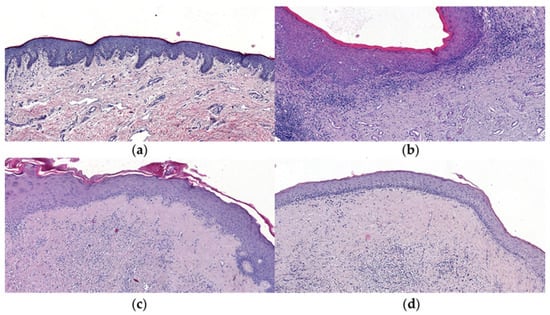
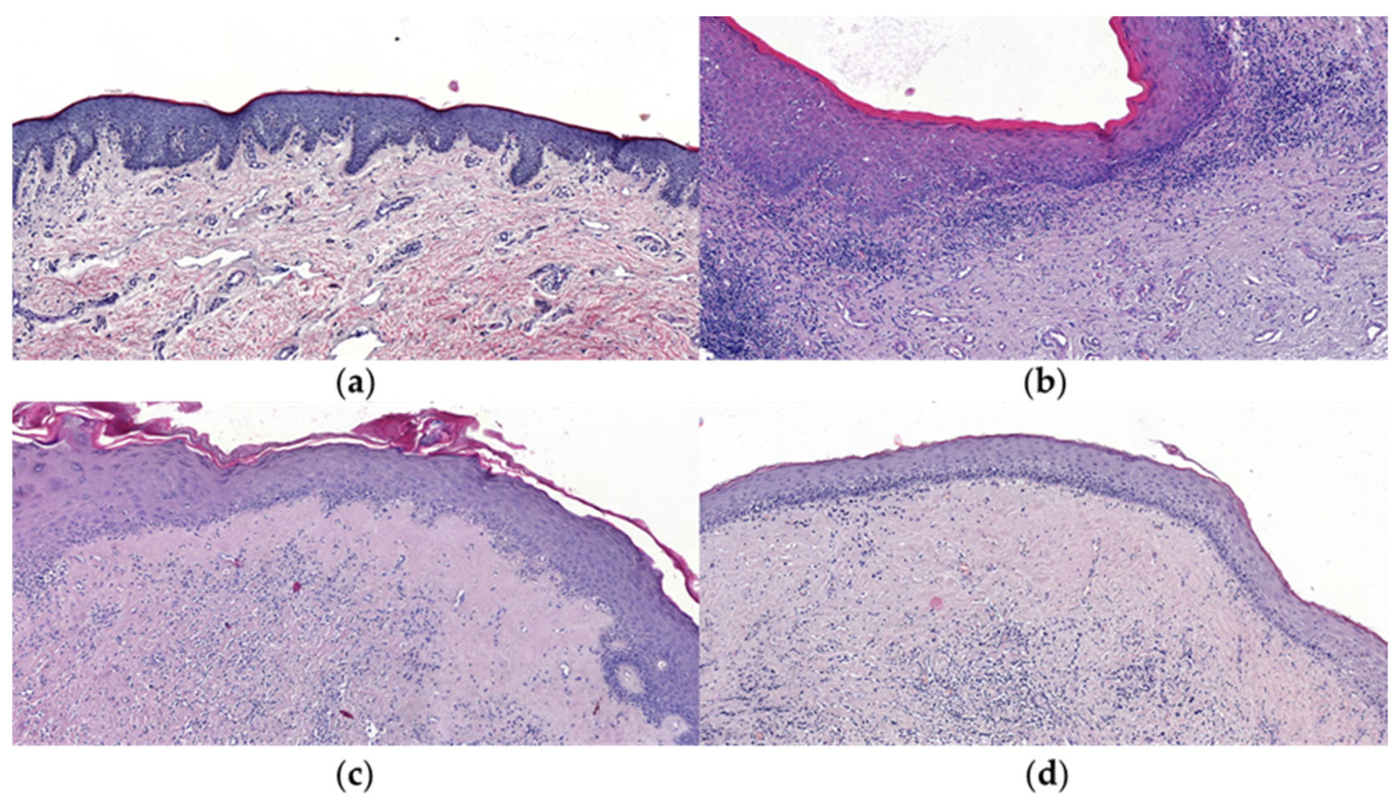
Based on histopathological examination, 10 patients were classified as having early-stage PLS, 26 as moderate-stage PLS, and 3 as severe-stage PLS. Healthy foreskin specimens obtained from 18 individuals served as controls (Figure 1).
Figure 1.
Hematoxylin and eosin (H&E) staining of representative sections of normal foreskin tissue and penile lichen sclerosus (PLS) at different stages: (a) normal foreskin (control); (b) early-stage PLS showing epidermal hyperkeratosis, loss of dermal papillae, and basal membrane degeneration with a dense inflammatory infiltrate located beneath the dermoepidermal junction; (c) moderate-stage PLS characterized by a narrow band of hyalinization separating the epidermis from a dense inflammatory infiltrate; (d) severe-stage PLS showing marked epidermal atrophy, extensive dermal hyalinization, and sparse inflammatory infiltrate in the deeper dermis. Magnification: 20×.
For macroscopic evaluation, each PLS specimen was divided into potentially affected (PA) and potentially unaltered (PH) regions. Consequently, the study included early PLS PA (n = 10), early PLS PH (n = 10), moderate PLS PA (n = 26), moderate PLS PH (n = 26), severe PLS PA (n = 3), and severe PLS PH (n = 3) samples. However, histopathological examination demonstrated that all PH samples exhibited microscopic features characteristic of lichen sclerosus, indicating that macroscopically unaltered tissue surrounding visible lesions was already involved in the disease process.
Representative histopathological features of PLS progression are presented in Figure 1. Early lesions were characterized by hyperkeratosis, degeneration of the basal cell layer, loss of dermal papillae, and dense inflammatory infiltrates located immediately beneath the dermoepidermal junction. Moderate lesions demonstrated the appearance of a distinct zone of dermal hyalinization separating the epidermis from inflammatory infiltrates. Severe lesions showed marked epidermal atrophy, extensive dermal sclerosis, and sparse inflammatory infiltrates confined primarily to deeper dermal layers.
3.2. Quantitative Assessment of Inflammatory Infiltrates
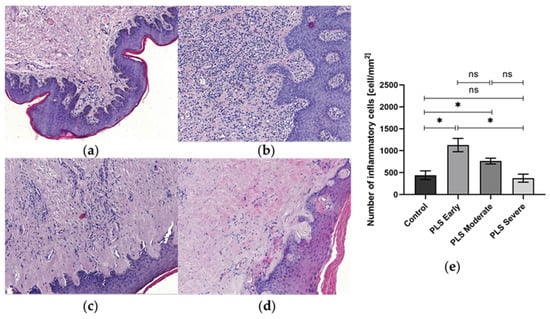
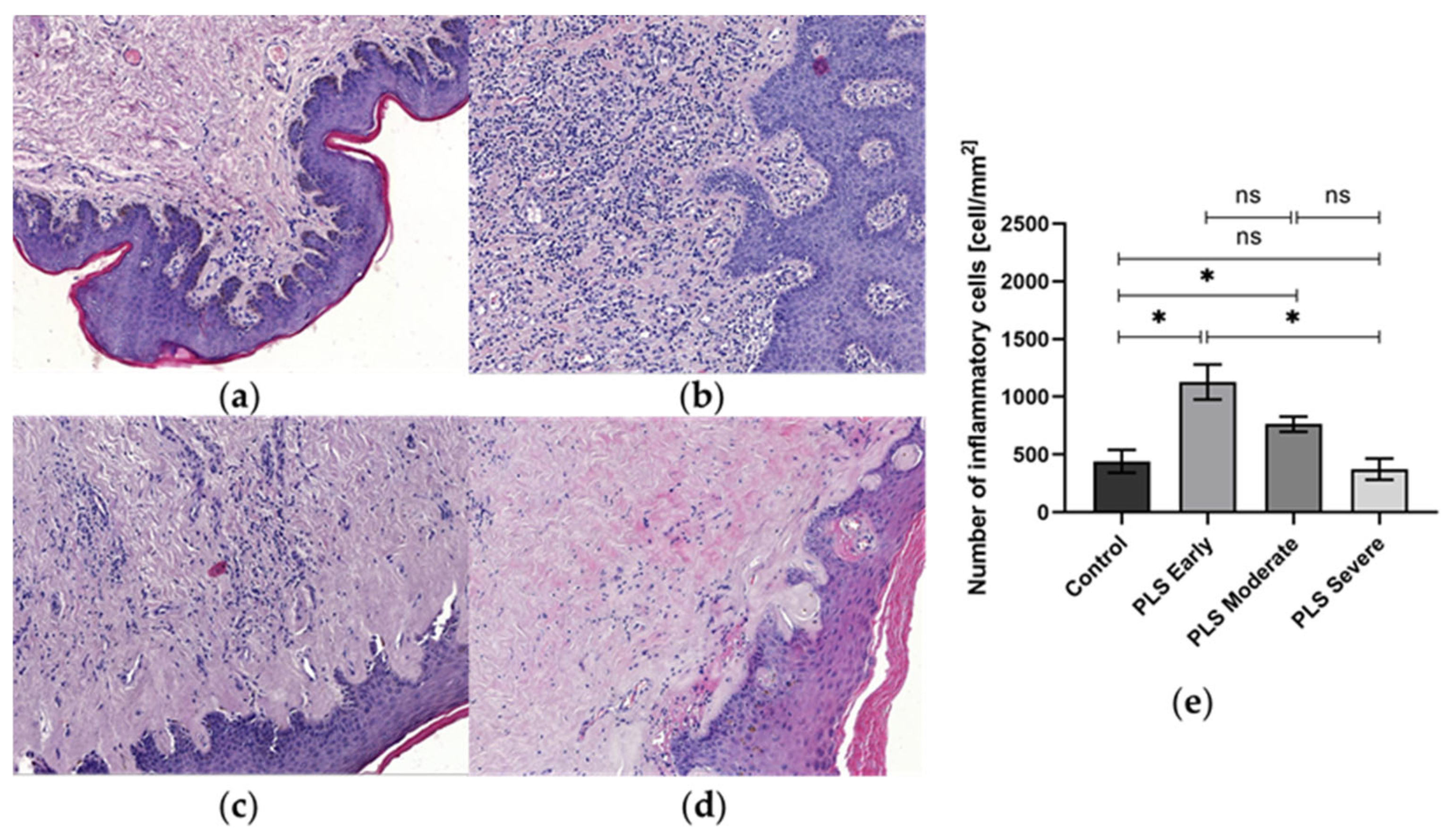
Quantitative analysis demonstrated differences in inflammatory cell density across the histopathological stages of PLS (Figure 2).
Figure 2.
Representative histological images illustrating inflammatory cell infiltration in normal foreskin tissue and PLS at different stages. H&E-stained histologic sections from (a) normal foreskin tissue (control), (b) early-stage PLS, (c) moderate-stage PLS, and (d) severe-stage PLS. (e) Quantitative analysis of inflammatory infiltrate density in regions of interest (ROI). Bars and whiskers represent the mean ± standard error of the mean (SEM) of inflammatory cell density (cells/mm2). Kruskal–Wallis test, p = 0.0002 (control vs. PLS early), p = 0.0197 (control vs. PLS moderate), p = 0.0168 (PLS early vs. PLS severe). Magnification: 10×. * indicates a statistically significant difference (p < 0.05), ns indicates no statistically significant difference.
The highest density of inflammatory cells was observed in early-stage lesions, whereas severe-stage PLS exhibited markedly reduced inflammatory infiltration. Statistical analysis demonstrated significant differences between control tissue and early-stage PLS (p = 0.0002), control tissue and moderate-stage PLS (p = 0.0197), and early-stage and severe-stage PLS (p = 0.0168).
These findings indicate that inflammatory cell density is higher in early-stage PLS and lower in more advanced stages, where fibrotic and tissue remodeling changes become more prominent histopathological features.
3.3. Immunohistochemical Localization of NF-κB p65
No NF-κB p65 immunoreactivity was observed within the cornified layer of the epidermis in either control or PLS specimens.
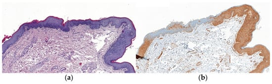
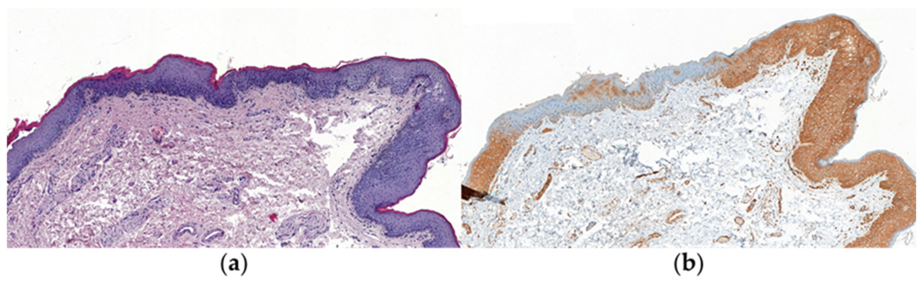
In normal foreskin samples, NF-κB p65 expression was predominantly detected within the epidermis, particularly in basal keratinocytes, where cytoplasmic–nuclear staining patterns were most frequently observed (Figure 3). Moderate immunoreactivity was also present in inflammatory cells located in the superficial dermis.
Figure 3.
H&E and immunohistochemical staining of NF-κB p65 in normal foreskin tissue: (a) H&E staining of normal foreskin tissue. (b) NF-κB p65 immunostaining showing a cytoplasmic–nuclear staining pattern in basal epithelial cells, with moderate cytoplasmic predominance. In this sample, cytoplasmic expression dominates, disappearing in places. Magnification: 10×.
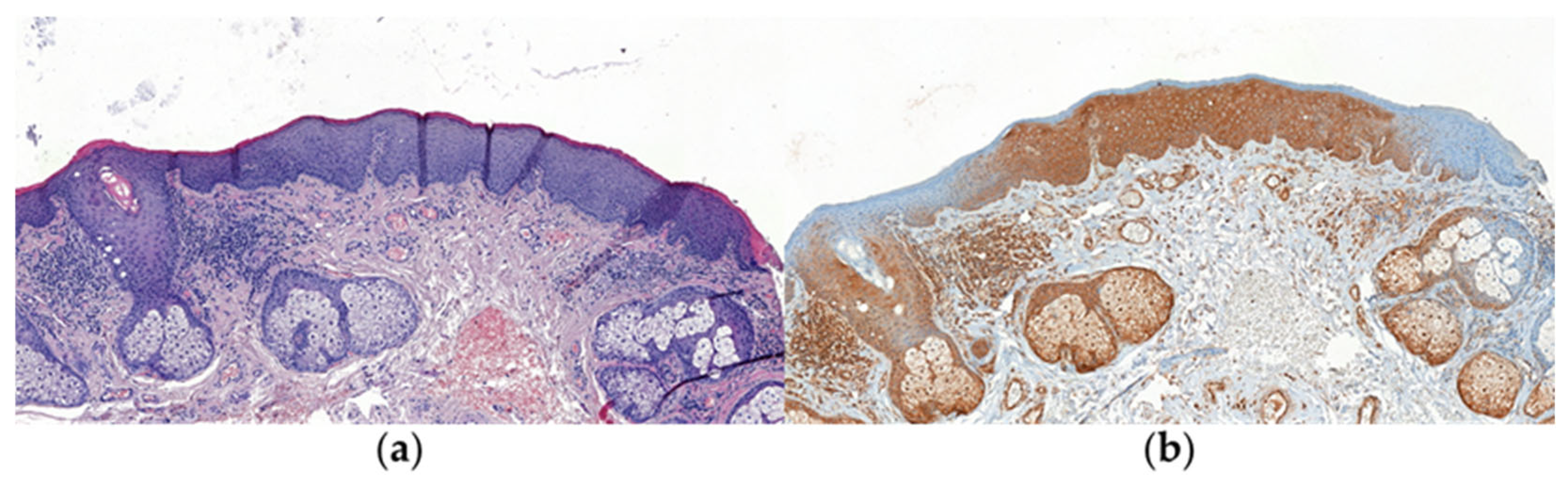
In early-stage PLS, strong cytoplasmic–nuclear NF-κB p65 expression was observed in basal keratinocytes and inflammatory infiltrates (Figure 4). Nuclear staining was also observed in inflammatory cells.
Figure 4.
Histological and immunohistochemical staining of NF-κB p65 expression in early-stage PLS (PA sample): (a) H&E staining showing dense inflammatory infiltrate beneath the epithelium; (b) NF-κB p65 immunostaining demonstrating a cytoplasmic–nuclear staining pattern in epithelial cells and inflammatory infiltrates, with cytoplasmic predominance. Magnification: 10×.
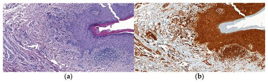
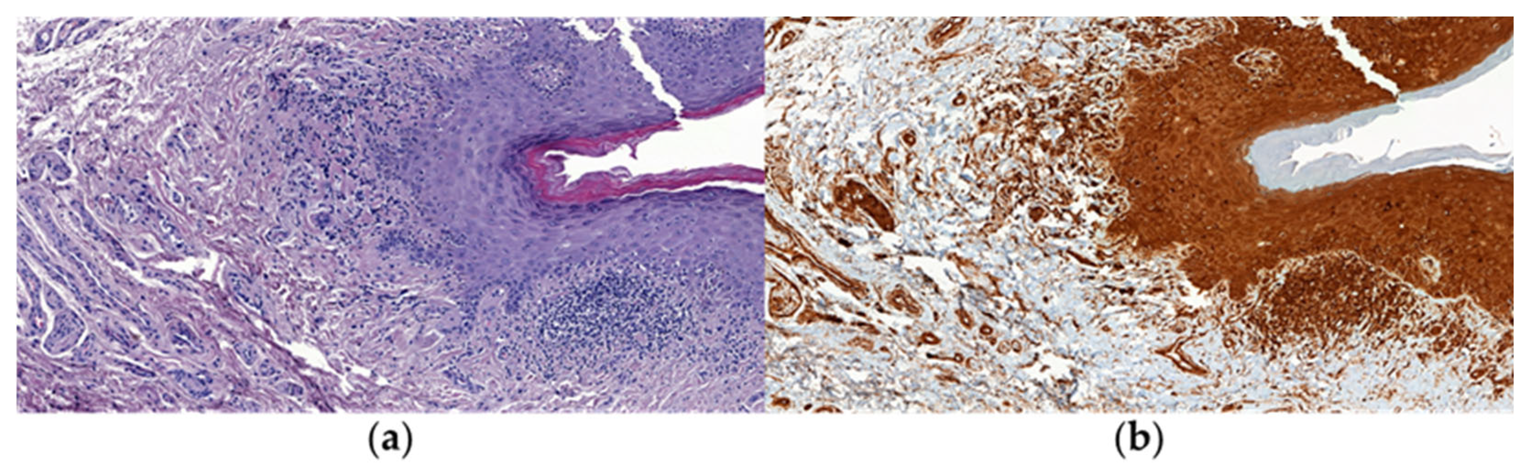
A similar expression pattern was observed in moderate-stage PLS (Figure 5). However, compared with early-stage lesions, staining appeared to show a greater cytoplasmic predominance despite persistent NF-κB p65 immunoreactivity.
Figure 5.
H&E and immunohistochemical staining of NF-κB p65 in moderate-stage PLS (PA sample): (a) H&E staining showing prominent inflammatory infiltrate in the upper dermis; (b) NF-κB p65 immunostaining showing a cytoplasmic–nuclear staining pattern in epithelial and inflammatory cells, with variable cytoplasmic predominance. Magnification: 10×.
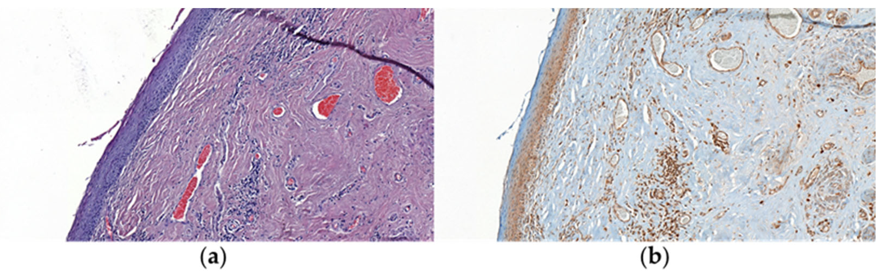
In severe-stage PLS, NF-κB p65 expression remained detectable but was characterized predominantly by cytoplasmic staining, with less frequent nuclear localization (Figure 6). This pattern was observed alongside reduced inflammatory cell infiltration and extensive dermal sclerosis.
Figure 6.
H&E and immunohistochemical staining of NF-κB p65 in severe-stage PLS (PA sample): (a) H&E staining showing marked dermal fibrosis and sparse inflammatory infiltrate; (b) NF-κB p65 immunostaining demonstrating predominantly cytoplasmic staining in epithelial and inflammatory cells, with reduced nuclear localization. Magnification: 10×.
No consistent differences in staining patterns were observed between PA and PH samples within the same disease stage. Importantly, histopathological examination confirmed microscopic PLS in all PH samples.
Overall, the descriptive findings showed a shift in NF-κB p65 staining from a cytoplasmic–nuclear pattern in early and moderate PLS toward predominantly cytoplasmic localization in severe-stage lesions. These observations should be interpreted cautiously, as subcellular localization was assessed descriptively rather than quantitatively.
3.4. Semi-Quantitative Evaluation of NF-κB p65 Expression
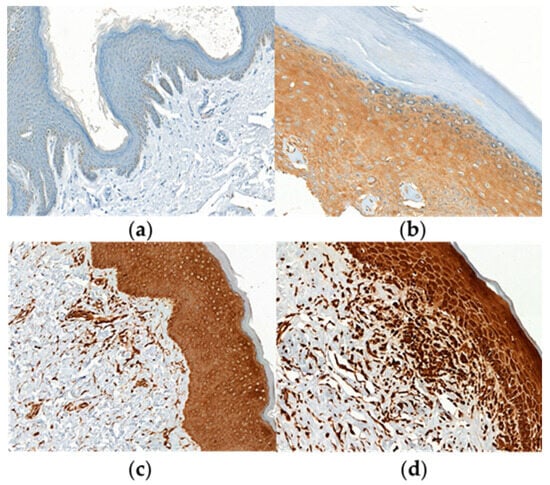
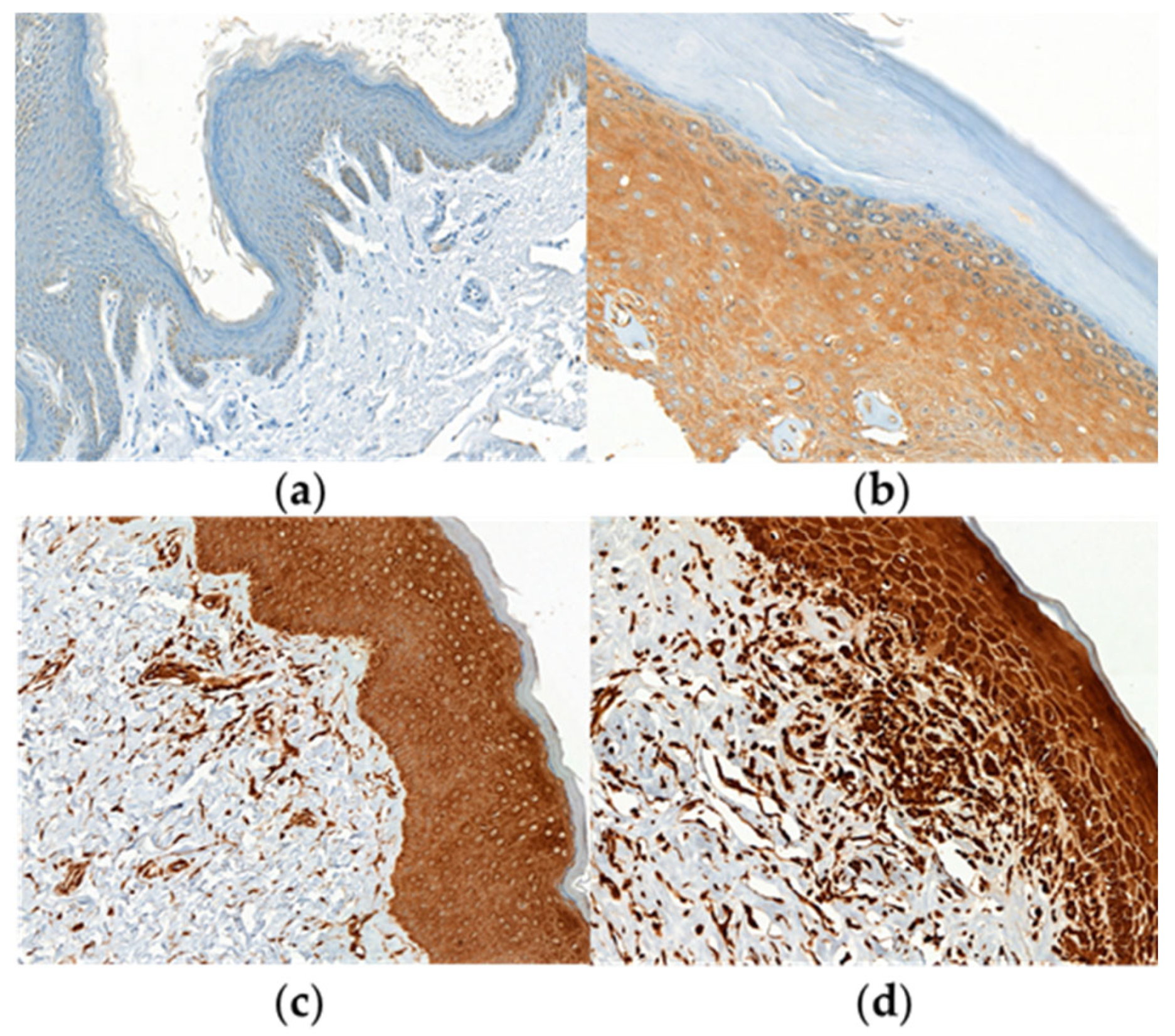
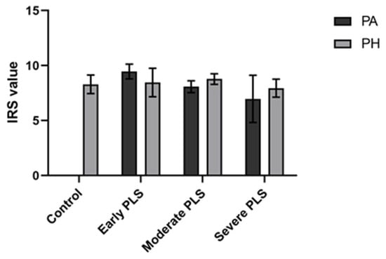
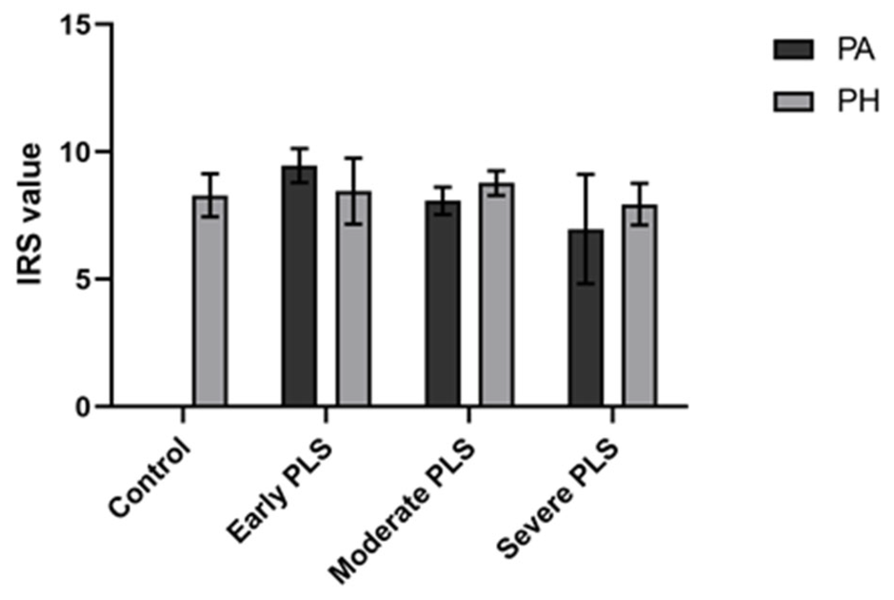
Semi-quantitative analysis using the immunoreactive score (IRS) system revealed variations in NF-κB p65 expression across the histopathological stages of PLS (Figure 7, Figure 8 and Figure 9).
Figure 7.
Representative examples of NF-κB p65 immunohistochemical staining intensity based on the immunoreactive score (IRS) system. Brown staining indicates positive immunoreactivity: (a) score 0—no staining; (b) score 1—weak staining; (c) score 2—moderate staining; (d) score 3—strong staining. All sections were counterstained with hematoxylin. Magnification: 20×.
Figure 8.
Immunoreactive score (IRS) analysis of NF-κB p65 expression in control, PLS PA, and PLS PH tissue samples. IRS evaluation of NF-κB protein expression was performed as described in the Materials and Methods. Bars and whiskers represent the mean ± standard error of the mean (SEM). Comparisons among the control, early-, moderate-, and severe-stage PLS groups were performed separately for PA and PH samples using the Kruskal–Wallis test, followed by Dunn’s multiple-comparison test. No statistically significant differences were observed. Abbreviations: Control, histologically confirmed normal foreskin tissue samples obtained from adult men undergoing partial circumcision; Early PLS, early stage of penile lichen sclerosus; Moderate PLS, moderate stage of penile lichen sclerosus; Severe PLS, severe stage of penile lichen sclerosus; PA, macroscopically altered tissue samples from patients with PLS; PH, macroscopically unaltered peripheral tissue samples from patients with PLS. Samples from each PLS stage were divided into PA and PH regions.
Figure 9.
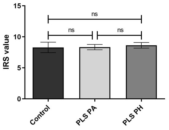
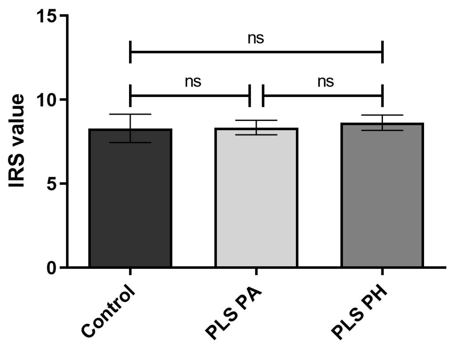
Comparative IRS analysis of NF-κB p65 expression in control, PLS PA, and PLS PH tissue samples. IRS evaluation of NF-κB protein expression was performed as described in the Materials and Methods. Bars and whiskers represent the mean ± standard error of the mean (SEM). Comparisons between the control group and PLS PA or PLS PH samples were performed using the Mann–Whitney U test, with Holm correction for multiple comparisons. The comparison between paired PLS PA and PLS PH samples was performed using the Wilcoxon matched-pair signed-rank test. No statistically significant differences were observed. Abbreviations: Control, histologically confirmed normal foreskin tissue samples obtained from adult men undergoing partial circumcision; PLS PA, macroscopically altered penile lichen sclerosus tissue samples from patients with PLS; PLS PH, macroscopically unaltered peripheral tissue samples from patients with PLS. ns indicates no statistically significant difference.
Although early- and moderate-stage lesions exhibited higher mean IRS values than severe-stage lesions, no statistically significant differences in NF-κB p65 IRS values were observed among the control, early-, moderate-, and severe-stage PLS groups in either PA or PH samples (Kruskal–Wallis test: PA, H = 2.126, p = 0.5467; PH, H = 0.8512, p = 0.8372). Dunn’s multiple-comparison test revealed no significant differences between any of the individual groups in either analysis (all adjusted p-values > 0.9999).
Furthermore, no statistically significant differences in NF-κB p65 IRS values were observed between the control group and PLS PA samples (Mann–Whitney U test, p = 0.6905; Holm-adjusted p = 1.000) or between the control group and PLS PH samples (Mann–Whitney U test, p = 0.9898; Holm-adjusted p = 1.000). Similarly, no statistically significant difference was found between paired PLS PA and PH samples (Wilcoxon matched-pair signed-rank test, p = 0.3697).
4. Discussion
Penile lichen sclerosus is a chronic inflammatory and fibrosing dermatosis whose pathogenesis remains incompletely understood. Although autoimmune mechanisms, chronic irritation, genetic susceptibility, and infectious triggers have all been proposed as contributing factors [1,6,18,19,20,21], increasing evidence supports a central role for immune dysregulation in disease initiation and progression [12,22].
The present study evaluated the immunohistochemical expression and tissue localization of NF-κB p65, a key transcriptional regulator of inflammatory signaling pathways, across different histopathological stages of PLS. Our findings demonstrate a reduction in inflammatory infiltrates with disease progression, whereas descriptive analysis of NF-κB p65 immunohistochemical staining showed a trend toward lower immunoreactivity and less frequent nuclear localization in advanced lesions. These observations raise the possibility of changes in NF-κB-related inflammatory activity during disease progression; however, they do not establish progressive attenuation of canonical NF-κB signaling.
The histopathological findings observed in the present study were consistent with previous descriptions of PLS progression [22,23,24]. Early lesions demonstrated dense inflammatory infiltrates immediately beneath the dermoepidermal junction, whereas advanced lesions were characterized by epidermal atrophy, extensive dermal hyalinization, and markedly reduced inflammatory cell infiltration. This pattern may be associated with the activation of profibrotic pathways, particularly transforming growth factor-β (TGF-β)-mediated fibroblast activation and extracellular matrix remodeling, which may contribute to progressive tissue sclerosis in chronic inflammatory conditions [23,24,25]. Quantitative analysis demonstrated significantly lower inflammatory cell density in severe-stage lesions compared with early-stage lesions, consistent with the observation that inflammatory infiltrates were more prominent in early disease, whereas fibrosis was more prominent in advanced lesions.
NF-κB signaling plays a central role in the regulation of innate and adaptive immune responses. Activation of NF-κB induces transcription of numerous pro-inflammatory mediators, including IL-1, IL-6, TNF-α, chemokines, adhesion molecules, and regulators of leukocyte recruitment [13,14]. Persistent or dysregulated NF-κB activation has been implicated in the pathogenesis of chronic inflammatory and autoimmune diseases [13,14,15].
Several observations support the involvement of inflammatory mechanisms in lichen sclerosus. Studies of vulvar lichen sclerosus have demonstrated increased expression of Th1-associated cytokines, including IFN-γ, IL-7, IL-15, and TNF-α, suggesting a predominantly cell-mediated immune response [24]. Similarly, our previous molecular analyses of PLS tissue demonstrated markedly elevated expression of IL1A, IL6, and IFNG genes, supporting the existence of a highly pro-inflammatory microenvironment within affected foreskin tissue [12].
Interestingly, our current findings suggest that this inflammatory response may vary across disease stages. Although NF-κB p65 expression was observed at all stages of PLS, descriptive analysis showed a trend toward lower overall immunoreactivity and less frequent nuclear localization of NF-κB p65 in advanced lesions, accompanied by a reduction in inflammatory infiltrates. Nuclear translocation of p65 is a critical step in canonical NF-κB activation. However, because subcellular localization was assessed descriptively, and differences in IRS did not reach statistical significance, the present findings cannot establish reduced pathway activity in advanced PLS.
An important methodological consideration is that the present study evaluated NF-κB p65 (RelA) protein expression by immunohistochemistry, whereas previous molecular studies have assessed NFKB1 and NFKB2 gene expression [26]. Therefore, the findings of these studies are not directly comparable and should be interpreted with caution. The NF-κB family comprises several subunits, including p65 (RelA), p50 (derived from NFKB1), and p52 (derived from NFKB2), which may be regulated differently at the transcriptional and post-transcriptional levels. Accordingly, the previously observed increase in NFKB1 mRNA expression does not necessarily contradict the descriptive trend toward lower NF-κB p65 immunoreactivity in advanced lesions. However, the present study did not assess the expression of other NF-κB subunits, and the biological basis of this difference remains unclear.
Another observation concerned the subcellular localization of NF-κB p65. In early and moderate lesions, cytoplasmic–nuclear staining was observed, whereas severe lesions showed predominantly cytoplasmic staining with less frequent nuclear localization. Since nuclear localization of p65 is associated with canonical NF-κB activation, this pattern may reflect differences in inflammatory signaling or inflammatory stimulation across disease stages. However, as subcellular localization was assessed descriptively rather than quantitatively, this interpretation remains preliminary. When considered together with the lower inflammatory cell density and more prominent fibrotic changes observed in advanced lesions, these findings may indicate a shift in histopathological features from predominantly inflammatory changes toward more pronounced fibrosis. Nevertheless, the relationship between these processes requires further investigation.
Our histopathological evaluation also demonstrated that all samples designated as potentially unaltered (PH) based on their macroscopic appearance exhibited microscopic features of lichen sclerosus. Thus, these samples represented histologically involved peripheral regions rather than unaffected tissue. This finding suggests that disease involvement may extend beyond visible altered lesion borders and highlights the importance of histopathological verification when interpreting findings obtained from macroscopically designated PH regions. The presence of histopathological changes in both PA and PH regions may also help explain the absence of statistically significant differences in NF-κB p65 immunoreactivity between these tissue samples.
Several limitations should be acknowledged. First, the number of patients with severe PLS was limited (n = 3), which reduces the reliability and statistical power of comparisons involving this group. Second, although NF-κB p65 immunoreactivity was assessed using a semi-quantitative IRS method, subcellular localization was evaluated descriptively. Therefore, immunohistochemistry provided semi-quantitative information on protein expression but did not directly assess the functional activation of NF-κB signaling. Consequently, the observed staining patterns cannot independently establish changes in NF-κB transcriptional activity. Third, the assessment of a single NF-κB family subunit (p65/RELA) does not allow comprehensive evaluation of the complex regulatory interactions among individual NF-κB subunits.
Future studies should combine immunohistochemical analyses with quantitative assessment of NF-κB p65 expression and nuclear localization, as well as characterization of inflammatory cell populations within PLS lesions. Investigation of phosphorylated NF-κB p65, NFKB1/p50, NFKB2/p52, and fibrosis-related signaling pathways may provide additional insight into the molecular mechanisms underlying disease progression. Furthermore, integration of transcriptomic, proteomic, and spatial immune profiling approaches may help identify novel biomarkers and therapeutic targets in PLS.
From a diagnostic perspective, the present findings provide a basis for further investigation of NF-κB p65 as a potential complementary marker in the histopathological assessment of PLS. Although the observed staining patterns may warrant further investigation in relation to inflammatory and fibrotic changes, the differences in NF-κB p65 immunoreactivity between disease groups did not reach statistical significance, and subcellular localization was assessed descriptively. Therefore, the present findings do not establish the diagnostic utility of NF-κB p65. In addition, the identification of microscopic PLS features in peripheral tissue designated as potentially unaltered (PH) based on its macroscopic appearance highlights the importance of histopathological assessment beyond visibly altered areas. At present, NF-κB p65 immunohistochemistry cannot be considered an independent diagnostic biomarker or a reliable tool for disease staging. Nevertheless, its potential value as a complementary marker in routine histopathological evaluation warrants further investigation in larger patient groups, particularly those including more cases of advanced PLS, to determine whether it can provide clinically meaningful information and contribute to a more comprehensive assessment of disease severity.
5. Conclusions
The present study demonstrated significantly lower inflammatory cell density in severe PLS lesions compared with early-stage lesions, consistent with a transition from predominantly inflammatory changes in early lesions toward more prominent fibrotic remodeling in advanced disease. Descriptive analysis of NF-κB p65 immunohistochemical staining showed a trend toward lower immunoreactivity and less frequent nuclear localization in advanced lesions. However, differences in IRS between disease stages did not reach statistical significance, and subcellular localization was not quantitatively assessed. Therefore, these findings do not establish progressive attenuation of canonical NF-κB signaling and should be interpreted as preliminary observations requiring further investigation.
Histopathological examination also identified microscopic PLS involvement in all samples designated as potentially unaltered (PH) based on macroscopic appearance, suggesting that disease-related changes may extend beyond visibly altered lesion margins. This finding highlights the potential importance of histopathological assessment of peripheral tissue, even when it appears unaffected on macroscopic examination. Although NF-κB p65 cannot currently be considered an independent diagnostic biomarker, its immunohistochemical expression pattern and subcellular localization may provide complementary information for the histopathological assessment of inflammatory and fibrotic changes in PLS. Further studies involving larger patient groups, particularly with more cases of severe PLS, and quantitative assessment of nuclear p65 localization and NF-κB pathway activity are needed to determine the clinical relevance of these findings and establish whether NF-κB p65 can contribute to a more comprehensive histopathological assessment of PLSS.
Author Contributions
Conceptualization, M.V., B.N., M.C. and L.K.; methodology, M.V. and K.J.; software, M.V. and A.K.-C.; validation, M.V., A.K.-C. and P.M.W.; formal analysis, M.V., A.K.-C. and P.M.W.; investigation, M.V.; resources, M.V., M.C. and P.M.W.; data curation, M.V.; writing—original draft preparation, M.V. and L.K.; writing—review and editing, A.K.-C., M.C., M.M., B.N., P.M.W. and L.K.; visualization, M.V.; supervision, L.K., M.M., B.N. and P.M.W.; project administration, M.V. and L.K.; funding acquisition, L.K. All authors have read and agreed to the published version of the manuscript.
Funding
This research was funded by the Ministry of Science and Higher Education of Poland, grant no. 4/SPUB/SP/0133/2026.
Institutional Review Board Statement
The present study protocol was reviewed and approved by the Institutional Review Board of the Medical University of Gdansk (decision no. NKBBN/369/2017).
Informed Consent Statement
Informed consent was obtained from all subjects involved in the study.
Data Availability Statement
The original contributions presented in the study are included in the article, further inquiries can be directed to the corresponding author. We confirm that all the figures and tables are the original work of the authors. All have been performed by the authors of this manuscript, have not been adapted from other authors, and do not have an online link.
Conflicts of Interest
The authors declare no conflicts of interest.
References
- Tran, D.A.; Tan, X.; Macri, C.J.; Goldstein, A.T.; Fu, S.W. Lichen Sclerosus: An Autoimmunopathogenic and Genomic Enigma with Emerging Genetic and Immune Targets. Int. J. Biol. Sci. 2019, 15, 1429–1439. [Google Scholar] [CrossRef] [Scilit]
- Clouston, D.; Hall, A.; Lawrentschuk, N. Penile Lichen Sclerosus (Balanitis Xerotica Obliterans). BJU Int. 2011, 108, 14–19. [Google Scholar] [CrossRef] [Scilit]
- Sander, C.S.; Ali, I.; Dean, D.; Thiele, J.J.; Wojnarowska, F. Oxidative Stress Is Implicated in the Pathogenesis of Lichen Sclerosus. Br. J. Dermatol. 2004, 151, 627–635. [Google Scholar] [CrossRef] [Scilit]
- Shah, V.S.; Jung, N.L.; Lee, D.K.; Nepple, K.G. Does Routine Pathology Analysis of Adult Circumcision Tissue Identify Penile Cancer? Urology 2015, 85, 1431–1435. [Google Scholar] [CrossRef] [Scilit]
- Czajkowski, M.; Żawrocki, A.; Czajkowska, K.; Kłącz, J.; Sokołowska-Wojdyło, M.; Biernat, W.; Matuszewski, M. Lichen Sclerosus and Phimosis—Discrepancies Between Clinical and Pathological Diagnosis and Its Consequences. Urology 2021, 148, 274–279. [Google Scholar] [CrossRef] [Scilit]
- Paulis, G.; Berardesca, E. Lichen Sclerosus: The Role of Oxidative Stress in the Pathogenesis of the Disease and Its Possible Transformation into Carcinoma. Res. Rep. Urol. 2019, 11, 223–232. [Google Scholar] [CrossRef] [Scilit]
- Nasca, M.R.; Innocenzi, D.; Micali, G. Penile Cancer among Patients with Genital Lichen Sclerosus. J. Am. Acad. Dermatol. 1999, 41, 911–914. [Google Scholar] [CrossRef] [Scilit]
- Bunker, C.B.; Shim, T. Male Genital Lichen Sclerosus. Indian J. Dermatol. 2015, 60, 111–117. [Google Scholar] [CrossRef] [Scilit]
- Ranjan, N.; Singh, S.K. Malignant Transformation of Penile Lichen Sclerosus: Exactly How Common Is It? Int. J. Dermatol. 2008, 47, 1308–1309. [Google Scholar] [CrossRef] [Scilit]
- Philippou, P.; Shabbir, M.; Ralph, D.J.; Malone, P.; Nigam, R.; Freeman, A.; Muneer, A.; Minhas, S. Genital Lichen Sclerosus/Balanitis Xerotica Obliterans in Men with Penile Carcinoma: A Critical Analysis. BJU Int. 2013, 111, 970–976. [Google Scholar] [CrossRef] [Scilit]
- Pietrzak, P.; Hadway, P.; Corbishley, C.M.; Watkin, N.A. Is the Association between Balanitis Xerotica Obliterans and Penile Carcinoma Underestimated? BJU Int. 2006, 98, 74–76. [Google Scholar] [CrossRef] [Scilit]
- Czajkowski, M.; Wierzbicki, P.; Kotulak-Chrząszcz, A.; Czajkowska, K.; Bolcewicz, M.; Kłącz, J.; Kreft, K.; Lewandowska, A.; Nedoszytko, B.; Sokołowska-Wojdyło, M.; et al. The Role of Occlusion and Micro-Incontinence in the Pathogenesis of Penile Lichen Sclerosus: An Observational Study of Pro-Inflammatory Cytokines’ Gene Expression. Int. Urol. Nephrol. 2022, 54, 763–772. [Google Scholar] [CrossRef] [Scilit]
- Liu, T.; Zhang, L.; Joo, D.; Sun, S.C. NF-κB Signaling in Inflammation. Signal Transduct. Target. Ther. 2017, 2, 17023. [Google Scholar] [CrossRef] [Scilit]
- Oeckinghaus, A.; Ghosh, S. The NF-κB Family of Transcription Factors and Its Regulation. Cold Spring Harb. Perspect. Biol. 2009, 1, a000034. [Google Scholar] [CrossRef] [Scilit]
- Sun, S.C.; Chang, J.H.; Jin, J. Regulation of NF-κB in Autoimmunity. Trends Immunol. 2013, 34, 282–289. [Google Scholar] [CrossRef] [Scilit]
- Kirtschig, G. Lichen Sclerosus—Diagnosis and Management. Dtsch. Arztebl. Int. 2016, 113, 337–343. [Google Scholar] [CrossRef] [Scilit]
- Remmele, W.; Stegner, H.E. Recommendation for uniform definition of an immunoreactive score (IRS) for immunohistochemical estrogen receptor detection (ER-ICA) in breast cancer tissue. Der Pathol. 1987, 8, 138–140. [Google Scholar]
- Godlewski, J.; Kiezun, J.; Krazinski, B.E.; Kozielec, Z.; Wierzbicki, P.M.; Kmiec, Z. The Immunoexpression of YAP1 and LATS1 Proteins in Clear Cell Renal Cell Carcinoma: Impact on Patients’ Survival. BioMed Res. Int. 2018, 2018, 2653623. [Google Scholar] [CrossRef] [Scilit]
- Bhambhani, D.; Bhambhani, S.; Pandya, N.K. Penile Lichen Sclerosus: A Surgical Perspective of Its Aetiology and Treatment. Cureus 2022, 14, e28418. [Google Scholar] [CrossRef] [Scilit]
- Fistarol, S.K.; Itin, P.H. Diagnosis and Treatment of Lichen Sclerosus: An Update. Am. J. Clin. Dermatol. 2013, 14, 27–47. [Google Scholar] [CrossRef] [Scilit]
- Oyama, N.; Hasegawa, M. Lichen Sclerosus: A Current Landscape of Autoimmune and Genetic Interplay. Diagnostics 2022, 12, 3070. [Google Scholar] [CrossRef] [Scilit]
- Regauer, S.; Liegl, B.; Reich, O. Early Vulvar Lichen Sclerosus: A Histopathological Challenge. Histopathology 2005, 47, 340–347. [Google Scholar] [CrossRef] [Scilit]
- De Luca, D.A.; Papara, C.; Vorobyev, A.; Staiger, H.; Bieber, K.; Thaçi, D.; Ludwig, R.J. Lichen Sclerosus: The 2023 Update. Front. Med. 2023, 10, 1106318. [Google Scholar] [CrossRef] [Scilit]
- Corazza, M.; Schettini, N.; Zedde, P.; Borghi, A. Vulvar Lichen Sclerosus: From Pathophysiology to Therapeutic Approaches. Biomedicines 2021, 9, 950. [Google Scholar] [CrossRef] [Scilit]
- Wynn, T.A.; Ramalingam, T.R. Mechanisms of fibrosis: Therapeutic translation for fibrotic disease. Nat. Med. 2012, 18, 1028–1040. [Google Scholar] [CrossRef] [Scilit]
- Wierzbicki, P.M.; Czajkowski, M.; Kotulak-Chrząszcz, A.; Bukowicz, J.; Dzieciuch, K.; Sokołowska-Wojdyło, M.; Kmieć, Z.; Matuszewski, M. Altered mRNA Expression of NFKB1 and NFKB2 Genes in Penile Lichen Sclerosus, Penile Cancer and Zoon Balanitis. J. Clin. Med. 2022, 11, 7254. [Google Scholar] [CrossRef] [Scilit]

Disclaimer/Publisher’s Note: The statements, opinions and data contained in all publications are solely those of the individual author(s) and contributor(s) and not of MDPI and/or the editor(s). MDPI and/or the editor(s) disclaim responsibility for any injury to people or property resulting from any ideas, methods, instructions or products referred to in the content. |
© 2026 by the authors. Licensee MDPI, Basel, Switzerland. This article is an open access article distributed under the terms and conditions of the Creative Commons Attribution (CC BY) license.