


Abstract
Background/Objectives: Accessory mandibular canals are frequent anatomical variations, and failure to recognize them may lead to incomplete anesthesia, bleeding, and neurosensory disturbances. Several systems have been proposed to classify bifid (BMCs) and trifid (TMCs) mandibular canals, but how consistently they are applied in cone-beam computed tomography (CBCT) research remains unclear. This scoping review aimed to map CBCT-based classification systems for BMCs and TMCs and to identify anatomical variants not addressed by existing systems. Methods: PubMed/MEDLINE, Scopus, Web of Science, and Embase were searched up to 31 May 2026. Original CBCT studies proposing, modifying, or applying a classification of accessory mandibular canals were included. Two reviewers independently screened records and charted data on the classification adopted, its modifications, and unclassified variants. Results: Sixty studies were included: forty-two applied a classification for BMCs, seventeen focused exclusively on retromolar canals (RMCs), and one on temporal crest canals (TCCs). The system by Naitoh et al. was the most frequently adopted, yet no study applied it without modification; changes included omission of subtypes, buccal/lingual subdivision, and the addition of superior, inferior, and trifid categories. Equivalent inferiorly directed canals were reported under three different names, RMCs and TCCs were classified using independent systems, and only one study proposed a dedicated subclassification for TMCs. Conclusions: CBCT-based classifications of accessory mandibular canals are fragmented and inconsistently applied. A conceptual three-level framework based on branching multiplicity, site of origin, and spatial trajectory is proposed to improve cross-study comparability; its reliability requires validation on independent CBCT datasets.
1. Introduction
The mandibular canal (MC), running through the body and ramus of the mandible, harbors the inferior alveolar nerve, artery, and vein, which collectively provide innervation and vascular supply to the lower teeth, alveolar bone, and lower lip/chin region [1]. Although historically described as a single osseous canal, extending from the mandibular foramen to the mental foramen with a terminal branch (incisive canal), several anatomical and radiological investigations demonstrated that accessory mandibular canals are frequent anatomical variations [2,3,4]. Since accessory canals may contain a neurovascular bundle, failure to recognize them prior to surgical procedures can lead to severe intraoperative and postoperative complications [5]. These include incomplete local anesthesia during inferior alveolar nerve blocks, intraoperative bleeding, neurosensory alterations, and traumatic neuroma development [6,7].
Depending on the number of accessory canals, bifid (BMCs) and trifid (TMCs) mandibular canals are described in the literature [8]. Although early studies were conducted on panoramic radiographs, traditional two-dimensional imaging modalities exhibited substantial limitations, including the overlapping of buccal and lingual bony structures, and dimensional distortion due to irregular magnification [9,10]. The widespread adoption of Cone-Beam Computed Tomography (CBCT) in clinical dentistry has overcome these limitations by providing high-resolution, three-dimensional, non-distorted multiplanar reconstructions [11]. Thus, CBCT allows clinicians and researchers to accurately define the point of origin, course and termination of the accessory canals, as well as to calculate angles, distances and measurements with high precision.
Multiple classification frameworks have been developed to categorize BMCs according to their anatomical course and spatial configuration: pioneer systems, including those formulated by Nortjé et al. and Langlais et al., relied on two-dimensional panoramic radiographs [12,13]; in contrast, Naitoh et al. introduced a dedicated classification scheme for BMCs based on CBCT images [14]. However, subsequent studies adopted this classification with varying degrees of modification, while other authors adopted older two-dimensional classification schemes for CBCT datasets [15]. In parallel, some studies proposed specific classifications restricted to localized accessory canal subtypes, such as retromolar canals (RMC) [16,17].
Previous reviews have described the classification systems available for mandibular canal branching [18] or summarized the morphological and quantitative characteristics of BMCs [1]. However, no review has systematically mapped how classification systems have been proposed, applied, modified, or extended across CBCT studies, nor identified the anatomical variants that these systems fail to accommodate. Therefore, a systematic mapping of the existing evidence is required to clarify how mandibular canal variations are currently categorized in CBCT research and to identify unresolved anatomical gaps.
The aim of this scoping review was to systematically map existing CBCT-based classification systems for BMCs and TMCs; by identifying the morphological patterns left unaddressed by current systems, this review provides the theoretical rationale for proposing an integrated classification framework.
2. Materials and Methods
This review was designed according to the standard recommendations of the Preferred Reporting Items for Systematic reviews and Meta-Analyses extension for Scoping Reviews (PRISMA-ScR) guidelines (https://www.equator-network.org/reporting-guidelines/prisma-scr/, accessed on 22 May 2026) [19], and the literature search was guided using the York framework proposed by Arksey and O’Malley [20]. A formal review protocol was not pre-registered with a public registry prior to commencing data extraction.
2.1. Focused Question
The primary question of this scoping review was as follows: “What classification systems have been proposed or applied in the literature to describe bifid and trifid mandibular canals using CBCT, and what anatomical variations remain unaddressed by existing classification systems?”
2.2. Eligibility Criteria
Studies were selected for inclusion in this scoping review based on the following criteria, structured according to the Population-Concept-Context (PCC) strategy:
- •
- Population: Studies involving human subjects who underwent CBCT evaluation of the mandible.
- •
- Concept: Studies describing bifid or trifid mandibular canals, including their anatomical subtypes.
- •
- Context: Studies that proposed a novel anatomical classification, applied or modified an existing classification system (e.g., Langlais et al. or Naitoh et al. [13,14]) to CBCT data, or categorized accessory mandibular canal variations according to explicitly defined criteria. A formally named classification system was not required, provided that the anatomical criteria distinguishing the reported categories were explicitly identifiable.
- •
- Study design: Original research studies, including observational (retrospective or prospective) cross-sectional studies, case series, and anatomical/cadaveric investigations using CBCT.
Studies were excluded if they met one or more of the following criteria:
- •
- Studies evaluating mandibular canal variations exclusively through 2D radiography (e.g., panoramic radiographs) or exclusively cadaveric evaluation.
- •
- Studies reporting solely prevalence or incidence rates of bifid/trifid canals without providing any anatomical classification or structured categorization into anatomical types or subtypes.
- •
- Single case reports lacking broader anatomical discussion or classification relevance.
- •
- Narrative reviews, systematic reviews, meta-analyses, conference abstracts, editorials, letters to the editor, textbooks, book chapters.
- •
- Animal or in vitro studies.
- •
- Studies not available in English.
2.3. Search Strategy
An electronic literature search was conducted up to 31 May 2026, in order to identify studies investigating bifid and trifid mandibular canals using CBCT, including those proposing new classification systems, modifying existing frameworks, or applying previously established classification schemes.
The search was conducted using the following scientific databases: PubMed/MEDLINE, Scopus, Web Of Science, and Embase. A combination of free-text keywords related to the mandibular canal and its anatomical variations was used, and the search syntax was adapted to each database using appropriate field tags (e.g., TITLE-ABS-KEY for Scopus, TS for Web of Science) and controlled vocabulary where applicable (e.g., MeSH terms). The search strategies were drafted by a reviewer (V.P.) and further refined through team discussion. The full electronic search strategies for each database are detailed in Supplementary Table S1.
2.4. Study Selection
All retrieved studies were imported into Rayyan (Rayyan Systems Inc., Cambridge, MA, USA), a web-based and mobile platform [21], and any duplicates were identified and subsequently removed. Prior to the screening process, a calibration exercise was performed between the two reviewers (V.P. and L.M.) using a pilot sample of 10% of the retrieved records to ensure complete alignment on the eligibility criteria. Analysis and selection of database search results were conducted independently by the two reviewers in a two-step process based on the eligibility criteria. Titles and abstracts of all studies were screened in the first step of selection. All potentially eligible articles were read in full during the second step of study selection to confirm inclusion. Articles that did not match the inclusion criteria were excluded from consideration. Discrepancies were resolved through discussion and consensus, with the intervention of a third reviewer when necessary (A.B.). For manuscripts without accessible full texts, the corresponding authors were contacted by email up to three times before exclusion.
2.5. Data Extraction
The data were independently extracted by two authors (V.P. and A.B.) using a pre-determined spreadsheet developed in Microsoft Excel (Microsoft Corporation, Redmond, WA, USA). Any disagreements were resolved through discussion.
The following information was systematically extracted from each eligible study:
- •
- General study characteristics: First author’s name, year of publication, country where the study was conducted, and study design.
- •
- Anatomical focus and targeted variants: The specific type of accessory bony canal addressed (e.g., BMCs or TMCs in general, or specific localized variants such as the RMC).
- •
- Classification framework: The specific classification system either adopted or modified or newly proposed by the authors, including the morphological criteria used.
- •
- Unclassified Anatomical Variations: Specific branching patterns or morphological variants identified in the results that did not fit into any existing category of the adopted classification system.
2.6. Data Synthesis
The included studies were initially grouped according to the types of accessory canals studied: BMCs, specific types of bifid canals (such as the RMC) and TMCs. The main findings were summarized in comparative tables and discussed accordingly. No data conversions or imputations were required, and outcomes were reported as presented in the original studies. For each study, the classification adopted was considered as reported in the corresponding results section (some studies may indicate in the materials and methods section that they used a classification present in the literature but present the results with a classification system with some modifications compared to the original).
3. Results
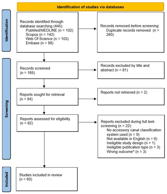
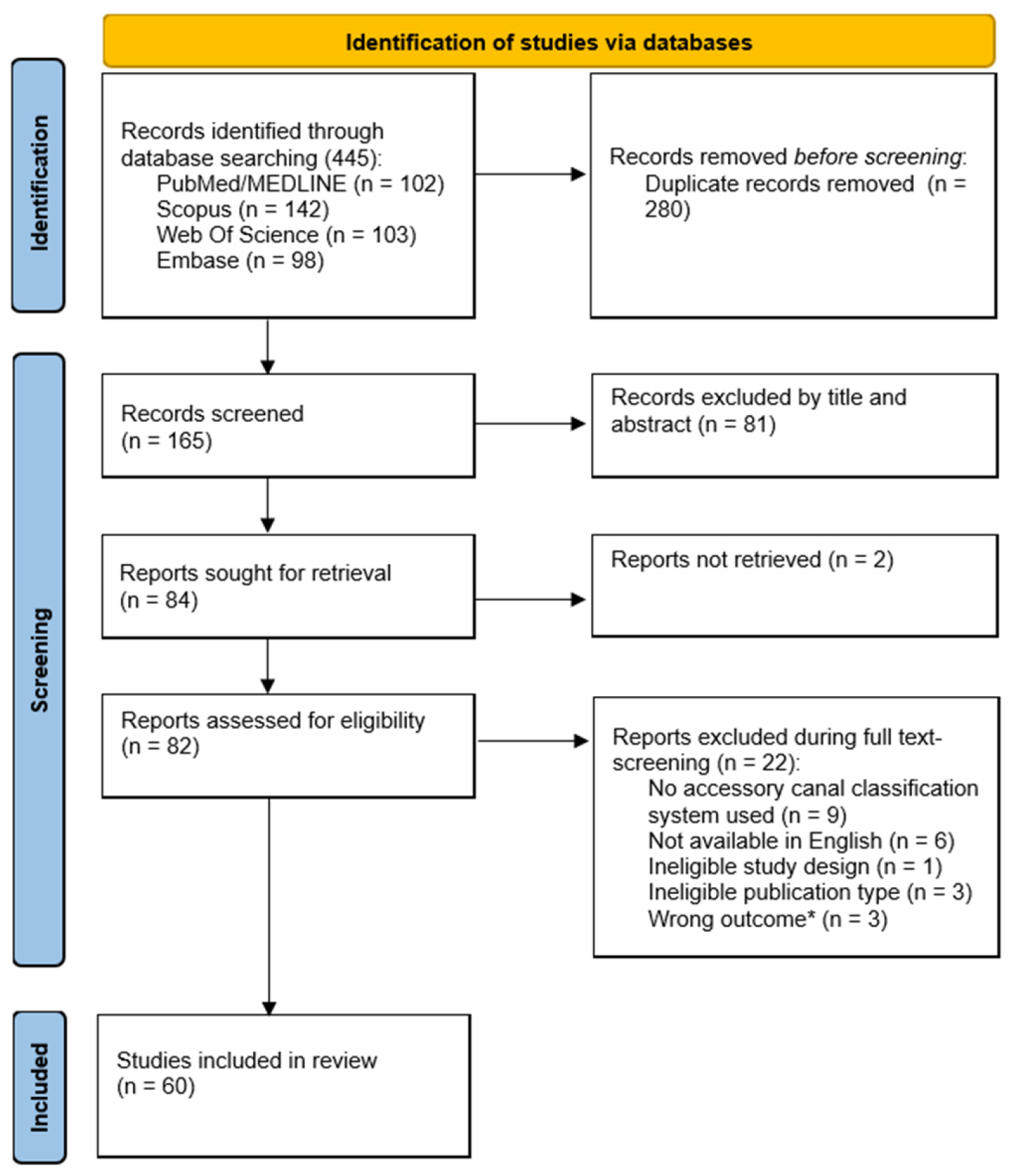
The article selection process is illustrated in Figure 1. A total of 445 records were retrieved through electronic database searching. Following duplicate removal, titles and abstracts of 165 articles were screened, leading to the exclusion of 81 studies. During the full-text retrieval phase, two studies could not be retrieved: despite contacting the respective corresponding authors, no response was received, resulting in their exclusion from eligibility assessment. Subsequently, 82 full-text articles were evaluated, of which 22 were excluded in accordance with the pre-determined exclusion criteria. Finally, a total of 60 articles met the inclusion criteria and were included for the qualitative analysis.
Figure 1.
Flowchart of article retrieval process. * Wrong outcome: Studies not providing a classification or morphological categorization specific to bifid/trifid mandibular canals or adopting an inappropriate classification approach.
The list of excluded full-text articles with reasons is reported in Supplementary Table S2.
Most of the included studies (42 of 60) adopted a specific classification scheme for BMCs [4,10,14,22,23,24,25,26,27,28,29,30,31,32,33,34,35,36,37,38,39,40,41,42,43,44,45,46,47,48,49,50,51,52,53,54,55,56,57,58,59,60]. Among these 42 studies, two additionally incorporated a secondary classification for a specific accessory canal subtype: Cuozzo et al. included a subclassification for RMCs [10], whereas Paul et al. used one for temporal crest canals (TCCs) [52].
Furthermore, 17 studies focused exclusively on classifications for RMCs [16,17,61,62,63,64,65,66,67,68,69,70,71,72,73,74,75], while one study adopted a specific classification exclusively for TCCs [76].
3.1. Classifications of BMCs
Forty-two studies evaluated accessory mandibular canals using specific classification systems for BMCs.
All studies employed a retrospective cross-sectional design. Additionally, the study by Sonneveld et al. included a case report [36], whereas Zhou et al. also conducted a macroscopic anatomical evaluation on dry mandibles [40].
These studies were conducted across Asia (Turkey, China, Japan, India, Saudi Arabia, Korea, Iran, Yemen, Bangladesh, and Thailand), Europe (Italy, Spain, United Kingdom, and Bosnia and Herzegovina), the Americas (Brazil and USA), and Africa (Egypt). A study was multicenter, involving institutions in two different countries (USA and Turkey) [47].
Sample sizes ranged from 28 to 2130 patients, with tomographic scans from 16,469 subjects analyzed overall. These numbers do not take into account the study by El Saket et al., which reported data strictly as the number of hemimandibles (n = 329) [43].
All 42 studies classified BMCs based on their morphologic/anatomical pathway. Four of these studies further classified BMCs according to their diameter, while one study proposed an additional classification based on the spatial relationship between the accessory canal and the third molar. The characteristics of the included studies focusing on BMC classifications are reported in Table 1.
Table 1.
Characteristics of studies adopting classifications of BMCs based on morphology and anatomical pathway of accessory canals.
3.1.1. Morphological/Anatomical Pathway
The most widely adopted scheme across the included literature was the classification system proposed by Naitoh et al. (2009) [14]. Based on CBCT images, this classification identified four main types of accessory canals and their respective subtypes:
- •
- Retromolar canal (Type 1): Branches off the mandibular canal in the ramus region and runs anterosuperiorly towards the retromolar region.
- •
- Dental canal (Type 2): Branches off the mandibular canal and reaches the root apex of the molars:
- ○
- Second molar subtype: Reaches the root apex of the second molar.
- ○
- Third molar subtype: Reaches the root apex of the third molar.
- •
- Forward canal (Type 3): Arises from the superior wall of the mandibular canal and extends anteriorly:
- ○
- With confluence: Rejoins the main mandibular canal.
- ○
- Without confluence: Does not rejoin the main mandibular canal.
- •
- Bucco-lingual canal (Type 4): Arises from the buccal or lingual wall of the mandibular canal.
However, none of the included studies adopted the original framework without modifications. The most frequent changes involved simplification by omitting subtypes for both dental and forward canals, subdivision of bucco-lingual canals into buccal and lingual variants, introduction of additional canal types (e.g., inferior, superior, accessory mandibular foramen canals), and inclusion of trifid canals.
Regarding RMCs, Menditti et al. omitted this type entirely as their study was restricted to the mandibular body segment [4]. Conversely, Zhou et al. defined three distinct retromolar subtypes: subtype 1, coursing directly to the surface of the bone; subtype 2, reaching the retromolar region with 1 crook, giving an impression of a “V”; subtype 3, 2 main crooks before ending at the retromolar region, simulating a “U” [40].
Finally, five studies intercepted canal subtypes not described in the adopted classification system: inferior canals [10,42,45], TCCs [10], quadrifid canals [50], v-shape canals (2 branches arise from the main canal, running forward and upward, forming a v shape) [32].
In contrast, only two studies adopted the original classification proposed by Nortjé et al. (1977) [23,31], originally developed from panoramic radiographs [12]. This framework categorizes BMCs into four anatomical patterns according to their origin and course (origin from the mandibular foramen or from an additional foramen, area of branching, direction of accessory canal, confluence in the main canal). No modifications to the original classification were reported.
Similarly, only three studies adopted the original classification proposed by Langlais et al. (1985) [25,35,59], also based on panoramic radiographs [13]. This system classified BMCs into four morphological patterns according to their branching configuration. Again, all included studies applied the original framework without modifications.
More recently, Türker et al. (2026) [60] proposed a novel CBCT-based classification system comprising five main canal categories with additional subtypes for dental and bucco-lingual canals, as well as a “complex type” encompassing multiple concurrent anatomical variations.
3.1.2. Accessory Canal Width
Four studies also classified bifid mandibular canals according to the diameter of the accessory canal relative to the main mandibular canal [23,27,45,50]. All adopted the original classification proposed by Kuribayashi et al. [23], which distinguished canals with a diameter ≥50% or <50% of that of the main mandibular canal. No modifications to the original framework were reported.
3.1.3. Spatial Relationship with the Third Molar
Only one study classified bifid mandibular canals according to their spatial relationship with the mandibular third molar [25]. The original classification comprises four categories describing the degree of anatomical proximity between the canal bifurcation and the third molar, ranging from no relationship to direct contact, with a separate category for cases in which the third molar was absent.
3.2. Classifications of RMCs
A total of eighteen studies adopted a specific classification system for RMCs, all focusing on their morphology and anatomical pathway: seventeen studies focused exclusively on RMCs [16,17,61,62,63,64,65,66,67,68,69,70,71,72,73,74,75], while one study investigated RMCs as a secondary subclassification within a broader BMC analysis [10].
Most studies employed a retrospective cross-sectional design [10,16,17,61,62,63,64,65,68,69,71,72,73,75]. Additionally, two studies also conducted a macroscopic anatomical evaluation on dry mandibles [66,74]. Two studies had a retrospective and prospective cross-sectional design [67,70]. These studies were conducted across Asia (India, Korea, Turkey, Iran, Japan, Saudi Arabia, Malaysia and Thailand) and Europe (Switzerland and Italy). Sample sizes ranged from 50 to 1008 patients, with tomographic scans from 5003 subjects analyzed overall. The characteristics of the included studies focusing on retromolar canal classifications are reported in Table 2.
Table 2.
Characteristics of studies adopting classifications of RMCs based on morphology and anatomical pathway of accessory canals.
The most widely adopted classification for RMCs was proposed by von Arx et al. (2011) [16]. Based on CBCT images, this classification identified three main morphological patterns of RMCs (A1, vertical; B1, curved; C, horizontal) with additional subtypes for vertical and curved canals (A2 and B2, presence of an additional horizontal branch).
Five studies adopted the original framework [61,62,63,64,65], whereas four studies applied a simplified version by omitting subtypes A2 and B2 (vertical and curved canals with an additional horizontal branch) [10,66,67,74].
In 2013, Patil et al. proposed a new classification for RMCs, also based on CBCT images [17]. This framework categorized RMCs into three main anatomical variants depending on location and point of origin: Type A (branching distal to the third molar, with vertical and curved subtypes), Type B (running between the retromolar fossa and the third molar roots), and Type C (originating from the mandibular foramen).
The original classification framework was subsequently adopted by two studies [68,69], while Patil et al. (2022) used a simplified version by omitting subclassification of type A RMCs [70].
Luangchana et al., instead, adopted this classification with some modifications [71]: they removed subclassification of type A RMCs, while type C was replaced with dental canal. Moreover, the authors identified canal subtypes not described in the adopted classification system: plexus types and forward canals with or without confluence.
Other standalone classification systems have been proposed in the literature, though none were subsequently adopted by other studies:
- •
- Ossenberg framework [77]: A 3-type panoramic-based classification distinguishing vertically curved, horizontally curved, and independent RMCs (canals with separate foramina in the mandibular ramus that run independently of the main mandibular canal). This classification was adopted by Han et al. in a study based on CBCT images [72].
- •
- Nikkerdar et al. system [75]: Extended Patil’s classification (2013) [17] in which the authors added RMCs originating from a foramen other than the mandibular foramen and coursing anteriorly toward the retromolar fossa (type IV).
- •
- Sisman et al. system [73]: A highly detailed 9-type classification categorizing complex course trajectories, horizontal branching, and periodontal space communications.
3.3. Classifications of TCCs
Only two studies adopted a specific classification system for TCCs (Table 3).
Table 3.
Characteristics of studies adopting classifications of TCCs.
Kawai et al. conducted a cross-sectional study on cadaveric mandibles and proposed a classification of TCCs based on its originating point [76]:
- •
- Type 1: Canal starting from the mandibular foramen.
- •
- Type 2: Canal starting from an accessory mandibular foramen.
Paul et al. conducted a retrospective cross-sectional study [52] and adopted Han’s classification, based on the curvature and width of the accessory canal [78]:
- •
- Type 1: Noticeably curved and increasingly narrow canal.
- •
- Type 2: Slightly curved and uniformly wide canal.
3.4. Classifications of Trifid Mandibular Canals
None of the included studies specifically investigated TMCs as their primary research focus. Nevertheless, nine studies incorporated TMCs as an additional category within modified versions of the framework by Naitoh et al. [28,34,37,39,42,48,50,52,55].
Among these, only one study proposed a dedicated structural subclassification for TMCs, categorizing them into five distinct morphological patterns based on the combination of accessory canal pathways [28]. This subclassification scheme was subsequently adopted by two other included studies [42,52].
4. Discussion
This scoping review systematically mapped and critically appraised the available evidence on CBCT-based classification systems for BMCs and TMCs, across 60 eligible observational studies. The primary finding emerging from this synthesis is that despite the widespread clinical integration of three-dimensional imaging, current diagnostic categorization strategies for accessory mandibular canals remain deeply fragmented, inconsistent, and methodologically variable. Most studies classify accessory canals based on overall spatial trajectory and morphological landmarks, incorporating criteria such as the bifurcation point relative to the main canal, the anatomic segment of the mandible involved, and the site of canal termination. Conversely, a minority of studies classify accessory canals based on other factors: four studies classified BMCs according to the relative diameter of the accessory canal compared to the main mandibular canal [23,27,45,50], whereas only a single study proposed a taxonomy based on the relationship between accessory canals and third molars [25].
Although the framework introduced by Naitoh et al. (2009) [14] represents the most widely referenced system for BMCs, not a single study utilized the original framework without modification. Authors systematically adapted, simplified, or expanded the original four-type classification to fit their specific observational findings.
While several groups introduced streamlined schemes by omitting specific subclassifications for dental or forward canals, others introduced major structural expansions. Specifically, ten studies expanded the dental canal category (Type II) by introducing a distinct subtype reaching the root apex of the first molar [4,24,27,37,46,47,48,53,54,57]. Furthermore, regarding the bucco-lingual canal (type IV), eighteen studies clearly reported a distinction between the buccal and lingual canals, depending on the position of the accessory canal relative to the main canal in cross-sectional and sagittal views [4,24,27,28,29,37,38,42,43,44,46,47,48,50,52,53,54,57].
Beyond modifying existing categories, several studies identified morphological variants that completely escaped original frameworks, prompting the ad hoc addition of novel canal types:
- •
- Muinelo-Lorenzo et al. and Sözen and Akpınar described superiorly directed branches originating from the main canal (superior canal) [26,56];
- •
- Six studies documented an accessory canal coursing toward the inferior border of the mandible, albeit under conflicting nomenclature. De Freitas et al. initially termed this variant the “base of the mandible canal” [29], whereas Zhang et al. and Dedeoglu et al. employed the term “bicanal” to refer to an accessory canal bifurcating from the inferior wall of the main canal at the origin [34,39]. Three additional studies incorporated this variant regardless of its bifurcation site (at the origin or not), designating it as an “inferior canal” [50,54,56]; among these, Göller Bulut further subclassified it into confluent and non-confluent subtypes [54].Notably, three studies reported inferiorly directed canals as fitting in none of the categories of the adopted classification [10,42,45]: Elnadoury et al. observed both canals that ran inferior to the main canal without rejoining it and canals that rejoined the main canal immediately after the branch point [45].
- •
- Allison and Carr added an accessory canal entering the mandible via an accessory mandibular foramen, located on the medial surface of the mandibular ramus and separate from the main mandibular foramen, which may or may not rejoin the main mandibular canal [33]. This pattern has also been described in the classifications by Nortjé et al. (Type III) and Langlais et al. (Type IV).
Regarding the classifications of Nortjé et al. and Langlais et al. [12,13], their application to volumetric multiplanar datasets represents an anatomical oversimplification that fails to leverage the superior diagnostic resolution of CBCT. However, they described types of accessory canals already analyzed previously. The sole exception lies in Nortjé’s Type I variant, which uniquely describes an accessory canal branching immediately at the mandibular foramen [12].
Similarly, the literature on RMCs exhibits parallel sub-specialized fragmentation. While systems by von Arx et al. (2011) [16] and Patil et al. (2013) [17] are widely applied, authors frequently alter them or apply standalone systems (e.g., Ossenberg, Sisman, Nikkerdar). A rigorous cross-study appraisal demonstrates a high degree of heterogeneity: all studies propose a distinction based on canal morphology (vertical, curved, or horizontal patterns); while all schemes differentiate RMCs based on trajectory, only select frameworks account for secondary branching patterns, and two studies include isolated retromolar pathways extending directly to third molar roots without connecting to the main mandibular canal [17,68,69,70,71,75]; finally, some studies distinguish the RMC based on its point of origin (some branch off from the mandibular canal, others originate directly from the mandibular foramen, and still others originate from an accessory mandibular foramen) [17,68,69,70,72,75].
This compartmentalization treats RMCs as an isolated entity rather than a specific regional anatomical expression of the broader accessory mandibular neurovascular network.
A similar lack of standardization affects TCCs, which run high in the ramus toward the temporal crest. In the analyzed literature, TCCs are inconsistently categorized as an RMC subtype [72,73], classified as a standalone entity [52,76], or dismissed as unclassifiable bifid or retromolar variants [10,69].
Finally, higher-order variations such as trifid mandibular canals (TMCs) are routinely relegated to descriptive footnotes in BMC studies. Although nine studies documented TMCs, only Rashsuren et al. formulated a structured subclassification [28].
Taken together, these findings point to four recurring failure modes. First, canal origin was captured inconsistently and only for isolated canal types (e.g., TCCs), despite recurring evidence that it discriminates clinically relevant variants (Nortjé’s Type I, Nikkerdar’s Type IV, Allison and Carr’s accessory-foramen canal). Second, anatomically equivalent variants were assigned divergent names across studies (the “base of the mandible canal,” “bicanal,” and “inferior canal” all describe the same inferiorly directed branch), impairing cross-study comparability. Third, region-specific systems for RMCs and TCCs were developed independently of the general BMC/TMC nomenclature, fragmenting a single anatomical continuum into isolated sub-literatures. Fourth, trifid and more complex configurations were never assigned a dedicated classificatory dimension, being absorbed as incidental additions to BMC-oriented schemes.
The discrepancies among classification systems are summarized in Table 4.
Table 4.
Cross-mapping of accessory canal variants across the main classification systems.
A comprehensive classification should therefore treat branching complexity as an independent dimension, decoupled from the morphology of any single branch; record where each accessory canal originates, rather than leaving this to isolated, type-specific descriptions; rest on one spatial-trajectory taxonomy broad enough to absorb the retromolar- and temporal-crest-specific systems as particular cases rather than parallel literatures; and apply equally to bifid, trifid, and more complex configurations, classifying each branch on its own terms.
4.1. Integrated Anatomical Framework
In order to evaluate the limitations of existing classification systems an Integrated Anatomical Framework for Accessory Mandibular Canals was outlined. This conceptual framework is designed to provide a basis for future empirical investigation, and therefore it should be regarded solely as an exploratory proposal rather than a validated classification system since its applicability, reproducibility, and clinical utility have not yet been established.
Operational definitions: An accessory canal is a corticated canal, visible in at least two orthogonal CBCT planes, that arises from the main mandibular canal or from the main or an accessory mandibular foramen and follows an independent course. A branch is a secondary canal arising from an accessory canal; it is recorded as a subtype and does not increase the multiplicity level. Bifid and trifid canals are therefore defined by the presence of one or two accessory canals, respectively.
Designed specifically for CBCT analysis, this system aims to provide a comprehensive and anatomically based taxonomy.
The proposed framework adopts a three-tiered hierarchical system:
- •
- Level 1: Global Branching Complexity (Multiplicity).It reflects the overall complexity of the mandibular canal based on the total number of accessory branches present:
- ○
- Single Mandibular Canal (MC): No accessory canals present.
- ○
- Bifid Mandibular Canal (BMC): Presence of 1 accessory canal.
- ○
- Trifid Mandibular Canal (TMC): Presence of 2 distinct accessory canals.
- ○
- Complex Mandibular Canal (CMC): Presence of 3 or more accessory canals.
- •
- Level 2: Anatomic Site of Origin (Origin Descriptor).To eliminate ambiguity regarding the entry point and inception of the accessory path, each accessory canal is assigned an origin descriptor:
- ○
- Intracanal Origin (O1): The accessory canal branches off directly from the main mandibular canal along its intraosseous course.
- ○
- Main Mandibular Foramen Origin (O2): The accessory canal originates directly at the main mandibular foramen, sharing the primary neurovascular entry point.
- ○
- Accessory Foramen Origin (O3): The accessory canal enters the mandible through an independent accessory foramen located on the medial or lateral aspect of the ramus or body.
- •
- Level 3: Spatial Trajectory and Destination (Anatomical Type).It categorizes each individual accessory canal based on its 3D spatial trajectory and anatomic destination on CBCT reconstructions (Table 5).For types presenting anatomical variations, subtypes are defined as follows:
- ○
- Type I (Retromolar Canal): Differentiated by trajectory into vertical (a1) or curved (b1), each also presenting a variant with an additional horizontal branch (a2 and b2, respectively). Subtype c accounts for canals with a purely horizontal course. Subtype d (Isolated Retromolar–Radicular Canal) accounts for canals coursing from the retromolar fossa directly toward the third molar roots without connecting to the main mandibular canal.
- ○
- Type II (Dental Canal): Subdivided according to the target tooth region into 3rd molar (IIa), 2nd molar (IIb), or 1st molar (IIc).
- ○
- Type III (Forward Canal) and Type VI (Inferior Canal): Categorized based on whether the accessory branch re-enters the main canal (with confluence, a) or terminates blindly/basally (without confluence, b).
- ○
- Type IV (Bucco-lingual Canal): Subdivided into buccal (a) or lingual (b) based on the transverse cortical projection.
- ○
- Type VII (Temporal Crest Canal).
For branches that change course or exhibit multiple destinations, canals are classified primarily based on their anatomical origin and ultimate exit portal (for cases with multiple destinations, the destination of the branch with the largest caliber is considered).
In multi-branched configurations (TMC and CMC), each individual accessory branch is classified independently by assigning its corresponding anatomical Type from Level 3.
Table 5.
New integrated anatomical framework.
To facilitate standardized reporting, findings can be expressed using the unified formula:
Multiplicity corresponds to the Level 1 category (MC, BMC, TMC, CMC): for example, a single mandibular canal (MC) is reported without brackets. Each accessory canal is reported as one entry within the brackets, combining its Level 2 origin descriptor (O1–O3) and its Level 3 type and subtype.
In multi-branched configurations, entries are separated by semicolons and listed from posterior to anterior according to their point of origin, so that the number of entries always matches the multiplicity level.
For Subtype Id, which has no origin, the origin descriptor is omitted.
Finally, in plexiform configurations, only individually traceable canals are reported. Worked examples are provided below:
- •
- BMC [O1–IIIa]: Forward canal arising from the main canal and rejoining it.
- •
- BMC [O1–Ib2]: Curved retromolar canal arising from the main canal, with an additional horizontal branch.
- •
- BMC [Id]: Isolated retromolar–radicular canal.
- •
- TMC [O2–Ia1; O1–IIb]: Vertical retromolar canal arising from the main mandibular foramen + dental canal to the second molar arising from the main canal.
- •
- TMC [O3–VII; O1–IVb]: Temporal crest canal entering through an accessory foramen + lingual canal arising from the main canal.
- •
- CMC [O1–Ia1; O1–IIa; O1–IIIb]: Vertical retromolar, dental to the third molar, forward without confluence.
The framework is intended for the CBCT assessment of adult dentate mandibles with unaltered anatomy, which reflects the populations of the included studies. Therefore, it may not be applicable to pediatric or edentulous patients, or to mandibles altered by surgery, trauma, or pathology, since several categories rely on dental landmarks and on the original course of the mandibular canal.
4.2. Limitations
This scoping review has a few limitations. First, limiting the search to English-language publications may have introduced language bias and excluded relevant studies. Furthermore, in line with scoping review methodology, a formal risk of bias assessment was not performed, as the primary objective was to map existing classifications and propose an integrated framework. Moreover, significant heterogeneity was observed among the included studies regarding CBCT acquisition parameters (e.g., voxel size, field of view, and slice thickness), which directly affect spatial resolution and the detection rate of smaller accessory canals. This heterogeneity may have influenced which anatomical variants were identified and, consequently, how they were classified across studies.
Beyond these methodological limitations of the review, the Integrated Anatomical Framework is currently a theoretical model derived from a narrative synthesis of the included literature rather than an empirically validated tool: its inter-rater reliability has not yet been tested, and its ability to comprehensively capture the full spectrum of anatomical variants has not been verified against an independent CBCT dataset. Prospective validation, including reliability testing between independent examiners applying the framework to real CBCT scans, is therefore warranted before its adoption in clinical or research practice.
5. Conclusions
This scoping review shows that, despite the widespread clinical adoption of CBCT, current classification systems for bifid and trifid mandibular canals remain fragmented, inconsistently applied, and frequently unable to accommodate recurring anatomical variants.
Considering the identified gaps, the three-level framework, which integrates branching multiplicity, anatomical origin, and spatial trajectory, is proposed. However, even if this framework may provide a foundation for future classification research, it should be noted that its applicability, reproducibility, and clinical relevance remain to be established through empirical validation using independent CBCT datasets.
Supplementary Materials
The following supporting information can be downloaded at https://www.mdpi.com/article/10.3390/diagnostics16223874/s1. Table S1: Full electronic search strategies used for each database; Table S2: Full Reference List of Excluded Studies after Full-Text Assessment with Reasons [79,80,81,82,83,84,85,86,87,88,89,90,91,92,93,94,95,96,97,98,99,100,101,102].
Author Contributions
Conceptualization, V.I.-S.; methodology, V.P., A.B. and L.M.; writing—original draft preparation, V.P., G.M. and E.R.; writing—review and editing, V.I.-S. and L.M.; supervision, L.R. All authors have read and agreed to the published version of the manuscript.
Funding
This research received no external funding.
Institutional Review Board Statement
Not applicable.
Informed Consent Statement
Not applicable.
Data Availability Statement
No new data were created or analyzed in this study. Data sharing is not applicable to this article.
Conflicts of Interest
The authors declare no conflicts of interest.
Abbreviations
The following abbreviations are used in this manuscript:
| MC | Mandibular Canal |
| BMC | Bifid Mandibular Canal |
| TMC | Trifid Mandibular Canal |
| CBCT | Cone-Beam Computed Tomography |
| RMC | Retromolar Canal |
| PCC | Population-Concept-Context |
| TCC | Temporal Crest Canal |
References
- Von Arx, T.; Bornstein, M.M. The Bifid Mandibular Canal in Three-Dimensional Radiography: Morphologic and Quantitative Characteristics. Swiss Dent. J. 2021, 131, 10–28. [Google Scholar] [CrossRef] [Scilit] [PubMed]
- Ayesha, R.T.; Pachipulusu, B.; Govindaraju, P. Assessment of Prevalence and Position of Mandibular Incisive Canal: A Cone Beam Computed Tomography Study. Tzu Chi Med. J. 2020, 32, 205–210. [Google Scholar] [CrossRef] [Scilit] [PubMed]
- Claeys, V.; Wackens, G. Bifid Mandibular Canal: Literature Review and Case Report. Dentomaxillofac. Radiol. 2005, 34, 55–58. [Google Scholar] [CrossRef] [Scilit] [PubMed]
- Menditti, D.; Mariani, P.; Di Mare, M.G.P.; Menditti, V.S.; Menditti, M.; Cappabianca, S.; Cicciù, M.; Reginelli, A. Variations in the Anatomy of the Mandibular Canal and Their Implications in Oral Surgery: A CBCT Study with a Proposed Anatomic-Clinical Classification. J. Craniofac. Surg. 2025, 36, 1746–1750. [Google Scholar] [CrossRef] [Scilit] [PubMed]
- Valenzuela-Fuenzalida, J.J.; Cariseo, C.; Gold, M.; Díaz, D.; Orellana, M.; Iwanaga, J. Anatomical Variations of the Mandibular Canal and Their Clinical Implications in Dental Practice: A Literature Review. Surg. Radiol. Anat. 2021, 43, 1259–1272. [Google Scholar] [CrossRef] [Scilit] [PubMed]
- Mizbah, K.; Gerlach, N.; Maal, T.J.; Bergé, S.J.; Meijer, G.J. The Clinical Relevance of Bifid and Trifid Mandibular Canals. Oral Maxillofac. Surg. 2012, 16, 147–151. [Google Scholar] [CrossRef] [Scilit] [PubMed]
- Truong, M.K.; He, P.; Adeeb, N.; Oskouian, R.J.; Tubbs, R.S.; Iwanaga, J. Clinical Anatomy and Significance of the Retromolar Foramina and Their Canals: A Literature Review. Cureus 2017, 9, e1781. [Google Scholar] [CrossRef] [Scilit] [PubMed]
- Iwanaga, J.; Takeshita, Y.; Matsushita, Y.; Hur, M.-S.; Ibaragi, S.; Tubbs, R.S. What Are the Retromolar and Bifid/Trifid Mandibular Canals as Seen on Cone-Beam Computed Tomography? Revisiting Classic Gross Anatomy of the Inferior Alveolar Nerve and Correcting Terminology. Surg. Radiol. Anat. 2022, 44, 147–156. [Google Scholar] [CrossRef] [Scilit] [PubMed]
- Angelopoulos, C.; Thomas, S.; Hechler, S.; Parissis, N.; Hlavacek, M. Comparison Between Digital Panoramic Radiography and Cone-Beam Computed Tomography for the Identification of the Mandibular Canal as Part of Presurgical Dental Implant Assessment. J. Oral Maxillofac. Surg. 2008, 66, 2130–2135. [Google Scholar] [CrossRef] [Scilit] [PubMed]
- Cuozzo, A.; Iorio-Siciliano, V.; Boariu, M.; Rusu, D.; Stratul, S.-I.; Galasso, L.; Pezzella, V.; Ramaglia, L. Prevalence and Anatomical Characteristics of Bifid and Trifid Mandibular Canals: A Computer Tomography Analysis. Oral Health Prev. Dent. 2024, 22, 301–308. [Google Scholar] [CrossRef] [Scilit] [PubMed]
- Chen, Y.; Liao, Q.; Wu, Y.; He, R.; Zhang, N.; Liao, Y.; Lian, K. Study of the Mandibular Canal and Its Surrounding Canals by Multi-View Cone-Beam Computed Tomography. Insights Imaging 2024, 15, 103. [Google Scholar] [CrossRef] [Scilit] [PubMed]
- Nortjé, C.J.; Farman, A.G.; Grotepass, F.W. Variations in the Normal Anatomy of the Inferior Dental (Mandibular) Canal: A Retrospective Study of Panoramic Radiographs from 3612 Routine Dental Patients. Br. J. Oral Surg. 1977, 15, 55–63. [Google Scholar] [CrossRef] [Scilit] [PubMed]
- Langlais, R.P.; Broadus, R.; Glass, B.J. Bifid Mandibular Canals in Panoramic Radiographs. J. Am. Dent. Assoc. 1985, 110, 923–926. [Google Scholar] [CrossRef] [Scilit] [PubMed]
- Naitoh, M.; Hiraiwa, Y.; Aimiya, H.; Ariji, E. Observation of Bifid Mandibular Canal Using Cone-Beam Computerized Tomography. Int. J. Oral Maxillofac. Implant. 2009, 24, 155–159. [Google Scholar]
- Aung, N.M.; Myint, K.K. Bifid Mandibular Canal: A Proportional Meta-Analysis of Computed Tomography Studies. Int. J. Dent. 2023, 2023, 9939076. [Google Scholar] [CrossRef] [Scilit] [PubMed]
- Von Arx, T.; Hänni, A.; Sendi, P.; Buser, D.; Bornstein, M.M. Radiographic Study of the Mandibular Retromolar Canal: An Anatomic Structure with Clinical Importance. J. Endod. 2011, 37, 1630–1635. [Google Scholar] [CrossRef] [Scilit] [PubMed]
- Patil, S.; Matsuda, Y.; Nakajima, K.; Araki, K.; Okano, T. Retromolar Canals as Observed on Cone-Beam Computed Tomography: Their Incidence, Course, and Characteristics. Oral Surg. Oral Med. Oral Pathol. Oral Radiol. 2013, 115, 692–699. [Google Scholar] [CrossRef] [Scilit] [PubMed]
- Castro, M.A.A.; Lagravere-Vich, M.O.; Amaral, T.M.P.; Abreu, M.H.G.; Mesquita, R.A. Classifications of Mandibular Canal Branching: A Review of Literature. World J. Radiol. 2015, 7, 531–537. [Google Scholar] [CrossRef] [Scilit] [PubMed]
- Tricco, A.C.; Lillie, E.; Zarin, W.; O’Brien, K.K.; Colquhoun, H.; Levac, D.; Moher, D.; Peters, M.D.J.; Horsley, T.; Weeks, L.; et al. PRISMA Extension for Scoping Reviews (PRISMA-ScR): Checklist and Explanation. Ann. Intern. Med. 2018, 169, 467–473. [Google Scholar] [CrossRef] [Scilit] [PubMed]
- Arksey, H.; O’Malley, L. Scoping Studies: Towards a Methodological Framework. Int. J. Soc. Res. Methodol. 2005, 8, 19–32. [Google Scholar] [CrossRef] [Scilit]
- Ouzzani, M.; Hammady, H.; Fedorowicz, Z.; Elmagarmid, A. Rayyan-a Web and Mobile App for Systematic Reviews. Syst. Rev. 2016, 5, 210. [Google Scholar] [CrossRef] [Scilit] [PubMed]
- Naitoh, M.; Nakahara, K.; Suenaga, Y.; Gotoh, K.; Kondo, S.; Ariji, E. Comparison between Cone-Beam and Multislice Computed Tomography Depicting Mandibular Neurovascular Canal Structures. Oral Surg. Oral Med. Oral Pathol. Oral Radiol. Endodontology 2010, 109, e25–e31. [Google Scholar] [CrossRef] [Scilit] [PubMed]
- Kuribayashi, A.; Watanabe, H.; Imaizumi, A.; Tantanapornkul, W.; Katakami, K.; Kurabayashi, T. Bifid Mandibular Canals: Cone Beam Computed Tomography Evaluation. Dentomaxillofac. Radiol. 2010, 39, 235–239. [Google Scholar] [CrossRef] [Scilit] [PubMed]
- Orhan, K.; Aksoy, S.; Bilecenoglu, B.; Sakul, B.U.; Paksoy, C.S. Evaluation of Bifid Mandibular Canals with Cone-Beam Computed Tomography in a Turkish Adult Population: A Retrospective Study. Surg. Radiol. Anat. 2011, 33, 501–507. [Google Scholar] [CrossRef] [Scilit] [PubMed]
- Correr, G.M.; Iwanko, D.; Leonardi, D.P.; Ulbrich, L.M.; de Araújo, M.R.; Deliberador, T.M. Classification of Bifid Mandibular Canals Using Cone Beam Computed Tomography. Braz. Oral Res. 2013, 27, 510–516. [Google Scholar] [CrossRef] [Scilit] [PubMed]
- Muinelo-Lorenzo, J.; Suárez-Quintanilla, J.A.; Fernández-Alonso, A.; Marsillas-Rascado, S.; Suárez-Cunqueiro, M.M. Descriptive Study of the Bifid Mandibular Canals and Retromolar Foramina: Cone Beam CT vs Panoramic Radiography. Dentomaxillofac. Radiol. 2014, 43, 20140090. [Google Scholar] [CrossRef] [Scilit] [PubMed]
- Kang, J.H.; Lee, K.S.; Oh, M.G.; Choi, H.Y.; Lee, S.R.; Oh, S.H.; Choi, Y.J.; Kim, G.T.; Choi, Y.S.; Hwang, E.H. The Incidence and Configuration of the Bifid Mandibular Canal in Koreans by Using Cone-Beam Computed Tomography. Imaging Sci. Dent. 2014, 44, 53–60. [Google Scholar] [CrossRef] [Scilit] [PubMed]
- Rashsuren, O.; Choi, J.W.; Han, W.J.; Kim, E.K. Assessment of Bifid and Trifid Mandibular Canals Using Cone-Beam Computed Tomography. Imaging Sci. Dent. 2014, 44, 229–236. [Google Scholar] [CrossRef] [Scilit] [PubMed]
- de Freitas, G.B.; de Freitas e Silva, A.; Morais, L.A.; Felippe Silva, M.B.; da Silva, T.C.G.; Manhães Júnior, L.R.C. Incidence and Classification of Bifid Mandibular Canals Using Cone Beam Computed Tomography. Braz. J. Oral Sci. 2015, 14, 294–298. [Google Scholar] [CrossRef] [Scilit]
- Borgonovo, A.E.; Taschieri, S.; Vavassori, V.; Re, D.; Francetti, L.; Corbella, S. Incidence and Characteristics of Mandibular Accessory Canals: A Radiographic Investigation. J. Investig. Clin. Dent. 2017, 8, e12260. [Google Scholar] [CrossRef] [Scilit] [PubMed]
- Karnasuta, P.; Plianrungsi, J.; Denkongpon, I.; Horsimasathaporn, N.; Chayutthanabun, P.; Weerachartwattana, J.; Boonchalermchai, P.; Charoenwathana, S.; Narongrat, N.; Jutipimarn, K.; et al. Cone-Beam Computed Tomography Investigation of Crucial Mandibular Canal Variations in Thais. Oral Radiol. 2017, 33, 219–226. [Google Scholar] [CrossRef] [Scilit]
- Yang, X.; Lyu, C.; Zou, D. Bifid Mandibular Canals Incidence and Anatomical Variations in the Population of Shanghai Area by Cone Beam Computed Tomography. J. Comput. Assist. Tomogr. 2017, 41, 535–540. [Google Scholar] [CrossRef] [Scilit] [PubMed]
- Allison, J.R.; Carr, A. A Prevalence Study of Bifid Mandibular Canals Using Cone Beam Computed Tomography. Oral Surg. 2017, 10, e55–e61. [Google Scholar] [CrossRef] [Scilit]
- Zhang, Y.Q.; Zhao, Y.N.; Liu, D.G.; Meng, Y.; Ma, X.C. Bifid Variations of the Mandibular Canal: Cone Beam Computed Tomography Evaluation of 1000 Northern Chinese Patients. Oral Surg. Oral Med. Oral Pathol. Oral Radiol. 2018, 126, e271–e278. [Google Scholar] [CrossRef] [Scilit] [PubMed]
- Dos Santos Oliveira, R.; Maria Gomes Oliveira, A.; Cintra Junqueira, J.L.; Kühl Panzarella, F. Association between the Anatomy of the Mandibular Canal and Facial Types: A Cone-Beam Computed Tomography Analysis. Int. J. Dent. 2018, 2018, 5481383. [Google Scholar] [CrossRef] [Scilit] [PubMed]
- Sonneveld, K.A.; Mai, P.T.; Hogge, M.; Choi, E.Y.; Portnof, J.E. Bifid Mandibular Canal: A Case Review and Retrospective Review of CBCTs. Implant Dent. 2018, 27, 682–686. [Google Scholar] [CrossRef] [Scilit] [PubMed]
- Okumuş, Ö.; Dumlu, A. Prevalence of Bifid Mandibular Canal According to Gender, Type and Side. J. Dent. Sci. 2019, 14, 126–133. [Google Scholar] [CrossRef] [Scilit] [PubMed]
- Nithya, J.; Aswath, N. Assessing the Prevalence and Morphological Characteristics of Bifid Mandibular Canal Using Cone-Beam Computed Tomography—A Retrospective Cross-Sectional Study. J. Clin. Imaging Sci. 2020, 10, 30. [Google Scholar] [CrossRef] [Scilit] [PubMed]
- Dedeoglu, N.; Duman, S.B. Prevalence of Bifid Mandibular Canals in Turkish Eastern Anatolia Population: A Retrospective Study. J. Clin. Diagn. Res. 2020, 14, ZC13–ZC17. [Google Scholar] [CrossRef] [Scilit]
- Zhou, X.; Gao, X.; Zhang, J. Bifid Mandibular Canals: CBCT Assessment and Macroscopic Observation. Surg. Radiol. Anat. 2020, 42, 1073–1079. [Google Scholar] [CrossRef] [Scilit] [PubMed]
- Qaid, N.; Aldilami, A.; Al-Jawfi, K.; Shamala, A. Prevalence and Morphological Assessment of Bifid Mandibular Canal Using Cone Beam Computed Tomography among a Group of Yemeni Adults. J. Indian Acad. Oral Med. Radiol. 2021, 33, 242–247. [Google Scholar] [CrossRef] [Scilit] [PubMed]
- Chanda, S.; Manoj, R.; Ramaswami, E.; Santosh, V.; Waghmare, M.; Vahanwala, S. Anatomical Variations of Mandibular Canal Using Cone-Beam Computed Tomography-A Retrospective Study. J. Indian Acad. Oral Med. Radiol. 2021, 33, 183–188. [Google Scholar] [CrossRef] [Scilit] [PubMed]
- El Saket, E.; Eiid, S.; Abd El Samad, A. The Prevalence and Anatomical Variations of Bifid Mandibular Canal in a Sample of Egyptian Population Using CBCT. A Cross-Section Study. Egypt. Dent. J. 2021, 67, 447–456. [Google Scholar] [CrossRef] [Scilit]
- Soman, C.; Wahass, T.; Alahmari, H.; Alamri, N.; Alhabashy, M.; Talha, A.; Alqhtani, N.; Albiebi, A. Prevalence and Characterization of Bifid Mandibular Canal Using Cone Beam Computed Tomography: A Retrospective Cross-Sectional Study in Saudi Arabia. Clin. Cosmet. Investig. Dent. 2022, 14, 297–306. [Google Scholar] [CrossRef] [Scilit] [PubMed]
- Elnadoury, E.A.; Gaweesh, Y.S.E.D.; Abu El Sadat, S.M.; Anwar, S.K. Prevalence of Bifid and Trifid Mandibular Canals with Unusual Patterns of Nerve Branching Using Cone Beam Computed Tomography. Odontology 2022, 110, 203–211. [Google Scholar] [CrossRef] [Scilit] [PubMed]
- Varvara, G.; Feragalli, B.; Turkyilmaz, I.; D’Alonzo, A.; Rinaldi, F.; Bianchi, S.; Piattelli, M.; Macchiarelli, G.; Bernardi, S. Prevalence and Characteristics of Accessory Mandibular Canals: A Cone-Beam Computed Tomography Study in a European Adult Population. Diagnostics 2022, 12, 1855. [Google Scholar] [CrossRef] [Scilit] [PubMed]
- Asar, N.V.; Çımen, T.; Duruel, O.; Goyushov, S.; Karabulut, E.; Tözüm, T.F. Inferior Alveolar Nerve Topography and Its Bifurcation Features: A Cone Beam Computed Tomography Evaluation. Minerva Dent. Oral Sci. 2022, 71, 149–154. [Google Scholar] [CrossRef] [Scilit] [PubMed]
- D’Agostino, S.; Valentini, G.; Baldini, A.; Ferrara, E.; Dolci, M. Cone-Beam Computed Tomography Assessment of Bifid and Trifid Mandibular Canals: A Cross-Sectional Study. Oral 2023, 3, 266–275. [Google Scholar] [CrossRef] [Scilit]
- Shokri, A.; Ehsani, A.; Yousefi, A. Prevalence of Bifid Variations of the Mandibular Canal in an Iranian Population Using Cone-Beam Computed Tomography. Oral Radiol. 2023, 39, 779–783. [Google Scholar] [CrossRef] [Scilit] [PubMed]
- Yang, M.; Yang, J.; Deng, S. Prevalence and Characteristics of Accessory Mandibular Canals in an Eastern Chinese Population by Cone Beam Computed Tomography. Surg. Radiol. Anat. 2023, 45, 1505–1514. [Google Scholar] [CrossRef] [Scilit] [PubMed]
- Khojastepour, L.; Nasiri, M.M.; Hashemi, K.; Ahrari, F. Mandibular Canal Branching Assessed with Cone-Beam Computed Tomography. J. Craniofac. Surg. 2023, 34, E767–E771. [Google Scholar] [CrossRef] [Scilit] [PubMed]
- Paul, S.; Pachipulusu, B.; Chandra, P.; Govindraju, P. Prevalence and Variations in the Mandibular Canal Assessed Using Cone Beam Computed Tomography: A Retrospective Observational Study. J. Clin. Diagn. Res. 2024, 18, ZC06–ZC11. [Google Scholar] [CrossRef] [Scilit]
- Alali, Y.; Mohammed, W.A.; Alabulkarim, M.; Alshahrani, A.; Almawh, A. Assessment of Bifid Mandibular Canals Using Cone Beam Computed Tomography in General Population: A Retrospective Evaluation. Eur. Rev. Med. Pharmacol. Sci. 2024, 28, 1741–1750. [Google Scholar] [CrossRef] [Scilit] [PubMed]
- Göller Bulut, D.; Kartal Yalçın, G.; Tanrıseven, Z.; Taşkın, B.; Aydın, B. Prevalence and Topography of Bifid and Trifid Mandibular Canal in Turkish Western Anatolia Population: Evaluation of the Inferior Alveolar Canal with CBCT. Surg. Radiol. Anat. 2024, 46, 1663–1672. [Google Scholar] [CrossRef] [Scilit] [PubMed]
- Alharbi, A.A.; Kolarkodi, S.H. Morphological Variations in the Inferior Alveolar Canal Detected through Cone Beam Computed Tomography (CBCT): A Retrospective Study. Bangladesh J. Med. Sci. 2025, 24, 109–116. [Google Scholar] [CrossRef] [Scilit]
- Sözen, E.; Akpınar, H. Is the Presence of Accessory Mandibular Canals Associated with the Dimensions of the Mandibular Canal? Acta Odontol. Scand. 2025, 84, 241–249. [Google Scholar] [CrossRef] [Scilit] [PubMed]
- Altındağ, A.; Yalın, H.; Yüksel, İ.B. Pattern Diversity and Prevalence of Bifid Mandibular Canal: A CBCT-Based Cross-Sectional Study: Evaluation of Bifid Mandibular Canal via CBCT. Oral Maxillofac. Surg. 2025, 29, 110. [Google Scholar] [CrossRef] [Scilit] [PubMed]
- Marin, S.; Abdihodžić, D.; Milinković, N.; Čelić, D.; Arbutina, A.; Đeri, A.; Trtić, N.; Radman, I.K. Bifid Mandibular Canal Assessment and Its Relevance to Local Anesthesia. J. Dent. Anesth. Pain Med. 2025, 25, 183. [Google Scholar] [CrossRef] [Scilit] [PubMed]
- Alsufyani, N.; Bukhamseen, R.; Alshehri, N.; Alzahrani, F.; AL-Omar, A. Evaluation of Bifid Mandibular Canals with Cone-Beam Computed Tomography in Saudi Population: A Retrospective Study. J. Maxillofac. Oral Surg. 2025, 24, 1441–1447. [Google Scholar] [CrossRef] [Scilit] [PubMed]
- Türker, N.; Kucuk Kurtgoz, M.; Cecen Erol, E.; Aktuna Belgin, C. Bifid Mandibular Canals: Anatomical Variations and Morphometric Analysis Based on a Modified CBCT Classification. Surg. Radiol. Anat. 2026, 48, 117. [Google Scholar] [CrossRef] [Scilit] [PubMed]
- Filo, K.; Schneider, T.; Kruse, A.L.; Locher, M.; Grätz, K.W.; Lübbers, H.-T. Frequency and Anatomy of the Retromolar Canal– Implications for the Dental Practice. Swiss Dent. J. 2015, 125, 278–285. [Google Scholar] [CrossRef] [Scilit] [PubMed]
- Jamalpour, M.; Shokri, A.; Falah-Koshki, S.; Zavareian, A. Evaluation of Retromolar Canals with Cone Beam-Computed Tomography in an Iranian Adult Population: A Retrospective Study. Int. J. Clin. Dent. 2016, 9, 233–240. [Google Scholar]
- Qurishi, A.A.; MBakri, M.; Mustafa, A.; Abdulaziz Altowarqi, S. Incidence And Evaluation Of Retromolar Canal with CBCT In The Population Of Jazan Saudi Arabia-Our Experience with Review. J. Pharm. Negat. Results 2022, 13, 1747–1750. [Google Scholar]
- Gozen, S.M.; Duran, H.; Memiş, S.; Sınanoglu, E.A. Evaluation of the Anatomical Structures and İncidence of Retromolar Canals in a Turkish Subpopulation Using Cone-Beam Computed Tomography. J. Maxillofac. Oral Surg. 2025. [Google Scholar] [CrossRef] [Scilit]
- Can, F.; Kalabalık, F.; Aytuğar, E. Investigation of the Prevalence and Characteristics of the Retromolar Canal Using Cone-Beam Computed Tomography in a Turkish Sample. Diagnostics 2025, 15, 2526. [Google Scholar] [CrossRef] [Scilit] [PubMed]
- Park, M.K.; Jung, W.; Bae, J.H.; Kwak, H.H. Anatomical and Radiographic Study of the Mandibular Retromolar Canal. J. Dent. Sci. 2016, 11, 370–376. [Google Scholar] [CrossRef] [Scilit] [PubMed]
- Suresh, N.; CJ, S.; Patil, K.; Suresh, N.; Anil, S. Prevalence and Morphometric Analysis of Retromolar Canals and Foramina: A Cone-Beam Computed Tomography Study. Oral Maxillofac. Surg. 2025, 29, 75. [Google Scholar] [CrossRef] [Scilit] [PubMed]
- Demirel, O.; Akbulut, A. Retromolar Canals and Mandibular Third Molar Position: Is There a Possible Connection? J. Anat. Soc. India 2022, 71, 47–53. [Google Scholar] [CrossRef] [Scilit] [PubMed]
- Howe, I.L.C.; Yue, L.; Chippagiri, P.; Mahdi, S.S.; Jnanendrappa, N. Retromolar Canals on Cone-Beam Computed Tomography: A Study of Incidence, Course, and Characteristics in Malaysian Chinese Population. J. Maxillofac. Oral Surg. 2026. [Google Scholar] [CrossRef] [Scilit]
- Patil, K.; Sanjay, C.J.; Devi, K.R.R.; Nagabhushana, D.; Viveka, S.; Girish, M.S. Retromolar Canal: A Classic Analysis with CBCT in South Indian Population. Eur. J. Anat. 2022, 26, 703–708. [Google Scholar] [CrossRef] [Scilit]
- Luangchana, P.; Pornprasertsuk-Damrongsri, S.; Kitisubkanchana, J.; Wongchuensoontorn, C. The Retromolar Canal and Its Variations: Classification Using Cone Beam Computed Tomography. Quintessence Int. 2018, 49, 61–67. [Google Scholar] [CrossRef] [Scilit] [PubMed]
- Han, S.S.; Hwang, Y.S. Cone Beam CT Findings of Retromolar Canals in a Korean Population. Surg. Radiol. Anat. 2014, 36, 871–876. [Google Scholar] [CrossRef] [Scilit] [PubMed]
- Sisman, Y.; Ercan-Sekerci, A.; Payveren-Arıkan, M.; Sahman, H. Diagnostic Accuracy of Cone-Beam CT Compared with Panoramic Images in Predicting Retromolar Canal during Extraction of Impacted Mandibular Third Molars. Med. Oral Patol. Oral Cir. Bucal 2015, 20, e74–e81. [Google Scholar] [CrossRef] [Scilit] [PubMed]
- Kim, H.J.; Kang, H.; Seo, Y.S.; Kim, D.K.; Yu, S.K. Anatomic Evaluation of the Retromolar Canal by Histologic and Radiologic Analyses. Arch. Oral Biol. 2017, 81, 192–197. [Google Scholar] [CrossRef] [Scilit] [PubMed]
- Nikkerdar, N.; Golshah, A.; Norouzi, M.; Falah-Kooshki, S. Incidence and Anatomical Properties of Retromolar Canal in an Iranian Population: A Cone-Beam Computed Tomography Study. Int. J. Dent. 2020, 2020, 9178973. [Google Scholar] [CrossRef] [Scilit] [PubMed]
- Kawai, T.; Asaumi, R.; Kumazawa, Y.; Sato, I.; Yosue, T. Observation of the Temporal Crest Canal in the Mandibular Ramus by Cone Beam Computed Tomography and Macroscopic Study. Int. J. Comput. Assist. Radiol. Surg. 2014, 9, 295–299. [Google Scholar] [CrossRef] [Scilit] [PubMed]
- Ossenberg, N.S. Retromolar Foramen of the Human Mandible. Am. J. Phys. Anthropol. 1987, 73, 119–128. [Google Scholar] [CrossRef] [Scilit] [PubMed]
- Han, S.-S.; Hwang, J.-J.; Park, C.-S. The Anomalous Canal between Two Accessory Foramina on the Mandibular Ramus: The Temporal Crest Canal. Dentomaxillofac. Radiol. 2014, 43, 20140115. [Google Scholar] [CrossRef] [Scilit] [PubMed]
- Kawai, T.; Asaumi, R.; Sato, I.; Kumazawa, Y.; Yosue, T. Observation of the Retromolar Foramen and Canal of the Mandible: A CBCT and Macroscopic Study. Oral Radiol. 2012, 28, 10–14. [Google Scholar] [CrossRef] [Scilit]
- de Oliveira-Santos, C.; Souza, P.H.C.; de Azambuja Berti-Couto, S.; Stinkens, L.; Moyaert, K.; Rubira-Bullen, I.R.F.; Jacobs, R. Assessment of Variations of the Mandibular Canal through Cone Beam Computed Tomography. Clin. Oral Investig. 2012, 16, 387–393. [Google Scholar] [CrossRef] [Scilit]
- Shen, E.-C.; Fu, E.; Fu, M.; Peng, M. Configuration and Corticalization of the Mandibular Bifid Canal in a Taiwanese Adult Population: A Computed Tomography Study. Int. J. Oral Maxillofac. Implant. 2014, 29, 893–897. [Google Scholar] [CrossRef] [Scilit]
- Yi, G.; Qiaohong, Z.; Xiaoqian, H. Analysis of Bifid Mandibular Canal via Cone Beam Computed Tomography. Hua Xi Kou Qiang Yi Xue Za Zhi 2015, 33, 158–160. [Google Scholar]
- Shen, E.C.; Fu, E.; Peng, M.; Hsieh, Y.D.; Tu, H.P.; Fu, M.W. Bifid Mandibular Canals and Their Cortex Thicknesses: A Comparison Study on Images Obtained from Cone-Beam and Multislice Computed Tomography. J. Dent. Sci. 2016, 11, 170–174. [Google Scholar] [CrossRef] [Scilit]
- Li, Y.; Yafei, C.; Jun, P.; Yuanyuan, L.; Shuqun, Q.; Jian, P. [Cone Beam Computed Tomography Evaluation of Bifid Mandibular Canals in the Adult Population in Sichuan Province]. Hua Xi Kou Qiang Yi Xue Za Zhi 2017, 35, 82–88. [Google Scholar]
- Tassoker, M.; Sener, S. Investigation of the Prevalence of Retromolar Canals: A Cone Beam CT Study. Int. J. Morphol. 2017, 35, 1298–1302. [Google Scholar] [CrossRef] [Scilit]
- Kabak, S.L.; Melnichenko, Y.M.; Savrasova, N.A.; Zhuravleva, N.V. Bifid Mandibular Canal. Stomatologiia (Mosk) 2018, 97, 63–66. [Google Scholar] [CrossRef] [Scilit]
- Wright, S. The Incidence and Assessment of Multiple Mandibular Canals Prior to Implant Placement. Oral Surg. 2018, 11, 4–8. [Google Scholar] [CrossRef] [Scilit]
- de Castro, M.A.A.; Barra, S.G.; Vich, M.O.L.; Abreu, M.H.G.; Mesquita, R.A. Mandibular Canal Branching Assessed with Cone Beam Computed Tomography. Radiol. Medica 2018, 123, 601–608. [Google Scholar] [CrossRef] [Scilit]
- Bernardi, S.; Bianchi, S.; Varvara, G.; Candido, E.; Feragalli, B.; Continenza, M.A.; Piattelli, M.; Macchiarelli, G. Prevalence and Feature of Bifid Mandibular Canals in a European: A Cbct Study. Surg. Radiol. Anat. 2019, 41, 1273–1274. [Google Scholar] [CrossRef] [Scilit]
- Luangchana, P.; Pornprasertsuk-Damrongsri, S.; Kitisubkanchana, J.; Wongchuensoontorn, C. Branching Patterns of the Inferior Alveolar Canal in a Thai Population: A Novel Classification Using Cone Beam Computed Tomography. Quintessence Int. 2019, 50, 224–231. [Google Scholar] [CrossRef] [Scilit]
- Orhan, B.K.; Yılmaz, D.; Ozemre, M.O.; Kamburoğlu, K.; Gulen, O.; Gulsahi, A. Evaluation of Mandibular Third Molar Region in a Turkish Population Using Cone-Beam Computed Tomography. Curr. Med. Imaging Former. Curr. Med. Imaging Rev. 2020, 16, 1105–1110. [Google Scholar] [CrossRef] [Scilit]
- Shen, Y.-W.; Chang, W.-C.; Huang, H.-L.; Tsai, M.-T.; Fuh, L.-J.; Hsu, J.-T. Assessment of the Retromolar Canal in Taiwan Subpopulation: A Cross-Sectional Cone-Beam Computed Tomography Study in a Medical Center. Tomography 2021, 7, 219–227. [Google Scholar] [CrossRef] [Scilit]
- Safi, Y.; Amid, R.; Kadkhodazadeh, M.; Rezaei, S.; Kazeminia, M. Anatomical Variations of the Mandibular Canal and Mental Foramen in Full Edentulous Iranian Subpopulation: A Cone-Beam Computed Tomographic Study. Shiraz E. Med. J. 2022, 23, e121613. [Google Scholar] [CrossRef] [Scilit]
- Ding, Q.; Feng, H.; Wei, J.; Ye, B.; Wang, Z. Imaging Study of the Mandibular Forward Canal in Adults Based on Cone Beam CT. J. Prev. Treat. Stomatol. Dis. 2022, 30, 200–206. [Google Scholar]
- Güneş, N.; Güler, R.; Demircan Ağın, H.; Dündar, S.; ERATİLLA, V. A Retrospective Evaluation of Bifid Mandibular Canal Prevalence of Southeastern Anatolia Population by Cone-Beam Computed Tomography. Cumhur. Dent. J. 2022, 25, 42–46. [Google Scholar] [CrossRef] [Scilit]
- Gong, Q.; Wang, J.; Fan, Y. Imaging Study of Mandibular Retromolar Canal Direction in Shanxi Population. J. Prev. Treat. Stomatol. Dis. 2023, 31, 40–46. [Google Scholar]
- González-Soler, E.M.; Puche-Roses, M.; Díaz-Tapiador, S.; Blasco-Serra, A.; Alfosea-Cuadrado, G.M.; Rams-Almenar, C.; Valverde-Navarro, A.A.; Puche-Torres, M. Radiological Anatomy of the Retromolar Canal and Its Clinical Importance. Surg. Radiol. Anat. 2023, 45, 1379. [Google Scholar] [CrossRef] [Scilit]
- Al-Siweedi, S.Y.A.; Ngeow, W.C.; Nambiar, P.; Abu-Hassan, M.I.; Ahmad, R.; Asif, M.K.; Chai, W.L. A New Classification System of Trifid Mandibular Canal Derived from Malaysian Population. Folia Morphol. 2023, 82, 315–324. [Google Scholar] [CrossRef] [Scilit]
- Xu, N.; Jing, W.; Wang, C.; Gao, Z.; Li, L.; Zhang, B.; Sun, J. Imaging of Mandibular Canal Branches in Adults Based on CBCT. J. Xi’an Jiaotong Univ. (Med. Sci.) 2025, 46, 497–503. [Google Scholar]
- Mehta, D.N.; Oza, D.; Gandhi, R.; Jha, M.; Singh, N.; Patel, S.J. Prevalence of Accessory Mandibular Canal Variations Using Cone Beam Computed Tomography in the North Gujarat Population: A Retrospective Study. Adv. Hum. Biol. 2025, 15, 118–121. [Google Scholar] [CrossRef] [Scilit]
- Koçak Akdeniz, E.; Duruel, O.; Ataman Duruel, E.T. Cone Beam Computed Tomography (CBCT) Analysis of the Mandibular Canal: A Determinative Study. J. Clin. Periodontol. 2025, 52, 323. [Google Scholar] [CrossRef] [Scilit]
- Menditti, D.; Mariani, P.; Di Mare, M.G.; Patanè, V.; Cappabianca, S.; Menditti, V.S.; De Luca, A.; Menditti, M.; Reginelli, A. Critical Analysis of Anatomy and Classification of Retromolar Canal: An Observational CBCT Study. Minerva Dent. Oral Sci. 2025, 74, 35–41. [Google Scholar] [CrossRef] [Scilit]

Disclaimer/Publisher’s Note: The statements, opinions and data contained in all publications are solely those of the individual author(s) and contributor(s) and not of MDPI and/or the editor(s). MDPI and/or the editor(s) disclaim responsibility for any injury to people or property resulting from any ideas, methods, instructions or products referred to in the content. |
© 2026 by the authors. Licensee MDPI, Basel, Switzerland. This article is an open access article distributed under the terms and conditions of the Creative Commons Attribution (CC BY) license.