


Abstract
Background/Objectives: Prenatal outcomes after mosaic embryo transfer are generally reassuring. However, evidence remains limited on how an abnormal first-trimester chorionic villus sampling (CVS) result should be interpreted. We evaluated first-trimester CVS findings after mosaic embryo transfer and examined their relationship with the original preimplantation genetic testing for aneuploidy (PGT-A) results and subsequent amniocentesis. Methods: This prospective observational study included 183 CVS specimens obtained from placentas corresponding to PGT-A-designated mosaic embryos. Uncultured chorionic villi were analyzed using the same next-generation sequencing platform used for PGT-A. Abnormal CVS findings were compared with the original PGT-A results, and all nine pregnancies with an abnormal CVS result underwent diagnostic amniocentesis. Results: Of the 183 CVS specimens, 174 (95.1%) were euploid and nine (4.9%; exact 95% confidence interval, 2.27–9.13%) were abnormal. Among the nine abnormal findings, three involved the same chromosome or chromosomal region reported by PGT-A, whereas six involved a different chromosome or region. Amniocentesis showed a euploid result in eight cases, which were classified as confirmed confined placental mosaicism. In the remaining case, mosaicism was also detected in amniotic fluid and was classified as confirmed fetal mosaicism. Conclusions: This is the largest published series specifically evaluating first-trimester CVS after mosaic embryo transfer. Abnormal CVS findings were uncommon, and eight of the nine abnormal findings were confined to the placenta. Furthermore, six involved a chromosome or chromosomal region different from that reported by PGT-A. Therefore, an abnormal CVS result does not necessarily indicate fetal involvement or persistence of the original PGT-A abnormality. Amniocentesis is required to clarify the fetal chromosomal status.
1. Introduction
A mosaic result on preimplantation genetic testing for aneuploidy (PGT-A) remains one of the most difficult findings to interpret in clinical in vitro fertilization (IVF). It is based on an intermediate copy-number signal detected in a limited trophectoderm biopsy and does not directly measure the chromosomal composition of the whole embryo [1]. Its interpretation may be influenced by embryo sampling, analytical method, laboratory-specific reporting thresholds, and the biological origin of the chromosomal abnormality [2,3,4,5]. A recent systematic review found that the diagnostic accuracy of mosaic PGT-A results was lower than that of clearly euploid or aneuploid results [6]. More recently, a large multicenter, double-blinded non-selection study found that reporting intermediate copy-number mosaicism had limited value for predicting live birth [7]. These findings have shifted the clinical focus from whether embryos classified as mosaic can result in healthy live births to how the original PGT-A findings should be interpreted in subsequent prenatal diagnostic testing.
Mosaic embryo transfer can result in healthy live births, although reproductive outcomes may differ from those after euploid embryo transfer [8,9]. Earlier studies from our center showed that both low- and high-level mosaic embryos could result in live birth and that prenatal karyotypes were normal in the pregnancies tested after single mosaic embryo transfer [10,11]. Available prenatal follow-up data are also generally reassuring. Most reported prenatal diagnostic results have been euploid, and persistence of the original PGT-A abnormality in the fetus appears uncommon [9,11,12,13]. In a recently published series of 328 pregnancies investigated because of a mosaic PGT-A result, none of the original mosaic abnormalities was confirmed by prenatal karyotyping or single nucleotide polymorphism (SNP) microarray analysis [13]. In another cohort, two abnormal amniocentesis findings were identified, but neither corresponded to the original PGT-A result [12]. These studies provide important information about fetal chromosomal risk after mosaic embryo transfer. However, most available evidence was obtained from amniocentesis or postnatal follow-up and does not explain how an abnormal first-trimester placental result should be interpreted.
This distinction is clinically important because PGT-A, chorionic villus sampling (CVS), and amniocentesis examine different samples. Trophectoderm biopsy and CVS both assess tissues related to the extraembryonic compartment, whereas amniotic fluid cells more directly reflect the fetal compartment [14,15]. CVS provides earlier prenatal information, but an abnormal CVS result may represent confined placental mosaicism (CPM) rather than fetal involvement. It may also involve a chromosome or chromosomal region different from that originally reported by PGT-A. Therefore, an abnormal CVS result after mosaic embryo transfer cannot automatically be interpreted as persistence of the original embryonic abnormality. Despite the growing number of prenatal follow-up studies, the frequency and clinical meaning of abnormal first-trimester CVS findings after mosaic embryo transfer remain insufficiently defined.
The present study was designed to address this specific gap. We prospectively evaluated 183 first-trimester CVS specimens obtained from placentas corresponding to PGT-A-designated mosaic embryos. PGT-A and CVS were analyzed using the same next-generation sequencing (NGS) platform, and all pregnancies with an abnormal CVS result underwent diagnostic amniocentesis. By linking the original PGT-A finding, the subsequent CVS result, and fetal confirmation within the same diagnostic pathway, we aimed to determine how often abnormal CVS findings occur, whether they correspond to the original PGT-A abnormality, and whether they represent confirmed CPM or fetal mosaicism.
2. Materials and Methods
2.1. Study Design and Participants
This prospective observational study was conducted at Lee Women’s Hospital, Taichung, Taiwan, from January 2019 to March 2023. Women were eligible when pregnancy was achieved after transfer of at least one blastocyst reported as mosaic by PGT-A and first-trimester CVS was subsequently performed. The primary analytic cohort included 183 CVS specimens obtained from chorionic villi corresponding to PGT-A-designated mosaic embryos. The individual CVS specimen was used as the unit of analysis because the study question concerned the chromosomal result of each sampled placenta.
In dichorionic twin pregnancies established after co-transfer of one mosaic and one euploid blastocyst, each placenta was sampled separately. CVS specimens were retrospectively matched to the transferred embryos using available testing information, including sex chromosome findings. Only specimens that could be linked to the corresponding embryo were included, and only CVS specimens corresponding to PGT-A-designated mosaic embryos were included in the primary analysis. Fifty CVS specimens corresponding to euploid co-twins were excluded from the primary analysis and were retained only as descriptive observations. Because these specimens arose from the same twin pregnancies and did not constitute an independent control group, no formal between-group hypothesis testing was performed.
The study protocol was approved by the Institutional Review Board of Chung Shan Medical University Hospital (approval No. CS18199), and written informed consent was obtained from all participants.
2.2. PGT-A and Mosaicism Definitions
Oocyte retrieval, IVF or intracytoplasmic sperm injection, embryo culture, trophectoderm biopsy, and frozen embryo transfer were performed according to the established laboratory protocol. Embryos were cultured to the blastocyst stage on day 5 or day 6. Approximately 5–10 trophectoderm cells were biopsied and analyzed by NGS-based PGT-A using the VeriSeq PGS workflow (Illumina, San Diego, CA, USA). Whole-genome amplification and library preparation were followed by sequencing on a MiSeq instrument. Sequencing runs were accepted at a cluster density of 900–1600 K/mm2 with at least 75% of clusters passing filter. Each sample required at least 200,000 filtered reads and a derivative log ratio below 0.4. Segmental copy-number variants of at least 10 Mb were reportable.
A mosaic result was assigned when the estimated abnormal cell fraction was greater than 20% and less than 80%. Low-level mosaicism was defined as greater than 20% and less than 50%, and high-level mosaicism as 50% or greater and less than 80%. For embryos with more than one mosaic finding, mosaic level was assigned according to the highest reported mosaic fraction. Mosaic type was classified as whole-chromosome, segmental, or complex mosaicism. Segmental mosaicism was defined as a subchromosomal copy-number gain or loss of at least 10 megabases. Complex mosaicism was defined as the concurrent presence of whole-chromosome and segmental mosaicism. Results by mosaic type were described as segmental versus non-segmental mosaicism, with the latter encompassing whole-chromosome and complex mosaicism.
2.3. Chorionic Villus Sampling and Prenatal Follow-Up
Following mosaic embryo transfer, all ongoing pregnancies received prenatal genetic counseling regarding the available options, including chorionic villus sampling, amniocentesis, or no invasive prenatal testing. The present study included patients who elected to undergo CVS at 10–13 weeks of gestation after counseling. CVS was not performed because of additional abnormal prenatal screening or ultrasonographic findings.
Transabdominal CVS was performed under continuous ultrasound guidance at 10–13 weeks of gestation. Uncultured chorionic villi were carefully separated from visible maternal decidual tissue and repeatedly washed before DNA extraction to minimize maternal cell contamination [16]. No additional short tandem repeat (STR)- or SNP-based maternal cell contamination testing was performed. DNA from uncultured chorionic villi was analyzed using the same VeriSeq PGS workflow and the same copy-number interpretation criteria, including mosaicism reporting thresholds, as those used for PGT-A. Direct analysis of uncultured tissue preserved the cellular composition of the sampled placental tissue and avoided culture-related cell selection, while retaining the known interpretive limitations of placental mosaicism [14,17].
These analytical benefits are balanced by known platform limitations: balanced rearrangements are not detected, and copy-neutral abnormalities require SNP- or trio-based analysis. CVS results were classified as euploid (46,XX or 46,XY), mosaic, or aneuploid. When an abnormal CVS result was identified, confirmatory amniocentesis at 16–18 weeks of gestation was recommended; all nine patients with abnormal CVS findings underwent amniocentesis. Amniotic fluid was analyzed by conventional karyotyping and array comparative genomic hybridization (array CGH). In Case 3, amniotic fluid was additionally analyzed by NGS, and the reported mosaic fraction was the NGS estimate. All pregnancies underwent targeted anatomic ultrasound and serial fetal growth surveillance.
2.4. Outcome Definitions
The primary outcomes were (1) an abnormal CVS finding, defined as any mosaic or aneuploid result detected in an uncultured chorionic villus specimen, and (2) the final diagnostic classification after an abnormal CVS finding. Confirmed CPM was defined as an abnormal CVS result followed by a euploid result on diagnostic amniocentesis. Confirmed fetal mosaicism was defined as a mosaic result detected in amniotic fluid after an abnormal CVS finding.
PGT-A-CVS concordance was assessed among specimens with an abnormal CVS result. A case was considered concordant when at least one chromosomal abnormality detected by CVS involved the same chromosome or chromosomal region reported in the original PGT-A result. Otherwise, the case was classified as non-concordant. Secondary descriptive outcomes among the abnormal CVS cases included gestational age at delivery and birth weight.
2.5. Statistical Analysis
All analyses were performed at the CVS specimen level. Categorical variables are presented as counts and percentages. Exact two-sided 95% confidence intervals were calculated using the Clopper–Pearson method for the primary cohort estimates of abnormal CVS findings, confirmed CPM, and confirmed fetal mosaicism. Confidence intervals were not calculated for proportions based on the nine abnormal CVS cases or for exploratory mosaic-type subgroups. The primary analysis described CVS findings and final diagnostic classifications among the 183 mosaic embryo-derived CVS specimens. Abnormal CVS findings were also summarized according to mosaic type as segmental or non-segmental; these subgroup results were considered exploratory.
Given the small number of abnormal CVS findings, no multivariable analysis or formal between-group hypothesis testing was performed. The 50 euploid co-twin specimens were not treated as an independent control group. As some women or pregnancies contributed more than one CVS specimen, the results were interpreted as specimen-level descriptive estimates and not as independent patient-level risks. Statistical analyses were performed using IBM SPSS Statistics, version 26.0 (IBM Corp., Armonk, NY, USA).
3. Results
3.1. Study Cohort
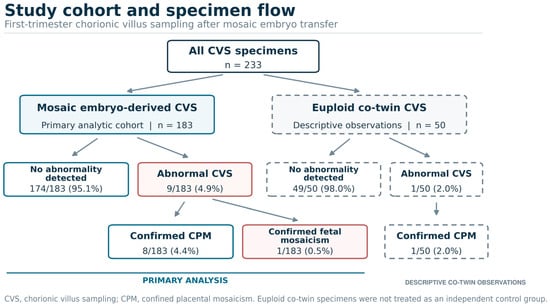
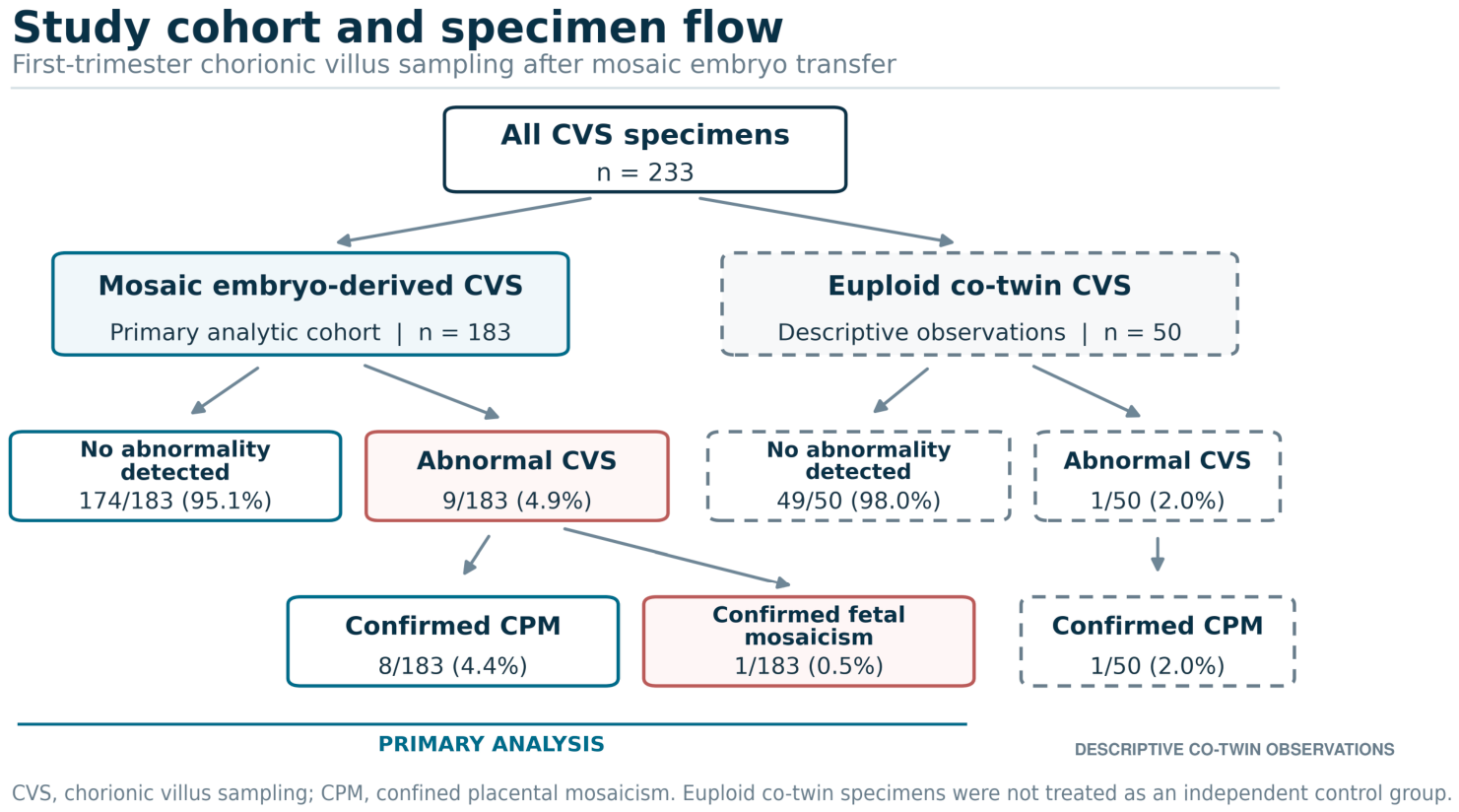
The primary analytic cohort comprised 183 CVS specimens derived from PGT-A-designated mosaic embryos (Figure 1). Trophectoderm biopsy was performed on day 5 for 131 embryos (71.6%) and on day 6 for 52 embryos (28.4%). Among the corresponding embryos, 148 (80.9%) were classified as segmental mosaic, 11 (6.0%) as whole-chromosome mosaic, and 24 (13.1%) as complex mosaic. Low-level mosaicism was reported in 121 embryos (66.1%) and high-level mosaicism in 62 (33.9%) (Table 1).
Figure 1.
Study cohort and specimen flow. The primary analysis included 183 CVS specimens corresponding to PGT-A-designated mosaic embryos. Fifty CVS specimens corresponding to euploid co-twins were excluded from the primary analysis. CPM, confined placental mosaicism; CVS, chorionic villus sampling.
Table 1.
PGT-A characteristics of embryos corresponding to the 183 first-trimester CVS specimens.
3.2. CVS Findings After Mosaic Embryo Transfer
Of the 183 mosaic embryo-derived CVS specimens, 174 (95.1%) had a euploid result and nine (4.9%; exact 95% confidence interval [CI], 2.27–9.13%) had an abnormal result. Abnormal CVS findings occurred in 6 of 148 segmental mosaic embryos (4.1%) and 3 of 35 non-segmental mosaic embryos (8.6%). These subgroup estimates were descriptive and were not used for between-group inference (Table 2).
Table 2.
First-trimester CVS findings and diagnostic outcomes after mosaic embryo transfer.
3.3. Diagnostic Confirmation of Abnormal CVS Findings
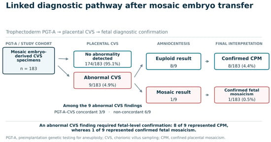
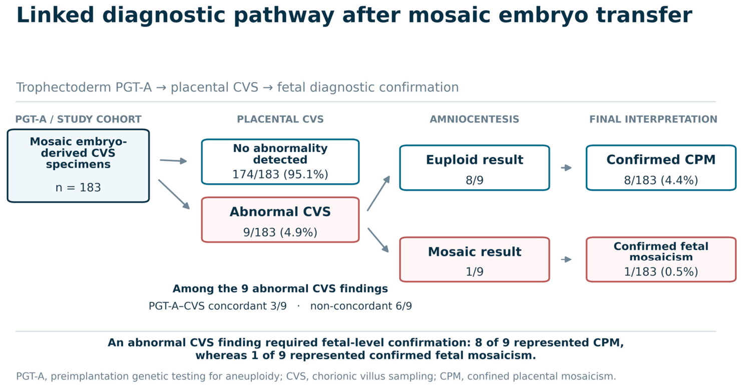
All nine cases with an abnormal CVS finding underwent diagnostic amniocentesis. Eight had a euploid amniocentesis result and were classified as confirmed CPM, corresponding to 8 of 183 specimens in the primary cohort (4.4%; exact 95% CI, 1.91–8.43%) and 8 of 9 abnormal CVS specimens (88.9%).
The remaining case showed mosaicism in amniotic fluid and was classified as confirmed fetal mosaicism, representing 1 of 183 specimens in the primary cohort (0.5%; exact 95% CI, 0.01–3.01%) and 1 of 9 abnormal CVS specimens (11.1%). The linked diagnostic pathway is shown in Figure 2.
Figure 2.
Linked diagnostic pathway after mosaic embryo transfer. All nine abnormal CVS findings underwent diagnostic amniocentesis. PGT-A–CVS concordance was defined as involvement of the same chromosome or chromosomal region, irrespective of the reported mosaic fraction. CPM, confined placental mosaicism; CVS, chorionic villus sampling; PGT-A, preimplantation genetic testing for aneuploidy.
3.4. Concordance Between PGT-A and CVS
Among the nine abnormal CVS cases, three (33.3%) were concordant with the original PGT-A finding, whereas six (66.7%) were non-concordant (Table 3).
Table 3.
Linked PGT-A, CVS, amniocentesis, and birth outcomes in nine pregnancies with an abnormal CVS result after mosaic embryo transfer.
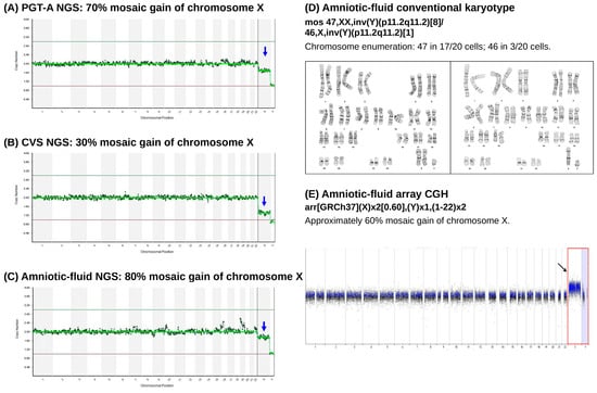
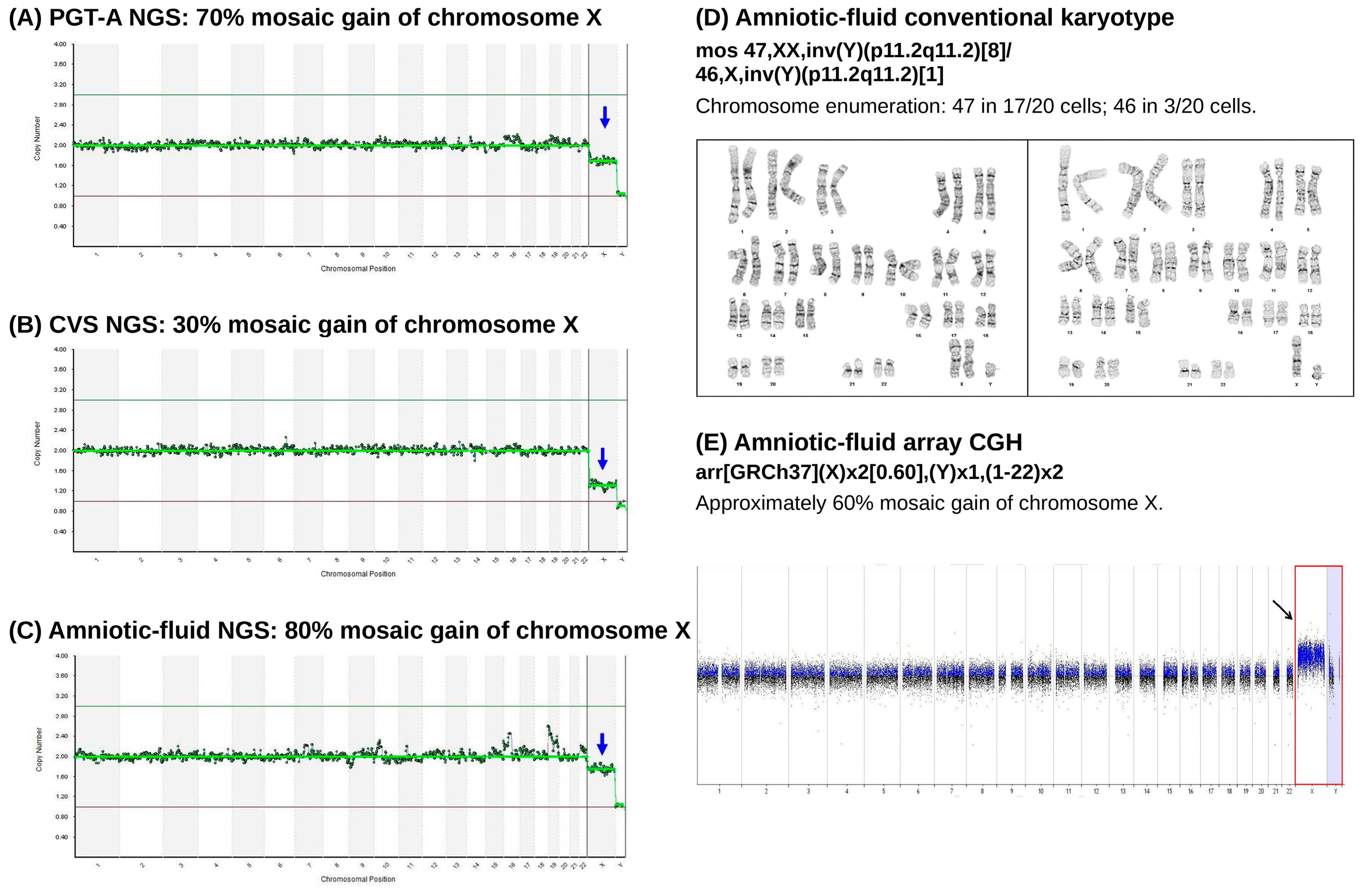
In Case 3, PGT-A reported an XY embryo with a 70% mosaic gain of chromosome X, and CVS showed a 30% mosaic gain of chromosome X. Amniotic-fluid NGS showed an estimated 80% mosaic gain of chromosome X. Conventional karyotyping of amniotic-fluid cells reported the following mosaic result: mos 47,XX,inv(Y)(p11.2q11.2)[8]/46,X,inv(Y)(p11.2q11.2)[1], while chromosome enumeration showed 47 chromosomes in 17 of 20 cells and 46 chromosomes in 3 of 20 cells. According to the original cytogenetic report, the [8]/[1] values corresponded to eight and one in situ culture colonies, respectively, obtained from three culture dishes; the 20-cell chromosome enumeration was documented separately. Array CGH showed arr[GRCh37](X)x2[0.60],(Y)x1,(1-22)x2. These findings confirmed fetal sex-chromosome mosaicism, although the mosaic estimates obtained from different tissues and analytical methods were not directly interchangeable (Figure 3). Detailed prenatal diagnostic findings for all nine abnormal CVS cases are provided in Supplementary Table S1.
Figure 3.
Sequential chromosomal assessment of the single case of confirmed fetal mosaicism after mosaic embryo transfer (Case 3). Sequential chromosomal findings across trophectoderm, uncultured chorionic villi, and amniotic fluid are shown. (A) Trophectoderm biopsy (PGT-A, VeriSeq NGS): approximately 70% mosaic gain of chromosome X. (B) Uncultured chorionic villi (CVS, VeriSeq NGS, 10–13 weeks): approximately 30% mosaic gain of chromosome X. (C) Amniotic fluid (NGS, 16–18 weeks): approximately 80% mosaic gain of chromosome X. (D) Conventional karyotyping of amniotic fluid reported mos 47,XX,inv(Y)(p11.2q11.2)[8]/46,X,inv(Y)(p11.2q11.2)[1]. Chromosome enumeration showed 47 chromosomes in 17 of 20 cells and 46 chromosomes in 3 of 20 cells. The original laboratory report describes the [8] and [1] findings as eight and one in situ culture colonies containing the 47-chromosome and 46-chromosome cell lines, respectively, among nine colonies obtained from three culture dishes. These colony-level observations were documented separately from the chromosome enumeration of 20 cells. (E) Array CGH of amniotic fluid showed arr[GRCh37](X)x2[0.60],(Y)x1,(1-22)x2, consistent with approximately 60% mosaic gain of chromosome X. Mosaic estimates obtained from different tissues and analytical methods should not be interpreted as directly interchangeable. The infant was delivered at 36 weeks with a birth weight of 2560 g. Available pediatric records indicated a favorable clinical outcome; no postnatal cytogenetic testing was performed.
3.5. Birth Outcomes of Abnormal CVS Cases
Birth outcomes were recorded for all nine abnormal CVS cases in the primary cohort. Gestational age at delivery ranged from 31+ to 40 weeks, and birth weight ranged from 1630 to 3785 g. Case 7 was delivered preterm at 31+ weeks following PPROM. Deliveries at 36 weeks occurred in twin pregnancies, and Case 9 was also a co-twin. No diagnosis of fetal growth restriction or small for gestational age was documented, and no abnormalities were recorded on routine prenatal, detailed anatomic, or fetal cardiac ultrasonography. Detailed fetal biometric percentiles and Doppler measurements were unavailable. These outcomes were reported descriptively, without comparative analysis.
4. Discussion
4.1. Clinical Interpretation of Abnormal CVS Findings
Abnormal first-trimester CVS findings after mosaic embryo transfer were uncommon, and most abnormal findings were classified as CPM. Most also involved a chromosome or chromosomal region different from that reported by PGT-A. An abnormal CVS result should therefore be interpreted as a placental finding, rather than as confirmation of the PGT-A result or fetal involvement.
PGT-A, CVS, and amniocentesis examine different samples at different developmental stages. PGT-A is based on a limited trophectoderm biopsy. CVS also samples extraembryonic tissue, whereas amniotic fluid cells more directly reflect the fetal compartment [1,2,3,4,14,15]. Non-uniform cell distribution, differences in sampling location, and laboratory reporting thresholds may explain discordant results. Using the same NGS platform for PGT-A and CVS provided methodological consistency, although biological and sampling-related differences remained. The mosaic fraction reported by PGT-A should not be used as an estimate of the proportion of abnormal cells in the fetus.
Case 3 further illustrates this limitation. The mosaic estimates differed among PGT-A, CVS, amniotic-fluid NGS, conventional karyotyping, and array CGH. These values were generated from different tissues and analytical methods and should not be interpreted as directly equivalent measurements of fetal mosaicism. The lower mosaic estimate in CVS therefore did not exclude fetal involvement, which required amniocentesis for confirmation.
Six of the nine abnormal CVS results involved a chromosome or chromosomal region different from that reported by PGT-A. This may reflect a separate placental cell line or a different distribution of abnormal cells between the original trophectoderm biopsy and the later placental specimen. We could not determine whether these abnormalities arose independently or were not sampled in the original biopsy. Each abnormal CVS result should therefore be compared with the original PGT-A report and interpreted separately.
4.2. Added Value for Prenatal Diagnosis and Counseling
Previous studies have reported generally reassuring prenatal and neonatal outcomes after mosaic embryo transfer, but most available data were obtained from amniocentesis or postnatal follow-up [9,11,12,13]. These studies provide information about the overall risk of fetal chromosomal abnormality, but little information on the interpretation of an abnormal first-trimester CVS result. Persistence of a PGT-A-detected mosaic abnormality in the fetus has been reported, although it appears to be rare [18]. Earlier studies from our center focused on reproductive outcomes and prenatal karyotyping after mosaic embryo transfer [10,11]. The present study extends this work by evaluating first-trimester placental findings and linking every abnormal CVS result to subsequent fetal diagnostic testing.
Professional guidance and genetic counseling literature recommend genetic counseling and discussion of prenatal testing after mosaic embryo transfer [19,20,21]. Patients should understand that CVS and amniocentesis provide different information. CVS gives earlier information about the placenta, whereas amniocentesis is more appropriate when the main purpose is to determine fetal chromosomal status. When CVS yields an abnormal result, confirmatory amniocentesis is recommended to evaluate potential fetal involvement. Patients choosing CVS should be informed that a second invasive procedure may be needed. This study did not compare CVS with direct amniocentesis and does not support one testing strategy for all patients. The choice should be individualized according to the purpose and timing of testing, the original PGT-A finding, and patient preference.
4.3. Obstetric Implications of Confirmed CPM
The euploid amniocentesis result is reassuring regarding fetal chromosomal status. However, confirmed CPM may still affect placental function and pregnancy outcome. Previous studies have associated CPM with impaired fetal growth, although the risk varies according to the chromosome involved, the placental cell lineage, and the degree of mosaicism [15,22,23]. Fetal growth surveillance may therefore still be considered when CPM is confirmed.
This study was not designed to quantify the obstetric risk associated with CPM. Only eight cases were classified as confirmed CPM, and there was no appropriate comparison group. The birth outcomes were descriptive. We cannot determine whether CPM after mosaic embryo transfer carries the same risk as CPM detected in other clinical settings. Based on the available CPM literature, serial assessment of fetal growth may be considered, but the intensity of surveillance should be individualized. Larger studies are needed to define follow-up after mosaic embryo transfer.
4.4. Strengths and Limitations
To our knowledge, this is the largest published series specifically evaluating first-trimester CVS after mosaic embryo transfer. The study included 183 mosaic embryo-derived CVS specimens, and all abnormal CVS findings were followed by amniocentesis. PGT-A and CVS were also analyzed using the same NGS platform. These features allowed direct comparison of PGT-A, placental, and fetal findings.
This study has several limitations. It was conducted at a single center using one laboratory platform and predefined mosaicism thresholds. Only ongoing pregnancies in which CVS was performed were included, so the findings cannot be used to estimate the overall pregnancy-level risk after mosaic embryo transfer. An independent cohort of pregnancies following single euploid embryo transfer and undergoing CVS was not available. Fetal diagnostic follow-up was not systematically obtained after a euploid CVS result. Only nine abnormal CVS findings were identified, which limited subgroup and obstetric analyses. The analysis was performed at the specimen level, and a euploid amniocentesis result cannot completely exclude low-level or tissue-limited fetal mosaicism.
5. Conclusions
This is the largest published series specifically evaluating first-trimester CVS after mosaic embryo transfer and linking the original PGT-A result with placental and fetal testing. Most abnormal CVS findings reflected CPM and often did not involve the same chromosome or chromosomal region reported by PGT-A. An abnormal CVS result should not be used to diagnose fetal involvement, and amniocentesis is needed for fetal confirmation. These results may help clinicians counsel patients about prenatal testing after mosaic embryo transfer.
Supplementary Materials
The following supporting information can be downloaded at: https://www.mdpi.com/article/10.3390/diagnostics16223755/s1, Table S1: Prenatal diagnostic findings and pregnancy outcomes in nine cases with abnormal chorionic villus sampling results after mosaic embryo transfer.
Author Contributions
Conceptualization, M.-S.L.; methodology, M.-S.L., E.-H.C. and Y.-P.P.; software, Y.-P.P.; validation, E.-H.C. and Y.-P.P.; formal analysis, Y.-P.P. and P.-Y.L.; investigation, M.-S.L., T.-H.L. and E.-H.C.; resources, M.-S.L. and C.-C.H.; data curation, Y.-P.P., P.-Y.L., T.-N.Y. and W.-C.L.; writing—original draft preparation, P.-Y.L.; writing—review and editing, P.-Y.L. and M.-S.L.; visualization, P.-Y.L. and Y.-P.P.; supervision, M.-S.L., P.-Y.L., C.-I.L., T.-H.L., T.-N.Y., M.-J.C. and E.-H.C.; project administration, C.-C.H., Y.-P.P. and E.-H.C. All authors have read and agreed to the published version of the manuscript.
Funding
This research received no external funding.
Institutional Review Board Statement
The study was conducted in accordance with the Declaration of Helsinki and approved by the Institutional Review Board of Chung Shan Medical University Hospital (approval No. CS18199; 19 November 2018).
Informed Consent Statement
Informed consent was obtained from all subjects involved in the study.
Data Availability Statement
The data presented in this study are available from the corresponding author upon reasonable request, subject to Institutional Review Board approval and institutional policies.
Conflicts of Interest
The authors declare no conflicts of interest.
References
- Munné, S.; Wells, D. Detection of mosaicism at blastocyst stage with the use of high-resolution next-generation sequencing. Fertil. Steril. 2017, 107, 1085–1091. [Google Scholar] [CrossRef] [Scilit] [PubMed]
- Treff, N.R.; Marin, D. The “mosaic” embryo: Misconceptions and misinterpretations in preimplantation genetic testing for aneuploidy. Fertil. Steril. 2021, 116, 1205–1211. [Google Scholar] [CrossRef] [Scilit] [PubMed]
- Popovic, M.; Borot, L.; Lorenzon, A.R.; Lopes, A.L.R.d.C.; Sakkas, D.; Lledó, B.; Morales, R.; Ortiz, J.A.; Polyzos, N.P.; Parriego, M.; et al. Implicit bias in diagnosing mosaicism amongst preimplantation genetic testing providers: Results from a multicenter study of 36,395 blastocysts. Hum. Reprod. 2024, 39, 258–274. [Google Scholar] [CrossRef] [Scilit] [PubMed]
- De Witte, L.; Baetens, M.; Tilleman, K.; Vanden Meerschaut, F.; Janssens, S.; Van Tongerloo, A.; Szymczak, V.; Stoop, D.; Dheedene, A.; Symoens, S.; et al. Aligning genotyping and copy number data in single trophectoderm biopsies for aneuploidy prediction: Uncovering incomplete concordance. Hum. Reprod. Open 2024, 2024, hoae056. [Google Scholar] [CrossRef] [Scilit] [PubMed]
- Zhang, Q.; Liu, G.; Xiang, Y.; Zou, Y.; Chen, Y.; Xia, Y.; Xiong, S.; Fu, T.; Wang, J.; Jiang, Y.; et al. Parental and cell-division origin analysis to reduce false-positives in mosaic embryos for preimplantation genetic testing. Hum. Reprod. Open 2025, 2025, hoaf075. [Google Scholar] [CrossRef] [Scilit] [PubMed]
- Bacal, V.; Li, A.; Shapiro, H.; Rana, U.; Zwingerman, R.; Avery, L.; Palermo, A.; Philipoppolous, E.; Chan, C. A systematic review and meta-analysis of the diagnostic accuracy after preimplantation genetic testing for aneuploidy. PLoS ONE 2025, 20, e0321859. [Google Scholar] [CrossRef] [Scilit] [PubMed]
- Gill, P.; Tao, X.; Zhan, Y.; Mulas, F.; Ottolini, C.S.; Picchetta, L.; Caroselli, S.; Babariya, D.; Wells, D.; Clark, G.; et al. Preimplantation genetic testing for aneuploidy mosaicism reporting lacks clinical predictive value for live birth in a multisite, double-blinded study with independent validation. Am. J. Obstet. Gynecol. 2026, 234, 1015–1041. [Google Scholar] [CrossRef] [Scilit] [PubMed]
- Dal Molin, E.A.; Pires, G.N.; Lazzari, V.M. Live birth and other reproductive outcomes of mosaic and euploid embryos: A systematic review and meta-analysis. J. Assist Reprod. Genet. 2025, 42, 2469–2481. [Google Scholar] [PubMed]
- Viotti, M.; Greco, E.; Grifo, J.A.; Madjunkov, M.; Librach, C.; Cetinkaya, M.; Kahraman, S.; Yakovlev, P.; Kornilov, N.; Corti, L.; et al. Chromosomal, gestational, and neonatal outcomes of embryos classified as a mosaic by preimplantation genetic testing for aneuploidy. Fertil. Steril. 2023, 120, 957–966. [Google Scholar] [CrossRef] [Scilit] [PubMed]
- Lin, P.-Y.; Lee, C.-I.; Cheng, E.-H.; Huang, C.-C.; Lee, T.-H.; Shih, H.-H.; Pai, Y.-P.; Chen, Y.-C.; Lee, M.-S. Clinical outcomes of single mosaic embryo transfer: High-level or low-level mosaic embryo, does it matter? J. Clin. Med. 2020, 9, 1695. [Google Scholar] [CrossRef] [Scilit] [PubMed]
- Lee, C.-I.; Cheng, E.-H.; Lee, M.-S.; Lin, P.-Y.; Chen, Y.-C.; Chen, C.-H.; Huang, L.-S.; Huang, C.-C.; Lee, T.-H. Healthy live births from transfer of low-mosaicism embryos after preimplantation genetic testing for aneuploidy. J. Assist Reprod. Genet. 2020, 37, 2305–2313. [Google Scholar] [CrossRef] [Scilit] [PubMed]
- Hong, Y.M.; Kim, S.H.; Park, H.J.; Ryu, H.M.; Cha, D.H.; Kim, M.Y.; Han, Y.J. Prenatal ultrasound findings and chromosomal outcomes of pregnancies with mosaic embryo transfer. Diagnostics 2024, 14, 2795. [Google Scholar] [CrossRef] [Scilit] [PubMed]
- Kline, L.A.; Nitibhon, V.A.; Dyr, B.; Soster, E.L.; Sussman, A.; Weinblatt, V.J.; Schwartz, S. Cytogenetic and microarray analysis follow-up of PGT-A mosaic and sex-discrepant embryos during pregnancy: Absence of confirmation and follow-up recommendations. Prenat. Diagn. 2026, 46, 1151–1158. [Google Scholar] [CrossRef] [Scilit] [PubMed]
- Lund, I.C.B.; Becher, N.; Christensen, R.; Petersen, O.B.; Steffensen, E.H.; Vestergaard, E.M.; Vogel, I. Prevalence of mosaicism in uncultured chorionic villus samples after chromosomal microarray and clinical outcome in pregnancies affected by confined placental mosaicism. Prenat. Diagn. 2020, 40, 244–259. [Google Scholar] [CrossRef] [Scilit] [PubMed]
- Eggenhuizen, G.M.; Go, A.; Koster, M.P.H.; Baart, E.B.; Galjaard, R.J. Confined placental mosaicism and the association with pregnancy outcome and fetal growth: A review of the literature. Hum. Reprod. Update 2021, 27, 885–903. [Google Scholar] [CrossRef] [Scilit] [PubMed]
- Nagan, N.; Faulkner, N.E.; Curtis, C.; Schrijver, I.; MCC Guidelines Working Group. Laboratory guidelines for detection, interpretation, and reporting of maternal cell contamination in prenatal analyses: A report of the Association for Molecular Pathology. J. Mol. Diagn. 2011, 13, 7–11. [Google Scholar] [CrossRef] [Scilit] [PubMed]
- Militaru, M.S.; Babliuc, I.M.; Bloaje-Florică, V.L.; Danci, V.A.; Filip-Deac, I.; Kutasi, E.; Simon, V.; Militaru, M.; Cătană, A. The impact of chromosomal mosaicisms on prenatal diagnosis and genetic counseling-a narrative review. J. Pers. Med. 2024, 14, 774. [Google Scholar] [CrossRef] [Scilit] [PubMed]
- Kahraman, S.; Cetinkaya, M.; Yuksel, B.; Yesil, M.; Cetinkaya, C.P. The birth of a baby with mosaicism resulting from a known mosaic embryo transfer: A case report. Hum. Reprod. 2020, 35, 727–733. [Google Scholar] [CrossRef] [Scilit] [PubMed]
- Practice Committees of the American Society for Reproductive Medicine and the Genetic Counseling Professional Group. Clinical management of mosaic results from preimplantation genetic testing for aneuploidy of blastocysts: A committee opinion. Fertil. Steril. 2023, 120, 973–982. [Google Scholar] [CrossRef] [Scilit] [PubMed]
- Leigh, D.; Cram, D.; Rechitsky, S.; Handyside, A.; Wells, D.; Munne, S.; Kahraman, S.; Grifo, J.; Katz-Jaffe, M.; Rubio, C.; et al. PGDIS position statement on the transfer of mosaic embryos 2021. Reprod. Biomed Online 2022, 45, 19–25. [Google Scholar] [CrossRef] [Scilit] [PubMed]
- Chen, K.; Darcy, D.; Boyd, A. Pregnancy from mosaic embryo transfer: Genetic counseling considerations. Curr. Opin. Obstet. Gynecol. 2021, 33, 100–105. [Google Scholar] [CrossRef] [Scilit] [PubMed]
- Spinillo, S.L.; Farina, A.; Sotiriadis, A.; Pozzoni, M.; Giglio, S.; Papale, M.; Candiani, M.; Cavoretto, P.I. Pregnancy outcome of confined placental mosaicism: Meta-analysis of cohort studies. Am. J. Obstet. Gynecol. 2022, 227, 714–727.e1. [Google Scholar] [CrossRef] [Scilit] [PubMed]
- Eggenhuizen, G.M.; Go, A.T.J.I.; Sauter, Z.; Hoffer, M.J.V.; Haak, M.C.; Geeven, G.; Diderich, K.E.M.; Joosten, M.; Born, M.v.D.; Srebniak, M.I.; et al. The role of confined placental mosaicism in fetal growth restriction: A retrospective cohort study. Prenat. Diagn. 2024, 44, 289–296. [Google Scholar] [CrossRef] [Scilit] [PubMed]

Disclaimer/Publisher’s Note: The statements, opinions and data contained in all publications are solely those of the individual author(s) and contributor(s) and not of MDPI and/or the editor(s). MDPI and/or the editor(s) disclaim responsibility for any injury to people or property resulting from any ideas, methods, instructions or products referred to in the content. |
© 2026 by the authors. Licensee MDPI, Basel, Switzerland. This article is an open access article distributed under the terms and conditions of the Creative Commons Attribution (CC BY) license.