


Abstract
Free air in the pericardial space is called pneumopericardium, a rare phenomenon. Increased intra-alveolar pressure may cause alveolar rupture, releasing air that travels to the pericardial space. This condition can occur following trauma or surgical interventions. In this case, we present a young female patient with pneumopericardium that developed early after open-heart surgery. Interestingly, the pleural spaces were not opened during the initial operation, and hemodynamic stability was maintained despite the massive mediastinal air that developed. The case was treated surgically due to the possible risk of infection associated with the Dacron graft in the mediastinum.
Figure 1.
Preoperative chest radiograph and computed tomographic images of the patient (the blue arrow indicates the pericardial air). A 17-year-old female patient with Marfan Syndrome underwent a Bentall operation with a 25–28 mm St. Jude valved conduit on cardiopulmonary bypass at 32 °C. The pleura was not opened during the operation, and mediastinal tube drains were placed. The patient’s mediastinal drains were removed on the second postoperative day. On the fourth postoperative day, a routine chest X-ray revealed mediastinal air (A). This was confirmed by computed tomography (B–D). The patient’s oxygen saturation and arterial blood pressure were normal. The asymptomatic patient underwent emergency reoperation. Written informed consent was obtained from the patient’s legal guardian.
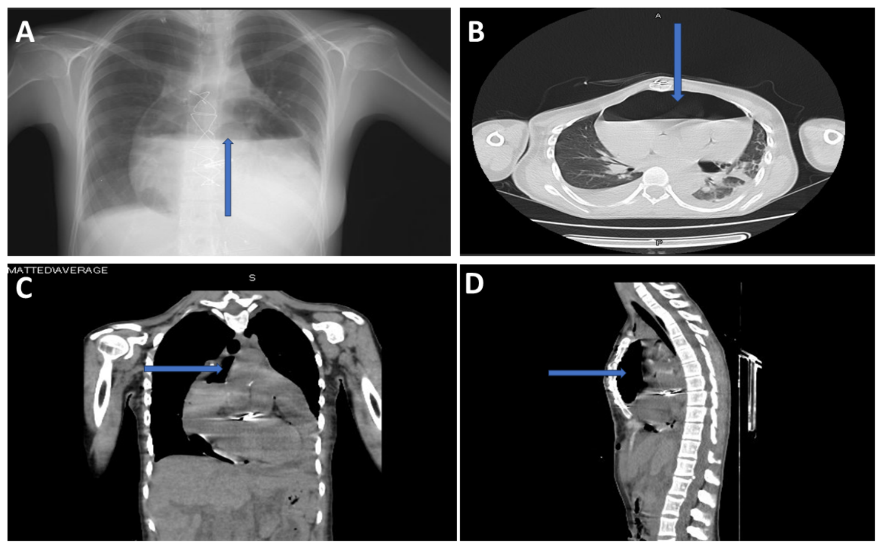
Figure 2.
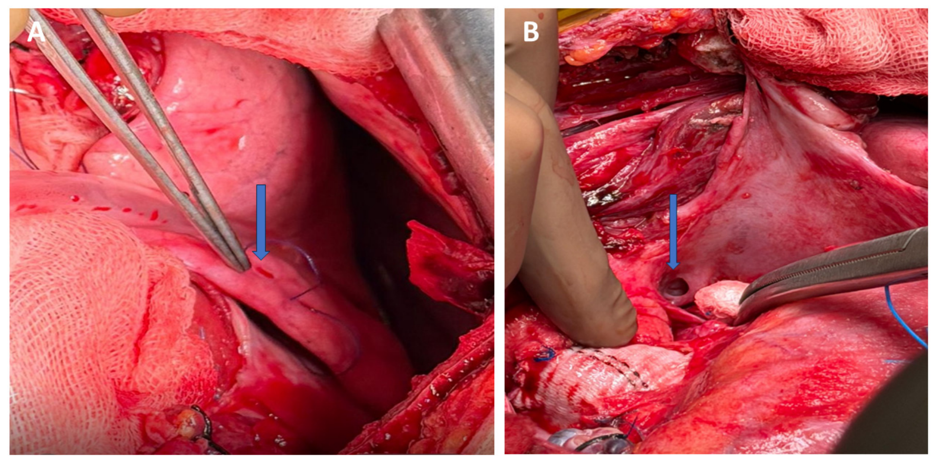
Operative images of the patient. (A) The blue arrow indicates the lung laceration. (B) The blue arrow indicates the pleural–mediastinal hole. Mediastinal air and fluids were drained. When the left pleura was opened, a small laceration without air leakage was observed in the left lung (A) and was primarily repaired. Additionally, a pleural–mediastinal hole was observed posterior to the pulmonary artery (B). Mediastinal and left thoracic drains were placed, and the operation was concluded. The patient was discharged on the 12th postoperative day. We hypothesized that this clinical condition may have resulted from air escaping from the laceration in the lung tissue (A), flowing through the pleural–mediastinal opening (B) into the mediastinum due to a pressure difference. The patient is currently in the 9th month of follow-up, and there is no pneumopericardium recurrence or complications. Free air in the pericardial space is called pneumopericardium, a rare phenomenon. Increased intra-alveolar pressure may cause alveolar rupture, releasing air that travels to the pericardial space. This condition can occur following trauma or surgical interventions. In the literature, a case has been reported involving a 26-year-old male patient who underwent robotic atrial septal defect (ASD) repair, in whom free air was observed spreading around the pericardium and mediastinal structures on the third postoperative day. This case was hemodynamically stable and treated medically [1]. Our patient was asymptomatic, and the air was confined to the pericardium. However, due to the high air content and the risk of infection [2] in early-term cardiac surgery, we treated her surgically. In a patient with a Dacron graft in the mediastinum, the consequences of any mediastinal infection that may develop can be extremely serious [3]. A case of pneumopericardium developing six hours postoperatively after ASD repair in a 13-month-old girl has also been reported in the literature. In this case, the tube drained the air [4].
Author Contributions
Conceptualization, M.E. and O.S.; methodology, M.E.; software, M.E.; validation, M.E. and U.A.; formal analysis, Y.A.; investigation, Y.A.; resources, Y.A.; data curation, M.E.; writing—original draft preparation, M.E.; writing—review and editing, M.E., O.S., U.A. and Y.A.; visualization, Y.A.; supervision, U.A. All authors have read and agreed to the published version of the manuscript.
Funding
This research received no external funding.
Institutional Review Board Statement
Ethical approval was waived according to the regulations of the Bursa Yuksek Ihtisas Training and Research Hospital, as a single case report does not require approval from the Ethics Committee.
Informed Consent Statement
Written informed consent was obtained from the patient’s legal guardian to publish this paper.
Data Availability Statement
The original contributions presented in this study are included in the article. Further inquiries can be directed to the corresponding author.
Conflicts of Interest
The authors declare no conflicts of interest.
References
- Erol, G.; Kubat, E.; Sicim, H.; Kadan, M.; Bolcal, C. An unexpected complication of robotic cardiac surgery: Pneumomediastinum. J. Saudi Heart Assoc. 2019, 31, 106–108. [Google Scholar] [CrossRef] [Scilit]
- Merz, A.; Ran, H.; Chiu, C.Y.; Dreger, H.; Morris, D.A.; Schneider-Reigbert, M. Iatrogenic Pneumopericardium After Pericardiocentesis: A Systematic Review and Case Report. J. Cardiovasc Dev. Dis. 2025, 12, 246. [Google Scholar] [CrossRef] [Scilit]
- Türköz, R.; Doğan, A.; Türkekul, Y.; Özker, E. Successful treatment of thoracic aortic graft infection by omental flap following vacuum-assisted closure therapy. J. Card. Surg. 2020, 35, 2857–2859. [Google Scholar] [CrossRef] [Scilit]
- Park, C.S.; Lee, C.H.; Lee, C.; Kwak, J.G.; Song, J.Y.; Jang, S.I.; Choi, E.Y.; Kim, Y.M. Tension pneumopericardium after cardiac surgery: A rare but lethal problem. Circulation 2010, 121, 2079–2080. [Google Scholar]
Disclaimer/Publisher’s Note: The statements, opinions and data contained in all publications are solely those of the individual author(s) and contributor(s) and not of MDPI and/or the editor(s). MDPI and/or the editor(s) disclaim responsibility for any injury to people or property resulting from any ideas, methods, instructions or products referred to in the content. |
© 2026 by the authors. Licensee MDPI, Basel, Switzerland. This article is an open access article distributed under the terms and conditions of the Creative Commons Attribution (CC BY) license.